
Regulation of Th2 responses by different cell types expressing ...
11
Saito et al. Allergy Asthma Clin Immunol (2017) 13:23 DOI 10.1186/s13223-017-0194-9 RESEARCH Regulation of Th2 responses by different cell types expressing the interleukin-31 receptor Saburo Saito 1* , Ayana Aoki 1,2 , Iwao Arai 1 , Shinya Takaishi 1,3 , Haruyasu Ito 4 , Nobutake Akiyama 1 and Hiroshi Kiyonari 5 Abstract Background: Interleukin-31 (IL-31) is a recently identified cytokine produced by Th2 cells that is involved in the development of atopic dermatitis-induced skin inflammation and pruritus. Its receptor, IL-31RA, is expressed by a number of cell types, including epithelial cells, eosinophils, and activated monocytes and macrophages. To date, how- ever, the regulation of Th2 responses by distinct cell types and tissues expressing IL-31RA has not been well studied. Methods: In this study, Cry j 2, one of the major allergens of Japanese cedar pollen, was administered to IL-31RA- deficient or wild-type (WT ) mice via nasal or intraperitoneal injection for induction of specific Th2 responses. Results: After nasal administration of Cry j 2, IL-31RA-deficient mice showed lower Cry j 2-specific CD4+ T cell proliferation, Th2 cytokine (IL-5 and IL-13) production, and Th2-mediated (IgE, IgG1, and IgG2b) antibody responses than WT mice. In contrast, IL-31RA-deficient mice administered Cry j 2 intraperitoneally showed stronger Th2 immune responses than WT mice. Conclusions: These results indicate that IL-31R signaling positively regulates Th2 responses induced by nasal admin- istration of Cry j 2, but negatively regulates these responses when Cry j 2 is administered intraperitoneally. Collectively, these data indicate that the induction of antigen-specific Th2 immune responses might depend on tissue-specific cell types expressing IL-31RA. Keywords: IL-31 receptor, Th2, IgE, Cry j 2, Deficient mice © The Author(s) 2017. This article is distributed under the terms of the Creative Commons Attribution 4.0 International License (http://creativecommons.org/licenses/by/4.0/), which permits unrestricted use, distribution, and reproduction in any medium, provided you give appropriate credit to the original author(s) and the source, provide a link to the Creative Commons license, and indicate if changes were made. The Creative Commons Public Domain Dedication waiver (http://creativecommons.org/ publicdomain/zero/1.0/) applies to the data made available in this article, unless otherwise stated. Background Interleukin-31 (IL-31) is a recently identified cytokine produced by activated CD4+ 2 cells that plays an important role in human T cell-mediated skin disease [1]. e expression of IL-31 has been shown to be correlated with the expression of the 2 cytokines IL-4 and IL-13 in human skin diseases [2], and serum IL-31 has been shown to be higher in patients with atopic dermatitis [3]. When overexpressed in transgenic mice, IL-31 induces severe pruritus, which resembles eczema in humans [2]. Investigation of IL-31 transgenic mice has shown that overexpression of IL-31 results in the development of atopic dermatitis (AD)-like lesions, and that IL-31 expression is highly associated with 2-skewed diseases [4]. ese results indicate that IL-31 plays a role in the development and exacerbation of the 2-associated dis- ease AD. In contrast to observations indicating that IL-31 is actively involved in the promotion of 2-type diseases, others suggest that IL-31–IL-31R signaling negatively regulates 2-type immune responses in the lungs fol- lowing Schistosoma mansoni egg-induced inflammation [5]. In this study, the authors showed that IL-31RA- deficient mice developed exacerbated S. mansoni egg- induced 2-type immune responses in the lungs and that loss of IL-31RA signaling resulted in enhanced anti- gen presentation by macrophages and increased 2 cytokine expression by CD4+ T cells [5]. Furthermore, they showed that in response to Trichuris infection, IL-31RA-deficient mice exhibited increased 2 cytokine responses in the mesenteric lymph nodes and elevated Open Access Allergy, Asthma & Clinical Immunology *Correspondence: [email protected] 1 Division of Molecular Immunology, Research Center for Medical Science, The Jikei University School of Medicine, 3-25-8 Nishi-shinbashi, Minato-ku, Tokyo 105-8461, Japan Full list of author information is available at the end of the article
-
Upload
khangminh22 -
Category
Documents
-
view
1 -
download
0
Transcript of Regulation of Th2 responses by different cell types expressing ...
Saito et al. Allergy Asthma Clin Immunol (2017) 13:23 DOI 10.1186/s13223-017-0194-9
RESEARCH
Regulation of Th2 responses by different cell types expressing the interleukin-31 receptorSaburo Saito1* , Ayana Aoki1,2, Iwao Arai1, Shinya Takaishi1,3, Haruyasu Ito4, Nobutake Akiyama1 and Hiroshi Kiyonari5
Abstract
Background: Interleukin-31 (IL-31) is a recently identified cytokine produced by Th2 cells that is involved in the development of atopic dermatitis-induced skin inflammation and pruritus. Its receptor, IL-31RA, is expressed by a number of cell types, including epithelial cells, eosinophils, and activated monocytes and macrophages. To date, how-ever, the regulation of Th2 responses by distinct cell types and tissues expressing IL-31RA has not been well studied.
Methods: In this study, Cry j 2, one of the major allergens of Japanese cedar pollen, was administered to IL-31RA-deficient or wild-type (WT) mice via nasal or intraperitoneal injection for induction of specific Th2 responses.
Results: After nasal administration of Cry j 2, IL-31RA-deficient mice showed lower Cry j 2-specific CD4+ T cell proliferation, Th2 cytokine (IL-5 and IL-13) production, and Th2-mediated (IgE, IgG1, and IgG2b) antibody responses than WT mice. In contrast, IL-31RA-deficient mice administered Cry j 2 intraperitoneally showed stronger Th2 immune responses than WT mice.
Conclusions: These results indicate that IL-31R signaling positively regulates Th2 responses induced by nasal admin-istration of Cry j 2, but negatively regulates these responses when Cry j 2 is administered intraperitoneally. Collectively, these data indicate that the induction of antigen-specific Th2 immune responses might depend on tissue-specific cell types expressing IL-31RA.
Keywords: IL-31 receptor, Th2, IgE, Cry j 2, Deficient mice
© The Author(s) 2017. This article is distributed under the terms of the Creative Commons Attribution 4.0 International License (http://creativecommons.org/licenses/by/4.0/), which permits unrestricted use, distribution, and reproduction in any medium, provided you give appropriate credit to the original author(s) and the source, provide a link to the Creative Commons license, and indicate if changes were made. The Creative Commons Public Domain Dedication waiver (http://creativecommons.org/publicdomain/zero/1.0/) applies to the data made available in this article, unless otherwise stated.
BackgroundInterleukin-31 (IL-31) is a recently identified cytokine produced by activated CD4+ Th2 cells that plays an important role in human T cell-mediated skin disease [1]. The expression of IL-31 has been shown to be correlated with the expression of the Th2 cytokines IL-4 and IL-13 in human skin diseases [2], and serum IL-31 has been shown to be higher in patients with atopic dermatitis [3]. When overexpressed in transgenic mice, IL-31 induces severe pruritus, which resembles eczema in humans [2]. Investigation of IL-31 transgenic mice has shown that overexpression of IL-31 results in the development of atopic dermatitis (AD)-like lesions, and that IL-31
expression is highly associated with Th2-skewed diseases [4]. These results indicate that IL-31 plays a role in the development and exacerbation of the Th2-associated dis-ease AD.
In contrast to observations indicating that IL-31 is actively involved in the promotion of Th2-type diseases, others suggest that IL-31–IL-31R signaling negatively regulates Th2-type immune responses in the lungs fol-lowing Schistosoma mansoni egg-induced inflammation [5]. In this study, the authors showed that IL-31RA-deficient mice developed exacerbated S. mansoni egg-induced Th2-type immune responses in the lungs and that loss of IL-31RA signaling resulted in enhanced anti-gen presentation by macrophages and increased Th2 cytokine expression by CD4+ T cells [5]. Furthermore, they showed that in response to Trichuris infection, IL-31RA-deficient mice exhibited increased Th2 cytokine responses in the mesenteric lymph nodes and elevated
Open Access
Allergy, Asthma & Clinical Immunology
*Correspondence: [email protected] 1 Division of Molecular Immunology, Research Center for Medical Science, The Jikei University School of Medicine, 3-25-8 Nishi-shinbashi, Minato-ku, Tokyo 105-8461, JapanFull list of author information is available at the end of the article

Page 2 of 11Saito et al. Allergy Asthma Clin Immunol (2017) 13:23
serum IgE and IgG1 levels as compared to WT mice [6]. In contrast, Bilsborough et al. reported that the suscep-tibility of IL-31RA KO mice [7] to exacerbated Th2-type diseases was an indirect result of IL-31RA deletion that causes an increased responsiveness to oncostatin M (OSM) and exacerbated production of OSM-inducible cytokines, such as IL-6, VEGF, and TIMP-1, during air-way sensitization and challenge. These results indicate that the differential effects of IL-31 in distinct tissues may influence the distinct patterns of Th2-type immune responses, with, for example, positive effects in the skin, but negative effects in the lung and intestine.
On the basis of these previous findings, we assumed that Th2 immune responses are specifically regulated by different types of cells or tissues expressing the IL-31 receptor. To examine whether the reported exacerbated Th2-type response in IL-31RA KO mice [5, 6] has tissue-specific mechanisms, we investigated the antigen-specific Th2 responses in IL-31RA-deficient mice administered an allergen nasally or intraperitoneally.
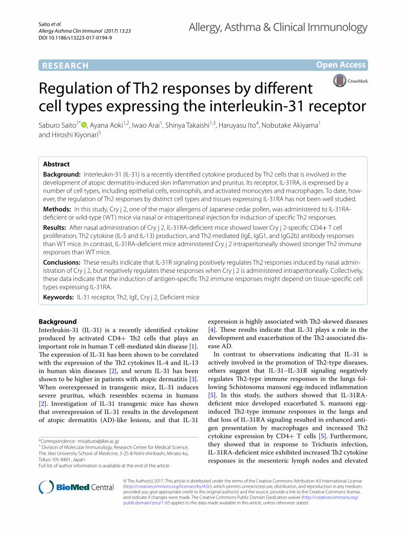
MethodsMiceC57BL/6 mice were purchased from Sankyo Laboratories (Tokyo, Japan) and were housed in our facilities under specific pathogen-free conditions. All experiments were performed following the Animal Experimentation Guide-lines of The Jikei University School of Medicine and the RIKEN Kobe Branch. The IL-31RA mutant (Acces-sion No. CDB1012K: http://www2.clst.riken.jp/arg/mutant%20mice%20list.html) was established as follows. To generate IL-31RA deficient mice, homologous recom-bination in embryonic stem (ES) cells was used to create a mutant allele in which exon 4 of the IL-31RA gene was replaced with a cassette expressing the selective marker neomycin transferase. In brief, homologous regions (5′5 8001 bp, 3′: 3074 bp) were subcloned into a knock-in vec-tor (DT-A-pA/lox71/LacZ-pA/frt/PGK-Neo/frt/loxP/pA: http://www2.clst.riken.jp/arg/cassette.html) and electroporated into mouse ES cells. Two recombinant ES cells were found to be IL-31RAtm1(LacZ). Homologously integrated ES cells were injected into 8-cell-stage zygotes (Fig. 1a). Chimeric mice were mated with C57BL/6 mice to produce mutant IL-31RA+/LacZ(+/−) progeny. The generation of mutant IL-31RA+/− mice was verified by Southern blot analysis (Fig. 1b). Digestion with ApaI fol-lowed by hybridization of membranes with probe 1 (a 900-bp genomic DNA fragment obtained by PCR: Gen-Bank AC154767.2, nt 49651–50550) yielded a 15,638-bp fragment for the WT allele and a 21,281-bp fragment
for the correctly targeted mutant allele. Similarly, diges-tion of genomic DNA with NheI followed by hybridiza-tion of membranes with probe 2 (a 800-bp genomic DNA fragment obtained by PCR: GenBank NC_000079.6, nt 112555274–112556073) yielded a 16,632-bp fragment for the WT allele and a 10,930-bp fragment for the correctly targeted mutant allele. To reduce heterozygosity, the IL-31RA+/− allele was then backcrossed to C57BL/6 mice for 15 more generations using IL-31RA+/− males. Dis-ruption of IL-31RA was identified by PCR using the cor-responding primers to give a 602-bp product (primer1: 5′-caaagcgccattcgccattcaggctgcgca-3′,primer2: 5′-tgtg-cattgtgagtgggtgagtggtatgca-3′). The presence of wild-type IL-31RA was identified as a 377-bp product (primer 2 and primer 3: 5′-tgaatttgcagaggaaagagaatgcccaca-3′).
Homozygous IL-31RA−/− and WT (IL-31RA+/+) litter-mates were generated by intercrossing IL-31RA+/− mice from the 15th generation of backcrossed mice. Heterozy-gous IL-31RA+/− and homozygous IL-31RA−/− litter-mates were generated by crossing IL-31RA+/− mice with homozygous IL-31RA−/− mice.
Immunohistochemistry for IL‑31RA and beta‑galactosidaseTo examine the expression of IL-31RA or beta-galactosi-dase (β-Gal) from WT C57BL/6 mice (IL-31RA+/+) and IL-31RA-deficient (IL-31RA−/−) mice, skin specimens were fixed for 3 days in 10% normal buffered formalin and embedded in paraffin using standard techniques. Five-micrometer sections were heated at 60 °C for 30 min for tissue adhesion. Slides were subsequently dewaxed by incubation in xylene and then rehydrated in 100, 95, and 70% EtOH. Finally, the slides were rinsed with Tris-buff-ered saline Tween buffer, and prepared as recommended by the manufacturer. Endogenous peroxidase was blocked with 4.5% H2O2 in MeOH for 30 min at room temperature. A protein block (phosphate-buffered saline block containing 10% normal goat serum) was applied overnight at 4 °C.
To examine the expression of IL-31RA, we prepared rabbit anti-mouse IL-31RA polyclonal antibodies as fol-lows. A partial peptide of murine IL-31RA, mGPL 65–86 (YSDNATEASYSFPRSCAMPPDI), was synthesized by referring to [8] and conjugated to keyhole limpet hemo-cyanin. Rabbits were immunized with the conjugates and Freund’s Complete Adjuvant. After booster shots, whole blood from immunized rabbits was collected and serum was separated. Serum antibodies that recognize IL-31RA were purified using a column containing Sepharose-bound mGPL 65–86 peptide and the antigen-binding activity of the purified antibodies to the peptide was

Page 3 of 11Saito et al. Allergy Asthma Clin Immunol (2017) 13:23
checked by ELISA. To examine the expression of β-Gal, an anti-beta β-Gal antibody (Abcam PLC, Cambridge, US) was used as a primary antibody and biotin-conju-gated goat anti-chicken IgY (H+L) polyclonal antibody (GeneTex Inc, CA, USA) was used as a secondary anti-body for β-Gal staining.
ImmunizationTo induce allergen-specific Th2 responses, mice were administered an allergen nasally or intraperitoneally. For nasal administration, 2.5 µg of Cry j 2 (Hayashibara Bio-chemical Laboratories, Okayama, Japan), one of the major allergens of Japanese cedar pollen, was dissolved in 4 μl of PBS, and simply administered intranasally 15 times (5 times per week). For intraperitoneal immunization, mice were intraperitoneally injected with 20 µg of Cry j 2 in 0.2 ml of PBS without any adjuvants once a week for 5 weeks.
T‑cell proliferation assaySeven days after the last immunization, the spleen and lymph node cells were collected. Cry j 2-specific T-cell proliferative responses were determined by an in vitro [3H] thymidine incorporation assay. RPMI-1640 medium (Thermo Fisher Scientific Inc., MA, USA) supple-mented with 1% normal mouse serum was used to sus-pend cells and 8 × 10⁵ cells were seeded into each well of 96-well plates (Nunc Microwell 96F; Thermo Fisher Scientific Inc.) and cultured with 2.5 µg/ml of Cry j 2 for 88 h. The cultures were pulsed with 0.5 µCi of [3H] thy-midine (American Radiolabeled Chemicals Inc., USA) for the last 16 h in Nunc Maxisorp plates (Kamnstrup, Denmark). The cells were harvested using a Micro 96 Harvester (Skatron Instruments, Norway), and [3H] thy-midine uptake by the cells was determined by measur-ing the radioactivity using a liquid scintillation counter
a
b c+/+ +/- +/+ +/-
KO 21281 bpWT 15638 bp
KO 16632 bp
WT 10930 bp
5’ probe 3’ probe
IL-31Rα(-/-) IL-31Rα(+/+)
Anti-β-galactosidaseantibody
Anti-IL-31RAantibody
Wt allele
Targeted allele PGK Neo pALacZ pA
2kb
ApaI ApaI
ApaI ApaI
NheI
NheI NheI
NheI
15638 bp
21281 bp
5’ Probe
5’ Probe
3’ Probe
3’ Probe
Fig. 1 Generation of IL-31RA-deficient mice. To generate C57BL/6-IL-31RAtLacZ/+ knock-out (KO) mice, we used homologous recombination in embryonic stem (ES) cells to create a mutant allele in which exon 4 of the IL-31RA gene was replaced by a cassette expressing the selective marker neomycin transferase (a). Two recombinant ES cells were found to be IL-31RAtm1(LacZ). Chimeric mice were mated with C57BL/6 mice to produce mutant IL-31RA+/LacZ(+/−) progeny. The generation of mutant IL-31RA+/− mice was verified by Southern blot analysis (b). The IL-31RA+/− allele was then backcrossed to C57BL/6 mice for 15 more generations using male IL-31RA+/−. Homozygous IL-31RA−/− and WT (IL-31RA+/+) littermates were generated by intercrossing IL-31RA+/− mice from the 15th generations of backcrossed mice. Heterozygous IL-31RA+/− and homozygous IL-31RA−/− littermates were generated by crossing IL-31RA+/− mice with homozygous IL-31RA−/− mice. The expression of IL-31RA or beta-galactosidase of skin from IL-31RA+/+ and IL-31RA−/− mice was revealed by immunohistological staining with antibodies against each antigen (c)
Page 4 of 11Saito et al. Allergy Asthma Clin Immunol (2017) 13:23
(LSC-6000; ALOKA, Tokyo, Japan). T-cell proliferation was expressed as a stimulation index (SI), which repre-sents the ratio of [3H] thymidine incorporation in cul-tures with and without antigen. Responses with SIs greater than 2.0 were considered positive.
Cytokine assaySpleen or lymph node cells were stimulated with 2.5 µg/ml of Cry j 2 and supernatants were collected at 72 h and stored at −20 °C for cytokine detection assays. The con-centrations of IL-5, IL-13, and IFN-γ were determined using standard sandwich ELISA protocols (IL-5: BD Bio-sciences, NJ, USA; IL-13: Thermo Fisher Scientific Inc.; IFN-γ: BD Biosciences).
ELISAFor the assay of Cry j 2-specific IgE, IgG1, IgG2a, and IgG2b, we used serum collected from mice 7 days after immunization. Antigen-specific IgE antibody titers were measured by IgE capture ELISA as previously described [9], with several modifications. Microtiter plates were incubated with anti-mouse IgE (BD Biosciences, CA, USA) capture antibody overnight at 4 °C, washed, and blocked with PBS containing 10% FCS for 1 h. The plates were incubated with serially diluted serum samples for 2 h. After washing, 0.3 µg/ml of biotinylated Cry j 2 was added to each well for further incubation for 1 h at room temperature. The plates were washed and strepta-vidin-horseradish peroxidase (HRP) conjugates (BD Bio-sciences) was added and incubated for 30 min. After 5 times washing, tetramethylbenzidine (TMB) substrate solution was added to each well and incubated for 30 min in the dark, and 2 N H2SO4 solution was added to each well to stop the reactions. The absorbance in each well was measured with a microplate reader (Bio-Rad, CA, USA). To calculate the anti-Cry j 2 IgE antibody titer, sera of five C57 BL/6 mice immunized five times with Cry j 2 in the peritoneal cavity were pooled and a standard curve was prepared to calculate anti-Cry j 2 IgE antibody by measuring total IgE. There was a correlation between total IgE and the Cry j 2-specific IgE antibody titer in the diluted samples from pooled sera. On the other hand, anti-Cry j 2 IgE antibody could not be detected in non-immunized mice (data not shown).
Antigen-specific IgG, IgG1, IgG2a, and IgG2b anti-body titers were measured by indirect ELISA. Micro-plates, which were coated with Cry j 2 (1 µg/mL), were blocked with FCS, and serum samples were added to the
wells and incubated for 2 h at room temperature. After the plates were washed, HRP-conjugated isotype-specific antibody was added to the wells. The bound antibod-ies were detected by adding TMB substrate solution, and color development was assessed by measuring the absorbance at 450 nm.
Statistical analysisMeans were compared using the unpaired or paired t test in GraphPad Prism version 6.02 (GraphPad Soft-ware Inc., San Diego, CA, USA). P values of <0.05 were regarded significant.
ResultsGeneration of IL‑31RA‑deficient miceTo investigate the function of endogenous IL-31–IL-31R interactions, IL-31RA-deficient mice were generated as IL-31RA knock-in mice by homologous recombination, as shown in Fig. 1a. Correct homologous recombination was confirmed by Southern blot analysis (Fig. 1b). The phenotypes were not significantly different in the two IL-31RA+/LacZ(−) knock-in mouse lines. Inheritance of WT and mutant alleles was followed by PCR analysis of genomic DNA obtained from tail biopsies with pairs of primers specific for WT or mutant alleles. Digestion with ApaI followed by hybridization of membranes with a 5′ probe (900-bp) of a genomic DNA fragment obtained by PCR with oligos and a 3′ probe (800-bp) yielded a 15,638-bp fragment for the WT allele and a 21,281-bp fragment for the correctly targeted mutant allele. The IL-31RA+/− allele was then backcrossed to C57BL/6 mice for 15 more generations using males IL-31RA+/−. Disruption of IL-31RA was identified by PCR with the corresponding primers.
Homozygous IL-31RA−/− and WT (IL-31RA+/+) litter-mates were generated by intercrossing IL-31RA+/− mice from the 15th generation of backcrossed mice. Heterozy-gous IL-31RA+/− and homozygous IL-31RA−/− litter-mates were generated by crossing IL-31RA+/− mice with homozygous IL-31RA−/− mice. As shown in Fig. 1c, the absence of IL-31RA expression in the skin of IL-31RA-deficient mice was confirmed by immunohistological staining with antibodies against IL-31RA or β-Gal. Stain-ing with the anti-IL-31RA antibody revealed that IL-31RA was expressed in the hair roots of the skin of the WT mice but not in that of IL-31RA−/− mice. In contrast, β-Gal was found to be expressed in the skin hair roots of the IL-31RA−/− mice but not in that of WT mice.
Page 5 of 11Saito et al. Allergy Asthma Clin Immunol (2017) 13:23
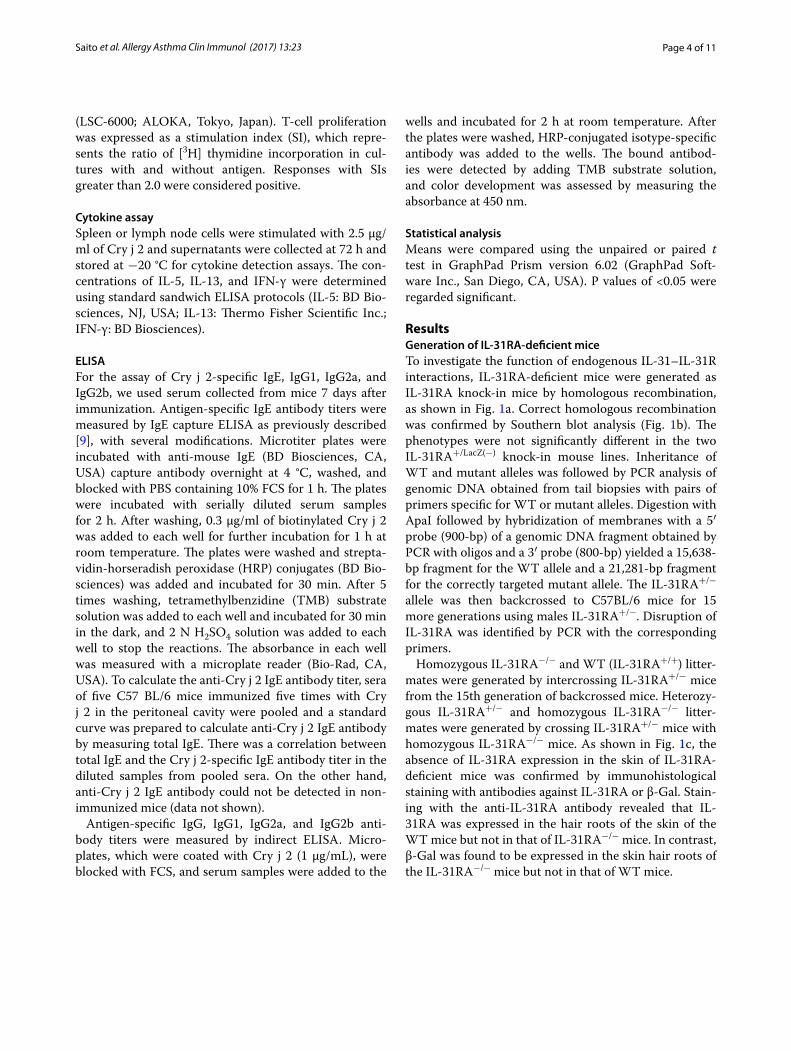
IL‑31RA‑deficient mice show decreased specific Th2 immune responses following nasal injection of Cry j 2In order to clearly analyze the differential effects of tis-sue-specific IL-31R signaling on Th2 immune responses, mice were injected nasally or intraperitoneally with the Th2 antigen Cry j 2. First, to examine the positive role of IL-31–IL-31R interactions in the skin on Th2 immune responses, IL-31RA-deficient and WT mice were injected nasally with Cry j 2, one of the major allergens of Japanese cedar pollen, which induces Th2 polarized and allergic inflammation [10]. Following repeated nasal administra-tion of Cry j 2, the Cry j 2-specific serum IgE and IgG1 levels in IL-31RA-deficient mice were significantly lower than those observed in the serum of WT mice injected nasally with Cry j 2 (Fig. 2a). To evaluate the effects of tis-sue-specific IL-31R signaling on Th2 immune responses more accurately, we used the Cry j 2 allergen as an immu-nogen without any adjuvants to induce Th2 immune responses, as Cry j 2 has protease activity similar to that
of Th2 adjuvants [11]. However, when Cry j 2 was admin-istered in a mixture with Alum, there was no difference in the level of induced Th2 immune response between IL-31RA-deficient and WT mice (data not shown), even though Alum adjuvants strongly induce antigen-specific Th2 immune responses [12]. To confirm our previous observations that IL-31–IL-31R interactions positively influence Th2 immune responses following nasal admin-istration of Cry j 2 (Fig. 2a), heterozygous IL-31RA+/− and homozygous IL-31RA−/− littermates were generated by crossing IL-31RA+/− with IL-31RA−/− mice. The lit-termates were nasally administered Cry j 2 and examined for Cry j 2-specific Th2 immune responses. Consistent with previous findings, in IL-31RA−/−, Cry j 2-specific IgE and IgG1 (Fig. 2b) serum levels were significantly lower than those in IL-31RA+/− mice. In addition, serum levels of IgG2b (Fig. 2b), which are influenced by Th2 immune responses [13], were also significantly lower in IL-31RA−/− mice than in IL-31RA+/− mice. In contrast, serum IgG2a (Fig. 2b), which is influenced by IFN-γ secreted by Th1-cells [14], did not differ between the two strains. Furthermore, in IL-31RA-deficient mice, not only antigen-specific proliferative responses (Fig. 3a) of spleen cells but also secretion levels of the Th2 cytokines IL-5 (Fig. 3b) and IL-13 (Fig. 3c) were significantly lower than those in WT mice injected nasally with Cry j 2. These results demonstrated that IL-31R signaling following nasal administration of an allergen can positively regu-late the Th2 immune response, which is consistent with the findings of numerous previous studies that have shown that IL-31R signaling exacerbates Th2-associated immune responses [1–3, 15–17].
Furthermore, spleen cells isolated from IL-31RA−/− mice nasally administered Cry j 2 showed significantly lower levels of Cry j 2-specific T-cell proliferative responses (Fig. 4a) and Th2 cytokine IL-5 and IL-13 secretion than did IL31 RA+/− mice nasally administered Cry j 2 (Fig. 4b, c). In addition, IL31 RA−/− mice nasally administered Cry j 2 in which submandibular lymph node cells were draining showed significantly lower lev-els of Cry j 2-specific Th2 cytokine IL-5 and IL-13 secre-tion than IL31 RA+/− mice nasally administered Cry j 2 (Fig. 5a, b), whereas IFN-γ secretion was not detected in either strain of immunized mice (Fig. 5c). Collectively, these results strongly demonstrated that IL-31R signaling following nasal administration of an allergen can posi-tively regulate Th2-associated immune responses.
IL‑31RA‑deficient mice intraperitoneally injected with Cry j 2 show heightened specific Th2 immune responsesTo investigate whether IL-31R signaling in differ-ent cells and tissues influences the patterns of Th2 immune responses, IL-31RA-deficient (IL-31RA−/−)
0
1
2
3
IL-31RA
U/m
l
+/+ -/-
IgE
810x 270x 90x0
1
2
3
4 IgG1
Dilution
O.D
. *
+/--/-
*
810x 270x 90x0
1
2
3
4*
IgG1
O.D
.
Dilution
+/+-/-
IgG2a
270x 90x 30x0.0
0.2
0.4
0.6
0.8
Dilution
O.D
.
*
O.D
.
270x 90x 30x0
1
2
3
*
Dilution
IgG2b+/--/-
a
0
2
4
6
8
IL-31RA+/- -/-
**U/m
l
IgEb
+/--/-
*
Fig. 2 IL-31RA−/− mice nasally injected with Cry j 2 exhibit decreased antigen-specific Th2-mediated antibody responses. Serum from WT (IL-31RA+/+, N = 6) and KO (IL-31RA−/−, N = 7) mice following 15 nasal administrations of Cry j 2 was assayed for antigen-specific IgE and IgG1 antibodies (a). Littermate IL-31RA−/− mice (N = 15) and IL-31RA+/− control mice (N = 10) were nasally administered Cry j 2 antigen five times a week for induction of specific Th2 responses. Serum from littermate mice following 15 nasal administrations was assayed for antigen-specific IgE, IgG1, IgG2a, and IgG2b levels (b) by ELISA. The data shown are of one experiment representative of three independent experiments. Data are presented as the mean ± SD. Unpaired t test; *P < 0.05. **P < 0.01

Page 6 of 11Saito et al. Allergy Asthma Clin Immunol (2017) 13:23
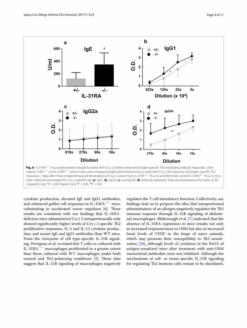
and IL-31RA+/− littermate mice were intraperitoneally administered Cry j 2. Following a repeated cycle of intra-peritoneal administration with Cry j 2, the level of Cry j 2-specific serum IgE (Fig. 6a), IgG1 (Fig. 6b), and IgG2a (Fig. 6c) in IL-31RA-deficient mice were significantly higher than IL-31RA+/− mice intraperitoneally admin-istered Cry j 2. On the other hand, the serum level of IgG2a (Fig. 6c) did not differ between the two strains. Furthermore, in IL-31RA-deficient mice, the secretion levels of Th2 cytokines IL-5 and IL-13 (Fig. 7a, b) were significantly higher than those in IL-31RA+/− mice injected intraperitoneally with Cry j 2. In contrast, IFN-γ production by Th1-cells did not differ between the two strains. These data are consistent with the increase in Th2
cytokines observed in the draining lymph node of the lungs in IL-31RA−/− mice following S. mansoni egg injec-tion [5], and support a role for IL-31–IL-31R interactions in limiting intestinal Th2 cytokine production following Trichuris infection [6]. These results demonstrate that IL-31R signaling in mice intraperitoneally administered Cry j 2 negatively regulates the Th2-associated immune responses, even though IL-31R signaling following nasal administration positively regulates the Th2 responses. Collectively, these data demonstrated that IL-31R sign-aling following nasal administration of an allergen posi-tively regulates, whereas that following intraperitoneal administration negatively regulates, antigen-specific Th2 responses. These results indicated that regulation of the
+/+ -/-0246810
IL-31RA
S.I
* P = 0.0204
+/+ -/-0
200
400
600
800
IL-31RA
IL-5
(pg/
ml)
** P = 0.0086
+/+ -/-0
500
1000
1500
2000
IL-31RA
IL-1
3 (p
g/m
l )
P = 0.0164*
a
b c
Fig. 3 IL-31RA−/− mice nasally injected with Cry j 2 show decreased antigen-specific proliferative and Th2 cytokine responses. 7 days after 15 nasal administrations of Cry j 2, spleen cells from WT (IL-31RA+/+, N = 6) and KO (IL-31RA−/−, N = 7) mice were cultured for 88 h in the presence or absence of Cry j 2 (0.1 μg/ml) and pulsed for the last 16 h with 0.5 μCi of tritiated thymidine. The results of T-cell proliferation (a) are expressed in terms of a stimulation index (SI) calculated by dividing counts per minute (cpm) in the presence of antigen with cpm in the absence of antigen. Supernatants obtained from a 72-h cell culture were assayed by ELISA for IL-13 (b) and IL-5 (c). Data are presented as the mean ± SD

Page 7 of 11Saito et al. Allergy Asthma Clin Immunol (2017) 13:23
Th2 immune response depends on the cell types express-ing IL-31RA, in which IL-31R signaling plays an impor-tant role.
DiscussionIn this study, in order to investigate the differential effects of cell- and tissue-specific IL-31R signaling on the induc-tion of antigen-specific Th2 immune responses, Cry j 2, one of the major allergens of Japanese cedar pollen, was administered either nasally or intraperitoneally into IL-31RA-deficient and WT mice. After repeated nasal administration of Cry j 2, IL-31RA-deficient mice showed significantly lower levels of Cry j 2-specific Th2 prolifera-tive responses, IL-5 and IL-13 cytokine production, and Th2-mediated Cry j 2-specific IgE, IgG1, and IgG2b anti-body responses than IL-31RA+/+ WT mice. Even when
compared with IL-31RA+/− littermate mice, IL-31RA-deficient mice nasally administered Cry j 2 showed lower levels of Th2 immune responses. In contrast, IL-31RA-deficient mice intraperitoneally administered Cry j 2 showed significantly higher levels of Cry j 2-specific Th2 proliferative responses, IL-5 and IL-13 cytokine produc-tion, and Th2-mediated Cry j 2-specific IgE, IgG1, and IgG2b antibody responses than did IL-31RA+/− litter-mates. In contrast, even if IL-31RA was deficient, Th1 cytokine (IFN-γ) production and Th1-mediated IgG2a antibody response were not affected. These data indicate that IL-31–IL-31R interactions in mice receiving nasal administration of an allergen positively regulate Th2 immune responses, whereas in those receiving intraperi-toneal administration, these interactions negatively regu-late Th2 immune responses.
- 0 .1 0 .5 2 .50
2
4
6
8
C ry j 2 (µg /m l)
S.I
IL 3 1 R A + /-
IL 3 1 R A -/-
P = 0 .0 1 5 3
P = 0 .0 4 6 5
P = 0 .0 4 0 2
*
*
*a
b c
+/- -/-0
200
400
600
IL-31RA
IL-1
3(pg
/ml)
IL-5
(pg/
ml)
+/- -/-0
200
400
600
800
1000
IL-31RA
* P=0.0208 * P=0.031
Fig. 4 Decreased antigen-specific proliferative and Th2 cytokine responses of spleen cells from IL-31RA−/− mice nasally injected with Cry j 2. Seven days after 19 nasal administrations of Cry j 2, spleen cells from six representative IL-31RA−/− and littermate control IL-31RA+/− mice were cultured for 88 h in the presence or absence of the indicated dose of Cry j 2 and pulsed for the last 16 h with 0.5 μCi of tritiated thymidine to analyze antigen-specific T-cell proliferative responses (a). Supernatants from 72-h cell cultures were assayed for IL-13 (b) and IL-5 (c). Data are presented as the mean ± SD

Page 8 of 11Saito et al. Allergy Asthma Clin Immunol (2017) 13:23
Our finding that IL-31R signaling following nasal admin-istration of an allergen positively regulates the Th2 immune responses may be explained by the findings of previous studies from the perspective of the cell type expressing IL-31RA involved in induction of the Th2 immune response [18, 19]. These studies showed that IL-31 induces cytokine and chemokine production from human bronchial epithe-lial cells through activation of mitogen-activated protein kinase signaling pathways related to an allergic response. Stott et al. [19] also indicated primary bronchial epithelial cells treated with IL-31 induced proinflammatory genes, such as CCL2 and granulocyte colony-stimulating factor, suggesting the proinflammatory role of IL-31 in amplifying existing Th2 reactions.
In contrast to our observation that IL-31R signaling following nasal administration of an allergen positively
regulates Th2 immune responses, we found that intra-peritoneal administration of Cry j 2 resulted in negative regulation of antigen-specific Th2 immune responses. Previous reports have also indicated that IL-31 negatively regulates the development of the Th2-type response [5, 6], which is in accordance with our findings regarding intraperitoneal administration. Perrigoue et al. showed that IL-31RA-deficient mice exhibited elevated expres-sion of Th2 cytokines and developed exacerbated airway inflammation following exposure to S. mansoni eggs, indicating a negative regulatory role for endogenous IL-31–IL-31R interactions in limiting type 2 inflam-mation in the lungs [5]. Furthermore, they showed that in the absence of IL-31RA expression, the magnitude of helminth-induced type 2 immune responses is sig-nificantly increased, as illustrated by heightened Th2
a b
c
+/- -/-0
50
100
150
200
IL-31RA
IL-1
3(pg
/ml)
** P=0.0010
+/- -/-0
100
200
300
400
500
IL-31RA
IL-5
(pg/
ml)
*** P=0.0004
+/- -/-0
100
200
300
400
500
IL-31RA
IFN
-γ(p
g/m
l)
N.D N.D
Fig. 5 Decreased antigen-specific Th2 cytokine responses of submandibular lymph node cells from IL-31RA−/− mice nasally injected with Cry j 2. 7 days after 19 nasal administrations of Cry j 2, submandibular lymph node cells from six representative IL-31RA−/− and littermate control IL-31RA+/− mice were pooled and cultured. Supernatants from a 72-h cell culture were assayed by ELISA for IL-5 (a), IL-13 (b), and IFN-γ (c). Data are presented as the mean ± SD
Page 9 of 11Saito et al. Allergy Asthma Clin Immunol (2017) 13:23
cytokine production, elevated IgE and IgG1 antibodies, and enhanced goblet cell responses in IL-31RA−/− mice, culminating in accelerated worm expulsion [6]. These results are consistent with our findings that IL-31RA-deficient mice administered Cry j 2 intraperitoneally only showed significantly higher levels of Cry j 2-specific Th2 proliferative responses, IL-5 and IL-13 cytokine produc-tion, and serum IgE and IgG1 antibodies than WT mice. From the viewpoint of cell type-specific IL-31R signal-ing, Perrigoue et al. revealed that T cells co-cultured with IL-31RA−/− macrophages proliferated to a greater extent than those cultured with WT macrophages under both neutral and Th2-polarizing conditions [5]. These data suggest that IL-31R signaling of macrophages negatively
regulates the T cell stimulatory function. Collectively, our findings lead us to propose the idea that intraperitoneal administration of an allergen negatively regulates the Th2 immune response through IL-31R signaling of abdomi-nal macrophages. Bilsborough et al. [7] indicated that the absence of IL-31RA expression in mice results not only in increased responsiveness to OSM but also in increased basal levels of VEGF in the lungs of naive animals, which may promote their susceptibility to Th2 sensiti-zation [20], although levels of cytokines in the BALF of antigen-sensitized mice after treatment with anti-OSM monoclonal antibodies were not inhibited. Although the mechanisms of cell- or tissue-specific IL-31R signaling for regulating Th2 immune cells remain to be elucidated,
*
0
200
400
600
IL-31RA+/- -/-
U/m
lIgE
0
1
2
3
4 +/--/-
5x25x125x625x
Dilution (x 104)
O.D
.
IgG1ba
0
1
2
3
4 IgG2a
O.D
.
810x 270x 90x 30x
Dilution
+/--/-
c
#
0
1
2
3
4 IgG2b
270x 90x 30x 10x
O.D
.
Dilution
+/--/-
d
##
Fig. 6 IL-31RA−/− mice administered intraperitoneally with Cry j 2 exhibit enhanced antigen-specific Th2-mediated antibody responses. Litter-mate IL-31RA−/− and IL-31RA+/− control mice were intraperitoneally administered once a week with Cry j 2 for induction of antigen-specific Th2 responses. 7 days after three intraperitoneal administrations of Cry j 2, serum from IL-31RA−/− (N = 5) and littermate control IL-31RA+/− (N = 5) mice were collected and assayed for Cry j 2-specific IgE (a), IgG1 (b), IgG2a (c), and IgG2b (d) antibody responses. Data are presented as the mean ± SD. Unpaired t test; *P < 0.05. Paired t test; #P < 0.05, ##P < 0.01

Page 10 of 11Saito et al. Allergy Asthma Clin Immunol (2017) 13:23
determining the tissue distribution of receptor variants of IL-31RA and OSMR [1] may be critical to our under-standing of the regulation of Th2 immune responses induced by different routes of administration.
ConclusionsThe results of the present study indicate that IL-31R sign-aling positively regulates Th2 responses induced by nasal administration of an allergen but negatively regulates these responses following intraperitoneal administra-tion. Collectively, the data suggest that regulation of Th2 immune responses might be dependent on tissue-specific cell types expressing IL-31RA.
AbbreviationsIL-31: interleukin-31; OSMR: oncostatin M receptor; KO: knock-out; IL-31R: IL-31 receptor; Cry j 2: cryptomeria japonica 2.
Authors’ contributionsSS was a major contributor in writing the manuscript. SS, NA, and HK contributed to the generation of the IL-31RA-deficient mice. AA, HI, and IA contributed to the generation of backcrossed mice. AA, SS, and ST
performed the experimental work. All authors read and approved the final manuscript.
Author details1 Division of Molecular Immunology, Research Center for Medical Science, The Jikei University School of Medicine, 3-25-8 Nishi-shinbashi, Minato-ku, Tokyo 105-8461, Japan. 2 Department of Dermatology, The Jikei University School of Medicine, 3-25-8 Nishi-shinbashi, Minato-ku, Tokyo, Japan. 3 Depart-ment of Otolaryngology, The Jikei University School of Medicine, 3-25-8 Nishi-shinbashi, Minato-ku, Tokyo, Japan. 4 Division of Rheumatology, Depart-ment of Internal Medicine, The Jikei University School of Medicine, 3-25-8 Nishi-shinbashi, Minato-ku, Tokyo, Japan. 5 Animal Resource Development Unit and Genetic Engineering Team, RIKEN Center for Life Science Technologies, 2-2-3 Minatojima-minamimachi, Chuou-ku, Kobe, Hyogo 650-0047, Japan.
AcknowledgementsWe would like to thank Mayumi Tsuda and Yoko Natake for technical assis-tance, and Akiko Itasaka for preparing the manuscript. We would like to thank Editage (http://www.editage.jp) for English language editing.
Competing interestsThe authors declare that they have no competing interests.
Availability of data and materialsThe dataset supporting the conclusions of this article is included within the article. Further data are available from the corresponding author on reason-able request.
+/- -/-0
2000
4000
6000
IL-31RA
IL-5
(pg/
ml)
* P=0.0113
0
20000
40000
60000
IL-1
3 (p
g/m
l)
P = 0.0458*
+/- -/-IL-31RA
ba
c
+/- -/-0
100
200
300
400
500
IL-31RA
IFN
-γ(p
g/m
l)
Fig. 7 IL-31RA−/− mice intraperitoneally administered Cry j 2 show enhanced antigen-specific proliferative and Th2 cytokine responses. For the analysis of Th2 cytokine responses, spleen cells were collected from mice 7 days after five intraperitoneal administrations of Cry j 2 and cultured with Cry j 2. Supernatants from 72-h cell cultures were assayed for IL-5 (a), IL-13 (b), and IFN-γ (c). The data shown are of one experiment representa-tive of five independent experiments with 4 to 10 mice per group. Data are presented as the mean ± SD

Page 11 of 11Saito et al. Allergy Asthma Clin Immunol (2017) 13:23
• We accept pre-submission inquiries
• Our selector tool helps you to find the most relevant journal
• We provide round the clock customer support
• Convenient online submission
• Thorough peer review
• Inclusion in PubMed and all major indexing services
• Maximum visibility for your research
Submit your manuscript atwww.biomedcentral.com/submit
Submit your next manuscript to BioMed Central and we will help you at every step:
Ethics approval and consent to participateAll animal studies were approved by the Institutional Animal Care and Use Committee of Jikei University (2015-134).
FundingThis work was supported by the Ministry of Education, Culture, Sports, Science and Technology of Japan (Grant Numbers, 19591329 and 22591230).
Publisher’s NoteSpringer Nature remains neutral with regard to jurisdictional claims in pub-lished maps and institutional affiliations.
Received: 16 November 2016 Accepted: 8 April 2017
References 1. Dillon SR, Sprecher C, Hammond A, Bilsborough J, Rosenfeld-Franklin M,
Presnell SR, et al. Interleukin 31, a cytokine produced by activated T cells, induces dermatitis in mice. Nat Immunol. 2004;5(7):752–60.
2. Bilsborough J, Leung DY, Maurer M, Howell M, Boguniewicz M, Yao L, et al. IL-31 is associated with cutaneous lymphocyte antigen-positive skin homing T cells in patients with atopic dermatitis. J Allergy Clin Immunol. 2006;117(2):418–25.
3. Ezzat MH, Hasan ZE, Shaheen KY. Serum measurement of interleukin-31 (IL-31) in paediatric atopic dermatitis: elevated levels correlate with sever-ity scoring. J Eur Acad Dermatol Venereol. 2011;25(3):334–9.
4. Neis MM, Peters B, Dreuw A, Wenzel J, Bieber T, Mauch C, et al. Enhanced expression levels of IL-31 correlate with IL-4 and IL-13 in atopic and aller-gic contact dermatitis. J Allergy Clin Immunol. 2006;118(4):930–7.
5. Perrigoue JG, Li J, Zaph C, Goldschmidt M, Scott P, de Sauvage FJ, et al. IL-31–IL-31R interactions negatively regulate type 2 inflammation in the lung. J Exp Med. 2007;204(3):481–7.
6. Perrigoue JG, Zaph C, Guild K, Du Y, Artis D. IL-31–IL-31R interactions limit the magnitude of Th2 cytokine-dependent immunity and inflammation following intestinal helminth infection. J Immunol. 2009;182(10):6088–94.
7. Bilsborough J, Mudri S, Chadwick E, Harder B, Dillon SR. IL-31 receptor (IL-31RA) knockout mice exhibit elevated responsiveness to oncostatin M. J Immunol. 2010;185(10):6023–30.
8. Diveu C, Lelievre E, Perret D, Lak-Hal AH, Froger J, Guillet C, et al. GPL, a novel cytokine receptor related to GP130 and leukemia inhibitory factor receptor. J Biol Chem. 2003;278(50):49850–9.
9. Sakaguchi M, Inouye S, Yasueda H, Irie T, Yoshizawa S, Shida T. Measure-ment of allergens associated with dust mite allergy. II. Concentrations of airborne mite allergens (Der I and Der II) in the house. Int Arch Allergy Appl Immunol. 1989;90(2):190–3.
10. Hosoki K, Boldogh I, Sur S. Innate responses to pollen allergens. Curr Opin Allergy Clin Immunol. 2015;15(1):79–88.
11. Chapman MD, Wunschmann S, Pomes A. Proteases as Th2 adjuvants. Curr Allergy Asthma Rep. 2007;7(5):363–7.
12. Brewer JM, Conacher M, Hunter CA, Mohrs M, Brombacher F, Alexander J. Aluminium hydroxide adjuvant initiates strong antigen-specific Th2 responses in the absence of IL-4- or IL-13-mediated signaling. J Immunol. 1999;163(12):6448–54.
13. Finkelman FD, Holmes J, Katona IM, Urban JF Jr, Beckmann MP, Park LS, et al. Lymphokine control of in vivo immunoglobulin isotype selection. Annu Rev Immunol. 1990;8:303–33.
14. Bossie A, Vitetta ES. IFN-gamma enhances secretion of IgG2a from IgG2a-committed LPS-stimulated murine B cells: implications for the role of IFN-gamma in class switching. Cell Immunol. 1991;135(1):95–104.
15. Takaoka A, Arai I, Sugimoto M, Yamaguchi A, Tanaka M, Nakaike S. Expres-sion of IL-31 gene transcripts in NC/Nga mice with atopic dermatitis. Eur J Pharmacol. 2005;516(2):180–1.
16. Kim S, Kim HJ, Yang HS, Kim E, Huh IS, Yang JM. IL-31 Serum protein and tissue mRNA levels in patients with atopic dermatitis. Ann Dermatol. 2011;23(4):468–73.
17. Raap U, Weissmantel S, Gehring M, Eisenberg AM, Kapp A, Folster-Holst R. IL-31 significantly correlates with disease activity and Th2 cytokine levels in children with atopic dermatitis. Pediatric Allergy Immunol Official Public Eur Soc Ped Allergy Immunol. 2012;23(3):285–8.
18. Ip WK, Wong CK, Li ML, Li PW, Cheung PF, Lam CW. Interleukin-31 induces cytokine and chemokine production from human bronchial epithelial cells through activation of mitogen-activated protein kinase signalling pathways: implications for the allergic response. Immunology. 2007;122(4):532–41.
19. Stott B, Lavender P, Lehmann S, Pennino D, Durham S, Schmidt-Weber CB. Human IL-31 is induced by IL-4 and promotes TH2-driven inflammation. J Allergy Clin Immunol. 2013;132(2):446–54.
20. Chen SH, Benveniste EN. Oncostatin M: a pleiotropic cytokine in the central nervous system. Cytokine Growth Factor Rev. 2004;15(5):379–91.