Selenium, Zinc, and Thyroid Hormones In Healthy Subjects
11
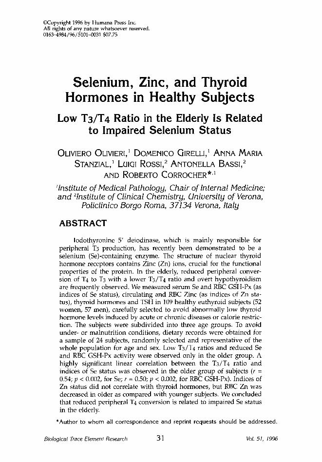
~Copyright 1996 by Humana Press Inc. All rights of any nature whatsoever reserved. 0163-4984/96/5101-0031 $07.75 Selenium, Zinc, and Thyroid Hormones in Healthy Subjects Low Ts/T4 Ratio in the Elderly Is Related to Impaired Selenium Status OLIVIERO OLIVIERI, 1 DOMENICO GIRELLI, 1 ANNA MARIA STANZIAL,1 LUIGI ROSSI, 2 ANTONELLA BASSi, 2 AND ROBERTO CORROCHER *'1 'Institute of Medical Pathology, Chair of Internal Medicine; and 2Institute of Clinical Chemistry, University of Verona, Policlinico Borgo Roma, 37134 Verona, Italy ABSTRACT Iodothyronine 5' deiodinase, which is mainly responsible for peripheral T3 production, has recently been demonstrated to be a selenium (Se)-containing enzyme. The structure of nuclear thyroid hormone receptors contains Zinc (Zn) ions, crucial for the functional properties of the protein. In the elderly, reduced peripheral conver- sion of T4 to T3 with a lower T3/T4 ratio and overt hypothyroidism are frequently observed. We measured serum Se and RBC GSH-Px (as indices of Se status), circulating and RBC Zinc (as indices of Zn sta- tus), thyroid hormones and TSH in 109 healthy euthyroid subjects (52 women, 57 men), carefully selected to avoid abnormally low thyroid hormone levels induced by acute or chronic diseases or calorie restric- tion. The subjects were subdivided into three age groups. To avoid under- or malnutrition conditions, dietary records were obtained for a sample of 24 subjects, randomly selected and representative of the whole population for age and sex. Low T3/T4 ratios and reduced Se and RBC GSH-Px activity were observed only in the older group. A highly significant linear correlation between the T3/T4 ratio and indices of Se status was observed in the older group of subjects (r = 0.54; p < 0.002, for Se; r = 0.50; p < 0.002, for RBC GSH-Px). Indices of Zn status did not correlate with thyroid hormones, but RBC Zn was decreased in older as compared with younger subjects. We concluded that reduced peripheral T4 conversion is related to impaired Se status in the elderly. *Author to whom all correspondence and reprint requests should be addressed. Biological Trace Element Research 31 Vol. 51, 1996
-
Upload
independent -
Category
Documents
-
view
1 -
download
0
Transcript of Selenium, Zinc, and Thyroid Hormones In Healthy Subjects
~Copyright 1996 by Humana Press Inc. All rights of any nature whatsoever reserved. 0163-4984/96/5101-0031 $07.75
Selenium, Zinc, and Thyroid Hormones in Healthy Subjects
Low Ts/T4 Ratio in the Elderly Is Related to Impaired Selenium Status
OLIVIERO OLIVIERI, 1 DOMENICO GIRELLI, 1 A N N A MARIA
STANZIAL,1 LUIGI ROSSI, 2 ANTONELLA BASSi, 2
AND ROBERTO CORROCHER *'1
'Institute of Medical Pathology, Chair of Internal Medicine; and 2Institute of Clinical Chemistry, University of Verona,
Policlinico Borgo Roma, 37134 Verona, Italy
ABSTRACT
Iodothyronine 5' deiodinase, which is mainly responsible for peripheral T3 production, has recently been demonstrated to be a selenium (Se)-containing enzyme. The structure of nuclear thyroid hormone receptors contains Zinc (Zn) ions, crucial for the functional properties of the protein. In the elderly, reduced peripheral conver- sion of T4 to T3 with a lower T3/T4 ratio and overt hypothyroidism are frequently observed. We measured serum Se and RBC GSH-Px (as indices of Se status), circulating and RBC Zinc (as indices of Zn sta- tus), thyroid hormones and TSH in 109 healthy euthyroid subjects (52 women, 57 men), carefully selected to avoid abnormally low thyroid hormone levels induced by acute or chronic diseases or calorie restric- tion. The subjects were subdivided into three age groups. To avoid under- or malnutrition conditions, dietary records were obtained for a sample of 24 subjects, randomly selected and representative of the whole population for age and sex. Low T3/T4 ratios and reduced Se and RBC GSH-Px activity were observed only in the older group. A highly significant linear correlation between the T3/T4 ratio and indices of Se status was observed in the older group of subjects (r = 0.54; p < 0.002, for Se; r = 0.50; p < 0.002, for RBC GSH-Px). Indices of Zn status did not correlate with thyroid hormones, but RBC Zn was decreased in older as compared with younger subjects. We concluded that reduced peripheral T4 conversion is related to impaired Se status in the elderly.
*Author to whom all cor re spondence and reprint requests should be addressed .
Biological Trace Element Research 31 Vol. 51, 1996
32 Olivieri et al
Index Entries: Selenium; glutathione peroxidase; zinc; aging; thyroid hormones; TSH.
INTRODUCTION
Many processes involved in growth, development, and metabolism in humans are under the direct or indirect control of the thyroid hor- mones. Full activity of the thyroid hormones requires the deiodination of thyroxine (T4) to tri-iodothyronine (T3), which functions through a spe- cific nuclear receptor protein (1) capable of modulating expression of spe- cific control regions of target genes (2). The thyroid hormone receptor has been cloned, and protein analysis has shown a region containing Zinc (Zn) ions crucial for binding the receptors to their target genes (3). Thus, it has been postulated that Zn status may be important for the full bio- logical functioning of T3 (4).
T3 is produced by the thyroid gland but results mainly from the peripheral deiodination of T4 catalyzed by iodothyronine 5' deiodinase (ID) (5). Three isoenzymes of iodothyronine 5' deiodinase have been identified (5): type I (ID-I) is found in the liver, kidney, and thyroid; ID- II is abundant in the brain, brown adipose tissue, and pituitary; and ID- III is present in the brain and placenta.
ID-I, which is mainly responsible for peripheral T3 production, has recently been demonstrated to be a selenium (Se)-containing enzyme (6,7); this observation agrees with previous reports that an Se-deficient diet inhibits ID-I in rats (8,9). Thus, after a long period of time in which the only known Se-containing enzyme was glutathione peroxidase (GSH- Px) and the metabolic function of Se was related to this enzymatic activ- ity, considerable interest now focuses on the relationships between Se status and thyroid metabolism. Most of the available data have been obtained in animals. In humans, attention has mainly been devoted to the possible role of impaired Se status in determining hypothyroidism, and particularly the detrimental effects of combined Se and iodine defi- ciency on thyroid activity (10-12).
No clinical studies relating Se status, thyroid function, and aging have been published to date. Advanced age is a well-recognized condi- tion characterized by important changes in thyroid function (13). In addi- tion to a high frequency of overt hypothyroidism (13), reduced iodine uptake, weight, colloid content, follicular volume of the gland, and reduced peripheral conversion of T4 to T3 with a lower T3/T4 ratio are usually observed in elderly euthyroid people (14). Numerous features of the aging process associated with impaired or slowed cellular functions mimic to some extent those of hypothyroidism, so that it is sometimes very difficult to clinically distinguish the manifestations of the illness from those related to aging per se. As a result of such similarities, the con- cept of aging as a sort of "tissue hypothyroidism" has also been pro- posed (14).
Biological Trace Element Research Vol. 51, 1996
Se and Zn Status and Thyroid Function 33
Since Se status tends to decline over 60-65 yr of age (15), reduced ID-I activity owing to impaired Se availability may explain the reduction in the serum T3/T4 ratio observed in elderly people. To test this hypoth- esis, we investigated the relationships between age, Se status, and thy- roid hormones in three groups of healthy subjects of different age, matched for sex distribution.
In view of the role of Zn as a constituent of T3 nuclear receptors, we also measured serum and red blood cell Zn in order to evaluate possible interactions with circulating levels of thyroid hormones.
MATERIAL AND METHODS
Subjects An initial age-based, sex-balanced selection of 500 subjects was
obtained by applying tables of random numbers to the population of Nove (a village near Vicenza, northern Italy) figuring on the electoral register.
A further selection was performed in order to study the conse- quences of aging without the confounding effects of chronic or acute dis- eases that might affect levels of both Se and circulating thyroid hormones. Thus, very strict criteria were adopted to define the "healthy" population. We therefore excluded all subjects known to be suffering from: hypertension; diabetes; hyperlipidaemia; liver, neoplastic, renal, endocrinological, or immunological diseases; coagulative disorders; or acute intercurrent illness. Three seemingly euthyroid subjects with TSH values higher than 5 mU/L were also excluded. One hundred and nine subjects (52 women, 57 men) were finally admitted to the study. None of them were institutionalized or on a special diet, and none were taking preparations containing multivitamins and/or trace elements. The study population was divided into three sex-matched groups, according to the following schedule:
Group I. 36 subjects (20-44 yr old); Group II. 36 subjects (45-65 yr old); and Group III. 37 subjects (over 65 yr old).
In order to exclude conditions of under- or malnutrition, a 7-d food record was obtained for a representative sample of the subjects (n = 24, eight per group), randomly selected from among the age groups. Informed consent was obtained from all participants according to the ethical guidelines of the Helsinki Declaration.
Biochemical Analysis Blood samples were collected after overnight fasting, transported to
the laboratories in Verona within 1 h, and processed immediately. Haemogram was automatically measured using the Technicon H-2 Sys- tem (Technicon Instruments, Tarrytown NY). Plasma triglycerides, total
Biological Trace Element Research Vol. 51, 1996
34 Olivieri et al
and HDL-cholesterol and serum albumin were determined using a Tech- nicon DAX 96 automated analyzer (Technicon). Serum T3, T4, FT4, and TSH were assayed by a chemiluminescence immunoassay system (LIA- mat, Byk-Sangtec Diagnostica, FRG) (16). To prepare haemolysates for enzyme assays, the red blood cells (RBC) were washed and then filtered in cellulose. For serum Se and Zn assay, blood was collected in trace-ele- ment-free vacutainer tubes for metal and metalloid determinations (Bec- ton Dickinson, Rutherford, NJ), containing no additives.
RBC GSH-Px Assay Activity of RBC glutathione peroxidase (GfiH-Px; EC 1.11.1.9), which
accurately reflects the Se intake over 2-3 mo (15), was measured spec- trophotometrically at 25~ in 50 gL of cell lysate, according to the Giin- zler method, using 0.2 mmol/L ter-butyl-hydroperoxide as acceptor substrate, as previously described (17); GSH-Px activity was expressed as IU, defined as gmol NADPH oxidized per minute per g Hb (IU/g Hb).
Se and Zn Assay
Serum fie was determined as previously described (17), using hydride generation atomic absorption spectrometry on serum stored at -20~ until analyzed. Briefly, 1 mL of serum was treated by wet digestion in nitric/hydrochloric acid (1.5/5 v/v). Se concentrations were deter- mined using a G.B.C. 902 atomic absorption spectrophotometer equipped with a hydride generation system and a selenium hollow cathode lamp. Sodium borohydride was used as reductant. A batch of Seronorm trace element serum (Nycomed Pharma As, Oslo, Norway) with a defined Se concentration (mean recommended value = 1.19 gmol/L, range 1.17-1.22; mean value obtained in our laboratory = 1.20 gmol/L) was used as a con- trol reference. The within- and between-run coefficients of variation were 1.7% and 4%, respectively (final concentration of the standard sample employed for these calculations = 0.06 gmol/L).
Concentrations of Zn in serum (1 mL) and in RBC lysates (2 mL of 1:20 dilution in double distilled water) were determined by flame atomic absorption spectrometry (FAAS) using a Perkin Elmer 372 double beam spectrophotometer equipped with an air-acetylene flame burner and hol- low cathode lamp (operated at 15 mA). Atomic absorption was measured at 213.9 nm. The spectral bandwidth was 0.7 nm. Standard solutions con- taining 30 gmol/L of Zn were prepared in double distilled water. All analyses were performed in duplicate. The coefficient of variation for serum and RBC Zn was < 5% (final concentration of the standard sam- ple employed for these calculations = 10 gmol/L). RBC Zn values were standardized for grams of Hb.
Statist ical Methods
Statistical analysis was carried out with an Apple Macintosh SE/30 computer using the Systat 5.0 statistical package. The data are given as
Biological Trace Element Research Vol. 51, 1996
Se and Zn Status and Thyroid Function 35
means _+ standard deviations. Comparison of the various parameters among groups of age was performed using one-way analysis of variance (ANOVA); differences at the 5% significance level (p < 0.05) were assessed by Tukey's post-hoc probability test for multiple comparisons. Simple correlations were determined using the Pearson correlation coef- ficient. Multiple linear regression analysis was used to evaluate the rela- tionships between T3/T4 ratio, Se, and age; multiple r (r), multiple squared r (r2), beta (~) and beta standardized (~ st) coefficients are reported in the results.
RESULTS
Table 1 gives the main features of the subjects, subdivided according to the various age groups. All our subjects had normal nutritional indices such as BMI, Hb, PCV, and serum albumin; dietary records showed no significant differences between the groups.
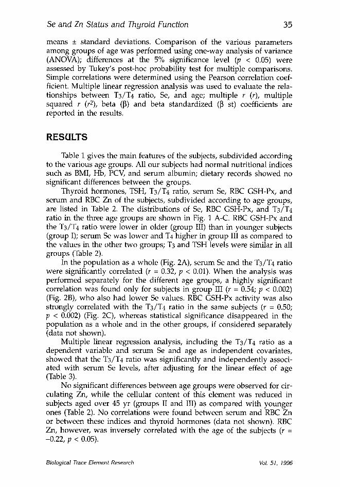
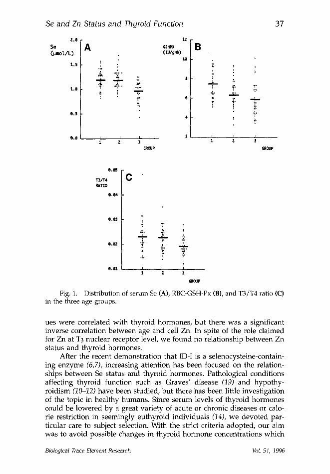
Thyroid hormones, TSH, T3/T4 ratio, serum Se, RBC GSH-Px, and serum and RBC Zn of the subjects, subdivided according to age groups, are listed in Table 2. The distributions of Se, RBC GSH-Px, and T3/T4 ratio in the three age groups are shown in Fig. 1 A-C. RBC GSH-Px and the T3/T4 ratio were lower in older (group III) than in younger subjects (group I); serum Se was lower and T4 higher in group III as compared to the values in the other two groups; T3 and TSH levels were similar in all groups (Table 2).
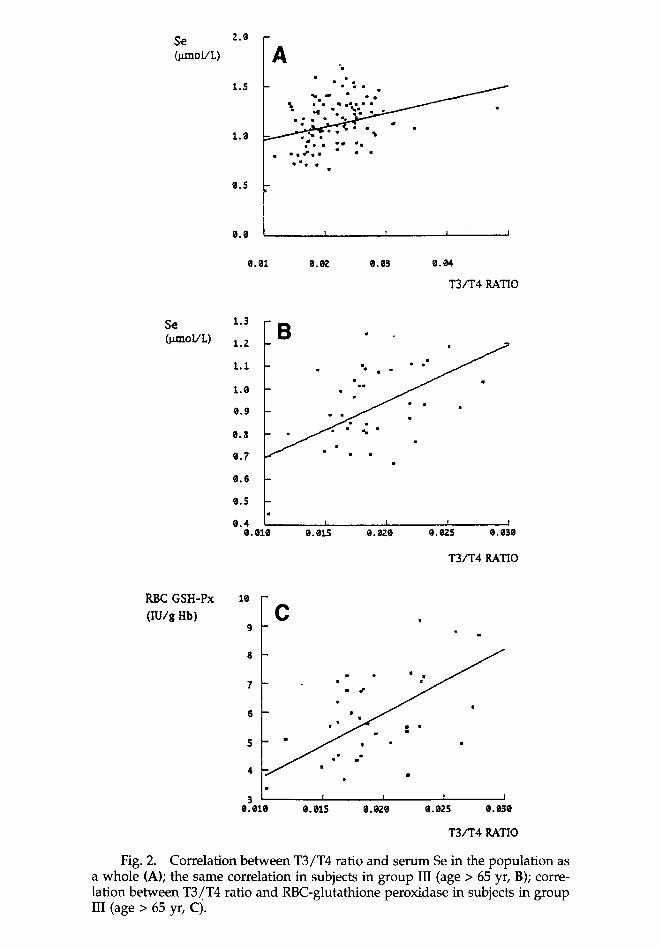
In the population as a whole (Fig. 2A), serum Se and the T3/T4 ratio were significantly correlated (r = 0.32, p < 0.01). When the analysis was performed separately for the different age groups, a highly significant correlation was found only for subjects in group III (r = 0.54; p < 0.002) (Fig. 2B), who also had lower Se values. RBC GSH-Px activity was also strongly correlated with the T3/T4 ratio in the same subjects (r = 0.50; p < 0.002) (Fig. 2C), whereas statistical significance disappeared in the population as a whole and in the other groups, if considered separately (data not shown).
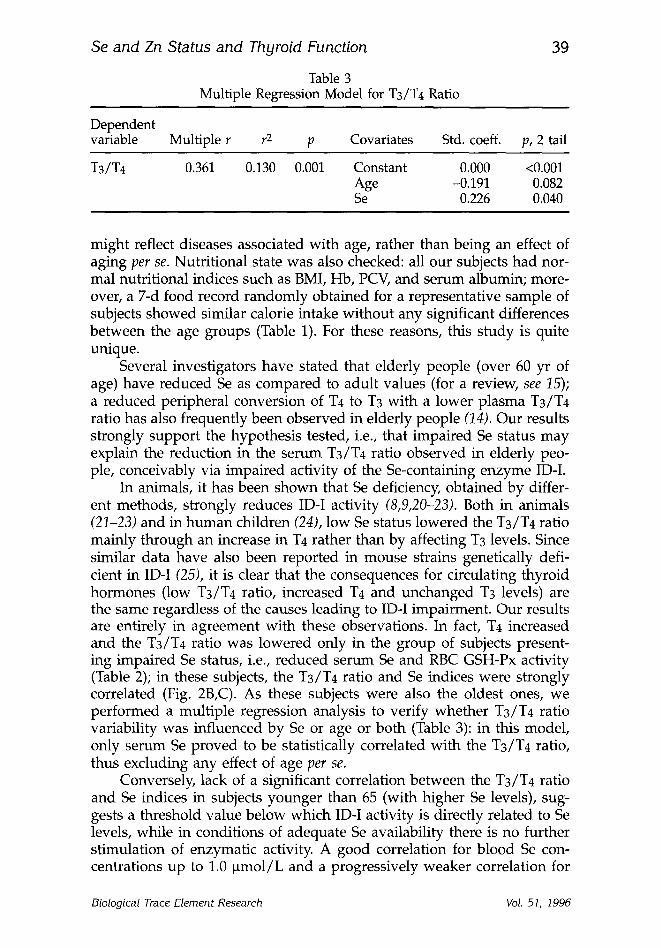
Multiple linear regression analysis, including the T3/T4 ratio as a dependent variable and serum Se and age as independent covariates, showed that the T3/T4 ratio was significantly and independently associ- ated with serum Se levels, after adjusting for the linear effect of age (Table 3).
No significant differences between age groups were observed for cir- culating Zn, while the cellular content of this element was reduced in subjects aged over 45 yr (groups II and III) as compared with younger ones (Table 2). No correlations were found between serum and RBC Zn or between these indices and thyroid hormones (data not shown). RBC Zn, however, was inversely correlated with the age of the subjects (r = -0.22, p < 0.05).
Biological Trace Element Research Vol. 51, 1996

36 Olivieri et al
Table 1 Main Features of the Subjects, Subdivided According to Age Groups
Group I Group II Group III
Parameters Age, yr 20-44 45-64 > 65 BMI, k g / m 2 23.7 + 3.2 a 25.8 + 2.5 a 24.6 -+ 3.2 Cholesterol, m m o l / L 4.85 + 1.08a, b 5.90 -+ 0.87a 6.29 _+ .19 b HDL-choI. , m m o l / L 1.52 + 0.38 1.45 _+ 0.23 1.53 _+ 0.51 Triglycerides, m m o l / L 1.16 + 0.6b 1.54 + 0.7 1.76 +_ 0.8b Albumin, g / L 43.6 + 2.32 43.2 + 2.23 41.6 _+ 4.6 Hemoglob in , g / d L 14.4 _ 2.1 14.15 _+ 2.2 13 _+ 2 . 6 Packed cell vo lume, % 41 _+ 4 41.6 _+ 2.3 38.4 _+ 4.4
Dietary intake, n = 24 Total calories, c a l / K g / d 32.4 + 4.1 32.3 _+ 7.4 33.4 _+ 12.01 Lipids, g / K g / d 0.99 + 0.16 1.09 _+ 0.2 1.04 + 0.5 Carbohydra tes , g / K g / d 4.6 + 1.05 4.3 _+ 1.7 3.6 + 1.8 Proteins, g / K g / d 1.2 _+ 0.3 1.3 _+ 0.3 1.4 _+ 0.6
ap < 0.05 for group I vs II. bp < 0.05 for group ! vs III. The comparisons between age groups were performed using analysis of variance
(ANOVA) and differences at the 5% significance level were assessed by Tukey's test. Means not sharing a common superscript symbol are not significantly different.
Table 2 Thyroid Parameters and Indices of Selenium and Zinc Status
Parameters Group I Group II Group III
Age, yr T3, n m o l / L T4, n m o l / L TSH, m U / L T3/T4 ratio Serum Se, ~tmol/L RBC GSH-Px, I U / g Hb Serum Zn, B m o l / L RBC Zn, n m o l / g Hb
20-44 45-64 > 65 1.87 + 0.34 1.96 _+ 0.38 1.95 _+ 0.41 85.1 _+ 16 a 92.9 +_ 6 b 104 + 25a, b 1.39 -+ 0.55 1.03 _+ 0.62 1.43 _+ 1.05
0.023 _+ 0.007a 0.022 _+ 0.004 0.019 _+ 0.004a 1.19 + 0.19a 1.20 + 0.19 b 0.94 _+ 0.18a,b 7.24 _+ 1.3 a 6.46 _+ 1.48 5.89 _+ 1.68 a 14.9 _ 1.68 15.11 _+ 1.68 13.75 + 1.99 637 _+ 106a, c 532 _+ 76 c 561 + 113a
ap < 0.05 for group I vs III. bp < 0.05 for group II vs III. cp < 0.05 for group I vs II. The comparisons between age groups were performed using analysis of variance
(ANOVA) and differences at the 5% significance level were assessed by Tukey's test. Means not sharing a common superscript symbol are not significantly different.
DISCUSSION
We f o u n d a r e d u c t i o n in ce l lu l a r Z n w i t h a d v a n c i n g a g e (Table 2). U n l i k e s e r u m leve ls , RBC Z n is c o n s i d e r e d a u s e f u l i n d i c a t o r o f Z n s ta - t u s in e u t h y r o i d sub j ec t s (18). N e i t h e r c i r c u l a t i n g l eve l s n o r RBC Z n va l -
Biological Trace Element Research Vol. 51, 1996
Se and Zn Status and Thyroid Function 37
2.0
Se
C~ol/L)
1.5
1.e
e.5
e.O
A GSHPX
C i U / g X b )
~149
o.1.o
.~
I I I
1 2 3
GROUP
12
1Q
13
4.
$
m u i m m .~
i - , i , -
4"
GROUP
T3/T4 RATIO
e.tl4
e.03
e. ;)2
e . a l
C
w
- , , . . ..'. ! "." ~o
";" t - : : : . . . .
o~ .~, '.~
1 2 3
GROUP
Fig. 1. Distribution of serum Se (A), RBC-GSH-Px (B), and T3/T4 ratio (C) in the three age groups.
ues were correlated with thyroid hormones, but there was a significant inverse correlation between age and cell Zn. In spite of the role claimed for Zn at T3 nuclear receptor level, we found no relationship between Zn status and thyroid hormones.
After the recent demonstration that ID-I is a selenocysteine-contain- ing enzyme (6,7), increasing attention has been focused on the relation- ships between Se status and thyroid hormones. Pathological conditions affecting thyroid function such as Graves' disease (19) and hypothy- roidism (10-12) have been studied, but there has been little investigation of the topic in healthy humans. Since serum levels of thyroid hormones could be lowered by a great variety of acute or chronic diseases or calo- rie restriction in seemingly euthyroid individuals (14), we devoted par- ticular care to subject selection. With the strict criteria adopted, our aim was to avoid possible changes in thyroid hormone concentrations which
Biological Trace Element Research Vol. 51, 1996
Se (gmoi/L)
1.5
0.5
O.O
A
�9 ,�9 - �9 ~ o�9 / J
i d o l �9 � 9 e l
n I I ~ 1 l ! I
�9 We �9
0.82 e.03 e.04
T 3 / T 4 RATIO
Se O.Lmol/L) 1.2
1.1
1.0
0 .9
0 .8
0 .7
0 .6
O.S
0.4 O.~lO
w
I T T I
O. 015 O. ;)20 g. 02S Q. ;)30
T 3 / T 4 RATIO
RBC GSH-Px (IU/g 1-15)
10
9
tl
7
6
5
4
3 O. elO
C �9
I
l l i I
O.elS e. ;!,?.0 0. ~)25 0.03fl
T 3 / T 4 RATIO
Fig. 2. Correlation between T3/T4 ratio and serum Se in the population as a whole (A); the same correlation in subjects in group III (age > 65 yr, B); corre- lation between T3/T4 ratio and RBC-glutathione peroxidase in subjects in group III (age > 65 yr, C).
Se and Zn Status and Thyroid Function
Table 3 Multiple Regression Model for T3/T4 Ratio
39
Dependent variable Multiple r r 2 p Covariates Std. coeff, p, 2 tail
T3/T4 0.361 0.130 0.001 Constant 0.000 <0.001 Age -0.191 0.082 Se 0.226 0.040
might reflect diseases associated with age, rather than being an effect of aging per se. Nutritional state was also checked: all our subjects had nor- mal nutritional indices such as BMI, Hb, PCV, and serum albumin; more- over, a 7-d food record randomly obtained for a representative sample of subjects showed similar calorie intake without any significant differences between the age groups (Table 1). For these reasons, this study is quite unique.
Several investigators have stated that elderly people (over 60 yr of age) have reduced Se as compared to adult values (for a review, see 15); a reduced peripheral conversion of T4 to T3 with a lower plasma T3/T4 ratio has also frequently been observed in elderly people (14). Our results strongly support the hypothesis tested, i.e., that impaired Se status may explain the reduction in the serum T3/T4 ratio observed in elderly peo- ple, conceivably via impaired activity of the Se-containing enzyme ID-I.
In animals, it has been shown that Se deficiency, obtained by differ- ent methods, strongly reduces ID-I activity (8,9,20-23). Both in animals (21-23) and in human children (24), low Se status lowered the T3/T4 ratio mainly through an increase in T4 rather than by affecting T3 levels. Since similar data have also been reported in mouse strains genetically defi- cient in ID-I (25), it is clear that the consequences for circulating thyroid hormones (low T3/T4 ratio, increased T4 and unchanged T3 levels) are the same regardless of the causes leading to ID-I impairment. Our results are entirely in agreement with these observations. In fact, T4 increased and the T3/T4 ratio was lowered only in the group of subjects present- ing impaired Se status, i.e., reduced serum Se and RBC GSH-Px activity (Table 2); in these subjects, the T3/T4 ratio and Se indices were strongly correlated (Fig. 2B,C). As these subjects were also the oldest ones, we performed a multiple regression analysis to verify whether T3/T4 ratio variability was influenced by Se or age or both (Table 3): in this model, only serum Se proved to be statistically correlated with the T3/T4 ratio, thus excluding any effect of age per se.
Conversely, lack of a significant correlation between the T3/T4 ratio and Se indices in subjects younger than 65 (with higher Se levels), sug- gests a threshold value below which IDq activity is directly related to Se levels, while in conditions of adequate Se availability there is no further stimulation of enzymatic activity. A good correlation for blood Se con- centrations up to 1.0 ~tmol/L and a progressively weaker correlation for
Biological Trace Element Research Vol. 51, 1996

40 Olivieri et al
values above 1.0 ~tmol/L had already been observed for the activity of the other Se-containing enzyme, GSH-Px (15).
Several lines of evidence have suggested a role for Se deficiency in favoring the process of aging. Se deficiency has been reported in Down's syndrome, a disorder characterized by premature aging (15). Similarly, decreased Se and GSH-Px activity in the blood of children with Kashin- Beck disease have been associated with the presenile changes observed in this situation (15). The protective action of Se has so far been attrib- uted to its antioxidant properties, according to the view whereby the aging process is the result of cell oxidative damage accumulated throughout life (26). The present work suggests an alternative or com- plementary hypothesis. Chronic impairment of ID-I activity following the decline in Se status with aging might reduce the cell availability of T3, with the result that a true tissue hypothyroidism ensues without TSH changes. In fact, in the present and other studies, TSH levels were not affected by impaired Se status (8,11,20,24), suggesting normal T3 and deiodinase activity within the pituitary, since inadequate T3 levels act as a signal for TSH secretion. Thus, the aging process could be equivalent to a progressive tissue hypothyroidism with "inappropriately" normal TSH. Long-term dietary Se supplementation, preventing the impairment of ID-I and at the same time slowing down some of the effects of aging, should allow us to verify whether or not this hypothesis is true.
ACKNOWLEDGMENTS
This work was supported by grants from National Research Council No. 91.00338.48, from the Ministry of the University and of Scientific and Technological Research 60%, and from the Veneto Region Department of Health.
REFERENCES
1. J. H. Oppenheimer, D. Koerner, H. L. Schwartz, and M. I. Surks, Specific nuclear tri- iodothyronine binding sites in rat liver and kidney, ]. Clin. Endocrinol. 35, 330-333 (1972).
2. G. A. Brent, D. D. Moore, and P. R. Larsen, Thyroid hormone regulation of gene expression, Annu. Rev. Physiol. 53, 17-35 (1991).
3. T. Miyamoto, A. Sakurai, and L. De Groot, Effects of zinc and other divalent metals on deoxyribonucleic acid binding and hormone-binding activity of human o~1 thy- roid hormone receptor expressed in Escherichia coli, Endocrinology 129, 3027-3033 (1991).
4. H. C. Freake, Molecular biological approaches to studying trace minerals: why should clinicians care?, J. Am. Coll. Nutr. 12, 294-302 (1993).
5. J. Kohrle, R. D. Hesch, and J. L. Leonard, Intracellular pathways of iodothyronine metabolism, in The Thyroid, L. E. Braverman and R. D. Utiger, eds., Lippincott, Philadelphia, pp. 144-189 (1991).
6. D. Behne, A. Kyriakopoulos, H. Meinhold, and J. Kohrle, Identification of type-I iodothyronine 5'-deiodinase as a selenoenzyme, Biochem. Biophys. Res. Commun. 173, 1143-1149 (1990).
Biological Trace Element Research Vol. 51, 1996
Se and Zn Status and Thyroid Function 41
7. M. J. Berry, L. Banu, and P. R. Larsen, Type-I iodothyronine deiodinase is a seleno- cysteine-containing enzyme, Nature 349, 438-440 (1991).
8. G. J. Beckett, S. E. Beddows, P. C. Morrice, E Nicol, and J. R. Arthur, Inhibition of hepatic deiodination of thyroxine is caused by selenium deficiency in rats, Biochem J. 248, 443-447 (1987).
9. G. J. Beckett, D. A. MacDougall, F. Nicol, and J. R. Arthur, Inhibition of type I and type II deiodinase activity in rat liver, kidney and brain produced by selenium defi- ciency, Biochem J. 259, 887-892 (1989).
10. B. Contempr6, J. E. Dumont, B. Ngo, C. H. Thilly, A. T. Diplock, and J. Vanderpas, Effects of selenium supplementation in hypothyroid subjects of an iodine and sele- nium deficient area: the possible danger of indiscriminate supplementation of iodine-deficient subjects with selenium, J. Clin. Endocrinol. Metab. 73, 213-215 (1991).
11. B. Contempr6, N. L. Duale, J. E. Dumont, B. Ngo, A. T. Diplock, and J. Vanderpas, Effect of selenium supplementation on thyroid hormone metabolism in an iodine and selenium deficient population, Clin. Endocrinol. 36, 579-583 (1992).
12. E. Roti, R. Minelli, E. Gardini, L. Bianconi, A. Ronchi, A. Gatti, and C. Minoia, Sele- nium administration does not cause thyroid insufficiency in subjects with mild iodine deficiency and sufficient selenium intake, J. Endocrinol. Invest. 16, 481-484 (1993).
13. P. Rae, J. Farrar, G. Beckett, and A. Toft, Assessment of thyroid status in elderly peo- ple, Br. Med. I. 307, 177-180 (1993).
14. S. H. Ingbar, The thyroid gland, in Textbook of Endocrinology, J. D. Wilson and D. W. Foster, eds., Saunders, Philadelphia, pp. 682-815 (1985).
15. G. Lockitch, Selenium: clinical significance and analytical concepts, Crit. Rev Clin. Lab. Sci. 27, 483-541 (1989).
16. G. C. Zucchelli, A. Pilo, S. Masini, M. R. Chiesa, and C. Prontera, A new chemilu- minescence immunoassay for triiodothyronine and thyroxine: evaluation using qual- ity control sera assayed in an interlaboratory survey, J. Clin. Chem. Clin. Biochem. 28, 193-197 (1990).
17. D. Girelli, O. Olivieri, A. M. Stanzial, M. Azzini, A. Lupo, P. Bernich C. Menini, L. Gammaro, and R. Corrocher, Low platelet glutathione peroxidase and serum sele- nium in chronic renal failure patients: relations to dialysis treatments, diet and car- diovascular complications, Clin. Sci. 84, 611-617 (1993).
18. N. W. Solomons, On the assessment of zinc and copper nutriture in man, Am. J. Clin. Nutr. 32, 856-871 (1979).
19. J. Reglinski, W. E. Smith, R. Wilson, D. J. Halls, J. H. McKillop, and J. A. Thomson, Selenium in Graves' disease (letter), Clin. Chim. Acta 211, 189-190 (1992).
20. J. R. Arthur, F. Nicol, and G. J. Beckett, Selenium deficiency, thyroid hormone metab- olism, and thyroid hormone deiodinases, Am. J. Clin. Nutr. 57(2 suppl.), 236S-239S (1993).
21. G.J. Beckett, F. Nicol, P. W. H. Rae, S. Beech, Y. Guo, and J. R. Arthur, Effects of com- bined iodine and selenium deficiency on thyroid hormone metabolism in rats, Am. J. Clin. Nutr. 57(2 suppl), 240S-243S (1993).
22. S. Vadhanavikit and H. E. Ganther, Selenium requirements of rats for normal hepatic and thyroidal 5'-deiodinase (type I) activities, J. Nutr. 123, 1124-1128 (1993).
23. J. Chanoine, L. E. Braverman, A. P. Farwell, M. Safran, S. Alex, S. Dubord, and J. L. Leonard, The thyroid gland is a major source of circulating T3 in the rat, J. Clin. Invest. 91, 2709-2713 (1993).
24. W. Terwolbeck, D. Behne, H. Meinhold, H. Menzel, and I. Lombeck, Increased plasma T4-1evels in children with low selenium state due to reduced type I iodothy- ronine 5'-deiodinase activity, J. Trace Elem. Electrolytes Health. Dis. 7, 53-55 (1993).
25. M. J. Berry, D. Grieco, B. A. Taylor, A. L. Maia, J. D. Kieffer, W. Beamer, E. Glover, A. Poland, and P. R. Larsen, Physiological and genetic analyses of inbred mouse strains with a type I iodothyronine 5'-deiodinase deficiency, J. Clin. Invest. 92, 1517-1528 (1993).
26. D. Hartman, The aging process, Proc. Natl. Acad. Sci. USA 78, 7124-7128 (1981).
Biological Trace Element Research Vol. 51, 1996