Selenium nanoparticles decorated with Ulva lactuca ...
15
Zhu et al. J Nanobiotechnol (2017) 15:20 DOI 10.1186/s12951-017-0252-y RESEARCH Selenium nanoparticles decorated with Ulva lactuca polysaccharide potentially attenuate colitis by inhibiting NF-κB mediated hyper inflammation Chenghui Zhu 1,2 , Shuimei Zhang 1 , Chengwei Song 1 , Yibo Zhang 1 , Qinjie Ling 1 , Peter R. Hoffmann 1,3 , Jun Li 1 , Tianfeng Chen 1,4 , Wenjie Zheng 1,4* and Zhi Huang 1* Abstract Background: Selenium (Se) is an essential micronutrient trace element and an established nutritional antioxidant. Low Se status exacerbates inflammatory bowel diseases progression, which involves hyper inflammation in the diges- tive tract. Se nanoparticles (SeNPs) exhibit anti-inflammatory activity accompanied by low toxicity, especially when decorated with natural biological compounds. Herein, we explored the beneficial effects of SeNPs decorated with Ulva lactuca polysaccharide (ULP) in mice subjected to the acute colitis model. Results: We constructed SeNPs coated with ULP (ULP-SeNPs) in average diameter ~130 nm and demonstrated their stability and homogeneity. Supplementation with ULP-SeNPs (0.8 ppm Se) resulted in a significant protective effect on DSS-induced acute colitis in mice including mitigation of body weight loss, and colonic inflammatory damage. ULP-SeNPs ameliorated macrophage infiltration as evidenced by decreased CD68 levels in colon tissue sections. The anti-inflammatory effects of ULP-SeNPs were found to involve modulation of cytokines including IL-6 and TNF-α. Mechanistically, ULP-SeNPs inhibited the activation of macrophages by suppressing the nuclear translocation of NF-κB, which drives the transcription of these pro-inflammatory cytokines. Conclusions: ULP-SeNPs supplementation may offer therapeutic potential for reducing the symptoms of acute coli- tis through its anti-inflammatory actions. Keywords: Selenium (Se), Selenium nanoparticles (SeNPs), Ulva lactuca polysaccharide (ULP), Inflammatory bowel diseases (IBD), Nuclear factor κ-B (NF-κB) © The Author(s) 2017. This article is distributed under the terms of the Creative Commons Attribution 4.0 International License (http://creativecommons.org/licenses/by/4.0/), which permits unrestricted use, distribution, and reproduction in any medium, provided you give appropriate credit to the original author(s) and the source, provide a link to the Creative Commons license, and indicate if changes were made. The Creative Commons Public Domain Dedication waiver (http://creativecommons.org/ publicdomain/zero/1.0/) applies to the data made available in this article, unless otherwise stated. Background e micronutrient trace element selenium (Se) is an established nutritional antioxidant. Se carries out its bio- logical effects mainly through the 21st amino acid, sele- nocysteine, which is incorporated into selenoproteins [1]. Se deficiency has been demonstrated in association with increased risk of chronic inflammatory diseases such as cardiovascular disease and inflammatory bowel diseases (IBD) [2]. IBD is characterized by hyper inflamma- tory conditions of the colon and small intestine includ- ing Crohn’s disease (CD) and ulcerative colitis (UC). Decreased levels of Se have been observed in both UC and CD patients [3]. Moreover, low Se status was found to be associated with exacerbated CD severity and colon cancer risk with an involvement of enhanced epithelial injury [4, 5]. Selenoproteins play important roles in the pathophysiological processes of fine-tuning immunity and inflammatory responses [1]. However, beneficial effects of many other types of dietary and supplemental Se such as Se nanoparticles (SeNPs) remain unclear for diseases like IBD. Open Access Journal of Nanobiotechnology *Correspondence: [email protected]; [email protected] 1 School of Life Science and Technology, Jinan University, Guangzhou 510632, Guangdong Province, China Full list of author information is available at the end of the article
-
Upload
khangminh22 -
Category
Documents
-
view
0 -
download
0
Transcript of Selenium nanoparticles decorated with Ulva lactuca ...
Zhu et al. J Nanobiotechnol (2017) 15:20 DOI 10.1186/s12951-017-0252-y
RESEARCH
Selenium nanoparticles decorated with Ulva lactuca polysaccharide potentially attenuate colitis by inhibiting NF-κB mediated hyper inflammationChenghui Zhu1,2, Shuimei Zhang1, Chengwei Song1, Yibo Zhang1, Qinjie Ling1, Peter R. Hoffmann1,3, Jun Li1, Tianfeng Chen1,4, Wenjie Zheng1,4* and Zhi Huang1*
Abstract
Background: Selenium (Se) is an essential micronutrient trace element and an established nutritional antioxidant. Low Se status exacerbates inflammatory bowel diseases progression, which involves hyper inflammation in the diges-tive tract. Se nanoparticles (SeNPs) exhibit anti-inflammatory activity accompanied by low toxicity, especially when decorated with natural biological compounds. Herein, we explored the beneficial effects of SeNPs decorated with Ulva lactuca polysaccharide (ULP) in mice subjected to the acute colitis model.
Results: We constructed SeNPs coated with ULP (ULP-SeNPs) in average diameter ~130 nm and demonstrated their stability and homogeneity. Supplementation with ULP-SeNPs (0.8 ppm Se) resulted in a significant protective effect on DSS-induced acute colitis in mice including mitigation of body weight loss, and colonic inflammatory damage. ULP-SeNPs ameliorated macrophage infiltration as evidenced by decreased CD68 levels in colon tissue sections. The anti-inflammatory effects of ULP-SeNPs were found to involve modulation of cytokines including IL-6 and TNF-α. Mechanistically, ULP-SeNPs inhibited the activation of macrophages by suppressing the nuclear translocation of NF-κB, which drives the transcription of these pro-inflammatory cytokines.
Conclusions: ULP-SeNPs supplementation may offer therapeutic potential for reducing the symptoms of acute coli-tis through its anti-inflammatory actions.
Keywords: Selenium (Se), Selenium nanoparticles (SeNPs), Ulva lactuca polysaccharide (ULP), Inflammatory bowel diseases (IBD), Nuclear factor κ-B (NF-κB)
© The Author(s) 2017. This article is distributed under the terms of the Creative Commons Attribution 4.0 International License (http://creativecommons.org/licenses/by/4.0/), which permits unrestricted use, distribution, and reproduction in any medium, provided you give appropriate credit to the original author(s) and the source, provide a link to the Creative Commons license, and indicate if changes were made. The Creative Commons Public Domain Dedication waiver (http://creativecommons.org/publicdomain/zero/1.0/) applies to the data made available in this article, unless otherwise stated.
BackgroundThe micronutrient trace element selenium (Se) is an established nutritional antioxidant. Se carries out its bio-logical effects mainly through the 21st amino acid, sele-nocysteine, which is incorporated into selenoproteins [1]. Se deficiency has been demonstrated in association with increased risk of chronic inflammatory diseases such as cardiovascular disease and inflammatory bowel diseases
(IBD) [2]. IBD is characterized by hyper inflamma-tory conditions of the colon and small intestine includ-ing Crohn’s disease (CD) and ulcerative colitis (UC). Decreased levels of Se have been observed in both UC and CD patients [3]. Moreover, low Se status was found to be associated with exacerbated CD severity and colon cancer risk with an involvement of enhanced epithelial injury [4, 5]. Selenoproteins play important roles in the pathophysiological processes of fine-tuning immunity and inflammatory responses [1]. However, beneficial effects of many other types of dietary and supplemental Se such as Se nanoparticles (SeNPs) remain unclear for diseases like IBD.
Open Access
Journal of Nanobiotechnology
*Correspondence: [email protected]; [email protected] 1 School of Life Science and Technology, Jinan University, Guangzhou 510632, Guangdong Province, ChinaFull list of author information is available at the end of the article
Page 2 of 15Zhu et al. J Nanobiotechnol (2017) 15:20
SeNPs appear to be more effective than that of other forms of Se at increasing selenoproteins expression, scavenging free radicals, and preventing oxidative DNA damage and have additional benefits such as low toxic-ity and acceptable bioavailability [6, 7]. Investigations in nanomedicine have shown that nanoparticles decorated with natural biological compounds exhibited therapeutic potential with low adverse effects through specific inter-actions with target cells [8, 9]. Several strategies to direct nanoparticles into the gut mucosa for treatment of IBD have also been documented, mainly for local (rectal) use [10, 11]. A recent study investigated how drug loaded polymeric nanoparticles targeted the site of inflamma-tion and analyzed the influence of different colon-specific delivery strategies [12]. We have found that some capping agents such as ATP and vitamin C on SeNPs can not only control the size and stability of SeNPs but also enhance cellular uptake and prolong circulation of SeNPs [13]. These effects are apparent despite the similar physical and chemical properties of decorated and undecorated SeNPs compounds and equivalent Se bioavailability [14].
Polysaccharides possess various pharmacological activities, including immune regulation, anti-oxidation, antiviral activities, anti-oncological activity, anti-coag-ulation, and anti-aging effects. Mounting evidence sug-gests that fabrication of nanomaterials with bioactive polysaccharide may have several advantages [15, 16]. Ulva lactuca polysaccharide (ULP) displays several phys-icochemical and biological features of interest for food, pharmaceutical, agricultural, and chemical applications. Previous studies have shown that ULP had potent effects on cholesterol lowering, immunomodulatory and anti-heptotoxic property in vivo and in vitro [17, 18]. ULP consisting of rhamnose, xylose, glucose, uronic acid, and sulfate was shown to stabilize the functional status of bio-membranes and act as an antioxidant and sur-factant [18–20]. Accordingly, we set out to design SeNPs decorated with ULP and hypothesized that these SeNPs would exhibit anti-inflammatory activity accompanied by low toxicity for functionally attenuating IBD.
In the present study, we constructed ULP-SeNPs of an average diameter ~130 nm. We explored the therapeu-tic effects of ULP-SeNPs on mice subjected to the DSS-induced colitis mouse model. We also investigated the function of ULP-SeNPs in inhibiting NF-κB activation in macrophages, which represents an important mechanism by which ULP-SeNPs reduce the inflammatory pathology that drives colitis.
ResultsPreparation and Characterization of ULP‑SeNPsNanoparticles with size ranging from 30 to 150 nm were produced to enhance the cellular uptake, with both size
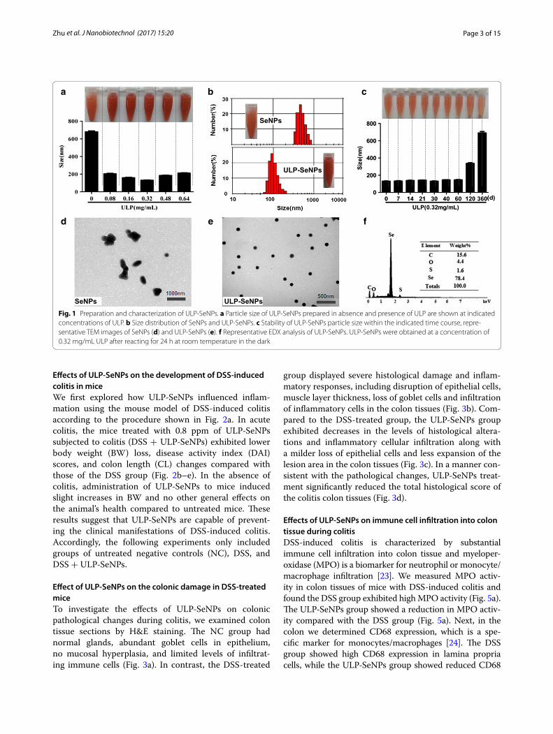
and stability being important [21, 22]. Size-controlled SeNPs were prepared in the redox reaction system of sel-enite acid and ascorbic acid, and for some of these parti-cles we added ULP to generate ULP-SeNPs. The particle size, stability and dispersity of SeNPs and ULP-SeNPs were measured as shown in Fig. 1. SeNPs without ULP decoration showed an average diameter of 680 nm, while addition of 0.32 mg/mL ULP generated ULP-SeNPs with significantly decreased particle diameters to 131 nm (Fig. 1a). The particle-size distribution was 305–900 nm and 58–205 nm for SeNPs and SeNPs-ULP (0.32 mg/mL ULP), respectively (Fig. 1b). There was a trend toward an increase in size of SeNPs with adding ULP above the level of 0.32 mg/mL, which implied that the particle size of SeNPs has something to do with the concentrations of ULP. It was also found that the ULP-SeNPs remained stable at least for 60 days when stored in 0.32 mg/mL ULP solutions (Fig. 1c). Moreover, the TEM images of these nanoparticles clearly revealed that SeNPs tended to aggregate and precipitate in absence of ULP (Fig. 1d), while SeNPs decorated with 0.32 mg/mL ULP appeared to monodisperse into homogeneous spherical struc-tures with an average diameter of 130 nm (Fig. 1e). The diagram from surface elemental composition analysis of ULP-SeNPs by EDX showed four signals, including a strong Se atom signal (78.4%) from SeNPs and minor sig-nals of C (15.6%), O (4.4) and S (1.6%) that likely origi-nated from the ULP (Fig. 1f ).
To monitor the physical characteristics of the ULP-SeNPs in solution, we examined the stability of ULP-SeNPs in mouse plasma and in digestion fluid after incubation for 24 h. The average size of ULP-SeNPs was maintained at ~130 nm in both plasma and digestion fluid indicating no aggregation of ULP-SeNPs (Additional file 1: Figure S1A, B). After centrifugation to remove the ULP-SeNPs, dissolved Se levels were only slightly altered in plasma, and Se levels increased by ~16% in the diges-tion fluid in comparison with controls (Additional file 1: Figure S1C, D). These data imply that the ULP-SeNPs can be deconstructed to some extent in the digestive tract, which is consistent with data below showing increased Se levels in colon and liver of mice supplementated with ULP-SeNPs. To compare the cellular uptake of ULP-SeNPs and SeNPs, intracellular Se concentrations were detected by ICP-MS after BMDMs were incubated with SeNPs or ULP-SeNPs for 24 h. Data showed that incuba-tion of BMDMs with ULP-SeNPs (0.5 μM) significantly increased the intracellular Se concentration from 0.36 to 16.25 ng/107 cells, which was ~2.4 times higher than that of SeNPs treatment (6.9 ng/107cells) (Additional file 1: Figure S2). Taken together, these results indicated that capping ULP on SeNPs not only controls the size and sta-bility of SeNPs but also enhances cellular uptake.
Page 3 of 15Zhu et al. J Nanobiotechnol (2017) 15:20
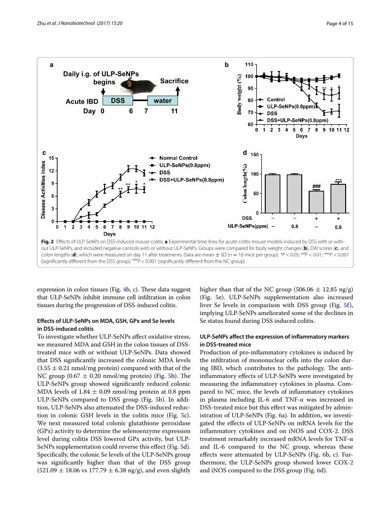
Effects of ULP‑SeNPs on the development of DSS‑induced colitis in miceWe first explored how ULP-SeNPs influenced inflam-mation using the mouse model of DSS-induced colitis according to the procedure shown in Fig. 2a. In acute colitis, the mice treated with 0.8 ppm of ULP-SeNPs subjected to colitis (DSS + ULP-SeNPs) exhibited lower body weight (BW) loss, disease activity index (DAI) scores, and colon length (CL) changes compared with those of the DSS group (Fig. 2b–e). In the absence of colitis, administration of ULP-SeNPs to mice induced slight increases in BW and no other general effects on the animal’s health compared to untreated mice. These results suggest that ULP-SeNPs are capable of prevent-ing the clinical manifestations of DSS-induced colitis. Accordingly, the following experiments only included groups of untreated negative controls (NC), DSS, and DSS + ULP-SeNPs.
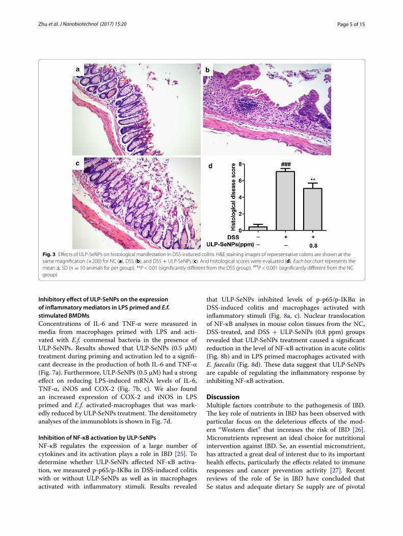
Effect of ULP‑SeNPs on the colonic damage in DSS‑treated miceTo investigate the effects of ULP-SeNPs on colonic pathological changes during colitis, we examined colon tissue sections by H&E staining. The NC group had normal glands, abundant goblet cells in epithelium, no mucosal hyperplasia, and limited levels of infiltrat-ing immune cells (Fig. 3a). In contrast, the DSS-treated
group displayed severe histological damage and inflam-matory responses, including disruption of epithelial cells, muscle layer thickness, loss of goblet cells and infiltration of inflammatory cells in the colon tissues (Fig. 3b). Com-pared to the DSS-treated group, the ULP-SeNPs group exhibited decreases in the levels of histological altera-tions and inflammatory cellular infiltration along with a milder loss of epithelial cells and less expansion of the lesion area in the colon tissues (Fig. 3c). In a manner con-sistent with the pathological changes, ULP-SeNPs treat-ment significantly reduced the total histological score of the colitis colon tissues (Fig. 3d).
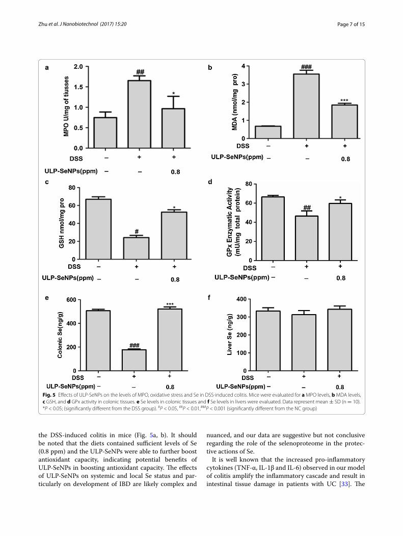
Effects of ULP‑SeNPs on immune cell infiltration into colon tissue during colitisDSS-induced colitis is characterized by substantial immune cell infiltration into colon tissue and myeloper-oxidase (MPO) is a biomarker for neutrophil or monocyte/macrophage infiltration [23]. We measured MPO activ-ity in colon tissues of mice with DSS-induced colitis and found the DSS group exhibited high MPO activity (Fig. 5a). The ULP-SeNPs group showed a reduction in MPO activ-ity compared with the DSS group (Fig. 5a). Next, in the colon we determined CD68 expression, which is a spe-cific marker for monocytes/macrophages [24]. The DSS group showed high CD68 expression in lamina propria cells, while the ULP-SeNPs group showed reduced CD68
b
ULP-SeNPs
SeNPs
a
d e f
SeNPs ULP-SeNPs
c
Fig. 1 Preparation and characterization of ULP-SeNPs. a Particle size of ULP-SeNPs prepared in absence and presence of ULP are shown at indicated concentrations of ULP. b Size distribution of SeNPs and ULP-SeNPs. c Stability of ULP-SeNPs particle size within the indicated time course, repre-sentative TEM images of SeNPs (d) and ULP-SeNPs (e). f Representative EDX analysis of ULP-SeNPs. ULP-SeNPs were obtained at a concentration of 0.32 mg/mL ULP after reacting for 24 h at room temperature in the dark
Page 4 of 15Zhu et al. J Nanobiotechnol (2017) 15:20
expression in colon tissues (Fig. 4b, c). These data suggest that ULP-SeNPs inhibit immune cell infiltration in colon tissues during the progression of DSS-induced colitis.
Effects of ULP‑SeNPs on MDA, GSH, GPx and Se levels in DSS‑induced colitisTo investigate whether ULP-SeNPs affect oxidative stress, we measured MDA and GSH in the colon tissues of DSS-treated mice with or without ULP-SeNPs. Data showed that DSS significantly increased the colonic MDA levels (3.55 ± 0.21 nmol/mg protein) compared with that of the NC group (0.67 ± 0.20 nmol/mg protein) (Fig. 5b). The ULP-SeNPs group showed significantly reduced colonic MDA levels of 1.84 ± 0.09 nmol/mg protein at 0.8 ppm ULP-SeNPs compared to DSS group (Fig. 5b). In addi-tion, ULP-SeNPs also attenuated the DSS-induced reduc-tion in colonic GSH levels in the colitis mice (Fig. 5c). We next measured total colonic glutathione peroxidase (GPx) activity to determine the selenoenzyme expression level during colitis DSS lowered GPx activity, but ULP-SeNPs supplementation could reverse this effect (Fig. 5d). Specifically, the colonic Se levels of the ULP-SeNPs group was significantly higher than that of the DSS group (521.09 ± 18.06 vs 177.79 ± 6.38 ng/g), and even slightly
higher than that of the NC group (506.06 ± 12.85 ng/g) (Fig. 5e). ULP-SeNPs supplementation also increased liver Se levels in comparison with DSS group (Fig. 5f ), implying ULP-SeNPs ameliorated some of the declines in Se status found during DSS induced colitis.
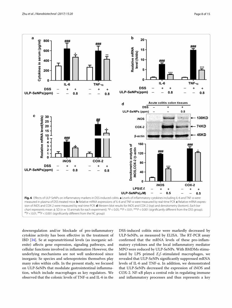
ULP‑SeNPs affect the expression of inflammatory markers in DSS‑treated miceProduction of pro-inflammatory cytokines is induced by the infiltration of mononuclear cells into the colon dur-ing IBD, which contributes to the pathology. The anti-inflammatory effects of ULP-SeNPs were investigated by measuring the inflammatory cytokines in plasma. Com-pared to NC mice, the levels of inflammatory cytokines in plasma including IL-6 and TNF-α was increased in DSS-treated mice but this effect was mitigated by admin-istration of ULP-SeNPs (Fig. 6a). In addition, we investi-gated the effects of ULP-SeNPs on mRNA levels for the inflammatory cytokines and on iNOS and COX-2. DSS treatment remarkably increased mRNA levels for TNF-α and IL-6 compared to the NC group, whereas these effects were attenuated by ULP-SeNPs (Fig. 6b, c). Fur-thermore, the ULP-SeNPs group showed lower COX-2 and iNOS compared to the DSS group (Fig. 6d).
a b
c d
0 6 7 11DayAcute IBD DSS
Daily i.g. of ULP-SeNPs begins
water
Sacrifice
Fig. 2 Effects of ULP-SeNPs on DSS-induced mouse colitis. a Experimental time lines for acute colitis mouse models induced by DSS with or with-out ULP-SeNPs, and included negative controls with or without ULP-SeNPs. Groups were compared for body weight changes (b), DAI scores (c), and colon lengths (d), which were measured on day 11 after treatments. Data are mean ± SD (n = 10 mice per group). *P < 0.05; **P < 0.01; ***P < 0.001 (significantly different from the DSS group). ###P < 0.001 (significantly different from the NC group)
Page 5 of 15Zhu et al. J Nanobiotechnol (2017) 15:20
Inhibitory effect of ULP‑SeNPs on the expression of inflammatory mediators in LPS primed and E.f. stimulated BMDMsConcentrations of IL-6 and TNF-α were measured in media from macrophages primed with LPS and acti-vated with E.f. commensal bacteria in the presence of ULP-SeNPs. Results showed that ULP-SeNPs (0.5 μM) treatment during priming and activation led to a signifi-cant decrease in the production of both IL-6 and TNF-α (Fig. 7a). Furthermore, ULP-SeNPs (0.5 μM) had a strong effect on reducing LPS-induced mRNA levels of IL-6, TNF-α, iNOS and COX-2 (Fig. 7b, c). We also found an increased expression of COX-2 and iNOS in LPS primed and E.f. activated-macrophages that was mark-edly reduced by ULP-SeNPs treatment. The densitometry analyses of the immunoblots is shown in Fig. 7d.
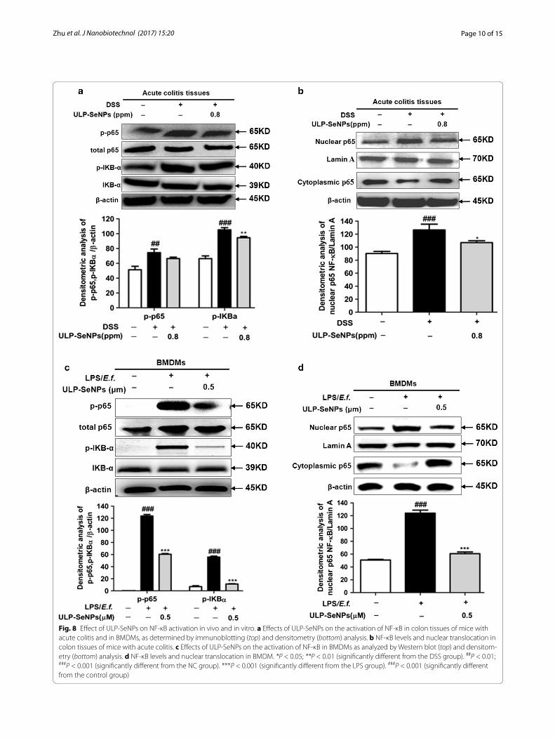
Inhibition of NF‑κB activation by ULP‑SeNPsNF-κB regulates the expression of a large number of cytokines and its activation plays a role in IBD [25]. To determine whether ULP-SeNPs affected NF-κB activa-tion, we measured p-p65/p-IKBα in DSS-induced colitis with or without ULP-SeNPs as well as in macrophages activated with inflammatory stimuli. Results revealed
that ULP-SeNPs inhibited levels of p-p65/p-IKBα in DSS-induced colitis and macrophages activated with inflammatory stimuli (Fig. 8a, c). Nuclear translocation of NF-κB analyses in mouse colon tissues from the NC, DSS-treated, and DSS + ULP-SeNPs (0.8 ppm) groups revealed that ULP-SeNPs treatment caused a significant reduction in the level of NF-κB activation in acute colitis (Fig. 8b) and in LPS primed macrophages activated with E. faecalis (Fig. 8d). These data suggest that ULP-SeNPs are capable of regulating the inflammatory response by inhibiting NF-κB activation.
DiscussionMultiple factors contribute to the pathogenesis of IBD. The key role of nutrients in IBD has been observed with particular focus on the deleterious effects of the mod-ern “Western diet” that increases the risk of IBD [26]. Micronutrients represent an ideal choice for nutritional intervention against IBD. Se, an essential micronutrient, has attracted a great deal of interest due to its important health effects, particularly the effects related to immune responses and cancer prevention activity [27]. Recent reviews of the role of Se in IBD have concluded that Se status and adequate dietary Se supply are of pivotal
Fig. 3 Effects of ULP-SeNPs on histological manifestation in DSS-induced colitis. H&E staining images of representative colons are shown at the same magnification (×200) for NC (a), DSS (b), and DSS + ULP-SeNPs (c). And histological scores were evaluated (d). Each bar chart represents the mean ± SD (n = 10 animals for per group). **P < 0.01 (significantly different from the DSS group). ###P < 0.001 (significantly different from the NC group)

Page 6 of 15Zhu et al. J Nanobiotechnol (2017) 15:20
importance to colonic inflammation [26, 28]. Nanopar-ticles that include elemental Se (SeNPs) as a novel form of this nutrient appear to be more effective than that of other Se sources because of their excellent bioavailabil-ity, biological activity, and low toxicity [29]. To address application of ULP-SeNPs to prevent IBD, we developed a stable, homogeneous formulation of ULP-SeNPs with consistent diameter of 130 nm. The mouse macrophage reporter cell line-RAW-Blue cells was used to determine the effects of SeNPs and ULP-SeNPs on the LPS/E.f. triggered NF-κB inflammatory signaling pathway. Data showed that ULP-SeNPs (0.5 μM) inhibited either LPS or E.f.-induced NF-κB hyper-activation, and did so in a more effective manner than ULP or SeNPs (Additional file 1: Figure S3). These data support the notion that ULP-SeNPs may represent a nutriceutical with promising anti-inflammation capacity.
In this study, we report for the first time that ULP-SeNPs led to a reduction in the severity of the sys-temic (e.g., BW loss and DAI scores) and local (e.g., CL shortened and HDS) symptoms of DSS-induced coli-tis. Moreover, ULP-SeNPs suppressed inflammatory cell infiltration and local inflammation, resulting in less pathological damage being induced by DSS in colon.
DSS-induced acute colitis and chronic colitis in mice reproduce the histological pathology of human IBD [30]. Optimization of dietary ULP-SeNPs supplementa-tion might offer a promising supportive therapy for IBD patients as long as safety can be demonstrated. It is con-ceivable that ULP-SeNPs would have limited side effects because Se nanoparticles (SeNPs) exhibit excellent bio-availability, biological activity, and low toxicity compared to organic or inorganic Se. Altogether, these results indi-cate that ULP-SeNPs might be a candidate as a potential therapeutic nanomedicine for prevention of IBD and other inflammatory diseases. However, further follow-up investigations of the safety and efficacy of ULP-SeNPs are necessary before it can be considered for the treatment of IBD patients.
Oxidative stress or cellular damage from free radicals, often involving profound lipid peroxidation are the hall-marks of UC [31]. Mounting evidence suggests that many Se metabolites exhibit antioxidative properties and anti-inflammatory roles for modulating disorders such as IBD, atherosclerosis, and cancer [2, 32]. In the present study, the results showed that ULP-SeNPs markedly increased the colonic GSH levels, one marker for oxidative sta-tus, and reduced the generation of MDA to attenuate
Fig. 4 Effects of ULP-SeNPs on immune cell infiltration into colon tissue during DSS colitis. Expression levels of CD68 (×200 magnification) were evaluated by immunohistochemistry in the colon tissue. Three groups including a NC, b DSS, c DSS + ULP-SeNPs and d CD68 positive cell numbers were counted and compared among three groups (n = 10). **P < 0.01; (significantly different from the DSS group). ###P < 0.001 (significantly differ-ent from the NC group)
Page 7 of 15Zhu et al. J Nanobiotechnol (2017) 15:20
the DSS-induced colitis in mice (Fig. 5a, b). It should be noted that the diets contained sufficient levels of Se (0.8 ppm) and the ULP-SeNPs were able to further boost antioxidant capacity, indicating potential benefits of ULP-SeNPs in boosting antioxidant capacity. The effects of ULP-SeNPs on systemic and local Se status and par-ticularly on development of IBD are likely complex and
nuanced, and our data are suggestive but not conclusive regarding the role of the selenoproteome in the protec-tive actions of Se.
It is well known that the increased pro-inflammatory cytokines (TNF-α, IL-1β and IL-6) observed in our model of colitis amplify the inflammatory cascade and result in intestinal tissue damage in patients with UC [33]. The
Fig. 5 Effects of ULP-SeNPs on the levels of MPO, oxidative stress and Se in DSS-induced colitis. Mice were evaluated for a MPO levels, b MDA levels, c GSH, and d GPx activity in colonic tissues. e Se levels in colonic tissues and f Se levels in livers were evaluated. Data represent mean ± SD (n = 10). *P < 0.05; (significantly different from the DSS group). #P < 0.05, ##P < 0.01,###P < 0.001 (significantly different from the NC group)
Page 8 of 15Zhu et al. J Nanobiotechnol (2017) 15:20
downregulation and/or blockade of pro-inflammatory cytokine activity has been effective in the treatment of IBD [34]. Se at supranutritional levels (as inorganic sel-enite) affects gene expression, signaling pathways, and cellular functions involved in inflammation However, the underlying mechanisms are not well understood since inorganic Se species and selenoproteins themselves play many roles within cells. In the present study, we focused on ULP-SeNPs that modulate gastrointestinal inflamma-tion, which include macrophages as key regulators. We observed that the colonic levels of TNF-α and IL-6 in the
DSS-induced colitis mice were markedly decreased by ULP-SeNPs, as measured by ELISA. The RT-PCR assay confirmed that the mRNA levels of these pro-inflam-matory cytokines and the local inflammatory mediator MPO were reduced by ULP-SeNPs. With BMDMs stimu-lated by LPS primed E.f.-stimulated macrophages, we revealed that ULP-SeNPs significantly suppressed mRNA levels of IL-6 and TNF-α. In addition, we demonstrated that ULP-SeNPs decreased the expression of iNOS and COX-2. NF-κB plays a central role in regulating immune and inflammatory processes and thus represents a key
Fig. 6 Effects of ULP-SeNPs on inflammatory markers in DSS-induced colitis. a Levels of inflammatory cytokines including IL-6 and TNF-α were measured in plasma of DSS-treated mice. b Relative mRNA expressions of IL-6 and TNF-α were measured by real-time PCR. c Relative mRNA expres-sion of iNOS and COX-2 were measured by real-time PCR. d Western blot results for iNOS and COX-2 (top) and densitometry (bottom). Each bar chart represents mean ± SD (n = 10 animals for each experiment). *P < 0.05; **P < 0.01; ***P < 0.001 (significantly different from the DSS group). ##P < 0.01; ###P < 0.001 (significantly different from the NC group)

Page 9 of 15Zhu et al. J Nanobiotechnol (2017) 15:20
target for developing novel treatments for inflammatory diseases [35]. Prior to activation, NF-κB is complexed with IκBα, an inhibitory protein keeping NF-κB inactive state in the cytoplasm. Induced by various stimuli, such as LPS and proinflammatory factors, NF-κB is released and translocates from cytoplasm into the nucleus due to IκBα phosphorylation, ubiquitinylation, and degrada-tion. Phosphorylated subunit p65 plays an important role in triggering the transcription of certain genes. Results in our study showed that ULP-SeNPs strongly inhibited the level of NF-κB activation in colitis colon tissues and stimulated macrophages by suppressing the phospho-rylation and degradation of IκBα and thus decreased
the phosphorylation of p65. However, inflammation also plays a part in restoring gut homeostasis through diverse pathways involving the colonic macrophage family (e.g. M1 and M2 types), [36] and mediators such as IL-10 and NO. Further mechanistic studies will improve our under-standing of the actions of ULP-SeNPs in IBD and identify specific mediators driving the beneficial Se effects dem-onstrated in our studies.
The data from our in vivo and in vitro experiments sug-gest that ULP-SeNPs might represent a modality that inhibits IBD. However, our study is limited by a lack of definitive molecules on which the increased Se acts. Altered redox tone in the colon and perhaps modulation
Fig. 7 Effect of ULP-SeNPs on of inflammatory marker levels in LPS-primed E.f.-activated BMDM. a IL-6 and TNF-α levels in cell-free supernatants were determined by ELISA. b Relative levels of IL-6 and TNF-α mRNA were measured by real-time PCR. c Relative levels of iNOS and COX-2 mRNA were measured by real-time PCR. d Analyses iNOS and COX-2 protein by Western blot (top) and densitometry (bottom). Data represent mean ± SD (n = 3 in independent experiments). *P < 0.05; **P < 0.01; ***P < 0.001 (significantly different from the LPS group). ##P < 0.01; ###P < 0.001 (signifi-cantly different from the control group)
Page 10 of 15Zhu et al. J Nanobiotechnol (2017) 15:20
Fig. 8 Effect of ULP-SeNPs on NF-κB activation in vivo and in vitro. a Effects of ULP-SeNPs on the activation of NF-κB in colon tissues of mice with acute colitis and in BMDMs, as determined by immunoblotting (top) and densitometry (bottom) analysis. b NF-κB levels and nuclear translocation in colon tissues of mice with acute colitis. c Effects of ULP-SeNPs on the activation of NF-κB in BMDMs as analyzed by Western blot (top) and densitom-etry (bottom) analysis. d NF-κB levels and nuclear translocation in BMDM. *P < 0.05; **P < 0.01 (significantly different from the DSS group). ##P < 0.01; ###P < 0.001 (significantly different from the NC group). ***P < 0.001 (significantly different from the LPS group). ###P < 0.001 (significantly different from the control group)
Page 11 of 15Zhu et al. J Nanobiotechnol (2017) 15:20
of immune cells are two ways that Se has been shown to affect inflammation [1]. The selenoprotein subfamilies of glutathione peroxidases and thioredoxin reductases may be involved in regulating these processes, [37] but other selenoproteins are likely also involved in mediating the effects of SeNPs. Another limitation of our data is that the DSS mouse model of chemically induced colitis is strongly macrophage driven inflammation and not com-pletely representative of human IBD [38]. Several genetic models of colitis have been generated [39] and it would be of interest to repeat these SeNPs experiments in those mice.
ConclusionsThe present data demonstrated that ULP-SeNPs sup-plementation exhibits the anti-inflammatory effects to reduce the symptoms of acute colitis through inhibition the hyper activation of NF-κB in colonic tissues and mac-rophages. Therefore, ULP-SeNPs may be a candidate for further evaluation as a potential therapeutic product for treatment IBD and other inflammatory diseases.
MethodsMaterials and chemicalsDSS was obtained from MP Biomedicals (Illkirch, France). The glutathione (GSH), glutathione peroxidase (GPx) and malondialdehyde (MDA) detection kits were purchased from Nanjing Jiancheng Bioengineering Insti-tute (Nanjing, China). The bicinchoninic acid (BCA) kits for protein assays were supplied by the Beyotime Insti-tute of Biotechnology (Shanghai, China). Ulva lactuca polysaccharide was purchased from Elicityl (Grenoble, France). NF-κB activation assay kits were supplied by FIVE photon Biochemicals (San Diego, CA, USA). Dul-becco’s modified Eagle’s medium (DMEM) and the anti-biotic mixture (penicillin/streptomycin) were purchased from Invitrogen (Carlsbad, CA, USA). Proteinase inhibi-tor cocktail and Laemmli buffer were purchased from Bio-Rad (Hercules, CA, USA). Antibodies purchased from Cell Signaling Technology (Danvers, MA, USA) included anti-NF-κB(p65), anti-p-p65, anti-p-IKBα, anti-IKBα, anti-iNOS, anti-COX-2, anti-β-actin, anti-lamin A. Secondary antibodies for western blots were obtained from Li-Cor (Lincoln, Nebraska, USA). Anti-CD68 was purchased from Santa Cruz Biotechnology (Dallas, Texas, USA). Hexadecyltrimethylammonium bromide, o-dianisidine, and lipopolysaccharide (LPS) purified from Escherichia coli O111:B4 were purchased from Sigma-Aldrich (St. Louis, MO, USA). Heat-killed Enterococcus faecalis (E.f.) was obtained from American Type Culture Collection. All of the solvents used were of high perfor-mance liquid chromatography (HPLC) grade. Chemicals
used were of analytical grade, including sodium selenite, sodium dodecyl sulfate (SDS), Tween 20, and other com-monly used reagents were obtained from Guangdong Guanghua Sci-Tech (Huada & JHD, Guangzhou, China). The ultrapure water used in all experiments was supplied by a Milli-Q water purification system from Millipore (Bedford, MA, USA).
Preparation of ULP‑SeNPsThe solution of ULP was prepared by dissolving 1 g of ULP powder in 100 mL of Milli-Q water. The solution of 40 mM ascorbic acid was freshly prepared. As a typi-cal procedure, varied volume of ULP solution was added dropwise into sodium selenite solution (1 mL, 100 mM) under magnetic stirring, and then 10 mL of 40 mM ascorbic acid solution was added into the mixture, and it was reconstituted to a final volume of 20 mL with Milli-Q water. The final concentration of Se was 5 mM, and the reactant concentrations of ULP were 0, 0.08, 0.16, 0.32, 0.48 and 0.64 mg/mL. The solution was dialyzed against Milli-Q water until no Se was detected in the outer solu-tion by ICP-MS analysis.
Characterization of ULP‑SeNPsThe ULP-SeNPs were characterized by microscopic and spectroscopic methods including transmission elec-tron microscopy (TEM), energy dispersive X-ray (EDX), dynamic light scattering (DLS). Briefly, TEM was carried out with a Hitachi (H-7650) at an acceleration voltage of 80 kV. The EDX were taken on a JEOL 2010 high-resolu-tion TEM operated at 200 kV. A Zetasizer Nano ZS par-ticle analyzer (Malvern Instruments Limited) was used to measure the particle size, size distribution, and stabil-ity of the nanoparticles in aqueous solution, plasma and digestion fluid by DLS measurement.
Animals and induction of DSS‑induced IBDMale C57BL/6 J mice (6–8 weeks old), SPF bedding, and certified diet were purchased from the Sun Yat-Sen Uni-versity Animal Breeding Unit (Guangzhou, China). All animals were fed standard laboratory chow housed in wire cages at 21 ± 3 °C and 55 ± 15% relative humidity with a 12 h light/dark cycle, and allowed water ad libi-tum. The Se content of the diet was 0.12 ± 0.01 ppm on average.
Acute colitis was induced by oral administration of 4% (w/v) DSS dissolved into drinking water, for 7 days according to the method employed in previous studies [40]. A cohort of mice was randomly divided into three groups (n = 10 per group) for acute treated as a normal control group (NC), a DSS-induced colitis group (DSS), and a DSS with 0.8 ppm of ULP-SeNPs treatment group
Page 12 of 15Zhu et al. J Nanobiotechnol (2017) 15:20
(DSS + ULP-SeNPs). Body weights (BW) were meas-ured daily. The daily weight changes were calculated by percent in relation to the initial weight measured. After 11 days, mice in each acute cohort had blood and colon tissues analyzed after being humanely euthanized. Blood was collected in a heparinized tube, immediately centri-fuged to separate plasma, and then kept at −80 °C. The colon was rapidly excised and placed in 4% paraformal-dehyde for histological assessment. The remaining colon samples were flash frozen and stored at −80 °C until fur-ther analysis.
Assessment of severity of colitisA general assessment of colitis was performed as described previously [41]. Briefly, the severity of the coli-tis was measured by evaluating the disease activity index through the scoring of weight loss, stool consistency, bleeding and coat roughness (grade from 0 to 4 on sever-ity of each index), general activity and bedding contami-nation by stool and blood (graded from 0 to 2 on severity of each index).
Histological analysisColon tissues were collected, fixed in 4% paraformalde-hyde, subjected to consecutive steps of alcohol–xylene changes, and embedded in paraffin from which 5 μm Sections were prepared. Standard hematoxylin and eosin (H&E) staining of paraffin-embedded tissue samples was conducted as previously described [42]. Five H&E-stained sections from each mouse were scored blindly, as previously described [43]. Images were captured using a Zeiss Axioskop 2 plus upright light microscope and cam-era (Zeiss, Oberkochen, Germany).
IHC stainingParafin-embedded tissues were cut into 5 μm-thick sections, deparaffinized in xylene, and rehydrated in graded ethanol. Sections were incubated with anti-CD68 antibodies overnight at 4 °C and then incubated with SignalStain®Boost IHC Detection Reagent (HRP, Rabbit) as indicated by the manufacturer (Cell Signaling Technol-ogy, Danvers, MA, USA) for 30 min at room temperature. Finally, the sections were stained with DAB + Liquid according to manufacturer’s protocol (DAKO, Glostrup, Denmark).
Determination of SeSe concentration in solutions and biological samples was determined by an inductively coupled plasma mass spec-trometry (ICP-MS) method described previously [44]. To prepare liver and colonic tissues for this method, the tis-sues were weighed and mineralized in HNO3 65% (ICP-MS grade) and then diluted 100 fold in deionized water.
MPO assayMPO activity, a marker of polymorphonuclear neutrophil primary granules, was measured in colonic tissues using the method by Bradley et al. [45]. Briefly, colon segments were homogenized at 50 mg/mL in phosphate buffer (50 mmol/L, pH 6.0) with 0.5% hexadecyltrimethylam-monium bromide. Samples were centrifuged at 30,000×g for 15 min at 4 °C after freezing and thawing 3 times. The supernatants were diluted 1/30 with 50 mmol/L phos-phate buffer (pH 6.0) containing 0.167 mg/mL o-diani-sine (Sigma) and 0.0005% H2O2. Changes in absorbance between 1 and 3 min at 450 nm were measured with a spectrophotometer. MPO was expressed in units per mil-ligram of wet tissue.
Glutathione content, GPx activity and lipid peroxidation assayColon tissue (100 mg) was washed and homogenized in cooled PBS. The samples were then homogenized fol-lowed by centrifugation at 3000 rpm for 15 min. Super-natant was collected for measuring total GSH, GPx activity, and lipid peroxidation of MDA using GSH, GPx and MDA detection kits, respectively. All assays were conducted according to the manufacturer’s instructions. The protein content was assayed by bicinchoninic acid (BCA) kits.
Cell cultureBone marrow-derived macrophages (BMDMs) were pre-pared as previously described [46]. On day 5 of culture, BMDMs were plated at a density of 2 × 10⁶ cells/well in six well plates in 2 mL DMEM with 5% FBS (Life Tech-nologies, Gaithersburg, MD) penicillin (100 U/mL), and streptomycin (50 U/mL) overnight at 37 °C in a humidi-fied incubator with a 5% CO2 atmosphere. The next day, cultured cells were pretreated with ULP-SeNPs at the indicated concentrations for 1 h and then were primed for 18 h with LPS (100 ng/mL), followed by 1 h stimu-lation with E. f. (1 μg/mL). Supernatants of the cultures were collected at the indicated time point for cytokine detection.
RNA isolation, cDNA synthesis, and real‑time PCRTo detect the effect of ULP-SeNPs on gene expression in colon tissues and LPS primed and E.f.-stimulated cells, Tis-sue samples were thawed and RNA extracted using RNeasy Mini kit and RNase-free DNase I (Qiagen, Hilden, Germany), in accordance with the manufacturer’s instructions. BMDMs were preincubated on six-well plates (1 × 10⁶ cell/mL) and were pretreated with ULP-SeNPs (0.5 μM) 1 h prior to LPS (100 ng/mL) primed and E. f. (1 μg/mL) treatment in a 37 °C, 5% CO2 incubator for 12 h. Total RNA from BMDMs was per-formed using RNeasy (Qiagen, Hilden, Germany) according
Page 13 of 15Zhu et al. J Nanobiotechnol (2017) 15:20
to the manufacturer’s instructions. The quantity and quality of RNA were determined by Nano Drop ND-1000 (Thermo Scientific, Waltham, MA, USA). The cDNA was synthesized using Superscript III (Invitrogen, Carlsbad, CA, USA) and oligo dT primer, with 2 µg of total RNA per 50 µL reaction. For real-time PCR, 1 µL of this cDNA was used in 10 µL reactions with Platinum SYBR Green qPCR SuperMix-UDG (Invitrogen). Reactions were carried out in a 9700HT thermal cycler (Applied Biosystems, Foster City, CA) using the following conditions: denaturation for 2 min at 50 °C, for 2 min at 95 °C, and 40 cycles of 95 °C at 15 s, 60 °C at 60 s. The mouse primers used were as follows: IL-6 (for-ward: 5′-AGAAGGAGTGGCTAAGGACCAA-3′; reverse: 5′-ACGCACTAGGTTTGCCGAGTA-3′), β-actin (forward: 5′-GCATTGTTACCAACTGGGACGA-3′; reverse: 5′-TGGCTGG GGTGTTGAAGGTC-3′), TNF-α forward: (5′-AGGACCCA GTGGTGGGAAGCT-3′; reverse: 5′-AAAGAGGAGGCAA GGTAGAGA-3′), IL-10 (forward: 5′-GCTCTTACTGACTGG CATGAG-3′; reverse: 5′-CGCAGCTCTAGGAGCATGT G-3′), COX-2 (forward: 5′-TTGCTGTACAAGCAGTGGCA AAGG-3′; reverse: 5′-AGGACAAACACCGGAGGGAATCT T-3′), iNOS (forward: 5′-CAGATCGAGCCCTGGAAG AC-3′; reverse: 5′-CTGGTCCATGCAGACAACCT-3′). Rela-tive gene expression was calculated according to the 2−��Ct method [47] using mouse β-actin as an endogenous control for normalization. Results were expressed as fold changes rela-tive to the negative control samples.
Enzyme‑linked immunosorbent assayPlasma samples and cell supernatants were analyzed for levels of inflammatory cytokines including TNF-a, IL-6 and IL-10 using commercially available enzyme-linked immunosorbent assay (ELISA) kits (R&D Systems Inc, USA), according to the manufacturer’s instructions.
Western blot analysisCells pellets were harvested and lysed in lysis buffer [150 mM NaCl, 5 mM EDTA, 50 mM Tris–HCl (pH 7.4), 1% Triton X-100, and 0.5% sodium deoxycholate] with a 1× proteinase inhibitor cocktail. Colon tissues were homogenized with lysis buffer on ice and then cen-trifuged at 13,000×g for 15 min at 4 °C. In some cases, nuclear and cytosolic fractions were separated from cells or colon tissues using the NF-κB activation assay kits. After separation on a 12% polyacrylamide gel, proteins were transferred to polyvinylidene difluoride (PVDF) membranes (Millipore) and then incubated using spe-cific antibodies to detect target proteins. After the pri-mary antibody reaction overnight at 4 °C, the PVDF membrane was washed three times with TBST (137 mM NaCl, 2.7 mM KCl, 19 mM Tris base, and 0.1% Tween 20) and incubated with secondary Abs from Li-Cor for 1 h. Membranes were washed with TBST, and signals were
detected with densitometry conducted using the Odys-sey imaging system (Li-Cor, Lincoln, NE). Densitometry of blots was analyzed and normalized by β-actin or lamin A using ImageJ (National Institutes of Health, Bethesda, MD).
Statistical analysisAll comparison analyses were conducted using the SPSS statistical software package (SPSS 13.0 for Windows, SPSS, Inc., Chicago, IL). A P value of <0.05 was considered sta-tistically significant. Comparison of mean values was done using analysis of variance (ANOVA). For the body weight change, DAI scores and colon lengths were analyzed using one-way ANOVA (Welch ANOVA in cases of unequal variance) followed by the Games–Howell post hoc test. All other analyses of equal sample sizes for each group and equal variances among the groups were compared using one-way ANOVA with the Tukey post hoc test.
AbbreviationsSeNPs: selenium nanoparticles; Se: selenium; ULP: Ulva lactuca polysaccharide; IBD: inflammatory bowel disease; DSS: dextran sulfate sodium; IL-6: interleu-kin-6; IL-10: interleukin-10; TNF-α: tumor necrosis factor-α; NF-κB: nuclear factor κ-B; p-NF-κB: phospho-nuclear factor κ-B; p-IKB-α: phospho-IKB-α; UC: ulcera-tive colitis; CD: Crohn’s disease; BCA: bicinchoninic acid; SDS–PAGE: sodium dodecyl sulfate–polyacrylamide gel electrophoresis; DAI: disease activity index; H&E: hematoxylin and eosin; MPO: myeloperoxidase; GSH: glutathione; MDA: malondialdehyde; ELISA: enzymelinked immunosorbent assay; GSH: glutathione; FBS: fetal bovine serum; LPS: lipopolysaccharide; E.f.: Enterococcus faecalis; iNOS: inducible nitric oxide synthase; SD: standard deviation.
Authors’ contributionsCZ, SZ, CS, YZ, and QL performed the experiments. CZ, WZ and ZH designed the study, analyzed the data and wrote the paper. JL, PRH and TC interpreted and polished the writing. All authors discussed the results and implications and commented on the manuscript at all stages. All authors have read and approved the final manuscript.
Author details1 School of Life Science and Technology, Jinan University, Guangzhou 510632, Guangdong Province, China. 2 College of Pharmacy, Jinan University, Guang-zhou 510632, Guangdong Province, China. 3 Department of Cell and Molecu-lar Biology, John A. Burns School of Medicine, University of Hawaii, Honolulu, HI, USA. 4 College of Chemistry and Material Science, Jinan University, Guang-zhou 510632, Guangdong Province, China.
AcknowledgementsWe are grateful to Mr.Yanqing Zhang and Mr.Xueran Lin for their assistance in sampling and ICP-MS analysis.
Competing interestsThe authors declare that they have no competing interests.
Availability of data and materialsAll data generated or analyzed during this study are included in the article.
Additional file
Additional file 1. Supplemental information of ULP-SeNPs concerns their stability in physiological solutions, uptake by BMDMs and effect on NF-κB activation.
Page 14 of 15Zhu et al. J Nanobiotechnol (2017) 15:20
Ethical approvalAll animal experiments were approved by the Animal Care and Ethics Com-mittee of Jinan University (20150306003) and carried out in accordance with the National Laboratory Animal Ethics Committee of China.
FundingThis work was supported by Grants of the National Nature Science Founda-tion of China (Nos. 81570397, 81372372), the Key Project of Natural Science Foundation of Guangdong (2014A030311026), the Project of Science and Technology of Guangdong (2013B010404029, 2014A050503044), the Key Pro-ject of Marine Fishery Science and Technology of Guangdong (A201501C07), the Major Project of Science and Technology of Guangzhou (201604020142) and the Fundamental Research Funds for the Central Universities of China (Jinan University, No. 21611611).
Received: 7 December 2016 Accepted: 22 February 2017
References 1. Huang Z, Rose AH, Hoffmann PR. The role of selenium in inflammation
and immunity: from molecular mechanisms to therapeutic opportunities. Antioxid Redox Signal. 2012;16:705–43.
2. Fairweather-Tait SJ, Bao Y, Broadley MR, Collings R, Ford D, Hesketh JE, Hurst R. Selenium in human health and disease. Antioxid Redox Signal. 2011;14:1337–83.
3. Geerling BJ, Badart-Smook A, Stockbrügger RW, Brummer RJ. Compre-hensive nutritional status in recently diagnosed patients with inflamma-tory bowel disease compared with population controls. Eur J Clin Nutr. 2000;54:514–21.
4. Gentschew L, Bishop KS, Han DY, Morgan AR, Fraser AG, Lam WJ, Karu-nasinghe N, Campbell B, Ferguson LR. Selenium, selenoprotein genes and Crohn’s disease in a case–control population from Auckland, New Zealand. Nutrients. 2012;4:1247–59.
5. Barrett CW, Singh K, Motley AK, Lintel MK, Matafonova E, Bradley AM, Ning W, Poindexter SV, Parang B, Reddy VK, et al. Dietary selenium defi-ciency exacerbates DSS-induced epithelial injury and AOM/DSS-induced tumorigenesis. PLoS ONE. 2013;8:e67845.
6. Peng D, Zhang J, Liu Q, Taylor EW. Size effect of elemental selenium nano-particles (Nano-Se) at supranutritional levels on selenium accumulation and glutathione S-transferase activity. J Inorg Biochem. 2007;101:1457–63.
7. Zhang J, Wang X, Xu T. Elemental selenium at nano size (Nano-Se) as a poten-tial chemopreventive agent with reduced risk of selenium toxicity: compari-son with se-methylselenocysteine in mice. Toxicol Sci. 2008;101:22–31.
8. Peer D, Karp JM, Hong S, Farokhzad OC, Margalit R, Langer R. Nanocar-riers as an emerging platform for cancer therapy. Nat Nanotechnol. 2007;2:751–60.
9. Moghimi SM, Peer D, Langer R. Reshaping the future of nanopharmaceu-ticals: ad iudicium. ACS Nano. 2011;5:8454–8.
10. Capurso NA, Fahmya TM. Development of a pH-responsive particulate drug delivery vehicle for localized biologic therapy in inflammatory bowel disease. Yale J Biol Med. 2011;84:285–8.
11. Collnot EM, Ali H, Lehr CM. Nano- and microparticulate drug carri-ers for targeting of the inflamed intestinal mucosa. J Control Release. 2012;161:235–46.
12. Coco R, Plapied L, Pourcelle V, Jerome C, Brayden DJ, Schneider YJ, Preat V. Drug delivery to inflamed colon by nanoparticles: comparison of differ-ent strategies. Int J Pharm. 2013;440:3–12.
13. Zhang Y, Li X, Huang Z, Zheng W, Fan C, Chen T. Enhancement of cell permeabilization apoptosis-inducing activity of selenium nanoparticles by ATP surface decoration. Nanomedicine. 2013;9:74–84.
14. Wu S, Sun K, Wang X, Wang D, Wan X, Zhang J. Protonation of epigallocat-echin-3-gallate (EGCG) results in massive aggregation and reduced oral bioavailability of EGCG-dispersed selenium nanoparticles. J Agric Food Chem. 2013;61:7268–75.
15. Yang F, Tang Q, Zhong X, Bai Y, Chen T, Zhang Y, Li Y, Zheng W. Surface decoration by Spirulina polysaccharide enhances the cellular uptake and anticancer efficacy of selenium nanoparticles. Int J Nanomed. 2012;7:835–44.
16. Wu H, Zhu H, Li X, Liu Z, Zheng W, Chen T, Yu B, Wong KH. Induction of apoptosis and cell cycle arrest in A549 human lung adenocarcinoma cells by surface-capping selenium nanoparticles: an effect enhanced by polysaccharide–protein complexes from Polyporus rhinocerus. J Agric Food Chem. 2013;61:9859–66.
17. Leiro JM, Castro R, Arranz JA, Lamas J. Immunomodulating activities of acidic sulphated polysaccharides obtained from the seaweed Ulva rigida C. Agardh. Int Immunopharmacol. 2007;7:879–88.
18. Devaki T, Sathivel A, BalajiRaghavendran HR. Stabilization of mito-chondrial and microsomal function by polysaccharide of Ulva lactuca on d-galactosamine induced hepatitis in rats. Chem Biol Interact. 2009;177:83–8.
19. Lahaye M, Robic A. Structure and functional properties of Ulvan, a poly-saccharide from green seaweeds. Biomacromolecules. 2007;8:1765–74.
20. Tian H, Yin X, Zeng Q, Zhu L, Chen J. Isolation, structure, and surfactant properties of polysaccharides from Ulva lactuca L. from South China Sea. Int J Biol Macromol. 2015;79:577–82.
21. Tsourkas DLJ, Tsourkas A. Size, charge and concentration dependent uptake of iron oxide particles by non-phagocytic cells. Biomaterials. 2008;29:3583–90.
22. Bakshi MS, Kaur H, Banipal TS, Singh N, Kaur G. Biomineralization of gold nanoparticles by lysozyme and cytochrome C and their applications in protein film formation. Langmuir. 2010;26:13535–44.
23. Malle E, Furtmuller PG, Sattler W, Obinger C. Myeloperoxidase: a target for new drug development? Br J Pharmacol. 2007;152:838–54.
24. Harris JA, Jain S, Ren Q, Zarineh A, Liu C, Ibrahim S. CD163 versus CD68 in tumor associated macrophages of classical Hodgkin lymphoma. Diagn Pathol. 2012;7:12.
25. Murano M, Maemura K, Hirata I, Toshina K, Nishikawa T, Hamamoto N, Sasaki S, Katsu OSK. Therapeutic effect of intracolonically admin-istered nuclear factor kB (p65) antisense oligonucleotide on mouse dextran sulphate sodium (DSS)-induced colitis. Clin Exp Immunol. 2000;120:51–8.
26. Speckmann B, Steinbrenner H. Selenium and selenoproteins in inflam-matory bowel diseases and experimental colitis. Inflamm Bowel Dis. 2014;20:1110–9.
27. Rayman MP. Selenium in cancer prevention: a review of the evidence and mechanism of action. Proc Nutr Soc. 2007;64:527–42.
28. Kudva AK, Shay AE, Prabhu KS. Selenium and inflammatory bowel dis-ease. Am J Physiol Gastrointest Liver Physiol. 2015;309:G71–7.
29. Zhang Y, Wang J, Zhang L. Creation of highly stable selenium nanopar-ticles capped with hyperbranched polysaccharide in water. Langmuir. 2010;26:17617–23.
30. Okayasu I, Hatakeyama S, Yamada M, Ohkusa T, Inagaki Y, Nakaya R. A novel method in the induction of reliable experimental acute and chronic ulcerative colitis in mice. Gastroenterology. 1990;98:694–702.
31. Dieleman LA, Palmen M, Akol H, Bloemena E, Peña AS, Meuwissen SG, Van Rees EP. Chronic experimental colitis induced by dextran sulphate sodium (DSS) is characterized by Th1 and Th2 cytokines. Clin Exp Immu-nol. 1998;114:385–91.
32. Papp LV, Lu J, Holmgren A, Khanna KK. From selenium to selenoproteins: synthesis, identity, and their role in human health. Antioxid Redox Signal. 2007;9:775–806.
33. Perrier C, Rutgeerts P. Cytokine blockade in inflammatory bowel diseases. Immunotherapy. 2011;3:1341–52.
34. Rutgeerts PSW, Feagan BG, Reinisch W, Olson A, Johanns J, Travers S, Rachmilewitz D, Hanauer SB, Lichtenstein GR. Infliximab for induc-tion and maintenance therapy for ulcerative colitis. N Engl J Med. 2005;353:2462–76.
35. Checker R, Patwardhan RS, Sharma D, Menon J, Thoh M, Bhilwade HN, Konishi T, Sandur SK. Schisandrin B exhibits anti-inflammatory activity through modulation of the redox-sensitive transcription factors Nrf2 and NF-kappaB. Free Radic Biol Med. 2012;53:1421–30.
36. Huang Z, Rose AH, Hoffmann FW, Hashimoto AS, Bertino P, Denk T, Takano J, Iwata N, Saido TC, Hoffmann PR. Calpastatin prevents NF-kappaB-mediated hyperactivation of macrophages and attenuates colitis. J Immunol. 2013;191:3778–88.
37. Hoffmann FW, Hashimoto AC, Shafer LA, Dow S, Berry MJ, Hoffmann PR. Dietary selenium modulates activation and differentiation of CD4+ T cells in mice through a mechanism involving cellular free thiols. J Nutr. 2010;140:1155–61.

Page 15 of 15Zhu et al. J Nanobiotechnol (2017) 15:20
• We accept pre-submission inquiries
• Our selector tool helps you to find the most relevant journal
• We provide round the clock customer support
• Convenient online submission
• Thorough peer review
• Inclusion in PubMed and all major indexing services
• Maximum visibility for your research
Submit your manuscript atwww.biomedcentral.com/submit
Submit your next manuscript to BioMed Central and we will help you at every step:
38. Elson CO, Sartor RB, Tennyson GS, Riddell RH. Experimental models of inflammatory bowel disease. Gastroenterology. 1995;109:1344–67.
39. Mizoguchi A, Takeuchi T, Himuro H, Okada T, Mizoguchi E. Genetically engineered mouse models for studying inflammatory bowel disease. J Pathol. 2016;238:205–19.
40. Aharoni R, Kayhan B, Brenner O, Domev H, Labunskay G, Arnon R. Immunomodulatory therapeutic effect of glatiramer acetate on several murine models of inflammatory bowel disease. J Pharmacol Exp Ther. 2006;318:68–78.
41. Bang B, Lichtenberger L. Methods of inducing inflammatory bowel disease in mice. Curr Protoc Pharmacol. 2016;72:5.58.1–58.42.
42. Hoffmann PR, Jourdan-Le Saux C, Hoffmann FW, Chang PS, Bollt O, He Q, Tam EK, Berry MJ. A role for dietary selenium and selenoproteins in allergic airway inflammation. J Immunol. 2007;179:3258–67.
43. Wirtz S, Neurath MF. Mouse models of inflammatory bowel disease. Adv Drug Deliv Rev. 2007;59:1073–83.
44. Huang Z, Pei Q, Sun G, Zhang S, Liang J, Gao Y, Zhang X. Low selenium status affects arsenic metabolites in an arsenic exposed population with skin lesions. Clin Chim Acta. 2008;387:139–44.
45. Bradley PP, Priebat DA, Christensen RD, Rothstein G. Measurement of cutaneous inflammation: estimation of neutrophil content with an enzyme marker. J Invest Dermatol. 1982;78:206–9.
46. Huang Z, Hoffmann FW, Norton RL, Hashimoto AC, Hoffmann PR. Selenoprotein K is a novel target of m-calpain, and cleavage is regulated by Toll-like receptor-induced calpastatin in macrophages. J Biol Chem. 2011;286:34830–8.
47. Livak KJ, Schmittgen TD. Analysis of relative gene expression data using real-time quantitative PCR and the 2(−ΔΔCt) method. Methods. 2001;25(25):402–8.