Exploiting 4-sulphate N -acetyl galactosamine decorated gelatin nanoparticles for effective...
14
1 Introduction ere are number of colloidal formulations such as polymeric nanoparticles that have attracted interest as drug carriers for achieving site specific drug delivery which offers an intelligent delivery system (Mitra et al., 2001). e colloidal formulation built up with excipients like gelatin has promising features, for instance they are biodegradable, non-antigenic and though they are often easy to prepare according to the required size distribution (Weber et al., 2000). e major advantage which can be executed through these colloidal drug carrier systems is the drug targeting to a particular site and enhancing the cellular uptake of a number of pharmaceutical active agents (Hans & Lowman, 2002, Shukla et al., 2010). e GNPs have a higher molecular weight, which enables them to get opsonised immediately after application (Owens & Peppas, 2006). Gelatin, in addition to its biodegradable properties, its primary structure offers room for chemical modification, which may be exploited to achieve enhanced stability and circulation time in vivo (Marty et al., 1978; Jahanshahi et al., 2008; Jameela & Jayakrishnan, 1995; Weber et al., 2000). GNPs, when surface modified with site-specific ligands, further facilitates uptake by receptor-mediated endocytosis (Chellat et al., 2005). Surface modification of these materials can be done by covalent coupling or by topography changes which imparts desirable properties to these particles which plays an important role during cell adhesion. GNPs have been successfully modified by poly ethylene glycol (PEG), mannose, pegylated mannose for intracellular RESEARCH ARTICLE Exploiting 4-sulphate N-acetyl galactosamine decorated gelatin nanoparticles for effective targeting to professional phagocytes in vitro and in vivo Pankaj Dwivedi, Shaswat Kansal, Monika Sharma, Rahul Shukla, Ashwini Verma, Prashant Shukla, Priyanka Tripathi, Pramod Gupta, Deepika Saini, Kiran Khandelwal, Rahul Verma, Anil Kumar Dwivedi, and Prabhat Ranjan Mishra Pharmaceutics Division, CSIR-Central Drug Research Institute, Lucknow, India Abstract The present study was focused on the development of surface modified gelatin nanoparticles (SGNPs) using novel ligand 4-sulfated N-acetyl galactosamine (4-SO 4 GalNAc) for specific targeting to macrophages. The gelatin has been modified with the potential targeting moiety 4-SO 4 GalNAc, which was further used for the preparation of modified nanoparticles. The nanoparticles have been prepared by two step desolvation method. The SGNPs and unmodified gelatin nanoparticles (GNPs) were loaded with doxorubicin (DxR) and its targeting potential was compared. Developed DxR-loaded SGNPs (DxR-SGNPs) were found to have negative zeta potential (−19.8 ± 0.22 mV) whereas DxR-loaded GNPs (DxR-GNPs) have the positive zeta potential of around +12.2 ± 0.36 mV. The mean particle size of DxR-SGNPs and DxR-GNPs was found to be 283 ± 7 and 134 ± 5 nm, respectively. Flow cytometric data confirmed the enhanced uptake of DxR-SGNPs in J774A.1 and PBMC when compared with DxR-GNPs. Intracellular localization studies indicate that the fluorescence intensity of DxR-SGNPs was significantly higher when compared to DxR-GNPs. DxR-SGNPs rendered significantly higher localization of DxR in liver and spleen as compared to DxR-GNPs after i.v. administration. The study stipulates that 4-SO 4 GalNAc assures for targeting resident macrophages. Keywords: 4-sulfated N-acetyl galactosamine, gelatin nanoparticles, mannose receptors, macrophages Address for Correspondence: Dr. P.R. Mishra, Scientist E-1, Pharmaceutics Division, CSIR-Central Drug Research Institute, Chattar Manzil palace, Lucknow-226-001. E-mail: [email protected], [email protected] (Received 30 April 2012; revised 18 August 2012; accepted 25 August 2012) Journal of Drug Targeting, 2012; Early Online: 1–14 © 2012 Informa UK, Ltd. ISSN 1061-186X print/ISSN 1029-2330 online DOI: 10.3109/1061186X.2012.725169 Journal of Drug Targeting Downloaded from informahealthcare.com by Central Drug Research Institute on 10/03/12 For personal use only.
-
Upload
independent -
Category
Documents
-
view
2 -
download
0
Transcript of Exploiting 4-sulphate N -acetyl galactosamine decorated gelatin nanoparticles for effective...
1
IntroductionThere are number of colloidal formulations such as polymeric nanoparticles that have attracted interest as drug carriers for achieving site specific drug delivery which offers an intelligent delivery system (Mitra et al., 2001). The colloidal formulation built up with excipients like gelatin has promising features, for instance they are biodegradable, non-antigenic and though they are often easy to prepare according to the required size distribution (Weber et al., 2000). The major advantage which can be executed through these colloidal drug carrier systems is the drug targeting to a particular site and enhancing the cellular uptake of a number of pharmaceutical active agents (Hans & Lowman, 2002, Shukla et al., 2010). The GNPs have a higher molecular weight, which enables them
to get opsonised immediately after application (Owens & Peppas, 2006). Gelatin, in addition to its biodegradable properties, its primary structure offers room for chemical modification, which may be exploited to achieve enhanced stability and circulation time in vivo (Marty et al., 1978; Jahanshahi et al., 2008; Jameela & Jayakrishnan, 1995; Weber et al., 2000). GNPs, when surface modified with site-specific ligands, further facilitates uptake by receptor-mediated endocytosis (Chellat et al., 2005). Surface modification of these materials can be done by covalent coupling or by topography changes which imparts desirable properties to these particles which plays an important role during cell adhesion. GNPs have been successfully modified by poly ethylene glycol (PEG), mannose, pegylated mannose for intracellular
RESEARCH ARTICLE
Exploiting 4-sulphate N-acetyl galactosamine decorated gelatin nanoparticles for effective targeting to professional phagocytes in vitro and in vivo
Pankaj Dwivedi, Shaswat Kansal, Monika Sharma, Rahul Shukla, Ashwini Verma, Prashant Shukla, Priyanka Tripathi, Pramod Gupta, Deepika Saini, Kiran Khandelwal, Rahul Verma, Anil Kumar Dwivedi, and Prabhat Ranjan Mishra
Pharmaceutics Division, CSIR-Central Drug Research Institute, Lucknow, India
AbstractThe present study was focused on the development of surface modified gelatin nanoparticles (SGNPs) using novel ligand 4-sulfated N-acetyl galactosamine (4-SO4GalNAc) for specific targeting to macrophages. The gelatin has been modified with the potential targeting moiety 4-SO4GalNAc, which was further used for the preparation of modified nanoparticles. The nanoparticles have been prepared by two step desolvation method. The SGNPs and unmodified gelatin nanoparticles (GNPs) were loaded with doxorubicin (DxR) and its targeting potential was compared. Developed DxR-loaded SGNPs (DxR-SGNPs) were found to have negative zeta potential (−19.8 ± 0.22 mV) whereas DxR-loaded GNPs (DxR-GNPs) have the positive zeta potential of around +12.2 ± 0.36 mV. The mean particle size of DxR-SGNPs and DxR-GNPs was found to be 283 ± 7 and 134 ± 5 nm, respectively. Flow cytometric data confirmed the enhanced uptake of DxR-SGNPs in J774A.1 and PBMC when compared with DxR-GNPs. Intracellular localization studies indicate that the fluorescence intensity of DxR-SGNPs was significantly higher when compared to DxR-GNPs. DxR-SGNPs rendered significantly higher localization of DxR in liver and spleen as compared to DxR-GNPs after i.v. administration. The study stipulates that 4-SO4GalNAc assures for targeting resident macrophages.Keywords: 4-sulfated N-acetyl galactosamine, gelatin nanoparticles, mannose receptors, macrophages
Address for Correspondence: Dr. P.R. Mishra, Scientist E-1, Pharmaceutics Division, CSIR-Central Drug Research Institute, Chattar Manzil palace, Lucknow-226-001. E-mail: [email protected], [email protected]
(Received 30 April 2012; revised 18 August 2012; accepted 25 August 2012)
Journal of Drug Targeting, 2012; Early Online: 1–14© 2012 Informa UK, Ltd.ISSN 1061-186X print/ISSN 1029-2330 onlineDOI: 10.3109/1061186X.2012.725169
Journal of Drug Targeting
00
00
1
14
30April2012
18August2012
25August2012
1061-186X
1029-2330
© 2012 Informa UK, Ltd.
10.3109/1061186X.2012.725169
2012
4-sulphate N-acetyl galactosamine decorated gelatin nanoparticles
P. Dwivedi et al.
Jour
nal o
f D
rug
Tar
getin
g D
ownl
oade
d fr
om in
form
ahea
lthca
re.c
om b
y C
entr
al D
rug
Res
earc
h In
stitu
te o
n 10
/03/
12Fo
r pe
rson
al u
se o
nly.
2 P. Dwivedi et al.
Journal of Drug Targeting
selective drug delivery which in turn increases the drug loading and also the cellular uptake by 2.7 times more as compared with unmodified GNPs. GNPs when coupled with mannose, significantly enhanced the uptake of drug by lung, liver, and lymph nodes (Kommareddy & Amiji, 2007; Kaul & Amiji, 2002). These surface modified nanoparticles could be used for effective delivery of biopharmaceuticals into the macrophages (Hallab et al., 1995; Nahar et al., 2010; Jain et al., 2008). All these studies reveals ability of GNPs to be taken up by the macrophage tissues preferentially without being toxic to cells even at high concentration of nanoparticles (Chawla et al., 2004). The surface engineered nanoparticles have better accumulation in macrophage rich organs, e.g. liver, spleen, and brain when compared with other colloidal systems due to their preferential phagocytosis for which they are known to target some parasitic diseases such as malaria, leishmaniasis and others (Mukhopadhyay & Basu, 2003; Ahsan et al., 2002; Jain et al., 2008). In the present investigation we have explored the potential role of 4-SO
4GalNAc in targeting professional macrophages of
liver and spleen which could be useful for the conditions of visceral leishmaniasis (VL). Macrophages generally plays a dual role in infection by killing ingested micro-organisms and reducing the risk of infection, but there are some pathogens for whom macrophages have been reported to be the focal target and they survive within it and use their intracellular space for survival. Leishmania parasites are one of the intracellular protozoan parasites which reside primarily in host mononuclear phagocytes and usually lethal if untreated (Phillips et al., 2010; Mosser & Brittingham, 1997). This condition shows the desirability of receptor mediated enhanced uptake by macrophages to kill the intracellular pathogens. Macrophages possess variety of surface receptors such as mannosyl, lectin and galactosyl called mannose receptors (MRs) and these MRs are over expressed in infected macrophages, which can efficiently be targeted with appropriate drug-delivery systems (Wadhwa & Rice, 1995; Nimje et al., 2009). MRs are known to bind foreign and host ligands through interaction with two portions having distinct carbohydrate recognition properties, that is amino terminal cysteine-rich domain (Cys-MR) and C-type lectin domain, which have a critical role in binding sulfated glycoprotein’s (N-acetyl glucosamine and fucose-terminating oligosaccharides) for immune response towards microbial pathogens and carbohydrate dependent uptake of infectious microorganism respectively. MRs can bind with the ligands by interacting with both sulfated and non-sulfated polysaccharide chain (Liu et al., 2001; Leteux et al., 2000). The 4-SO
4GalNAc is closely related to the macrophage MRs
both antigenically and structurally (Fiete & Baenziger, 1997). Cys-MR domain binds glycoprotein’s bearing sulfated sugars by hydrogen bonding between sulphate group of 4-SO
4GalNAc and cystein group on MRs (Harris
et al., 1992; Fiete & Baenziger, 1997; Fiete et al., 1991; Fiete et al., 1998; Liu et al., 2009). These receptors present
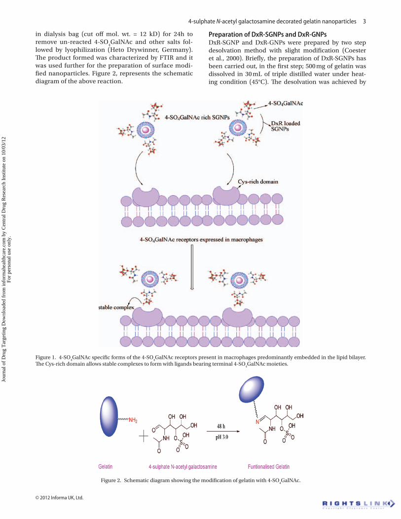
on the surface of macrophage have the responsibility of identification and endocytosis of a particulate carrier. Schematic diagram (Figure 1) shows the specific binding sites for targeted sulphate moiety present preferably on macrophages. These facts about the macrophages, its receptors and the modification of the chemical and physical properties of the ligands attached to the carriers such as mannosyl, immunoglobulin, fibronectin and galactosyl are exploited for better phagocytosed than carriers without such ligands (Shao & Hochmuth, 1997). This enhanced uptake gives us better opportunity to develop a delivery system with properties of targeting at particular site, with increased specificity, potency of the drug and decreasing the therapeutic dose of even the potent drug and thus by reducing the toxicity of the drug (Chen et al., 2008).
DxR was chosen as a model drug which showed sig-nificant efficacy both in vitro and in vivo against pro-mastigote as well as amastigotes and has been proposed as the second-line therapeutic agents for leishmaniasis chemotherapy (Kansal et al., 2012; Sett et al., 1992, 1993; Gupta et al., 2009). Within this preview, we have explored the targeting potential of DxR-SGNPs with resident macrophages. This novel 4-SO
4GalNAc moiety provides
an alternative for targeting specifically MRs on mac-rophages, which can be easily decorated on GNPs by a simple method.
Materials and methodsMaterialsGelatin, type A was purchased from sigma chemical company (St. Louis, USA). 4-SO
4GalNAc, dialysis bag
(cut off mol. wt. = 12 kD) and Trypsin from Bovine pan-creas were purchased from Sigma-Aldrich (St. Louis, USA). Glutaraldehyde 25% was purchased from S.D. Fine chemical ltd (India, Mumbai). Other reagents and chemicals used were of analytical grade. For cell culture, RPMI-1640 medium, fetal bovine serum (FBS) and anti-biotic solution (penicillin/streptomycin, 0.1% v/v), MTT (3-[4,5-dimethylthiazol-2-yl]-2,5-diphenyl tetrazolium bromide) and 4′,6-diamidino-2-phenylindole(DAPI) were purchased from Sigma. Well plates for cytotoxicity and uptake studies were purchased from Greiner Bio One Frickenhausen (Germany). The triple distilled water was used in all experiments by a three-stage Millipore Milli-Q plus 185 purification system (Bedford, MA, USA).
Surface modification of gelatin with 4-SO4GalNAc
The gelatin modification with 4-SO4GalNAc has been
carried out with the method which has been reported earlier with slight modifications (Mitchell et al., 1999). Briefly 4-SO
4GalNAc (10 mM) was dissolved in 0.1 M
acetate buffer (pH = 3.0) and added to aqueous solu-tion of gelatin (0.5 mM). The mixture was agitated at ambient temperature (30 ± 2°C) for 2 days for comple-tion of reaction. Resulting solution was concentrated and purified by dialyzing against triple distilled water
Jour
nal o
f D
rug
Tar
getin
g D
ownl
oade
d fr
om in
form
ahea
lthca
re.c
om b
y C
entr
al D
rug
Res
earc
h In
stitu
te o
n 10
/03/
12Fo
r pe
rson
al u
se o
nly.
4-sulphate N-acetyl galactosamine decorated gelatin nanoparticles 3
© 2012 Informa UK, Ltd.
in dialysis bag (cut off mol. wt. = 12 kD) for 24h to remove un-reacted 4-SO
4GalNAc and other salts fol-
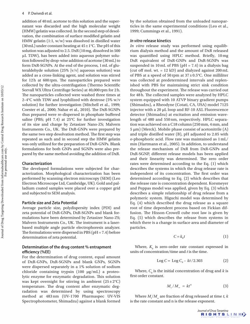
lowed by lyophilization (Heto Drywinner, Germany). The product formed was characterized by FTIR and it was used further for the preparation of surface modi-fied nanoparticles. Figure 2, represents the schematic diagram of the above reaction.
Preparation of DxR-SGNPs and DxR-GNPsDxR-SGNP and DxR-GNPs were prepared by two step desolvation method with slight modification (Coester et al., 2000). Briefly, the preparation of DxR-SGNPs has been carried out, in the first step; 500 mg of gelatin was dissolved in 30 mL of triple distilled water under heat-ing condition (45°C). The desolvation was achieved by
Figure 1. 4-SO4GalNAc specific forms of the 4-SO
4GalNAc receptors present in macrophages predominantly embedded in the lipid bilayer.
The Cys-rich domain allows stable complexes to form with ligands bearing terminal 4-SO4GalNAc moieties.
Figure 2. Schematic diagram showing the modification of gelatin with 4-SO4GalNAc.
Jour
nal o
f D
rug
Tar
getin
g D
ownl
oade
d fr
om in
form
ahea
lthca
re.c
om b
y C
entr
al D
rug
Res
earc
h In
stitu
te o
n 10
/03/
12Fo
r pe
rson
al u
se o
nly.
4 P. Dwivedi et al.
Journal of Drug Targeting
addition of 40 mL acetone to this solution and the super-natant was discarded and the high molecular weight (HMW) gelatin was collected. In the second step of desol-vation, the combination of surface modified gelatin and HMW gelatin (1:1, w/w) was dissolved in distilled water (30 mL) under constant heating at 45 ± 1°C. The pH of this solution was adjusted to 2.5. DxR (10 mg, dissolved in 500 μL TDW), has been added into aqueous polymer solu-tion followed by drop-wise addition of acetone (30 mL) to form DxR-SGNPs. At the end of the process, 1 mL of glu-teraldehyde solution (25% [v/v] aqueous solution) was added as a cross-linking agent, and solution was stirred for 12 h at 600 rpm. The nanoparticles prepared were collected by the ultra-centrifugation (Thermo Scientific Sorvall WX Ultra Centrifuge Series) at 40,000 rpm for 2 h. The nanoparticles collected were washed three times at 2–4°C with TDW and lyophilized with dextrose (5% w/v solution) for further investigation (Mitchell et al., 1999; Coester et al., 2000; Nahar et al., 2010). The DxR-SGNPs thus prepared were re-dispersed in phosphate buffered saline (PBS; pH 7.4) at 25°C for further investigation of its size and charge by Zetasizer Nano-ZS; Malvern Instruments Co., UK. The DxR-GNPs were prepared by the same two step desolvation method. The first step was repeated as such and in second step the HMW gelatin was only utilized for the preparation of DxR-GNPs. Blank formulations for both GNPs and SGNPs were also pre-pared by the same method avoiding the addition of DxR.
CharacterizationThe developed formulations were subjected for char-acterization. Morphological characterization has been performed by scanning electron microscopy (SEM) (Leo Electron Microscope Ltd, Cambridge, UK). Gold and pal-ladium coated samples were placed over a copper grid and subjected to SEM analysis.
Particle size and Zeta PotentialAverage particle size, polydispersity index (PDI) and zeta potential of DxR-GNPs, DxR-SGNPs and blank for-mulations have been determined by Zetasizer Nano-ZS; Malvern Instruments Co., UK. The instrument is a laser-based multiple angle particle electrophoresis analyzer. The formulations were dispersed in PBS (pH = 7.4) before determination of zeta potential.
Determination of the drug content % entrapment efficiency (%EE)For the determination of drug content, equal amount of DxR-GNPs, DxR-SGNPs and blank GNPs, SGNPs were dispersed separately in a 1% solution of sodium chloride containing trypsin (100 µg/mL) a proteo-lytic enzyme for enzymatic degradation. This solution was kept overnight for stirring in ambient (25 ± 2°C) temperature. The drug content after enzymatic deg-radation was determined by using spectroscopy method at 483 nm (UV-1700 Pharmaspec UV-VIS Spectrophotometer, Shimadzu) against a blank formed
by the solution obtained from the unloaded nanopar-ticles in the same experimental conditions (Leo et al., 1999; Cummings et al., 1991).
In vitro release kineticsIn vitro release study was performed using equilib-rium dialysis method and the amount of DxR released was quantified using HPLC method. Briefly, 10 mg DxR equivalent of DxR-GNPs and DxR-SGNPs was suspended in 10 mL of PBS (pH = 7.4) in a dialysis bag (cut off mol. wt. = 12 kD) and dialyzed against 200 mL of PBS at a speed of 50 rpm at 37 ± 0.5°C. One milliliter was collected at predetermined intervals and replen-ished with PBS for maintaining strict sink condition throughout the experiment. The release was carried out for 48 h. The collected samples were analyzed by HPLC system equipped with 10 ATVP binary gradient pumps (Shimadzu), a Rheodyne (Cotati, CA, USA) model 7125 injector with a 20 µL loop and RF-10 AXL Fluorescence detector (Shimadzu) at excitation and emission wave-length of 480 and 550 nm, respectively. HPLC separa-tion was achieved on a C18 column (250 mm, 4 mm, and 5 µm) (Merck). Mobile phase consist of acetonitrile (A) and triple distilled water (B), pH adjusted to 2.05 with o-phosphoric acid. Flow rate was maintained as 1 mL/min (Hartmann et al., 2005). In addition, to understand the release mechanism of DxR from DxR-GNPs and DxR-SGNP, different release models has been applied and their linearity was determined. The zero order rates were determined according to the Eq. (1) which describes the systems in which the drug release rate is independent of its concentration. The first order was determined according to Eq. (2) which describes that the release rate is concentration dependent. Korsmeyer and Peppas model was applied, given by Eq. (3) which describes a simple relationship of drug release from a polymeric system. Higuchi model was determined by Eq. (4) which described the drug release as a square root of time dependent process based on Fickian dif-fusion. The Hixson-Crowell cube root law is given by Eq. (5) which describes the release from systems in which there is a change in surface area and diameter of particles.
C k t= 0 (1)
Where, K0 is zero-order rate constant expressed in
units of concentration/time and t is the time.
Log Log C C kt= 0 2 303– / . (2)
Where, C0 is the initial concentration of drug and k is
first order constant.
M M kt nt / ∞ = (3)
Where Mt/M
∞ are fraction of drug released at time t, k
is the rate constant and n is the release exponent.
Jour
nal o
f D
rug
Tar
getin
g D
ownl
oade
d fr
om in
form
ahea
lthca
re.c
om b
y C
entr
al D
rug
Res
earc
h In
stitu
te o
n 10
/03/
12Fo
r pe
rson
al u
se o
nly.
4-sulphate N-acetyl galactosamine decorated gelatin nanoparticles 5
© 2012 Informa UK, Ltd.
Q = Kt1 2/
(4)
Where, K is the constant reflecting the design variables of the system.
–/ /W W Ktt01 3 1 3 = (5)
Where, Wt is the amount of drug released at time t, W
0
is the initial amount of the drug in nanoparticles and K is the rate constant for Hixson-Crowell rate equation.
Stability studiesThe stability of DxR-GNPs and DxR-SGNPs has been assessed by measuring the particle size, zeta potential and % EE. The formulations were stored and evaluated at 5 ± 3°C and ambient room temperature (25 ± 2°C, 60 ± 5 RH).
Macrophage uptake studies in J774A.1 and peripheral blood monocyte cells (PBMC)The uptake studies of the formulations has been car-ried out on both adherent mouse macrophage cell line J774A.1 and PBMC, and evaluated using fluorescence activated cell sorter (FACS) instrument (BD Biosciences, FACS Aria, Germany).
Uptake studies in J774A.1Aliquots (100 μL) containing J774A.1 (1 × 10⁵) was sus-pended in 0.9 mL of fresh RPMI-1640 medium supple-mented with penicillin 10 U/mL, 10% FBS, 100μg/mL streptomycin, 1 mM sodium pyruvate and 10 mM HEPES (4-(2-hydroxyethyl)-1-piperazineethanesulfonic acid) medium. These were transferred into 24-well plates con-taining fresh medium and suspended in a 37°C humidi-fied incubator with 5% CO
2 atmosphere. After 24 h the
culture medium was replaced with fresh culture medium. DxR, DxR-GNPs and DxR-SGNPs were added to it in trip-licate such that the amount of DxR remains constant and incubated for 4 h at room temperature in dark. The cells were scraped at various time intervals and the extent of nanoparticles uptake in vitro was examined by flow cytometry. For separation of the internalized and surface bound nanoparticles, the cells were washed three times with acetate buffer (pH = 4.0). The cell-associated fluo-rescence was measured by FACS at an excitation wave-length of 480 nm and an emission wavelength of 550 nm.
Isolation of PBMC and uptake studiesFor PBMC uptake studies it has been first isolated and cultured. Isolation of PBMC has been carried out by a Ficoll-Hypaque gradient method (Rettig et al., 2010). Briefly, 10 mL human blood (anti-coagulated with EDTA) was obtained from healthy volunteer and was diluted with an equal volume of 0.9% NaCl and poured over the surface of 10 mL ficoll solution (Histopaque 1077, Sigma, St Louis, USA) by layering in 50 mL Falcon tubes (Tarson, India). The tubes were centrifuged at 800 rpm for 20 min at room temperature. After centrifugation, three distinct layers of different cell population were obtained; PBMC,
Polymorphonuclear Neutrophil Granulocytes and Red Blood cells. The buffy coat ring was carefully aspirated and transferred to a 50 mL tube, and washed three times with PBS. Cells were counted using a hemocytometer and adjusted to 106 cells mL−1 in RPMI 1640 (Sigma, St Louis USA) cell culture medium supplemented 10% fetal calf serum (FCS; Gibco, Invitrogen GmbH), and antibiotic-antimycotic (100 U/mL penicillin and 100 mg/mL strep-tomycin; Gibco, Berlin, Germany) in 25-cm3 culture flask. After 24 h nonadherent cells were removed by repeated washing and cells were cultured with fresh medium.
For uptake studies, peripheral monocyte derived macrophages (1 × 10⁶ cells) were plated on 6-well culture plates (NUNC GmbH, Wiesbaden, Germany). DxR, DxR-GNPs and DxR-SGNPs were added to it in triplicate such that the amount of DxR remains constant and were incu-bated for 4 h. After incubation cells were washed with PBS, carefully detached with Grenier cell scraper and were washed two times. These cells were re-suspending in 500 μL PBS for the measurement of fluorescence directly using FACS. For each measurement, 10,000 cells events were acquired. The data were analyzed with CELLQuest software (Becton Dickinson).
Confocal microscopy for phagocytosis evaluationConfocal microscopy has been carried out by using Carl Zeiss confocal micro scanning microscope (CLSM-510 META) by using plan apochromat objective lens, Germany. In brief poly-l-lysine-coated slides were pre-pared and cells were adhered on the slides. Cells seeded at a density of 1 × 10⁶ cells/well on poly-l-lysine coated glass cover slip in 6 well plate for 12 h, then macrophage monolayer’s adherent were washed and incubated in fresh medium. Free or nano-encapsulated drug were added to triplicate wells. DxR-loaded nanoparticles were suspended in Dulbecco Modified Eagle Medium (DMEM) and added to macrophages and incubated 4h at room temperature in dark. After incubation, mono-layers were washed repeatedly to remove non-adherent nanoparticles and cells were fixed in 10% formalin (in Phosphate buffer saline). Nucleus of the macrophages has been stained using 10 µL of 1 mg/mL DAPI, a nucleus staining dye and mounted with glycerol. Macrophages internalized nanoparticles were observed using confocal microscopy of triplicate cover slips for each experimental condition.
Cytotoxicity studiesThe in-vitro cellular viability of DxR-GNPs, DxR-GNPs and blank GNPs, SGNPs with plain DxR has been mea-sured by the MTT [3-(4, 5 dimethyl-thiazol-2-yl-2, 5 biphenyl) tetrazolium bromide] proliferation assay (calo-rimetric assay) on J774A.1 macrophage cell line. Viable cells are capable to reduce MTT into colored formazan and the amount of MTT-formazan produced was an indi-cator for cell viability. J774A.1 cells (mouse macrophage cell line) was cultured in DMEM supplemented with 10% FCS and 1% antimycotic-antibiotic mixture culture plates
Jour
nal o
f D
rug
Tar
getin
g D
ownl
oade
d fr
om in
form
ahea
lthca
re.c
om b
y C
entr
al D
rug
Res
earc
h In
stitu
te o
n 10
/03/
12Fo
r pe
rson
al u
se o
nly.
6 P. Dwivedi et al.
Journal of Drug Targeting
at 37°C and 5% CO2 humidified incubator Cells seeded
at a density of 1 × 10⁵ cells/well in 96 well plate for 24 h then, cells were washed and incubated in fresh medium. Formulations (DxR-GNPs, DxR-GNPs, GNPs and SGNPs with plain DxR) have been added at different concentra-tion (0.1, 1, 5 and 10 µg/mL) to triplicate wells for 24 h, after which cells were washed three times with PBS. After washing, 20 µL of MTT solution (5 mg/mL stock solution) was added to each well and the plates were incubated for an additional 4 h. The un-reacted MTT dye and medium were discarded, and 100 µL of DMSO was added to each well to solubilize the MTT formazan crystals. The contents of the plates were mixed for 15 min to achieve complete solubilization of the formazan crystals and the optical density of each well was measured at 570 nm with a micro plate spectrophotometer. The optical density was measured using a multi-well scanning spectrophotom-eter (MRX Micro plate Reader, Dynatech Laboratories Inc., Chantilly, VA, US) at a wavelength of 570 nm.
In vivo studiesThe studies were carried out according to the guidelines of Council for the Purpose of Control and Supervision of Experiments on Animals (CPCSEA), Ministry of Social Justice and Empowerment, Government of India. Experiments were conducted after ethical clearance by the Institutional Animal Ethics Committee of the insti-tute (IAEC approval no. 70/11/Pharmaceutics/IAEC).
Organ distribution studiesWistar rats weighing about 150–200 g were divided into three groups of six rats each. DxR solution, DxR-SGNPs and DxR-GNPs containing equivalent doses of DxR (1 mg/kg body weight) were administered intravenously to different groups. Animals from each group were sacri-ficed at 0.5, 1, 2, 4, 6 and 24h after administration of the formulations. Blood samples were collected by cardiac puncture while liver and spleen was excised after dis-section. The liver and spleen were washed with distilled water and dried by blotting on tissue papers. Drug con-tent in the blood and organs was determined by HPLC using the method reported previously with slight modi-fication. Drug targeting index (DTI) has been estimated in order to evaluate the targeting efficacy of the surface modified drug carrier. DTI was calculated based on area under the concentration time curve at target organ and systemic site (plasma) using following equation.
DTI AUC AUC AUC AUCLiver Liver Plasma DxR SGNP Liver Plasma= ( ) −/ / /(( )
= ( )−
−
DxR GNP
Spleen Spleen Plasma DxR SGNP SplDTI AUC AUC AUC/ / eeen Plasma DxR GNPAUC/( )
−
Measurement of hemolysisThe degree of hemolysis induced by blank SGNPs and GNPs was determined on fresh citrated blood of both human and wistar rat using the method reported previ-ously with slight modification (Wang et al., 2006). SGNPs and GNPs were incubated with 1 mL blood of both
human and Wistar rat at 37°C for 2h, and then centri-fuged at 10,000 rpm for 20 min. The hemoglobin released in the supernatant was measured spectrophotometri-cally at 540 nm. For the negative control, blood was simi-larly incubated with 0.9% (w/v) saline. Blood incubated with the same volume of distilled water (100% hemolysis) was taken as positive control. The percentage hemolysis was determined from the hemoglobin released into the supernatant.
Statistical analysisAll results have been expressed as mean ± SD (n = 3). Differences between formulations were compared using one-way analysis of variance (ANOVA) followed by the Turkey-Kramer multiple comparison test, using Graph Pad Instat software (Graph Pad Software Inc. CA, USA). p < 0.05 denotes significance in all cases.
ResultsSurface modification of gelatin with 4-SO
4GalNAc and its characterization
The gelatin has been modified by 4-SO4GalNAc as rep-
resented schematically in Figure 2. The preparation of 4-SO
4GalNAc modified gelatin was characterized by
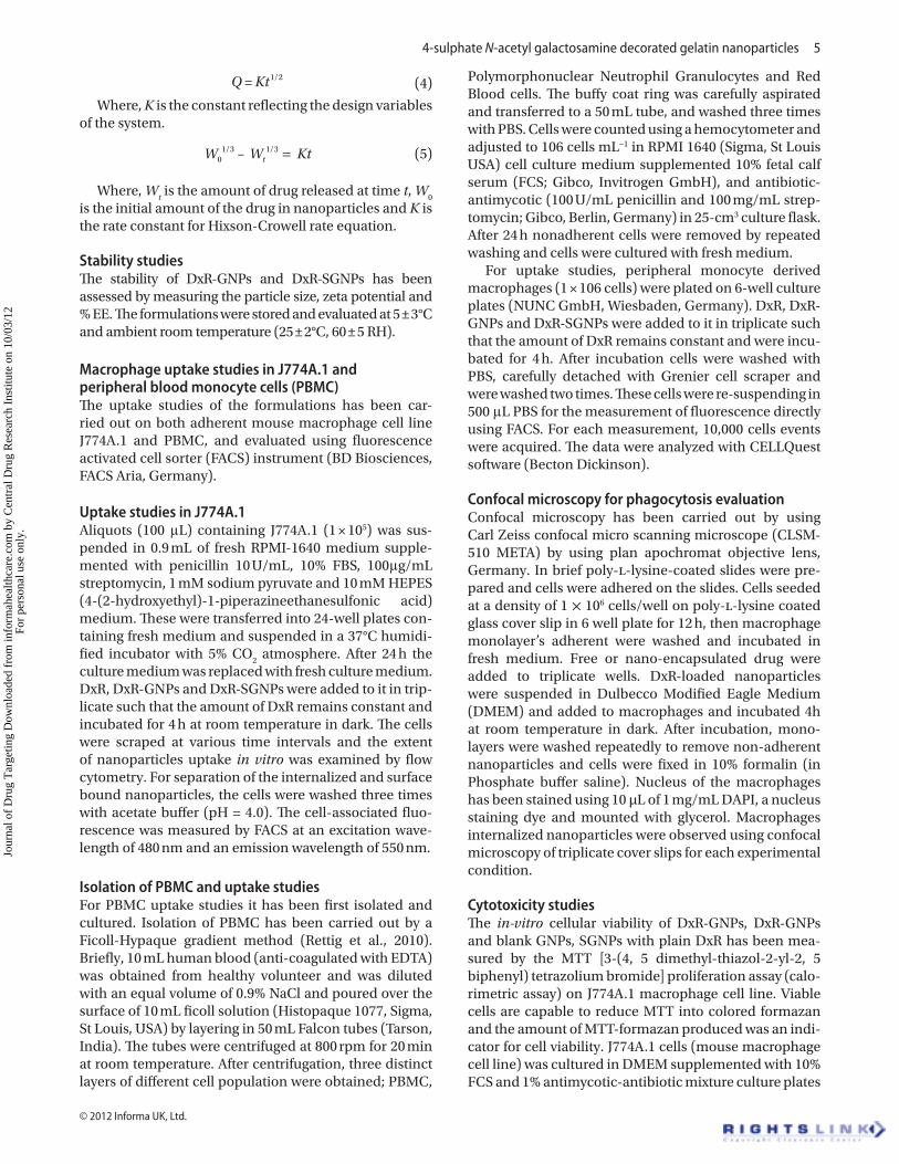
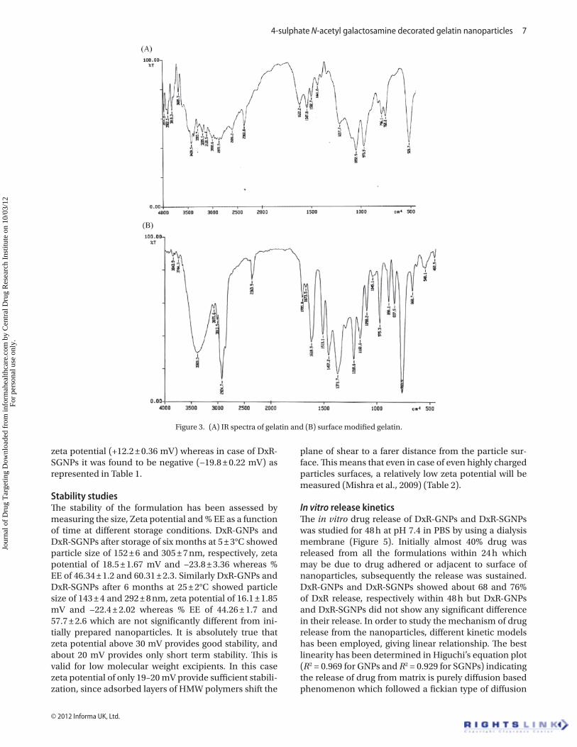
FTIR. The data was compared with unmodified gelatin and the formation of Schiff’s base (–C=N–) between the amino terminal of gelatin and the ligands was confirmed, which reveals Schiff’s base C=N stretch at 1457 cm−1. The broad intense O-H stretch of galactosamine at 3383 cm−1 and C=O stretch at 1618 cm−1 (shift of C=O might be due to the presence of N acetyl), N-H bending of secondary amines at 1513 cm−1 was also present in the modified gelatin which was not present in gelatin itself. Other characteristic peaks identified in modified gelatin was C–N stretch at 1218 cm−1, S=O stretch at 1371 cm−1, strong C–O stretch at 1098 cm−1. This data provide evidences for the formation of an imine bond between gelatin and 4-SO
4GalNAc. (Figure 3A and 3B)
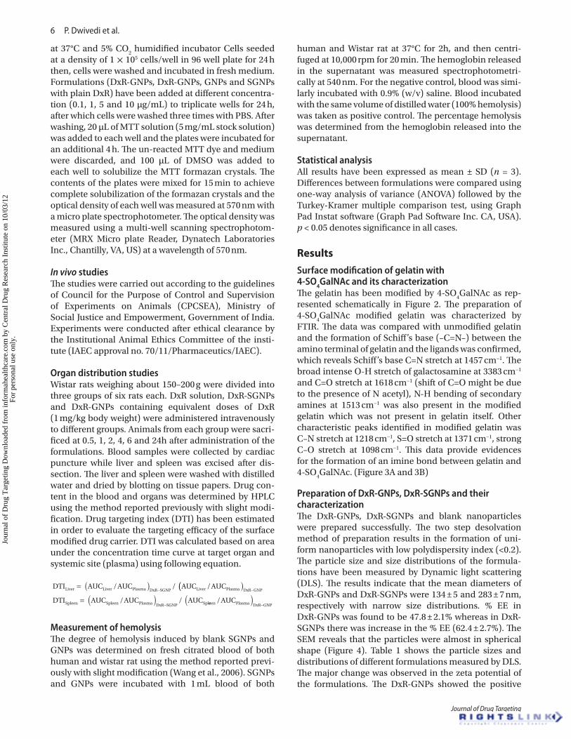
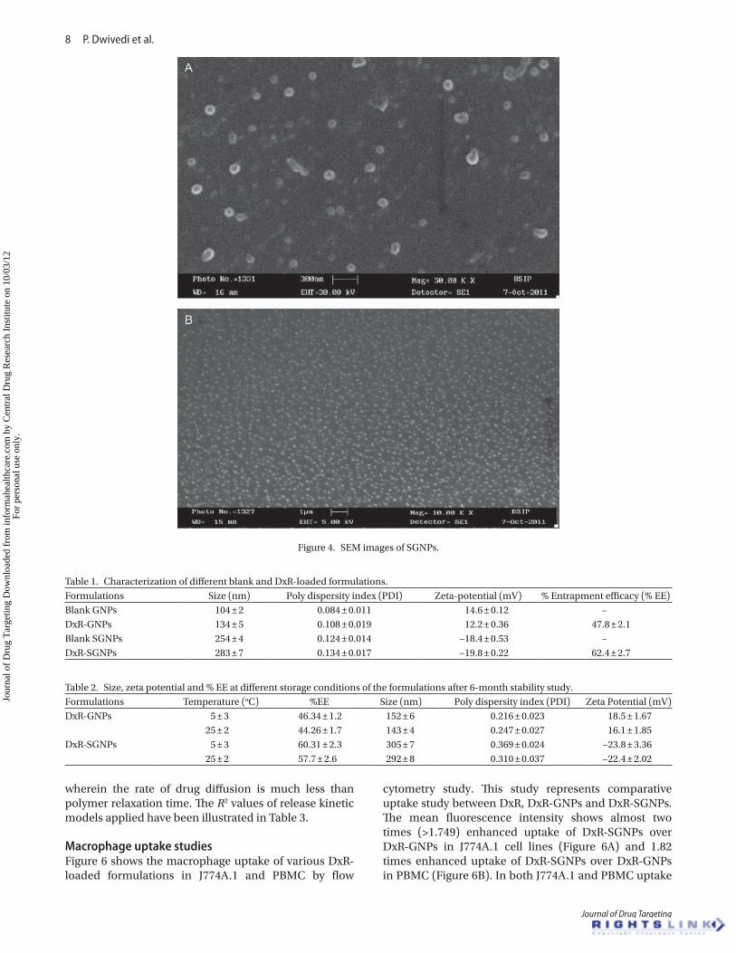
Preparation of DxR-GNPs, DxR-SGNPs and their characterizationThe DxR-GNPs, DxR-SGNPs and blank nanoparticles were prepared successfully. The two step desolvation method of preparation results in the formation of uni-form nanoparticles with low polydispersity index (<0.2). The particle size and size distributions of the formula-tions have been measured by Dynamic light scattering (DLS). The results indicate that the mean diameters of DxR-GNPs and DxR-SGNPs were 134 ± 5 and 283 ± 7 nm, respectively with narrow size distributions. % EE in DxR-GNPs was found to be 47.8 ± 2.1% whereas in DxR-SGNPs there was increase in the % EE (62.4 ± 2.7%). The SEM reveals that the particles were almost in spherical shape (Figure 4). Table 1 shows the particle sizes and distributions of different formulations measured by DLS. The major change was observed in the zeta potential of the formulations. The DxR-GNPs showed the positive
Jour
nal o
f D
rug
Tar
getin
g D
ownl
oade
d fr
om in
form
ahea
lthca
re.c
om b
y C
entr
al D
rug
Res
earc
h In
stitu
te o
n 10
/03/
12Fo
r pe
rson
al u
se o
nly.
4-sulphate N-acetyl galactosamine decorated gelatin nanoparticles 7
© 2012 Informa UK, Ltd.
zeta potential (+12.2 ± 0.36 mV) whereas in case of DxR-SGNPs it was found to be negative (−19.8 ± 0.22 mV) as represented in Table 1.
Stability studiesThe stability of the formulation has been assessed by measuring the size, Zeta potential and % EE as a function of time at different storage conditions. DxR-GNPs and DxR-SGNPs after storage of six months at 5 ± 3°C showed particle size of 152 ± 6 and 305 ± 7 nm, respectively, zeta potential of 18.5 ± 1.67 mV and −23.8 ± 3.36 whereas % EE of 46.34 ± 1.2 and 60.31 ± 2.3. Similarly DxR-GNPs and DxR-SGNPs after 6 months at 25 ± 2°C showed particle size of 143 ± 4 and 292 ± 8 nm, zeta potential of 16.1 ± 1.85 mV and −22.4 ± 2.02 whereas % EE of 44.26 ± 1.7 and 57.7 ± 2.6 which are not significantly different from ini-tially prepared nanoparticles. It is absolutely true that zeta potential above 30 mV provides good stability, and about 20 mV provides only short term stability. This is valid for low molecular weight excipients. In this case zeta potential of only 19–20 mV provide sufficient stabili-zation, since adsorbed layers of HMW polymers shift the
plane of shear to a farer distance from the particle sur-face. This means that even in case of even highly charged particles surfaces, a relatively low zeta potential will be measured (Mishra et al., 2009) (Table 2).
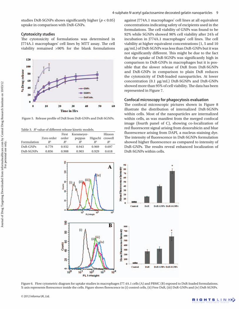
In vitro release kineticsThe in vitro drug release of DxR-GNPs and DxR-SGNPs was studied for 48 h at pH 7.4 in PBS by using a dialysis membrane (Figure 5). Initially almost 40% drug was released from all the formulations within 24 h which may be due to drug adhered or adjacent to surface of nanoparticles, subsequently the release was sustained. DxR-GNPs and DxR-SGNPs showed about 68 and 76% of DxR release, respectively within 48 h but DxR-GNPs and DxR-SGNPs did not show any significant difference in their release. In order to study the mechanism of drug release from the nanoparticles, different kinetic models has been employed, giving linear relationship. The best linearity has been determined in Higuchi’s equation plot (R2 = 0.969 for GNPs and R2 = 0.929 for SGNPs) indicating the release of drug from matrix is purely diffusion based phenomenon which followed a fickian type of diffusion
Figure 3. (A) IR spectra of gelatin and (B) surface modified gelatin.
Jour
nal o
f D
rug
Tar
getin
g D
ownl
oade
d fr
om in
form
ahea
lthca
re.c
om b
y C
entr
al D
rug
Res
earc
h In
stitu
te o
n 10
/03/
12Fo
r pe
rson
al u
se o
nly.
8 P. Dwivedi et al.
Journal of Drug Targeting
wherein the rate of drug diffusion is much less than polymer relaxation time. The R2 values of release kinetic models applied have been illustrated in Table 3.
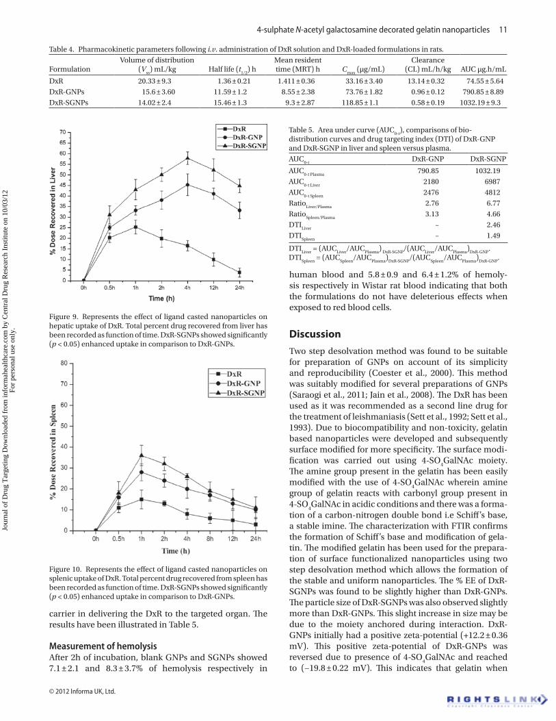
Macrophage uptake studiesFigure 6 shows the macrophage uptake of various DxR-loaded formulations in J774A.1 and PBMC by flow
cytometry study. This study represents comparative uptake study between DxR, DxR-GNPs and DxR-SGNPs. The mean fluorescence intensity shows almost two times (>1.749) enhanced uptake of DxR-SGNPs over DxR-GNPs in J774A.1 cell lines (Figure 6A) and 1.82 times enhanced uptake of DxR-SGNPs over DxR-GNPs in PBMC (Figure 6B). In both J774A.1 and PBMC uptake
Figure 4. SEM images of SGNPs.
Table 1. Characterization of different blank and DxR-loaded formulations. Formulations Size (nm) Poly dispersity index (PDI) Zeta-potential (mV) % Entrapment efficacy (% EE)Blank GNPs 104 ± 2 0.084 ± 0.011 14.6 ± 0.12 –DxR-GNPs 134 ± 5 0.108 ± 0.019 12.2 ± 0.36 47.8 ± 2.1Blank SGNPs 254 ± 4 0.124 ± 0.014 −18.4 ± 0.53 –DxR-SGNPs 283 ± 7 0.134 ± 0.017 −19.8 ± 0.22 62.4 ± 2.7
Table 2. Size, zeta potential and % EE at different storage conditions of the formulations after 6-month stability study. Formulations Temperature (oC) %EE Size (nm) Poly dispersity index (PDI) Zeta Potential (mV)DxR-GNPs 5 ± 3 46.34 ± 1.2 152 ± 6 0.216 ± 0.023 18.5 ± 1.67
25 ± 2 44.26 ± 1.7 143 ± 4 0.247 ± 0.027 16.1 ± 1.85DxR-SGNPs 5 ± 3 60.31 ± 2.3 305 ± 7 0.369 ± 0.024 −23.8 ± 3.36
25 ± 2 57.7 ± 2.6 292 ± 8 0.310 ± 0.037 −22.4 ± 2.02
Jour
nal o
f D
rug
Tar
getin
g D
ownl
oade
d fr
om in
form
ahea
lthca
re.c
om b
y C
entr
al D
rug
Res
earc
h In
stitu
te o
n 10
/03/
12Fo
r pe
rson
al u
se o
nly.
4-sulphate N-acetyl galactosamine decorated gelatin nanoparticles 9
© 2012 Informa UK, Ltd.
studies DxR-SGNPs shows significantly higher (p < 0.05) uptake in comparison with DxR-GNPs.
Cytotoxicity studiesThe cytotoxicity of formulations was determined in J774A.1 macrophages’ cell lines by MTT assay. The cell viability remained >90% for the blank formulations
against J774A.1 macrophages’ cell lines at all equivalent concentrations indicating safety of excipients used in the formulations. The cell viability of GNPs was found to be 92% while SGNPs showed 90% cell viability after 24 h of incubation in J774A.1 macrophages’ cell lines. The cell viability at higher equivalent concentrations (1, 5 and 10 µg/mL) of DxR-SGNPs was less than DxR-GNPs but it was not significantly different. This might be due to the fact that the uptake of DxR-SGNPs was significantly high in comparison to DxR-GNPs in macrophages but it is pos-sible that the slower release of DxR from DxR-SGNPs and DxR-GNPs in comparison to plain DxR reduces the cytotoxicity of DxR-loaded nanoparticles. At lower concentration (0.1 µg/mL) DxR-SGNPs and DxR-GNPs showed more than 95% of cell viability. The data has been represented in Figure 7.
Confocal microscopy for phagocytosis evaluationThe confocal microscopic pictures shown in Figure 8 illustrate the distribution of internalized DxR-SGNPs within cells. Most of the nanoparticles are internalized within cells, as was manifest from the merged confocal image (fourth panel of C), showing co-localization of red fluorescent signal arising from doxorubicin and blue fluorescence arising from DAPI, a nucleus-staining dye. The intensity of fluorescence in DxR-SGNPs formulation showed higher fluorescence as compared to intensity of DxR-GNPs. The results reveal enhanced localization of DxR-SGNPs within cells.
Figure 5. Release profile of DxR from DxR-GNPs and DxR-SGNPs.
Table 3. R2 value of different release kinetic models.
FormulationZero order
R2
First order
R2
Korsmeyer-peppas
R2
Higuchi R2
Hixson crowell
R2
DxR-GNPs 0.779 0.932 0.943 0.969 0.697DxR-SGNPs 0.856 0.908 0.903 0.929 0.618
Figure 6. Flow cytometric diagram for uptake studies in macrophages J77.4A.1 cells (A) and PBMC (B) exposed to DxR-loaded formulations. X-axis represents florescence inside the cells. Figure shows florescence in (i) control cells, (ii) Free DxR, (iii) DxR-GNPs and (iv) DxR-SGNPs.
Jour
nal o
f D
rug
Tar
getin
g D
ownl
oade
d fr
om in
form
ahea
lthca
re.c
om b
y C
entr
al D
rug
Res
earc
h In
stitu
te o
n 10
/03/
12Fo
r pe
rson
al u
se o
nly.

10 P. Dwivedi et al.
Journal of Drug Targeting
Pharmacokinetics of DxR formulation in ratsThe area under curve (AUC) of DxR in case of DxR-SGNPs (AUC = 1032.19 ± 2.32 h·µg/mL) was significantly higher (p < 0.05) than DxR-GNPs (AUC = 790.85 ± 2.89 h·µg/mL) and DxR (AUC = 74.55 ± 5.64 h·µg/mL). High
mean residence time (MRT = 9.3 ± 2.87h) and lower clear-ance (CL = 0.58 ± 0.19mL/h/kg) was observed in DxR-SGNPs as compared to DxR-GNPs (MRT = 8.55 ± 2.38 h, CL = 0.96 ± 0.12 mL/h/kg), and DxR (MRT = 1.411 ± 0.36h, CL = 13.14 ± 0.32 mL/h/kg). The pharmacokinetic param-eters of different formulations have been shown in Table 4.
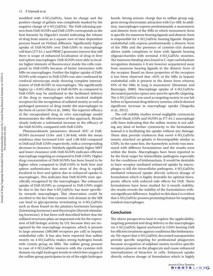
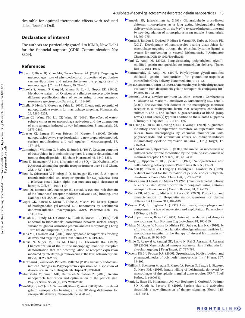
Liver and spleen distribution of different DxR formulationDxR concentration in liver and spleen after i.v. injec-tion of different DxR formulations has been shown in Figures 9 and 10. The drug localization was significantly higher in DxR-SGNPs and DxR-GNPs as compared to DxR at all time points (p < 0.05). Similarly DxR-SGNPs showed enhanced drug distribution compared to DxR-GNPs in liver and spleen. The efficacy of DxR-SGNPs in terms of DTI has been quantified as the ratio of the DxR reach-ing the target organ through the drug carrier complex and the amount of drug in the plasma. The DTI of DxR in the liver by DxR-SGNP was about 2.46-folds compared to that of DxR associated with DxR-GNP whereas DxR in the spleen by DxR-SGNP was about 1.49 folds compared to that of DxR associated with DxR-GNP. The results indicate the critical role of 4-SO
4GalNAc associated drug
Figure 8. Confocal fluorescence image of single XY optical section of tissue culture cells. The red fluorescent signals were arising from DxR and blue florescent signals were arising from DAPI, a nucleus staining dye. (A) Shows the image of J774A.1 cells of control. (B) Shows the incubation of the cells with DxR-GNPs. (C) Shows the DxR-SGNPs treated cells. The first, second, third and fourth panels reveals the topographic view of the control cells, red fluorescence of internalized DxR, blue florescence of DAPI stained nucleus and merged confocal image respectively. Most of the nanoparticles are internalized within cells, as was manifest from the merged confocal image (fourth panel of C), showing the co-localization of red fluorescent signal arising from doxorubicin and blue fluorescence arising from DAPI, a nucleus-staining dye.
Figure 7. MTT assay results of formulations showing the cell viability at different concentration of DxR.
Jour
nal o
f D
rug
Tar
getin
g D
ownl
oade
d fr
om in
form
ahea
lthca
re.c
om b
y C
entr
al D
rug
Res
earc
h In
stitu
te o
n 10
/03/
12Fo
r pe
rson
al u
se o
nly.
4-sulphate N-acetyl galactosamine decorated gelatin nanoparticles 11
© 2012 Informa UK, Ltd.
carrier in delivering the DxR to the targeted organ. The results have been illustrated in Table 5.
Measurement of hemolysisAfter 2h of incubation, blank GNPs and SGNPs showed 7.1 ± 2.1 and 8.3 ± 3.7% of hemolysis respectively in
human blood and 5.8 ± 0.9 and 6.4 ± 1.2% of hemoly-sis respectively in Wistar rat blood indicating that both the formulations do not have deleterious effects when exposed to red blood cells.
DiscussionTwo step desolvation method was found to be suitable for preparation of GNPs on account of its simplicity and reproducibility (Coester et al., 2000). This method was suitably modified for several preparations of GNPs (Saraogi et al., 2011; Jain et al., 2008). The DxR has been used as it was recommended as a second line drug for the treatment of leishmaniasis (Sett et al., 1992; Sett et al., 1993). Due to biocompatibility and non-toxicity, gelatin based nanoparticles were developed and subsequently surface modified for more specificity. The surface modi-fication was carried out using 4-SO
4GalNAc moiety.
The amine group present in the gelatin has been easily modified with the use of 4-SO
4GalNAc wherein amine
group of gelatin reacts with carbonyl group present in 4-SO
4GalNAc in acidic conditions and there was a forma-
tion of a carbon-nitrogen double bond i.e Schiff’s base, a stable imine. The characterization with FTIR confirms the formation of Schiff’s base and modification of gela-tin. The modified gelatin has been used for the prepara-tion of surface functionalized nanoparticles using two step desolvation method which allows the formation of the stable and uniform nanoparticles. The % EE of DxR-SGNPs was found to be slightly higher than DxR-GNPs. The particle size of DxR-SGNPs was also observed slightly more than DxR-GNPs. This slight increase in size may be due to the moiety anchored during interaction. DxR-GNPs initially had a positive zeta-potential (+12.2 ± 0.36 mV). This positive zeta-potential of DxR-GNPs was reversed due to presence of 4-SO
4GalNAc and reached
to (−19.8 ± 0.22 mV). This indicates that gelatin when
Table 4. Pharmacokinetic parameters following i.v. administration of DxR solution and DxR-loaded formulations in rats.
FormulationVolume of distribution
(Vss
) mL/kg Half life (t1/2
) hMean resident time (MRT) h C
max (µg/mL)
Clearance (CL) mL/h/kg AUC µg.h/mL
DxR 20.33 ± 9.3 1.36 ± 0.21 1.411 ± 0.36 33.16 ± 3.40 13.14 ± 0.32 74.55 ± 5.64DxR-GNPs 15.6 ± 3.60 11.59 ± 1.2 8.55 ± 2.38 73.76 ± 1.82 0.96 ± 0.12 790.85 ± 8.89DxR-SGNPs 14.02 ± 2.4 15.46 ± 1.3 9.3 ± 2.87 118.85 ± 1.1 0.58 ± 0.19 1032.19 ± 9.3
Figure 9. Represents the effect of ligand casted nanoparticles on hepatic uptake of DxR. Total percent drug recovered from liver has been recorded as function of time. DxR-SGNPs showed significantly (p < 0.05) enhanced uptake in comparison to DxR-GNPs.
Figure 10. Represents the effect of ligand casted nanoparticles on splenic uptake of DxR. Total percent drug recovered from spleen has been recorded as function of time. DxR-SGNPs showed significantly (p < 0.05) enhanced uptake in comparison to DxR-GNPs.
Table 5. Area under curve (AUC0-t
), comparisons of bio-distribution curves and drug targeting index (DTI) of DxR-GNP and DxR-SGNP in liver and spleen versus plasma. AUC
0-tDxR-GNP DxR-SGNP
AUC0-t Plasma
790.85 1032.19AUC
0-t Liver2180 6987
AUC0-t Spleen
2476 4812Ratio
Liver/Plasma2.76 6.77
RatioSpleen/Plasma
3.13 4.66DTI
Liver– 2.46
DTISpleen
– 1.49
DTILiver
= (AUCLiver
/AUCPlasma
) DxR-SGNP
/(AUCLiver
/AUCPlasma
)DxR-GNP
.DTI
Spleen = (AUC
Spleen/AUC
Plasma)
DxR-SGNP/(AUC
Spleen/AUC
Plasma)
DxR-GNP.
Jour
nal o
f D
rug
Tar
getin
g D
ownl
oade
d fr
om in
form
ahea
lthca
re.c
om b
y C
entr
al D
rug
Res
earc
h In
stitu
te o
n 10
/03/
12Fo
r pe
rson
al u
se o
nly.
12 P. Dwivedi et al.
Journal of Drug Targeting
modified with 4-SO4GalNAc, loses its charge and the
positive charge of gelatin was completely masked by the negative charge of 4-SO
4GalNAc. The DxR releasing pat-
tern from DxR-SGNPs and DxR-GNPs corresponds as the best linearity by Higuchi’s model indicating the release of drug from matrix as a square root of time dependent process based on Fickian diffusion. Significantly higher uptake of DxR-SGNPs over DxR-GNPs in macrophage cell lines (J774A.1 and PBMC) generates interest that still there is scope of enhanced localization of drug in liver and spleen macrophages. DxR-SGNPs were able to local-ize higher intensity of fluorescence inside the cells com-pared to DxR-GNPs because of better interaction with MRs on macrophages. Further the higher uptake of DxR-SGNPs with respect to DxR-GNPs was also confirmed by confocal microscopy study showing complete internal-ization of DxR-SGNPs in macrophages. The significantly higher (p < 0.05) efficacy of DxR-SGNPs as compared to DxR-GNPs may be attributed to the facilitated delivery of the drug to macrophages which involved multiple receptors for the recognition of sulfated moiety as well as prolonged presence of drug inside the macrophages in the form of carrier (Wu et al., 2006). The superior efficacy of the encapsulated drug in vitro macrophage model demonstrates the effectiveness of this approach. Results clearly indicate a substantial increase in the efficacy of the DxR-SGNPs, compared with DxR-GNPs.
Pharmacokinetic parameters showed AUC of DxR-SGNPs increased 13.84- and 1.30-fold, while the mean residence time increased 6.64- and 1.08-fold compared to DxR and DxR-GNPs respectively, with a corresponding decrease in clearance. Similarly significantly higher MRT and lower clearance with DxR-SGNPs indicates efficient macrophage targeting as compared to DxR-GNPs. Higher drug concentration of DxR-SGNPs has been found to be higher when compared to DxR-GNPs in both liver and spleen authenticates that most of the drug is rapidly localized in liver and spleen due to enhanced uptake in macrophages. This indicates that DxR-SGNPs were spe-cifically recognized by the macrophages. The enhanced uptake of DxR-SGNPs as compared to DxR-GNPs might be due to the fact that 4-SO
4GalNAc has more specific-
ity towards macrophages. This observation could be ascribed to the fact that cysteine-rich domain in the MR can bind to glycoproteins terminating in 4-SO
4GalNAc
such as those found on the pituitary hormones lutropin (luteinizing hormone) and thyrotropin (thyroid stimulat-ing hormone). It has been well described before that the sulfated structures plays an important role for the expres-sion of full biologic activity by LH, because they are rec-ognized by the macrophage receptors, which is present in large amounts (500,000 receptors per cell) in hepatic endothelial cells. It has also been reported that sulfate moiety on 4-SO
4GalNAc makes strong hydrogen bonds
with cystein group on MRs. The sulfate group present in case of 4-SO
4GalNAc interacts with the cysteine-rich
domain via eight hydrogen bonds in which free oxygen of the sulfate group participates in six of the eight hydrogen
bonds. Strong anionic charge due to sulfate group sug-gests strong electrostatic attraction with Cys-MR. In addi-tion, macrophages express predominantly a monomeric and dimeric form of the MRs in which monomeric form is specific for mannose bearing ligands and dimeric form is responsible for 4-SO
4GalNAc bearing ligands. Hepatic
endothelial cells express predominantly a dimeric form of the MRs and the presence of cysteine-rich domain allows stable complexes to form with ligands bearing oligosaccharides with terminal 4-SO
4GalNAc moieties.
The mannose binding sites found in C-type carbohydrate recognition domains 4–8 are however sequestered away from mannose bearing ligands in the dimeric form of the receptor. Based on these properties of the receptors it has been observed that >62% of the MRs in hepatic endothelial cells is present in the dimer form whereas 93% of the MRs in lung is monomeric (Roseman and Baenziger, 2000). Macrophage uptake of 4-SO
4GalNAc
decorated particles opens new area for specific targeting. The 4-SO
4GalNAc was used as targeting moiety in our lab
before in liposomal drug delivery systems, which showed significant increase in macrophage uptake (Singodia et al., 2012).
The cell viability studies reveal negligible cytotoxicity of both blank GNPs and SGNPs in J77.4A.1 macrophage cells lines indicating that the sulfate moiety is not add-ing any kind of further cytotoxicity to the formulation instead it is facilitating the uptake without any damage. These data provide evidences that novel 4-SO
4GalNAc
moiety attached on gelatin is biocompatible similar to GNPs. In the same line, the haemolytic activity was mea-sured with different formulations and the results were within the limits. Macrophages have been reported to be the focal target for intracellular pathogens especially for the conditions of leishmaniasis. It would be desirable to have receptor mediated enhanced uptake by macro-phages to kill the intracellular pathogens. 4-SO
4GalNAc
mediated enhanced uptake directly reduces dosage of formulation which is highly desirable for optimal thera-peutic effects with reduced side effects for DxR. These formulations have been studied for 6-month stability, the results reveals the stability of the formulations with-out significant difference. Implicit to this data it is evident that 4-SO
4GalNAc possess promising feature for targeting
resident macrophages.
ConclusionThe above perspectives lead to explore the applicability, targeting potential and drug delivery to the macrophages via 4-SO
4GalNAc ligand anchored in GNPs bearing DxR
for efficient treatment against conditions like leishmania-sis. We report that 4-SO
4GalNAc decorated nanoparticles
provide specific targeting for professional phagocytes because recognition of sulphate moiety involves specific receptors present on the phagocyte and cause enhanced internalization of bioactive in cells. Enhanced uptake directly reduces dosage of formulation which is highly
Jour
nal o
f D
rug
Tar
getin
g D
ownl
oade
d fr
om in
form
ahea
lthca
re.c
om b
y C
entr
al D
rug
Res
earc
h In
stitu
te o
n 10
/03/
12Fo
r pe
rson
al u
se o
nly.
4-sulphate N-acetyl galactosamine decorated gelatin nanoparticles 13
© 2012 Informa UK, Ltd.
desirable for optimal therapeutic effects with reduced side effects for DxR.
Declaration of interestThe authors are particularly grateful to ICMR, New Delhi for the financial support (CDRI Communication No: 8309).
ReferencesAhsan F, Rivas IP, Khan MA, Torres Suarez AI. (2002). Targeting to
macrophages: role of physicochemical properties of particulate carriers–liposomes and microspheres–on the phagocytosis by macrophages. J Control Release, 79, 29–40.
Chawla S, Kumar S, Garg M, Kumar R, Roy R, Gupta RK. (2004). Metabolite pattern of Cysticercus cellulosae metacestode from different predilection sites of swine using proton magnetic resonance spectroscopy. Parasite, 11, 161–167.
Chellat F, Merhi Y, Moreau A, Yahia L. (2005). Therapeutic potential of nanoparticulate systems for macrophage targeting. Biomaterials, 26, 7260–7275.
Chen CL, Wang YM, Liu CF, Wang JY. (2008). The effect of water-soluble chitosan on macrophage activation and the attenuation of mite allergen-induced airway inflammation. Biomaterials, 29, 2173–2182.
Coester CJ, Langer K, van Briesen H, Kreuter J. (2000). Gelatin nanoparticles by two step desolvation–a new preparation method, surface modifications and cell uptake. J Microencapsul, 17, 187–193.
Cummings J, Willmott N, Marley E, Smyth J. (1991). Covalent coupling of doxorubicin in protein microspheres is a major determinant of tumour drug disposition. Biochem Pharmacol, 41, 1849–1854.
Fiete D, Baenziger JU. (1997). Isolation of the SO4-4-GalNAcbeta1,4Glc
NAcbeta1,2Manalpha-specific receptor from rat liver. J Biol Chem, 272, 14629–14637.
Fiete D, Srivastava V, Hindsgaul O, Baenziger JU. (1991). A hepatic reticuloendothelial cell receptor specific for SO
4-4GalNAc beta
1,4GlcNAc beta 1,2Man alpha that mediates rapid clearance of lutropin. Cell, 67, 1103–1110.
Fiete DJ, Beranek MC, Baenziger JU. (1998). A cysteine-rich domain of the “mannose” receptor mediates GalNAc-4-SO
4 binding. Proc
Natl Acad Sci USA, 95, 2089–2093.Gupta GK, Kansal S, Misra P, Dube A, Mishra PR. (2009). Uptake
of biodegradable gel-assisted LBL nanomatrix by Leishmania donovani-infected macrophages. AAPS PharmSciTech, 10, 1343–1347.
Hallab NJ, Bundy KJ, O’Connor K, Clark R, Moses RL. (1995). Cell adhesion to biomaterials: correlations between surface charge, surface roughness, adsorbed protein, and cell morphology. J Long Term Eff Med Implants, 5, 209–231.
Hans ML, Lowman AM. (2002). Biodegradable nanoparticles for drug delivery and targeting. Curr Opin Solid St M, 6, 319–327.
Harris N, Super M, Rits M, Chang G, Ezekowitz RA. (1992). Characterization of the murine macrophage mannose receptor: demonstration that the downregulation of receptor expression mediated by interferon-gamma occurs at the level of transcription. Blood, 80, 2363–2373.
Hartmann G, Vassileva V, Piquette-Miller M. (2005). Impact of endotoxin-induced changes in P-glycoprotein expression on disposition of doxorubicin in mice. Drug Metab Dispos, 33, 820–828.
Jahanshahi M, Sanati MH, Hajizadeh S, Babaei Z. (2008). Gelatin nanoparticle fabrication and optimization of the particle size. Physica Status Solidi (a), 205, 2898–2902.
Jain SK, Gupta Y, Jain A, Saxena AR, Khare P, Jain A. (2008). Mannosylated gelatin nanoparticles bearing an anti-HIV drug didanosine for site-specific delivery. Nanomedicine, 4, 41–48.
Jameela SR, Jayakrishnan A. (1995). Glutaraldehyde cross-linked chitosan microspheres as a long acting biodegradable drug delivery vehicle: studies on the in vitro release of mitoxantrone and in vivo degradation of microspheres in rat muscle. Biomaterials, 16, 769–775.
Kansal S, Tandon R, Dwivedi P, Misra P, Verma PR, Dube A, Mishra PR. (2012). Development of nanocapsules bearing doxorubicin for macrophage targeting through the phosphatidylserine ligand: a system for intervention in visceral leishmaniasis. J Antimicrob Chemother. DOI: 10.1093/jac/dks286.
Kaul G, Amiji M. (2002). Long-circulating poly(ethylene glycol)-modified gelatin nanoparticles for intracellular delivery. Pharm Res, 19, 1061–1067.
Kommareddy S, Amiji M. (2007). Poly(ethylene glycol)-modified thiolated gelatin nanoparticles for glutathione-responsive intracellular DNA delivery. Nanomedicine, 3, 32–42.
Leo E, Cameroni R, Forni F. (1999). Dynamic dialysis for the drug release evaluation from doxorubicin-gelatin nanoparticle conjugates. Int J Pharm, 180, 23–30.
Leteux C, Chai W, Loveless RW, Yuen CT, Uhlin-Hansen L, Combarnous Y, Jankovic M, Maric SC, Misulovin Z, Nussenzweig MC, Feizi T. (2000). The cysteine-rich domain of the macrophage mannose receptor is a multispecific lectin that recognizes chondroitin sulfates A and B and sulfated oligosaccharides of blood group Lewis(a) and Lewis(x) types in addition to the sulfated N-glycans of lutropin. J Exp Med, 191, 1117–1126.
Liu J, Teng L, Liu C, Hu L, Wang Y, Liu H, Wang F. (2009). Augmented inhibitory effect of superoxide dismutase on superoxide anion release from macrophages by chemical modification with polysaccharide and attenuation effects on radiation-induced inflammatory cytokine expression in vitro. J Drug Target, 17, 216–224.
Liu Y, Misulovin Z, Bjorkman PJ. (2001). The molecular mechanism of sulfated carbohydrate recognition by the cysteine-rich domain of mannose receptor. J Mol Biol, 305, 481–490.
Marty JJ, Oppenheim RC, Speiser P. (1978). Nanoparticles–a new colloidal drug delivery system. Pharm Acta Helv, 53, 17–23.
Mitchell JP, Roberts KD, Langley J, Koentgen F, Lambert JN. (1999). A direct method for the formation of peptide and carbohydrate dendrimers. Bioorg Med Chem Lett, 9, 2785–2788.
Mitra S, Gaur U, Ghosh PC, Maitra AN. (2001). Tumour targeted delivery of encapsulated dextran-doxorubicin conjugate using chitosan nanoparticles as carrier. J Control Release, 74, 317–323.
Mishra PR, Al Shaal L, Müller RH, Keck CM. (2009). Production and characterization of Hesperetin nanosuspensions for dermal delivery. Int J Pharm, 371, 182–189.
Mosser DM, Brittingham A. (1997). Leishmania, macrophages and complement: a tale of subversion and exploitation. Parasitology, 115 Suppl, S9–23.
Mukhopadhyay A, Basu SK. (2003). Intracellular delivery of drugs to macrophages. Adv Biochem Eng Biotechnol, 84, 183–209.
Nahar M, Dubey V, Mishra D, Mishra PK, Dube A, Jain NK. (2010). In vitro evaluation of surface functionalized gelatin nanoparticles for macrophage targeting in the therapy of visceral leishmaniasis. J Drug Target, 18, 93–105.
Nimje N, Agarwal A, Saraogi GK, Lariya N, Rai G, Agrawal H, Agrawal GP. (2009). Mannosylated nanoparticulate carriers of rifabutin for alveolar targeting. J Drug Target, 17, 777–787.
Owens DE 3rd, Peppas NA. (2006). Opsonization, biodistribution, and pharmacokinetics of polymeric nanoparticles. Int J Pharm, 307, 93–102.
Phillips R, Svensson M, Aziz N, Maroof A, Brown N, Beattie L, Signoret N, Kaye PM. (2010). Innate killing of Leishmania donovani by macrophages of the splenic marginal zone requires IRF-7. PLoS Pathog, 6, e1000813.
Rettig L, Haen SP, Bittermann AG, von Boehmer L, Curioni A, Krämer SD, Knuth A, Pascolo S. (2010). Particle size and activation threshold: a new dimension of danger signaling. Blood, 115, 4533–4541.
Jour
nal o
f D
rug
Tar
getin
g D
ownl
oade
d fr
om in
form
ahea
lthca
re.c
om b
y C
entr
al D
rug
Res
earc
h In
stitu
te o
n 10
/03/
12Fo
r pe
rson
al u
se o
nly.
14 P. Dwivedi et al.
Journal of Drug Targeting
Roseman DS, Baenziger JU. (2000). Molecular basis of lutropin recognition by the mannose/GalNAc-4-SO
4 receptor. Proc Natl
Acad Sci USA, 97, 9949–9954.Saraogi GK, Sharma B, Joshi B, Gupta P, Gupta UD, Jain NK, Agrawal
GP. (2011). Mannosylated gelatin nanoparticles bearing isoniazid for effective management of tuberculosis. J Drug Target, 19, 219–227.
Sett R, Basu N, Ghosh AK, Das PK. (1992). Potential of doxorubicin as an antileishmanial agent. J Parasitol, 78, 350–354.
Sett R, Sarkar K, Das PK. (1993). Macrophage-directed delivery of doxorubicin conjugated to neoglycoprotein using leishmaniasis as the model disease. J Infect Dis, 168, 994–999.
Shao JY, Hochmuth RM. (1997). The resistance to flow of individual human neutrophils in glass capillary tubes with diameters between 4.65 and 7.75 microns. Microcirculation, 4, 61–74.
Shukla P, Gupta G, Singodia D, Shukla R, Verma AK, Dwivedi P, Kansal S, Mishra PR. (2010). Emerging trend in nano-engineered
polyelectrolyte-based surrogate carriers for delivery of bioactives. Expert Opin Drug Deliv, 7, 993–1011.
Singodia D, Verma A, Verma RK, Mishra PR. (2012). Investigations into an alternate approach to target mannose receptors on macrophages using 4-sulfated N-acetyl galactosamine more efficiently in comparison with mannose-decorated liposomes: an application in drug delivery. Nanomedicine, 8, 468–477.
Wadhwa MS, Rice KG. (1995). Receptor mediated glycotargeting. J Drug Target, 3, 111–127.
Wang JJ, Sung KC, Hu OY, Yeh CH, Fang JY. (2006). Submicron lipid emulsion as a drug delivery system for nalbuphine and its prodrugs. J Control Release, 115, 140–149.
Weber C, Coester C, Kreuter J, Langer K. (2000). Desolvation process and surface characterisation of protein nanoparticles. Int J Pharm, 194, 91–102.
Wu Y, Tibrewal N, Birge RB. (2006). Phosphatidylserine recognition by phagocytes: a view to a kill. Trends Cell Biol, 16, 189–197.
Jour
nal o
f D
rug
Tar
getin
g D
ownl
oade
d fr
om in
form
ahea
lthca
re.c
om b
y C
entr
al D
rug
Res
earc
h In
stitu
te o
n 10
/03/
12Fo
r pe
rson
al u
se o
nly.