Phospholipidome of Candida : Each Species of Candida Has Distinctive Phospholipid Molecular Species
Phospholipid Membranes Decorated by Cholesterol-Based Oligonucleotides as Soft HybridNanostructures
Martina Banchelli,‡ Francesca Betti,‡ Debora Berti,*,‡ Gabriella Caminati,‡Francesca Baldelli Bombelli,‡ Tom Brown,† L. Marcus Wilhelmsson,§ Bengt Norden,§ andP. Baglioni*,‡
Department of Chemistry and CSGI, UniVersity of Florence, Via della Lastruccia 3, Sesto Fiorentino,50019 Florence, Italy, , Department of Chemical and Biological Engineering, Physical Chemistry, ChalmersUniVersity of Technology, SE-41296 Gothenburg, Sweden, , School of Chemistry, UniVersity of Southampton,Highfield, Southampton SO17 1BJ, U.K.
ReceiVed: March 19, 2008; ReVised Manuscript ReceiVed: May 16, 2008
DNA monomers and oligomers are currently showing great promise as building blocks for supramoleculararrays that can self-assemble in a fashion preprogrammed by the base pairing code. The design and build-upof hybrid DNA/amphiphilic self-assemblies can expand the range of possible architectures and enhance theselectivity toward a well-specified geometry. We report on the self-assembly properties in aqueous solutionof a cholesteryl-tetraethylenglycol single stranded 18-mer oligonucleotide (ON1TEG-Chol) and on itsspontaneous insertion in fluid phospholipid membranes. Up to 500 units of these lipophilic ss-oligonucleotidescan be incorporated in the outer leaflet of 350 Å radius POPC vesicle. The insertion and hybridization withthe complementary oligonucleotide are monitored through light scattering as an increase of hydrodynamicthickness, which is interpreted in terms of average distance between anchoring sites. The conformation of thess-oligonucleotidic portion is strongly dependent on surface coverage, passing from a quasi-random coil to amore rigid configuration, as concentration increases. Interestingly, conformational details affect in astraightforward fashion the hybridization kinetics. Liposomes with single- and double-strand decorations remainstable within the experimental time window (about one week). The structure represents an example of successfuland stable amphiphile/DNA supramolecular hybrid, where a DNA guest is held in a membrane by hydrophobicinteractions. The lipophilic oligonucleotide under investigation is therefore a suitable building block that caneffectively serve as a hydrophobic anchor in the fluid bilayer to assemble supramolecular constructs based onthe DNA digital code.
Introduction
Amphiphilic self-assembly is an attractive bottom-up strategyto produce materials and devices with control at the nanometerlength scale. The main advantage over traditional supramolecularcovalent architectures lies in the thermodynamic control (andtherefore reversibility) of size and shape of the resultantnanostructures and the facile preparation.1,2 These characteristicscan be further exploited through the appropriate incorporationof complex structural motifs in the amphiphilic building blocks.Specific functions can be encoded in the molecular subunits toengineer supramolecular functional constructs with predictablesize, shape and physical properties. This approach has led tothe relatively new field of functional surfactants, where ad-ditional control parameters (i.e., thermoresponsivity, photosen-sitivity, redox activity, etc.) can be chemically included in theamphiphile.3,4
Among the many examples of structural motifs from Nature,DNA monomers and oligomers stand out as the best choice.They are currently showing great promise due to their chemicalstability, well understood and predictable physico/chemical
properties and highly selective molecular recognition code.These properties, together with the natural occurrence ofenzymes that interact with these biopolymers with greatspecificity (e.g., restriction endonucleases) open up the pos-sibility of building 2-D and 3-D networks with addressabilityat the Å scale.5 Some of the authors of this paper have recentlydevised a strategy for self-assembly of DNA networks usinglinear and novel branched three-way oligonucleotide building-blocks, and the formation of the fundamental cell of the network,a DNA hexagon with 4 nm sides (the smallest so far reported)has been demonstrated.6
The spontaneous self-organization of amphiphiles can beintegrated with this naturally inspired strategy through conjuga-tion of oligonucleotides to amphiphilic assemblers to yieldbuilding blocks with tailored properties for controlled self-assembly.
We have pioneered the investigation of self-assemblies ofphospholipid-based anionic nucleolipids,7-11 where one singleRNA base is enzymatically attached to the polar head of alecithin, substituting a choline headgroup. Both hydrophophicportions and bases have been changed in order to build self-assembled objects decorated by nucleic functionalities withvarying interfacial curvature and chemical selectivity. Recentlythe evidence of binding of polynucleotides to small globularmicelles formed by 1,2-dioctanoylphosphatidyladenosine12 andof the sandwiching and ordering of polynucleotides and oligo-
* Corresponding authors. E-mail: (P.B.) [email protected]; (D.B.)[email protected].
‡ Department of Chemistry and CSGI, University of Florence.† School of Chemistry, University of Southampton.§ Department of Chemical and Biological Engineering, Physical Chem-
istry, Chalmers University of Technology.
J. Phys. Chem. B 2008, 112, 10942–1095210942
10.1021/jp802415t CCC: $40.75 2008 American Chemical SocietyPublished on Web 08/09/2008
nucleotides between lamellar phases of 1-oloeyl, 2-palmi-toylphosphatidyladenosine have been reported.13 These examplesdemonstrate that fluid amphiphilic interfaces can induce basepairing processes between amphiphilic single-base derivativespromoting the interaction with similarly charged oligonucle-otides. However, the molecular recognition motif of nucleolipids(one single base) is too short to efficiently serve as a buildingblock for DNA-based supramolecular chemistry.
The binding of chemically modified oligonucleotides to”hard” nanoparticles and their subsequent hybridization has beenused as a tool to organize nanoparticles into arrays.14,15 Morechallenging is the anchoring of oligonucleotides to soft surfaces,such as phospholipid bilayers, that can be either planar (SLM,supported lipid membranes) or curved (liposomes). Such am-phiphilic self-assemblies can serve as scaffolds to providestructural skeletons, enhanced local concentration, and/or im-mobilization and/or favorable orientations for the guest oligo-nucleotides.
More recently, in the framework of DNA-based nanotech-nology, the same derivatives have attracted considerable atten-tion from material scientists as possible building blocks to formsupramolecular structures with amphiphilic support. Xu andcoauthors have reported an interfacial characterization ofcholesteryl-oligonucleotides and demonstrated their incorpora-tion at fluid interfaces.16 The same derivatives (with differentss-oligonucleotides) have been incorporated in planar mem-branes by Hook and co-workers and hybridization and tetheringof vesicles decorated with the complementary sequence has beenachieved.17 More recently, Boxer and co-workers used lipophilicoligonucleotide pairing to tether intact liposomes to solid-supported planar bilayers.18
Some authors have reported that if a linker (PEG, n ) 100)is inserted between the cholesteryl anchor and the oligonucle-otides, the derivative can insert in a negatively chargedmembrane and hybridize with the complementary unmodifiedoligonucleotide.19
We have designed and characterized a family of amphiphilicoligonucleotides, whose first member is a cholesteryl-tetraeth-ylenglycol single stranded 18-mer oligonucleotide, henceforthcalled ON1TEG-Chol. Our aim is to identify a robust anchorfor the insertion in a lipidic environment. Therefore we have
varied both the degree of hydrophobic functionalizations (oneor more cholesterol), their position within the sequence, andthe length of the hydrophilic spacer, to test the affinity for thebilayer and the hybridization efficiency.20 The insertion of thehydrophobically modified oligonucleotide in a lipid bilayer isthe first step to build-up more complex DNA-based architecturessupported onto membranes. The knowledge of the vesicleloading capacity of DNA, the conformation of the oligonucleo-tidic portion and its accessibility for biorecognition of thecomplementary strand in solution are fundamental to producedigitally addressable architectures.
The adsorption of this ON1TEG-Chol onto hydrophobic SU-8surfaces and subsequent hybridization has been recentlyreported.21,22
In this paper we investigate the aggregation and surfaceproperties of the ON1TEG-Chol and characterize its insertioninto liquid crystalline bilayers, as a function of surface coverage.The hybridization with the complementary nonhydrophobicoligonucleotide (ON2-FAM) is also studied both from a kineticand a thermodynamic point of view. The results are discussedin terms of the oligonucleotide conformation influenced by itslipophilic anchoring in a bilayer.
This paper is organized as follows: (i) the experimental resultson the self-assembly of ON1TEG-Chol in solution are presented;(ii) the insertion of the lipophilic oligonucleotide in a modelbilayer is investigated through scattering and spectroscopictechniques as a function of time, concentration and temperature;(iii) the hybridization with the complementary sequence has beenmonitored with the same techniques.
Experimental Methods
Synthesis of Cholesterol-TEG-ss-DNA-18mers (ON1TEG-Chol) and ss-DNA-18mer FAM (ON2-FAM). Standard DNAphosphoramidites, solid supports and additional reagents includ-ing the 5′-FAM monomer (6-fluoresceinamidohexyl phosphora-midite) were purchased from Link Technologies and AppliedBiosystems Ltd. The cholesterol-TEG phosphoramidite mono-mer was purchased from Glen Research Inc. An AppliedBiosystems 394 automated DNA/RNA synthesizer was used andmultiple 1.0 µMole phosphoramidite cycles of acid-catalyzed
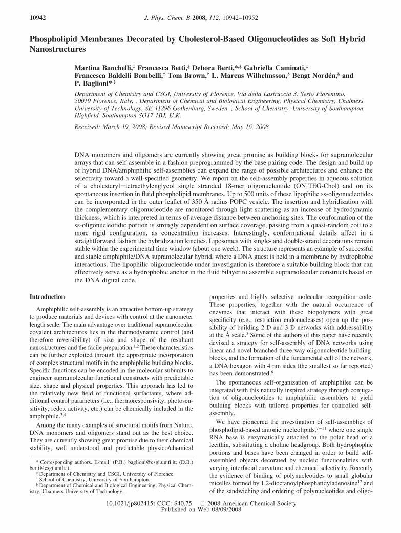
SCHEME 1: Chemical Structure and Simple Geometric Representation of ON1TEG-Chol Showing TEG-Chol Attachedto the 5′-Thymidine Nucleotidea
a The molecular size was estimated assuming a compact shape and using the contour lengths for the TEG23 and oligonucleotidic moieties.24-26
The order of magnitude of the oligonucleotide cross-section is coarsely estimated as the diameter of a B-DNA double helix.
Addressable Soft Nanostructures J. Phys. Chem. B, Vol. 112, No. 35, 2008 10943
detritylation, coupling, capping and iodine oxidation wereemployed to produce the required oligonucleotides on largescale.27 5′-Cholesterol-TEG-modified oligonucleotides and 5′-FAM-modified oligonucleotides were synnthesized “trityl-on”and unmodified oligonucleotides were synthesized “trityl-off”.Normal monomers (A, G, C, and T) were allowed to couplefor 25 s and the cholesterol-TEG and FAM monomers for anadditional 300 s. Stepwise coupling efficiencies and overallyields of monomers were determined by measuring trityl cationconductivity and in all cases these were >98.0%. Cleavage ofthe oligonucleotides from the solid support was carried out inconcentrated aqueous ammonia at 55 °C for 6 h. After ammoniadeprotection the cholesterol-TEG oligonucleotides were evapo-rated to dryness then treated with acetic acid/water (80/20) for30 min at room temperature to remove the DMT-group fromthe cholesteryl moiety. After evaporation of the solvent theoligonucleotides were dissolved in water (2.5 mL), extractedwith diethyl ether (3 × 5 mL) then purified by reversed-phaseHPLC. All other oligonucleotides were purified by reversed-phase HPLC directly after ammonia deprotection and evapora-tion of aqueous ammonia (HPLC results are available asSupporting Information).
Purification of Oligonucleotides. Purification of oligonucle-otides was carried out by reversed phase HPLC on a Gilsonsystem using a Brownlee Aquapore column (C8), 8 mm × 250mm, pore size 300 Å. The following protocol was used: runtime 30 min, flow rate 3 mL per minute, binary system gradient(time in minutes (% buffer B);0 (0); 3(0); 5(20); 21 (100);25(100); 27 (0); 30(0)). Elution buffer A: 100 mM ammoniumacetate, pH 7.0, buffer B: 100 mM ammonium acetate with 70%acetonitrile pH 7.0 (cholesterol-TEG oligomers), buffer B: 100mM ammonium acetate with 25% acetonitrile pH 7.0 (unmodi-fied and FAM oligomers). Elution was monitored by ultravioletabsorption at 295 nm. After HPLC purification, oligonucleotideswere desalted using disposable NAP 10 Sephadex columns(Pharmacia) using the manufacturer’s instructions, aliquoted intoEppendorf tubes and stored at -20 °C in distilled deionizedwater.
Phospholipid Vesicles. POPC (1-palmitoyl-2-oleoyl-sn-glyc-ero-3-phosphocholine) was purchased from Avanti Polar LipidsInc. (Alabama) and used as received. All other chemicals (TRISbase, NaCl) were purchased from Fluka, (Milan, Italy) and usedas received. Vesicles were prepared by evaporation of thesolvent from a CHCl3/MeOH solution of the lipid; the dry lipidicfilm was solvated by addition of a 50 mM TRIS, 100 mM NaCl(pH 7.5, TBS) solution. The suspension was vortexed,,frozen-thawed (liquid nitrogen-40 °C water bath) six timesand sized down by repeated extrusion (extruder by LipexBiomembranes Inc., Vancouver, Canada) through stacks ofNucleopore polycarbonate membranes (200, 100 and 50 nm poresize), yielding a narrow sized distribution of unilamellar vesicles.Different liposomal preparations resulted in slightly differentinitial sizes, hydrodynamic radii ranging from 330 to 350 Å.The total membrane area and the number of lipid molecule inthe outer leaflet were determined from the hydrodynamic radiusconsidering an area per POPC of 70 Å2,28 and a membranethickness of 40 Å, for the bilayer in the liquid crystalline state,respectively.
Phospholipid Vesicles Decorated with Oligonucleotides.A stock vesicular dispersion was diluted to 1.3 mM withON1TEG-Chol/TRIS buffer varying ON1TEG-Chol concentra-tion in the diluting buffer. The samples considered, with differentlipid/ON1TEG-Chol ratios, are summarized in Table 1. To testhybridization, the complementary sequence, ON2-FAM was also
addedina1:1ratiowithrespect to thecholesteryl-oligonucleotide.ON1TEG-Chol translocation across the bilayer during theexperimental time window (5 days) is negligible.29
The distribution of guest molecules among colloidal hostsfollows a Poisson distribution.30 Since ⟨N⟩ is quite high, theprobability of oligonucleotide occupancy P(⟨N⟩) can be wellapproximated by a Gaussian distribution function centered at⟨N⟩. If we consider that the oligonucleotide derivative is entirelydistributed in the outer vesicular leaflet, and therefore considerthe stoichiometry with respect to POPC in the outer leaflet, theknowledge of liposomal size and the narrow size distributionallow a fairly accurate estimate of this number. These ⟨N⟩ values,reported in the second column of Table 1, can be convertedinto an “average distance” (Γ-1/2) between anchoring sites ontovesicular surface. It should be stressed here that anchoring isdue to intermolecular interactions of hydrophobic nature betweenthe cholesterol of the ON1TEG-Chol and the phospholipidbilayer.
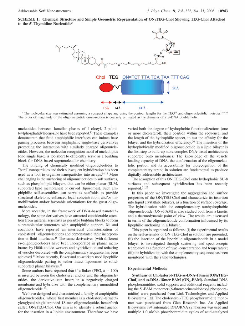
Membrane Incorporation. A quantitative determination ofthe ON1TEG-Chol incorporation in lipid bilayer was assessedby gel-filtration on Sephadex G-50 (Pharmacia Fine Chemicals,Uppsala, Sweden). The liposomes were separated from unin-corporated ON1TEG-Chol by a mini-column centrifugationmethod,31 which has the advantage that the liposomes can berecovered without dilution. This method is appropriate forseparating ON1TEG-Chol (MW ) 6193) from larger POPCliposomes (∼30000 KDa), but it should be mentioned thatpossible ON1TEG-Chol oligomers are also excluded.
The Sephadex powder (10 g) was hydrated with 120 mL ofTBS and stored for 24 h at 4 °C before use. The mini-columngel filtration was performed under the same conditions on threedifferent samples simultaneously: POPC liposomes and ON1TEG-Chol (4.1 µM, ⟨N⟩) 125), POPC liposomes as a blank, freeON1TEG-Chol as a control. Fractions of 0.2 mL were collectedand subjected to light scattering and UV analysis in order toquantify ON1TEG-Chol concentration relatively to liposomes.QELS measurements confirm that liposomes are recovered inthe first fraction eluted with virtually no dilution (same valueof the intensity of scattered light). The light scattering contribu-tion to the UV absorption spectra was evaluated by polynomialfitting of the absorption curve (ln(I/Io) ) Cλ-g) in the spectralregion between 600 and 350 nm, where no absorption from thenucleobases is expected.32
Figure 1 shows the results for ON1TEG-Chol 4.1 µM. Thefirst fraction eluted from the mixed liposome-ON1TEG-Cholsystem contains the major amount of ON1TEG-Chol, (95.8%),almost absent in the subsequent fractions. The 4.2% loss canbe due to some free monomers entrapped inside the gel poresthat are partially recovered by repeating the elution. The control
TABLE 1: Composition of the Lipid/ON1TEG-CholSamplesa
ON1TEG-Chol [µM] ⟨N⟩ Γ-1/2 [Å]
0.3 9 4102.0 60 1584.1 125 1108.3 250 7816.6 500 5517.5 525 5357.0 1700 30
a POPC has a fixed concentration of 1.3 mM. The meanoccupancy number ⟨N⟩ is defined as the average number ofON1TEG-Chol per liposome. To evaluate Γ-1/2, the average distancebetween anchoring sites, the cholesteryl-oligonucleotides have beenconsidered only in the outer vesicular leaflet.
10944 J. Phys. Chem. B, Vol. 112, No. 35, 2008 Banchelli et al.
experiment performed with ON1TEG-Chol/TBS shows that26.5% is inside the first eluted fraction, consistent with theoccurrence of some aggregates which do not obviously form inthe presence of liposomes.
The presence of liposome therefore significantly alters therelative abundance of ON1TEG-Chol monomers/oligomers.However, as already highlighted the relatively low cutoff ofthe pore size is sufficient to exclude dimers, we should mentionthat the exact nature of such aggregates cannot be determinedby this experiment.
Quasi Elastic Light Scattering. QELS experiments werecarried out with a Brookhaven Instrument apparatus, New York,USA (BI 9000AT correlator card and BI 200 SM goniometer).The signal was detected by an EMI 9863B/350 photomultiplier.The light source was the doubled frequency of a CoherentInnova diode pumped Nd:YAG laser, (λ ) 532 nm, 20 mW),or alternatively a JDS Uniphase He-Ne (λ ) 633 nm, 5 mW).The laser long-term power stability was (0.5%. Self-beatingdetection was recorded using decahydronaphthalene (thermo-statted by a water circulating system) as index matching liquid.A temperature probe was inserted in the sample to monitor Twhile simultaneously recording autocorrelation functions. Acontrol experiment in the same temperature range explored formelting curves has been performed with an aqueous latexdispersion normally used as a standard for QELS calibration.Measurements have been performed in the range 15-55 °C on0.5 mL samples previously transferred into cylindrical Hellmascattering cells and flushed with N2 to avoid bubble formation.For each sample at least four separate measurements wereperformed at three different angles (i.e., 70, 90 and 120°respectively) corresponding to three different scattering vectorsq ) 4πn/[λ sin(θ/2); n is the refractive index of the mediumequal to 1.33. The data analysis was performed as describedelsewhere.11
Stopped Flow Experiments. The time evolution of the 260nm absorbance after mixing the oligo-loaded vesicles with thecomplementary strand solution, was followed with an AppliedPhotophysics stopped-flow instrument. At least three traces wererecorded for each concentration. In order not to saturate thesignal and to explore a meaningful range of liposomal coverage,the oligonucleotide concentration has been kept constant at 2µM, while the lipid content has been varied between 0.125 and4 mg/mL to scan the interval between 15 and 440 oligos in theouter leaflet respectively. The hybridization rate of a nonlipo-somal sample for the same oligonucleotide concentration hasbeen also measured as a reference. These solutions were mixed(within approximately 10-3 s) via syringe injection into a 1 cmpath-length cuvette in a stopped-flow apparatus and hybridiza-tion was monitored at fixed temperature, i.e. 25 °C.The timecourse of the absorbance was recorded between 0.5 to 20 s fromtime 0, i.e. when the injected solution volume reaches theinstrumental beam height in the cuvette.
� Potential. The electrophoretic mobility of the vesicles (plainand decorated with ss and ds oligonucleotides) in TBS wasdetermined with a Zetasizer Nano ZS (Malvern Instruments).
Fluorescence. Steady-state fluorescence was measured witha LS50B spectrofluorimeter (Perkin-Elmer, Italy). The fluores-cence spectra of pyrene were recorded in the corrected spectrummode with excitation wavelength set at 335 and 2.5 nm slit, atleast 10 scans were averaged for each spectrum. The ratio ofthe intensities I1/I3, which reflects the local polarity sensed bythe fluorophore in the organized system,33 was determined fromthe first (372 nm) and the third (382 nm) vibronic peaks ofpyrene emission spectrum. Pyrene concentration is 1 µM in allsamples.
UV-Vis Absorption. UV-vis spectra were recorded witha Lambda 900 spectrophotomer (Perkin-Elmer, Italy) and a Cary100 spectrometer (Varian, Italy). Thermal cycles from 10 to 70°C were performed at 0.5 or 0.1 °C/min.
Linear Dichroism. UV-vis linear dichroism (LD) measure-ments were recorded on a JASCO J-720 CD spectropolarimeterequipped with an Oxley prism to obtain linearly polarized light.34
Spectra were recorded from 200 to 450 nm in a Couette cell(path length 1 mm) with a shear gradient of 3100 s-1 andwithout applied shear (no orientation of the sample) subtractedas baseline. Isotropic absorption of the same samples wasmeasured on a Cary 4000 (Varian, Italy) spectrophotometer.
LD is defined as the difference in absorption of linearlypolarized light and perpendicular polarized light relative to amacroscopic orientation axis (flow direction).
LD)A|-A⊥ (1)
To observe linear dichroism, a sample oriented with respectto the incident light is required. Liposomes in a Couette cell,subjected to a nearly constant flow gradient, are slightlydeformed into elongate ellipsoid-shaped particles that willposition their axes preferentially along the flow direction,thereby orienting bound molecules.35
Flow LD experiments were measured in 50% w/w bufferedsucrose solution at [POPC] ) 0.17 mM at different lipid/ON1TEG-Chol ratios (⟨N⟩ ) 125, 250, 500), ON2-FAM wasalsoaddedina1:1ratiowithrespecttothecholesterol-oligonucleotide.The sucrose addition matches the refractive index of theliposome solution reducing the unwanted scattering effects andalso provides a better alignment of the particles increasing theviscosity of the solution.34 Retinoic acid was added as an internalreference ([POPC]/[ret.ac.] ) 100:1) to establish the degree oforientation of the liposomes and hence their orientationparameter.36,37 Provided that the orientation parameter of thesystem is known, from the reduced linear dichroism, LDr,defined as the ratio between LD and isotropic absorption, it ispossible to evaluate the tilt angle of the molecule with respectto the membrane surface.38
Results and Discussion
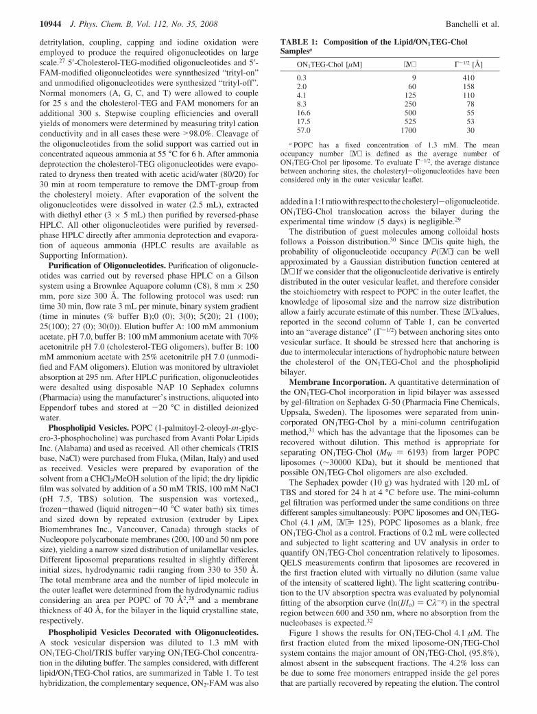
ON1TEG-Chol: Aggregation in Aqueous Medium. Figure2 shows the intensity of scattered light at 90° with respect tothe incoming beam, normalized by toluene scattering in the sameconditions, as a function of the oligonucleotide concentration.In the same plot, the results obtained using pyrene as afluorescent probe are also shown.
The scattering intensitiy curve shows a clear slope changeas the concentration is raised. For amphiphilic solutions thisbehavior marks the onset of aggregation. This trend is consistentwith the presence of scattering particles in the concentrationrange 15 µM/800 µM, but we cannot rule out the occurrence
Figure 1. Relative values of UV Absorbance % at 263 nm of thethree filtered fractions (Fr 1, Fr 2, Fr 3) with respect to the absorptionvalues before filtration. Left column: liposome-ON1TEG-Chol. Rightcolumn: ON1TEG-Chol.
Addressable Soft Nanostructures J. Phys. Chem. B, Vol. 112, No. 35, 2008 10945
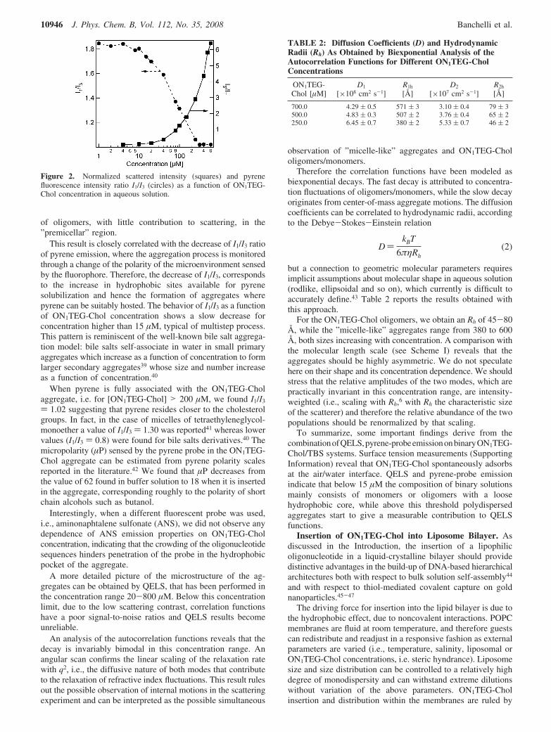
of oligomers, with little contribution to scattering, in the”premicellar” region.
This result is closely correlated with the decrease of I1/I3 ratioof pyrene emission, where the aggregation process is monitoredthrough a change of the polarity of the microenvironment sensedby the fluorophore. Therefore, the decrease of I1/I3, correspondsto the increase in hydrophobic sites available for pyrenesolubilization and hence the formation of aggregates wherepyrene can be suitably hosted. The behavior of I1/I3 as a functionof ON1TEG-Chol concentration shows a slow decrease forconcentration higher than 15 µM, typical of multistep process.This pattern is reminiscent of the well-known bile salt aggrega-tion model: bile salts self-associate in water in small primaryaggregates which increase as a function of concentration to formlarger secondary aggregates39 whose size and number increaseas a function of concentration.40
When pyrene is fully associated with the ON1TEG-Cholaggregate, i.e. for [ON1TEG-Chol] > 200 µM, we found I1/I3
) 1.02 suggesting that pyrene resides closer to the cholesterolgroups. In fact, in the case of micelles of tetraethyleneglycol-monoether a value of I1/I3 ) 1.30 was reported41 whereas lowervalues (I1/I3 ) 0.8) were found for bile salts derivatives.40 Themicropolarity (µP) sensed by the pyrene probe in the ON1TEG-Chol aggregate can be estimated from pyrene polarity scalesreported in the literature.42 We found that µP decreases fromthe value of 62 found in buffer solution to 18 when it is insertedin the aggregate, corresponding roughly to the polarity of shortchain alcohols such as butanol.
Interestingly, when a different fluorescent probe was used,i.e., aminonaphtalene sulfonate (ANS), we did not observe anydependence of ANS emission properties on ON1TEG-Cholconcentration, indicating that the crowding of the oligonucleotidesequences hinders penetration of the probe in the hydrophobicpocket of the aggregate.
A more detailed picture of the microstructure of the ag-gregates can be obtained by QELS, that has been performed inthe concentration range 20-800 µM. Below this concentrationlimit, due to the low scattering contrast, correlation functionshave a poor signal-to-noise ratios and QELS results becomeunreliable.
An analysis of the autocorrelation functions reveals that thedecay is invariably bimodal in this concentration range. Anangular scan confirms the linear scaling of the relaxation ratewith q2, i.e., the diffusive nature of both modes that contributeto the relaxation of refractive index fluctuations. This result rulesout the possible observation of internal motions in the scatteringexperiment and can be interpreted as the possible simultaneous
observation of ”micelle-like” aggregates and ON1TEG-Chololigomers/monomers.
Therefore the correlation functions have been modeled asbiexponential decays. The fast decay is attributed to concentra-tion fluctuations of oligomers/monomers, while the slow decayoriginates from center-of-mass aggregate motions. The diffusioncoefficients can be correlated to hydrodynamic radii, accordingto the Debye-Stokes-Einstein relation
D)kBT
6πηRh(2)
but a connection to geometric molecular parameters requiresimplicit assumptions about molecular shape in aqueous solution(rodlike, ellipsoidal and so on), which currently is difficult toaccurately define.43 Table 2 reports the results obtained withthis approach.
For the ON1TEG-Chol oligomers, we obtain an Rh of 45-80Å, while the ”micelle-like” aggregates range from 380 to 600Å, both sizes increasing with concentration. A comparison withthe molecular length scale (see Scheme I) reveals that theaggregates should be highly asymmetric. We do not speculatehere on their shape and its concentration dependence. We shouldstress that the relative amplitudes of the two modes, which arepractically invariant in this concentration range, are intensity-weighted (i.e., scaling with Rh,6 with Rh the characteristic sizeof the scatterer) and therefore the relative abundance of the twopopulations should be renormalized by that scaling.
To summarize, some important findings derive from thecombinationofQELS,pyrene-probeemissiononbinaryON1TEG-Chol/TBS systems. Surface tension measurements (SupportingInformation) reveal that ON1TEG-Chol spontaneously adsorbsat the air/water interface. QELS and pyrene-probe emissionindicate that below 15 µM the composition of binary solutionsmainly consists of monomers or oligomers with a loosehydrophobic core, while above this threshold polydispersedaggregates start to give a measurable contribution to QELSfunctions.
Insertion of ON1TEG-Chol into Liposome Bilayer. Asdiscussed in the Introduction, the insertion of a lipophilicoligonucleotide in a liquid-crystalline bilayer should providedistinctive advantages in the build-up of DNA-based hierarchicalarchitectures both with respect to bulk solution self-assembly44
and with respect to thiol-mediated covalent capture on goldnanoparticles.45-47
The driving force for insertion into the lipid bilayer is due tothe hydrophobic effect, due to noncovalent interactions. POPCmembranes are fluid at room temperature, and therefore guestscan redistribute and readjust in a responsive fashion as externalparameters are varied (i.e., temperature, salinity, liposomal orON1TEG-Chol concentrations, i.e. steric hyndrance). Liposomesize and size distribution can be controlled to a relatively highdegree of monodispersity and can withstand extreme dilutionswithout variation of the above parameters. ON1TEG-Cholinsertion and distribution within the membranes are ruled by
Figure 2. Normalized scattered intensity (squares) and pyrenefluorescence intensity ratio I1/I3 (circles) as a function of ON1TEG-Chol concentration in aqueous solution.
TABLE 2: Diffusion Coefficients (D) and HydrodynamicRadii (Rh) As Obtained by Biexponential Analysis of theAutocorrelation Functions for Different ON1TEG-CholConcentrations
ON1TEG-Chol [µM]
D1
[×108 cm2 s-1]R1h
[Å]D2
[×107 cm2 s-1]R2h
[Å]
700.0 4.29 ( 0.5 571 ( 3 3.10 ( 0.4 79 ( 3500.0 4.83 ( 0.3 507 ( 2 3.76 ( 0.4 65 ( 2250.0 6.45 ( 0.7 380 ( 2 5.33 ( 0.7 46 ( 2
10946 J. Phys. Chem. B, Vol. 112, No. 35, 2008 Banchelli et al.
thermal equilibrium, which guarantees self-repair from defectsthat can be generated under kinetic control.
ON1TEG-Chol has been added to a preformed liposomalsuspension (final [POPC]) 1.3 mM) in the concentration range0.3-52 µM. For the interval 4.1-16 µM hybridization withthe complementary strand has also been investigated. Asdiscussed in the previous section, below 15 µM ON1TEG-Cholis not aggregated.
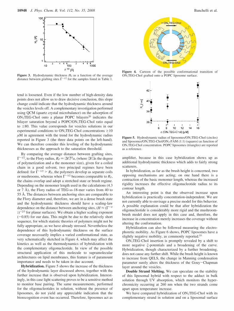
If the ON1TEG portion of the molecules protrudes outwardfrom the membrane, the insertion should cause an increase ofhydrodynamic radius of the liposomes, due to the addedhydrodynamic thickness. This can be monitored using QELSto follow the time evolution of intensity autocorrelation func-tions. Therefore intensity autocorrelation functions were re-corded at different equilibration times after dilution of thevesicular suspension with TBS containing the oligonucleotides,showing, on a log-log scale a slope variation that can be directlycorrelated with a size variation.
Table 3 reports the hydrodynamic radii and the polydispersityas determined through a second order cumulant fitting of theautocorrelation functions.
The decrease of the decay rate, observed in the autocorrelationfunctions, corresponds to an increase in hydrodynamic radiuswith time (column 4, Table 3), until equilibrium is reachedwithin 6 h. The size invariance observed in the reference sample,POPC at the same final concentration and temperature, providesclear-cut evidence that the increase in hydrodynamic radius ofthe scattering objects is a direct consequence of ON1TEG-Cholincorporation into liposomes. The observed radius increasecorresponds to an increase of the hydrodynamic thickness aroundthe vesicle, due to the hydrophilic portion of the guest molecules.This behavior has been previously observed with DNA-coatedpolystyrene particles and has been taken as the indication ofcoupling between the nanoparticle and the single strand oligo-nucleotide.48 An interesting observation concerns the small, butreproducible, decrease in polydispersity that follows ON1TEG-Chol insertion. This rules out that the observed increase in sizeis due to partial vesicle fusion, which would enlarge sizedistribution and skew the average size toward higher values.We believe that the origin of this effect lies in the membranestiffening and in the consequent decrease of thermally activatedshape deformation of vesicles.49
The gradual achievement of the final hydrodynamic thickness(six hours in this case) can be correlated with the insertionkinetics of ON1TEG-Chol in the membrane; this means thatthe final radius of the vesicles decorated by the oligonucleotideis dependent upon the oligonucleotide/lipid ratio. Therefore, wehave investigated the equilibrium thickness increase as a functionof the added ON1TEG-Chol. The contribution of the oligo-nucleotide does not depend on the initial size of the vesicles,whose radius of curvature (330-350 Å) is higher than the fullyextended length of the ON1TEG (≈90 Å).
The concentration of lipophilic oligonucleotides has anunambiguous effect on the equilibrium hydrodynamic size. Theincrease of hydrodynamic thickness, termed H0, is reported inFigure 3 as a function of the average distance between graftingsites, calculated as specified in the experimental part. Thedependence of the thickness of the hydrodynamic layer onsurface coverage indicates a conformational transition of theoligonucleotidic chain as the surface coverage is increased andthe average distance between anchoring sites on the vesicularsurface is decreased.
The physical properties of polymers and polyelectrolytesgrafted to planar and globular surfaces have been the subjectof considerable theoretical and experimental efforts. Most ofthe advances in the field are due to Alexander and De Gennesfor neutral polymers50,51 and to Pincus, Zhulina, and Birshtein,52,53
among others, for polyelectrolytes, whose high charge densityconfines counterions in the brush and determines conformationalproperties totally different from the neutral counterparts. Theosmotic pressure due to confined counterions stretches the chainsagainst entropic elasticity. The conformation of the polyelec-trolytes results from this balance. The natural gauge to quantifythe charge density for a polyelectrolyte is the Manningcondensation parameter, (q0 ) lB/l) which compares the chargespacing along the backbone, l, with the solvent Bjerrum lengthlB (7.14 Å for water). If this ratio (4.2 for dsDNA, 1-1.7 forssDNA) is higher than unity, then counterion condensationoccurs and renormalizes charge spacing to this distance.
In our experimental conditions the parameters for ssDNA arenot unanimously defined in the literature. Several lengths forthe monomer size have been reported, varying from 4.3 to 7 Å;moreover the persistence length, between 7.5 Å and 35 Å,24 iscomparable to the monomer size. These data suggest a ratherflexible conformation if compared to the dsDNA, which is avery stiff and highly charged polyelectrolyte, with a persistencelength of ∼150 base pairs (lp ) 500 Å) in physiological buffer.The significant difference in flexibility, which can be modulatedsimply by addition of the complementary strand, is in fact oneof the most attractive properties of this molecular machineryfor supramolecular chemistry.
Even if we are aware that the polyelectrolyte theory is notstrictly appropriate for such short oligonucleotides, it is worthto tentatively apply it to our case to infer a characteristic sizefor the chain protruding from the vesicles.
In view of the previous considerations and of the fact thatthe relatively high ionic strength should render the brush quasi-neutral, we will consider the segment TEG-ssDNA as a swollencoil characterized by its Flory radius, while we will considerdsDNA as a rigid rod.54 Therefore the oligonucleotides (eitheranchored to a bilayer or organized at the air/water interface)will occupy a hemisphere with radius corresponding to theswollen coil Flory radius for the ssDNA or to the contour lengthfor dsDNA. Within these assumptions one can therefore applythe scaling theory51 describing the conformation of flexiblepolymer chains grafted to a planar surface and immersed in agood solvent, which predicts the structural state of the polymericchains. The TEG-18-mer oligonucleotide portion is highlysoluble in the aqueous buffer used in this study and the vesicleshell can be reasonably approximated as a planar surface,considering that the particle diameters are very large comparedto the maximum chain extension.
Below 15 µM there is a clear dependence of hydrodynamicthickness on surface coverage and the observed trend follows apower law, represented by a linear trend in the doublelogarithmic plot. For high surface coverage (g15 µM) this linear
TABLE 3: Hydrodynamic Radii and Polydispersity Valuesfor POPC Liposome Solution upon Addition ofON1TEG-Chol 4.1 µM at Different Times
POPC Liposomes
reference sample with ON1TEG-Chol 4.1 µM
time [h] Rh [Å] polydispersity Rh [Å] polydispersity
1 328 ( 5 0.066 347 ( 5 0.0662 327 ( 6 0.067 349 ( 5 0.0666 330 ( 5 0.072 351 ( 6 0.06824 331 ( 4 0.073 352 ( 5 0.060
Addressable Soft Nanostructures J. Phys. Chem. B, Vol. 112, No. 35, 2008 10947
tend is loosened. Even if the low number of high-density datapoints does not allow us to draw decisive conclusion, this slopechange could indicate that the hydrodynamic thickness aroundthe vesicles levels off. A complementary investigation performedusing QCM (quartz crystal microbalance) on the adsorption ofON1TEG-Chol onto a planar POPC bilayers20 indicates thebilayer saturation beyond a POPC/ON1TEG-Chol ratio equalto e80. This value corresponds for vesicles solutions in ouresperimental conditions to ON1TEG-Chol concentrations g10µM in agreement with the trend for the hydrodynamic radiusreported in Figure 3 (the three data points on the left-hand).We can therefore consider this leveling of the hydrodynamicthicknesses as the approach to the saturation threshold.
By comparing the average distance between grafting sites,Γ-1/2, to the Flory radius, RF ) ⟨N⟩3/5a, (where ⟨N⟩ is the degreeof polymerization and a the monomer size), given for a coiledchain in a good solvent, two principal regimes have beendefined: for Γ-1/2 > RF, the polymers develop as separate coilsor mushrooms, whereas when Γ-1/2 becomes comparable to RF,the chains overlap and adopt a stretched state or brush regime.Depending on the monomer length used in the calculations (4.3or 7 Å), the Flory radius of TEG-ss-18-mer varies from 40 to55 Å. The distances between anchoring sites are comparable tothe Flory diameter and, therefore, we are in a dense brush stateand the hydrodynamic thickness should have a scaling-lawdependence on the distance between anchoring sites, (H ≈ (Γ-1/
2)-2/3 for planar surfaces). We obtain a higher scaling exponent(-0.85) for our data. This might be due to the relatively shortsequence, for which statistic theories of polymers might not befully appropriate, as we have already stressed. Nevertheless thedependence of this hydrodynamic thickness on the surfacecoverage necessarily implies a varied conformational state, asvery schematically sketched in Figure 4, which may affect thekinetics as well as the thermodynamics of hybridization withthe complementary oligonucleotide. In view of the possiblestructural application of this molecule to supramoleculararchitectures on lipid membranes, this feature is of paramountimportance and needs to be taken in due account.
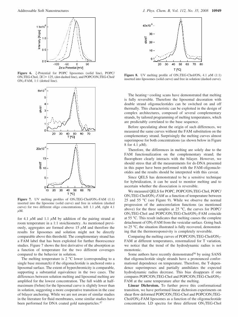
Hybridization. Figure 5 shows the increase of the thicknessof the hydrodynamic layer discussed above, together with thefurther increase that is observed upon hybridization. Interest-ingly, in this case light scattering proves to be a sensitive methodto monitor base pairing. The same measurements, performedfor the oligonucleotides in solution, without the presence ofliposomes, do not yield any appreciable indication that thebiorecognition event has occurred. Therefore, liposomes act as
amplifier, because in this case hybridization shows up asadditional hydrodynamic thickness which adds to fairly strongscatterers.
In hybridization, as far as the brush height is concerned, twoopposing mechanisms are acting; on one hand there is acontraction of the basic monomer length, whereas the increasedrigidity increases the effective oligonucleotide radius to itscontour length.
An interesting point is that the observed increase uponhybridization is practically concentration-independent. We arenot currently able to envisage a precise model for this behavior.A possible explanation could be that after hybridization theoligonucleotide is considerably more rigid and the mushroom-brush model does not apply in this case and, therefore, theincrease in concentration merely increases the coverage withoutaltering the conformation.
Hybridization can also be followed measuring the electro-phoretic mobility. As Figure 6 shows, POPC liposomes have aslightly negative mobility, as commonly reported.55
ON1TEG-Chol insertion is promptly revealed by a shift tomore negative �-potentials and a broadening of the curve.Hybridization, though characterized by a further broadening,does not cause any further shift. While the brush height is knownto increase from QELS, the change in Manning condensationparameter surely alters the thickness of the Gouy-Chapmanlayer around the vesicles.
Double Strand Melting. We can speculate on the stabilityof this liposomal hybrid with respect to the adduct in bulksolution through UV absorption, which monitors the hyper-chromicity occurring at 260 nm when the two strands comeapart upon temperature increase.
We have compared hybridization of ON1TEG-Chol with itscomplementary strand in solution and on a liposomal surface
Figure 3. Hydrodynamic thickness H0 as a function of the averagedistance between grafting sites Γ-1/2 for the samples listed in Table 1.
Figure 4. Cartoon of the possible conformational transition ofON1TEG-Chol grafted onto a POPC liposome surface.
Figure 5. Hydrodynamic radius of liposomes/ON1TEG-Chol (circles)and liposomes/ON1TEG-Chol/ON2-FAM (1:1) (squares) as function ofON1TEG-Chol concentration. POPC liposomes (triangles) are reportedas a reference.
10948 J. Phys. Chem. B, Vol. 112, No. 35, 2008 Banchelli et al.
for 4.1 µM and 1.1 µΜ by addition of the pairing strand atroom temperature in a 1:1 stoichiometry. As mentioned previ-ously, aggregates are formed above 15 µM and therefore theresults for liposomes and solution might not be directlycomparable above this threshold. The complementary strand hasa FAM label that has been exploited for further fluorescencestudies. Figure 7 shows the first derivative of the absorption asa function of temperature for the two different coveragescompared to the behavior in solution.
The melting temperature is 2 °C lower (corresponding to asingle base mismatch) if the oligonucleotide is anchored onto aliposomal surface. The extent of hyperchromicity is comparable,supporting a substantial equivalence in the two cases. Thedifferences between solution melting and liposomal melting areamplified for the lowest concentration. The full width at half-maximum (fwhm) for the liposomal curve is slightly lower thanin solution, suggesting a more cooperative transition in the caseof bilayer anchoring. While we are not aware of similar studiesin the literature for fluid membranes, some similar studies havebeen performed for DNA coated gold nanoparticles.47
The heating-cooling scans have demonstrated that meltingis fully reversible. Therefore the liposomal decoration withdouble strand oligonucleotides can be switched on and offthermally. This characteristic can be exploited in the design ofcomplex architectures, composed of several complementarystrands, by tailored programming of melting temperatures, whichare predictably correlated to the base sequence.
Before speculating about the origin of such differences, wemeasured the same curves without the FAM substitution on thecomplementary strand. Surprisingly the melting curves almostsuperimpose for both concentrations (as shown below in Figure8 for 4.1 µM).
Therefore, the differences in melting are solely due to theFAM functionalization on the complementary strand; thefluorophore clearly interacts with the bilayer. However, weshould stress that all the measurements for ds-DNA presentedin this paper have been performed with the FAM-oligonucle-otides and the results should be interpreted with this caveat.
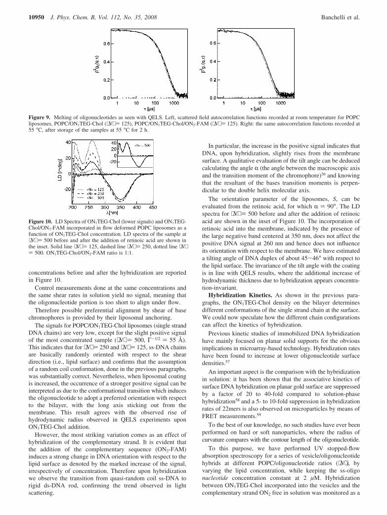
Since QELS has demonstrated to be a sensitive techniquefor hybridization, it can be used to monitor melting and toascertain whether the dissociation is reversible.
We measured QELS for POPC, POPC/ON1TEG-Chol, POPC/ON1TEG-Chol/ON2-FAM as a function of temperature between25 and 55 °C (see Figure 9). While we observe the normalprogression of the autocorrelation functions (as mentionedabove) for the three samples at 25 °C, the curves for POPC/ON1TEG-Chol and POPC/ON1TEG-Chol/ON2-FAM coincideat 55 °C. This result indicates that melting causes the completedetachment of ON2-FAM from the vesicular surface. Going backto 25 °C, the situation illustrated is fully recovered, demonstrat-ing that the thermoresponsivity is completely reversible.
Comparing the melting curves of POPC/ON1TEG-Chol/ON2-FAM at different temperatures, renormalized for T variation,we notice that the trend of the hydrodynamic radius is notmonotonic.
Some authors have recently demonstrated56 by using SANSthat oligonucleotide single strands have a pronounced confor-mational dependence on temperature. Therefore, the T-depen-dence superimposes and partially annihilates the expectedhydrodynamic radius decrease. This bias disappears if onecompares POPC/ON1TEG-Chol and POPC/ON1TEG-Chol/ON2-FAM at the same temperature after the melting.
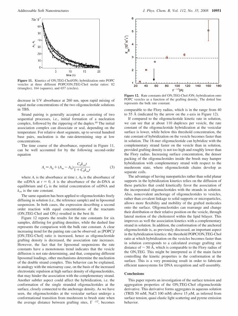
Linear Dichroism. To further prove this conformationaltransition, we have performed linear dichroism experiments onshear flow deformed POPC/ON1TEG-Chol and POPC/ON1TEG-Chol/ON2-FAM liposomes as a function of the oligonucleotideconcentration. LD spectra for three different ON1TEG-Chol
Figure 6. �-Potential for POPC liposomes (solid line), POPC/ON1TEG-Chol, ⟨N⟩ ) 125, (dot-dashed line), and POPC/ON1TEG-Chol/ON2-FAM, 1:1 (dotted line).
Figure 7. UV melting profiles of ON1TEG-Chol/ON2-FAM (1:1)inserted into the liposome (solid curve) and free in solution (dashedcurve) for two different oligo concentrations, left 1.1 µM, right 4.1µM.
Figure 8. UV melting profile of ON1TEG-Chol/ON2 4.1 µM (1:1)inserted into liposomes (solid curve) and free in solution (dashed curve).
Addressable Soft Nanostructures J. Phys. Chem. B, Vol. 112, No. 35, 2008 10949
concentrations before and after the hybridization are reportedin Figure 10.
Control measurements done at the same concentrations andthe same shear rates in solution yield no signal, meaning thatthe oligonucleotide portion is too short to align under flow.
Therefore possible preferential alignment by shear of basechromophores is provided by their liposomal anchoring.
The signals for POPC/ON1TEG-Chol liposomes (single strandDNA chains) are very low, except for the slight positive signalof the most concentrated sample (⟨N⟩ ) 500, Γ-1/2 ) 55 Å).This indicates that for ⟨N⟩ ) 250 and ⟨N⟩ ) 125, ss-DNA chainsare basically randomly oriented with respect to the sheardirection (i.e., lipid surface) and confirms that the assumptionof a random coil conformation, done in the previous paragraphs,was substantially correct. Nevertheless, when liposomal coatingis increased, the occurrence of a stronger positive signal can beinterpreted as due to the conformational transition which inducesthe oligonucleotide to adopt a preferred orientation with respectto the bilayer, with the long axis sticking out from themembrane. This result agrees with the observed rise ofhydrodynamic radius observed in QELS experiments uponON1TEG-Chol addition.
However, the most striking variation comes as an effect ofhybridization of the complementary strand. It is evident thatthe addition of the complementary sequence (ON2-FAM)induces a strong change in DNA orientation with respect to thelipid surface as denoted by the marked increase of the signal,irrespectively of concentration. Therefore upon hybridizationwe observe the transition from quasi-random coil ss-DNA torigid ds-DNA rod, confirming the trend observed in lightscattering.
In particular, the increase in the positive signal indicates thatDNA, upon hybridization, slightly rises from the membranesurface. A qualitative evaluation of the tilt angle can be deducedcalculating the angle R (the angle between the macroscopic axisand the transition moment of the chromophore)36 and knowingthat the resultant of the bases transition moments is perpen-dicular to the double helix molecular axis.
The orientation parameter of the liposomes, S, can beevaluated from the retinoic acid, for which R ) 90°. The LDspectra for ⟨N⟩ ) 500 before and after the addition of retinoicacid are shown in the inset of Figure 10. The incorporation ofretinoic acid into the membrane, indicated by the presence ofthe large negative band centered at 350 nm, does not affect thepositive DNA signal at 260 nm and hence does not influenceits orientation with respect to the membrane. We have estimateda tilting angle of DNA duplex of about 45-46° with respect tothe lipid surface. The invariance of the tilt angle with the coatingis in line with QELS results, where the additional increase ofhydrodynamic thickness due to hybridization appears concentra-tion-invariant.
Hybridization Kinetics. As shown in the previous para-graphs, the ON1TEG-Chol density on the bilayer determinesdifferent conformations of the single strand chain at the surface.We could now speculate how the different chain configurationscan affect the kinetics of hybridization.
Previous kinetic studies of immobilized DNA hybridizationhave mainly focused on planar solid supports for the obviousimplications in microarray-based technology. Hybridization rateshave been found to increase at lower oligonucleotide surfacedensities.57
An important aspect is the comparison with the hybridizationin solution: it has been shown that the associative kinetics ofsurface DNA hybridization on planar gold surface are suppressedby a factor of 20 to 40-fold compared to solution-phasehybridization58 and a 5- to 10-fold suppression in hybridizationrates of 22mers is also observed on microparticles by means ofFRET measurements.59
To the best of our knowledge, no such studies have ever beenperformed on hard or soft nanoparticles, where the radius ofcurvature compares with the contour length of the oligonucleotide.
To this purpose, we have performed UV stopped-flowabsorption spectroscopy for a series of vesicle/oligonucleotidehybrids at different POPC/oligonucleotide ratios (⟨N⟩), byvarying the lipid concentration, while keeping the ss-oligonucleotide concentration constant at 2 µM. Hybridizationbetween ON1TEG-Chol incorporated into the vesicles and thecomplementary strand ON2 free in solution was monitored as a
Figure 9. Melting of oligonucleotides as seen with QELS. Left, scattered field autocorrelation functions recorded at room temperature for POPCliposomes, POPC/ON1TEG-Chol (⟨N⟩ ) 125), POPC/ON1TEG-Chol/ON2-FAM (⟨N⟩ ) 125). Right: the same autocorrelation functions recorded at55 °C, after storage of the samples at 55 °C for 2 h.
Figure 10. LD Spectra of ON1TEG-Chol (lower signals) and ON1TEG-Chol/ON2-FAM incorporated in flow deformed POPC liposomes as afunction of ON1TEG-Chol concentration. LD spectra of the sample at⟨N⟩ ) 500 before and after the addition of retinoic acid are shown inthe inset. Solid line ⟨N⟩ ) 125, dashed line ⟨N⟩ ) 250, dotted line ⟨N⟩) 500. ON1TEG-Chol/ON2-FAM ratio is 1:1.
10950 J. Phys. Chem. B, Vol. 112, No. 35, 2008 Banchelli et al.
decrease in UV absorbance at 260 nm, upon rapid mixing ofequal molar concentrations of the two oligonucleotide solutionsin TBS.
Strand pairing is generally accepted as consisting of twosequential processes, i.e., initial formation of a nucleationcomplex, followed by the zippering of the duplex.60 The initialassociation complex can dissociate or seal, depending on thetemperature. For relative short segments, up to several hundredbase pairs, nucleation is the rate-determining step at lowconcentrations.
The time course of the absorbance, reported in Figure 11,can be well accounted for by the following second-orderequation
At )A0 + (A∞ -A0)C0kont
1+C0kont(3)
where At is the absorbance at time t, A0 is the absorbance ofthe ssDNA at t ) 0, A is the absorbance of the ds-DNA atequilibrium and C0 is the initial concentration of ssDNA andkon is the rate constant.
The same equation has been applied to oligonucleotides freelydiffusing in solution (i.e., the reference sample) and in liposomalsuspension. In both cases, the expression describing a secondorder reaction with equal concentrations of the reactants(ON1TEG-Chol and ON2) resulted in the best fit.
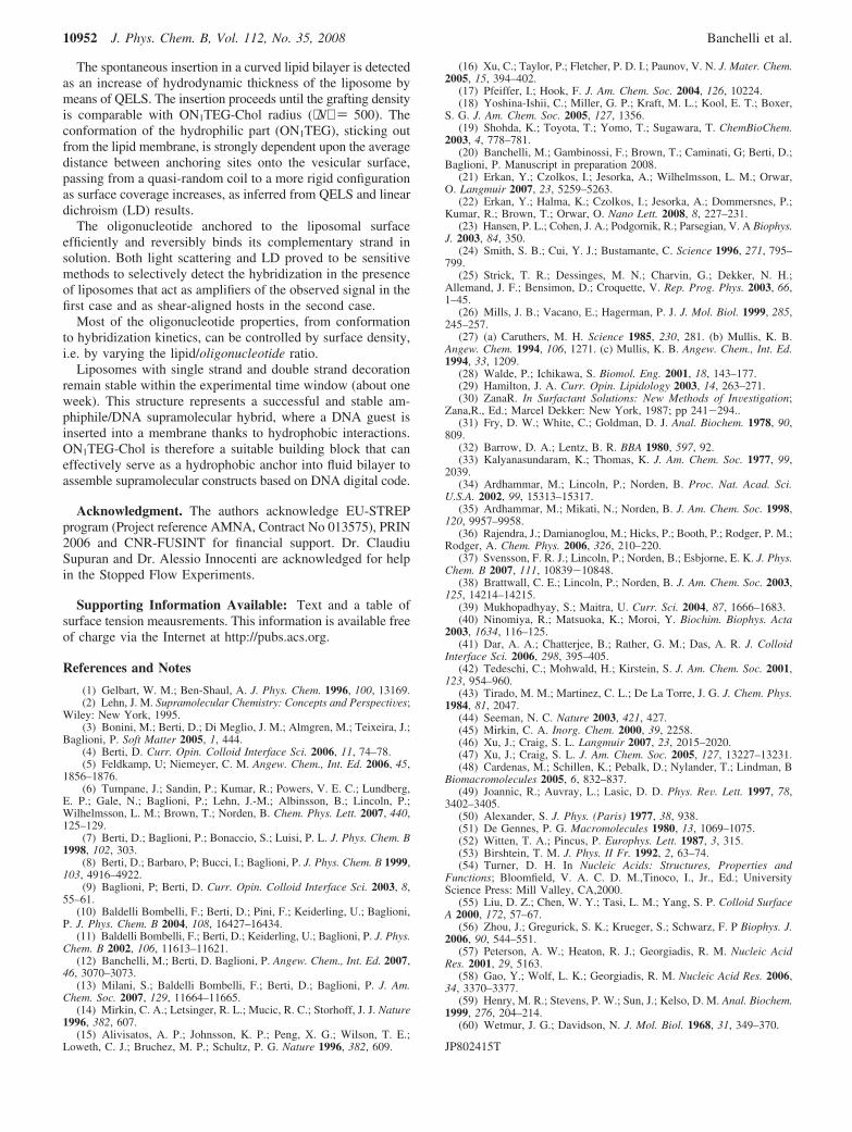
Figure 12 reports the results for the rate constants for sixsamples, differing for grafting densities, while the dashed linerepresents the comparison with the bulk rate constant. A clearincreasing trend for the pairing rate can be observed: as [POPC]/[ON1TEG-Chol] ratio is increased, hence as oligonucleotidegrafting density is decreased, the association rate increases.However, the fact that for liposomal suspensions the rateconstants have a monotonous trend indicates that the vesiclediffusion is not rate-determining, and that, comparing differentliposomal loadings, other mechanisms determine the nucleationof the double strand complex. This behavior can be explained,in analogy with the microarray case, on the basis of the increasedelectrostatic repulsion at high surface density of oligonucleotides,that may hinder the association with the complementary strand.Another subtler aspect could affect the hybridization, i.e. theconformation of the single stranded oligonucleotides at thesurface, closely connected to the anchorage density. As we haveseen, the oligonucleotides at the vesicular surface undergo aconformational transition from mushroom to brush state whenthe average distance between grafting sites, Γ -1/2, becomes
comparable to the Flory radius, which is in the range from 40to 55 Å (indicated by the arrow on the x-axis in Figure 12).
If compared to the oligonucleotide kinetic rate in solution,we can see that at about 110 duplexes per vesicle, the rateconstant of the oligonucleotide hybridization at the vesicularsurface is lower, while below this threshold concentration, therate constant of hybridization on the vesicle becomes faster thanin solution. The 18-mer oligonucleotide can hybridize with thecomplementary strand faster on the vesicle than in solution,provided grafting density is not too high and roughly lower thanthe Flory radius. Increasing surface concentration, the denserpacking of the oligonucleotides inside the brush may hamperhybridization with complementary strand with respect to themushroom state, where oligonucleotide chains develop asseparate coils.
The advantage of having nanoparticles rather than solid planarsupports in the hybridization kinetics relies on the diffusion ofthese particles that could kinetically favor the association ofthe incorporated oligonucleotides with the strands in solution.Also, noncovalent anchorage of oligonucleotides to vesicles,rather than covalent linkage to solid supports or microparticles,allows more flexibility and mobility of the grafted moleculesonto the surface. Oligonucleotides can rearrange and changetheir distribution or their relative position on the vesicle, throughlateral motion of the cholesterol within the lipid bilayer. Thisimproves as well the association kinetics with a complementarystrand in solution. In addition, the conformation of the anchoredoligonucleotide is, as previously discussed, an important aspectin the hybridization kinetics: the threshold POPC/ON1TEG-Cholratio at which hybridization on the vesicles becomes faster thanin solution corresponds to a calculated average grafting sitedistance of ∼ 50 Å, which is comparable to the Flory radius ofthe ON1TEG. This might be interpreted as if the main factorcontrolling the kinetic properties is the conformation at thesurface. This is a very promising result in order to fabricateefficient nanosystems for DNA recognition and self-assembly.
Conclusions
This paper reports an investigation of the surface tension andaggregation properties of the ON1TEG-Chol oligonucleotidederivative. This derivative forms aggregates in aqueous solution(TRIS 50 mM, NaCl 100 mM) above 15 µM, as inferred fromsurface tension, quasi elastic light scattering and pyrene emissionbehavior.
Figure 11. Kinetics of ON1TEG-Chol/ON2 hybridization onto POPCvesicles at three different POPC/ON1TEG-Chol molar ratios: 82(triangles), 164 (squares), and 657 (circles).
Figure 12. Rate constants dof ON1TEG-Chol /ON2 hybridization ontoPOPC vesicles as a function of the grafting density. The dotted linerepresents the bulk rate constant.
Addressable Soft Nanostructures J. Phys. Chem. B, Vol. 112, No. 35, 2008 10951
The spontaneous insertion in a curved lipid bilayer is detectedas an increase of hydrodynamic thickness of the liposome bymeans of QELS. The insertion proceeds until the grafting densityis comparable with ON1TEG-Chol radius (⟨N⟩ ) 500). Theconformation of the hydrophilic part (ON1TEG), sticking outfrom the lipid membrane, is strongly dependent upon the averagedistance between anchoring sites onto the vesicular surface,passing from a quasi-random coil to a more rigid configurationas surface coverage increases, as inferred from QELS and lineardichroism (LD) results.
The oligonucleotide anchored to the liposomal surfaceefficiently and reversibly binds its complementary strand insolution. Both light scattering and LD proved to be sensitivemethods to selectively detect the hybridization in the presenceof liposomes that act as amplifiers of the observed signal in thefirst case and as shear-aligned hosts in the second case.
Most of the oligonucleotide properties, from conformationto hybridization kinetics, can be controlled by surface density,i.e. by varying the lipid/oligonucleotide ratio.
Liposomes with single strand and double strand decorationremain stable within the experimental time window (about oneweek). This structure represents a successful and stable am-phiphile/DNA supramolecular hybrid, where a DNA guest isinserted into a membrane thanks to hydrophobic interactions.ON1TEG-Chol is therefore a suitable building block that caneffectively serve as a hydrophobic anchor into fluid bilayer toassemble supramolecular constructs based on DNA digital code.
Acknowledgment. The authors acknowledge EU-STREPprogram (Project reference AMNA, Contract No 013575), PRIN2006 and CNR-FUSINT for financial support. Dr. ClaudiuSupuran and Dr. Alessio Innocenti are acknowledged for helpin the Stopped Flow Experiments.
Supporting Information Available: Text and a table ofsurface tension meausrements. This information is available freeof charge via the Internet at http://pubs.acs.org.
References and Notes
(1) Gelbart, W. M.; Ben-Shaul, A. J. Phys. Chem. 1996, 100, 13169.(2) Lehn, J. M. Supramolecular Chemistry: Concepts and PerspectiVes;
Wiley: New York, 1995.(3) Bonini, M.; Berti, D.; Di Meglio, J. M.; Almgren, M.; Teixeira, J.;
Baglioni, P. Soft Matter 2005, 1, 444.(4) Berti, D. Curr. Opin. Colloid Interface Sci. 2006, 11, 74–78.(5) Feldkamp, U; Niemeyer, C. M. Angew. Chem., Int. Ed. 2006, 45,
1856–1876.(6) Tumpane, J.; Sandin, P.; Kumar, R.; Powers, V. E. C.; Lundberg,
E. P.; Gale, N.; Baglioni, P.; Lehn, J.-M.; Albinsson, B.; Lincoln, P.;Wilhelmsson, L. M.; Brown, T.; Norden, B. Chem. Phys. Lett. 2007, 440,125–129.
(7) Berti, D.; Baglioni, P.; Bonaccio, S.; Luisi, P. L. J. Phys. Chem. B1998, 102, 303.
(8) Berti, D.; Barbaro, P; Bucci, I.; Baglioni, P. J. Phys. Chem. B 1999,103, 4916–4922.
(9) Baglioni, P; Berti, D. Curr. Opin. Colloid Interface Sci. 2003, 8,55–61.
(10) Baldelli Bombelli, F.; Berti, D.; Pini, F.; Keiderling, U.; Baglioni,P. J. Phys. Chem. B 2004, 108, 16427–16434.
(11) Baldelli Bombelli, F.; Berti, D.; Keiderling, U.; Baglioni, P. J. Phys.Chem. B 2002, 106, 11613–11621.
(12) Banchelli, M.; Berti, D. Baglioni, P. Angew. Chem., Int. Ed. 2007,46, 3070–3073.
(13) Milani, S.; Baldelli Bombelli, F.; Berti, D.; Baglioni, P. J. Am.Chem. Soc. 2007, 129, 11664–11665.
(14) Mirkin, C. A.; Letsinger, R. L.; Mucic, R. C.; Storhoff, J. J. Nature1996, 382, 607.
(15) Alivisatos, A. P.; Johnsson, K. P.; Peng, X. G.; Wilson, T. E.;Loweth, C. J.; Bruchez, M. P.; Schultz, P. G. Nature 1996, 382, 609.
(16) Xu, C.; Taylor, P.; Fletcher, P. D. I.; Paunov, V. N. J. Mater. Chem.2005, 15, 394–402.
(17) Pfeiffer, I.; Hook, F. J. Am. Chem. Soc. 2004, 126, 10224.(18) Yoshina-Ishii, C.; Miller, G. P.; Kraft, M. L.; Kool, E. T.; Boxer,
S. G. J. Am. Chem. Soc. 2005, 127, 1356.(19) Shohda, K.; Toyota, T.; Yomo, T.; Sugawara, T. ChemBioChem.
2003, 4, 778–781.(20) Banchelli, M.; Gambinossi, F.; Brown, T.; Caminati, G; Berti, D.;
Baglioni, P. Manuscript in preparation 2008.(21) Erkan, Y.; Czolkos, I.; Jesorka, A.; Wilhelmsson, L. M.; Orwar,
O. Langmuir 2007, 23, 5259–5263.(22) Erkan, Y.; Halma, K.; Czolkos, I.; Jesorka, A.; Dommersnes, P.;
Kumar, R.; Brown, T.; Orwar, O. Nano Lett. 2008, 8, 227–231.(23) Hansen, P. L.; Cohen, J. A.; Podgornik, R.; Parsegian, V. A Biophys.
J. 2003, 84, 350.(24) Smith, S. B.; Cui, Y. J.; Bustamante, C. Science 1996, 271, 795–
799.(25) Strick, T. R.; Dessinges, M. N.; Charvin, G.; Dekker, N. H.;
Allemand, J. F.; Bensimon, D.; Croquette, V. Rep. Prog. Phys. 2003, 66,1–45.
(26) Mills, J. B.; Vacano, E.; Hagerman, P. J. J. Mol. Biol. 1999, 285,245–257.
(27) (a) Caruthers, M. H. Science 1985, 230, 281. (b) Mullis, K. B.Angew. Chem. 1994, 106, 1271. (c) Mullis, K. B. Angew. Chem., Int. Ed.1994, 33, 1209.
(28) Walde, P.; Ichikawa, S. Biomol. Eng. 2001, 18, 143–177.(29) Hamilton, J. A. Curr. Opin. Lipidology 2003, 14, 263–271.(30) ZanaR. In Surfactant Solutions: New Methods of InVestigation;
Zana,R., Ed.; Marcel Dekker: New York, 1987; pp 241-294..(31) Fry, D. W.; White, C.; Goldman, D. J. Anal. Biochem. 1978, 90,
809.(32) Barrow, D. A.; Lentz, B. R. BBA 1980, 597, 92.(33) Kalyanasundaram, K.; Thomas, K. J. Am. Chem. Soc. 1977, 99,
2039.(34) Ardhammar, M.; Lincoln, P.; Norden, B. Proc. Nat. Acad. Sci.
U.S.A. 2002, 99, 15313–15317.(35) Ardhammar, M.; Mikati, N.; Norden, B. J. Am. Chem. Soc. 1998,
120, 9957–9958.(36) Rajendra, J.; Damianoglou, M.; Hicks, P.; Booth, P.; Rodger, P. M.;
Rodger, A. Chem. Phys. 2006, 326, 210–220.(37) Svensson, F. R. J.; Lincoln, P.; Norden, B.; Esbjorne, E. K. J. Phys.
Chem. B 2007, 111, 10839-10848.(38) Brattwall, C. E.; Lincoln, P.; Norden, B. J. Am. Chem. Soc. 2003,
125, 14214–14215.(39) Mukhopadhyay, S.; Maitra, U. Curr. Sci. 2004, 87, 1666–1683.(40) Ninomiya, R.; Matsuoka, K.; Moroi, Y. Biochim. Biophys. Acta
2003, 1634, 116–125.(41) Dar, A. A.; Chatterjee, B.; Rather, G. M.; Das, A. R. J. Colloid
Interface Sci. 2006, 298, 395–405.(42) Tedeschi, C.; Mohwald, H.; Kirstein, S. J. Am. Chem. Soc. 2001,
123, 954–960.(43) Tirado, M. M.; Martinez, C. L.; De La Torre, J. G. J. Chem. Phys.
1984, 81, 2047.(44) Seeman, N. C. Nature 2003, 421, 427.(45) Mirkin, C. A. Inorg. Chem. 2000, 39, 2258.(46) Xu, J.; Craig, S. L. Langmuir 2007, 23, 2015–2020.(47) Xu, J.; Craig, S. L. J. Am. Chem. Soc. 2005, 127, 13227–13231.(48) Cardenas, M.; Schillen, K.; Pebalk, D.; Nylander, T.; Lindman, B
Biomacromolecules 2005, 6, 832–837.(49) Joannic, R.; Auvray, L.; Lasic, D. D. Phys. ReV. Lett. 1997, 78,
3402–3405.(50) Alexander, S. J. Phys. (Paris) 1977, 38, 938.(51) De Gennes, P. G. Macromolecules 1980, 13, 1069–1075.(52) Witten, T. A.; Pincus, P. Europhys. Lett. 1987, 3, 315.(53) Birshtein, T. M. J. Phys. II Fr. 1992, 2, 63–74.(54) Turner, D. H. In Nucleic Acids: Structures, Properties and
Functions; Bloomfield, V. A. C. D. M.,Tinoco, I., Jr., Ed.; UniversityScience Press: Mill Valley, CA,2000.
(55) Liu, D. Z.; Chen, W. Y.; Tasi, L. M.; Yang, S. P. Colloid SurfaceA 2000, 172, 57–67.
(56) Zhou, J.; Gregurick, S. K.; Krueger, S.; Schwarz, F. P Biophys. J.2006, 90, 544–551.
(57) Peterson, A. W.; Heaton, R. J.; Georgiadis, R. M. Nucleic AcidRes. 2001, 29, 5163.
(58) Gao, Y.; Wolf, L. K.; Georgiadis, R. M. Nucleic Acid Res. 2006,34, 3370–3377.
(59) Henry, M. R.; Stevens, P. W.; Sun, J.; Kelso, D. M. Anal. Biochem.1999, 276, 204–214.
(60) Wetmur, J. G.; Davidson, N. J. Mol. Biol. 1968, 31, 349–370.
JP802415T
10952 J. Phys. Chem. B, Vol. 112, No. 35, 2008 Banchelli et al.
Copyright © 2022 FDOKUMEN