
Experiences with Thermal Spray Zinc Duplex Coatings ... - MDPI
Upload
independentCategory
view
2download
0
Biochemistry 1994, 33, 15321 - 15328 15321
Fluorescence Energy Transfer between Two Triple Helix-Forming Oligonucleotides Bound to Duplex DNA
Jean-Louis Mergny,*st ThCrbse Garestier,t Michel RougCe,$ Alexandre V. Lebedev,§ Marcel Chassignol,” Nguyen T. Thuong,Il and Claude HClbnet
Laboratoire de Biophysique, MusCum National d’Histoire Naturelle, INSERM U201, CNRS UA481, 43, rue Cuvier 75005 Paris, France, Institute of Bioorganic Chemistry, Novosibirsk, Russia 630090, and Centre de Biophysique molCculaire,
45071 OrlCans cedex 02, France
Received May I S , 1994; Revised Manuscript Received August 24, 1994@
ABSTRACT: An 1 1-mer oligopyrimidine was covalently linked via its 5’-phosphate to an acridine derivative (acridine- 1 1-mer), and a 13-mer was covalently linked via its 3’-phosphate to an ethidium derivative (13-mer-ethidium). Each of them formed a triple helix with a 31-bp DNA fragment containing two oligopurine-oligopyrimidine sequences, 11 and 13 bp in length, separated by a variable number of base pairs. When both oligonucleotides were bound to the 3 1-bp DNA fragment, fluorescence energy transfer (FET) from acridine to ethidium was observed, as revealed by a quenching of acridine fluorescence and a sensitized ethidium emission. FET was temperature-dependent and occurred only when both oligonucleotides were simultaneously bound to the DNA matrix. A single base-pair change in one of the target sequences strongly reduced the energy-transfer efficiency. This method was used to discriminate between a fully complementary and a mismatched target sequence.
Pyrimidine oligodeoxynucleotides bind to the major groove of double-helical DNA at oligopurine-oligopyrimidine sequences, forming a local triple helix (Le Doan et al., 1987; Moser & Dervan, 1987; Franqois et al., 1988; Lyamichev et al., 1988; Praseuth et al., 1988; Povsic & Dervan, 1989). Intermolecular triplex formation occurs upon binding of the third strand to the major groove of double-stranded DNA, in a parallel orientation with respect to the purine strand of the duplex. Thus, recognition of the sequence is achieved without denaturation of the double-stranded helix. Sequence specificity is achieved by Hoogsteen hydrogen bonding interactions of thymine with T-A and of protonated cytosine with C G Watson-Crick base pairs, allowing us to design sequence-specific DNA-binding reagents. Alternatively, the third strand may contain G and T or G and A and form CG*G and T*A*T or CG*G and T*A*A base triplets, respectively [for a review, see Thuong and HClbne (1993)l. Triple helix-forming oligonucleotides may be used as tools in molecular biology, for example, as artificial endonucleases in gene mapping on chromosomes [for reviews, see Dervan (1992) and HCl&ne (1993)], or as agents for site-directed mutagenesis (Havre et al., 1993). If two oligopurine sequences are close to each other on the same strand of a DNA duplex, two oligonucleotides can be brought in close proximity on the DNA matrix (Strobe1 & Dervan, 1989; Luebke & Dervan, 199 1). Triple helix-forming oligonucle- otides could artificially control gene expression by competing for protein-binding sites on DNA (Cooney et al., 1988; Franqois et al., 1989; Maher et al., 1989, 1990; Collier et al., 1991a; Postel et al., 1991; Grigoriev et al., 1992). All these applications require a precise knowledge of the actual
* To whom correspondence should be addressed. Email: faucon@ mnhr.fr.
Museum National d’Histoire Naturelle. 5 Institute of Bioorganic Chemistry. I’ Centre de Biophysique moltculaire. @ Abstract published in Advance ACS Abstracts, November 15, 1994.
specificity of triple helix formation, as the presence of secondary sites of fixation on a slightly different sequence could seriously hamper the use of triple helix-forming oligonucleotides.
Fluorescence excitation energy transfer, hereinafter ab- breviated as FET, is a dipole-dipole resonance interaction between two close molecules, where one molecule, called the “donor”, transfers its excitation energy to the other, called the “acceptor“ (Forster, 1948). FET has given valuable information on nucleic acid-drug interactions or the structure of nucleic acids because of its distance and orientation dependence (Cooper & Hagerman, 1989; Murchie et al., 1989; Clegg et al., 1993, 1994). Hybridization of two fluorescent oligonucleotides to adjacent single-stranded sequences has been shown to lead to fluorescence energy transfer from a donor molecule to an acceptor, thus allowing detection of adjacent sequences (Cardullo et al., 1988; Morrisson et al., 1989; Oser & Valet, 1990; Mergny et al., 1994). Here we show that hybridization of two fluorescent oligonucleotides to neighboring double-stranded sequences can be detected by fluorescence energy transfer, using a donor chromophore attached to the 5’-end of one oligonucle- otide and an acceptor attached to the 3’-end of the other oligonucleotide. Application of fluorescence energy transfer to discriminate between fully complementary and singly mismatched sequences is then discussed.
MATERIALS AND METHODS
Oligonucleotides. All unmodified oligonucleotides were synthesized by Institut Pasteur (Paris) on the 0.2 p-mol scale. They were suspended in 200 pL of twice-distilled water, precipitated with 5 vol of ethanol in the presence of 0.2 M sodium acetate, and washed with ethanol. The pellet was lyophilized, resuspended in twice-distilled water, then gel- filtered on a Sephadex Quick Spin G25 column. The oligonucleotide was then reprecipitated and resuspended in
0006-2960/94/0433-15321$04.50/0 0 1994 American Chemical Society
15322 Biochemistry, Vol. 33, No. 51, 1994
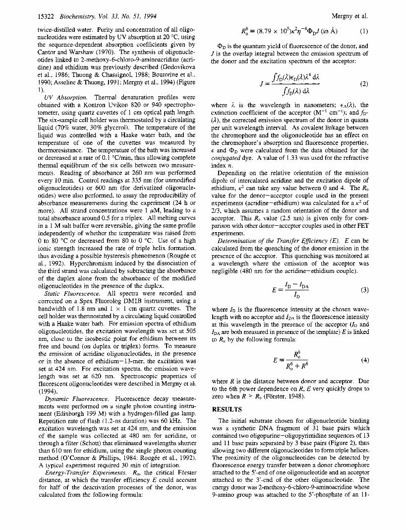
twice-distilled water. Purity and concentration of all oligo- nucleotides were estimated by UV absorption at 20 "C, using the sequence-dependent absorption coefficients given by Cantor and Warshaw (1970). The synthesis of oligonucle- otides linked to 2-methoxy-6-chloro-9-aminoacridine (acri- dine) and ethidium was previously described (Godovikova et al., 1986; Thuong & Chassignol, 1988; Boutorine et al., 1990; Asseline & Thuong, 1991; Mergny et al., 1994) (Figure 1).
Thermal denaturation profiles were obtained with a Kontron Uvikon 820 or 940 spectropho- tometer, using quartz cuvettes of 1 cm optical path length. The six-sample cell holder was thermostated by a circulating liquid (70% water, 30% glycerol). The temperature of the liquid was controlled with a Haake water bath, and the temperature of one of the cuvettes was measured by thermoresistance. The temperature of the bath was increased or decreased at a rate of 0.1 "C/min, thus allowing complete thermal equilibrum of the six cells between two measure- ments. Reading of absorbance at 260 nm was performed every 10 min. Control readings at 335 nm (for unmodified oligonucleotides) or 600 nm (for derivatized oligonucle- otides) were also performed, to assay the reproducibility of absorbance measurements during the experiment (24 h or more). All strand concentrations were 1 pM, leading to a total absorbance around 0.5 for a triplex. All melting curves in a 1 M salt buffer were reversible, giving the same profile independently of whether the temperature was raised from 0 to 80 "C or decreased from 80 to 0 "C. Use of a high ionic strength increased the rate of triple helix formation, thus avoiding a possible hysteresis phenomenon (RougCe et al., 1992). Hyperchromism induced by the dissociation of the third strand was calculated by subtracting the absorbance of the duplex alone from the absorbance of the modified oligonucleotides in the presence of the duplex.
All spectra were recorded and corrected on a Spex Fluorolog DMlB instrument, using a bandwidth of 1.8 nm and 1 x 1 cm quartz cuvettes. The cell holder was thermostated by a circulating liquid controlled with a Haake water bath. For emission spectra of ethidium oligonucleotides, the excitation wavelength was set at 505 nm, close to the isosbestic point for ethidium between its free and bound (on duplex or triplex) forms. To measure the emission of acridine oligonucleotides, in the presence or in the absence of ethidium- 13-mer, the excitation was set at 424 nm. For excitation spectra, the emission wave- length was set at 620 nm. Spectroscopic properties of fluorescent oligonucleotides were described in Mergny et al. (1994).
Dynamic Fluorescence. Fluorescence decay measure- ments were performed on a single photon counting instru- ment (Edinburgh 199 M) with a hydrogen-filled gas lamp. Repetition rate of flash (1.2-11s duration) was 60 kHz. The excitation wavelength was set at 424 nm, and the emission of the sample was collected at 480 nm for acridine, or through a filter (Schott) that eliminated wavelengths shorter than 610 nm for ethidium, using the single photon counting method (O'Connor & Phillips, 1984; RougCe et al., 1992). A typical experiment required 30 min of integration.
R,, the critical Forster distance, at which the transfer efficiency E could account for half of the deactivation processes of the donor, was calculated from the following formula:
UV Absorption.
Static Fluorescence.
Energy-Transfer Experiments.
Mergny et al.
(1)
Cy, is the quantum yield of fluorescence of the donor, and J is the overlap integral between the emission spectrum of the donor and the excitation spectrum of the acceptor:
R6, = (8.79 x 10⁵)~2f4@pd (in A)
where il is the wavelength in nanometers; €,(A), the extinction coefficient of the acceptor (M-' cm-'); and fD-
(A), the corrected emission spectrum of the donor in quanta per unit wavelength interval. As covalent linkage between the chromophore and the oligonucleotide has an effect on the chromophore's absorption and fluorescence properties, 6 and @D were calculated from the data obtained for the conjugated dye. A value of 1.33 was used for the refractive index n.
Depending on the relative orientation of the emission dipole of intercalated acridine and the excitation dipole of ethidium, K' can take any value between 0 and 4. The R, value for the donor-acceptor couple used in the present experiments (acridine-ethidium) was calculated for a K* of 213, which assumes a random orientation of the donor and acceptor. This R, value (2.5 nm) is given only for com- parison with other donor-acceptor couples used in other FET experiments.
Determination of the Transfer EfJiciency (E). E can be calculated from the quenching of the donor emission in the presence of the acceptor. This quenching was monitored at a wavelength where the emission of the acceptor was negligible (480 nm for the acridine-ethidium couple).
ID - IDA E=- (3)
where ID is the fluorescence intensity at the chosen wave- length with no acceptor and IDA is the fluorescence intensity at this wavelength in the presence of the acceptor ( ID and IDA are both measured in presence of the template) E is linked to R, by the following formula:
0 6 A0 E=-
R: + R6 (4)
where R is the distance between donor and acceptor. Due to the 6th power dependence on R, E very quickly drops to zero when R > R, (Forster, 1948).
RESULTS
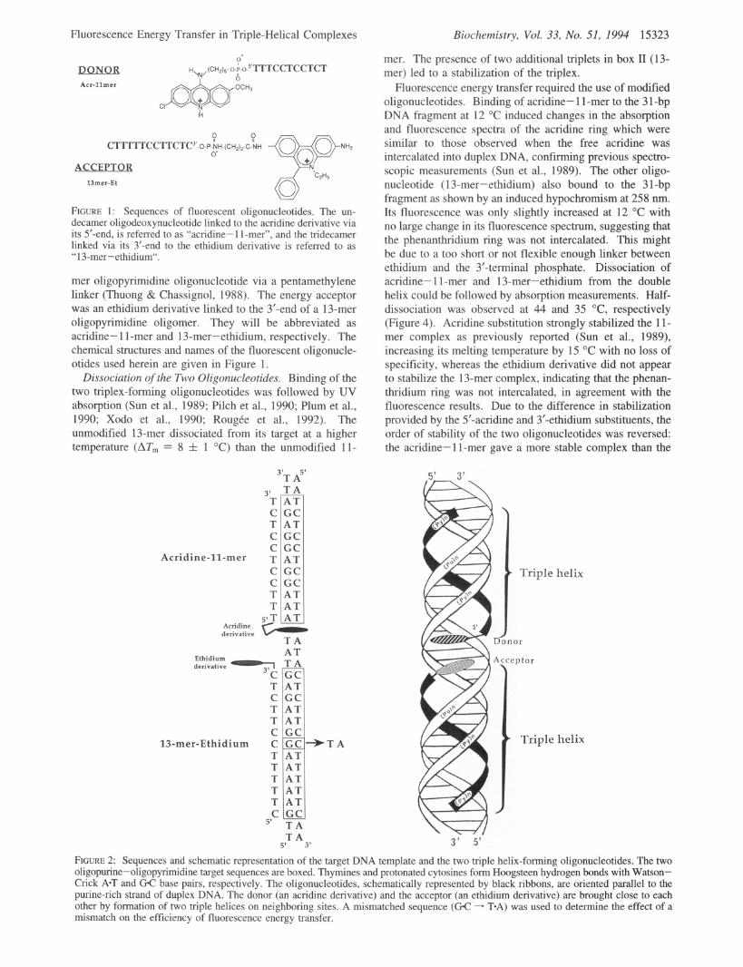
The initial substrate chosen for oligonucleotide binding was a synthetic DNA fragment of 31 base pairs which contained two oligopurine-oligopyrimidine sequences of 13 and 11 base pairs separated by 3 base pairs (Figure 2), thus allowing two different oligonucleotides to form triple helices. The proximity of the oligonucleotides can be detected by fluorescence energy transfer between a donor chromophore attached to the 5'-end of one oligonucleotide and an acceptor attached to the 3'-end of the other oligonucleotide. The energy donor was 2-methoxy-6-chloro-9-aminoacridine whose 9-amino group was attached to the 5'-phosphate of an 11-
Fluorescence Energy Transfer in Triple-Helical Complexes
HW(CH2)~-o-P-o-S’TTTCCTCCTCT ?- K DONOR
Acr-l lmer
CI k
ACCEPTOR 13mer-Et
FIGURE 1 : Sequences of fluorescent oligonucleotides. The un- decamer oligodeoxynucleotide linked to the acridine derivative via its 5’-end, is referred to as “acridine- 1 1-mer”, and the tridecamer linked via its 3’-end to the ethidium derivative is referred to as “13-mer-ethidium“.
mer oligopyrimidine oligonucleotide via a pentamethylene linker (Thuong & Chassignol, 1988). The energy acceptor was an ethidium derivative linked to the 3’-end of a 13-mer oligopyrimidine oligomer. They will be abbreviated as acridine- 1 1-mer and 13-mer-ethidium, respectively. The chemical structures and names of the fluorescent oligonucle- otides used herein are given in Figure 1.
Dissociation of the Two Oligonucleotides. Binding of the two triplex-forming oligonucleotides was followed by UV absorption (Sun et al., 1989; Pilch et al., 1990; Plum et al., 1990; Xodo et al., 1990; Roug6e et al., 1992). The unmodified 13-mer dissociated from its target at a higher temperature (ATm = 8 f 1 “C) than the unmodified 11-
3’ T C T C C
Acridine-11-mer T C C T T
3’T A5’ TA A T G C A T G C G C A T G C G C A T A T A T
A T T A Ethidium
derivative GCI
T C T T C
13-mer-Ethidium C T T T T T C
5’
AT G C A T A T
T A T A
5’ 3’
Biochemistry, Vol. 33, No. 51, 1994 15323
mer. The presence of two additional triplets in box I1 (13- mer) led to a stabilization of the triplex.
Fluorescence energy transfer required the use of modified oligonucleotides. Binding of acridine- 1 1 -mer to the 3 1 -bp DNA fragment at 12 “C induced changes in the absorption and fluorescence spectra of the acridine ring which were similar to those observed when the free acridine was intercalated into duplex DNA, confirming previous spectro- scopic measurements (Sun et al., 1989). The other oligo- nucleotide (13-mer-ethidium) also bound to the 3 1-bp fragment as shown by an induced hypochromism at 258 nm. Its fluorescence was only slightly increased at 12 “C with no large change in its fluorescence spectrum, suggesting that the phenanthridium ring was not intercalated. This might be due to a too short or not flexible enough linker between ethidium and the 3’-terminal phosphate. Dissociation of acridine- 1 1-mer and 13-mer-ethidium from the double helix could be followed by absorption measurements. Half- dissociation was observed at 44 and 35 “C, respectively (Figure 4). Acridine substitution strongly stabilized the 11- mer complex as previously reported (Sun et al., 1989), increasing its melting temperature by 15 “C with no loss of specificity, whereas the ethidium derivative did not appear to stabilize the 13-mer complex, indicating that the phenan- thridium ring was not intercalated, in agreement with the fluorescence results. Due to the difference in stabilization provided by the 5’-acridine and 3’-ethidium substituents, the order of stability of the two oligonucleotides was reversed: the acridine- 1 1-mer gave a more stable complex than the
D
Triple helix
or
\w - 3 ’ 5’
FIGURE 2: Sequences and schematic representation of the target DNA template and the two triple helix-forming oligonucleotides. The two oligopurine-oligopyrimidine target sequences are boxed. Thymines and protonated cytosines form Hoogsteen hydrogen bonds with Watson- Crick A T and GC base pairs, respectively. The oligonucleotides, schematically represented by black ribbons, are oriented parallel to the purine-rich strand of duplex DNA. The donor (an acridine derivative) and the acceptor (an ethidium derivative) are brought close to each other by formation of two triple helices on neighboring sites. A mismatched sequence (GC - T*A) was used to determine the effect of a mismatch on the efficiency of fluorescence energy transfer.
15324 Biochemistry, Vol. 33, No. 51, 1994
1
Mergny et al.
t
r i - .- x
E E
01 z e a i:
0 . 5
0 380 430 4 8 0 530
Wavelength (nm)
1 -1 h
450 500 5 5 0 6 0 0 6 5 0 700 Wavelength (nm)
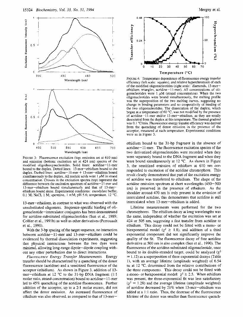
FIGURE 3: Fluorescence excitation (top; emission set at 610 nm) and emission (bottom; excitation set at 424 nm) spectra of the modified oligodeoxynucleotides. Solid lines: acridine- 1 1 -mer bound to the duplex. Dotted lines: 13-mer-ethidium bound to the duplex. Dashed lines: acridine- 1 I-mer + 13-mer-ethidium bound simultaneously to the duplex. All nucleic acids were 1 pM in strand concentration. Crosses in the excitation spectra (top) represent the difference between the excitation spectrum of acridine- 1 1-mer and 13-mer-ethidium bound simultaneously and that of 13-mer- ethidium bound alone. Experimental conditions: cacodylate buffer, 0.1 M; NaCI, 1 M; spermine, 1 mM; pH 5.6; temperature, 12 "C.
13-mer-ethidium, in contrast to what was observed with the unsubstituted oligomers. Sequence-specific binding of oli- gonucleotide-intercalator conjugates has been demonstrated for acridine-substituted oligonucleotides (Sun et al., 1989; Collier et al., 1991b) as well as other derivatives (Perrouault et al., 1990).
With the 3-bp spacing of the target sequence, no interaction between acridine- 1 1-mer and 13-mer-ethidium could be evidenced by thermal dissociation experiments, suggesting that physical interactions between the two dyes were minimal, allowing long-range dipole-dipole coupling with- out any other perturbation due to direct interactions.
Fluorescence Energy Transfer Measurements. Energy transfer should be characterized by a quenching of the donor fluorescence (acridine) and a sensitized fluorescence of the acceptor (ethidium). As shown in Figure 3, addition of 13- mer-ethidium at 12 "C to the 31-bp DNA fragment (1:l molar ratio, strand concentration) bound to acridine- 1 1-mer led to 45% quenching of the acridine fluorescence. Further addition of the acceptor, up to a 2:l molar excess, did not affect the donor emission. An increased fluorescence of ethidium was also observed, as compared to that of 13-mer-
0.48 100
X Y
cp r( n
P a 9
r .i
.i U
u(
w 50 ; 2 0.24
0) rn
G
i- - 0 0 0 10 20 30 40 50 60 70
I.
B y.l v)
2 8
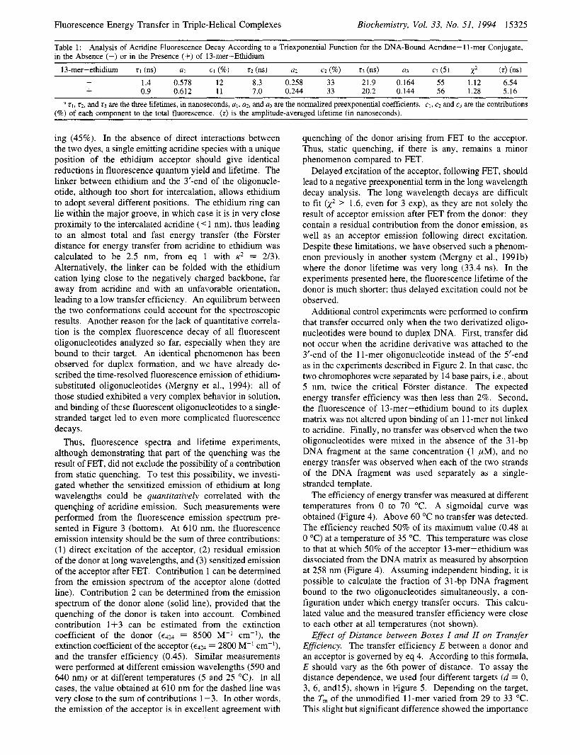
Temperature ("C) FIGURE 4: Temperature dependence of fluorescence energy transfer efficiency (left scale: squares), and relative hyperchromism of each of the modified oligonucleotides (right scale: diamonds, 13-mer- ethidium; triangles, acridine- 1 1 -mer). All concentrations of oli- gonucleotides were 1 pM (strand concentration). When the two oligonucleotides were bound simultaneously, the melting profile was the superposition of the two melting curves, suggesting no change in binding parameters and no cooperativity of binding of the two oligonucleotides. The dissociation of the duplex, which began at a temperature of 60 "C, was not modified by the presence of acridine-I I-mer and/or 13-mer-ethidium, as they are totally dissociated from the duplex at this temperature. The thermal gradient was 0.1 "C/min. Fluorescence energy transfer efficiency was derived from the quenching of donor emission in the presence of the acceptor, measured at each temperature. Experimental conditions were as in Figure 3.
ethidium bound to the 31-bp fragment in the absence of acridine- 1 1 -mer. The fluorescence excitation spectra of the two derivatized oligonucleotides were recorded when they were separately bound to the DNA fragment and when they were bound simultaneously at 12 "C. As shown in Figure 3, the sensitized emission of ethidium at 610 nm cor- responded to excitation of the acridine chromophore. This result clearly demonstrated that part of the excitation energy of acridine was transferred to ethidium. The shape of the acridine emission spectrum at short wavelengths (450-500 nm) is preserved in the presence of ethidium. As the shoulder around 470 nm is only present in the emission of intercalated acridine, this demonstrates that acridine is still intercalated when 13-mer-ethidium is added.
Lifetime measurements were performed for the two chromophores. The ethidium decay at long wavelengths was the same, independent of whether the excitation was set at 424 or 505 nm, suggesting a fast transfer from acridine to ethidium. This decay could not be fitted with a mono- or biexponential model k2 2 1.8), and addition of a third exponential component did not significantly improve the quality of the fit. The fluorescence decay of free acridine derivative at 500 nm is also complex (Sun et al., 1990). The fluorescence of the acridine-substituted oligonucleotide, once bound to its double-stranded target, could be analyzed k2 = 1.12) as a superposition of three exponential decays (Table l), with an average lifetime (amplitude weighted) of 6.54 ns at 12 "C, determined from the relative contributions of the three components. This decay could not be fitted with a mono- or biexponential model: x2 2 2.5. When ethidium was present, the three-exponential fit was less satisfactory k2 = 1.28) and the average lifetime (amplitude weighted) of acridine decreased by 21% when 13-mer-ethidium was added at a 1 : 1 ratio. Thus, the decrease of the fluorescence lifetime of the donor was smaller than fluorescence quench-
Fluorescence Energy Transfer in Triple-Helical Complexes Biochemistry, Vol. 33, No. 51, 1994 15325 ~~
Table 1: in the Absence (-) or in the Presence (+) of 13-mer-Ethidium
Analysis of Acridine Fluorescence Decay According to a Triexponential Function for the DNA-Bound Acridine- 1 l-mer Conjugate,
13-mer-ethidium 7 1 (ns) a1 CI (%) r2 (ns) a2 cz (96) r3 (ns) a3 c3 ( 5 ) x2 ( r ) (ns) - 1.4 0.578 12 8.3 0.258 33 21.9 0.164 55 1.12 6.54 + 0.9 0.612 11 7.0 0.244 33 20.2 0.144 56 1.28 5.16
q , t 2 . and 7 3 are the three lifetimes, in nanoseconds, al, a2, and a3 are the normalized preexponential coefficients. c1, c2 and c3 are the contributions (%) of each component to the total fluorescence. (7) is the amplitude-averaged lifetime (in nanoseconds).
ing (45%). In the absence of direct interactions between the two dyes, a single emitting acridine species with a unique position of the ethidium acceptor should give identical reductions in fluorescence quantum yield and lifetime. The linker between ethidium and the 3'-end of the oligonucle- otide, although too short for intercalation, allows ethidium to adopt several different positions. The ethidium ring can lie within the major groove, in which case it is in very close proximity to the intercalated acridine (< 1 nm), thus leading to an almost total and fast energy transfer (the Forster distance for energy transfer from acridine to ethidium was calculated to be 2.5 nm, from eq 1 with K* = 2/3). Alternatively, the linker can be folded with the ethidium cation lying close to the negatively charged backbone, far away from acridine and with an unfavorable orientation, leading to a low transfer efficiency. An equilibrum between the two conformations could account for the spectroscopic results. Another reason for the lack of quantitative correla- tion is the complex fluorescence decay of all fluorescent oligonucleotides analyzed so far, especially when they are bound to their target. An identical phenomenon has been observed for duplex formation, and we have already de- scribed the time-resolved fluorescence emission of ethidium- substituted oligonucleotides (Mergny et al., 1994): all of those studied exhibited a very complex behavior in solution, and binding of these fluorescent oligonucleotides to a single- stranded target led to even more complicated fluorescence decays.
Thus, fluorescence spectra and lifetime experiments, although demonstrating that part of the quenching was the result of FET, did not exclude the possibility of a contribution from static quenching. To test this possibility, we investi- gated whether the sensitized emission of ethidium at long wavelengths could be quantitatively correlated with the quenching of acridine emission. Such measurements were performed from the fluorescence emission spectrum pre- sented in Figure 3 (bottom). At 610 nm, the fluorescence emission intensity should be the sum of three contributions: (1) direct excitation of the acceptor, (2) residual emission of the donor at long wavelengths, and (3) sensitized emission of the acceptor after FET. Contribution 1 can be determined from the emission spectrum of the acceptor alone (dotted line). Contribution 2 can be determined from the emission spectrum of the donor alone (solid line), provided that the quenching of the donor is taken into account. Combined contribution 1+3 can be estimated from the extinction coefficient of the donor (€424 = 8500 M-' cm-I), the extinction coefficient of the acceptor (€424 = 2800 M-' cm-'), and the transfer efficiency (0.45). Similar measurements were performed at different emission wavelengths (590 and 640 nm) or at different temperatures (5 and 25 "C). In all cases, the value obtained at 610 nm for the dashed line was very close to the sum of contributions 1-3. In other words, the emission of the acceptor is in excellent agreement with
quenching of the donor arising from FET to the acceptor. Thus, static quenching, if there is any, remains a minor phenomenon compared to FET.
Delayed excitation of the acceptor, following FET, should lead to a negative preexponential term in the long wavelength decay analysis. The long wavelength decays are difficult to fit k2 > 1.6, even for 3 exp), as they are not solely the result of acceptor emission after FET from the donor: they contain a residual contribution from the donor emission, as well as an acceptor emission following direct excitation. Despite these limitations, we have observed such a phenom- enon previously in another system (Mergny et al., 1991b) where the donor lifetime was very long (33.4 ns). In the experiments presented here, the fluorescence lifetime of the donor is much shorter; thus delayed excitation could not be observed.
Additional control experiments were performed to confirm that transfer occurred only when the two derivatized oligo- nucleotides were bound to duplex DNA. First, transfer did not occur when the acridine derivative was attached to the 3'-end of the 1 1-mer oligonucleotide instead of the 5'-end as in the experiments described in Figure 2. In that case, the two chromophores were separated by 14 base pairs, Le., about 5 nm, twice the critical Forster distance. The expected energy transfer efficiency was then less than 2%. Second, the fluorescence of 13-mer-ethidium bound to its duplex matrix was not altered upon binding of an 1 l-mer not linked to acridine. Finally, no transfer was observed when the two oligonucleotides were mixed in the absence of the 31-bp DNA fragment at the same concentration (1 pM), and no energy transfer was observed when each of the two strands of the DNA fragment was used separately as a single- stranded template.
The efficiency of energy transfer was measured at different temperatures from 0 to 70 "C. A sigmoidal curve was obtained (Figure 4). Above 60 "C no transfer was detected. The efficiency reached 50% of its maximum value (0.48 at 0 "C) at a temperature of 35 "C. This temperature was close to that at which 50% of the acceptor 13-mer-ethidium was dissociated from the DNA matrix as measured by absorption at 258 nm (Figure 4). Assuming independent binding, it is possible to calculate the fraction of 31-bp DNA fragment bound to the two oligonucleotides simultaneously, a con- figuration under which energy transfer occurs. This calcu- lated value and the measured transfer efficiency were close to each other at all temperatures (not shown).
Effect of Distance between Boxes I and II on Transfer Efficiency. The transfer efficiency E between a donor and an acceptor is governed by eq 4. According to this formula, E should vary as the 6th power of distance. To assay the distance dependence, we used four different targets (d = 0, 3, 6, andl5), shown in Figure 5. Depending on the target, the T, of the unmodified 11-mer varied from 29 to 33 "C. This slight but significant difference showed the importance
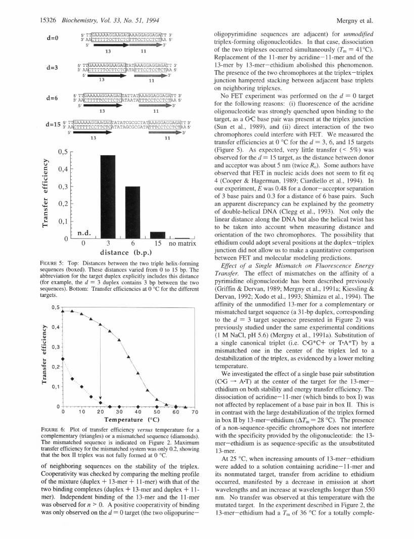
15326 Biochemistry, Vol. 33, No. 51, 1994 Mergny et al.
d=O
13 11
d=3 3'
11
5' - 13
0.5 r
" 0 3 6 15 no matrix distance (b.p.)
FIGURE 5: Top: Distances between the two triple helix-forming sequences (boxed). These distances varied from 0 to 15 bp. The abbreviation for the target duplex explicitly includes this distance (for example, the d = 3 duplex contains 3 bp between the two sequences). Bottom: Transfer efficiencies at 0 "C for the different targets.
095 1
*A.
0 10 20 30 4.0 50 6 0 Temperature ("C)
FIGURE 6: Plot of transfer efficiency versus temperature for a complementary (triangles) or a mismatched sequence (diamonds). The mismatched sequence is indicated on Figure 2. Maximum transfer efficiency for the mismatched system was only 0.2, showing that the box 11 triplex was not fully formed at 0 "C.
of neighboring sequences on the stability of the triplex. Cooperativity was checked by comparing the melting profile of the mixture (duplex + 13-mer + 1 1-mer) with that of the two binding complexes (duplex + 13-mer and duplex + 1 1- mer). Independent binding of the 13-mer and the 11-mer was observed for n > 0. A positive cooperativity of binding was only observed on the d = 0 target (the two oligopurine-
oligopyrimidine sequences are adjacent) for unmodified triplex-forming oligonucleotides. In that case, dissociation of the two triplexes occurred simultaneously ( T m = 41°C). Replacement of the 1 1 -mer by acridine- 1 1 -mer and of the 13-mer by 13-mer-ethidium abolished this phenomenon. The presence of the two chromophores at the triplex-triplex junction hampered stacking between adjacent base triplets on neighboring triplexes.
No FET experiment was performed on the d = 0 target for the following reasons: (i) fluorescence of the acridine oligonucleotide was strongly quenched upon binding to the target, as a GC base pair was present at the triplex junction (Sun et al., 1989), and (ii) direct interaction of the two chromophores could interfere with FET. We measured the transfer efficiencies at 0 "C for the d = 3, 6, and 15 targets (Figure 5). As expected, very little transfer (< 5%) was observed for the d = 15 target, as the distance between donor and acceptor was about 5 nm (twice RJ. Some authors have observed that FET in nucleic acids does not seem to fit eq 4 (Cooper & Hagerman, 1989; Ciardiello et al., 1994). In our experiment, E was 0.48 for a donor-acceptor separation of 3 base pairs and 0.3 for a distance of 6 base pairs. Such an apparent discrepancy can be explained by the geometry of double-helical DNA (Clegg et al., 1993). Not only the linear distance along the DNA but also the helical twist has to be taken into account when measuring distance and orientation of the two chromophores. The possibility that ethidium could adopt several positions at the duplex- triplex junction did not allow us to make a quantitative comparison between FET and molecular modeling predictions.
Effect of a Single Mismatch on Fluorescence Energy Transfer. The effect of mismatches on the affinity of a pyrimidine oligonucleotide has been described previously (Griffin & Dervan, 1989; Mergny et al., 1991a; Kiessling & Dervan, 1992; Xodo et al., 1993; Shimizu et al., 1994). The affinity of the unmodified 13-mer for a complementary or mismatched target sequence (a 3 1-bp duplex, corresponding to the d = 3 target sequence presented in Figure 2) was previously studied under the same experimental conditions (1 M NaC1, pH 5.6) (Mergny et al., 1991a). Substitution of a single canonical triplet (Le. C*G*C+ or T-A*T) by a mismatched one in the center of the triplex led to a destabilization of the triplex, as evidenced by a lower melting temperature.
We investigated the effect of a single base pair substitution (CG --+ A-T) at the center of the target for the 13-mer- ethidium on both stability and energy transfer efficiency. The dissociation of acridine- 1 1-mer (which binds to box I) was not affected by replacement of a base pair in box 11. This is in contrast with the large destabilization of the triplex formed in box II by 13-mer-ethidium (AT, = 28 "C). The presence of a non-sequence-specific chromophore does not interfere with the specificity provided by the oligonucleotide: the 13- mer-ethidium is as sequence-specific as the unsubstituted 13-mer.
At 25 "C, when increasing amounts of 13-mer-ethidium were added to a solution containing acridine- 1 1-mer and its nonmutated target, transfer from acridine to ethidium occurred, manifested by a decrease in emission at short wavelengths and an increase at wavelengths longer than 550 nm. No transfer was observed at this temperature with the mutated target. In the experiment described in Figure 2, the 13-mer-ethidium had a Tm of 36 "C for a totally comple-
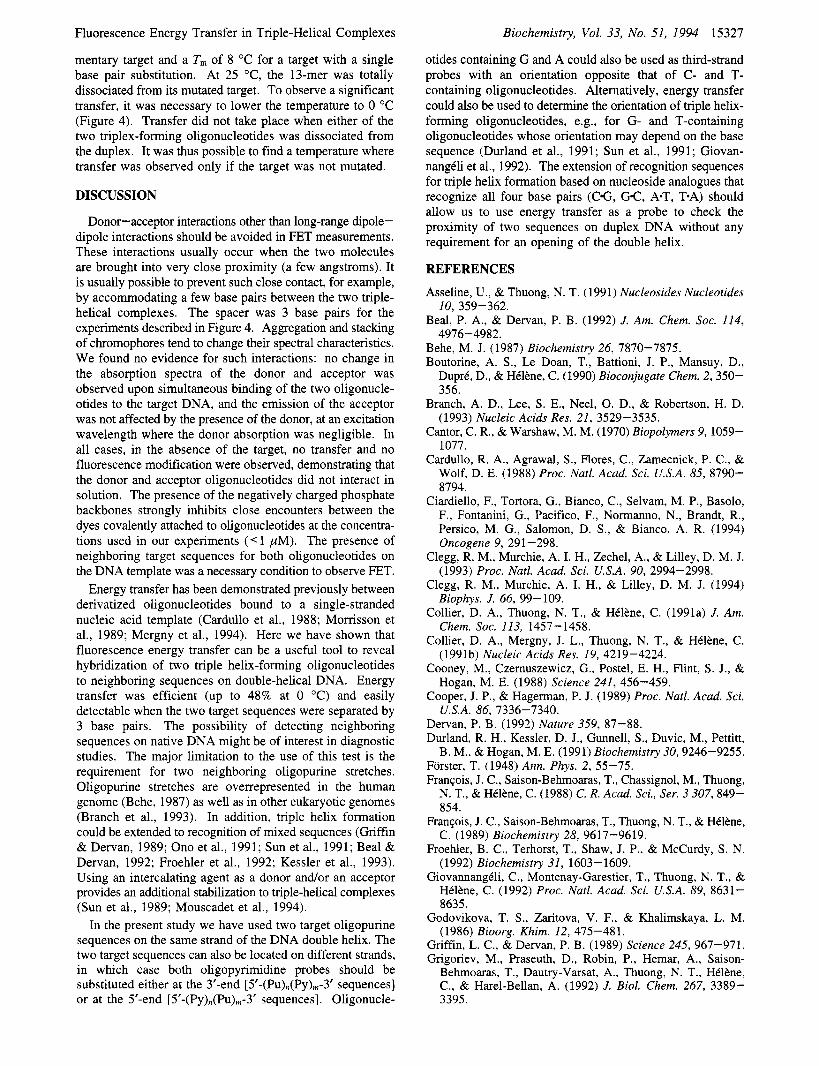
Fluorescence Energy Transfer in Triple-Helical Complexes
mentary target and a T, of 8 "C for a target with a single base pair substitution. At 25 "C, the 13-mer was totally dissociated from its mutated target. To observe a significant transfer, it was necessary to lower the temperature to 0 "C (Figure 4). Transfer did not take place when either of the two triplex-forming oligonucleotides was dissociated from the duplex. It was thus possible to find a temperature where transfer was observed only if the target was not mutated.
DISCUSSION
Donor-acceptor interactions other than long-range dipole- dipole interactions should be avoided in FET measurements. These interactions usually occur when the two molecules are brought into very close proximity (a few angstroms). It is usually possible to prevent such close contact, for example, by accommodating a few base pairs between the two triple- helical complexes. The spacer was 3 base pairs for the experiments described in Figure 4. Aggregation and stacking of chromophores tend to change their spectral characteristics. We found no evidence for such interactions: no change in the absorption spectra of the donor and acceptor was observed upon simultaneous binding of the two oligonucle- otides to the target DNA, and the emission of the acceptor was not affected by the presence of the donor, at an excitation wavelength where the donor absorption was negligible. In all cases, in the absence of the target, no transfer and no fluorescence modification were observed, demonstrating that the donor and acceptor oligonucleotides did not interact in solution. The presence of the negatively charged phosphate backbones strongly inhibits close encounters between the dyes covalently attached to oligonucleotides at the concentra- tions used in our experiments ( e l pM). The presence of neighboring target sequences for both oligonucleotides on the DNA template was a necessary condition to observe FET.
Energy transfer has been demonstrated previously between derivatized oligonucleotides bound to a single-stranded nucleic acid template (Cardullo et al., 1988; Morrisson et al., 1989; Mergny et al., 1994). Here we have shown that fluorescence energy transfer can be a useful tool to reveal hybridization of two triple helix-forming oligonucleotides to neighboring sequences on double-helical DNA. Energy transfer was efficient (up to 48% at 0 "C) and easily detectable when the two target sequences were separated by 3 base pairs. The possibility of detecting neighboring sequences on native DNA might be of interest in diagnostic studies. The major limitation to the use of this test is the requirement for two neighboring oligopurine stretches. Oligopurine stretches are overrepresented in the human genome (Behe, 1987) as well as in other eukaryotic genomes (Branch et al., 1993). In addition, triple helix formation could be extended to recognition of mixed sequences (Griffin & Dervan, 1989; Ono et al., 1991; Sun et al., 1991; Beal & Dervan, 1992; Froehler et al., 1992; Kessler et al., 1993). Using an intercalating agent as a donor and/or an acceptor provides an additional stabilization to triple-helical complexes (Sun et al., 1989; Mouscadet et al., 1994).
In the present study we have used two target oligopurine sequences on the same strand of the DNA double helix. The two target sequences can also be located on different strands, in which case both oligopyrimidine probes should be substituted either at the 3'-end [5'-(P~)~(Py),-3' sequences] or at the 5'-end [5'-(P~)~(F'u),-3' sequences]. Oligonucle-
Biochemistry, Vol. 33, No. 51, 1994 15327
otides containing G and A could also be used as third-strand probes with an orientation opposite that of C- and T- containing oligonucleotides. Alternatively, energy transfer could also be used to determine the orientation of triple helix- forming oligonucleotides, e.g., for G- and T-containing oligonucleotides whose orientation may depend on the base sequence (Durland et al., 1991; Sun et al., 1991; Giovan- nangCli et al., 1992). The extension of recognition sequences for triple helix formation based on nucleoside analogues that recognize all four base pairs (CG, G-C, A-T, F A ) should allow us to use energy transfer as a probe to check the proximity of two sequences on duplex DNA without any requirement for an opening of the double helix.
REFERENCES
Asseline, U., & Thuong, N. T. (1991) Nucleosides Nucleotides
Beal, P. A., & Dervan, P. B. (1992) J. Am. Chem. SOC. 114,
Behe, M. J. (1987) Biochemistry 26, 7870-7875. Boutorine, A. S., Le Doan, T., Battioni, J. P., Mansuy, D.,
DuprC, D., & HCBne, C. (1990) Bioconjugate Chem. 2,350- 356.
Branch, A. D., Lee, S. E., Neel, 0. D., & Robertson, H. D. (1993) Nucleic Acids Res. 21, 3529-3535.
Cantor, C. R., & Warshaw, M. M. (1970) Biopolymers 9, 1059- 1077.
Cardullo, R. A., Agrawal, S., Flores, C., Zamecnick, P. C., & Wolf, D. E. (1988) Proc. Natl. Acad. Sci. U.S.A. 85, 8790- 8794.
Ciardiello, F., Tortora, G., Bianco, C., Selvam, M. P., Basolo, F., Fontanini, G., Pacifico, F., Normanno, N., Brandt, R., Persico, M. G., Salomon, D. S., & Bianco, A. R. (1994) Oncogene 9, 291-298.
Clegg, R. M., Murchie, A. I. H., Zechel, A., & Lilley, D. M. J. (1993) Proc. Natl. Acad. Sci. U S A . 90, 2994-2998.
Clegg, R. M., Murchie, A. I. H., & Lilley, D. M. J. (1994) Biophys. J. 66, 99-109.
Collier, D. A., Thuong, N. T., & HCltne, C. (1991a) J. Am. Chem. SOC. 113, 1457-1458.
Collier, D. A., Mergny, J. L., Thuong, N. T., & HCltne, C. (1991b) Nucleic Acids Res. 19, 4219-4224.
Cooney, M., Czemuszewicz, G., Postel, E. H., Flint, S. J., & Hogan, M. E. (1988) Science 241, 456-459.
Cooper, J. P., & Hagerman, P. J. (1989) Proc. Natl. Acad. Sci. U.S.A. 86, 7336-7340.
Dervan, P. B. (1992) Nature 359, 87-88. Durland, R. H., Kessler, D. J., Gunnell, S., Duvic, M., Pettitt,
B. M., & Hogan, M. E. (1991) Biochemistry 30,9246-9255. Forster, T. (1948) Ann. Phys. 2 , 55-75. FranGois, J. C., Saison-Behmoaras, T., Chassignol, M., Thuong,
N. T., & HBltne, C. (1988) C. R. Acad. Sci., Ser. 3 307, 849- 854.
FranGois, J. C., Saison-Behmoaras, T., Thuong, N. T., & HCltne, C. (1989) Biochemistry 28, 9617-9619.
Froehler, B. C., Terhorst, T., Shaw, J. P., & McCurdy, S. N. (1992) Biochemistry 31, 1603-1609.
GiovannangCli, C., Montenay-Garestier, T., Thuong, N. T., & HClBne, C. (1992) Proc. Natl. Acad. Sci. U.S.A. 89, 8631- 8635.
Godovikova, T. S., Zaritova, V. F., & Khalimskaya, L. M. (1986) Bioorg. Khim. 12, 475-481.
Griffin, L. C., & Dervan, P. B. (1989) Science 245, 967-971. Grigoriev, M., Praseuth, D., Robin, P., Hemar, A., Saison-
Behmoaras, T., Dautry-Varsat, A., Thuong, N. T., HClkne, C., & Harel-Bellan, A. (1992) J. Biol. Chem. 267, 3389- 3395.
10, 359-362.
4976-4982.

15328 Biochemistry, Vol. 33, No. 51, 1994
Havre, P. A., Gunther, E. J., Gasparro, F. P., & Glazer, P. M.
HClbne, C. (1993) Curr. Opin. Biotechnol. 4 , 29-36. Kessler, D. J., Pettitt, B. M., Cheng, Y. K., Smith, S. R.,
Jayaraman, K., Vu, H. M., & Hogan, M. E. (1993) Nucleic Acids Res. 21, 4810-4815.
Kiessling, L. L., & Dervan, P. B. (1992) Biochemistry 31, 2829-2834.
Le Doan, T., Perrouault, L., Praseuth, D., Habhoub, N., Decout, J.-L., Thuong, N. T., Lhomme, J . , & Htlbne, C. (1987) Nucleic Acids Res. 15, 7749-7760.
Luebke, K. J., & Dervan, P. B. (1991) J. Am. Chem. SOC. 113, 7447 -7448.
Lyamichev, V. L., Mirkin, S. M., Frank-Kamenetskii, M. D., & Cantor, C. R. (1988) Nucleic Acids Res. 16, 2165-2178.
Maher, L. J., 111, Wold, B., & Dervan, P. B. (1989) Science 245, 725-730.
Maher L. J., 111, Dervan, P. B., & Wold, B. J. (1990) Biochemistry 29, 8820-8826.
Mergny, J. L., Sun, J. S., RougCe, M., Montenay-Garestier, T., Barcelo, F., Chomilier, J., & HClbne, C. (1991a) Biochemistry 30, 9791-9798.
Mergny, J. L., Slama-Schwock, A., RougCe, M., Montenay- Garestier, T., & HClbne, C. (1991b) Photochem. Phorobiol. 53, 555-558.
Mergny, J. L., Boutorine, A. S., Montenay-Garestier, T., Belloc, F., RougCe, M., Lebedev, A. V., Valeur, B., & HClbne, C. (1994) Nucleic Acids Res, 22, 920-928.
Morrisson, L. E., Halder, T. C., & Stols, L. M. (1989) Anal. Biochem. 183, 23 1-244.
Moser, H. E., & Dervan, P. B. (1987) Science 238, 645-650. Mouscadet, J. F., KetterlC, C., Goulaouic, H., Carteau, S., Subra,
F., Le Bret, M., & Auclair, C. (1994) Biochemistry 33,4187- 4196.
Murchie, A. I. H., Clegg, R. M., Kitzing, E., Duckett, D. R., Diekmann, S., & Lilley, D. M. J. (1989) Nature 341, 763- 766.
O’Connor, D. V., & Phillips, D. (1984). Time-correlated single photon counting, Academic Press, London.
Ono, A,, Chen, C. N., & Kan, L. S. (1991) Biochemistry 30, 9914-9921.
(1993) Proc. Natl. Acad. Sci. U.S.A. 90, 7879-7883.
Mergny et al.
Oser, A., & Valet, G . (1990) Angew. Chem., Int. Ed. Engl. 29,
Perrouault, L., Asseline, U., Rivalle, C., Thuong, N. T., Bisagni, E., GiovannangCli, C., Le Doan, T., & Htlbne, C. (1990) Nature 344, 358-360.
Pilch, D. S., Brousseau, R., & Shafer, R. H. (1990) Nucleic Acids Res. 18, 5743-5750.
Plum, G. E., Park, Y. W., Singleton, S. F., Dervan, P. B., & Breslauer, K. J. (1990) Proc. Natl. Acad. Sci. U.S.A. 87,
Postel, E. H., Flint, S. J., Kessler, D. J., & Hogan, M. E. (1991) Proc. Natl. Acad. Sci. U.S.A. 88, 8227-8231.
Povsic, T. J., & Dervan, P. B. (1989) J. Am. Chem. SOC. 1 1 1 , 3059-3060.
Praseuth, D., Perrouault, L., Le Doan, T., Chassignol, M., Thuong, N. T., & HClbne, C. (1988) Proc. Natl. Acad. Sci. U.S.A. 85, 1349-1353.
RougCe, M., Faucon, B., Mergny, J. L., Barcelo, F., Giovan- nangCli, C., Garestier, T., & HClbne, C. (1992) Biochemistry 31, 9269-9278.
Shimizu, M., Inoue, H., & Ohtsuka, E. (1994) Biochemistry 33, 606-61 3.
Strobel, S. A., & Dervan, P. B. (1989) J. Am. Chem. SOC. 111 , 7286-7287.
Sun, J. S., FranGois, J. C., Montenay-Garestier, T., Saison- Behmoaras, T., Roig, V., Chassignol, M., Thuong, N. T., & HClbne, C. (1989) Proc. Natl. Acad. Sci. U.S.A. 86, 9198- 9202.
Sun, J. S., RougCe, M., Delarue, M., Montenay-Garestier, T., & HClbne, C. (1990) J. Phys. Chem. 94, 968-977.
Sun, J. S., De Bizemont, T., Duval-Valentin, G., Montenay- Garestier, T., & HClbne, C. (1991) C.R. Acad. Sci., Ser. 3 313, 585-590.
Thuong, N. T., & Chassignol, M. (1988) Tetrahedron Lett. 29, 5905-5908.
Thuong, N . T., & Htlbne, C. (1993) Angew. Chem., Int. Ed. 32, 666-690.
Xodo, L. E., Manzini, G., & Quadrifoglio, F. (1990) Nucleic Acids Res. 18, 3557-3564.
Xodo, L. E., Alunni-Fabbroni, M., Manzini, G., & Quadrifoglio, F. (1993) Eur. J. Biochem. 212, 395-401.
1167-1169.
9436-9440.
Copyright © 2022 FDOKUMEN