Uncertainty Measures and Limiting Distributions for Filament Estimation
Upload
independentCategory
view
2download
0
Optical Materials xxx (2014) xxx–xxx
Contents lists available at ScienceDirect
Optical Materials
journal homepage: www.elsevier .com/locate /optmat
Optical limiting and nonlinear optical properties of gold-decoratedgraphene nanocomposites
http://dx.doi.org/10.1016/j.optmat.2014.11.0230925-3467/� 2014 Elsevier B.V. All rights reserved.
⇑ Corresponding author.E-mail addresses: [email protected] (R. Podila), vsaimuthukumar@sssihl.
edu.in (V. Sai Muthukumar).
Please cite this article in press as: P. Pradhan et al., Opt. Mater. (2014), http://dx.doi.org/10.1016/j.optmat.2014.11.023
Prabin Pradhan a, Ramakrishna Podila c,⇑, Muralikrishna Molli a, Adarsh Kaniyoor b, V. Sai Muthukumar a,⇑,S. Siva Sankara Sai a, S. Ramaprabhu b, A.M. Rao c
a Department of Physics, Sri Sathya Sai Institute of Higher Learning, Prashanthi Nilayam 515134, Indiab Alternative Energy and Nanotechnology Laboratory (AENL), Nano Functional Materials Technology Centre (NFMTC), Department of Physics,Indian Institute of Technology Madras (IITM), Chennai 600036, Indiac Department of Physics and Astronomy, Clemson Nanomaterials Center, Center for Optical Materials Science & Engineering Technologies, Clemson University, Clemson, SC 29634, USA
a r t i c l e i n f o a b s t r a c t
Article history:Received 7 July 2014Received in revised form 11 November 2014Accepted 12 November 2014Available online xxxx
Keywords:Optical limiterMetal nanoparticlesNonlinear opticsZ-scanLaser ablation
Metal nanoparticle-decorated low dimensional materials can synergistically combine the nonlinearoptical properties of metallic/inorganic nanostructures for enhancing the optical limiting performance.While many materials exhibit excellent optical limiting performance at a relatively higher fluence(>9 J/cm2), there is a still a dearth of optical limiting materials for protecting low damage threshold(<1 J/cm2) photonic devices. Although metal nanoparticle-decorated graphene hybrids are expected toresolve this issue, the rehybridization of metal d-orbitals and graphene p-orbitals often lead to undesir-able changes in graphene’s electronic structure which adversely affect the nonlinear optical performance.Here, we demonstrate that d-orbitals of Au nanoparticles exhibit little or no rehybridization with graph-ene and result in an enhanced optical limiting behavior at a low fluence of �0.4 J/cm2, which is lowerthan most metal decorated graphene, carbon nanotube nanocomposites and metal nanoparticles. Thisoptical limiting performance at a lower fluence is attributed to the excellent photo-absorption of Aunanoparticles combined with rapid thermalization of excited carriers by graphene.
� 2014 Elsevier B.V. All rights reserved.
1. Introduction
The wide spread use of lasers in applications ranging from mod-ern defense weaponry to biomedical treatment necessitate safetylimits on laser exposure to avoid undesired laser-induced damage[1]. The unavailability of ideal optical limiters to protect detectorsand imaging devices and sensor (in particular, the human eye) witha low damage threshold exacerbates the probability of irreversiblephysical damage during intentional or accidental exposure tounsafe levels of laser radiation [2]. Nanostructured nonlinear opti-cal (NLO) materials [3–11] have been used as passive and effectiveoptical limiters to reduce the optical transmittance of the incidentlaser radiation by virtue of their intrinsic optical properties (e.g.,multi-photon absorption in C60). Of specific interest to this workis the use of noble metal nanoparticle-decorated low dimensionalmaterials that can synergistically combine the NLO properties ofmetallic/inorganic nanostructures (e.g., Au, Ag, Pd & Pt nanoparti-cles (NPs)) and carbon nanomaterials (e.g., graphene, carbon
nanotubes) for realizing efficient optical limiting (OL) materialsfor low fluence radiation (�0.4 J/cm2). Previously, it has been dem-onstrated that nanohybrids such as graphene/Ag2S organic glasses[4], Pt/Pd decorated on graphene [5], Ag decorated graphene [6],graphene/ZnO organic glasses [7], fullerene or graphene–metalporphyrin composites [8,9] and graphene-oligothiophene hybrids[10] exhibit enhanced OL performance. Although these studiesdemonstrated an improved OL threshold (the input fluence atwhich the transmittance falls to 50% of its initial value) at higherinput fluences >9 J/cm2, they exhibited poor OL capability in thelow fluence regime (<1 J/cm2), which is critical for practical realiza-tion of OL devices [2,11]. Metal nanoparticle (NP)–decoratedgraphene, e.g., graphene/Pt NPs, are expected to resolve the issueof OL in the low fluence regime. However, the rehybridization ofmetal d-orbitals and graphene p-orbitals often lead to undesirablechanges in graphene’s electronic structure, which adversely affectthe NLO performance [13]. Unlike in the case of Pt NP-decoratedgraphene, the electronic/optical properties of graphene are not sig-nificantly perturbed because the d-orbitals of Au NPs are morethan 2 eV below graphene’s Fermi energy level [13]. Indeed, Sunet al., have recently proposed that addition Au nanoparticles(NPs) to graphene could enhance the OL performance [12].
2 P. Pradhan et al. / Optical Materials xxx (2014) xxx–xxx
Here, we present a facile one-pot synthesis of Au NP-decoratedchemically exfoliated graphene (Au–graphene) using the laserablation technique for improved OL performance in low fluenceregime. Our synthesis approach is unique in that it obviates theneed for reducing and capping agents that are essential for prepar-ing highly stable aqueous suspension of Au–graphene. Our resultsshow that the absence of rehybridized orbitals between Au andgraphene and increased electrostatic interaction between chargedAu NPs and oxygen functionalities on chemically modifiedgraphene, enhanced OL performance. Notably, the OL thresholdof our Au–graphene system is lowered from 9 J/cm2 (OL for bareAu NPs) to 0.4 J/cm2 – a major improvement in low-fluencenanomaterials.
2. Material and methods
2.1. Synthesis of Au–graphene
In our method, the Au–graphene is prepared using the so-calledfunctionalized hydrogen exfoliated graphene (referred henceforthas exf-graphene) as the starting material. The detailed synthesisof exf-graphene can be found in Ref. [14]. Briefly, exf-graphenewas prepared by the reduction and hydrogen gas induced exfolia-tion of graphite oxide (GO) (rather than the conventional thermalexfoliation method [15]). The GO used in this study was preparedby a modified Hummers’ method [16]. Exf-graphene is hydrophilicin nature due to the presence of –COOH and –OH functionalgroups. Such hydrophilic groups are important in the preparationof homogenous and stable dispersions of Au–graphene in water.An aqueous dispersion was prepared by first ultrasonically dispers-ing 1 mg of exf-graphene in 25 ml of millipore water. Next, a highpurity Au strip (purity �99.99%) of dimension (2 cm � 1 cm) wasultrasonically cleaned in 5 ml piranha solution and placed in a25 ml glass beaker containing 3 ml of aqueous solution of exf-graphene. A Nd:YAG (Surelite III, 50 mJ, rep. rate of 10 Hz) withits fundamental harmonic at 1064 nm and 15 ns pulse–widthwas used to ablate the Au strip to generate Au NPs which deco-rated the suspended exf-graphene yielding Au–graphene [17–20].The laser beam was focused into a 30 micron diameter spot ontothe Au strip using a convex lens of focal length 10 cm (Fig. 1a). Asimilar procedure was followed for preparing pristine/bare AuNPs in plain millipore water. The weight of the gold strip wasdetermined before and after the laser ablation process to assessthe net amount of gold released into the solution (�0.3 mg).
2.2. Material characterization
We studied the morphology and distribution of Au NPs on exf-graphene using a high resolution transmission electron microscopy(HRTEM – TECNAI F–20, S–TWIN, 200 kV). In order to probe theelectronic structure of the as- synthesized nanostructures, wemeasured their optical density using UV–Visible absorption spec-troscopy (Shimadzu 2450), and in-plane crystallite size (La) fromthe ratio of the so-called D & G band intensities using micro-Raman spectroscopy (Dilor X–Y, 514 nm excitation wavelength)[21].
3. Experimental
To gauge the NLO performance of Au NPs, exf-graphene, andAu–graphene, we used a conventional open aperture Z-scantechnique [22] which allows for the measurement of nonlinearabsorption (NLA) and nonlinear scattering (NLS) of the incidentintensity. In our automated Z-scan experimental set-up, we usedthe second harmonic (532 nm) of a high power Nd:YAG laser
Please cite this article in press as: P. Pradhan et al., Opt. Mater. (2014), http://
(Surelite III, Continuum Lasers) with 15 ns pulse width at a repeti-tion rate of 1 Hz. In the Z-scan experiment, a converging lens witha focal length of 20 cm was used to focus the incident laser beamon to the stable dispersion that was held in a 1 mm quartz cuvetteand translated across the focal plane in the beam direction. At eachZ position, the position dependent transmittance was measuredusing a calibrated photodetector (PIN10D, UDT sensor). The NLSintensity was measured simultaneously by another identical pho-todetector that was placed behind the focal plane at an angle of45� relative to the beam axis. The linear transmittance of each sus-pension (held in quartz cuvette of 1 mm path length) was adjustedto 70% at 532 nm.
4. Results and discussion
4.1. Morphological characterization and UV–Vis spectroscopic studies
Representative high-resolution transmission electron micro-scope (HRTEM) images of exf-graphene prior to the laser ablationof the Au strip, Au–graphene, and bare Au NPs are shown inFig. 1b–d, respectively. The average diameter of Au NPs generatedby the laser ablation process was found to be �22 ± 5 nm (Fig. 1e).The Au NPs were observed to be highly crystalline with a (111) lat-tice fringe spacing of �0.24 nm (Fig. 1d). As expected, the opticalabsorption spectra of as-synthesized Au NPs (green spectrum inFig. 1f) exhibited the localized surface plasmon resonance (LSPR)peak at �520 nm (2.38 eV). The absorption bands at 224 nm areattributed to the presence of Au clusters (Au3+ or Au+ ions) [23].Likewise, the exf-graphene (red spectrum in Fig. 1f) exhibited the273 nm (4.5 eV) peak which arises from the p to p* transitions ingraphene [24]. The black spectrum in Fig. 1f in which the LSPRand plasmon peaks are evident at �523 and 275 nm, respectively,is indicative of the formation of the Au–graphene, and consistentwith the HRTEM image in Fig. 1c.
4.2. Raman spectroscopy
Fig. 2 shows the Raman spectra of exf-graphene and Au–graph-ene, which show the presence of the D (�1350 cm�1), G(1585 cm�1) and 2D (�2700 cm�1) bands. A Lorentzian line shapeanalysis of the experimental Raman data yielded ID/IG ratios of0.4 and 0.9 for exf-graphene and Au–graphene, respectively, whereI represents the integrated area of a Raman band. These ratioscorrespond to an average crystallite size (La) of 42 and 18 nm,respectively [21]. In addition, the D and G band frequencies areslightly upshifted by �3 and �4 cm�1 respectively, which is indic-ative of a plausible absence of charge transfer between the lasergenerated Au NPs and exf-graphene. Indeed, such a small shift inthe G band frequency confirms our hypothesis that there is verylittle or no rehybridization between Au d-orbitals and exf-graphene p-orbitals. Furthermore, the presence of a broader 2Dband (Fig. 2b) in Au–graphene compared to exf-graphene alsomay imply possible stress in graphene sheets due to the loadingof Au NPs.
4.3. Z-scan and optical limiting measurements
The open aperture Z-scan data contains information from differ-ent kinds of NLA processes (e.g., multi-photon absorption (MPA)and excited state absorption (ESA)), which can be elicited, fromnumerical analysis as described below. At times, both of theseprocesses compete and contribute collectively to the OL response.Accordingly, we fitted the Z-scan data in Fig. 3 to nonlineartransmission equations involving two-photon absorption (2PA),three-photon absorption (3PA) and saturable absorption (SA).
dx.doi.org/10.1016/j.optmat.2014.11.023
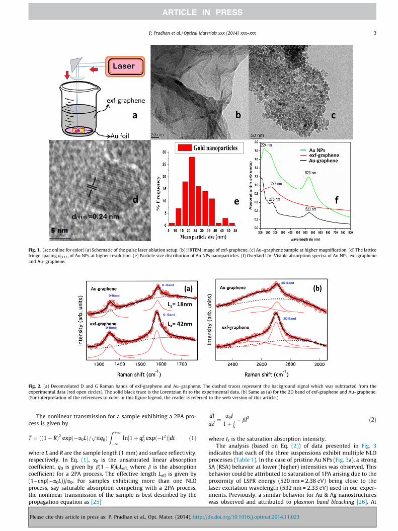
Fig. 1. (see online for color) (a) Schematic of the pulse laser ablation setup. (b) HRTEM image of exf-graphene. (c) Au–graphene sample at higher magnification. (d) The latticefringe spacing d(111) of Au NPs at higher resolution. (e) Particle size distribution of Au NPs nanoparticles. (f) Overlaid UV–Visible absorption spectra of Au NPs, exf-grapheneand Au–graphene.
Fig. 2. (a) Deconvoluted D and G Raman bands of exf-graphene and Au–graphene. The dashed traces represent the background signal which was subtracted from theexperimental data (red open circles). The solid black trace is the Lorentzian fit to the experimental data. (b) Same as (a) for the 2D band of exf-graphene and Au–graphene.(For interpretation of the references to color in this figure legend, the reader is referred to the web version of this article.)
P. Pradhan et al. / Optical Materials xxx (2014) xxx–xxx 3
The nonlinear transmission for a sample exhibiting a 2PA pro-cess is given by
T ¼ ðð1� RÞ2 expð�a0LÞ=ppq0ÞZ þ1
�1ln½1þ q2
0 expð�t2Þ�dt ð1Þ
where L and R are the sample length (1 mm) and surface reflectivity,respectively. In Eq. (1), a0 is the unsaturated linear absorptioncoefficient, q0 is given by b(1 � R)I0Leff, where b is the absorptioncoefficient for a 2PA process. The effective length Leff is given by(1�exp(�a0L))/a0. For samples exhibiting more than one NLOprocess, say saturable absorption competing with a 2PA process,the nonlinear transmission of the sample is best described by thepropagation equation as [25]
Please cite this article in press as: P. Pradhan et al., Opt. Mater. (2014), http://
dIdz0¼ a0I
1þ IIs
� bI2 ð2Þ
where Is is the saturation absorption intensity.The analysis (based on Eq. (2)) of data presented in Fig. 3
indicates that each of the three suspensions exhibit multiple NLOprocesses (Table 1). In the case of pristine Au NPs (Fig. 3a), a strongSA (RSA) behavior at lower (higher) intensities was observed. Thisbehavior could be attributed to saturation of 1PA arising due to theproximity of LSPR energy (520 nm = 2.38 eV) being close to thelaser excitation wavelength (532 nm = 2.33 eV) used in our exper-iments. Previously, a similar behavior for Au & Ag nanostructureswas observed and attributed to plasmon band bleaching [26]. At
dx.doi.org/10.1016/j.optmat.2014.11.023
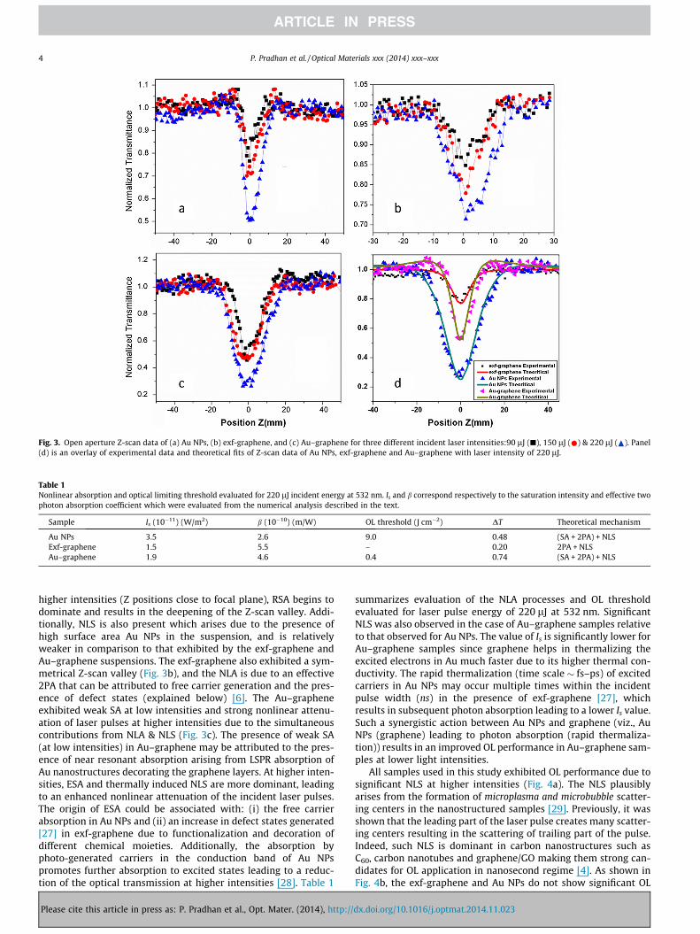
Fig. 3. Open aperture Z-scan data of (a) Au NPs, (b) exf-graphene, and (c) Au–graphene for three different incident laser intensities:90 lJ (j), 150 lJ ( ) & 220 lJ ( ). Panel(d) is an overlay of experimental data and theoretical fits of Z-scan data of Au NPs, exf-graphene and Au–graphene with laser intensity of 220 lJ.
Table 1Nonlinear absorption and optical limiting threshold evaluated for 220 lJ incident energy at 532 nm. Is and b correspond respectively to the saturation intensity and effective twophoton absorption coefficient which were evaluated from the numerical analysis described in the text.
Sample Is (10�11) (W/m2) b (10�10) (m/W) OL threshold (J cm�2) DT Theoretical mechanism
Au NPs 3.5 2.6 9.0 0.48 (SA + 2PA) + NLSExf-graphene 1.5 5.5 – 0.20 2PA + NLSAu–graphene 1.9 4.6 0.4 0.74 (SA + 2PA) + NLS
4 P. Pradhan et al. / Optical Materials xxx (2014) xxx–xxx
higher intensities (Z positions close to focal plane), RSA begins todominate and results in the deepening of the Z-scan valley. Addi-tionally, NLS is also present which arises due to the presence ofhigh surface area Au NPs in the suspension, and is relativelyweaker in comparison to that exhibited by the exf-graphene andAu–graphene suspensions. The exf-graphene also exhibited a sym-metrical Z-scan valley (Fig. 3b), and the NLA is due to an effective2PA that can be attributed to free carrier generation and the pres-ence of defect states (explained below) [6]. The Au–grapheneexhibited weak SA at low intensities and strong nonlinear attenu-ation of laser pulses at higher intensities due to the simultaneouscontributions from NLA & NLS (Fig. 3c). The presence of weak SA(at low intensities) in Au–graphene may be attributed to the pres-ence of near resonant absorption arising from LSPR absorption ofAu nanostructures decorating the graphene layers. At higher inten-sities, ESA and thermally induced NLS are more dominant, leadingto an enhanced nonlinear attenuation of the incident laser pulses.The origin of ESA could be associated with: (i) the free carrierabsorption in Au NPs and (ii) an increase in defect states generated[27] in exf-graphene due to functionalization and decoration ofdifferent chemical moieties. Additionally, the absorption byphoto-generated carriers in the conduction band of Au NPspromotes further absorption to excited states leading to a reduc-tion of the optical transmission at higher intensities [28]. Table 1
Please cite this article in press as: P. Pradhan et al., Opt. Mater. (2014), http://
summarizes evaluation of the NLA processes and OL thresholdevaluated for laser pulse energy of 220 lJ at 532 nm. SignificantNLS was also observed in the case of Au–graphene samples relativeto that observed for Au NPs. The value of Is is significantly lower forAu–graphene samples since graphene helps in thermalizing theexcited electrons in Au much faster due to its higher thermal con-ductivity. The rapid thermalization (time scale � fs–ps) of excitedcarriers in Au NPs may occur multiple times within the incidentpulse width (ns) in the presence of exf-graphene [27], whichresults in subsequent photon absorption leading to a lower Is value.Such a synergistic action between Au NPs and graphene (viz., AuNPs (graphene) leading to photon absorption (rapid thermaliza-tion)) results in an improved OL performance in Au–graphene sam-ples at lower light intensities.
All samples used in this study exhibited OL performance due tosignificant NLS at higher intensities (Fig. 4a). The NLS plausiblyarises from the formation of microplasma and microbubble scatter-ing centers in the nanostructured samples [29]. Previously, it wasshown that the leading part of the laser pulse creates many scatter-ing centers resulting in the scattering of trailing part of the pulse.Indeed, such NLS is dominant in carbon nanostructures such asC60, carbon nanotubes and graphene/GO making them strong can-didates for OL application in nanosecond regime [4]. As shown inFig. 4b, the exf-graphene and Au NPs do not show significant OL
dx.doi.org/10.1016/j.optmat.2014.11.023
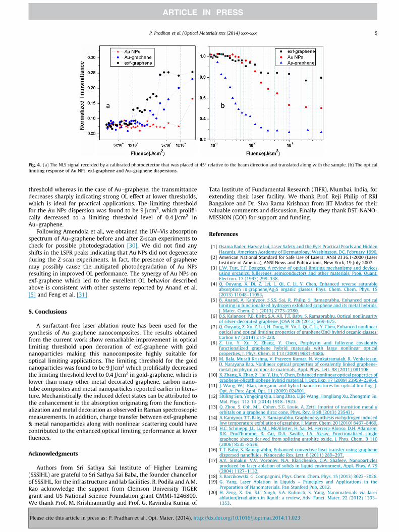
Fig. 4. (a) The NLS signal recorded by a calibrated photodetector that was placed at 45� relative to the beam direction and translated along with the sample. (b) The opticallimiting response of Au NPs, exf-graphene and Au–graphene dispersions.
P. Pradhan et al. / Optical Materials xxx (2014) xxx–xxx 5
threshold whereas in the case of Au–graphene, the transmittancedecreases sharply indicating strong OL effect at lower thresholds,which is ideal for practical applications. The limiting thresholdfor the Au NPs dispersion was found to be 9 J/cm2, which prolifi-cally decreased to a limiting threshold level of 0.4 J/cm2 inAu–graphene.
Following Amendola et al., we obtained the UV–Vis absorptionspectrum of Au–graphene before and after Z-scan experiments tocheck for possible photodegradation [30]. We did not find anyshifts in the LSPR peaks indicating that Au NPs did not degenerateduring the Z-scan experiments. In fact, the presence of graphenemay possibly cause the mitigated photodegradation of Au NPsresulting in improved OL performance. The synergy of Au NPs onexf-graphene which led to the excellent OL behavior describedabove is consistent with other systems reported by Anand et al.[5] and Feng et al. [31]
5. Conclusions
A surfactant-free laser ablation route has been used for thesynthesis of Au–graphene nanocomposites. The results obtainedfrom the current work show remarkable improvement in opticallimiting threshold upon decoration of exf-graphene with goldnanoparticles making this nanocomposite highly suitable foroptical limiting applications. The limiting threshold for the goldnanoparticles was found to be 9 J/cm2 which prolifically decreasedthe limiting threshold level to 0.4 J/cm2 in gold-graphene, which islower than many other metal decorated graphene, carbon nano-tube composites and metal nanoparticles reported earlier in litera-ture. Mechanistically, the induced defect states can be attributed tothe enhancement in the absorption originating from the function-alization and metal decoration as observed in Raman spectroscopicmeasurements. In addition, charge transfer between exf-graphene& metal nanoparticles along with nonlinear scattering could havecontributed to the enhanced optical limiting performance at lowerfluences.
Acknowledgments
Authors from Sri Sathya Sai Institute of Higher Learning(SSSIHL) are grateful to Sri Sathya Sai Baba, the founder chancellorof SSSIHL, for the infrastructure and lab facilities. R. Podila and A.M.Rao acknowledge the support from Clemson University TIGERgrant and US National Science Foundation grant CMMI-1246800.We thank Prof. M. Krishnamurthy and Prof. G. Ravindra Kumar of
Please cite this article in press as: P. Pradhan et al., Opt. Mater. (2014), http://
Tata Institute of Fundamental Research (TIFR), Mumbai, India, forextending their laser facility. We thank Prof. Reji Philip of RRIBangalore and Dr. Siva Rama Krishnan from IIT Madras for theirvaluable comments and discussion. Finally, they thank DST-NANO-MISSION (GOI) for support and funding.
References
[1] Osama Bader, Harvey Lui, Laser Safety and the Eye: Practical Pearls and HiddenHazards, American Academy of Dermatology, Washington, DC, February 1996.
[2] American National Standard for Safe Use of Lasers: ANSI Z136.1-2000 (LaserInstitute of America), ANSI News and Publications, New York, 19 July 2007.
[3] L.W. Tutt, T.F. Boggess, A review of optical limiting mechanisms and devicesusing organics, fullerenes, semiconductors and other materials, Prog. Quant.Electron. 17 (1993) 299–338.
[4] Q. Ouyang, X. Di, Z. Lei, L. Qi, C. Li, Y. Chen, Enhanced reverse saturableabsorption in graphene/Ag2S organic glasses, Phys. Chem. Chem. Phys. 15(2013) 11048–11053.
[5] B. Anand, A. Kaniyoor, S.S.S. Sai, R. Philip, S. Ramaprabhu, Enhanced opticallimiting in functionalized hydrogen exfoliated graphene and its metal hybrids,J. Mater. Chem. C 1 (2013) 2773–2780.
[6] B.S. Kalanoor, P.B. Bisht, S.A. Ali, T.T. Baby, S. Ramaprabhu, Optical nonlinearityof silver-decorated graphene, JOSA B 29 (2012) 669–675.
[7] Q. Ouyang, Z. Xu, Z. Lei, H. Dong, H. Yu, L. Qi, C. Li, Y. Chen, Enhanced nonlinearoptical and optical limiting properties of graphene/ZnO hybrid organic glasses,Carbon 67 (2014) 214–220.
[8] Z. Liu, Y. Xu, X. Zhang, Y. Chen, Porphyrin and fullerene covalentlyfunctionalized graphene hybrid materials with large nonlinear opticalproperties, J. Phys. Chem. B 113 (2009) 9681–9686.
[9] M. Bala, Murali Krishna, V. Praveen Kumar, N. Venkatramaiah, R. Venkatesan,D. Narayana Rao, Nonlinear optical properties of covalently linked graphene-metal porphyrin composite materials, Appl. Phys. Lett. 98 (2011) 081106.
[10] X. Zhang, X. Zhao, Z. Liu, Y. Liu, Y. Chen, Enhanced nonlinear optical properties ofgraphene-oligothiophene hybrid material, J. Opt. Exp. 17 (2009) 23959–23964.
[11] J. Wang, W.J. Blau, Inorganic and hybrid nanostructures for optical limiting, J.Opt. A: Pure Appl. Opt. 11 (2009) 024001.
[12] Shiling Sun, Yongqing Qiu, Liang Zhao, Lijie Wang, Hongliang Xu, Zhongmin Su,Mol. Phys. 112 14 (2014) 1918–1923.
[13] Q. Zhou, S. Coh, M.L. Cohen, S.G. Louie, A. Zettl, Imprint of transition metal dorbitals on a graphene dirac cone, Phys. Rev. B 88 (2013) 235431.
[14] A. Kaniyoor, T.T. Baby, S. Ramaprabhu, Graphene synthesis via hydrogen inducedlow temperature exfoliation of graphite, J. Mater. Chem. 20 (2010) 8467–8469.
[15] H.C. Schniepp, J.L. Li, M.J. McAllister, H. Sai, M. Herrera-Alonso, D.H. Adamson,R.K. Prud’homme, R. Car, D.A. Saville, I.A. Aksay, Functionalized singlegraphene sheets derived from splitting graphite oxide, J. Phys. Chem. B 110(2006) 8535–8539.
[16] T.T. Baby, S. Ramaprabhu, Enhanced convective heat transfer using graphenedispersed nanofluids, Nanoscale Res. Lett. 6 (2011) 289–297.
[17] A.V. Simakin, V.V. Voronov, N.A. Kkirichenko, G.A. Shafeev, Nanoparticlesproduced by laser ablation of solids in liquid environment, Appl. Phys. A 79(2004) 1127–1132.
[18] S. Barcikowski, G. Compagnini, Phys. Chem. Chem. Phys. 15 (2013) 3022–3026.[19] G. Yang, Laser Ablation in Liquids – Principles and Applications in the
Preparation of Nanomaterials, Pan Stanford Pub, 2012.[20] H. Zeng, X. Du, S.C. Singh, S.A. Kulinich, S. Yang, Nanomaterials via laser
ablation/irradiation in liquid: a review, Adv. Funct. Mater. 22 (2012) 1333–1353.
dx.doi.org/10.1016/j.optmat.2014.11.023
6 P. Pradhan et al. / Optical Materials xxx (2014) xxx–xxx
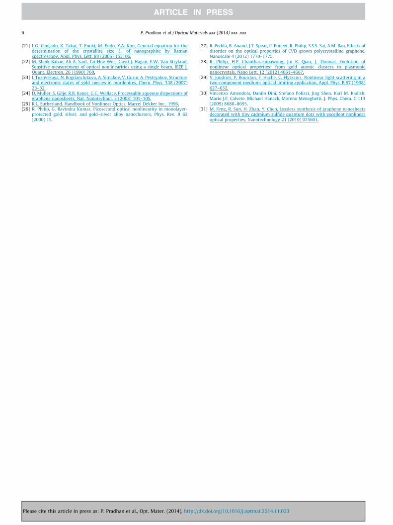
[21] L.G. Cançado, K. Takai, T. Enoki, M. Endo, Y.A. Kim, General equation for thedetermination of the crystallite size La of nanographite by Ramanspectroscopy, Appl. Phys. Lett. 88 (2006) 163106.
[22] M. Sheik-Bahae, Ali A. Said, Tai-Hue Wei, David J. Hagan, E.W. Van Stryland,Sensitive measurement of optical nonlinearities using a single beam, IEEE J.Quant. Electron. 26 (1990) 760.
[23] I. Tuzovskaya, N. Bogdanchikova, A. Simakov, V. Gurin, A. Pestryakov, Structureand electronic states of gold species in mordenites, Chem. Phys. 338 (2007)23–32.
[24] D. Muller, S. Gilje, R.B. Kaner, G.G. Wallace, Processable aqueous dispersions ofgraphene nanosheets, Nat. Nanotechnol. 3 (2008) 101–105.
[25] R.L. Sutherland, Handbook of Nonlinear Optics, Marcel Dekker Inc., 1996.[26] R. Philip, G. Ravindra Kumar, Picosecond optical nonlinearity in monolayer-
protected gold, silver, and gold–silver alloy nanoclusters, Phys. Rev. B 62(2000) 15.
Please cite this article in press as: P. Pradhan et al., Opt. Mater. (2014), http://
[27] R. Podila, B. Anand, J.T. Spear, P. Puneet, R. Philip, S.S.S. Sai, A.M. Rao, Effects ofdisorder on the optical properties of CVD grown polycrystalline graphene,Nanoscale 4 (2012) 1770–1775.
[28] R. Philip, H.P. Chantharasupawong, Jin R. Qian, J. Thomas, Evolution ofnonlinear optical properties: from gold atomic clusters to plasmonicnanocrystals, Nano Lett. 12 (2012) 4661–4667.
[29] V. Joudrier, P. Bourdon, F. Hache, C. Flytzanis, Nonlinear light scattering in atwo-component medium: optical limiting application, Appl. Phys. B 67 (1998)627–632.
[30] Vincenzo Amendola, Danilo Dini, Stefano Polizzi, Jing Shen, Karl M. Kadish,Mario J.F. Calvete, Michael Hanack, Moreno Meneghetti, J. Phys. Chem. C 113(2009) 8688–8695.
[31] M. Feng, R. Sun, H. Zhan, Y. Chen, Lossless synthesis of graphene nanosheetsdecorated with tiny cadmium sulfide quantum dots with excellent nonlinearoptical properties, Nanotechnology 21 (2010) 075601.
dx.doi.org/10.1016/j.optmat.2014.11.023
Copyright © 2022 FDOKUMEN