Effectiveness of Boundary Structures in Limiting Residential Encroachment into Urban Forests

Downloaded from www.microbiologyresearch.org by
IP: 174.129.184.250
On: Tue, 16 Aug 2016 09:33:53
Analysis of the Pseudomonas putida CA-3proteome during growth on styrene undernitrogen-limiting and non-limiting conditions
Jasmina Nikodinovic-Runic,1 Michelle Flanagan,2 Aisling R. Hume,1
Gerard Cagney2 and Kevin E. O’Connor1
Correspondence
Kevin E. O’Connor
1School of Biomolecular and Biomedical Science, Centre for Synthesis and Chemical Biology,University College Dublin, Belfield, Dublin 4, Ireland
2Conway Institute, University College Dublin, Belfield, Dublin 4, Ireland
Received 28 May 2009
Revised 8 July 2009
Accepted 9 July 2009
Pseudomonas putida CA-3 is a styrene-degrading bacterium capable of accumulating medium-
chain-length polyhydroxyalkanoate (mclPHA) when exposed to limiting concentrations of a
nitrogen source in the growth medium. Using shotgun proteomics we analysed global proteome
expression in P. putida CA-3 supplied with styrene as the sole carbon and energy source under
N-limiting (condition permissive for mclPHA synthesis) and non-limiting (condition non-permissive
for mclPHA accumulation) growth conditions in order to provide insight into the molecular
response of P. putida CA-3 to limitation of nitrogen when grown on styrene. A total of 1761
proteins were identified with high confidence and the detected proteins could be assigned to
functional groups including styrene degradation, energy, nucleotide metabolism, protein
synthesis, transport, stress response and motility. Proteins involved in the upper and lower styrene
degradation pathway were expressed throughout the 48 h growth period under both nitrogen
limitation and excess. Proteins involved in polyhydroxyalkanoate (PHA) biosynthesis, nitrogen
assimilation and amino acid transport, and outer membrane proteins were upregulated under
nitrogen limitation. PHA accumulation and biosynthesis were only expressed under nitrogen
limitation. Nitrogen assimilation proteins were detected on average at twofold higher amounts
under nitrogen limitation. Expression of the branched-chain amino acid ABC transporter was up to
16-fold higher under nitrogen-limiting conditions. Branched chain amino acid uptake by nitrogen-
limited cultures was also higher than that by non-limited cultures. Outer membrane lipoproteins
were expressed at twofold higher levels under nitrogen limitation. This was confirmed by Western
blotting (immunochemical detection) of cells grown under nitrogen limitation. Our study provides
the first global description of protein expression changes during growth of any organism on
styrene and accumulating mclPHA (nitrogen-limited growth).
INTRODUCTION
Pseudomonas putida CA-3 has been reported to utilizestyrene as a sole source of carbon and energy and toaccumulate the biodegradable polymer medium-chain-length polyhydroxyalkanoate (mclPHA) when nitrogen(N) becomes limited in the growth medium (O’Connoret al., 1996; Ward et al., 2005). These two abilities havebeen employed in the two-step chemo-biotechnological
conversion of polystyrene, a non-biodegradable polymer,to mclPHA at laboratory scale (Ward et al., 2006). Theprocess has been improved through the controlled feedingof N to the growth medium, which results in a twofoldincrease in biomass and a 1.4-fold increase in mclPHAaccumulation (Goff et al., 2007).
Molecular investigations of the styrene degradationpathway in this organism have been reported (Mooneyet al., 2006b; O’Connor et al., 2001). Styrene metabolism inP. putida CA-3 proceeds via initial side chain oxidation andinvolves an upper pathway converting styrene to phenyl-acetic acid (PA) (O’Connor et al., 1995), and a lowerpathway initiated via activation of PA to phenylacetyl-CoA.The further metabolism of phenylacetyl-CoA involvesoxidation of the aromatic nucleus, followed by ringcleavage and oxidation of the alicyclic compound,
Abbreviations: CDW, cell dry weight; mclPHA, medium-chain-lengthpolyhydroxyalkanoate; nano-LC MS/MS, nano-electrospray liquid chro-matography MS; OMP, outer membrane protein; PA, phenylacetic acid,PHA, polyhydroxyalkanoate; PHB, polyhydroxybutyrate.
Two supplementary tables, listing proteins and peptides identified in thestudy and spectral count abundance measurements, are available withthe online version of this paper.
Microbiology (2009), 155, 3348–3361 DOI 10.1099/mic.0.031153-0
3348 031153 G 2009 SGM Printed in Great Britain
Downloaded from www.microbiologyresearch.org by
IP: 174.129.184.250
On: Tue, 16 Aug 2016 09:33:53
eventually yielding TCA cycle intermediates (Martinez-Blanco et al., 1990; Mooney et al., 2006b; O’Leary et al.,2002b; Olivera et al., 1998). The pha operon in P. putidaCA-3 consists of two class II MCL-PHA synthases (phaC1and phaC2) flanking the PHA depolymerase-encodingphaZ gene (O’Leary et al., 2005).
While traditional metabolic studies have providedinformation on single or a small number of moleculartargets, high-throughput experimental technologies such asproteomics can generate comprehensive datasets thatfacilitate understanding of the wider metabolic networkof the organism (Park & Lee, 2005). Such system-levelanalyses are currently changing the strategy of engineering.Bacterial proteome profiling enables elucidation of meta-bolic networks in bacteria grown under relevant conditions(Krayl et al., 2003; VerBerkmoes et al., 2006). Weperformed this study with the view to gaining a broaderand deeper insight into the response of P. putida CA-3 to Nlimitation when grown on styrene. We compared theproteome of P. putida CA-3 grown on styrene undermclPHA accumulating and non-accumulating conditions,expecting that the broad overviews of the proteinexpression patterns will facilitate identification of proteinsand/or pathways possibly affecting styrene metabolism andPHA accumulation. A number of tests were performedwith whole cells and extracts of bacterial cells to confirmcertain observations arising from the proteomic data andto quantify those responses biochemically.
METHODS
P. putida CA-3 growth. P. putida CA-3 (NCIMB 41162) had been
isolated previously from a bioreactor containing styrene (O’Connor
et al., 1995). Cultures were grown in a 5 l stirred tank reactor
(Electrolab) with the styrene supplied continuously as a vapour, as
described previously (Goff et al., 2007). The growth medium used
was minimal mineral salts (MSM) containing, per litre: 9 g
Na2HPO4.12H2O, 1.5 g KH2PO4, 0.2 g MgSO4.7H2O, 0.002 g CaCl2and 1 ml trace element solution (Schlegel et al., 1961). The N source
was NH4Cl (Sigma-Aldrich). For mclPHA accumulation, N was
limited with a starting concentration of 65 mg l21 and a feeding rate
of 1.5 mg l21 h21 (Goff et al., 2007). To prevent mclPHA
accumulation, N was supplied at an initial concentration of 525 mg
l21 and maintained above 400 mg l21 using a N feeding rate of 45 mg
l21 h21. Fermentations were performed over a 48 h period at 30 uC,
at an agitation rate of 500 r.p.m. and a controlled pH of 6.8. Samples
(40 ml) were taken for analysis at 6, 20, 30 and 48 h (T6, T20, T30
and T48, respectively). From each sample, 10 ml was used for protein
preparation, another 10 ml was used for the determination of amino
acid uptake rate, and the remainder (20 ml) for mclPHA analysis.
Cell growth was monitored spectrophotometrically (OD540; Unicam
Helios d UV/VIS spectrophotometer) and cell dry weight (CDW) was
obtained via a calibration curve of OD540 versus dry weight, where cell
suspensions with optical densities ranging from 0.1 to 0.8 were
filtered through previously weighed glass fibre membranes
(Whatman) and dried at 90 uC until a constant weight was achieved.
Protein sample preparation. Cells from 10 ml culture aliquots
taken from the fermenter at different time points over the 48 h period
of incubation were collected by centrifugation (5000 g for 10 min at
4 uC) and washed twice with an equal volume of phosphate buffer(50 mM, pH 7). Cells were then resuspended in phosphate buffer toan equal concentration of CDW of 1.2 g l21 and centrifuged again.Cell pellets were then resuspended in 500 ml BugBuster (primaryamine-free, Novagen). Benzonase nuclease (Novagen) and PMSF(Sigma) were added to the suspension at 2 U ml21 and 0.3 mg ml21,respectively. Cell suspensions were incubated at 30 uC with gentleshaking (50 r.p.m.) and the cell debris and unbroken cells removed bycentrifugation (5000 g for 10 min at 4 uC). The protein concentrationof the supernatant was determined by the Lowry method using BSA asa standard. Protein samples (30 ml) were resuspended in double thevolume of 26 non-reduced SDS-PAGE sample buffer (80 mM Tris/HCl, pH 6.8, 10 % b-mercaptoethanol, 2 % SDS, 10 %, v/v, glycerol,0.1 % Bromophenol Blue) and heated at 100 uC for 5 min. Denaturedproteins were separated on a 10 % (w/v) polyacrylamide SDS gel andstained with Coomassie Brilliant Blue (Sigma-Aldrich).
Mass spectrometric peptide analysis. SDS-PAGE gels were cutinto bands (16 bands per time point) and the proteins digested in-gelwith trypsin according to the method of Shevchenko et al. (1996). Theresulting peptide mixtures were resuspended in 1 % formic acid andanalysed by nano-electrospray liquid chromatography MS (nano-LCMS/MS). An HPLC instrument (Dionex) was interfaced with an LTQion trap mass spectrometer (ThermoFinnigan). Chromatographybuffer solutions (Buffer A, 5 % acetonitrile and 0.1 % formic acid;Buffer B, 80 % acetonitrile and 0.1 % formic acid) were used to delivera 60 min gradient (35 min to 45 % Buffer B, 10 min to 90 %, hold10 min, 3 min to 5 %, hold for 15 min). A flow rate of 2 ml min21
was used at the electrospray source. Spectra were searched using theSequest algorithm (Craig & Beavis, 2004) against the UniProtdatabase restricted to P. putida entries (downloaded January 7,2007). Proteins with (a) a Peptide Prophet probability score greaterthan 0.99 (Keller et al., 2002), and (b) identified by a minimum oftwo different peptide spectra were automatically accepted, whilespectra for the minority of proteins identified by single spectra weremanually checked for quality. Spectral counts were automaticallycalculated for each protein across different fractions using in-housescripts.
mclPHA extraction and analysis. Cells collected at different timepoints of fermentation were harvested by centrifugation (5000 g for10 min at 4 uC) and washed twice with an equal volume of phosphatebuffer (50 mM, pH 7). Cells were centrifuged again (5000 g for10 min at 4 uC) and freeze-dried. The polymer content wasdetermined by subjecting 5–10 mg lyophilized whole cells to acidicmethanolysis according to published protocols (Brandl et al., 1988;Lageveen et al., 1988). This method degrades the intracellular PHA tomethyl esters of its constituent 3-hydroxyalkanoic acids. The 3-hydroxyalkanoic acid methyl esters were assayed by GC using aHewlett Packard HP6890 chromatograph equipped with a BP-20capillary column (30 m by 0.25 mm, 0.25 mm film thickness; J & WScientific) and a flame-ionization detector (FID). A temperatureprogramme of 60 uC for 3 min, temperature ramp of 5 uC per min,200 uC for 1 min was used. For the peak identification, methyl estersof 3-hydroxyalkanoic acid were prepared in a similar manner andPHA standards from P. putida CA-3 were used (Ward et al., 2005).
Nitrogen assay. The nitrogen concentration in the growth mediumwas determined by the indophenol method, as described by Scheiner(1976).
Phenylacyl-CoA ligase enzyme assay. The phenylacetyl-CoAligase activity was determined by measuring the rate of formationof phenylacetylhydroxamate in the presence of cell lysates (200 ml;prepared as described in the protein sample preparation section),ATP, CoA, PA and neutral hydroxylamine, as previously described(Martinez-Blanco et al., 1990; Ward & O’Connor, 2005). Phenylacyl-
P. putida CA-3 proteome when grown on styrene
http://mic.sgmjournals.org 3349
Downloaded from www.microbiologyresearch.org by
IP: 174.129.184.250
On: Tue, 16 Aug 2016 09:33:53
CoA ligase activities were measured at 30 uC over a 30 min period.
The molar extinction coefficient of phenylacetylhydroxamate in the
presence of ferric chloride under these conditions is 0.9 mM cm21
(Martinez-Blanco et al., 1990; Ward & O’Connor, 2005).
Branched chain amino acid uptake. For the measurement of
leucine and valine uptake rates, 10 ml samples of cells taken from the
fermenter at various time points were harvested by centrifugation
(5000 g for 10 min at 4 uC), washed and resuspended in deionized
H2O to OD540 4. Cell suspensions (2 ml) were then supplemented
with 1 mM leucine and/or 1 mM valine and incubated at 30 uC with
shaking (200 r.p.m.). Samples (50 ml) were removed every 30 min
over a 2 h period. The amino acid detection was based on
derivatization with isobutyl chloroformate followed by GC–MS, as
described elsewhere (Sobolevsky et al., 2004). Briefly, reaction
mixture (50 ml) was mixed well with deionized H2O (50 ml),
isobutanol (30 ml), pyridine (10 ml) and isobutyl chloroformate
(30 ml). The derivatives were extracted into chloroform (500 ml) by
vigorous shaking and subsequent centrifugation (10 000 g for 1 min).
A 1 ml volume of the chloroform layer was injected in the splitless
mode onto an HP-5 column (20 m by 0.25 mm, 0.33 mm filmthickness; J & W Scientific) using an Agilent 6890 gas chromatograph
coupled to a 5973 mass-selective detector. The column was held at
50 uC for 2 min then heated to 300 uC at a rate of 10 uC min21.
Under the above conditions, N-isobutoxycarbonyl isobutyl esters of
leucine and valine had retention times of 14.37 and 15.12 min,
respectively.
Immunoblotting of outer membrane protein (OMP). Crude cell
lysates, prepared as described in Methods, Protein sample prepara-
tion, and used for the mass peptide analysis were separated by SDS-
PAGE (10 %, w/v) and transferred to a nitrocellulose membrane
(Hybond ECL, Amersham Biosciences). For Western blotting, the
membrane was probed with mAb 7.3 [mouse antibodies against P.
putida KT2440 outer membrane lipoprotein as described by Ramos-Gonzalez et al. (1992)]. Peroxidase-conjugated goat anti-mouse
immunoglobulins (Merck) were used for the chemiluminescent
detection. The blots were developed using Immobilon Western
chemiluminescent HRP substrate (Millipore) according to the
manufacturer’s protocol. Immunoluminescence was detected and
evaluated using the chemiluminescence-compatible FluorChem
imaging system equipped with AlphaEase FC2 software (Alpha
Innotech).
RESULTS AND DISCUSSION
In order to gain an insight into the effect of N limitation onthe general cellular response, mclPHA accumulation andstyrene metabolism, P. putida CA-3 was grown in abioreactor with styrene as the sole source of carbon andenergy under two different growth conditions (limited Nand non-limited N). The proteomes were profiled overtime (T6, T20, T30 and T48) using nano-LC MS/MS(Washburn et al., 2003). This shotgun technique isdesigned to sample the proteome at a broad level, anduses spectral counts (the frequency with which eachprotein is detected in the mass spectrometer) as asemiquantitative measure of differential protein expressionlevels (Cagney et al., 2005). The high-throughput proteinannotation and metabolic pathway prediction done in thisstudy were enabled by the availability of the genomesequence of P. putida KT2440 (Nelson et al., 2002). Themajor observations arising from the proteomic analysis
were then confirmed by assay and analysis of whole cells,cell extracts and cytoplasmic contents.
Growth characteristics of P. putida CA-3
P. putida CA-3 was grown in the stirred tank bioreactor aspreviously described (Goff et al., 2007) using styrene as thesole carbon and energy source over 48 h, during whichtime biomass, N utilization and PHA production weremonitored (Fig. 1). After a 48 h fermentation, duringwhich the nitrogen (supplied as NH4Cl) concentration waskept above 400 mg l21, a final biomass of 7.2 g l21 (total35.8 g from 5 l culture) was obtained (Fig. 1). No PHA wasaccumulated under these growth conditions. When the Nsupply was limited (65 mg N l21 at the start of fermenta-tion), a final CDW of 6.5 g l21 was achieved (total 30.3 gCDW). However, this weight consisted of 52 % PHA(w/w), thus total biomass was 15 g (a total of 10.2 g ofmclPHA was obtained from 5 l culture). Thus, Nlimitation resulted in a 2.4-fold decrease in biomass yield.During the exponential phase (T6–T25), the growth ratesfor non-limiting and limiting N growth conditions were0.37 and 0.11 g CDW l21 h21, respectively. Therefore, weexpected that the different growth rates and concentrationsof N source during fermentation would most likely affectthe protein expression pattern (Ferenci, 1999).
Shotgun proteomics of P. putida CA-3 grown in abioreactor on styrene under N limitation(permissive for PHA accumulation) and under Nexcess (not permissive for PHA accumulation)
P. putida CA-3 cultures were grown as described above andharvested at four time points: 6, 20, 30 and 48 h post-inoculation (Fig. 1). In order to produce an extensivedataset of protein expression, the lysate was fractionated bySDS-PAGE and analysed by LC-MS. Over 75 000 peptidetandem mass spectra were used to identify 9033 uniquepeptides, in turn generating 1761 high-confidence (¢0.99Protein Prophet score) protein identifications (Supple-mentary Table S1). This represents a substantial proportion(33 %) of the predicted P. putida proteome, and reflects theextensive MS analysis at different time points underdifferent growth conditions. Moreover, the proteinsexhibited a wide range of annotated biophysical (molecularmass, isoelectric point), biochemical (functional annota-tions) and structural (domains) properties, suggesting thatthe analysis was not strongly biased in favour of or againstany protein class.
Spectral counts were used as a semiquantitative measure ofprotein abundance (Cagney et al., 2005; Liu et al., 2004;Washburn et al., 2003). Using this analysis, it was possibleto follow the expression of individual proteins in thedifferent samples (Supplementary Table S2, Fig. 2).Comparison of our results with a preliminary experimentusing P. putida CA-3 cultures suggested that the percentageof proteins detected in two MS analyses of independently
J. Nikodinovic-Runic and others
3350 Microbiology 155

Downloaded from www.microbiologyresearch.org by
IP: 174.129.184.250
On: Tue, 16 Aug 2016 09:33:53
grown cultures is 70–80 % (data not shown). Analysis of 20ribosomal proteins, whose expression might be assumed toremain relatively independent of the time points andexperimental treatment, showed that all 20 were detected inall fractions (% CV for spectra counts50.34). Identifiedproteins were next organized into functional pathwaygroups (e.g. ‘nitrogen assimilation’, ‘amino acid uptake’)using annotations from the Gene Ontology database(Raghava, 2006). High-throughput protein annotationand metabolic pathway prediction were enabled by theavailability of the genome sequence of P. putida KT2440(Nelson et al., 2002). The spectral counts, reflecting therelative abundance of all the proteins engaged in therespective pathways, were then totalled. In this way,potentially up- or downregulated pathways could beobserved (Table 1). Changes in the relative abundance ofindividual proteins or whole pathways are discussed below.
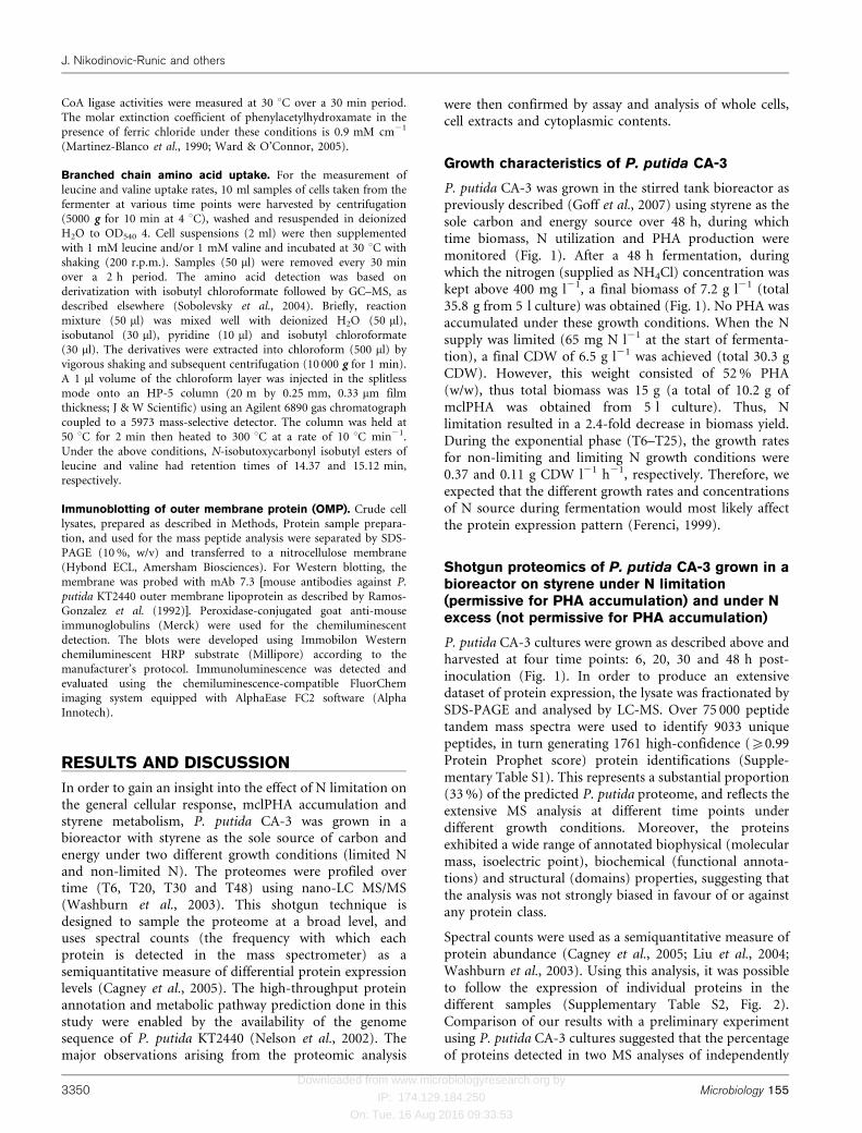
Styrene degradation proteins
P. putida CA-3 is a styrene-degrading bacterium thatmetabolizes styrene aerobically, employing enzymes orga-nized in two distinct catabolic pathways (O’Leary et al.,2002b). An upper pathway involves oxidation of thestyrene side chain, leading to epoxystyrene, which issubsequently isomerized to phenylacetaldehyde and thenoxidized to PA. The lower pathway involves the conversion
of the PA through CoA intermediates into aliphaticcompounds that can enter the Krebs cycle (Fig. 2a).
The genes encoding the upper pathway are organized onthe styABCD operon, which is regulated at the transcrip-tional level by the StySR two-component sensory apparatusin this strain (O’Leary et al., 2001) and other species suchas Pseudomonas fluorescens ST (Beltrametti et al., 1997) andPseudomonas sp. Y2 (Bartolome-Martin et al., 2004;Velasco et al., 1998). The genes encoding the lowerpathway (PACoA catabolon) are typically organized infive contiguous operons: paaABCEF, paaGHIJK, paaLMN,paaX and paaY (Luengo et al., 2001). Proteins encoded bythese genes can be classified into functional units: atransport system (PaaL and PaaM); a phenylacetyl-CoA-activating enzyme (PaaF); a ring-hydroxylating enzymiccomplex (PaaGHIJK); a ring-fission protein (PaaN); a b-oxidation system (PaaABCE); and two regulatory proteins,a repressor (PaaX) and a putative regulator (PaaY) (Ariaset al., 2008; Olivera et al., 1998).
While the typical PACoA catabolon has been suggested tobe present in P. putida CA-3 (O’Leary et al., 2005), this isthe first time, to our knowledge, that all of the enzymeswere detected and their relative amounts observed overtime when this strain was grown on styrene (Fig. 2b). All ofthe enzymes of the upper and lower pathways could bedetected throughout the 48 h fermentation under both
Fig. 1. Growth (biomass, g l”1) of P. putida CA-3 in minimal salts medium supplied with styrene in the bioreactor under N-limiting (65 mg N l”1 at T0, &) and non-limiting growth conditions (.500 mg N l”1 at T0, h). N concentration (mg l”1) wastracked over time under N-limiting (m) and non-limiting growth conditions (g). PHA (as a percentage of CDW) wasaccumulated under N-limiting conditions only (grey bars). Samples for proteome analysis were taken at 4, 20, 30 and 48 hpost-inoculation, as indicated by arrows.
P. putida CA-3 proteome when grown on styrene
http://mic.sgmjournals.org 3351
Downloaded from www.microbiologyresearch.org by
IP: 174.129.184.250
On: Tue, 16 Aug 2016 09:33:53
N-limiting and non-limiting conditions. However, therelative amounts of the styrene degradation enzymesranged from 18–22 % under N excess to 11–18 % underN-limited conditions with respect to the total proteome.StyA and StyD were the most abundant proteins expressed
over time in the upper pathway. StyA was expressed tohigher levels at time 6 h under N limitation but to lowerlevels after this time point (Fig. 2). This observation is inagreement with our previous finding that the catabolicefficiency of styrene degradation dramatically decreases
Fig. 2. Styrene catabolic pathway of P. putida CA-3. (a) Styrene upper and lower catabolic pathways, adapted from Nogales et
al. (2007) and O’Leary et al. (2002b). (b) Proteins and their relative abundance in cells grown on styrene under no nutrientlimitation (white bars) and under N limitation (grey bars) in the bioreactor. Enzymes and proteins are: StyAB, styrenemonooxygenase; StyC, epoxystyrene isomerase; StyD, phenylacetaldehyde dehydrogenase; PaaF, phenylacetyl-CoA ligase;PaaGHIJK, multicomponent phenylacetyl-CoA oxygenase; PaaN and PaaABCDE, enzymes involved in ring-cleavage- and b-oxidation-like reactions of the aliphatic-CoA intermediates. The consensus nomenclature for the paa genes proposed byLuengo et al. (2001) is used.
J. Nikodinovic-Runic and others
3352 Microbiology 155
Downloaded from www.microbiologyresearch.org by
IP: 174.129.184.250
On: Tue, 16 Aug 2016 09:33:53
under PHA-accumulating (N-limiting) conditions (Wardet al., 2005).
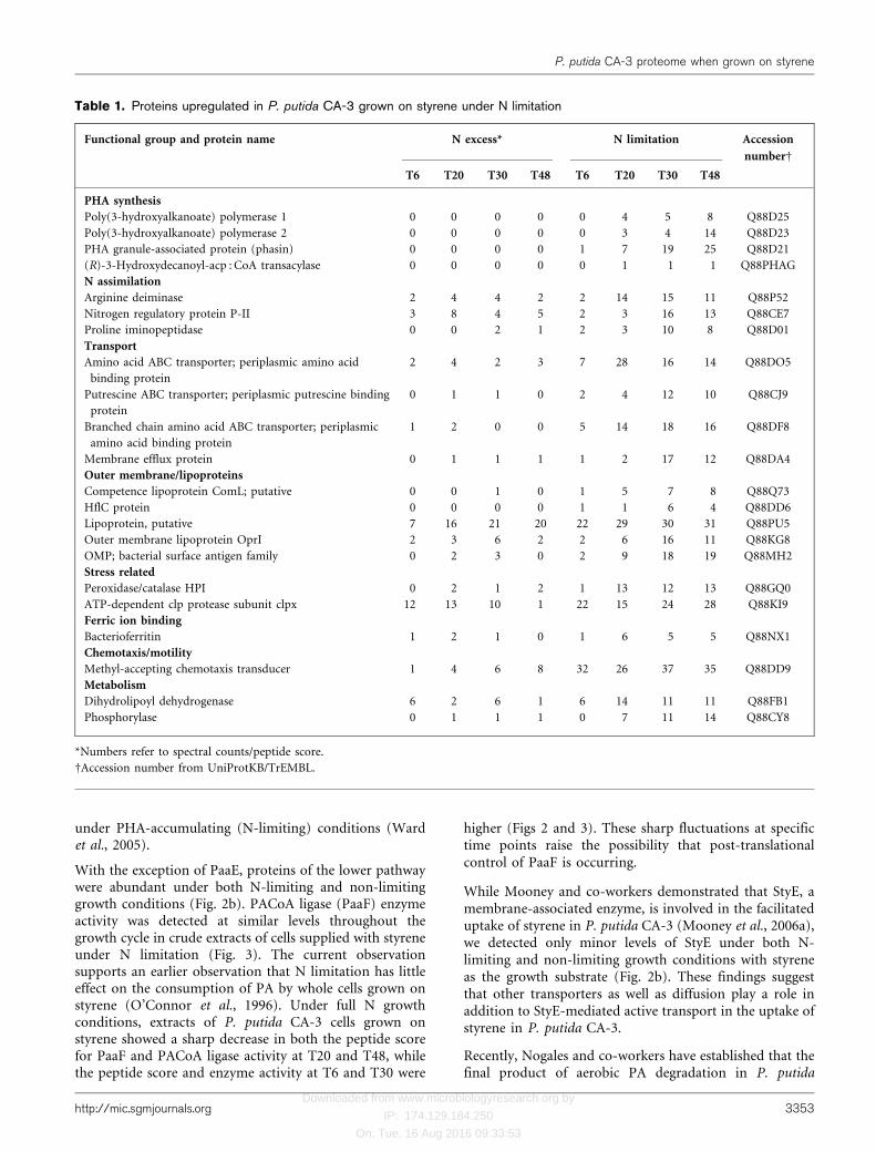
With the exception of PaaE, proteins of the lower pathwaywere abundant under both N-limiting and non-limitinggrowth conditions (Fig. 2b). PACoA ligase (PaaF) enzymeactivity was detected at similar levels throughout thegrowth cycle in crude extracts of cells supplied with styreneunder N limitation (Fig. 3). The current observationsupports an earlier observation that N limitation has littleeffect on the consumption of PA by whole cells grown onstyrene (O’Connor et al., 1996). Under full N growthconditions, extracts of P. putida CA-3 cells grown onstyrene showed a sharp decrease in both the peptide scorefor PaaF and PACoA ligase activity at T20 and T48, whilethe peptide score and enzyme activity at T6 and T30 were
higher (Figs 2 and 3). These sharp fluctuations at specifictime points raise the possibility that post-translationalcontrol of PaaF is occurring.
While Mooney and co-workers demonstrated that StyE, amembrane-associated enzyme, is involved in the facilitateduptake of styrene in P. putida CA-3 (Mooney et al., 2006a),we detected only minor levels of StyE under both N-limiting and non-limiting growth conditions with styreneas the growth substrate (Fig. 2b). These findings suggestthat other transporters as well as diffusion play a role inaddition to StyE-mediated active transport in the uptake ofstyrene in P. putida CA-3.
Recently, Nogales and co-workers have established that thefinal product of aerobic PA degradation in P. putida
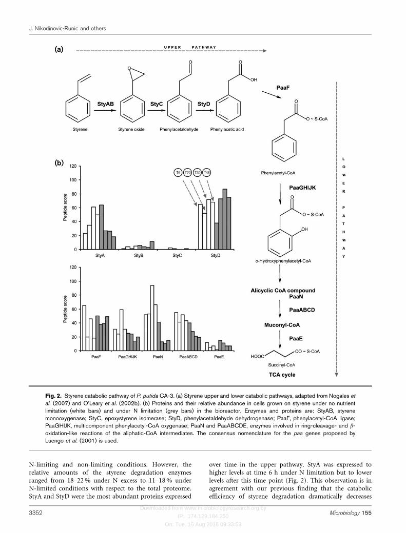
Table 1. Proteins upregulated in P. putida CA-3 grown on styrene under N limitation
Functional group and protein name N excess* N limitation Accession
numberD
T6 T20 T30 T48 T6 T20 T30 T48
PHA synthesis
Poly(3-hydroxyalkanoate) polymerase 1 0 0 0 0 0 4 5 8 Q88D25
Poly(3-hydroxyalkanoate) polymerase 2 0 0 0 0 0 3 4 14 Q88D23
PHA granule-associated protein (phasin) 0 0 0 0 1 7 19 25 Q88D21
(R)-3-Hydroxydecanoyl-acp : CoA transacylase 0 0 0 0 0 1 1 1 Q88PHAG
N assimilation
Arginine deiminase 2 4 4 2 2 14 15 11 Q88P52
Nitrogen regulatory protein P-II 3 8 4 5 2 3 16 13 Q88CE7
Proline iminopeptidase 0 0 2 1 2 3 10 8 Q88D01
Transport
Amino acid ABC transporter; periplasmic amino acid
binding protein
2 4 2 3 7 28 16 14 Q88DO5
Putrescine ABC transporter; periplasmic putrescine binding
protein
0 1 1 0 2 4 12 10 Q88CJ9
Branched chain amino acid ABC transporter; periplasmic
amino acid binding protein
1 2 0 0 5 14 18 16 Q88DF8
Membrane efflux protein 0 1 1 1 1 2 17 12 Q88DA4
Outer membrane/lipoproteins
Competence lipoprotein ComL; putative 0 0 1 0 1 5 7 8 Q88Q73
HflC protein 0 0 0 0 1 1 6 4 Q88DD6
Lipoprotein, putative 7 16 21 20 22 29 30 31 Q88PU5
Outer membrane lipoprotein OprI 2 3 6 2 2 6 16 11 Q88KG8
OMP; bacterial surface antigen family 0 2 3 0 2 9 18 19 Q88MH2
Stress related
Peroxidase/catalase HPI 0 2 1 2 1 13 12 13 Q88GQ0
ATP-dependent clp protease subunit clpx 12 13 10 1 22 15 24 28 Q88KI9
Ferric ion binding
Bacterioferritin 1 2 1 0 1 6 5 5 Q88NX1
Chemotaxis/motility
Methyl-accepting chemotaxis transducer 1 4 6 8 32 26 37 35 Q88DD9
Metabolism
Dihydrolipoyl dehydrogenase 6 2 6 1 6 14 11 11 Q88FB1
Phosphorylase 0 1 1 1 0 7 11 14 Q88CY8
*Numbers refer to spectral counts/peptide score.
DAccession number from UniProtKB/TrEMBL.
P. putida CA-3 proteome when grown on styrene
http://mic.sgmjournals.org 3353
Downloaded from www.microbiologyresearch.org by
IP: 174.129.184.250
On: Tue, 16 Aug 2016 09:33:53
KT2440 is succinyl-CoA (Luengo et al., 2004; Nogales et al.,2007). They also demonstrated, through generation ofrandom insertion mutants and biochemical analysis, thatthis last step of PA degradation is catalysed by b-ketoadypil-CoA thiolase (PaaE). PaaE was also detectedin P. putida CA-3 when grown on styrene under both N-limiting and non-limiting growth conditions (Fig. 2b).
The regulatory proteins StyS, StyR, PaaX and PaaY weredetected at low levels, while PA specific permease (PaaL)was not observed in the proteome of P. putida CA-3 grownon styrene. O’Leary and co-workers observed that thetranscription of the styS gene (upper pathway regulatorysensor kinase) was growth condition-dependent (O’Learyet al., 2002a). Indeed, we observed higher amounts of StySpresent under N-limiting conditions (Supplementary TableS1). Del Peso-Santos and co-workers have demonstrated arole for PaaX as a major regulatory protein in thephenylacetyl-CoA catabolon (del Peso-Santos et al.,2006). They recently revealed that phenylacetyl-CoA bindsto PaaX, and inactivates PaaX-mediated repression of boththe paa genes and the styABCD operon in P. putida Y2 (delPeso-Santos et al., 2008). In agreement with this, PaaX wasthe most abundant of the regulators detected at T6 afterinoculation under non-limiting N conditions (data notshown).
Proteins upregulated under N limitation
The expression of proteins involved in energy/metabolism,amino acid biosynthesis, DNA/RNA biosynthesis andprocessing, transcription and translation was quite similarunder both N excess and N limited conditions(Supplementary Table S1) in P. putida CA-3 over time.Most of the N (NH4Cl) supplied was consumed within20 h of inoculation under N limitation. Under N limitation(2 mg N l21) the biomass (CDW) yield was 2.4 g l21 lowerthan under N excess conditions (Fig. 1). The lack of any
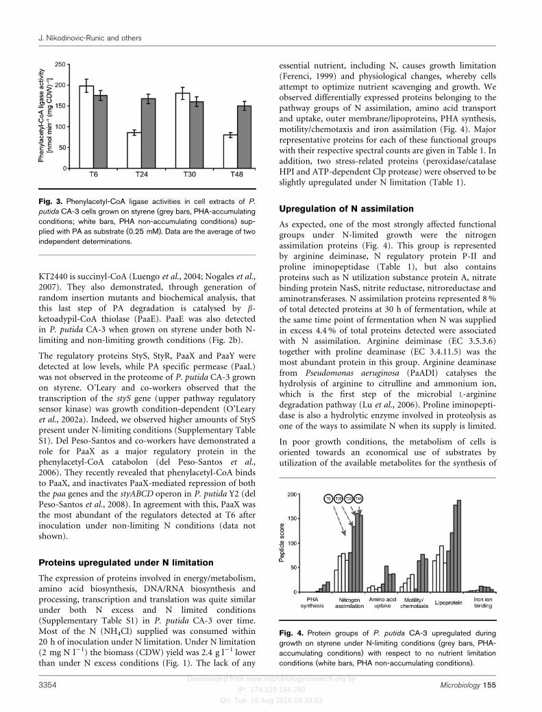
essential nutrient, including N, causes growth limitation(Ferenci, 1999) and physiological changes, whereby cellsattempt to optimize nutrient scavenging and growth. Weobserved differentially expressed proteins belonging to thepathway groups of N assimilation, amino acid transportand uptake, outer membrane/lipoproteins, PHA synthesis,motility/chemotaxis and iron assimilation (Fig. 4). Majorrepresentative proteins for each of these functional groupswith their respective spectral counts are given in Table 1. Inaddition, two stress-related proteins (peroxidase/catalaseHPI and ATP-dependent Clp protease) were observed to beslightly upregulated under N limitation (Table 1).
Upregulation of N assimilation
As expected, one of the most strongly affected functionalgroups under N-limited growth were the nitrogenassimilation proteins (Fig. 4). This group is representedby arginine deiminase, N regulatory protein P-II andproline iminopeptidase (Table 1), but also containsproteins such as N utilization substance protein A, nitratebinding protein NasS, nitrite reductase, nitroreductase andaminotransferases. N assimilation proteins represented 8 %of total detected proteins at 30 h of fermentation, while atthe same time point of fermentation when N was suppliedin excess 4.4 % of total proteins detected were associatedwith N assimilation. Arginine deiminase (EC 3.5.3.6)together with proline deaminase (EC 3.4.11.5) was themost abundant protein in this group. Arginine deaminasefrom Pseudomonas aeruginosa (PaADI) catalyses thehydrolysis of arginine to citrulline and ammonium ion,which is the first step of the microbial L-argininedegradation pathway (Lu et al., 2006). Proline iminopepti-dase is also a hydrolytic enzyme involved in proteolysis asone of the ways to assimilate N when its supply is limited.
In poor growth conditions, the metabolism of cells isoriented towards an economical use of substrates byutilization of the available metabolites for the synthesis of
Fig. 3. Phenylacetyl-CoA ligase activities in cell extracts of P.
putida CA-3 cells grown on styrene (grey bars, PHA-accumulatingconditions; white bars, PHA non-accumulating conditions) sup-plied with PA as substrate (0.25 mM). Data are the average of twoindependent determinations.
Fig. 4. Protein groups of P. putida CA-3 upregulated duringgrowth on styrene under N-limiting conditions (grey bars, PHA-accumulating conditions) with respect to no nutrient limitationconditions (white bars, PHA non-accumulating conditions).
J. Nikodinovic-Runic and others
3354 Microbiology 155
Downloaded from www.microbiologyresearch.org by
IP: 174.129.184.250
On: Tue, 16 Aug 2016 09:33:53
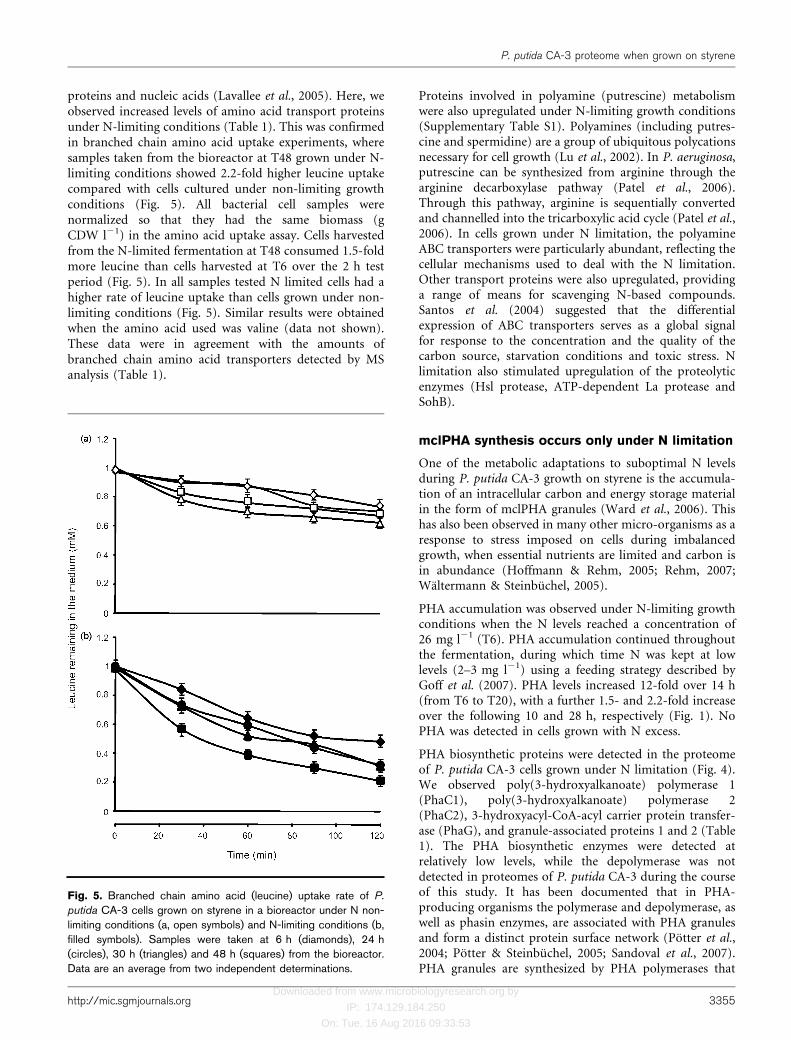
proteins and nucleic acids (Lavallee et al., 2005). Here, weobserved increased levels of amino acid transport proteinsunder N-limiting conditions (Table 1). This was confirmedin branched chain amino acid uptake experiments, wheresamples taken from the bioreactor at T48 grown under N-limiting conditions showed 2.2-fold higher leucine uptakecompared with cells cultured under non-limiting growthconditions (Fig. 5). All bacterial cell samples werenormalized so that they had the same biomass (gCDW l21) in the amino acid uptake assay. Cells harvestedfrom the N-limited fermentation at T48 consumed 1.5-foldmore leucine than cells harvested at T6 over the 2 h testperiod (Fig. 5). In all samples tested N limited cells had ahigher rate of leucine uptake than cells grown under non-limiting conditions (Fig. 5). Similar results were obtainedwhen the amino acid used was valine (data not shown).These data were in agreement with the amounts ofbranched chain amino acid transporters detected by MSanalysis (Table 1).
Proteins involved in polyamine (putrescine) metabolismwere also upregulated under N-limiting growth conditions(Supplementary Table S1). Polyamines (including putres-cine and spermidine) are a group of ubiquitous polycationsnecessary for cell growth (Lu et al., 2002). In P. aeruginosa,putrescine can be synthesized from arginine through thearginine decarboxylase pathway (Patel et al., 2006).Through this pathway, arginine is sequentially convertedand channelled into the tricarboxylic acid cycle (Patel et al.,2006). In cells grown under N limitation, the polyamineABC transporters were particularly abundant, reflecting thecellular mechanisms used to deal with the N limitation.Other transport proteins were also upregulated, providinga range of means for scavenging N-based compounds.Santos et al. (2004) suggested that the differentialexpression of ABC transporters serves as a global signalfor response to the concentration and the quality of thecarbon source, starvation conditions and toxic stress. Nlimitation also stimulated upregulation of the proteolyticenzymes (Hsl protease, ATP-dependent La protease andSohB).
mclPHA synthesis occurs only under N limitation
One of the metabolic adaptations to suboptimal N levelsduring P. putida CA-3 growth on styrene is the accumula-tion of an intracellular carbon and energy storage materialin the form of mclPHA granules (Ward et al., 2006). Thishas also been observed in many other micro-organisms as aresponse to stress imposed on cells during imbalancedgrowth, when essential nutrients are limited and carbon isin abundance (Hoffmann & Rehm, 2005; Rehm, 2007;Waltermann & Steinbuchel, 2005).
PHA accumulation was observed under N-limiting growthconditions when the N levels reached a concentration of26 mg l21 (T6). PHA accumulation continued throughoutthe fermentation, during which time N was kept at lowlevels (2–3 mg l21) using a feeding strategy described byGoff et al. (2007). PHA levels increased 12-fold over 14 h(from T6 to T20), with a further 1.5- and 2.2-fold increaseover the following 10 and 28 h, respectively (Fig. 1). NoPHA was detected in cells grown with N excess.
PHA biosynthetic proteins were detected in the proteomeof P. putida CA-3 cells grown under N limitation (Fig. 4).We observed poly(3-hydroxyalkanoate) polymerase 1(PhaC1), poly(3-hydroxyalkanoate) polymerase 2(PhaC2), 3-hydroxyacyl-CoA-acyl carrier protein transfer-ase (PhaG), and granule-associated proteins 1 and 2 (Table1). The PHA biosynthetic enzymes were detected atrelatively low levels, while the depolymerase was notdetected in proteomes of P. putida CA-3 during the courseof this study. It has been documented that in PHA-producing organisms the polymerase and depolymerase, aswell as phasin enzymes, are associated with PHA granulesand form a distinct protein surface network (Potter et al.,2004; Potter & Steinbuchel, 2005; Sandoval et al., 2007).PHA granules are synthesized by PHA polymerases that
Fig. 5. Branched chain amino acid (leucine) uptake rate of P.
putida CA-3 cells grown on styrene in a bioreactor under N non-limiting conditions (a, open symbols) and N-limiting conditions (b,filled symbols). Samples were taken at 6 h (diamonds), 24 h(circles), 30 h (triangles) and 48 h (squares) from the bioreactor.Data are an average from two independent determinations.
P. putida CA-3 proteome when grown on styrene
http://mic.sgmjournals.org 3355
Downloaded from www.microbiologyresearch.org by
IP: 174.129.184.250
On: Tue, 16 Aug 2016 09:33:53
utilize CoA thioesters of the respective 3-hydroxy fattyacids as substrates. Granules are mobilized (degraded) byPHA depolymerases, and formation of the granulesdepends on the activity of several granule-associatedproteins (phasins).
Lipoproteins, chemotaxis-related proteins andiron ion transport proteins are upregulated underN limitation
In cells accumulating PHA (grown under N-limitingconditions) we observed upregulation of other OMPs,such as lipoproteins and lipoprotein-releasing proteins (Lolproteins), that target and anchor lipoproteins to theperiplasmic surface of either the inner or the outermembrane (Miyamoto & Tokuda, 2007; Narita &Tokuda, 2006). It has been shown that nutrient limitationinduces the expression of proteins in the outer membraneof bacterial cells such as Pseudomonas species undercarbon, N and phosphorus limitation (Kragelund &Nybroe, 1994). Lipoproteins have been shown to haveemulsifying properties that increase surface area and henceenhance the bioavailability of hydrophobic substrates suchas styrene (Kuiper et al., 2004; Whang et al., 2008). Theyare also known to play a role in the bacterial adaptation tochanges in environmental conditions such as osmotic stress(Guyard-Nicodeme et al., 2008). Among OMPs, OmpA, G,H, E3, F and I were detected in the current study, but onlyOmpF and OmpA porins were twofold to fourfoldupregulated by N limitation (Table 1, SupplementaryTable S1). OmpF is a major membrane protein in P.aeruginosa, where it has a non-specific porin function(Hancock & Brinkman, 2002). In fact, Li et al. (1995) havedemonstrated that an ompF2 mutant of P. aeruginosa wastoluene-tolerant, and therefore it was proposed thattoluene enters the cell through the OmpF channel inwild-type cells. In contrast, Volkers and co-workers foundthat OmpF is downregulated in the presence of tolueneexcess in a chemostat-based culture of P. putida S12 andproposed that this strain is capable of shutting down thischannel upon toluene stress (Volkers et al., 2006). In thesame study, OmpH was dramatically upregulated (12-fold), while we observed more moderate (twofold)upregulation of this OMP. The higher levels of OmpAobserved in this study are in agreement with the findings ofBenndorf and co-workers, who identified OmpA as beingupregulated in P. putida KT2440 in response to lipophilicherbicides (Benndorf et al., 2006).
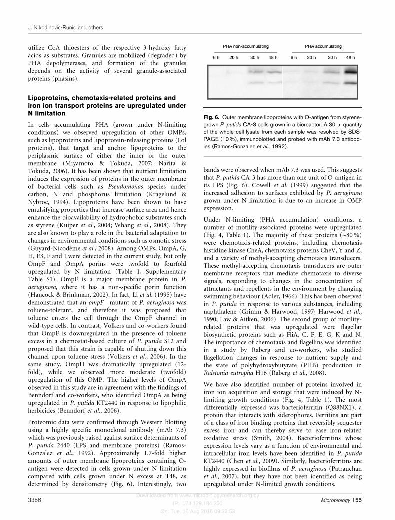
Proteomic data were confirmed through Western blottingusing a highly specific monoclonal antibody (mAb 7.3)which was previously raised against surface determinants ofP. putida 2440 (LPS and membrane proteins) (Ramos-Gonzalez et al., 1992). Approximately 1.7-fold higheramounts of outer membrane lipoproteins containing O-antigen were detected in cells grown under N limitationcompared with cells grown under N excess at T48, asdetermined by densitometry (Fig. 6). Interestingly, two
bands were observed when mAb 7.3 was used. This suggeststhat P. putida CA-3 has more than one unit of O-antigen inits LPS (Fig. 6). Cowell et al. (1999) suggested that theincreased adhesion to surfaces exhibited by P. aeruginosagrown under N limitation is due to an increase in OMPexpression.
Under N-limiting (PHA accumulation) conditions, anumber of motility-associated proteins were upregulated(Fig. 4, Table 1). The majority of these proteins (~80 %)were chemotaxis-related proteins, including chemotaxishistidine kinase CheA, chemotaxis proteins CheV, Y and Z,and a variety of methyl-accepting chemotaxis transducers.These methyl-accepting chemotaxis transducers are outermembrane receptors that mediate chemotaxis to diversesignals, responding to changes in the concentration ofattractants and repellents in the environment by changingswimming behaviour (Adler, 1966). This has been observedin P. putida in response to various substances, includingnaphthalene (Grimm & Harwood, 1997; Harwood et al.,1990; Law & Aitken, 2006). The second group of motility-related proteins that was upregulated were flagellarbiosynthetic proteins such as FliA, C, F, E, G, K and N.The importance of chemotaxis and flagellins was identifiedin a study by Raberg and co-workers, who studiedflagellation changes in response to nutrient supply andthe state of polyhydroxybutyrate (PHB) production inRalstonia eutropha H16 (Raberg et al., 2008).
We have also identified number of proteins involved iniron ion acquisition and storage that were induced by N-limiting growth conditions (Fig. 4, Table 1). The mostdifferentially expressed was bacterioferritin (Q88NX1), aprotein that interacts with siderophores. Ferritins are partof a class of iron binding proteins that reversibly sequesterexcess iron and can thereby serve to ease iron-relatedoxidative stress (Smith, 2004). Bacterioferritins whoseexpression levels vary as a function of environmental andintracellular iron levels have been identified in P. putidaKT2440 (Chen et al., 2009). Similarly, bacterioferritins arehighly expressed in biofilms of P. aeruginosa (Patrauchanet al., 2007), but they have not been identified as beingupregulated under N-limited growth conditions.
Fig. 6. Outer membrane lipoproteins with O-antigen from styrene-grown P. putida CA-3 cells grown in a bioreactor. A 30 ml quantityof the whole-cell lysate from each sample was resolved by SDS-PAGE (10 %), immunoblotted and probed with mAb 7.3 antibod-ies (Ramos-Gonzalez et al., 1992).
J. Nikodinovic-Runic and others
3356 Microbiology 155
Downloaded from www.microbiologyresearch.org by
IP: 174.129.184.250
On: Tue, 16 Aug 2016 09:33:53
Solvent tolerance response by P. putida CA-3 tothe presence of styrene
When grown on styrene, P. putida CA-3 cells face aparadox. On the one hand, styrene represents a carbonsource that can be metabolized to yield carbon and energyfor growth. On the other hand, with an octanol/waterpartition coefficient [log(POW)] of 3, styrene falls into acategory of organic solvents extremely toxic to micro-organisms, due to their ability to bind to the cells,disturbing membranes by removing lipids and proteinsand causing cell lysis (Ramos et al., 2001; Taylor et al.,2008). It is well documented that solvent-tolerant pseudo-monads possess three membrane-associated solvent tol-erance mechanisms: (A) active efflux of organic solvents,(B) outer membrane changes and (C) cytoplasmicmembrane changes (Volkers et al., 2006).
(A) Active efflux of organic solvents. Several laboratorieshave identified efflux pumps belonging to the resistance–nodulation–cell division (RND) family as being the mostimportant mechanism of solvent tolerance (Kieboom et al.,1998; Ramos et al., 2001). They are energy-dependentactive efflux pumps, which export toxic organic solvents tothe external medium. This mechanism has also beenidentified in P. putida KT2440 (Benndorf et al., 2006).These efflux pumps were present throughout thefermentation of P. putida CA-3 under both N-limitingand N-excess growth conditions. (e.g. UniProtKB/TrEMBLQ88DA4, homologous to toluene efflux pump).Nevertheless, these proteins were present at considerablyhigher levels (by a factor of 10) by T30 under N-limitingconditions (Table 1). It has been determined elsewhere thatthe operation of these efflux pumps seems to be coupled tothe proton motive force, that they have dual pumpingcapacity, and that specific and global regulators controltheir expression at the transcriptional level (Kieboom et al.,1998; Ramos et al., 2002).
(B) Outer membrane changes. An integral part of theefflux systems described above are OMPs, which representan outer membrane channel by which the pumpedmolecule is released into the medium (Kieboom & DeBont, 2001). OMPs associated with the efflux pumps(TolC, OmpJ) were detected in the proteome of P. putidaCA-3 grown on styrene (Supplementary Table S1). Asdescribed above in Results and Discussion, Lipoproteins,chemotaxis-related proteins and iron ion transportproteins upregulated under N limitation, a putativelipoprotein is expressed by cells grown on styrene, butincreased outer membrane lipoprotein and bacterialsurface antigen expression occurs under N limitation.
(C) Cytoplasmic membrane changes. An immediateresponse of micro-organisms to harsh environmentalconditions (chemical or physical stresses) is thereadjustment of cell-membrane fluidity by the alterationof their phospholipid composition. One of the key
elements in this response is the cis to trans isomerizationof unsaturated fatty acids by the Cti isomerase, whichcauses membrane rigidity (Heipieper et al., 1996; vonWallbrunn et al., 2003). However, in this study, Ctiisomerase (which is located in the periplasm) was notdetected. In agreement with this finding, Junker and co-workers described a P. putida DOT-T1E Cti null mutantthat is still able to survive at high toluene concentrations,indicating that although important, cis/trans isomerizationis not essential for bacterial solvent resistance (Junker &Ramos, 1999).
Given that the Cti fatty acid isomerase was not detected inthis study, it would appear that efflux pumps and outermembrane lipoproteins (i.e. Q88MH2, Table 1) are morelikely to be involved in the maintenance and integrity ofthe cell membrane in P. putida CA-3 grown on styrene,especially under N-limiting conditions.
Stress-related proteins
A number of proteins involved in detoxification, oxidativestress response and protein folding mechanisms wereidentified with higher spectral counts in cells grown underN limitation, e.g. peroxidase/catalase HPI and ATP-dependent Clp protease (Table 1). It is thought that thecatalase/peroxidase subfamily provides protection underoxidative stress in bacteria (Welinder, 1991). Increasedcatalase levels have been detected in the proteome ofPseudomonas sp. M1 and KT2440 when grown on or in thepresence of phenol (Santos et al., 2004, 2007). Multiplestress-related proteins, such as AphC, SodB and otherantioxidants, are induced in P. putida KT2440 cells in thepresence of a range of aromatic substrates (Kim et al.,2006). The presence of stress-related proteins in cells grownon styrene supports the claim that styrene has a toxic effecton bacteria cells (Ramos et al., 2001). Bacteria accumulat-ing PHB have been previously reported to upregulate heatshock (HspA) proteins in response to stress (Kang et al.,2008; Tessmer et al., 2007). The authors propose that HspAcan act like a PHA granule-associated protein (phasin)which affects PHA granule coalescence (Tessmer et al.,2007). In the current study, Clp protease (subunit ClpX)was upregulated in P. putida CA-3 cells under N limitation(Table 1). Recently, we generated a transposon mutantnegatively affected in PHA accumulation disrupted in theclpA gene, suggesting that the activity of the ClpP proteaseis important for PHA accumulation in P. putida CA-3(Goff et al., 2009). While some members of the Clp familyare involved in proteolysis regulation, this family also hasmany attributes of molecular chaperones (Squires &Squires, 1992). ClpXP is involved in DNA damage repair,stationary-phase gene expression and protein qualitycontrol (Neuwald et al., 1999), and to date more than 50proteins, including transcription factors, metabolicenzymes, and proteins involved in the starvation andoxidative stress responses, have been identified as sub-strates (Flynn et al., 2003). Clp protease in Bacillus subtilis
P. putida CA-3 proteome when grown on styrene
http://mic.sgmjournals.org 3357
Downloaded from www.microbiologyresearch.org by
IP: 174.129.184.250
On: Tue, 16 Aug 2016 09:33:53
ClpP is thought to play a role during heat shock as well asoxidative and salt stress (Volker et al., 1994), while in P.fluorescens (O’Toole & Kolter, 1998), it is linked to biofilmformation, which is a bacterial response to specificenvironmental triggers/stresses.
Conclusions
A wide variety of proteins are expressed in P. putida CA-3in response to the presence of styrene and limitingconcentrations of N in the growth medium. The broadrange of these proteins indicates the complex and variedresponse of this bacterium to the presence of a lipophilicsubstrate (styrene) and to the decrease in the concentrationof an essential inorganic nutrient (N). The internal andsurface protein expression response also reveals the multi-faceted nature of the physiological response of P. putidaCA-3 to environmental stimuli. The data generated herewill allow us to investigate specific molecular targets toexamine the effect of gene disruption on styrene metabol-ism, N assimilation and mclPHA accumulation by P.putida CA-3. The data also form a basis for the design andevaluation of future metabolic engineering efforts, whereanalysis of multiple pathways and global responses willincrease as this field moves towards systems biologymethods (Park et al., 2008).
ACKNOWLEDGEMENTS
We thank Dr Juan L. Ramos and Dr Maribel Ramos (CSIC, EstacionExperimental del Zaidin, Granada, Spain) for providing mAb 7.3monoclonal antibodies. This project has been funded under a grantfrom the Environmental Protection Agency of Ireland (ERTDI 2005-ET-LS-9-M3).
REFERENCES
Adler, J. (1966). Chemotaxis in bacteria. Science 153, 708–715.
Arias, S., Olivera, E. R., Arcos, M., Naharro, G. & Luengo, J. M. (2008).Genetic analyses and molecular characterization of the pathwaysinvolved in the conversion of 2-phenylethylamine and 2-phenyletha-nol into phenylacetic acid in Pseudomonas putida U. EnvironMicrobiol 10, 413–432.
Bartolome-Martin, D., Martinez-Garcia, E., Mascaraque, V., Rubio, J.,Perera, J. & Alonso, S. (2004). Characterization of a secondfunctional gene cluster for the catabolism of phenylacetic acid inPseudomonas sp. strain Y2. Gene 341, 167–179.
Beltrametti, F., Marconi, A. M., Bestetti, G., Colombo, C., Galli, E.,Ruzzi, M. & Zennaro, E. (1997). Sequencing and functional analysis ofstyrene catabolism genes from Pseudomonas fluorescens ST. ApplEnviron Microbiol 63, 2232–2239.
Benndorf, D., Thiersch, M., Loffhagen, N., Kunath, C. & Harms, H.(2006). Pseudomonas putida KT2440 responds specifically to chlor-ophenoxy herbicides and their initial metabolites. Proteomics 6, 3319–3329.
Brandl, H., Gross, R. A., Lenz, R. W. & Fuller, R. C. (1988).Pseudomonas oleovorans as a source of poly(b-hydroxyalkanoates)for potential applications as biodegradable polyesters. Appl EnvironMicrobiol 54, 1977–1982.
Cagney, G., Park, S., Chung, C., Tong, B., O’Dushlaine, C., Shields,D. C. & Emili, A. (2005). Human tissue profiling with multi-
dimensional protein identification technology. J Proteome Res 4,
1757–1767.
Chen, S., Bleam, W. F. & Hickey, W. J. (2009). Simultaneous analysis
of bacterioferritin gene expression and intracellular iron status in
Pseudomonas putida KT2440 by using a rapid dual luciferase reporter
assay. Appl Environ Microbiol 75, 866–868.
Cowell, B. A., Willcox, M. D. P., Herbert, B. & Schneider, R. P. (1999).Effect of nutrient limitation on adhesion characteristics of Pseudo-
monas aeruginosa. J Appl Microbiol 86, 944–954.
Craig, R. & Beavis, R. C. (2004). TANDEM: matching proteins with
tandem mass spectra. Bioinformatics 20, 1466–1467.
del Peso-Santos, T., Bartolome-Martin, D., Fernandez, C., Alonso, S.,Garcia, J. L., Diaz, E., Shingler, V. & Perera, J. (2006). Coregulation by
phenylacetyl-coenzyme A-responsive PaaX integrates control of the
upper and lower pathways for catabolism of styrene by Pseudomonas sp.
strain Y2. J Bacteriol 188, 4812–4821.
del Peso-Santos, T., Shingler, V. & Perera, J. (2008). The styrene-
responsive StyS/StyR regulation system controls expression of an
auxiliary phenylacetyl-coenzyme A ligase: implications for rapid
metabolic coupling of the styrene upper- and lower-degradative
pathways. Mol Microbiol 69, 317–330.
Ferenci, T. (1999). Regulation by nutrient limitation. Curr Opin
Microbiol 2, 208–213.
Flynn, J. M., Neher, S. B., Kim, Y. I., Sauer, R. T. & Baker, T. A. (2003).Proteomic discovery of cellular substrates of the ClpXP protease
reveals five classes of ClpX-recognition signals. Mol Cell 11,
671–683.
Goff, M., Ward, P. G. & O’Connor, K. E. (2007). Improvement of the
conversion of polystyrene to polyhydroxyalkanoate through the
manipulation of the microbial aspect of the process: a nitrogen
feeding strategy for bacterial cells in a stirred tank reactor. J Biotechnol
132, 283–286.
Goff, M., Nikodinovic-Runic, J. & O’Connor, K. (2009). Characteri-
sation of temperature sensitive and lipopolysaccharide over-
producing transposon mutants of Pseudomonas putida CA-3 affected
in PHA accumulation. FEMS Microbiol Lett 292, 297–305.
Grimm, A. C. & Harwood, C. S. (1997). Chemotaxis of Pseudomonas
spp. to the polyaromatic hydrocarbon naphthalene. Appl Environ
Microbiol 63, 4111–4115.
Guyard-Nicodeme, M., Bazire, A., Hemery, G., Meylheuc, T.,Molle, D., Orange, N., Fito-Boncompte, L., Feuilloley, M., Haras, D.& other authors (2008). Outer membrane modifications of
Pseudomonas fluorescens MF37 in response to hyperosmolarity.
J Proteome Res 7, 1218–1225.
Hancock, R. E. & Brinkman, F. S. (2002). Function of Pseudomonas
porins in uptake and efflux. Annu Rev Microbiol 56, 17–38.
Harwood, C. S., Parales, R. E. & Dispensa, M. (1990). Chemotaxis of
Pseudomonas putida toward chlorinated benzoates. Appl Environ
Microbiol 56, 1501–1503.
Heipieper, H. J., Meulenbeld, G., van Oirschot, Q. & de Bont, J. A. M.(1996). Effect of environmental factors on the trans/cis ratio of
unsaturated fatty acids in Pseudomonas putida S12. Appl Environ
Microbiol 62, 2773–2777.
Hoffmann, N. & Rehm, B. H. A. (2005). Nitrogen-dependent
regulation of medium-chain length polyhydroxyalkanoate biosyn-
thesis genes in pseudomonads. Biotechnol Lett 27, 279–282.
Junker, F. & Ramos, J. L. (1999). Involvement of the cis/trans
isomerase Cti in solvent resistance of Pseudomonas putida DOT-T1E.
J Bacteriol 181, 5693–5700.
J. Nikodinovic-Runic and others
3358 Microbiology 155
Downloaded from www.microbiologyresearch.org by
IP: 174.129.184.250
On: Tue, 16 Aug 2016 09:33:53
Kang, Z., Wang, Q., Zhang, H. & Qi, Q. (2008). Construction of a
stress-induced system in Escherichia coli for efficient polyhydroxyalk-
anoates production. Appl Microbiol Biotechnol 79, 203–208.
Keller, A., Nesvizhskii, A. I., Kolker, E. & Aebersold, R. (2002).Empirical statistical model to estimate the accuracy of peptide
identifications made by MS/MS and database search. Anal Chem 74,
5383–5392.
Kieboom, J. & De Bont, J. A. M. (2001). Identification and molecular
characterization of an efflux system involved in Pseudomonas putida
S12 multidrug resistance. Microbiology 147, 43–51.
Kieboom, J., Dennis, J. J., Zylstra, G. J. & De Bont, J. A. M. (1998).Active efflux of organic solvents by Pseudomonas putida S12 is
induced by solvents. J Bacteriol 180, 6769–6772.
Kim, Y. H., Cho, K., Yun, S.-H., Kim, J. Y., Kwon, K.-H., Yoo, J. S. &Kim, S. I. (2006). Analysis of aromatic catabolic pathways in
Pseudomonas putida KT 2440 using a combined proteomic approach:
2-DE/MS and cleavable isotope-coded affinity tag analysis. Proteomics
6, 1301–1318.
Kragelund, L. & Nybroe, O. (1994). Culturability and expression of
outer membrane proteins during carbon, nitrogen, or phosphorus
starvation of Pseudomonas fluorescens DF57 and Pseudomonas putida
DF14. Appl Environ Microbiol 60, 2944–2948.
Krayl, M., Benndorf, D., Loffhagen, N. & Babel, W. (2003). Use of
proteomics and physiological characteristics to elucidate ecotoxic
effects of methyl tert-butyl ether in Pseudomonas putida KT2440.
Proteomics 3, 1544–1552.
Kuiper, I., Lagendijk, E. L., Pickford, R., Derrick, J. P., Lamers, G. E. M.,Thomas-Oates, J. E., Lugtenberg, B. J. J. & Bloemberg, G. V. (2004).Characterization of two Pseudomonas putida lipopeptide biosurfac-
tants, putisolvin I and II, which inhibit biofilm formation and break
down existing biofilms. Mol Microbiol 51, 97–113.
Lageveen, R. G., Huisman, G. W., Preusting, H., Ketelaar, P.,
Eggink, G. & Witholt, B. (1988). Formation of polyesters by
Pseudomonas oleovorans: effect of substrates on formation and
composition of poly-(R)-3-hydroxyalkanoates and poly-(R)-3-hydro-
xyalkenoates. Appl Environ Microbiol 54, 2924–2932.
Lavallee, B., Lessard, P. & Vanrolleghem, P. A. (2005). Review of
procaryote metabolism in view of modeling microbial adaptation
from fast growth to starvation conditions. J Environ Eng Sci 4, 517–
532.
Law, A. M. J. & Aitken, M. D. (2006). The effect of oxygen on
chemotaxis to naphthalene by Pseudomonas putida G7. Biotechnol
Bioeng 93, 457–464.
Li, L., Komatsu, T., Inoue, A. & Horikoshi, K. (1995). A toluene-
tolerant mutant of Pseudomonas aeruginosa lacking the outer
membrane protein F. Biosci Biotechnol Biochem 59, 2358–2359.
Liu, H., Sadygov, R. G. & Yates, J. R. I. (2004). A model for random
sampling and estimation of relative protein abundance in shotgun
proteomics. Anal Chem 76, 4193–4201.
Lu, C.-D., Itoh, Y., Nakada, Y. & Jiang, Y. (2002). Functional analysis
and regulation of the divergent spuABCDEFGH-spuI operons for
polyamine uptake and utilization in Pseudomonas aeruginosa PAO1.
J Bacteriol 184, 3765–3773.
Lu, X., Li, L., Wu, R., Feng, X., Li, Z., Yang, H., Wang, C., Guo, H.,Galkin, A. & other authors (2006). Kinetic analysis of Pseudomonas
aeruginosa arginine deiminase mutants and alternate substrates
provides insight into structural determinants of function.
Biochemistry 45, 1162–1172.
Luengo, J. M., Garcia, J. L. & Olivera, E. R. (2001). The phenylacetyl-
CoA catabolon: a complex catabolic unit with broad biotechnological
applications. Mol Microbiol 39, 1434–1442.
Luengo, J. M., Arias, S., Sandoval, A., Arias-Barrau, E., Arcos, M.,Naharro, G. & Olivera, E. R. (2004). From aromatics to bioplastics: the
phenylacetyl-CoA catabolon as a model of catabolic convergence. Rec
Res Dev Bioph Biochem 4, 257–292.
Martinez-Blanco, H., Reglero, A., Rodriguez-Aparicio, L. B. &Luengo, J. M. (1990). Purification and biochemical characterization
of phenylacetyl-CoA ligase from Pseudomonas putida: a specific
enzyme for the catabolism of phenylacetic acid. J Biol Chem 265,
7084–7090.
Miyamoto, S. & Tokuda, H. (2007). Diverse effects of phospholipids
on lipoprotein sorting and ATP hydrolysis by the ABC transporter
LolCDE complex. Biochim Biophys Acta 1768, 1848–1854.
Mooney, A., O’Leary, N. D. & Dobson, A. D. (2006a). Cloning and
functional characterization of the styE gene, involved in styrene
transport in Pseudomonas putida CA-3. Appl Environ Microbiol 72,
1302–1309.
Mooney, A., Ward, P. G. & O’Connor, K. (2006b). Microbial
degradation of styrene: biochemistry, molecular genetics, and
perspectives for biotechnological applications. Appl Microbiol
Biotechnol 72, 1–10.
Narita, S.-i. & Tokuda, H. (2006). An ABC transporter mediating the
membrane detachment of bacterial lipoproteins depending on their
sorting signals. FEBS Lett 580, 1164–1170.
Nelson, K. E., Weinel, C., Paulsen, I. T., Dodson, R. J., Hilbert, H.,Martins dos Santos, V. A., Fouts, D. E., Gill, S. R., Pop, M. & otherauthors (2002). Complete genome sequence and comparative analysis
of the metabolically versatile Pseudomonas putida KT2440. Environ
Microbiol 4, 799–808.
Neuwald, A. F., Aravind, L., Spouge, J. L. & Koonin, E. V. (1999). A
class of chaperone-like ATPases associated with the assembly,
operation, and disassembly of protein complexes. Genome Res 9,
27–43.
Nogales, J., Macchi, R., Franchi, F., Barzaghi, D., Fernandez, C.,Garcıa, J. L., Bertoni, G. & Dıaz, E. (2007). Characterization of the last
step of the aerobic phenylacetic acid degradation pathway.
Microbiology 153, 357–365.
O’Connor, K., Buckley, C. M., Hartmans, S. & Dobson, A. D. W.(1995). Possible regulatory role for nonaromatic carbon sources in
styrene degradation by Pseudomonas putida CA-3. Appl Environ
Microbiol 61, 544–548.
O’Connor, K. E., Duetz, W., Wind, B. & Dobson, A. D. W. (1996). The
effect of nutrient limitation on styrene metabolism in Pseudomonas
putida CA-3. Appl Environ Microbiol 62, 3594–3599.
O’Connor, K. E., Witholt, B. & Duetz, W. (2001). p-Hydroxy-
phenylacetic acid metabolism in Pseudomonas putida F6. J Bacteriol
183, 928–933.
O’Leary, N. D., O’Connor, K. E., Duetz, W. & Dobson, A. D. W. (2001).Transcriptional regulation of styrene degradation in Pseudomonas
putida CA-3. Microbiology 147, 973–979.
O’Leary, N. D., Duetz, W. A., Dobson, A. D. & O’Connor, K. E. (2002a).Induction and repression of the sty operon in Pseudomonas putida
CA-3 during growth on phenylacetic acid under organic and
inorganic nutrient-limiting continuous culture conditions. FEMS
Microbiol Lett 208, 263–268.
O’Leary, N. D., O’Connor, K. E. & Dobson, A. D. W. (2002b).
Biochemistry, genetics and physiology of microbial styrene degrada-
tion. FEMS Microbiol Rev 26, 403–417.
O’Leary, N. D., O’Connor, K. E., Ward, P., Goff, M. & Dobson, A. D. W.
(2005). Genetic characterization of accumulation of polyhydroxyalk-
anoate from styrene in Pseudomonas putida CA-3. Appl Environ
Microbiol 71, 4380–4387.
P. putida CA-3 proteome when grown on styrene
http://mic.sgmjournals.org 3359
Downloaded from www.microbiologyresearch.org by
IP: 174.129.184.250
On: Tue, 16 Aug 2016 09:33:53
Olivera, E. R., Minambres, B., Garcia, B., Muntiz, C., Moreno, M. A.,Ferrandez, A., Diaz, E., Garcia, J. L. & Luengo, J. M. (1998). Molecularcharacterization of the phenylacetic acid catabolic pathway inPseudomonas putida U: the phenylacetyl-CoA catabolon. Proc NatlAcad Sci U S A 95, 6419–6424.
O’Toole, G. A. & Kolter, R. (1998). Initiation of biofilm formation inPseudomonas fluorescens WCS365 proceeds via multiple, convergentsignalling pathways: a genetic analysis. Mol Microbiol 28, 449–461.
Park, S. J. & Lee, S. Y. (2005). Systems biological approach for theproduction of various polyhydroxyalkanoates by metabolicallyengineered Escherichia coli. Macromol Symp 224, 1–10.
Park, J. H., Lee, S. Y., Kim, T. Y. & Kim, H. U. (2008). Application ofsystems biology for bioprocess development. Trends Biotechnol 26,404–412.
Patel, C. N., Wortham, B. W., Lines, J. L., Fetherston, J. D., Perry, R. D.& Oliveira, M. A. (2006). Polyamines are essential for the formation ofplague biofilm. J Bacteriol 188, 2355–2363.
Patrauchan, M. A., Sarkisova, S. A. & Franklin, M. J. (2007). Strain-specific proteome responses of Pseudomonas aeruginosa to biofilm-associated growth and to calcium. Microbiology 153, 3838–3851.
Potter, M. & Steinbuchel, A. (2005). Poly(3-hydroxybutyrate)granule-associated proteins: impacts on poly(3-hydroxybutyrate)synthesis and degradation. Biomacromolecules 6, 552–560.
Potter, M., Muller, H., Reinecke, F., Wieczorek, R., Fricke, F.,Bowien, B., Friedrich, B. & Steinbuchel, A. (2004). The complexstructure of polyhydroxybutyrate (PHB) granules: four orthologousand paralogous phasins occur in Ralstonia eutropha. Microbiology 150,2301–2311.
Raberg, M., Reinecke, F., Reichelt, R., Malkus, U., Konig, S.,Potter, M., Fricke, W. F., Pohlmann, A., Voigt, B. & other authors(2008). Ralstonia eutropha H16 flagellation changes according tonutrient supply and state of poly(3-hydroxybutyrate) accumulation.Appl Environ Microbiol 74, 4477–4490.
Raghava, G. P. S. (2006). MANGO: prediction of Genome Ontology(GO) class of a protein from its amino acid and dipeptidecomposition using nearest neighbor approach. CASP7 93.
Ramos, J. L., Gallegos, M.-T., Marques, S., Ramos-Gonzalez, M.-I.,Espinosa-Urgel, M. & Segura, A. (2001). Responses of Gram-negativebacteria to certain environmental stressors. Curr Opin Microbiol 4,166–171.
Ramos, J. L., Duque, E., Gallegos, M.-T., Godoy, P., Ramos-Gonzalez, M. I., Rojas, A., Teran, W. & Segura, A. (2002).Mechanisms of solvent tolerance in Gram-negative bacteria. AnnuRev Microbiol 56, 743–768.
Ramos-Gonzalez, M.-I., Ruiz-Cabello, F., Brettrar, I., Garrido, F. &Ramos, J. L. (1992). Tracking genetically engineered bacteria:monoclonal antibodies against surface determinants of the soilbacterium Pseudomonas putida 2440. J Bacteriol 174, 2978–2985.
Rehm, B. H. (2007). Biogenesis of microbial polyhydroxyalkanoategranules: a platform technology for the production of tailor-madebioparticles. Curr Issues Mol Biol 9, 41–62.
Sandoval, A., Arias-Barrau, E., Arcos, M., Naharro, G., Olivera, E. R. &Luengo, J. M. (2007). Genetic and ultrastructural analysis of differentmutants of Pseudomonas putida affected in the poly-3-hydroxy-n-alkanoate gene cluster. Environ Microbiol 9, 737–751.
Santos, P. M., Benndorf, D. & Sa-Correia, I. (2004). Insights intoPseudomonas putida KT2440 response to phenol-induced stress byquantitative proteomics. Proteomics 4, 2640–2652.
Santos, P. M., Roma, V., Benndorf, D., von Bergen, M., Harms, H. &Sa-Correia, I. (2007). Mechanistic insights into the global response tophenol in the phenol-biodegrading strain Pseudomonas sp. M1revealed by quantitative proteomics. OMICS 11, 233–251.
Scheiner, D. (1976). Determination of ammonia and Kjeldahl
nitrogen by indophenol method. Water Res 10, 31–36.
Schlegel, H. G., Kaltwasser, H. & Gottschalk, G. (1961). A
submersion method for culture of hydrogen-oxidizing bacteria:growth physiological studies. Arch Mikrobiol 38, 209–222 in German.
Shevchenko, A., Wilm, M., Vorm, O. & Mann, M. (1996). Mass
spectrometric sequencing of proteins silver-stained polyacrylamidegels. Anal Chem 68, 850–858.
Smith, J. L. (2004). The physiological role of ferritin-like compoundsin bacteria. Crit Rev Microbiol 30, 173–185.
Sobolevsky, T. G., Revelsky, A. I., Revelsky, I. A., Miller, B. &Oriedo, V. (2004). Simultaneous determination of fatty, dicarboxylicand amino acids based on derivatization with isobutyl chloroformate
followed by gas chromatography-positive ion chemical ionization
mass spectrometry. J Chromatogr B Analyt Technol Biomed Life Sci800, 101–107.
Squires, C. & Squires, C. L. (1992). The Clp proteins: proteolysisregulators or molecular chaperones? J Bacteriol 174, 1081–1085.
Taylor, M., Tuffin, M., Burton, S., Eley, K. & Cowan, D. (2008).Microbial responses to solvent and alcohol stress. Biotechnol J 3,1388–1397.
Tessmer, N., Konig, S., Malkus, U., Reichelt, R., Potter, M. &Steinbuchel, A. (2007). Heat-shock protein HspA mimics the
function of phasins sensu stricto in recombinant strains of
Escherichia coli accumulating polythioesters or polyhydroxyalkano-
ates. Microbiology 153, 366–374.
Velasco, A., Alonso, S., Garcıa, J. L., Perera, J. & Diaz, E. (1998).Genetic and functional analysis of the styrene catabolic cluster ofPseudomonas sp. strain Y2. J Bacteriol 180, 1063–1071.
VerBerkmoes, N. C., Shah, M. B., Lankford, P. K., Pelletier, D. A.,Strader, M. B., Tabb, D. L., McDonald, W. H., Barton, J. W., Hurst, G. B.& other authors (2006). Determination and comparison of the
baseline proteomes of the versatile microbe Rhodopseudomonas
palustris under its major metabolic states. J Proteome Res 5,287–298.
Volker, U., Engelmann, S., Maul, B., Roethdorf, S., Voelker, A. &Schmid, R. (1994). Analysis of the induction of stress proteins of
Bacillus subtilis. Microbiology 140, 741–752.
Volkers, R. J. M., de Jong, A. L., Hulst, A. G., van Baar, B. L. M., deBont, J. A. M. & Wery, J. (2006). Chemostat-based proteomic analysis
of toluene-affected Pseudomonas putida S12. Environ Microbiol 8,
1674–1679.
von Wallbrunn, A., Richnow, H. H., Neumann, G., Meinhardt, F. &Heipieper, H. J. (2003). Mechanism of cis–trans isomerization ofunsaturated fatty acids in Pseudomonas putida. J Bacteriol 185, 1730–
1733.
Waltermann, M. & Steinbuchel, A. (2005). Neutral lipid bodies inprokaryotes: recent insights into structure, formation, and relation-
ship to eukaryotic lipid depots. J Bacteriol 187, 3607–3619.
Ward, P. G. & O’Connor, K. E. (2005). Induction and quantification of
phenylacyl-CoA ligase enzyme activities in Pseudomonas putida CA-3
grown on aromatic carboxylic acids. FEMS Microbiol Lett 251, 227–232.
Ward, P. G., de Roo, G. & O’Connor, K. E. (2005). Accumulation of
polyhydroxyalkanoate from styrene and phenylacetic acid byPseudomonas putida CA-3. Appl Environ Microbiol 71, 2046–2052.
Ward, P. G., Goff, M., Donner, M., Kaminsky, W. & O’Connor, K. E.(2006). A two step chemo-biotechnological conversion of polystyrene
to a biodegradable thermoplastic. Environ Sci Technol 40, 2433–2437.
Washburn, M. P., Ulaszek, R. R. & Yates, J. R. I. (2003).Reproducibility of quantitative proteomic analyses of complex
J. Nikodinovic-Runic and others
3360 Microbiology 155
Downloaded from www.microbiologyresearch.org by
IP: 174.129.184.250
On: Tue, 16 Aug 2016 09:33:53
biological mixtures by multi-dimensional protein identificationtechnology. Anal Chem 75, 5054–5061.
Welinder, K. G. (1991). Bacterial catalase-peroxidases are geneduplicated members of the plant peroxidase superfamily. BiochimBiophys Acta 1080, 215–220.
Whang, L.-M., Liu, P.-W. G., Ma, C.-C. & Cheng, S.-S. (2008). Applicationof biosurfactants, rhamnolipid, and surfactin, for enhanced biodegrada-tion of diesel-contaminated water and soil. J Hazard Mater 151, 155–163.
Edited by: M. A. Kertesz
P. putida CA-3 proteome when grown on styrene
http://mic.sgmjournals.org 3361
Copyright © 2022 FDOKUMEN