Dr. Jekyll and Mr. Hyde - Distinctiveness and plasticity of mononuclear phagocytes in the mouse skin
181
Dr. Jekyll and Mr. Hyde - Distinctiveness and plasticity of mononuclear phagocytes in the mouse skin Dr. Jekyll and Mr. Hyde - Onderscheid en plasticiteit van mononucleaire fagocyten in de huid van de muis
-
Upload
independent -
Category
Documents
-
view
0 -
download
0
Transcript of Dr. Jekyll and Mr. Hyde - Distinctiveness and plasticity of mononuclear phagocytes in the mouse skin

Dr. Jekyll and Mr. Hyde - Distinctiveness and plasticity of mononuclear phagocytes in the
mouse skin
Dr. Jekyll and Mr. Hyde - Onderscheid en plasticiteit van mononucleaire fagocyten in de huid van de muis

ISBN: 978-90-73436-85-5
No part of this thesis may be reproduced or transmitted in any form by any means, elec-tronic or mechanical, including photocopying, recording or any information storage and retrieval system, without permission in writing from the publisher (M. Dupasquier, Department of Immunology, Erasmus MC, P.O. Box 2040, 3000 CA Rotterdam, The Netherlands).

Dr. Jekyll and Mr. Hyde - Distinctiveness and plasticity of mononuclear phagocytes in the
mouse skin
Dr. Jekyll and Mr. Hyde - Onderscheid en plasticiteit van mononucleaire fagocyten in de huid van de muis
PROEFSCHRIFT
ter verkrijging van de graad van doctor aan deErasmus Universiteit Rotterdam
op gezag van derector magnificus
prof. dr. S.W.J. Lambertsen volgens besluit van het College voor Promoties.
De openbare verdediging zal plaatsvinden op20 november 2008 om 09.00 uur
door
Marcel Dupasquier
geboren te Bazel, Zwitserland
PROMOTIECOMMISSIE
Promotor: prof. dr. R. Benner
Overige leden: prof. dr. R. Fodde prof. dr. H. van Loveren prof. dr. E.P. Prens
Copromotor: dr. P.J.M. Leenen
The studies described in this thesis were performed at the Department of Immunology, Erasmus MC, Rotterdam, The Netherlands.
The studies were financially supported by the former Interuniversitary Institute for Radiation Pathology and Radiation Protection (IRS), Leiden.
The printing of the thesis was supported by the Dutch Arthritis Association (Reumafonds), Novartis Pharma B.V., Hycult Biotechnology B.V., Harlan Netherlands B.V., Eurogentec B.V., the Erasmus University Rotterdam and the Department of Immunology, Erasmus MC, Rotterdam.
Illustrations: Marcel Dupasquier and Tar van OsPrinting: Ridderprint B.V., RidderkerkCover: Marcel Dupasquier and Tar van OsLay-out: Wendy Netten, Marcia IJdo and Daniëlle Korpershoek

Dr. Jekyll and Mr. Hyde - Distinctiveness and plasticity of mononuclear phagocytes in the
mouse skin
Dr. Jekyll and Mr. Hyde - Onderscheid en plasticiteit van mononucleaire fagocyten in de huid van de muis
CONTENTS
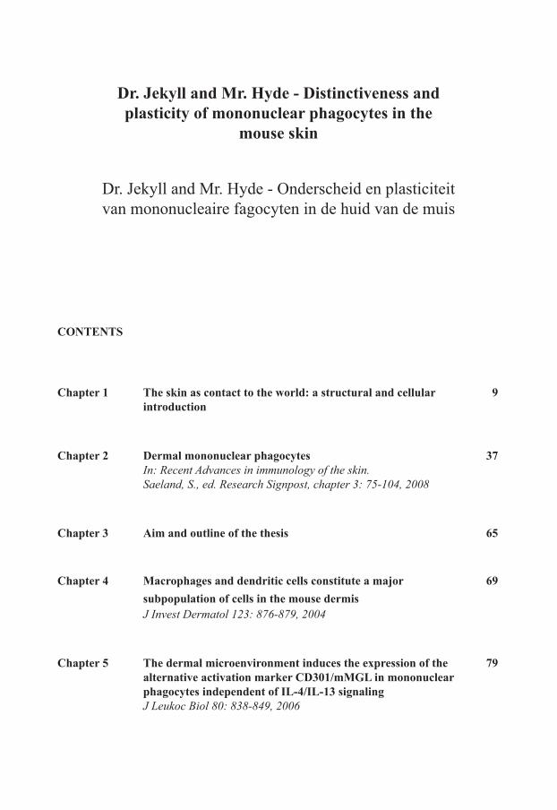
Chapter 1 The skin as contact to the world: a structural and cellular 9 introduction
Chapter 2 Dermal mononuclear phagocytes 37 In: Recent Advances in immunology of the skin. Saeland, S., ed. Research Signpost, chapter 3: 75-104, 2008
Chapter 3 Aim and outline of the thesis 65
Chapter 4 Macrophages and dendritic cells constitute a major 69 subpopulation of cells in the mouse dermis
J Invest Dermatol 123: 876-879, 2004
Chapter 5 The dermal microenvironment induces the expression of the 79 alternative activation marker CD301/mMGL in mononuclear
phagocytes independent of IL-4/IL-13 signaling J Leukoc Biol 80: 838-849, 2006
Chapter 6 Dermal macrophages develop into genuine lymph node 101 dendritic cells upon emigration from the skin Submitted for publication
Chapter 7 Minimal effects of single erythemal UV-irradiation on 121 antigen-presenting cell subsets in the mouse skin
Chapter 8 General discussion 141
Summary 161
Samenvatting 163
Abbreviations 166
Acknowledgements 167
Curriculum vitae 169
List of publications 171
Appendix 172


CHAPTER 1
THE SKIN AS CONTACT TO THE WORLD:A STRUCTURAL AND CELLULAR INTRODUCTION
Chapter 1
10
THE SKIN AS CONTACT TO THE WORLD
Structure of the skinThe skin forms the largest organ of the body. Consequently, it plays a central role in
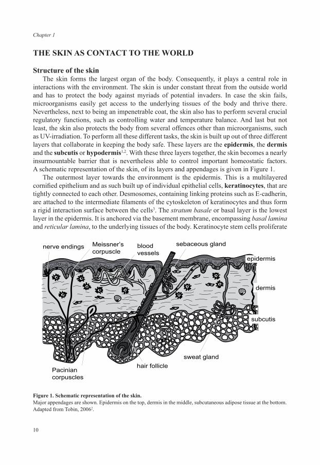
interactions with the environment. The skin is under constant threat from the outside world and has to protect the body against myriads of potential invaders. In case the skin fails, microorganisms easily get access to the underlying tissues of the body and thrive there. Nevertheless, next to being an impenetrable coat, the skin also has to perform several crucial regulatory functions, such as controlling water and temperature balance. And last but not least, the skin also protects the body from several offences other than microorganisms, such as UV-irradiation. To perform all these different tasks, the skin is built up out of three different layers that collaborate in keeping the body safe. These layers are the epidermis, the dermis and the subcutis or hypodermis1,2. With these three layers together, the skin becomes a nearly insurmountable barrier that is nevertheless able to control important homeostatic factors. A schematic representation of the skin, of its layers and appendages is given in Figure 1.
The outermost layer towards the environment is the epidermis. This is a multilayered cornified epithelium and as such built up of individual epithelial cells, keratinocytes, that are tightly connected to each other. Desmosomes, containing linking proteins such as E-cadherin, are attached to the intermediate filaments of the cytoskeleton of keratinocytes and thus form a rigid interaction surface between the cells3. The stratum basale or basal layer is the lowest layer in the epidermis. It is anchored via the basement membrane, encompassing basal lamina and reticular lamina, to the underlying tissues of the body. Keratinocyte stem cells proliferate
hair follicle
sebaceous gland
sweat gland
nerve endings Meissner’scorpuscle
Paciniancorpuscles
bloodvessels
dermis
epidermis
subcutis
Figure 1. Schematic representation of the skin.Major appendages are shown. Epidermis on the top, dermis in the middle, subcutaneous adipose tissue at the bottom. Adapted from Tobin, 20062.

11
A skin deep introduction
in the stratum basale and give rise to descendants that keep a limited capacity to proliferate. Once keratinocytes start to move upwards, they detach from the basement membrane and consequently mature4. During their maturation, which takes place in the stratum spinosum or spinous layer and in the stratum granulosum or granular layer, keratinocytes upregulate the expression of specialized keratins, intermediate filaments of the cytoskeleton that give the cells stability but that are also important intracellular signaling molecules5. Moreover, maturing keratinocytes exchange their lipid bilayer cell membrane with a cornified envelope, a thick, stable structure that consists of multiple cross-linked proteins and lipid components. In the course of this cornification, the cellular envelope is cross-linked with the keratin cytoskeleton, conferring the structure its stability. In addition, keratinocytes will also exude free lipids into the extracellular space to seal off the layers. Once this is accomplished, keratinocytes have fulfilled their lives as viable cells; they will lose their organelles and eventually die. Nevertheless, their strongly cross-linked cell skeletons will remain as stratum lucidum or clear layer and stratum corneum or cornified layer, forming the strong, water-insoluble layers that will keep water inside as well as outside6,7. As a final step, these layers will get sloughed off by mechanical forces and be replaced by newly formed layers that will move up.
Intermingled between the keratinocytes of the basal and suprabasal layers of the epidermis occur other cells that enable the epidermis to perform additional physiological functions. Melanocytes produce and export melanin to neighboring keratinocytes, thereby making them less vulnerable to UV-irradiation that would otherwise cause significant DNA damage also in deeper skin layers8. Yet, the amount of melanin that is present in keratinocytes has to be strictly regulated in order to allow efficient vitamin D3 production by keratinocytes in the basal layer9. Consequently, melanin production in the skin has been optimized during evolution to the ambient UV irradiation9. Nerve cells perform another crucial role in the sensory function of the skin. Merkel cells, which may or may not be nerve cells themselves, connect directly to free sensory nerve endings and are localized in the basal layer of the epidermis10. They are thought to function as mechanoreceptors, although clear neurophysiological evidence is still lacking11. Multiple neurons protrude endings into the epidermis. Aβ-fibers, Aδ-fibers and C-fibers contact multiple cells in the epidermis, receiving constantly information from the outside and thus transforming the skin into the largest sensory organ of the body12.
In the mouse, dendritic epidermal T cells (DETC), in contrast to human epidermal T cells, possess a canonical T cell receptor formed by unusual γ- and δ-chains and showing no junctional diversity. These cells possess a limited receptor repertoire. With it, it has been postulated that they recognize a specific ligand on damaged or stressed keratinocytes, contributing to wound healing and immunosurveillance, to prevent stressed keratinocytes to develop into cancer cells13,14. Human epidermal T cells show more heterogeneity, most of them being αβ T cells and only few γδ T cells15,16. Moreover, their repertoire is not as strictly limited as in the mouse17. This seems to be due to the fact that humans lack the Skint1 molecule that is important for selecting the canonical γδ T cells in the mouse18. Consequently, human epidermal T cells seem to be genuine T cells that recognize antigens, whereas mouse epidermal T cells possess a more general role in skin homeostasis not necessarily associated with their utilization of the T cell receptor. The last epidermal cells to be introduced here are the Langerhans cells (LC), the dendritic cells (DC) of the epidermis.

Chapter 1
12
As such, these cells constitute the sentinels of the immune system that can recognize invading microorganisms. Upon activation, they can rapidly induce an efficient immune response. Moreover, under steady-state conditions, thus in the healthy skin, they can also induce tolerance19-21.
LC form the paradigm of DC biology and thus constitute the gold standard to which the dermal cells that have been studied in this thesis are compared. Therefore, the functions of LC will be discussed in more detail in a later section in this chapter.
Underneath the epidermis, the dermis constitutes the connective tissue of the skin. As such, it mainly consists of extracellular matrix, predominately collagens. The dermis forms not only a strong base onto which the epidermis is attached, but also anchors several skin appendages, e.g. hair follicles, sebaceous glands and sweat glands (see Figure 1). Thereby, the dermis gives the skin its stability. Blood vessels ending in fine capillaries are located throughout the dermis; they are not only important in bringing nutrients to the dermis and epidermis but also in regulating the body’s heat flow through the skin. Upon dilation, blood vessels allow the transport of more heat to be emitted via the skin. Sweat glands also help to adjust the body temperature by excreting sweat. When this sweat evaporates, the underlying skin is cooled down. Next to blood vessels, lymph vessels start in the dermis draining redundant body fluid back into the circulation1,2.
Scattered throughout the extracellular matrix of the dermis are interstitial cells. On the one hand, these cells comprise fibroblasts22, which produce and maintain the extracellular matrix and therefore are considered to be the prototype connective tissue cell. On the other hand, additional, specialized cells can be found dispersed in the extracellular matrix of the dermis. Nerve cells are located here, with Meissner’s corpuscles lying just beneath the epidermis whereas Pacinian corpuscles and Ruffini endings lie deeper23. Additionally, the dermis possesses its own repertoire of hematopoietic cells, thus cells of the immune system: mast cells, macrophages and DC reside embedded in the dermal extracellular matrix, ready to react upon entry of invaders.
Also recirculating T cells can be found patrolling, entering via blood vessels and exiting again via lymph vessels24. These T cells have been quantified and, remarkably, it was estimated that the number of T cells residing in the skin is approximately twice the number of cells that can be found in the circulation24. Nevertheless, hematopoietic cells are thought to constitute only minor dermal subpopulations, compared to the fibroblasts (cf. recent histology text books).
Below the dermis lies the subcutis or hypodermis, a layer that is mainly composed of adipose tissue. The function of this layer, besides that the stored fats can be used as nutritients, is to be a thermal insulation layer. Therefore, this layer together with the dermis regulates the body temperature. Far less is known about the cell composition of the subcutis, compared to the two layers above. Nevertheless, adipocytes and also macrophages can be found in the subcutis in large amounts. Directly below the subcutis is usually a layer of muscle tissue.
Cutaneous immune responsesThe water-insoluble keratin layer of the epidermis forms such an impermeable layer that
it is virtually impossible for pathogens to gain access to the body. Nevertheless, pathogens can readily enter through injuries, which occur steadily caused by daily wear and tear. To be

13
A skin deep introduction
prepared for such a case, the skin constitutes not only a passive barrier, but also possesses the ability to react actively to invaders. Therefore, the skin contains a high density of specialized cells of the immune system.
The skin harbors an abundance of cells of the innate immune system. Macrophages and DC, together indicated as mononuclear phagocytes26, as well as mast cells27 reside in the skin and can readily react to any infection or other insult. Mononuclear phagocytes can phagocytose invaders and destroy them, a function that is mainly attributed to the macrophages. Moreover, they can release a multitude of soluble mediators, such as antimicrobial agents to hit pathogens directly, but they can also release proinflammatory mediators to attract more players of the immune system to assist them.
As such, polymorphonuclear neutrophils (PMN) and monocytes can be recruited from the blood to help the local-resident cells in their stride.
Mononuclear phagocytes in the skin take endocytosed samples of the invader and transport them to the draining lymph nodes. When they arrive there, they present pieces of the invaders to the lymphoid cells that wait there to be activated. Consequently, by activating naïve T cells in draining lymph nodes, mononuclear phagocytes form a bridge between the pathogen-infected skin and the draining lymph nodes. Those T cells that will recognize the pathogen will then migrate to the affected side and help the cells there to rid the body of invaders. By forming a memory compartment, the immune system will also be prepared for future encounters with the same invader.
Large numbers of such memory T cells reside in or patrol through the skin25. They easily become activated again should they re-encounter the same antigen. They will quickly develop into effector cells and eliminate the invader, before a full-blown immune response is initiated.
The mononuclear phagocytes of the skin are thus located at a pivotal position to induce a potent immune response. On the one hand, they mount a first, local reaction, which in some cases is already enough to repel an invader. On the other hand, they bring antigens to the draining lymph nodes to trigger a full-blown systemic immune response. Cells of the adaptive immune system will then complement them in the skin for an efficient antigen-specific immune response. Moreover, they form memory cells, patrolling the skin prepared to react again rapidly to the same pathogen. The circle that is formed by migrating mononuclear phagocytes and recirculating T cells is depicted schematically in Figure 2.
Of the cutaneous mononuclear phagocytes, epidermal LC have been investigated most extensively. Therefore, they currently serve as the paradigm of DC residing in a peripheral tissue and bringing antigens to draining lymph nodes to activate naïve T cells28. In contrast to LC, mononuclear phagocyte subpopulations of the dermis are much less well characterized. Therefore, the studies described in this thesis were intended to investigate the dermal counterparts of the epidermal LC that can perform similar functions. To gain further insight into the biology of these cells, current knowledge of different cutaneous mononuclear phagocyte subpopulations will be introduced in more detail in the following sections.

Chapter 1
14
PHENOTYPE AND FUNCTION OF SKIN MONONUCLEAR PHAGOCYTES
Langerhans cells, the sentinels of the epidermisLC originally have been discovered by Paul Langerhans in 1868 by staining epidermal
tissues with a technique that was used at that time to identify nerve cells29. As it was not possible to characterize these cells functionally, they were suggested to be nerve cells ending in the epidermis. Later, LC were connected with melanin production and thought to represent melanocytes that had lost their capacities to produce melanin. Finally, in the late 1970s and early 1980s LC were characterized to possess ATPase activity and to express MHC class II molecules and receptors for antibodies (FcR) and complement30-32. Additionally, they were determined to develop from bone marrow precursor cells, classifying them thus as hematopoetic cells33,34. It was shown that they are the only epidermal cells capable of stimulating T cell proliferation35. Consequently, they were categorized as a cutaneous mononuclear phagocyte subpopulation. In 1985 LC were portrayed to change their phenotype after four days of culture to resemble the recently discovered splenic dendritic cells36. Comparable to these cells, cultured LC were highly efficient in stimulating allogeneic T cells, whereas freshly isolated LC were less so. Therefore, LC were recognized as the dendritic cell subpopulation of the epidermis.
activation
migration, maturation
stimulation
recruitment
IFN-�,IL-17,…
activation
migration, maturation
stimulation
recruitment
IFN-�,IL-17,…
Figure 2. Representation of the induction of an immune response via the skin. Skin immature dendritic cell precursors, thus epidermal Langerhans cells and dermal mononuclear phagocytes, are activated when they recognize foreign antigens. Upon activation, they mature and migrate to skin-draining lymph nodes. There, they activate naïve antigen-specific T cells. Activated T cells subsequently migrate through the blood to the side of antigen entry. Adapted from Vukmanovic et al., 200625.

15
A skin deep introduction
Due to their easy accessibility, since then LC have been used to investigate various aspects of dendritic cell biology. Nowadays LC serve as a paradigm of these professional antigen-presenting cells (APC)28.
LC are the sentinels of the immune system in the epidermis. At this location, they reside in an immature state, where they express high levels of molecules that are involved in antigen trapping (e.g. Langerin/CD207 or FcγRII/CD32). In contrast, they hardly express molecules that are involved in T cell stimulation, such as CD40, CD80 or CD86. Moreover, they express MHC class II molecules only intracellularly (see e.g. ref. 37). Consequently, their predominant role in this state is to overview their surroundings and to phagocytose and accumulate extracellular matter. In the healthy state, they may use this material later to present it to T cells to induce tolerance. Upon activation, however, their phenotype will change dramatically.
One the one hand, LC may be activated by recognizing pathogens directly via their expressed Toll-like receptors (TLR), such as TLR1, TLR2, TLR3, TLR5, TLR6 and TLR938-41. Interestingly, LC seem to be negative for TLR4 expression, the receptor that signals upon LPS recognition. Therefore, certain pathogens may not be recognized by LC, but by other surrounding cells, such as keratinocytes that express themselves a repertoire of TLR39,42-44. Consequently, pathogen recognition by LC or keratinocytes will lead to mutual activation of both cell types, caused by TNF-α produced by keratinocytes, IL-1β produced exclusively by LC in the murine epidermis and IL-18 produced by both LC and keratinocytes45-47. Other proinflammatory cytokines might be involved in this activation of epidermal cells after pathogen activation as well, such as IL-20 and IL28/IL-2948,49. On the other hand, LC constitutively become activated as well at low frequency in the steady-state epidermis. Although their function will be to induce tolerance rather than immunity in this state, their phenotype nevertheless will change as if they had been activated by recognizing pathogens50. The exact trigger of this activation in the healthy skin is not known so far.
Common to both activation pathways is that LC will downregulate their endocytic receptors and upregulate molecules that are essential for their function as APC. Consequently, stimulatory MHC class I and MHC class II molecules and costimulatory CD40, CD5851, CD80 and CD8637 molecules can be found expressed at high levels on the surface of activated LC. Activated LC will breakdown ingested particles and assemble the breakdown products into complexes with MHC class II molecules, and with MHC class I molecules for cross presentation. These loaded molecules will be displayed on their surface for T cells to recognize them with their T cell receptors, thus resulting in efficient T cell stimulation.
LC activation leads not only to this change in functional phenotype from antigen-phagocytosing to APC, indicated in short as LC maturation. LC activation also initiates the migration of LC from the epidermis, the site of pathogen recognition, to skin-draining lymph nodes, the site of T cell stimulation. Consequently, maturing LC will also upregulate the expression of the chemokine receptor CCR7/CD197. CCR7 is required to guide maturing LC towards the chemokines MIP-3β/CCL19 and SLC/CCL21, which are produced in lymphoid organs, and in the case of SLC/CCL21 also by lymphatic endothelial cells. Therefore, these chemokines will lead LC on their way to the lymphatic vessels and finally into skin-draining lymph nodes52. As LC activated by pathogen-recognition and LC activated in the steady-state will have undergone the same maturation process, they have a highly
Chapter 1
16
similar phenotype of mature LC50. Their function will nevertheless differ significantly, as can be measured for example through their cytokine production, such as IL-1β, IL-6 or IL-1053. LC activated in the steady-state are therefore sometimes referred to as semi-mature LC, in order to distinguish them from the functionally different mature LC54.
The migration of LC from the epidermis into skin-draining lymph nodes by itself poses a challenge for LC. First they have to lose their interactions with the surrounding keratinocytes to be able to push themselves through the basal layer of keratinocytes. Thereafter they have to traverse the basement membrane of the epidermis and the connective tissue of the dermis, both assembled by tightly interacting extracellular matrix components, until they reach lymphatic vessels. There, they interact with the lymphatic endothelium to get access to the inside of the vessels. Once inside, they can resume their journey floating passively in the lymphatic fluid, which will carry them directly into the lymph nodes55. As a consequence, to migrate successfully, maturing LC need to downregulate molecules like the cell adhesion molecule E-cadherin/CD324, which anchors them to neighboring keratinocytes in the epidermis56, and need to upregulate molecules that are required for the migration process rather than for antigen presentation. These include, for instance, the matrix metalloproteinases 2 and 9 (MMP-2 and MMP-9), which are upregulated on maturing LC and enable them to digest basement membrane and dermal extracellular matrix to gain a way through57.
Migrating LC also need cell adhesion molecules to interact with cells and with the extracellular matrix for efficient migration. α6β1 integrins, which bind to laminin, are downregulated on maturing LC; yet they seem to be involved in LC migration, presumably in interaction with the basement membrane, as LC are retained in the epidermis upon application of an α6 blocking antibody58. α4β1 integrins, on the other hand, binding to the extracellular matrix component fibronectin, are upregulated on maturing LC59, but seem not to be involved in LC migration as α4 blocking antibodies had no effect on migration58. Another adhesion molecule that is upregulated during LC maturation is CD4460. CD44 primarily binds to hyaluronan, a glycosaminoglycan containing repeating disaccharide units of N-acetylglucosamine and glucuronic acid, but also binds to other extracellular matrix components, such as collagens and fibronectin. CD44 knock-out mice showed a diminished migration of LC into skin-draining lymph nodes61,62. Nevertheless, LC still normally left the epidermis, suggesting that interaction of LC with hyaluronan or another ligand takes place later during LC migration and is therefore required for LC to cross the dermis efficiently.
The role of the β2 integrins in LC migration remains controversial. Grabbe et al. showed that β2 integrins do not play a role in the migration of LC into lymph nodes in CD18 knock-out mice63. In contrast, Xu et al. showed that ICAM-1/CD54, the prime ligand for LFA-1 (αLβ2; CD11a-CD18) on lymphatic endothelium, is required for LC migration64. Wild-type DC migrated less efficiently into lymph nodes in ICAM-1 knock-out mice, compared to CD18 knock-out DC in wild type recipients. This suggests a role for interaction between β2 integrins on LC and ICAM-1 on endothelial cells64. Nevertheless, LFA-1 (αLβ2; CD11a-CD18) is upregulated on LC relatively late and requires a strong maturation stimulus, comparable to the CD8α upregulation65. This kinetics suggests therefore that LFA-1 plays a role in LC – T cell interaction rather than in migration to lymph nodes. Mac-1/complement receptor 3 (αMβ2; CD11b-CD18) is downregulated upon LC maturation66, suggesting that this receptor has his function more likely in antigen uptake. p150,95 (αXβ2; CD11c-CD18), on the other hand,
17
A skin deep introduction
has been demonstrated to bind to ICAM-167,68. Moreover, it has been shown that this integrin is involved in the interaction of p150,95-expressing cells with inflamed endothelium68. Therefore, p150,95 on migrating LC might indeed bind to ICAM-1 on lymphatic endothelial cells. Alternatively, another so far unknown receptor on LC might be involved in this interaction. ICAM-1 itself is upregulated on LC upon maturation and contributes to LC - T cell interaction51. Also ICAM-2 and ICAM-3 are involved in DC functions. Although less relevant for LC, as it is not expressed by epidermal LC but by dermal DC69,70 (see also Chapter 2), the DC-specific lectin DC-SIGN has been shown to bind to ICAM-2 and ICAM-3. Thereby, the interaction with ICAM-3 has been shown to be important in DC - T cell interactions71, whereas the ICAM-2 interaction was suggested to be required for blood DC to interact with endothelial cells to extravasate into peripheral tissues and lymph nodes72.
Due to their significant change in phenotype and function, post-migration mature LC almost can be considered as a novel cell type. For long it has been impossible to identify mature LC unequivocally among the DC subpopulations in skin-draining lymph nodes. Only the discovery of the LC-specific lectin Langerin, or CD20773, enabled this specific identification of LC after maturation in lymph nodes74. Although LC downregulate Langerin expression from the surface during maturation, they remain intracellularly positive75 (cf. Chapter 5). The specificity of Langerin-promoter usage by LC enabled the construction of a mouse model wherein LC specifically express the green-fluorescent protein (GFP) and the diphtheria-toxin receptor (DTR). In these mice, LC can be depleted by the application of the diphtheria-toxin in vivo, thus enabling studies of the contribution of other cutaneous mononuclear phagocyte subpopulations to a variety of skin-related processes76-78. Therefore, LC biology in skin-draining lymph nodes is just starting to be unraveled.
The many names and faces of dermal mononuclear phagocytesAnalogous to the epidermis, the dermis, as the second layer of the skin, comparably
harbors a sizeable population of mononuclear phagocytes. There is much more heterogeneity among these cells concerning marker expression than in the epidermis. As a consequence, it remains controversial whether they represent different developmental stages of one cell type or different cell types. Accordingly, different nomenclatures are used describing them. In the section below, I will delineate the origins of this disagreement. A detailed idea on the relationship between different dermal mononuclear phagocytes based on our current knowledge and insights will then be presented in Chapter 2.
Fixed, i.e. resident cells in soft (connective) tissues capable of ingesting particles have initially been termed histiocytes79,80. Originally, they have been assigned to the reticuloendothelial system (RES), together with endothelial cells, fibrocytes, reticular cells, reticuloendothelial cells and monocytes81. These cell types have been grouped after observations that they all could take up injected foreign particles, such as colloidal carbon. It was not clear in those days, however, whether they possessed any other relationship except this common function. After the discovery that most of those cell types only endocytosed via micropinocytosis after carbon overloading, the real phagocytic cells that could take up also larger particles were newly assigned to the mononuclear phagocyte system (MPS)82. These cells included precursors in the bone marrow (monoblasts and promonocytes) and in peripheral blood (monocytes) as well as mature cells in peripheral tissues, such as bone

Chapter 1
18
(osteoclasts), lung (alveolar macrophages), liver (Kupffer cells), the peritoneal cavity (peritoneal macrophages) and connective tissue (histiocytes). As the inclusion criteria for cells belonging to the MPS were i) firm adherence to a glass surface and ii) avid phagocytosis, the DC, the professional APC, were excluded from the MPS, as the paradigm cells at that time, spleen and lymph node DC, did not show efficient phagocytosis83-85. Consequently, a dichotomous view emerged that considered the professional phagocytic cells and the professional APC as two different and unrelated cell types.
Soon, it became evident that DC were more related to macrophages than previously thought. First, it was shown that macrophages and DC can not only be generated from the same bone marrow precursors86,87, but also from a common precursor in the blood, the monocyte88. Moreover, it was demonstrated that immature DC are more adherent than mature DC and acquire antigen via receptor-mediated phagocytosis89, fulfilling thereby all the requirements for DC to be members of the mononuclear phagocyte system. Consequently, macrophages and DC are nowadays considered to constitute two extreme stages of development within the MPS, rather than being independent entities of their own. In this respect, DC are the professional APC and macrophages the professional endocytic and degrading cells. However, a clear overlap exists between these functions and consequently also between the cell types90-93
(see also Chapter 2). Nevertheless, distinction of mononuclear phagocytes into macrophages and DC is still commonly applied these days (see e.g. ref. 94). I will indicate cells for which a consensus assignment exists accordingly, so for example LC as DC. In contrast, I will label APC that so far lack a clear assignment, for example the dermal representatives, mononuclear phagocytes.
The existence of dermal mononuclear phagocytes has been known for quite a while when the studies described in this thesis were initiated. Nevertheless, they remained quite enigmatic, as their heterogeneity made it difficult to draw conclusions on their identity and functions. In recent years, increasing understanding has been gathered on dermal mononuclear phagocytes, also because of the studies described in this thesis. A unifying picture of dermal mononuclear phagocytes in mouse and man will be introduced in the next chapter (Chapter 2).
Lectins on non-lymphoid cells in the immune system: more than mere markersAs described above, lectins such as DC-SIGN or Langerin are commonly used as unique
markers for various cell subtypes. Lectins are more than just flags on cells for us to identify them. They possess widespread functions in the immune system but also beyond, as all lectins are proteins that bind glycan moieties, thus sugar structures. Since several lectins expressed by mononuclear phagocytes play an important role in the experimental parts of this thesis, they merit a further introduction.
Since the 1880s, it has been known that extracts from certain plants can cause agglutination of red blood cells95 (reviewed in ref. 96). In the 1940s, agglutinins were discovered which could “select” different types of cells based on their blood groups. Hence, the term “lectin” was originally defined to stand for such agglutinins that can discriminate between different types of red blood cells. Over the years, the meaning has changed so that the term is used now in a much more general sense and simply means proteins that bind glycans, and that are not immunoglobulins or enzymes, regardless of their ability to agglutinate cells and regardless

19
A skin deep introduction
of their origins96,97. Therefore it comes as no surprise that lectins can be derived from many different species, including plants, animals, fungi, bacteria and viruses. Lectins can be shed by cells into the extracellular space, they can be expressed on the cell surface and they can be expressed intracellularly in the cytoplasm or even in the nucleoplasm. There, they perform functions that can be as diverse as the lectins themselves. Different lectins may thus share the common property of binding to defined glycan structures, but once they have bound their glycosylated ligands, they cause completely different reactions.
The molecular structure of lectins is highly diverse as they possess many different, unrelated domains. Based on the different three-dimensional structures of lectins, they are classified into different families97. Most animal lectins originally have been grouped into two families, the C-type lectins, whose members are dependent on Ca2+ for sugar binding, and the S-type lectins, whose members are sulfhydryl-dependent98. The few exceptions known at that time were a heterogeneous group referred to as N-type lectins (not C or S). Yet, with the primary amino acid sequence of many more lectins becoming available, it has been recognized that many Ca2+-dependent lectins do not possess a typical sequence related to C-type lectins, and that not all S-type lectins, now known as galectins, are sulfhydryl-dependent97. Later on, many more families were introduced. Consequently, there are now more than fourteen families defined. Different families are subdivided into different subfamilies. Elaborating them all would go far beyond the scope of this thesis. Nevertheless, three different subfamilies of C-type lectins are of particular interest when considering mononuclear phagocytes; the type II, type IV and type VI C-type lectins. Therefore these subfamilies will be discussed in more detail in the following paragraphs. Their molecular structure is schematically represented in Figure 3.
COOH
NH2
COOH
NH2
CRD
linker domain
signaling domain
CRDs andCTLDs
cystein-richdomaintype II FNdomain
EGFdomain
CRD
consensusdomains(2 – 9)
type II type VI type IV / selectins
COOH
NH2
COOH
NH2NH2
COOH
NH2
COOH
C-type lectins (CTL)
Figure 3. Representation of three classes of C-type lectins (CTL).Type II, type VI and type IV (selectins) are shown. Abbreviations: COOH, carboxy-terminus; CRD, carbohydrate-recognition domain; CTLD, C-type lectin-like domain, indicating non-sugar binding CRD; EGF domain, epidermal growth factor-like domain; FN fibronectin, NH2, amino-terminus.

Chapter 1
20
• Type II C-type lectins possess a single carbohydrate recognition domain (CRD) at the C-terminus of the protein. Thereafter, a linker domain follows, containing several leucine-rich zippers. The linker domain is important for the oligomerization of type II C-type lectins. After the transmembrane (TM) domain, a signaling domain at the cytoplasmatic tail follows at the N-terminus97,99. As type II TM proteins, type II C-type lectins must thus be produced completely in the cytoplasm before they can be exported with the C-terminus first into the cell membrane. Possessing only one CRD, type II C-type lectin can bind only a single glycan residue. To gain nevertheless binding specificity and affinity, type II C-type lectins are generally expressed as oligomers100. Many members of this family are known nowadays and, interestingly, a significant number is expressed cell type-specific by mononuclear phagocytes. Type II C-type lectins expressed on macrophages and DC includes BDCA-2/CD303, CLEC-1, CLEC-2, DC-IR, DC-SIGN/CD209, Dectin-1 (the β-glucan receptor), Dectin-2, Langerin/CD207, MGL/CD301 and Mincle. Functionally, type II C-type lectins have been proposed to act as receptors that are used to recognize and take up specific glycosylated pathogens but also self-antigens97,101. It has been proposed that they may act as antipodes to the TLR by inducing tolerance when triggered alone without simultaneous TLR signaling102. In contrast to this view, it has been demonstrated that Dectin-1 possesses an immunostimulatory domain, whereas Dectin-2 has been portrayed to associate with the common FcRγ chain103,104. Consequently, triggering of both type II C-type lectins will lead to an activating signal. In addition to the function as endocytic receptors, type II C-type lectins may perform several other functions by interacting with sugar moieties on binding partners. It has been shown, for instance, that DC-SIGN participates in the formation of the immunological synapse between DC and T cells71 and in the adherence to and subsequent crossing of the endothelial cell layer72, thus enabling DC to migrate and to induce effective immune responses.
• Type VI C-type lectins consist of an N-terminal cysteine-rich (CR) domain, a domain containing fibronectin type two repeats (FNII), multiple CRD, a TM domain and a short cytoplasmatic tail. Type VI C-type lectins are thus also type I TM proteins. The most prominent members of this group are the mannose receptor (MR)/CD206 and DEC-205/CD205, containing eight and six CRD, respectively. Two more members are known, phospholipase A2 receptor and Endo180, which possess eight CRD. The functions of these lectins are more complex, as they contain next to their CRD additional, unrelated domains. With those, they are suggested to perform additional functions next to binding to glycan moieties. Moreover, not all their CRD can recognize sugar residues. The non-sugar binding CRD are called C-type lectin-like carbohydrate recognition domain (CTLD)106. MR can also be shed from the cell surface and function extracellularly, contributing to the versatility of this receptor. Nevertheless both MR and CD205 have a demonstrated function as endocytic receptors, thus being able to recognize and internalize glycosylated molecules. For the MR, its CRD have been shown to bind to mannose, fucose and N-acetylglucosamine (GlcNAc)-terminated ligands. The glycan specificities of CD205 are still unknown107.
• The type IV C-type lectins are also known as selectins. They consist of an N-terminal CRD, an epidermal growth factor (EGF) domain, several short consensus repeat

21
A skin deep introduction
domains, a single TM domain, and a cytoplasmic tail at the C-terminus. Therefore they are type I TM proteins. They are expressed by leukocytes (L-selectin) and endothelial cells (E-selectin, P-selectin) to enable initial adhesive interactions between these two cell types. As a consequence, leukocytes can roll over endothelium cells and scan them for homing signals. Once they encounter the right signal, they will adhere more tightly to and eventually cross the endothelium to reach the perivascular spaces105.
Lectins bind thus specific glycosylated ligands and enable unique interactions of cells with their environment. Consequently, they belong to the most exclusively expressed molecules. As such, they can be used to recognize specific cell subpopulations. In the human skin, the type II C-type lectin Langerin/CD207 is solely expressed by LC, whereas dermal mononuclear phagocytes uniquely express DC-SIGN69,70 and the MR/CD20669 (see also Chapter 2). Both these subpopulations express the type IV C-type lectin CD20569,70, whereas only about 20% of all human dermal CD68+ mononuclear phagocytes express MGL (hMGL)/CD301108. Only few BDCA-2/CD303 expressing plasmacytoid DC can be found in the healthy skin70.
In the mouse skin, Langerin/CD207 comparably can be used to identify LC uniquely74. No staining patterns for mouse DC-SIGN/CD209 or BDCA-2/CD303 have been described so far, whereas SIGN-R1, a mouse DC-SIGN homologue, is expressed only by a few cells in the mouse dermis (Chapter 4). Mouse MGL (mMGL)/CD301 and MR/CD206, in contrast, have been shown to be expressed by numerous mononuclear phagocytes in the dermis, but not by LC in the epidermis109-111. In our studies, we sought to identify a unique marker that recognizes all mouse dermal mononuclear phagocytes without recognizing epidermal LC. Given their cell type-specific expression pattern, a lectin was prime candidate in this search. As an antibody against mMGL/CD301 had been produced previously in the lab112,113, this antibody was chosen to analyze its expression by dermal mononuclear phagocytes and by LC. These experiments revealed that mMGL/CD301 is expressed by all dermal mononuclear phagocytes (Chapter 4), but also by LC under certain conditions (Chapter 5). As MR/CD206 as well is expressed by all dermal mononuclear phagocytes, but not by LC (Chapter 5), this molecule probably is more suitable for the specific identification of dermal mononuclear phagocytes.
Since extracellular molecules, either or not cell-bound, are generally glycosylated, the binding of a specific glycan residue will be the first step in the interaction of a cell with its environment. This is followed by more extensive and high affinity interactions. Therefore, identification of a specific lectin expressed on an exclusive cell subpopulation is just the first step in the line of understanding specific functions of the lectin. Further experiments should then be carried out to unravel the significance of the interaction between the lectin and its ligand, and its consequences for the lectin-expressing cell. Mouse knock-out studies can help in this. Yet, all three knock-out mice, lacking either MR/CD206114,115, mMGL1/CD301a116 or Langerin/CD20776 have failed so far to show specific phenotypes. This hints to the fact that there might be significant redundancy concerning lectin expression and functions.
THE IMMUNOLOGICAL CONSEQUENCES OF UV-IRRADIATION
Initial findings showing immunological effects of UV-irradiation in the mouseEven though the skin possesses large numbers of immunologically potent mononuclear

Chapter 1
22
phagocytes that have been described in the previous paragraphs, conditions occur where skin contact with strong immunogenic antigens does not lead to the elicitation of an immune response. In such situations, the immune system is referred to as being suppressed. Applying cyclosporin systemically or pimecrolimus locally, for example, inhibits T cell activation and consequently impairs skin immune responses. UV-irradiation is another mediator that is nowadays well known to cause the skin to lose its ability to initiate an immune response, thus to cause immunosuppression. This was discovered rather incidentally in the early seventies117,118. Then, chronic UV-irradiation, as were chemical agents, was used in oncological studies to cause DNA damage and consequently to induce skin cancer. It was realized quickly that when those UV-induced tumors were transplanted into non-irradiated syngeneic mice, they were readily rejected. They were thus highly antigenic in contrast to chemically induced skin cancers. Transplanting UV-induced tumors into UV-irradiated mice, which did not yet develop their own cancers, resulted in the outgrowth of these tumors. Moreover, transplanting these tumor cells into chemically immunosuppressed recipient mice led to a comparable progression of the tumors. Therefore, it was concluded that UV-irradiation does not only induce skin cancers but also propagates them by suppressing appropriate defense responses by the immune system117,118.
In an unrelated set of experiments, it had been found that UV-irradiation depletes LC from the epidermis119. This finding was applied to assess the function of LC in the steady-state epidermis by irradiating mice with low doses of UV-irradiation to deplete them of LC120. Subsequently, UV-irradiated mice were immune-sensitized via the LC-depleted skin by a hapten, i.e. a small molecule that becomes immunogenic by crosslinking to endogenous proteins. In normal, non-irradiated mice, the application of the same hapten after a short recovery time onto another side of the body, with mice preferentially onto their ears, leads to a strong inflammation caused by a type IV hypersensivity reaction, or contact hypersensitivity (CHS) reaction. In UV-irradiated, thus LC-depleted mice, this CHS response appeared to be suppressed. As APC were known to be important in the process of sensitization, the lack thereof in UV-irradiated skin was taken as proof that LC are indeed epidermal APC120.
Additional experiments indicated, however, that the immunological effects of UV-irradiation are more far-reaching than just the induction of immune suppression due to the lack of LC. Resensitizing previously UV-irradiated mice onto never irradiated skin parts of their body with the same hapten did not sensitize for a strong CHS response either. This in spite of the fact that there were now sufficient LC present to induce an immune reaction against the hapten. It was thus realized that sensitization of skin exposed to relatively low doses of UV-irradiation induced a silent, suppressed immune status that does not allow the induction of an inflammatory immune response in the future, thus what is now referred to as tolerance. Tolerance at this time was already quite well characterized as being caused by antigen-specific T cells that would inhibit the activation of effector T cells. These T cells could be isolated from secondary lymphoid organs and transplanted into untreated mice that would show then the same tolerance as the original mice121,122. Due to their function, these T cells were called suppressor T cells and are nowadays referred to as regulatory T cells to avoid the confusion raised in the past about their identity and existence123. And indeed, the presence of such regulatory T cells appeared to explain the outgrowth of UV-induced skin tumors in

23
A skin deep introduction
UV-irradiated recipients since transplantation of T cells from UV-irradiated mice into unirradiated mice causes tolerance for UV-induced skin cancers124 and haptens125.
Due to technical reasons, the immunological effects of UV-irradiation mostly have been assigned to the more energy-rich wavelengths of the UV-B spectrum126. Nowadays, increasing numbers of studies are performed with solar-simulated UV radiation (SSR), which, similar to the ambient sunlight, delivers twenty times more energy in the UV-A spectrum than UV-B radiation. Depending on dose and irradiation regimen, the UV-A radiation has been shown either to reinforce or to counteract the immunosuppressive effects of UV-B radiation127,128.
UV-irradiation induces systemic immunosuppressionIn the previous section it was observed that transplanted UV-induced skin tumors can
grow in UV-irradiated mice, independent of whether they had been transplanted into the irradiated side or into an unirradiated side of the body118. It was realized that UV-irradiation not only altered directly the functionality of the skin that had been irradiated, but also caused a systemic alteration of the organism’s immune system118. Similarly, UV-irradiation suppressed the induction of a CHS response, whether the hapten was applied to an irradiated side or to an unirradiated side129.
Subsequently it was shown that UV-irradiation affects systemically all APC subpopulations throughout the body to induce a more potent response of regulatory T cells compared to effector cells. This aberrant antigen-presenting function can thus not only be measured in the lymph nodes draining the irradiated skin, but also in other secondary lymphoid organs, such as in lymph nodes draining unirradated skin or even in the spleen130,131. In order to reach distant organs in the body, soluble mediators that are produced in the UV-irradiated skin that diffuse through the lymph and the blood are thought to be crucial for UV-induced systemic immunosuppression132,133. Table 1 gives an overview of soluble factors that have so far been identified, primarily in experimental animal studies, to be involved in UV-induced immunosuppression.
Table 1. Soluble factors connected with UV-induced systemic immunosuppression.
Mediator Primary references
cis-urocanic acid (cis-UCA) De Fabo and Noonan, 1983134; Moodycliffe et al., 1993135
IL-10 Rivas and Ullrich, 1992136; Rivan and Ullrich, 1994137; Shreedhar et al., 1998138; Kurimoto et al., 2000139
IL-4 Rivan and Ullrich, 1994137; Shreedhar et al., 1998138
IL-12p40 homodimers Schmitt and Ullrich, 2000140
prostaglandin E2 (PGE2) Chung et al., 1986141; Shreedhar et al., 1998138
nitric oxide (NO) Halliday et al., 1999142; Kuchel et al., 2003143
platelet-activating factor (PAF) Walterscheid et al., 2002144
histamine Jaksic et al., 1995145; Hart et al., 2001146
vitamin D3 (vit D3) Reichrath and Rappl, 2003147

Chapter 1
24
The process of UV-induced systemic immunosuppression, however, is far from being completely understood. On the one hand, no significant changes have been observed in the levels of serum cytokines in human subjects after whole-body irradiation dosages known to cause systemic immunosuppressive effects, such as an inhibition in NK cell activity or peripheral blood mononuclear cell (PBMC) cytokine production148. Therefore, it might be that immunosuppressive mediators differ between mice and humans. Moreover, the assumed role of the DC as the APC inducing regulatory T cells after UV-irradiation has been questioned149,150. In UV-irradiated mice, B cells seem to play a role as the APC mediating immunosuppression, possibly via production of IL-10149,151. Moreover, it has also been suggested that the migration of mast cells from the irradiated skin to skin-draining lymph nodes is crucial for systemic immunosuppression152.
The role of skin-resident and skin-recruited APC subsets in inducing local immunosuppression after UV-irradiation
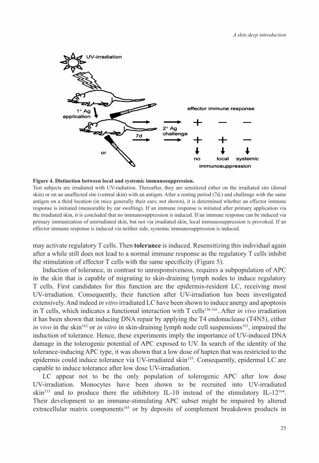
When lower UV-irradiation dosage regimens were applied, it was observed that systemic immunosuppression was not induced. However, UV-induced immunosuppression and tolerance could still be obtained when the hapten was applied directly onto the UV-irradiated skin site132,153-155. Therefore, it was appreciated that low dosages of UV-irradiation did not cause systemic but only local immunosuppression in the UV-irradiated skin. This local UV-induced immunosuppression is limited to UV-irradiated skin only. In order to distinguish between the two forms of immunosuppression, specific experimental read-out systems were developed (see Figure 4). In this setting, UV-irradiated mice are sensitized onto UV-irradiated skin (in the figure on the back) or onto non-irradiated skin (in the figure on the abdomen). After a resting period allowing the primary immune response to occur, the mice are challenged (normally onto their ears; not shown in the figure) to be able to determine the degree of either sensitization, or immunosuppression. A state of UV-induced local immunosuppression has been observed when application of the hapten to the non-irradiated skin, but not to the UV-irradiated side, gives rise to efficient sensitization.
It is likely that APC populations present in the UV-irradiated skin or the local draining lymph node are responsible for the immunosuppression120,132,133. Two processes have to be distinguished thereby that both result in local immunosuppression, namely immunological unresponsiveness and tolerance, depicted schematically in Figure 5. In the case of unresponsiveness, these local cells are thought to be impaired in their antigen-presenting function. They can be aberrant in their antigen-uptake abilities, in their maturation, migration, or T cell interaction capacities, or they might have too much DNA damage so that they undergo apoptosis. In any case, they will not be able to interact actively with T cells in skin-draining lymph nodes. As a result, immunological unresponsiveness is observed. Resensitizing the same organism after a while with the same antigen on healthy skin will result in a normal immune response. Unresponsiveness implies that temporarily skin-resident APC are affected in their function. Therefore, local epidermal as well as dermal mononuclear phagocytes are impaired153.
On the other hand, APC may be able to migrate normally from UV-irradiated skin to draining lymph nodes and to interact there with T cells in an antigen-specific manner. The UV-irradiation may have impaired their function to induce effector cells. Instead they
25
A skin deep introduction
may activate regulatory T cells. Then tolerance is induced. Resensitizing this individual again after a while still does not lead to a normal immune response as the regulatory T cells inhibit the stimulation of effector T cells with the same specificity (Figure 5).
Induction of tolerance, in contrast to unresponsiveness, requires a subpopulation of APC in the skin that is capable of migrating to skin-draining lymph nodes to induce regulatory T cells. First candidates for this function are the epidermis-resident LC, receiving most UV-irradiation. Consequently, their function after UV-irradiation has been investigated extensively. And indeed in vitro irradiated LC have been shown to induce anergy and apoptosis in T cells, which indicates a functional interaction with T cells156-161. After in vivo irradiation it has been shown that inducing DNA repair by applying the T4 endonuclease (T4N5), either in vivo in the skin162 or in vitro in skin-draining lymph node cell suspensions163, impaired the induction of tolerance. Hence, these experiments imply the importance of UV-induced DNA damage in the tolerogenic potential of APC exposed to UV. In search of the identity of the tolerance-inducing APC type, it was shown that a low dose of hapten that was restricted to the epidermis could induce tolerance via UV-irradiated skin155. Consequently, epidermal LC are capable to induce tolerance after low dose UV-irradiation.
LC appear not to be the only population of tolerogenic APC after low dose UV-irradiation. Monocytes have been shown to be recruited into UV-irradiated skin153 and to produce there the inhibitory IL-10 instead of the stimulatory IL-12164. Their development to an immune-stimulating APC subset might be impaired by altered extracellular matrix components165 or by deposits of complement breakdown products in
effector immune response
UV-irradiation
1° Agapplication –
+––
local systemicimmunosuppression
++
noor
7d
2° Agchallenge
effector immune response
UV-irradiation
1° Agapplication –
+––
local systemicimmunosuppression
++
noor
7d
2° Agchallenge
Figure 4. Distinction between local and systemic immunosuppression.Test subjects are irradiated with UV-radiation. Thereafter, they are sensitized either on the irradiated site (dorsal skin) or on an unaffected site (ventral skin) with an antigen. After a resting period (7d.) and challenge with the same antigen on a third location (in mice generally their ears; not shown), it is determined whether an effector immune response is initiated (measurable by ear swelling). If an immune response is initiated after primary application via the irradiated skin, it is concluded that no immunosuppression is induced. If an immune response can be induced via primary immunization of unirradiated skin, but not via irradiated skin, local immunosuppression is provoked. If an effector immune response is induced via neither side, systemic immunosuppression is induced.

Chapter 1
26
UV-irradiated skin166. These recruited inflammatory monocytes have been shown to migrate to skin-draining lymph nodes after hapten application onto UV-irradiated skin, where they, comparable to their activity in the skin, produce IL-10 but no IL-12167. Therefore, also recruited APC subsets are able to induce local UV-induced tolerance.
In contrast to the populations mentioned above, resident dermal mononuclear phagocytes have never been investigated for their capacities to induce tolerance after a low dose UV-irradiation. Therefore, dermal mononuclear phagocytes await their characterization in the process of local UV-induced immunosuppression and the role of DNA damage therein.
The meaning of local and systemic UV-induced tolerance for the bodySince placental mammals have lost the photolyase enzyme during their evolution, higher
mammals have been deprived of the most efficient manner to repair DNA damage168. Therefore, they are much more prone to UV-induced immunosuppression, as the photolyase enzyme in lower marsupials can inhibit this UV-mediated impairment of the immune system169. UV-induced immunosuppression persisted during the evolution up to humans. Consequently, one wonders what the beneficial effects of UV-induced immunosuppression might be. Clearly, systemic UV-induced immunosuppression has harmful aspects for the body as general immune reactions are suppressed170,171. Cyclosporin, via a completely different mechanism, also induces immunosuppression, with major consequences for immune surveillance and carcinogenesis172. Systemic UV-irradiation, to a certain degree, has similar effects, also in view of the fact that UV-irradiation by itself can cause DNA damage and therewith cancer.
UV-irradiation
secondaryapplication
no effector immune response is induced
Ag application
effector immune response
–tolerance
+unresponsiveness
primaryapplication
at primary antigen application
Figure 5. Distinction between immunological unresponsiveness and tolerance. Test subjects are irradiated with UV-radiation, sensitized with an antigen and challenged with the same antigen. When no effector response is obtained, the mice are secondarily immunized with the same antigen. If in this case an effector immune response is induced, then immunological tolerance is absent and the skin was unresponsive at the first sensitisation. If, however, still no immune response occurs, then the subject was tolerized.

27
A skin deep introduction
UV-induced local immunosuppression might also be beneficial to the body, as immune reactions to UV-induced neo-antigens might be inhibited. This is exemplified by the notion that aberrancies in the ability to induce local UV-immunosuppression might lead to autoimmune diseases such as polymorphic light eruption (PLE) or cutaneous lupus erythematosus (CLE)173. IL-10+, IL-12– monocytes migrating to skin-draining lymph nodes have only been observed after the application of a hapten in adjuvant onto UV-irradiated skin, but not after the UV-irradiation itself167. Thus, low-dose UV-irradiation might require the occurrence of an immunostimulatory antigen to lead to local UV-induced tolerance. Consequently, the body might indeed use this mechanism to inhibit reactions to neo-antigens that would otherwise lead to sensitization for self-antigens.
If there is a physiological role for local UV-induced tolerance, is then the systemic immunosuppression an unrelated, contra-productive interference with physiological functions or does systemic immunosuppression just represent an exaggerated form of the local tolerance induction? Soluble mediators produced locally in the skin can affect the resident APC subpopulations. Therefore, the difference between low dose local and high dose systemic immunosuppression might indeed lie in the enhanced induction of these mediators by higher dosages of UV-irradiation. On the other hand, different regulatory T cells have been described to be induced after UV-irradiation: CD4+, CTLA-4+ (ref. 174), CD25+, Dectin-2-binding175 Tr1 cells were induced after a local tolerance protocol174,175, while CD4+ DX5+ NKT cells with regulatory potential were observed after a systemic tolerance protocol176. Therefore, different regulatory T cells might mediate immunosuppression and tolerance, respectively133. Yet, this notion remains to be proven. Consequently, the relationship between local and systemic UV-induced immunosuppression remains open for additional experimental approaches.
Taken together, UV-irradiation of the skin may have distinct and complex immunological consequences as it may lead to two different kinds of immunosuppression as well as tolerance, depending on the characteristics and dosage of the applied UV radiation. At a low dosage, immunosuppression and tolerance are induced only for antigens that are applied locally onto the UV-irradiated skin. Local resident and recruited APC are most probably responsible for this phenomenon. At higher dosages, a systemic state of immunosuppression and tolerance is induced. This is connected with the production of soluble mediators in the irradiated skin and their distribution all over the body. Moreover, systemic leukocyte populations become suppressed. In this situation the induction of tolerance for a cutaneously applied antigen has been connected with aberrant mast cells and B cells. So far, the relationship between these two UV-induced immune deviations remains unclear, although they clearly possess commonalities.
REFERENCES
1. Kanitakis, J. Anatomy, histology and immunohistochemistry of normal human skin. Eur J Dermatol 12, 390-399; quiz 400-391 (2002).
2. Tobin, D.J. Biochemistry of human skin--our brain on the outside. Chem Soc Rev 35, 52-67 (2006).3. Green, K.J. & Simpson, C.L. Desmosomes: new perspectives on a classic. J Invest Dermatol 127, 2499-2515
(2007).4. Blanpain, C. & Fuchs, E. Epidermal stem cells of the skin. Annu Rev Cell Dev Biol 22, 339-373 (2006).
Chapter 1
28
5. Kirfel, J., Magin, T.M. & Reichelt, J. Keratins: a structural scaffold with emerging functions. Cell Mol Life Sci 60, 56-71 (2003).
6. Kalinin, A.E., Kajava, A.V. & Steinert, P.M. Epithelial barrier function: assembly and structural features of the cornified cell envelope. Bioessays 24, 789-800 (2002).
7. Madison, K.C. Barrier function of the skin: “la raison d’etre” of the epidermis. J Invest Dermatol 121, 231-241 (2003).
8. Lin, J.Y. & Fisher, D.E. Melanocyte biology and skin pigmentation. Nature 445, 843-850 (2007).9. Jablonski, N.G. & Chaplin, G. The evolution of human skin coloration. J Hum Evol 39, 57-106 (2000).10. Boulais, N. & Misery, L. Merkel cells. J Am Acad Dermatol 57, 147-165 (2007).11. Moll, I., Roessler, M., Brandner, J.M., Eispert, A.C., Houdek, P. & Moll, R. Human Merkel cells--aspects of cell
biology, distribution and functions. Eur J Cell Biol 84, 259-271 (2005).12. Boulais, N. & Misery, L. The epidermis: a sensory tissue. Eur J Dermatol 18, 119-127 (2008).13. Jameson, J.M., Cauvi, G., Witherden, D.A. & Havran, W.L. A keratinocyte-responsive gamma delta TCR is
necessary for dendritic epidermal T cell activation by damaged keratinocytes and maintenance in the epidermis. J Immunol 172, 3573-3579 (2004).
14. Strid, J., Roberts, S.J., Filler, R.B., Lewis, J.M., Kwong, B.Y., Schpero, W., Kaplan, D.H., Hayday, A.C. & Girardi, M. Acute upregulation of an NKG2D ligand promotes rapid reorganization of a local immune compartment with pleiotropic effects on carcinogenesis. Nat Immunol 9, 146-154 (2008).
15. Foster, C.A., Yokozeki, H., Rappersberger, K., Koning, F., Volc-Platzer, B., Rieger, A., Coligan, J.E., Wolff, K. & Stingl, G. Human epidermal T cells predominantly belong to the lineage expressing alpha/beta T cell receptor. J Exp Med 171, 997-1013 (1990).
16. Spetz, A.L., Strominger, J. & Groh-Spies, V. T cell subsets in normal human epidermis. Am J Pathol 149, 665-674 (1996).
17. Dunn, D.A., Gadenne, A.S., Simha, S., Lerner, E.A., Bigby, M. & Bleicher, P.A. T-cell receptor V beta expression in normal human skin. Proc Natl Acad Sci U S A 90, 1267-1271 (1993).
18. Boyden, L.M., Lewis, J.M., Barbee, S.D., Bas, A., Girardi, M., Hayday, A.C., Tigelaar, R.E. & Lifton, R.P. Skint1, the prototype of a newly identified immunoglobulin superfamily gene cluster, positively selects epidermal gammadelta T cells. Nat Genet 40, 656-662 (2008).
19. Romani, N., Holzmann, S., Tripp, C.H., Koch, F. & Stoitzner, P. Langerhans cells - dendritic cells of the epidermis. Apmis 111, 725-740 (2003).
20. Romani, N., Ebner, S., Tripp, C.H., Flacher, V., Koch, F. & Stoitzner, P. Epidermal Langerhans cells--changing views on their function in vivo. Immunol Lett 106, 119-125 (2006).
21. Romani, N., Ebner, S., Flacher, V., Tripp, C.H., Heufler, C., Clausen, B.E. & Stoitzner, P. Langerhans cells - dendritic cells of the epidermis and other epithelia, in Recent Advances in Skin Immunology, Vol. 37/661. (ed. S. Saeland) (Research Signpost, Kerala, India; 2008).
22. Dunphy, J.E. The fibroblast – a ubiquitous friend of the surgeon. N Engl J Med. 268, 1367-1377 (1963).23. Macefield, V.G. Physiological characteristics of low-threshold mechanoreceptors in joints, muscle and skin in
human subjects. Clin Exp Pharmacol Physiol 32, 135-144 (2005).24. Clark, R.A., Chong, B., Mirchandani, N., Brinster, N.K., Yamanaka, K., Dowgiert, R.K. & Kupper, T.S. The
vast majority of CLA+ T cells are resident in normal skin. J Immunol 176, 4431-4439 (2006).25. Vukmanovic-Stejic, M., Reed, J.R., Lacy, K.E., Rustin, M.H. & Akbar, A.N. Mantoux Test as a model for a
secondary immune response in humans. Immunol Lett 107, 93-101 (2006).26. Valladeau, J. & Saeland, S. Cutaneous dendritic cells. Semin Immunol 17, 273-283 (2005).27. Gersch, C., Dewald, O., Zoerlein, M., Michael, L.H., Entman, M.L. & Frangogiannis, N.G. Mast cells and
macrophages in normal C57/BL/6 mice. Histochem Cell Biol 118, 41-49 (2002).28. Villadangos, J.A. & Heath, W.R. Life cycle, migration and antigen presenting functions of spleen and lymph
node dendritic cells: limitations of the Langerhans cells paradigm. Semin Immunol 17, 262-272 (2005).29. Langerhans, P. Über die Nerven der menschlichen Haut. [Virchows] Archiv für pathologische Anatomie und
Physiologie, und für klinische Medicin, Berlin 44, 325-337 (1868).30. Stingl, G., Wolff-Schreiner, E.C., Pichler, W.J., Gschnait, F., Knapp, W. & Wolff, K. Epidermal Langerhans cells
bear Fc and C3 receptors. Nature 268, 245-246 (1977).31. Rowden, G., Lewis, M.G. & Sullivan, A.K. Ia antigen expression on human epidermal Langerhans cells. Nature
268, 247-248 (1977).

29
A skin deep introduction
32. Klareskog, L., Tjernlund, U., Forsum, U. & Peterson, P.A. Epidermal Langerhans cells express Ia antigens. Nature 268, 248-250 (1977).
33. Katz, S.I., Tamaki, K. & Sachs, D.H. Epidermal Langerhans cells are derived from cells originating in bone marrow. Nature 282, 324-326 (1979).
34. Frelinger, J.G., Hood, L., Hill, S. & Frelinger, J.A. Mouse epidermal Ia molecules have a bone marrow origin. Nature 282, 321-323 (1979).
35. Stingl, G., Katz, S.I., Clement, L., Green, I. & Shevach, E.M. Immunologic functions of Ia-bearing epidermal Langerhans cells. J Immunol 121, 2005-2013 (1978).
36. Schuler, G. & Steinman, R.M. Murine epidermal Langerhans cells mature into potent immunostimulatory dendritic cells in vitro. J Exp Med 161, 526-546 (1985).
37. Berthier-Vergnes, O., Bermond, F., Flacher, V., Massacrier, C., Schmitt, D. & Peguet-Navarro, J. TNF-alpha enhances phenotypic and functional maturation of human epidermal Langerhans cells and induces IL-12 p40 and IP-10/CXCL-10 production. FEBS Lett 579, 3660-3668 (2005).
38. Mitsui, H., Watanabe, T., Saeki, H., Mori, K., Fujita, H., Tada, Y., Asahina, A., Nakamura, K. & Tamaki, K. Differential expression and function of Toll-like receptors in Langerhans cells: comparison with splenic dendritic cells. J Invest Dermatol 122, 95-102 (2004).
39. Flacher, V., Bouschbacher, M., Verronese, E., Massacrier, C., Sisirak, V., Berthier-Vergnes, O., de Saint-Vis, B., Caux, C., Dezutter-Dambuyant, C., Lebecque, S. & Valladeau, J. Human Langerhans cells express a specific TLR profile and differentially respond to viruses and Gram-positive bacteria. J Immunol 177, 7959-7967 (2006).
40. van der Aar, A.M., Sylva-Steenland, R.M., Bos, J.D., Kapsenberg, M.L., de Jong, E.C. & Teunissen, M.B. Loss of TLR2, TLR4, and TLR5 on Langerhans cells abolishes bacterial recognition. J Immunol 178, 1986-1990 (2007).
41. Peiser, M., Koeck, J., Kirschning, C.J., Wittig, B. & Wanner, R. Human Langerhans cells selectively activated via Toll-like receptor 2 agonists acquire migratory and CD4+T cell stimulatory capacity. J Leukoc Biol 83, 1118-1127 (2008).
42. Kawai, K., Shimura, H., Minagawa, M., Ito, A., Tomiyama, K. & Ito, M. Expression of functional Toll-like receptor 2 on human epidermal keratinocytes. J Dermatol Sci 30, 185-194 (2002).
43. Song, P.I., Park, Y.M., Abraham, T., Harten, B., Zivony, A., Neparidze, N., Armstrong, C.A. & Ansel, J.C. Human keratinocytes express functional CD14 and toll-like receptor 4. J Invest Dermatol 119, 424-432 (2002).
44. Kollisch, G., Kalali, B.N., Voelcker, V., Wallich, R., Behrendt, H., Ring, J., Bauer, S., Jakob, T., Mempel, M. & Ollert, M. Various members of the Toll-like receptor family contribute to the innate immune response of human epidermal keratinocytes. Immunology 114, 531-541 (2005).
45. Cumberbatch, M., Dearman, R.J., Griffiths, C.E. & Kimber, I. Langerhans cell migration. Clin Exp Dermatol 25, 413-418 (2000).
46. Cumberbatch, M., Dearman, R.J., Antonopoulos, C., Groves, R.W. & Kimber, I. Interleukin (IL)-18 induces Langerhans cell migration by a tumour necrosis factor-alpha- and IL-1beta-dependent mechanism. Immunology 102, 323-330 (2001).
47. Kimber, I., Cumberbatch, M., Dearman, R.J., Bhushan, M. & Griffiths, C.E. Cytokines and chemokines in the initiation and regulation of epidermal Langerhans cell mobilization. Br J Dermatol 142, 401-412 (2000).
48. Sugita, K., Kabashima, K., Atarashi, K., Shimauchi, T., Kobayashi, M. & Tokura, Y. Innate immunity mediated by epidermal keratinocytes promotes acquired immunity involving Langerhans cells and T cells in the skin. Clin Exp Immunol 147, 176-183 (2007).
49. Wolk, K., Witte, K., Witte, E., Proesch, S., Schulze-Tanzil, G., Nasilowska, K., Thilo, J., Asadullah, K., Sterry, W., Volk, H.D. & Sabat, R. Maturing dendritic cells are an important source of IL-29 and IL-20 that may cooperatively increase the innate immunity of keratinocytes. J Leukoc Biol 83, 1181-1193 (2008).
50. Stoitzner, P., Tripp, C.H., Douillard, P., Saeland, S. & Romani, N. Migratory Langerhans cells in mouse lymph nodes in steady state and inflammation. J Invest Dermatol 125, 116-125 (2005).
51. Teunissen, M.B., Rongen, H.A. & Bos, J.D. Function of adhesion molecules lymphocyte function-associated antigen-3 and intercellular adhesion molecule-1 on human epidermal Langerhans cells in antigen-specific T cell activation. J Immunol 152, 3400-3409 (1994).
Chapter 1
30
52. Saeki, H., Moore, A.M., Brown, M.J. & Hwang, S.T. Cutting edge: secondary lymphoid-tissue chemokine (SLC) and CC chemokine receptor 7 (CCR7) participate in the emigration pathway of mature dendritic cells from the skin to regional lymph nodes. J Immunol 162, 2472-2475 (1999).
53. Menges, M., Rossner, S., Voigtlander, C., Schindler, H., Kukutsch, N.A., Bogdan, C., Erb, K., Schuler, G. & Lutz, M.B. Repetitive injections of dendritic cells matured with tumor necrosis factor alpha induce antigen-specific protection of mice from autoimmunity. J Exp Med 195, 15-21 (2002).
54. Lutz, M.B. & Schuler, G. Immature, semi-mature and fully mature dendritic cells: which signals induce tolerance or immunity? Trends Immunol 23, 445-449 (2002).
55. Stoitzner, P., Pfaller, K., Stossel, H. & Romani, N. A close-up view of migrating Langerhans cells in the skin. J Invest Dermatol 118, 117-125 (2002).
56. Tang, A., Amagai, M., Granger, L.G., Stanley, J.R. & Udey, M.C. Adhesion of epidermal Langerhans cells to keratinocytes mediated by E-cadherin. Nature 361, 82-85 (1993).
57. Ratzinger, G., Stoitzner, P., Ebner, S., Lutz, M.B., Layton, G.T., Rainer, C., Senior, R.M., Shipley, J.M., Fritsch, P., Schuler, G. & Romani, N. Matrix metalloproteinases 9 and 2 are necessary for the migration of Langerhans cells and dermal dendritic cells from human and murine skin. J Immunol 168, 4361-4371 (2002).
58. Price, A.A., Cumberbatch, M., Kimber, I. & Ager, A. Alpha 6 integrins are required for Langerhans cell migration from the epidermis. J Exp Med 186, 1725-1735 (1997).
59. Aiba, S., Nakagawa, S., Ozawa, H., Miyake, K., Yagita, H. & Tagami, H. Up-regulation of alpha 4 integrin on activated Langerhans cells: analysis of adhesion molecules on Langerhans cells relating to their migration from skin to draining lymph nodes. J Invest Dermatol 100, 143-147 (1993).
60. Osada, A., Nakashima, H., Furue, M. & Tamaki, K. Up-regulation of CD44 expression by tumor necrosis factor-alpha is neutralized by interleukin-10 in Langerhans cells. J Invest Dermatol 105, 124-127 (1995).
61. Mummert, D.I., Takashima, A. & Mummert, M.E. Langerhans cells in CD44-deficient mice emigrate from the epidermis but fail to reach the lymph nodes after hapten application. J Invest Dermatol 122, 846-847 (2004).
62. Mummert, M.E., Mohamadzadeh, M., Mummert, D.I., Mizumoto, N. & Takashima, A. Development of a peptide inhibitor of hyaluronan-mediated leukocyte trafficking. J Exp Med 192, 769-779 (2000).
63. Grabbe, S., Varga, G., Beissert, S., Steinert, M., Pendl, G., Seeliger, S., Bloch, W., Peters, T., Schwarz, T., Sunderkotter, C. & Scharffetter-Kochanek, K. Beta2 integrins are required for skin homing of primed T cells but not for priming naive T cells. J Clin Invest 109, 183-192 (2002).
64. Xu, H., Guan, H., Zu, G., Bullard, D., Hanson, J., Slater, M. & Elmets, C.A. The role of ICAM-1 molecule in the migration of Langerhans cells in the skin and regional lymph node. Eur J Immunol 31, 3085-3093 (2001).
65. Anjuere, F., Martinez del Hoyo, G., Martin, P. & Ardavin, C. Langerhans cells acquire a CD8+ dendritic cell phenotype on maturation by CD40 ligation. J Leukoc Biol 67, 206-209 (2000).
66. Teunissen, M.B., Wormmeester, J., Krieg, S.R., Peters, P.J., Vogels, I.M., Kapsenberg, M.L. & Bos, J.D. Human epidermal Langerhans cells undergo profound morphologic and phenotypical changes during in vitro culture. J Invest Dermatol 94, 166-173 (1990).
67. Blackford, J., Reid, H.W., Pappin, D.J., Bowers, F.S. & Wilkinson, J.M. A monoclonal antibody, 3/22, to rabbit CD11c which induces homotypic T cell aggregation: evidence that ICAM-1 is a ligand for CD11c/CD18. Eur J Immunol 26, 525-531 (1996).
68. Sadhu, C., Ting, H.J., Lipsky, B., Hensley, K., Garcia-Martinez, L.F., Simon, S.I. & Staunton, D.E. CD11c/CD18: novel ligands and a role in delayed-type hypersensitivity. J Leukoc Biol 81, 1395-1403 (2007).
69. Turville, S.G., Cameron, P.U., Handley, A., Lin, G., Pohlmann, S., Doms, R.W. & Cunningham, A.L. Diversity of receptors binding HIV on dendritic cell subsets. Nat Immunol 3, 975-983 (2002).
70. Ebner, S., Ehammer, Z., Holzmann, S., Schwingshackl, P., Forstner, M., Stoitzner, P., Huemer, G.M., Fritsch, P. & Romani, N. Expression of C-type lectin receptors by subsets of dendritic cells in human skin. Int Immunol 16, 877-887 (2004).
71. Geijtenbeek, T.B., Torensma, R., van Vliet, S.J., van Duijnhoven, G.C., Adema, G.J., van Kooyk, Y. & Figdor, C.G. Identification of DC-SIGN, a novel dendritic cell-specific ICAM-3 receptor that supports primary immune responses. Cell 100, 575-585 (2000).
72. Geijtenbeek, T.B., Krooshoop, D.J., Bleijs, D.A., van Vliet, S.J., van Duijnhoven, G.C., Grabovsky, V., Alon, R., Figdor, C.G. & van Kooyk, Y. DC-SIGN-ICAM-2 interaction mediates dendritic cell trafficking. Nat Immunol 1, 353-357 (2000).

31
A skin deep introduction
73. Valladeau, J., Ravel, O., Dezutter-Dambuyant, C., Moore, K., Kleijmeer, M., Liu, Y., Duvert-Frances, V., Vincent, C., Schmitt, D., Davoust, J., Caux, C., Lebecque, S. & Saeland, S. Langerin, a novel C-type lectin specific to Langerhans cells, is an endocytic receptor that induces the formation of Birbeck granules. Immunity 12, 71-81 (2000).
74. Stoitzner, P., Holzmann, S., McLellan, A.D., Ivarsson, L., Stossel, H., Kapp, M., Kammerer, U., Douillard, P., Kampgen, E., Koch, F., Saeland, S. & Romani, N. Visualization and characterization of migratory Langerhans cells in murine skin and lymph nodes by antibodies against Langerin/CD207. J Invest Dermatol 120, 266-274 (2003).
75. Douillard, P., Stoitzner, P., Tripp, C.H., Clair-Moninot, V., Ait-Yahia, S., McLellan, A.D., Eggert, A., Romani, N. & Saeland, S. Mouse lymphoid tissue contains distinct subsets of langerin/CD207 dendritic cells, only one of which represents epidermal-derived Langerhans cells. J Invest Dermatol 125, 983-994 (2005).
76. Kissenpfennig, A., Ait-Yahia, S., Clair-Moninot, V., Stossel, H., Badell, E., Bordat, Y., Pooley, J.L., Lang, T., Prina, E., Coste, I., Gresser, O., Renno, T., Winter, N., Milon, G., Shortman, K., Romani, N., Lebecque, S., Malissen, B., Saeland, S. & Douillard, P. Disruption of the langerin/CD207 gene abolishes Birbeck granules without a marked loss of Langerhans cell function. Mol Cell Biol 25, 88-99 (2005).
77. Bennett, C.L., van Rijn, E., Jung, S., Inaba, K., Steinman, R.M., Kapsenberg, M.L. & Clausen, B.E. Inducible ablation of mouse Langerhans cells diminishes but fails to abrogate contact hypersensitivity. J Cell Biol 169, 569-576 (2005).
78. Kaplan, D.H., Jenison, M.C., Saeland, S., Shlomchik, W.D. & Shlomchik, M.J. Epidermal langerhans cell-deficient mice develop enhanced contact hypersensitivity. Immunity 23, 611-620 (2005).
79. Wiseman, B.K. The origin of the white blood cell. JAMA 103, 1523 (1934).80. Headington, J.T. The histiocyte. In memoriam. Arch Dermatol 122, 532-533 (1986).81. Aschoff, L. Das reticulo-endotheliale System. Ergeb Inn Med Kinderheilkd 26, 1-119 (1924).82. van Furth, R., Cohn, Z.A., Hirsch, J.G., Humphrey, J.H., Spector, W.G. & Langevoort, H.L. The mononuclear
phagocyte system: a new classification of macrophages, monocytes, and their precursor cells. Bull World Health Organ 46, 845-852 (1972).
83. Steinman, R.M., Adams, J.C. & Cohn, Z.A. Identification of a novel cell type in peripheral lymphoid organs of mice. IV. Identification and distribution in mouse spleen. J Exp Med 141, 804-820 (1975).
84. Steinman, R.M. & Cohn, Z.A. Identification of a novel cell type in peripheral lymphoid organs of mice. I. Morphology, quantitation, tissue distribution. J Exp Med 137, 1142-1162 (1973).
85. Steinman, R.M. & Cohn, Z.A. Identification of a novel cell type in peripheral lymphoid organs of mice. II. Functional properties in vitro. J Exp Med 139, 380-397 (1974).
86. de Bruijn, M.F., Slieker, W.A., van der Loo, J.C., Voerman, J.S., van Ewijk, W. & Leenen, P.J. Distinct mouse bone marrow macrophage precursors identified by differential expression of ER-MP12 and ER-MP20 antigens. Eur J Immunol 24, 2279-2284 (1994).
87. Nikolic, T., de Bruijn, M.F., Lutz, M.B. & Leenen, P.J. Developmental stages of myeloid dendritic cells in mouse bone marrow. Int Immunol 15, 515-524 (2003).
88. Sallusto, F. & Lanzavecchia, A. Efficient presentation of soluble antigen by cultured human dendritic cells is maintained by granulocyte/macrophage colony-stimulating factor plus interleukin 4 and downregulated by tumor necrosis factor alpha. J Exp Med 179, 1109-1118 (1994).
89. Sallusto, F., Cella, M., Danieli, C. & Lanzavecchia, A. Dendritic cells use macropinocytosis and the mannose receptor to concentrate macromolecules in the major histocompatibility complex class II compartment: downregulation by cytokines and bacterial products. J Exp Med 182, 389-400 (1995).
90. Goerdt, S., Kodelja, V., Schmuth, M., Orfanos, C.E. & Sorg, C. The mononuclear phagocyte-dendritic cell dichotomy: myths, facts, and a revised concept. Clin Exp Immunol 105, 1-9 (1996).
91. Hume, D.A., Ross, I.L., Himes, S.R., Sasmono, R.T., Wells, C.A. & Ravasi, T. The mononuclear phagocyte system revisited. J Leukoc Biol 72, 621-627 (2002).
92. Nikolic, T. & Leenen, P.J. Histiocyte function and development in the normal immune system., in Histiocytic Disorders of Children and Adults. (eds. S. Weitzman & R.M. Egeler) 40-65 (Cambridge University Press, Cambridge, UK; 2005).
93. Hume, D.A. The mononuclear phagocyte system. Curr Opin Immunol 18, 49-53 (2006).94. Vakkila, J., Lotze, M.T., Riga, C. & Jaffe, R. A basis for distinguishing cultured dendritic cells and macrophages
in cytospins and fixed sections. Pediatr Dev Pathol 8, 43-51 (2005).

Chapter 1
32
95. Stillmark, P.H. Über Ricin, ein giftiges Ferment aus den Samen von Ricinus comm. L. und einigen anderen Euphorbiaceen, Inaugural Dissertation (Schnakenburg›s Buchdruckerei, Tartu (Dorpat); 1888).
96. Sharon, N. & Lis, H. History of lectins: from hemagglutinins to biological recognition molecules. Glycobiology 14, 53R-62R (2004).
97. Kilpatrick, D.C. Animal lectins: a historical introduction and overview. Biochim Biophys Acta 1572, 187-197 (2002).
98. Drickamer, K. Two distinct classes of carbohydrate-recognition domains in animal lectins. J Biol Chem 263, 9557-9560 (1988).
99. Kanazawa, N., Tashiro, K. & Miyachi, Y. Signaling and immune regulatory role of the dendritic cell immunoreceptor (DCIR) family lectins: DCIR, DCAR, dectin-2 and BDCA-2. Immunobiology 209, 179-190 (2004).
100. Mitchell, D.A., Fadden, A.J. & Drickamer, K. A novel mechanism of carbohydrate recognition by the C-type lectins DC-SIGN and DC-SIGNR. Subunit organization and binding to multivalent ligands. J Biol Chem 276, 28939-28945 (2001).
101. McGreal, E.P., Martinez-Pomares, L. & Gordon, S. Divergent roles for C-type lectins expressed by cells of the innate immune system. Mol Immunol 41, 1109-1121 (2004).
102. t Hart, B.A. & van Kooyk, Y. Yin-Yang regulation of autoimmunity by DCs. Trends Immunol 25, 353-359 (2004).
103. Ariizumi, K., Shen, G.L., Shikano, S., Xu, S., Ritter, R., 3rd, Kumamoto, T., Edelbaum, D., Morita, A., Bergstresser, P.R. & Takashima, A. Identification of a novel, dendritic cell-associated molecule, dectin-1, by subtractive cDNA cloning. J Biol Chem 275, 20157-20167 (2000).
104. Sato, K., Yang, X.L., Yudate, T., Chung, J.S., Wu, J., Luby-Phelps, K., Kimberly, R.P., Underhill, D., Cruz, P.D., Jr. & Ariizumi, K. Dectin-2 is a pattern recognition receptor for fungi that couples with the Fc receptor gamma chain to induce innate immune responses. J Biol Chem 281, 38854-38866 (2006).
105. Rosen, S.D. Ligands for L-selectin: homing, inflammation, and beyond. Annu Rev Immunol 22, 129-156 (2004).
106. Drickamer, K. C-type lectin-like domains. Curr Opin Struct Biol 9, 585-590 (1999).107. Taylor, P.R., Gordon, S. & Martinez-Pomares, L. The mannose receptor: linking homeostasis and immunity
through sugar recognition. Trends Immunol 26, 104-110 (2005).108. Higashi, N., Morikawa, A., Fujioka, K., Fujita, Y., Sano, Y., Miyata-Takeuchi, M., Suzuki, N. & Irimura, T.
Human macrophage lectin specific for galactose/N-acetylgalactosamine is a marker for cells at an intermediate stage in their differentiation from monocytes into macrophages. Int Immunol 14, 545-554 (2002).
109. Mizuochi, S., Akimoto, Y., Imai, Y., Hirano, H. & Irimura, T. Unique tissue distribution of a mouse macrophage C-type lectin. Glycobiology 7, 137-146 (1997).
110. Sato, K., Imai, Y. & Irimura, T. Contribution of dermal macrophage trafficking in the sensitization phase of contact hypersensitivity. J Immunol 161, 6835-6844 (1998).
111. Linehan, S.A. The mannose receptor is expressed by subsets of APC in non-lymphoid organs. BMC Immunol 6, 4 (2005).
112. Leenen, P.J., de Bruijn, M.F., Voerman, J.S., Campbell, P.A. & van Ewijk, W. Markers of mouse macrophage development detected by monoclonal antibodies. J Immunol Methods 174, 5-19 (1994).
113. Jansen, A., Homo-Delarche, F., Hooijkaas, H., Leenen, P.J., Dardenne, M. & Drexhage, H.A. Immunohistochemical characterization of monocytes-macrophages and dendritic cells involved in the initiation of the insulitis and beta-cell destruction in NOD mice. Diabetes 43, 667-675 (1994).
114. Lee, S.J., Zheng, N.Y., Clavijo, M. & Nussenzweig, M.C. Normal host defense during systemic candidiasis in mannose receptor-deficient mice. Infect Immun 71, 437-445 (2003).
115. Akilov, O.E., Kasuboski, R.E., Carter, C.R. & McDowell, M.A. The role of mannose receptor during experimental leishmaniasis. J Leukoc Biol 81, 1188-1196 (2007).
116. Onami, T.M., Lin, M.Y., Page, D.M., Reynolds, S.A., Katayama, C.D., Marth, J.D., Irimura, T., Varki, A., Varki, N. & Hedrick, S.M. Generation of mice deficient for macrophage galactose- and N-acetylgalactosamine-specific lectin: limited role in lymphoid and erythroid homeostasis and evidence for multiple lectins. Mol Cell Biol 22, 5173-5181 (2002).
117. Kripke, M.L. Antigenicity of murine skin tumors induced by ultraviolet light. J Natl Cancer Inst 53, 1333-1336 (1974).
33
A skin deep introduction
118. Kripke, M.L. & Fisher, M.S. Immunologic parameters of ultraviolet carcinogenesis. J Natl Cancer Inst 57, 211-215 (1976).
119. Zelickson, A.S. & Mottaz, J. The effect of sunlight on human epidermis. A quantitative electron microscopic study of dendritic cells. Arch Dermatol 101, 312-315 (1970).
120. Toews, G.B., Bergstresser, P.R. & Streilein, J.W. Epidermal Langerhans cell density determines whether contact hypersensitivity or unresponsiveness follows skin painting with DNFB. J Immunol 124, 445-453 (1980).
121. Gershon, R.K. & Kondo, K. Cell interactions in the induction of tolerance: the role of thymic lymphocytes. Immunology 18, 723-737 (1970).
122. Gershon, R.K. & Kondo, K. Infectious immunological tolerance. Immunology 21, 903-914 (1971).123. Bopp, T., Jonuleit, H. & Schmitt, E. Regulatory T cells--the renaissance of the suppressor T cells. Ann Med 39,
322-334 (2007).124. Fisher, M.S. & Kripke, M.L. Suppressor T lymphocytes control the development of primary skin cancers in
ultraviolet-irradiated mice. Science 216, 1133-1134 (1982).125. Elmets, C.A., Bergstresser, P.R., Tigelaar, R.E., Wood, P.J. & Streilein, J.W. Analysis of the mechanism of
unresponsiveness produced by haptens painted on skin exposed to low dose ultraviolet radiation. J Exp Med 158, 781-794 (1983).
126. El-Ghorr, A.A., Pierik, F. & Norval, M. Comparative potency of different UV sources in reducing the density and antigen-presenting capacity of Langerhans cells in C3H mice. Photochem Photobiol 60, 256-261 (1994).
127. Byrne, S.N., Spinks, N. & Halliday, G.M. Ultraviolet a irradiation of C57BL/6 mice suppresses systemic contact hypersensitivity or enhances secondary immunity depending on dose. J Invest Dermatol 119, 858-864 (2002).
128. Phan, T.A., Halliday, G.M., Barnetson, R.S. & Damian, D.L. Spectral and dose dependence of ultraviolet radiation-induced immunosuppression. Front Biosci 11, 394-411 (2006).
129. Noonan, F.P., De Fabo, E.C. & Kripke, M.L. Suppression of contact hypersensitivity by ultraviolet radiation: an experimental model. Springer Semin Immunopathol 4, 293-304 (1981).
130. Kitazawa, T. & Streilein, J.W. Studies on delayed systemic effects of ultraviolet B radiation on the induction of contact hypersensitivity, 3. Dendritic cells from secondary lymphoid organs are deficient in interleukin-12 production and capacity to promote activation and differentiation of T helper type 1 cells. Immunology 99, 296-304 (2000).
131. Boonstra, A., van Oudenaren, A., Barendregt, B., An, L., Leenen, P.J. & Savelkoul, H.F. UVB irradiation modulates systemic immune responses by affecting cytokine production of antigen-presenting cells. Int Immunol 12, 1531-1538 (2000).
132. Schwarz, T. Photoimmunosuppression. Photodermatol Photoimmunol Photomed 18, 141-145 (2002).133. Ullrich, S.E. Mechanisms underlying UV-induced immune suppression. Mutat Res 571, 185-205 (2005).134. De Fabo, E.C. & Noonan, F.P. Mechanism of immune suppression by ultraviolet irradiation in vivo. I. Evidence
for the existence of a unique photoreceptor in skin and its role in photoimmunology. J Exp Med 158, 84-98 (1983).
135. Moodycliffe, A.M., Norval, M., Kimber, I. & Simpson, T.J. Characterization of a monoclonal antibody to cis-urocanic acid: detection of cis-urocanic acid in the serum of irradiated mice by immunoassay. Immunology 79, 667-672 (1993).
136. Rivas, J.M. & Ullrich, S.E. Systemic suppression of delayed-type hypersensitivity by supernatants from UV-irradiated keratinocytes. An essential role for keratinocyte-derived IL-10. J Immunol 149, 3865-3871 (1992).
137. Rivas, J.M. & Ullrich, S.E. The role of IL-4, IL-10, and TNF-alpha in the immune suppression induced by ultraviolet radiation. J Leukoc Biol 56, 769-775 (1994).
138. Shreedhar, V., Giese, T., Sung, V.W. & Ullrich, S.E. A cytokine cascade including prostaglandin E2, IL-4, and IL-10 is responsible for UV-induced systemic immune suppression. J Immunol 160, 3783-3789 (1998).
139. Kurimoto, I., Kitazawa, T. & Streilein, J.W. Studies of delayed systemic effects of ultraviolet B radiation (UVR) on the induction of contact hypersensitivity, 2. Evidence that interleukin-10 from UVR-treated epidermis is the critical mediator. Immunology 99, 134-140 (2000).
140. Schmitt, D.A. & Ullrich, S.E. Exposure to ultraviolet radiation causes dendritic cells/macrophages to secrete immune-suppressive IL-12p40 homodimers. J Immunol 165, 3162-3167 (2000).
141. Chung, H.T., Burnham, D.K., Robertson, B., Roberts, L.K. & Daynes, R.A. Involvement of prostaglandins in the immune alterations caused by the exposure of mice to ultraviolet radiation. J Immunol 137, 2478-2484 (1986).
Chapter 1
34
142. Halliday, G.M., Russo, P.A., Yuen, K.S. & Robertson, B.O. Effect of inhibitors of oxygen radical and nitric oxide formation on UV radiation-induced erythema, immunosuppression and carcinogenesis. Redox Rep 4, 316-318 (1999).
143. Kuchel, J.M., Barnetson, R.S. & Halliday, G.M. Nitric oxide appears to be a mediator of solar-simulated ultraviolet radiation-induced immunosuppression in humans. J Invest Dermatol 121, 587-593 (2003).
144. Walterscheid, J.P., Ullrich, S.E. & Nghiem, D.X. Platelet-activating factor, a molecular sensor for cellular damage, activates systemic immune suppression. J Exp Med 195, 171-179 (2002).
145. Jaksic, A., Finlay-Jones, J.J., Watson, C.J., Spencer, L.K., Santucci, I. & Hart, P.H. Cis-urocanic acid synergizes with histamine for increased PGE2 production by human keratinocytes: link to indomethacin-inhibitable UVB-induced immunosuppression. Photochem Photobiol 61, 303-309 (1995).
146. Hart, P.H., Grimbaldeston, M.A. & Finlay-Jones, J.J. Sunlight, immunosuppression and skin cancer: role of histamine and mast cells. Clin Exp Pharmacol Physiol 28, 1-8 (2001).
147. Reichrath, J. & Rappl, G. Ultraviolet light (UV)-induced immunosuppression: is vitamin D the missing link? J Cell Biochem 89, 6-8 (2003).
148. McLoone, P., Man, I., Yule, S., Fluitman, A., Van Loveren, H., Norval, M. & Gibbs, N.K. Whole-body UVB (TL-01) or UVA-1 irradiation does not alter the levels of immunomodulatory cytokines in the serum of human volunteers. Photodermatol Photoimmunol Photomed 20, 76-80 (2004).
149. Byrne, S.N. & Halliday, G.M. B cells activated in lymph nodes in response to ultraviolet irradiation or by interleukin-10 inhibit dendritic cell induction of immunity. J Invest Dermatol 124, 570-578 (2005).
150. Gorman, S., Tan, J.W., Thomas, J.A., Townley, S.L., Stumbles, P.A., Finlay-Jones, J.J. & Hart, P.H. Primary defect in UVB-induced systemic immunomodulation does not relate to immature or functionally impaired APCs in regional lymph nodes. J Immunol 174, 6677-6685 (2005).
151. Matsumura, Y., Byrne, S.N., Nghiem, D.X., Miyahara, Y. & Ullrich, S.E. A role for inflammatory mediators in the induction of immunoregulatory B cells. J Immunol 177, 4810-4817 (2006).
152. Byrne, S.N., Limon-Flores, A.Y. & Ullrich, S.E. Mast cell migration from the skin to the draining lymph nodes upon ultraviolet irradiation represents a key step in the induction of immune suppression. J Immunol 180, 4648-4655 (2008).
153. Hammerberg, C., Duraiswamy, N. & Cooper, K.D. Temporal correlation between UV radiation locally-inducible tolerance and the sequential appearance of dermal, then epidermal, class II MHC+CD11b+ monocytic/macrophagic cells. J Invest Dermatol 107, 755-763 (1996).
154. Vink, A.A., Shreedhar, V., Roza, L., Krutmann, J. & Kripke, M.L. Cellular target of UVB-induced DNA damage resulting in local suppression of contact hypersensitivity. J Photochem Photobiol B 44, 107-111 (1998).
155. Kurimoto, I., Kitazawa, T. & Streilein, J.W. Suprathreshold doses of hapten are required to induce both contact hypersensitivity and tolerance. J Dermatol Sci 24, 48-59 (2000).
156. Simon, J.C., Tigelaar, R.E., Bergstresser, P.R., Edelbaum, D. & Cruz, P.D., Jr. Ultraviolet B radiation converts Langerhans cells from immunogenic to tolerogenic antigen-presenting cells. Induction of specific clonal anergy in CD4+ T helper 1 cells. J Immunol 146, 485-491 (1991).
157. Weiss, J.M., Renkl, A.C., Denfeld, R.W., de Roche, R., Spitzlei, M., Schopf, E. & Simon, J.C. Low-dose UVB radiation perturbs the functional expression of B7.1 and B7.2 co-stimulatory molecules on human Langerhans cells. Eur J Immunol 25, 2858-2862 (1995).
158. Rattis, F.M., Peguet-Navarro, J., Courtellemont, P., Redziniak, G. & Schmitt, D. In vitro effects of ultraviolet B radiation on human Langerhans cell antigen-presenting function. Cell Immunol 164, 65-72 (1995).
159. Rattis, F.M., Concha, M., Dalbiez-Gauthier, C., Courtellemont, P., Schmitt, D. & Peguet-Navarro, J. Effects of ultraviolet B radiation on human Langerhans cells: functional alteration of CD86 upregulation and induction of apoptotic cell death. J Invest Dermatol 111, 373-379 (1998).
160. Denfeld, R.W., Tesmann, J.P., Dittmar, H., Weiss, J.M., Schopf, E., Weltzien, H.U. & Simon, J.C. Further characterization of UVB radiation effects on Langerhans cells: altered expression of the costimulatory molecules B7-1 and B7-2. Photochem Photobiol 67, 554-560 (1998).
161. Mizuno, K., Okamoto, H. & Horio, T. Ultraviolet B radiation suppresses endocytosis, subsequent maturation, and migration activity of langerhans cell-like dendritic cells. J Invest Dermatol 122, 300-306 (2004).
162. Vink, A.A., Strickland, F.M., Bucana, C., Cox, P.A., Roza, L., Yarosh, D.B. & Kripke, M.L. Localization of DNA damage and its role in altered antigen-presenting cell function in ultraviolet-irradiated mice. J Exp Med 183, 1491-1500 (1996).

35
A skin deep introduction
163. Vink, A.A., Moodycliffe, A.M., Shreedhar, V., Ullrich, S.E., Roza, L., Yarosh, D.B. & Kripke, M.L. The inhibition of antigen-presenting activity of dendritic cells resulting from UV irradiation of murine skin is restored by in vitro photorepair of cyclobutane pyrimidine dimers. Proc Natl Acad Sci U S A 94, 5255-5260 (1997).
164. Kang, K., Hammerberg, C., Meunier, L. & Cooper, K.D. CD11b+ macrophages that infiltrate human epidermis after in vivo ultraviolet exposure potently produce IL-10 and represent the major secretory source of epidermal IL-10 protein. J Immunol 153, 5256-5264 (1994).
165. Yoshida, Y., Kang, K., Chen, G., Gilliam, A.C. & Cooper, K.D. Cellular fibronectin is induced in ultraviolet-exposed human skin and induces IL-10 production by monocytes/macrophages. J Invest Dermatol 113, 49-55 (1999).
166. Yoshida, Y., Kang, K., Berger, M., Chen, G., Gilliam, A.C., Moser, A., Wu, L., Hammerberg, C. & Cooper, K.D. Monocyte induction of IL-10 and down-regulation of IL-12 by iC3b deposited in ultraviolet-exposed human skin. J Immunol 161, 5873-5879 (1998).
167. Toichi, E., Lu, K.Q., Swick, A.R., McCormick, T.S. & Cooper, K.D. Skin-Infiltrating Monocytes/Macrophages Migrate to Draining Lymph Nodes and Produce IL-10 After Contact Sensitizer Exposure to UV-Irradiated Skin. J Invest Dermatol (2008).
168. Kato, T., Jr., Todo, T., Ayaki, H., Ishizaki, K., Morita, T., Mitra, S. & Ikenaga, M. Cloning of a marsupial DNA photolyase gene and the lack of related nucleotide sequences in placental mammals. Nucleic Acids Res 22, 4119-4124 (1994).
169. Applegate, L.A., Ley, R.D., Alcalay, J. & Kripke, M.L. Identification of the molecular target for the suppression of contact hypersensitivity by ultraviolet radiation. J Exp Med 170, 1117-1131 (1989).
170. Yoshikawa, T., Rae, V., Bruins-Slot, W., Van den Berg, J.W., Taylor, J.R. & Streilein, J.W. Susceptibility to effects of UVB radiation on induction of contact hypersensitivity as a risk factor for skin cancer in humans. J Invest Dermatol 95, 530-536 (1990).
171. de Vries, E., Louwman, M., Bastiaens, M., de Gruijl, F. & Coebergh, J.W. Rapid and continuous increases in incidence rates of basal cell carcinoma in the southeast Netherlands since 1973. J Invest Dermatol 123, 634-638 (2004).
172. Penn, I. Post-transplant malignancy: the role of immunosuppression. Drug Saf 23, 101-113 (2000).173. Palmer, R.A. & Friedmann, P.S. Ultraviolet radiation causes less immunosuppression in patients with
polymorphic light eruption than in controls. J Invest Dermatol 122, 291-294 (2004).174. Schwarz, A., Beissert, S., Grosse-Heitmeyer, K., Gunzer, M., Bluestone, J.A., Grabbe, S. & Schwarz, T.
Evidence for functional relevance of CTLA-4 in ultraviolet-radiation-induced tolerance. J Immunol 165, 1824-1831 (2000).
175. Aragane, Y., Maeda, A., Schwarz, A., Tezuka, T., Ariizumi, K. & Schwarz, T. Involvement of dectin-2 in ultraviolet radiation-induced tolerance. J Immunol 171, 3801-3807 (2003).
176. Moodycliffe, A.M., Nghiem, D., Clydesdale, G. & Ullrich, S.E. Immune suppression and skin cancer development: regulation by NKT cells. Nat Immunol 1, 521-525 (2000).

CHAPTER 2
DERMAL MONONUCLEAR PHAGOCYTES
Marcel Dupasquier1, Errol P. Prens2 and Pieter J.M. Leenen1
1 Department of Immunology and 2 Department of Dermatology Erasmus MC, University Medical Center, Rotterdam, The Netherlands
Saeland, S., ed. Research Signpost, chapter 3: 75-104, 2008

Chapter 2
38
ABSTRACT
In immune responses of the skin, the connective tissue environment of the dermis plays a decisive role. Large numbers of resident mononuclear phagocytes are located here and these are thought to be crucial for the initiation and regulation of such responses. Surprisingly, the characterization of dermal mononuclear phagocytes, often distinguished as macrophages and dendritic cells (DC), has been limited compared to those in other immune organs and to their Langerhans cell counterparts in the epidermis. This is likely explained by the difficulty to obtain dermal cells in large quantity. In this chapter, we will provide an overview of the current insights on dermal mononuclear phagocytes, using the different technical approaches to study these cells as a guideline. For practical purposes we will focus primarily on the steady-state situation and discuss this for human and mouse skin. In situ analysis using skin sections has indicated that mononuclear phagocytes represent a remarkably large proportion of nucleated cells in the dermis, comprising multiple, phenotypically distinct subsets. Using skin explant cultures or freshly isolated cells from dermal tissue, single cells can be obtained. These approaches confirm the extensive heterogeneity of the dermal mononuclear phagocytes. Interpreting the available data, we propose that a developmental relationship may exist between the major subsets. While the cells migrate upwards from the deeper layers in the dermis they mature and change from endocytic macrophage-like cells to cells with an immunostimulatory DC phenotype, which may leave the dermis via afferent lymphatics to interact with the immune system in skin-draining lymph nodes.
INTRODUCTION
The skin establishes the body’s largest boundary towards the environment and consequently has to form a strong, resilient layer to seal the body’s homeostatic system. Nonetheless, such a separation can never be absolute. Be it from injuries or from the daily wear and tear, the skin’s barrier is breached frequently and this offers pathogenic invaders the opportunity to infiltrate the body. In order to provide protection also under such conditions, the skin does not only have to supply the prevention of severe damage but also the cure by initiating an appropriate host defense response. To this end, the skin harbors large populations of mononuclear phagocytes that not only can react directly to invaders by phagocytosis and production of soluble immune mediators, but also can function as antigen-presenting cells by shuttling of invaders or -derived molecules to draining lymph nodes and eliciting a specific immune response. In this respect, the epidermal Langerhans cells (LC) have long been considered as the predominant skin-associated antigen-presenting cells, and thus have been investigated in most detail so far. Nevertheless, new evidence increases the notion that also dermal subpopulations of potential antigen-presenting cells are important in the response of the skin to repel invading pathogens. Yet, the typification of dermal mononuclear phagocytes always has lagged behind the characterization of epidermal LC. This can be attributed mainly to two features of dermal mononuclear phagocytes, as on the one hand they are far more heterogeneous, and on the other hand they are more difficult to obtain as isolated single cells compared to LC.

39
Dermal mononuclear phagocytes
In this chapter we will outline the characteristics of the different dermal mononuclear phagocyte populations identified so far, focusing primarily on the situation in the steady-state. First, we deem it important to discuss the differences and commonalities of macrophages and dendritic cells as the distinction between these cell types is usually made in studies characterizing heterogeneous mononuclear phagocyte populations, but in reality may be a source of confusion. Further, we will follow the different technical approaches used to study the cells in the dermis as a lead for the discussion as these have led to quite distinct characterizations of these cell populations. These approaches comprise study of the cells (i) in situ in tissue sections, (ii) ex vivo obtained by emigration from skin explants, and (iii) upon direct isolation from skin by enzymatic and mechanical means. From these various results, we will work out an integrated picture of the steady-state dermal mononuclear phagocyte subpopulations in human and mouse. Finally, we will briefly summarize the unique populations of phagocytes that can be found in the dermis during inflammation.
Skin antigen-presenting cells: epidermal Langerhans cells vs. dermal mononuclear phagocytes
Langerhans cells (LC) were the first cutaneous antigen-presenting cells to be discovered. Although morphologically identified now 140 years ago1, their function in the skin was elucidated only about 110 years later, when they were characterized as the antigen-presenting cells of the epidermis2. These LC resembled, at least after a few days of culture, the previously described dendritic cells (DC), identified as the professional antigen-presenting cells of spleen and lymph nodes3. Nowadays, the function of epidermal LC as antigen-presenting cells is well known and they currently serve as paradigm for tissue-resident immature DC4. More details about LC can be found in the previous two chapters.
As a natural consequence of their early description and their accessibility and relative ease of isolation, epidermal LC often have been at the center of interest in dermatological studies. Accordingly, involvement of LC has been suggested in multiple pathologies such as contact hypersensitivity or irritant contact dermatitis (see e.g. refs. 5-7), atopic dermatitis8,9, psoriasis10-13, cutaneous leishmaniasis14,15, Herpes simplex virus infection16,17, UV-B induced immunosuppression18-20 and polymorphous light eruption21. However, the awareness has raised that all distinct anatomical locations possess their own immature DC populations as sentinels and local outposts of the immune system. Accordingly, the presence of dermal antigen-presenting cells has been shown few years after the identification of epidermal LC as DC of the epidermis. Streilein showed that removal of the epidermis by tape stripping did not interfere with the induction of a contact hypersensitivity reaction, indicating the presence of functional dermal antigen-presenting cells22. Subsequently, Tse and Cooper showed that isolated dermal MHC class II+ cells were fully capable of carrying an antigen into the draining lymph nodes and inducing there a contact hypersensitivity response23.
Consequently, it would make sense also to consider the functional involvement of dermal antigen-presenting cells in immune responses induced via the skin. Nevertheless, dermal antigen-presenting cells proved to be much more elusive than their epidermal counterparts. In conjunction with their less accessible location, they were more difficult to isolate and appeared to be much more heterogeneous than LC. As a consequence, they remained poorly characterized for a long time and meanwhile the potential of the skin to induce adaptive immune responses

Chapter 2
40
has been assigned primarily to the epidermal LC as the main cutaneous antigen-presenting cell type. In recent years, it has become increasingly clear, however, that the role of epidermal LC in initiation of immune responses is more restricted than previously thought. In accordance with the early results by Streilein et al.22, it has been confirmed recently in the LC knock-out mouse model that LC are indeed not essential for the induction of a contact hypersensitivity response24-26. Moreover, LC do not stimulate T cells with a subcutaneously injected model-antigen27, nor are they required for vaccination induced by gene gun immunization28. But also when the induction of immune responses against pathogens is considered, LC seem to be dispensable in multiple occasions, such as in immunity against Herpes simplex virus29,30, Leishmania major31,32 and Schistosoma mansoni33. An important factor in this lack of LC involvement in resistance against infection may be that LC lack the Toll-like receptors (TLR)-2, -4 and -5 and thus poorly recognize extracellular bacteria34. In contrast, dermal antigen-presenting cells readily respond to these pathogens.
Together, the data mentioned above imply that epidermal and dermal antigen-presenting cells possess different, i.e. specialized functions, next to their commonalities shared with other mononuclear phagocytes. Speculatively, epidermal LC thus might possess more important roles during steady-state, or in minimally invasive trauma when the epidermal integrity is essentially maintained. In contrast, dermal antigen-presenting cells might possess a more important function during severe skin injuries when the dermis is directly exposed to the environment. Whatever different functions the antigen-presenting cells of the two different compartments will execute, dermal antigen-presenting cells clearly need to be taken into account when cutaneous immune responses are investigated.
From phagocytosis to antigen presentation: macrophages vs. dendritic cellsOriginally, the mononuclear phagocytes of the dermis, and more generally those in
connective tissues, were indicated with the unfortunate historic term “histiocytes”35,36. The notion that mononuclear phagocytes comprise dendritic cells (DC) and macrophages, and that functional specialization exists between these cells urged the distinction of these cell types in the dermis (see for example refs. 37, 38), despite their close relationship39-42. Per definition, dermal macrophages were the cells that ingested particles and stimulated effector T cells during an ongoing immune response, while dendritic cells were considered to be unique in their activation of naïve T cells in lymph nodes43. Meanwhile, however, it has been shown that (i) macrophages and DC, including LC, can be derived from a common precursor in the blood, the monocyte44,45, (ii) that immature DC acquire antigen via receptor-mediated endocytosis, and are not, as previously thought, inactive for phagocytosis46-48 and (iii) that at least some macrophages can activate naïve T cells49,50.
Therefore, it seems that DC and macrophages, rather than being two entities of their own, are two extremes of a spectrum of phagocytosing and antigen-presenting cells. At least in some stages of their development, both DC and macrophages phagocytose and present antigen, they only do so to different extents. Macrophages efficiently endocytose large amounts of material within short time and consequently have to degrade this rather quickly. In contrast, DC present antigen very efficiently, but only when they have completed their maturation, which usually coincides with their arrival in draining lymph nodes51,52. Conversely, their phagocytic capacity is significantly down-regulated upon final maturation. Therefore, DC keep the rather limited

41
Dermal mononuclear phagocytes
amount of engulfed material long intact and only start to break it down once they mature. This holds true in particular for the protein antigens they acquire53.
As a consequence of this subtle difference in function, which is furthermore dependent on developmental stage and subtype, there is also an intrinsic difficulty in defining distinguishing markers for macrophages and DC. Undoubtedly, preferential expression of certain markers, related to their specific function, exists. However, as both cell types express the same molecules for the same functions, markers that were initially thought to be exclusively expressed by DC are often found on subsets of macrophages and vice versa (e.g. refs. 54, 55). Furthermore, changes of phenotypically defined DC or macrophages observed in some studies has led to the conclusion that transdifferentiation occurs (e.g. refs. 56, 57). In our view, this relates to a shift in functional specialization of the cells, which is expressed in their molecular profile, rather than to a switch in hematopoietic lineage.
The quantitative differences in functional capacity between DC and macrophages are difficult to define in terms of expression levels of molecules, especially by immunohistochemistry in situ (see below). Moreover, for cells in peripheral tissues, such as the dermis, both cell types would be expected to endocytose but not to present58. Consequently, the strict classification of dermal mononuclear phagocytes into dermal macrophages and DC is highly dependent on the definition of these cell types on the basis of commonly accepted markers and as such a matter of nomenclature, but it suggests a dichotomy that lacks satisfactory justification. Yet, as many authors still distinguish macrophages and DC on the basis of such markers that are considered typical for these cells (e.g. CD14 and CD68 for macrophages vs. MHC class II, CD11c, CD83 or CD206/DC-SIGN for DC), we will retain this nomenclature for easier reference to the literature. Nevertheless, we feel that this absolute distinction is artificial and strongly influenced by the methodological approach chosen, such as studying cells in situ or after isolation (see below).
Ways to characterize dermal mononuclear phagocytesThere are basically three ways to characterize dermal antigen-presenting cells. On the one
hand, they can be stained in situ by immunohistochemistry or immunofluorescence. On the other hand, they can be isolated by letting them migrate out of the intact dermal tissue in skin explant cultures. And third, they can be obtained from the dermis by digesting isolated dermal tissue. All three of these techniques posses their advantages and disadvantages that need to be taken into consideration when dermal antigen-presenting cells are characterized (Table 1).
With the first method, dermal cells are analyzed in situ in skin tissue sections. Advantage of this technique is that cells are analyzed in the unmanipulated skin and therefore represent the total population of cells as it is present in vivo. On the other hand, this technique has the disadvantage that it is primarily useful for studies where specific cellular marker expression profiles are determined or phenotypically different subpopulations of cells are distinguished. Functional assays are hardly possible by this approach, however.
Second, dermal antigen-presenting cells can be obtained and subsequently analyzed by letting them migrate out of the dermis in skin explant cultures. Advantage of this method is that the natural function of these cells, i.e. to migrate out of the dermis after activation is used and the cells are only manipulated in the sense that they are activated and do what they also would do in vivo. Disadvantage of this technique is that the cells are matured and thus do not

Chapter 2
42
represent the original developmental state they have been in the dermis. Therefore, conclusions about the dermal cells they have been derived from can only be drawn to a limited degree. Nevertheless, activated dermal antigen-presenting cells can be obtained with this technique in a more or less physiological way, and these cells can then be analyzed functionally as well as phenotypically.
In the third method, cells are isolated from the dermis by digesting the dermal tissue with enzymes such as collagenase, dispase or liberase. Single cells are then obtained by filtering them from undigested dermal residues. With this approach, in theory, immature cells with an activation state like those in situ in the dermis are obtained. Those cells then cannot only be analyzed phenotypically but also functionally. In practice, however, this isolation procedure does have an impact on the cells, and the isolated cells are already somewhat activated. Moreover, due to the fibrous nature of the dermis, only a part of the cells is liberated and they also loose part of their expressed markers as a consequence of the enzymatic procedures (see below). Therefore, these cells do not represent the immature cells in situ but are a selection of dermal cells which are probably slightly activated. As such, this method is less suitable to analyze the composition of the dermal interstitial cell population. The method possesses nonetheless its value when a clearly defined cell subpopulation is isolated and functionally characterized afterwards.
To summarize, different approaches have thus their own, specific outcomes, which emphasize distinct aspects of the dermal cell populations. In the following parts, we will discuss the characterization of the dermal antigen-presenting cells in human and mouse skin using these different methods.
Dermal mononuclear phagocyte populations in situ
Human dermal mononuclear phagocytes in situUsing monoclonal antibodies to stain skin sections, a wealth of information has accumulated
Table 1. Advantages and disadvantages of different methods to characterize dermal mononuclear phagocytes.
method advantages disadvantages example studies
in situ cells are unmanipulated no selection occurs
limited capacity for functional assays
quantification is difficult
38,59,60
skin emigrants cells can be manipulated after isolation
functional assays are possible
cells change upon emigration only a selection of cells is
obtained
61,62
isolated cells cells can be manipulated after isolation
functional assays are possible
cellular status more closely reflects the status in situ
only a selection of cells is obtained
treatment-sensitive markers might be cleaved off
63,64

43
Dermal mononuclear phagocytes
about dermal antigen-presenting cells. The main message from these studies is that, in contrast to epidermal LC, dermal mononuclear phagocytes display a vast heterogeneity. Multiple markers, such as the typical LC marker CD1a or the coagulation factor XIIIa (FXIIIa) have been shown in various studies to be expressed by dermal antigen-presenting cells, but the relationship of these subpopulations remained obscure for a long time. Co-staining with different antibodies has resulted in an emerging picture of three main subpopulations of dermal mononuclear phagocytes (Table 2), next to LC that are present in the dermis and that can now be identified unequivocally by their CD207/Langerin expression65.
Early on it has been observed that the typical LC marker CD1a was also expressed by dermal cells66. Moreover, it was noted that these cells co-expressed CD1c. However, it was also reported that CD1c was expressed more broadly in the dermis, implying the existence of a population of CD1a– CD1c+ cells67-69. Additionally, CD1a+ cells were portrayed to co-express CD8370, FcεRI, and RFD168, and appeared to be localized in the upper dermis often associated with podoplanin+ lymphatic endothelial cells71.
CD14 expression, on the other hand, was described to be mutually exclusive to CD1a expression38,59. Moreover, CD14+ cells were characterized to co-express CD11b, CD11c, CD36, CD45, CD68, MHC class II73,74, RFD775, the C-type lectins CD206/mannose receptor and CD209/DC-SIGN76,77 and the Toll-like receptors (TLR)-2 and -470,71.
However, the CD14+ population of dermal cells appears to comprise two distinct subpopulations. On the one hand, there are the CD14+ cells that co-express CD1c, FcεRI,
Table 2. Mononuclear phagocyte subpopulations in the human dermis.
dermal population CD207+ 1 CD1a+ 1,2 superficial
CD14+ 1,2,3lower lyingCD14+ 1,2,3
relative quantity 1 3 3
phenotype
CD1ahigh
CD1c+
CD11b-
CD11c+
CD14-
CD68-
CD83-/+
CD163?
CD206-
CD207+
CD208+
CD209-
CCR7+
FcεRI+
FXIIIa-
RFD1+
RFD7-
TLR-2-
TLR-4-
MHC class II+
CD1a+
CD1c+
CD11b+
CD11c+
CD14-
CD68-
CD83+
CD163?
CD206-
CD207-
CD208-
CD209-
CCR7+
FcεRI+
FXIIIa-?
RFD1+
RFD7-
TLR-2-
TLR-4+
MHC class II+
CD1a-
CD1c+
CD11b+
CD11c+
CD14+
CD68+
CD83+
CD163-
CD206+
CD207-
CD208-
CD209+
CCR7-
FcεRI+
FXIIIa-
RFD1+
RFD7+
TLR-2-
TLR-4+
MHC class II+
CD1a-
CD1c-
CD11b+
CD11c-
CD14+
CD68+
CD83-
CD163+
CD206+
CD207-
CD208-
CD209+
CCR7-
FcεRI-
FXIIIa+
RFD1-
RFD7+
TLR-2+
TLR-4+
MHC class II–/low
The relative quantity gives the approximate ratio of these different subpopulations in the human skin in situ. TLR: toll-like receptor.Major references for phenotypic comparison: (1)70; (2)68; (3)72

Chapter 2
44
RFD168, CD11c72 and CD8370. These cells can be found quite concentrated closely underneath the epidermis and are therefore termed “superficial” CD14+ cells. Additionally, few of these CD14+ CD1c+ cells were seen to co-express the C-type lectin CD205/DEC-205 or CD208/DC-LAMP72. On the other hand, there is the subset of CD14+ cells that expresses FXIIIa, the scavenger receptor CD16372 and TLR-2 as distinctive markers70. These cells, however, can be found more broadly distributed through the dermis. Moreover, whereas the superficial CD14+ CD1c+ and the lower lying CD14+ FXIIIa+ cells can be found in relatively equal amounts, the dermal CD1a+ subpopulation is only one third of this size30,59,68.
The significance of the observed expression of the coagulation factor XIIIa by a subset of the dermal cells remains unknown to this date. Activated FXIIIa is able to cross-link extracellular matrix fibers such as fibronectin to collagen78. Additionally, it was reported that dermal FXIIIa+ cells themselves produce collagen79. Therefore, a role of these cells in extracellular matrix remodeling seems likely.
In addition to these three dermal antigen-presenting subpopulations, also CD207/Langerin+ mononuclear phagocytes can be found in the dermis. These cells represent epidermal LC migrating to the skin-draining lymph nodes, but possibly also genuine dermis-resident CD207/Langerin+ DC. However, the existence of this latter subset so far has been reported recently only for the mouse80-82 (see previous chapter).
Human antigen-presenting cell subpopulations that can be found in the dermis in situ can thus be categorized into four or possibly five subpopulations; two CD14+ dermal mononuclear phagocyte subpopulations, one CD1a+ CD207/Langerin– subpopulation and one or two CD207/Langerin+ subpopulations. The latter represent migrating LC and, possibly, dermis-resident LC. Their typical marker expressions can be found summarized in Table 2.
Mouse dermal mononuclear phagocytes in situIn contrast to the human dermis, the mouse dermis is much less extensively investigated
with respect to the presence of different mononuclear phagocyte populations. Since mice do not possess the CD1a gene, and the CD14 protein cannot be detected efficiently in the mouse, subpopulations cannot be defined according to these two markers that are instrumental for the distinction between subsets in the human dermis. The only descriptions available until recently demonstrated single markers expressed by mouse dermal mononuclear phagocytes, such as MHC class II, CD11b63, F4/8083 or CD301/mMGL84. Nevertheless, the issues of co-expression of markers or existence of distinct subpopulations of dermal mononuclear phagocytes in situ had never been addressed thoroughly. Recently, we have characterized dermal mononuclear cells in the mouse dermis at the histological level. For this purpose, we double labeled skin tissue sections using immunofluorescence for different mononuclear phagocyte markers60. The summary of this characterization is represented in Table 3.
Our analyses led to the identification of an unexpectedly large population of cells in the mouse dermis that expressed these markers; in fact more than 60% of all interstitial cells in the mouse dermis appeared to represent macrophages or dendritic cells60. Moreover, we found that a vast majority of mononuclear phagocytes homogenously expressed the molecules F4/80, CD11b, CD45, CD68, CD206/mannose receptor, CD301/mMGL and Dectin-160,85. Using CD301/mMGL as a universal marker, we found that these cells exhibited avid phagocytosis.

45
Dermal mononuclear phagocytes
Furthermore, we observed that cells lying deeper in the dermis expressed CD169/ sialoadhesin/MOMA-1, whereas superficial cells did not60. These two subpopulations of superficial and deeper lying cells occurred in about equal sizes and their location suggests a similarity with the human subpopulations of CD14+ cells that differ in CD1c and CD163 expression (Table 2). Concerning the potential of the mouse dermal mononuclear phagocytes to present antigen, we found that all CD301/mMGL+ cells in the back dermis expressed MHC class II molecules, whereas in the ear dermis only about a third of these cells were MHC class II+. The reason for this divergent expression pattern comparing back to ear dermis remains unknown so far.
Various markers directed against mononuclear phagocyte subsets identified minor subpopulations in the mouse dermis. About a third of the CD301/mMGL+ dermal mononuclear phagocytes was labeled by the ER-HR3 antibody, which detects an epitope on Langerhans cells and a selected subset of tissue macrophages86. Furthermore, in contrast to the broadly expressed human CD209/DC-SIGN, the homologous mouse SIGN-R1 was only found on a small subpopulation of cells in the dermis. CD11c expression as well was observed only on a small subpopulation of cells as only about 10% of CD301/mMGL+ cells expressed this marker60. CD205/DEC-205 likewise only stained a small subpopulation of dermal cells. Whether these markers found on minor subsets of dermal mononuclear phagocytes are expressed by migrating CD207/Langerin+ LC, or by dermis-resident CD301/mMGL+ cells has not been resolved yet.
Taken together, the characteristics of the large majority of dermal mononuclear phagocytes, typified by avid phagocytosis, widespread expression of macrophage markers and limited expression of typical DC markers beyond MHC class II imply that these cells can be classified as macrophages rather than DC. This categorization, however, is only of use to describe the status of these cells in situ as they will change their phenotype drastically upon emigration (see below).
Table 3. Mononuclear phagocyte subpopulations in the mouse dermis.
dermalpopulation CD207+ CD207–
CD11c+
superficialmononuclear phagocytes
lower lying mononuclear phagocytes
relative quantity 1 2 8 4
phenotype
CD11c+
CD11b+
CD45+
CD169–
CD206–
CD207+
CD301– /+
Dectin-1–
F4/80+
CD11c+
CD11b+
CD45+
??
CD207–
CD301+
?F4/80+
CD11c–
CD11b+
CD45+
CD169–
CD206+
CD207–
CD301+
Dectin-1+
F4/80+
CD11c–
CD11b+
CD45+
CD169+
CD206+
CD207–
CD301+
Dectin-1+
F4/80+
The relative quantity gives the approximate ratio of these different subpopulations in the mouse skin in situ. References: 60,85

Chapter 2
46
Maturation of dermal mononuclear phagocytes upon in vitro emigration from skin explants
Human dermal emigrantsThe skin explant culture system was reported for the first time in 1990 by Larsen et al.87.
Using this method, they cultured skin sheets and observed the emigration of epidermal LC through the dermis into the culture medium. They observed that cells accumulated in dermal lymphatics and formed cords. This buildup was most probably due to the artificial culture nature where lymph flow ceased. Moreover, emigrated cells that had left the dermis and accumulated in the medium continued to mature over the next couple of days, like cells that were isolated first and subsequently cultured3. This is, amongst others, indicated by their high level expression of MHC and costimulatory molecules such as CD40, CD80 and CD86 (Table 4).
Since then, this approach has been accepted as the method of choice to obtain matured dermis-derived cells emigrating out of the dermis without any further manipulation. Thereby, three major subpopulations are obtained, differing in their expression levels of CD1a and CD14. Intriguingly, different percentages of these three different subpopulations have been observed in different studies, probably depending on differences in specific culture conditions (Table 5).
On the one hand, a homogenous subpopulation of CD1a+ cells has been described to emigrate from the dermis, co-expressing also CD1c, CD11a, CD11c, CD18, CD45, CD54, the DC maturation markers CD40, CD80, CD83, CD86, CD208/DC-LAMP and CCR7, the C-type lectins CD205/DEC-205, CD206/DC-SIGN as well as high levels of MHC class II, but no CD14 or CD68 molecules (Table 4; refs. 61,76). These dermis-derived cells expressed these markers at similar levels as did mature epidermis-derived LC, differing only by their lower CD1a expression, their CD36 positivity and their lack of Lag antigen (CD207/Langerin; ref. 65) expression. In other studies also CD1a– CD14+ cells, next to double negative cells have been obtained from dermal explant cultures. Whereas the CD1a– CD14– cells expressed a similar mature DC phenotype as the CD1a+ CD14– subset, the CD14+ subpopulation showed a less mature phenotype, indicated by lower levels of MHC class II, CD40, CD86 and CCR7 and mostly lacking CD83 expression62,71,88. It seems likely that the culture-derived
Table 4. Phenotype of mononuclear phagocytes emigrating from dermis in human skin explant culture.
universally expressed markers variably expressed markers1 absent markers
CD1c+, CD5+, CD11a+, CD11b+, CD11c+, CD13+, CD18+, CD24+, CD25+, CD32+, CD33+, CD40+, CD45+, CD54+, CD58+, CD80+, CD83+, CD86+, CD205+, CD208+, CCR7+, RFD1+, MHC class IIhigh
CD1avar, CD14var, CD36var, CD68var, CD209var, FXIIIavar
CD1b–, CD2–, CD3–, CD4–, CD7–, CD8–, CD15–, CD16–, CD19–, CD20–, CD21–, CD22–, CD23–, CD28–, CD34–, CD35–, CD57–, CD64–, CD207–, CD303–,E-selectin–, L-selectin–, FVIII–, VCAM-1–
1 indicates that expression differences between different culture batches have been observed (see Table 5)Major references: 61,62,76,88

47
Dermal mononuclear phagocytes
populations differing in CD1a and CD14 expression derive from the corresponding populations demonstrated in situ. However, the maturation of cells induced by the culture conditions may well involve phenotypic transition of cells as discussed below.
The various studies generally agree in the functional features of the distinct CD1a and CD14 expressing subpopulations even though strongly varying percentages have been obtained from the explant cultures. The CD1a+ CD14– and the CD1a– CD14– dermis-derived mononuclear phagocytes possess equally potent capabilities to stimulate a mixed leukocyte reaction (MLR). This antigen-presenting capacity is also comparable to that of epidermis-derived LC, and much higher than of peripheral blood-derived monocytes61,62,71,88. The CD1a– CD14+ subset, on the other hand, is much less active and only reaches the stimulating level of peripheral blood-derived monocytes in the MLR62,71,88. This is correlated with a higher IL-10 and TGF-β1 production and a lower IL-23p19 and IL-12/23p40 expression, compared to the CD1a-positive and double negative subsets88. IL-12p70 is produced by none of the dermal subpopulations. Moreover, the CD1a– CD14+ cells displayed less migration toward the CCR7 ligands CCL19 and CCL21, corresponding to their lower CCR7 expression71.
The fate of these CD1a– CD14+ cells remains unclear. They seem to retain the capability to mature fully. Upon prolonged culturing of these cells with allogeneic T cells in the MLR, it has been demonstrated that they stimulate an efficient MLR, as do the other two subsets, hinting to the fact that they mature to a fully functional stage during this longer MLR culture time88. In addition, it has been described that they can develop into CD207/Langerin+ LC89, although it is uncertain whether these CD1a–CD14+ cells represent the genuine LC precursor. This route of LC generation might also reflect the plasticity of the mononuclear phagocyte system. In comparison, it has been demonstrated recently that GM-CSF + TGF-β stimulated the conversion of CD14+, CD209/DC-SIGN+, FXIIIa+ cells, which had been derived from CD34+ progenitors using M-CSF, into CD207/Langerin+ and Birbeck granules+ LC90. Therefore, the
Table 5. Distribution of dermal emigrant subpopulations differing in CD1a and CD14 expression reported in different studies.
dermal population
reference culture time
CD1a+
CD14–CD1a–
CD14+CD1a–
CD14–
Nestle et al., 1993 2-3d 12-40% 10-15% 45-80%
Lenz et al., 1993 3 d most few few
Richters et al., 1994 24h 60-75% 5-9% unknown
Larregina et al., 2001 3d 2-5% 25-35% 55-75%
Morelli et al., 2005 2-3d 8% 22% 70%
Angel et al., 2006 1 18h 52% 24% 17%
de Gruijl et al., 2006 2 2d 57% 18% 13%1 A small subpopulation of CD1a+CD14+ double positive cells (7%) was observed.2 In this study skin emigrants were evaluated for the effect of cytokines that were injected into the skin biopsies before culture. Data included in the table represent values without cytokines injected. A small CD1a+CD14+ double positive subpopulation was observed as well.

Chapter 2
48
full functional capacities of these CD1a– CD14+ cells and their relationship to the other two subsets of dermis-derived antigen-presenting cells remain to be elucidated.
Taken together, three different subpopulations of mononuclear phagocytes can be obtained after in vitro migration out of the human dermis in explant cultures. These subsets differ in their expression level of CD1a and CD14 molecules where the CD14-expressing cells appear to be less mature than the CD1a-single positive and double negative subpopulations. These subsets also seem to be partially comparable to those that can be found in the human dermis in situ, although a double negative population is lacking there. The phenotype and functional maturity of these cells suggests that they might derive in vitro from the superficial CD1c+ CD14+ population (Table 2). Mouse dermal emigrants
Similar to the situation in situ, the dermal emigrants in explant cultures of mouse skin are much less well characterized, compared to the human situation. Early descriptions reported the detection of some DC markers, such as MHC class II and CD205/DEC-205, as well as the M342 and 2A1 antigens on dermal emigrants 61. Moreover, in repeating the experiments by Larsen et al. with mouse skin, it was observed that 2A1+ and CD86+ cells accumulated in dermal cords and in the medium, similar to human skin explant cultures. About half of those cells in lymphatic vessels displayed Birbeck granules and expressed CD207/Langerin, and were on this basis thought to be epidermis-derived Langerhans cells while the remaining cells thus were dermis-derived antigen-presenting cells91,92. However, both cell subpopulations in the lymphatic vessels and in the medium still displayed CD74 reactivity, indicating the presence of MHC class II-associated invariant chain as a sign of DC immaturity. By culturing epidermal and dermal sheets for one day in parallel, Anjuère et al. observed very similar phenotypes for mononuclear phagocytes obtained from both tissues93. Epidermis-derived LC and dermis-derived mononuclear phagocytes expressed CD11b/Mac-1, CD11c, CD32/16/FcγRII/III, CD40, CD80 and CD205/DEC-205, but no CD8, or CD11a/LFA-1. Consequently, a distinction of these two subpopulations in skin-draining lymph nodes with these markers proved impossible.
In a recent series of experiments, we have characterized dermal emigrants from mouse ear halves and demonstrated that a majority of the dermal CD301/mMGL+ mononuclear phagocytes emigrated. About two third of these cells were seen to leave the dermis, which thus represented more than only a subpopulation of these cells (Dupasquier et al., manuscript submitted). This massive emigration of CD301/mMGL+ dermal cells is in agreement with previous findings84. We additionally observed that a majority of dermis-derived emigrants expressed high levels of MHC class II, CD40, CD80, CD86 molecules and were positive for CD11c and CCR7. The phenotypic change of these emigrating dermal mononuclear phagocytes is striking. While they expressed primarily a macrophage phenotype in situ, their egress from the dermal compartment coincides with the conversion into a phenotype characteristic of DC. In this respect, dermis-derived cells displayed similar levels of these DC markers as epidermis-derived LC. CD8 expression was not observed on dermis-derived emigrants, whereas ER-HR3 expression was seen as heterogeneous as it was noticed before for dermal cells in situ60.

49
Dermal mononuclear phagocytes
The similarity in immunophenotype of LC and dermis-derived DC raises the question whether selective markers can be identified that differentiate between these cells, additional to LC-specific CD207/Langerin. In agreement with the notion expressed by Iwasaki, 200394, we also found CD11b expression to be relatively higher on dermis-derived DC and CD205/DEC-205 relatively higher on LC. Nevertheless, these differences were insufficient to distinguish dermis-derived DC from LC unambiguously. Epidermis- and dermis-derived cells nonetheless could be distinguished elegantly by their expression of C-type lectins that distinguishes them already in the skin in situ: LC emigrants displayed high (mostly intracellular) levels of CD207/Langerin, whereas dermis-derived cells uniquely showed high (also mostly intracellular) expression levels of CD206/mannose receptor. CD301/mMGL is also expressed at high levels by dermis-derived cells, but present at low levels in a subset of the CD207/Langerin+ DC as well85. As we observed constant fractions of CD207/Langerin+ emigrants to be CD301/mMGL+ and CD301/mMGL– over four days of culture, it is tempting to speculate that one subpopulation might represent the migrating LC from the epidermis, while the other might represent the dermis-resident CD207/Langerin+ cells. This remains to be determined, however.
Functionally, we observed that dermis-derived CD301/mMGLhi cells constituted the majority of cells carrying skin-painted FITC into draining lymph nodes (Dupasquier et al., manuscript submitted). Moreover, dermis- and epidermis-derived antigen-presenting cells appeared to be equally efficient in stimulating an MLR. In contrast to Kissenpfennig et al.26, we did not find these populations to be located in different subcompartments of the paracortex in the lymph nodes.
As was seen in the human culture system, we also observed the emigration of a less mature subpopulation of dermis-derived cells. These emigrants expressed lower levels of MHC class II, CD40, CD80 and CD86. Moreover, they did not express CD11c or CCR7 and still displayed CD206/mannose receptor and CD301/mMGL reactivity on their cell surface. The frequency of these cells among the emigrants also differed between culture batches. Similar to their immature human equivalents, these dermis-derived cells presented less efficiently in the MLR.
To summarize the findings on the dermal cells obtained from mouse skin explant cultures, we have observed that two subpopulations of dermal mononuclear phagocytes differing in maturation stage emigrate from the dermis, similar to the situation in human. The phenotypes of these two subpopulations are summarized in Table 6.
Dermal mononuclear phagocyte populations isolated from fresh skin biopsies In addition to studying dermal cells emigrating from skin explant cultures in vitro or in
tissue sections in situ, they can also be isolated from tissue by enzymatic and mechanical means. As already mentioned before, besides its potential this approach has several drawbacks that make obtained results hard to interpret. On the one hand, quantification is difficult since never the whole dermis is digested, leaving open the question what exactly is liberated and what remains behind in the undigested dermis. It might be that some cell types are more easily liberated from the tissue than others, therefore a dermal single cell suspension might not represent cells as they can be found in the dermis but only a selection of cells that can easily be liberated. This is clearly illustrated by the finding that the population

Chapter 2
50
of cells isolated from mouse dermis comprises ~4% leukocytes63, a percentage that highly deviates from the population of 70% leukocytes found in the dermis by in situ analysis60. This significant difference is primarily explained by the presence of contaminating keratinocytes in the isolates, which probably originate from hair follicles extending deeply into the dermis. But also by using similar isolation techniques to obtain human dermal single cell suspensions, divergent frequencies of leukocytes have been reported (6% CD45+ leukocytes;64 vs. 39% CD45+ leukocytes;95). Therefore, minor differences in protocols appear to cause significant differences in outcome.
Another disadvantage of this technique is that some surface markers are highly sensitive to enzymatic digestion and become cleaved off during the isolation procedure. This has been shown for CD1a96 and CD205/DEC-20576,97. And last but not least, cells may become already activated during the isolation procedure. For example, dermal mononuclear phagocytes and endothelial cells showed up-regulated surface expression of MHC class II molecules after an 18h digestion protocol at 37°C98. Moreover, in order to reconstitute the marker expression profile on the cell surface, isolated cells are sometimes cultured additionally, which will undoubtedly cause further maturation and activation of the cells. Hence, these cells will to some extent reflect the in vitro changes observed with dermal emigrants.
Taken together, the technical caveats inherent to the technique imply that caution has to be exercised when deriving conclusions concerning the quantity or quality of cells obtained from dermal isolates. Nonetheless, this technique can be a valuable tool when a clearly defined dermal cell subpopulation is isolated and analyzed thereafter functionally.
Isolated dermal mononuclear phagocytes from human skinCharacterizing cells isolated from human dermis by flowcytometry, three subpopulations
have been described, comparable to those observed in situ64 (cf. Table 2). Whereas all three
Table 6. Phenotype of mononuclear phagocyte subpopulations emigrating from dermis in mouse skin explant culture.
mature subpopulation immature subpopulation
relativequantity 4 1
phenotype
CD8–
CD11b+
CD11c+
MHC class IIhi
CD40hi
CD80hi
CD86hi
CD205low
surface CD206–, intracellular CD206+
CD207–
surface CD301–, intracellular CD301+
CCR7+
F4/80+
CD8–
CD11b+
CD11clow
MHC class IIlow
CD40+
CD80+
CD86+
CD205 ?surface CD206+
intracellular CD206+
CD207–
surface CD301+
intracellular CD301+
CCR7low
F4/80+

51
Dermal mononuclear phagocytes
subpopulations were portrayed as expressing MHC class II, CD11b and CD45, one was shown to co-express CD1a, CD1b, CD1c, and CD11c molecules at a high level, one co-expressed CD1c and CD11c at an intermediate level, while the last one expressed no CD1 molecules and CD11c only at a low level. Instead, this last subpopulation, corresponding to the mononuclear phagocytes lying deeper in the dermis, was depicted as expressing the class B scavenger receptor CD36. Functionally, the CD1c– subset did not activate T cells efficiently in an MLR, whereas the CD1c+ subpopulations did. In agreement with the technical difficulties outlined above, each of these three subpopulations was estimated to amount to only about 2% of all dermal interstitial cells. Besides the putative presence of contaminating keratinocytes, this could indicate that the dermal mononuclear phagocytes might be anchored more tightly to their extracellular environment than other cells.
Isolated dermal mononuclear phagocytes from mouse skinReports characterizing the cells isolated from mouse dermis are scarce. Duraiswamy et
al.63 observed that in a cell suspension prepared from mouse dermis, four subpopulations of cells can be found differing in expression of MHC class II, CD11b and Gr-1/Ly-6G/C. These subpopulations were defined as LC-like antigen-presenting cells (MHC class II+, CD11b–, Gr-1–), monocytes (MHC class II+, CD11b+, Gr-1–), macrophages (MHC class II–, CD11b+, Gr-1–) and neutrophils (MHC class II–, CD11b+, Gr-1+). Together, they corresponded to only 3% of all interstitial cells obtained. Since this frequency and composition barely reflects the situation in situ, it seems that, as in humans, resident mouse mononuclear phagocytes mostly remain in the undigested remnants of the dermal tissue.
In a more recent report, Jacobs et al.99 investigated the composition of dermal interstitial cells during Vaccinia virus infection. Although they do not investigate specific marker profiles, they state that before infection about 30% of these cells express CD45. Therefore, this result confirms that different protocols indeed exert decisive effects on the composition of the dermal interstitial cell isolate.
How to interpret the heterogeneity of dermal mononuclear phagocytesIn contrast to the epidermal LC, dermal mononuclear phagocytes represent a much
more heterogeneous population of cells. Since this has been elaborated for the human dermis to greater detail compared to the mouse, we will focus this part of the discussion on the situation in the human skin. Combining the information obtained from the different approaches used to study dermal cells, the CD207/Langerin– cells can be subdivided into three different subpopulations according to their marker expression, i.e. the CD1a+CD14–, CD1a–CD14+CD1c+ and CD1a–CD14+FXIIIa+ cells (see below). In addition, recent evidence indicates that two CD207/Langerin+ subpopulations appear to be present at least transiently in the dermis, representing migrating epidermis-derived LC and the recently discovered dermis-resident CD207/Langerin+ cells. Consequently, five subpopulations of mononuclear phagocytes can be distinguished in the dermis according to their marker profiles.
The significance of this heterogeneity of dermal mononuclear phagocytes is still a matter of debate. Technical issues might contribute significantly to the differential staining of cells. Different staining techniques possess their own detection limits, as can be demonstrated with the detection of CD301/mMGL on a subset of murine CD207/Langerin+ LC. This expression is

Chapter 2
52
undetectable by immunofluorescence but clearly noticeable by flowcytometry85. The expression of some markers by dermal cells might thus be close to the detection limit and therefore only a subset of cells might be recognized as positive, introducing an artificial dichotomy. Nevertheless, we do not think that this is the main reason for the observed heterogeneity, as similar subpopulations have been identified using different methodological approaches. For instance, even though only a low number of mononuclear phagocytes have been isolated from the dermis by digestion, Meunier et al.64 have described similarly heterogeneous phenotypes as observed in situ. Hence it seems that the different dermal antigen-presenting subsets in the dermis are genuine.
The relationships between the three CD207/Langerin– subpopulations remain to be determined unequivocally, however. In this respect, the suggested explanation that immature dermal cells migrate from the lower dermis towards the upper dermis and mature during this process sounds appealing70. Accordingly, molecules that typify the immature antigen presenting cell function of particle uptake, such as CD36 and CD163, are expressed by cells deep in the dermis. Those molecules then subsequently are down-regulated while other molecules important in stimulating T cells, such as CD1a, CD1b, CD1c and MHC class II are up-regulated upon migration of these cells towards the upper dermis (Figure 1). This migration and maturation pattern adequately corresponds with the actual localization of these cells, with the most mature cells being localized primarily in the upper dermis and in close proximity to dermal lymphatic endothelial cells.
The finding that greatly different percentages of CD1a+ and CD14+ cells emigrate from dermal explant cultures as reported in different studies (Table 3) may provide further clues to the relationship between these subsets. Possibly, different factors during the cultures influence to what extent dermal mononuclear phagocytes mature from the CD14+ to the CD1a+ subset during their emigration. It has been reported that only CD1a+ cells but hardly CD14+ or CD68+ cells can be found in the skin-derived lymph under steady-state conditions100, after ultraviolet (UV) irradiation101 and after allergic contact dermatitis sensitization102. Therefore, it seems that under steady-state as well as under inflammatory conditions, only the matured CD1a+ cells migrate into skin-draining lymph nodes103.
Recently, de Gruijl et al.104 performed a study investigating the importance of the local microenvironment on human skin DC maturation. For this purpose, they injected different cytokines into the dermis of explant culture preparations and characterized thereafter the emigrating cells. In this investigation, they found that GM-CSF or GM-CSF plus IL-4 injected intradermally before the culture caused an enhanced percentage of the CD1a+ CD14– cells and a decreased percentage of CD1a– CD14+ cells among the dermal emigrants. Vice versa, IL-10 injection led to a decrease of the CD1a+ CD14– subpopulation and an increase of CD1a– CD14+ cells. Additionally, de Gruijl et al. demonstrated the developmental relationship of these subsets, in that the CD1a+ CD14– cells emigrating into the unconditioned medium or after IL-10 injection transformed into CD1a– CD14+ cells after several additional days of culture in the medium. Therefore, it seems that the CD1a+ CD14– and the CD1a– CD14+
cells are developmentally related, since the CD1a– CD14+ cells in the dermis acquire in vivo a CD1a+ CD14– phenotype before they reach the lymphatic vessels and emigrate. An immunosuppressive microenvironment, i.e. relatively IL-10-rich, during culture and maybe also in vivo leads to an arrest or even reversed conversion into the CD1a– CD14+
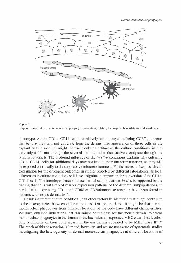
53
Dermal mononuclear phagocytes
phenotype. As the CD1a– CD14+ cells repetitively are portrayed as being CCR7–, it seems that in vivo they will not emigrate from the dermis. The appearance of these cells in the explant culture medium might represent only an artifact of the culture conditions, in that they might fall out through the severed dermis, rather than actively emigrate through the lymphatic vessels. The profound influence of the in vitro conditions explains why culturing CD1a– CD14+ cells for additional days may not lead to their further maturation, as they will be exposed continually to the suppressive microenvironment. Furthermore, it also provides an explanation for the divergent outcomes in studies reported by different laboratories, as local differences in culture conditions will have a significant impact on the conversion of the CD1a– CD14+ cells. The interdependence of these dermal subpopulations in vivo is supported by the finding that cells with mixed marker expression patterns of the different subpopulations, in particular co-expressing CD1a and CD68 or CD206/mannose receptor, have been found in patients with atopic dermatitis75.
Besides different culture conditions, can other factors be identified that might contribute to the discrepancies between different studies? On the one hand, it might be that dermal mononuclear phagocytes from different locations of the body have different characteristics. We have obtained indications that this might be the case for the mouse dermis. Whereas mononuclear phagocytes in the dermis of the back skin all expressed MHC class II molecules, only a minority of their counterparts in the ear dermis appeared to be MHC class II+ 60. The reach of this observation is limited, however, and we are not aware of systematic studies investigating the heterogeneity of dermal mononuclear phagocytes at different locations of
CD14+ FXIIIa+
lymphatic vessel
epidermis
CD1c+ CD14+
CD1a+ CD1c+
Figure 1. Proposed model of dermal mononuclear phagocyte maturation, relating the major subpopulations of dermal cells.
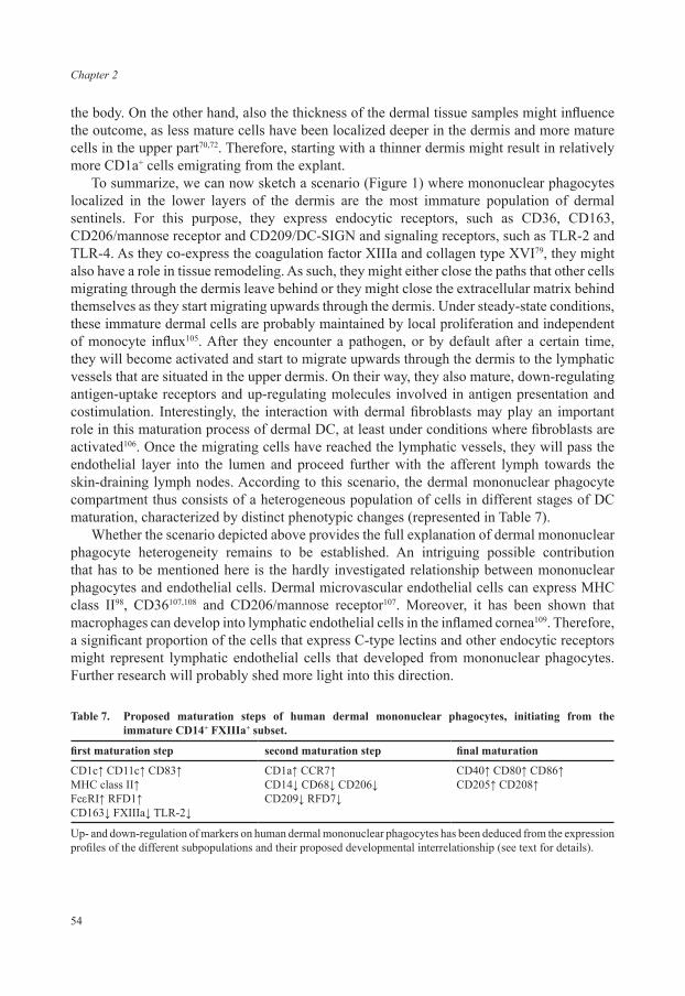
Chapter 2
54
the body. On the other hand, also the thickness of the dermal tissue samples might influence the outcome, as less mature cells have been localized deeper in the dermis and more mature cells in the upper part70,72. Therefore, starting with a thinner dermis might result in relatively more CD1a+ cells emigrating from the explant.
To summarize, we can now sketch a scenario (Figure 1) where mononuclear phagocytes localized in the lower layers of the dermis are the most immature population of dermal sentinels. For this purpose, they express endocytic receptors, such as CD36, CD163, CD206/mannose receptor and CD209/DC-SIGN and signaling receptors, such as TLR-2 and TLR-4. As they co-express the coagulation factor XIIIa and collagen type XVI79, they might also have a role in tissue remodeling. As such, they might either close the paths that other cells migrating through the dermis leave behind or they might close the extracellular matrix behind themselves as they start migrating upwards through the dermis. Under steady-state conditions, these immature dermal cells are probably maintained by local proliferation and independent of monocyte influx105. After they encounter a pathogen, or by default after a certain time, they will become activated and start to migrate upwards through the dermis to the lymphatic vessels that are situated in the upper dermis. On their way, they also mature, down-regulating antigen-uptake receptors and up-regulating molecules involved in antigen presentation and costimulation. Interestingly, the interaction with dermal fibroblasts may play an important role in this maturation process of dermal DC, at least under conditions where fibroblasts are activated106. Once the migrating cells have reached the lymphatic vessels, they will pass the endothelial layer into the lumen and proceed further with the afferent lymph towards the skin-draining lymph nodes. According to this scenario, the dermal mononuclear phagocyte compartment thus consists of a heterogeneous population of cells in different stages of DC maturation, characterized by distinct phenotypic changes (represented in Table 7).
Whether the scenario depicted above provides the full explanation of dermal mononuclear phagocyte heterogeneity remains to be established. An intriguing possible contribution that has to be mentioned here is the hardly investigated relationship between mononuclear phagocytes and endothelial cells. Dermal microvascular endothelial cells can express MHC class II98, CD36107,108 and CD206/mannose receptor107. Moreover, it has been shown that macrophages can develop into lymphatic endothelial cells in the inflamed cornea109. Therefore, a significant proportion of the cells that express C-type lectins and other endocytic receptors might represent lymphatic endothelial cells that developed from mononuclear phagocytes. Further research will probably shed more light into this direction.
Table 7. Proposed maturation steps of human dermal mononuclear phagocytes, initiating from the immature CD14+ FXIIIa+ subset.
first maturation step second maturation step final maturation
CD1c↑ CD11c↑ CD83↑ MHC class II↑ FcεRI↑ RFD1↑ CD163↓ FXIIIa↓ TLR-2↓
CD1a↑ CCR7↑CD14↓ CD68↓ CD206↓CD209↓ RFD7↓
CD40↑ CD80↑ CD86↑CD205↑ CD208↑
Up- and down-regulation of markers on human dermal mononuclear phagocytes has been deduced from the expression profiles of the different subpopulations and their proposed developmental interrelationship (see text for details).

55
Dermal mononuclear phagocytes
Phagocyte populations recruited to the inflamed skinCompared to the heterogeneity of the dermal mononuclear phagocytes in healthy skin
elaborated above, the inflamed skin contains an even bigger repertoire of phagocytes (summarized in Table 8). So far, five additional subsets of potential antigen-presenting cells have been defined in the inflamed skin, producing unique profiles of soluble inflammatory mediators and skewing T cell stimulation into different directions. Moreover, different skin pathologies are characterized by distinct profiles of recruited phagocyte subsets (Table 9). As many of these skin diseases will be discussed in more detail in the following chapters, we will just give here a broad overview of the involved cells.
Monocytes have been known for long to infiltrate inflamed tissues and to develop there into macrophages and DC. Thus, they contribute to the clearance of pathogens and to the resolution of inflammation110,111. Several types of DC, including inflammatory dendritic epidermal cells (IDEC), Tip-DC (TNF- and iNOS-producing DC) and plasmacytoid DC (pDC), have been described more recently in the inflamed skin that may or may not derive directly from infiltrating monocytes. IDEC have been portrayed to produce a myriad of different cytokines, among others IL-12 and IL-18, with which they create a Th1-inducing environment112.
Table 8. Phenotypes of additional phagocyte subpopulations in the inflamed human skin.MoDC 1 IDEC 2 Tip-DC 3 pDC 4 PMN 5
CD1a+ CD1a+ CD1a– CD1a–
CD1c– CD1c–
CD11bvar CD11b+ CD11b– CD11b+
CD11c+ CD11c+ CD11c– CD11c–
CD14– CD14– CD14– CD14–
CD23+
CD36+ CD36–
CD68+
CD40+
CD66b+
CD80+ CD80+
CD83+
CD86+ CD86+ CD86+
CD123+
CD206+ CD206–
CD207– CD207– CD207–
CD208+
CD303+
FcεRI+ FcεRI+
iNOS+
MHC class II+ MHC class II+ MHC class II+ MHC class II+ MHC class II+
elastase+
TNF-α+
IDEC - inflammatory dendritic epidermal cell; MoDC - monocyte-derived dendritic cell; PMN - polymorphonuclear neutrophilic granulocyte; pDC - plasmacytoid dendritic cell; TipDC - TNF- and iNOS-producing dendritic cell Main references: (1)121, (2)112, (3)113, (4)114, (5)117

Chapter 2
56
Tip-DC are major TNF-α and iNOS producers in the inflamed skin113, while plasmacytoid DC (also known as type 1 interferon producing cells; IPC) are characterized as principal type 1 interferon (IFN-α, β) producing cells114. Furthermore, polymorphonuclear neutrophils (PMN) have been known for long to infiltrate inflamed tissues. They are the first phagocytes to arrive there, even before monocytes. However, for long they have been considered only as phagocytes that non-specifically engulf pathogens, cell remnants and tissue debris, but do not contribute further to an immune response. Only more recently have their immune-stimulating capacities become apparent115,116. Furthermore, they have been shown to infiltrate ultraviolet-irradiated skin and to produce large amounts of IL-4 and IL-10, thus contributing to immunosuppression and photoageing117,118. Moreover, the recent finding that neutrophils may transdifferentiate into macrophages119,120 opens additional, unexpected pathways for the contribution of these cells to inflammatory processes in the skin.
With all these potential antigen-presenting cell populations accumulating in the inflamed skin, one starts to wonder which contribute predominantly to the induction of the adaptive immune response. After the discovery that LC are not the main immunostimulatory cells in the skin under all conditions, the same fate may await the dermis-resident mononuclear phagocytes. An illustrative example in this respect concerns the initiation of the immune response upon Leishmania infection in the mouse model. After it was determined that epidermal LC are not shuttling any Leishmania-antigens into the draining lymph nodes31, dermal antigen-presenting cells have been the prime candidates for this function32. Nevertheless, the newest results now suggest that none of the resident antigen-presenting cell subpopulations is responsible for the stimulation of Leishmania antigen-specific T cells in vivo. It appears that monocyte-derived DC126 or lymph node-resident DC127 are major candidates to perform this function. A similar scenario has been proposed for the initiation of the mouse immune response against Herpex simplex, where CD8α+ DC, which are most probably lymph node resident, have been shown to activate T cells 29. But even the PMN are not excluded anymore as it has been shown that PMN shuttle Mycobacterium bovis bacilli into skin-draining lymph nodes128. There, they might induce the immune response themselves upon acquisition of the appropriate stimulating molecules or transfer antigens to resident DC.
The notion that a number of antigen-presenting cell populations exists in the skin that are potentially equipped to stimulate T cell responses suggests that the categorization of a single subpopulation as main presenter seems to be outdated. Rather, multiple different
Table 9. Inflammatory phagocytes recruited to the skin under different pathologic conditions.
cell type involved in disease reference
IDEC contact dermatitis, atopic dermatitis, atopic eczema, allergic contact eczema, psoriasis vulgaris, lichen planus, cutaneous T-cell lymphoma; absent in lupus erythematosus
1,2
Tip-DC psoriasis vulgaris 3pDC psoriasis vulgaris, contact dermatitis, lupus erythematosus; absent in atopic
dermatitis, UV-irradiated skin, polymorphic light eruption2,4,5
IDEC - inflammatory dendritic epidermal cell; pDC - plasmacytoid dendritic cell; TipDC - TNF- and iNOS-producing dendritic cell Main references: (1)122; (2)123; (3)113; (4)124; (5)125.

57
Dermal mononuclear phagocytes
subpopulations appear to carry antigens into lymph nodes and induce there specific immune responses, with subtle differences depending on the nature of the involved cells. This redundancy clearly has selective advantage in evolution. On the one hand, more antigens will be transported, differently processed on the basis of the cellular machinery, and presented. This has been hinted recently in a study, where both CD11b+ Gr-1+ (PMN) and CD11b+ Gr-1-
cells (mononuclear phagocytes) have been demonstrated to rescue partially the deficiency of a CCR6 knock-out mouse to induce adaptive immune responses against pathogen-associated antigens129. On the other hand, pathogens might also block one antigen-presenting pathway or another in order to inhibit the host response upon infection. Consequently, being prepared and possessing more than a single antigen-presenting subpopulation might turn out to be crucial for the body to induce a life-saving adaptive immune response against a variety of pathogens. Thereby, it might still turn out that also dermis-resident antigen-presenting cells play an important role in this, and that, depending on the pathogen involved and the site and stage of infection, different cell types contribute differently to the host defense.
REFERENCES
1. Langerhans, P. Über die Nerven der menschlichen Haut. [Virchows] Archiv für pathologische Anatomie und Physiologie, und für klinische Medizin 44, 325-337 (1868).
2. Stingl, G., Katz, S.I., Clement, L., Green, I. & Shevach, E.M. Immunologic functions of Ia-bearing epidermal Langerhans cells. J Immunol 121, 2005-2013 (1978).
3. Schuler, G. & Steinman, R.M. Murine epidermal Langerhans cells mature into potent immunostimulatory dendritic cells in vitro. J Exp Med 161, 526-546 (1985).
4. Villadangos, J.A. & Heath, W.R. Life cycle, migration and antigen presenting functions of spleen and lymph node dendritic cells: limitations of the Langerhans cells paradigm. Semin Immunol 17, 262-272 (2005).
5. Mikulowska, A. & Falck, B. Distributional changes of Langerhans cells in human skin during irritant contact dermatitis. Arch Dermatol Res 286, 429-433 (1994).
6. Oka, D., Nakagawa, S., Oota, T. & Ueki, H. Distribution of 2,4-dinitrophenyl groups on the epidermal Langerhans cells of guinea pigs following skin painting with 2,4-dinitrochlorobenzene. Dermatologica 172, 12-17 (1986).
7. Toews, G.B., Bergstresser, P.R. & Streilein, J.W. Epidermal Langerhans cell density determines whether contact hypersensitivity or unresponsiveness follows skin painting with DNFB. J Immunol 124, 445-453 (1980).
8. Callard, R.E. & Harper, J.I. The skin barrier, atopic dermatitis and allergy: a role for Langerhans cells? Trends Immunol 28, 294-298 (2007).
9. Semper, A.E., Heron, K., Woollard, A.C., Kochan, J.P., Friedmann, P.S., Church, M.K. & Reischl, I.G. Surface expression of Fc epsilon RI on Langerhans› cells of clinically uninvolved skin is associated with disease activity in atopic dermatitis, allergic asthma, and rhinitis. J Allergy Clin Immunol 112, 411-419 (2003).
10. Cumberbatch, M., Singh, M., Dearman, R.J., Young, H.S., Kimber, I. & Griffiths, C.E. Impaired Langerhans cell migration in psoriasis. J Exp Med (2006).
11. Demidem, A., Taylor, J.R., Grammer, S.F. & Streilein, J.W. T-lymphocyte-activating properties of epidermal antigen-presenting cells from normal and psoriatic skin: evidence that psoriatic epidermal antigen-presenting cells resemble cultured normal Langerhans cells. J Invest Dermatol 97, 454-460 (1991).
12. Morhenn, V.B. Langerhans cells may trigger the psoriatic disease process via production of nitric oxide. Immunol Today 18, 433-436 (1997).
13. Prens, E.P., Benne, K., van Joost, T. & Benner, R. The autologous mixed epidermal cell-T lymphocyte reaction is elevated in psoriasis: a crucial role for epidermal HLA-DR+/CD1a– antigen-presenting cells. J Invest Dermatol 96, 880-887 (1991).

Chapter 2
58
14. Moll, H., Fuchs, H., Blank, C. & Rollinghoff, M. Langerhans cells transport Leishmania major from the infected skin to the draining lymph node for presentation to antigen-specific T cells. Eur J Immunol 23, 1595-1601 (1993).
15. Will, A., Blank, C., Rollinghoff, M. & Moll, H. Murine epidermal Langerhans cells are potent stimulators of an antigen-specific T cell response to Leishmania major, the cause of cutaneous leishmaniasis. Eur J Immunol 22, 1341-1347 (1992).
16. Sprecher, E. & Becker, Y. Skin Langerhans cells play an essential role in the defense against HSV-1 infection. Arch Virol 91, 341-349 (1986).
17. Sprecher, E. & Becker, Y. Langerhans cell density and activity in mouse skin and lymph nodes affect herpes simplex type 1 (HSV-1) pathogenicity. Arch Virol 107, 191-205 (1989).
18. Aberer, W., Schuler, G., Stingl, G., Honigsmann, H. & Wolff, K. Ultraviolet light depletes surface markers of Langerhans cells. J Invest Dermatol 76, 202-210 (1981).
19. Dai, R. & Streilein, J.W. Sensitizing capacity of Langerhans› cells obtained from ultraviolet-B-exposed murine skin. Immunology 86, 661-667 (1995).
20. Humm, S.A. & Cole, S. Changes with time in Langerhans cell number, ATPase reactivity and morphology in murine epidermis after exposure to UVB. Photodermatol 3, 174-178 (1986).
21. Kolgen, W., van Meurs, M., Jongsma, M., van Weelden, H., Bruijnzeel-Koomen, C.A., Knol, E.F., van Vloten, W.A., Laman, J. & de Gruijl, F.R. Differential expression of cytokines in UV-B-exposed skin of patients with polymorphous light eruption: correlation with Langerhans cell migration and immunosuppression. Arch Dermatol 140, 295-302 (2004).
22. Streilein, J.W. Antigen-presenting cells in the induction of contact hypersensitivity in mice: evidence that Langerhans cells are sufficient but not required. J Invest Dermatol 93, 443-448 (1989).
23. Tse, Y. & Cooper, K.D. Cutaneous dermal Ia+ cells are capable of initiating delayed type hypersensitivity responses. J Invest Dermatol 94, 267-272 (1990).
24. Bennett, C.L., van Rijn, E., Jung, S., Inaba, K., Steinman, R.M., Kapsenberg, M.L. & Clausen, B.E. Inducible ablation of mouse Langerhans cells diminishes but fails to abrogate contact hypersensitivity. J Cell Biol 169, 569-576 (2005).
25. Kaplan, D.H., Jenison, M.C., Saeland, S., Shlomchik, W.D. & Shlomchik, M.J. Epidermal langerhans cell-deficient mice develop enhanced contact hypersensitivity. Immunity 23, 611-620 (2005).
26. Kissenpfennig, A., Henri, S., Dubois, B., Laplace-Builhe, C., Perrin, P., Romani, N., Tripp, C.H., Douillard, P., Leserman, L., Kaiserlian, D., Saeland, S., Davoust, J. & Malissen, B. Dynamics and function of Langerhans cells in vivo: dermal dendritic cells colonize lymph node areas distinct from slower migrating Langerhans cells. Immunity 22, 643-654 (2005).
27. Itano, A.A., McSorley, S.J., Reinhardt, R.L., Ehst, B.D., Ingulli, E., Rudensky, A.Y. & Jenkins, M.K. Distinct dendritic cell populations sequentially present antigen to CD4 T cells and stimulate different aspects of cell-mediated immunity. Immunity 19, 47-57 (2003).
28. Stoecklinger, A., Grieshuber, I., Scheiblhofer, S., Weiss, R., Ritter, U., Kissenpfennig, A., Malissen, B., Romani, N., Koch, F., Ferreira, F., Thalhamer, J. & Hammerl, P. Epidermal langerhans cells are dispensable for humoral and cell-mediated immunity elicited by gene gun immunization. J Immunol 179, 886-893 (2007).
29. Allan, R.S., Smith, C.M., Belz, G.T., van Lint, A.L., Wakim, L.M., Heath, W.R. & Carbone, F.R. Epidermal viral immunity induced by CD8alpha+ dendritic cells but not by Langerhans cells. Science 301, 1925-1928 (2003).
30. Zhao, X., Deak, E., Soderberg, K., Linehan, M., Spezzano, D., Zhu, J., Knipe, D.M. & Iwasaki, A. Vaginal submucosal dendritic cells, but not Langerhans cells, induce protective Th1 responses to herpes simplex virus-2. J Exp Med 197, 153-162 (2003).
31. Ritter, U., Meissner, A., Scheidig, C. & Korner, H. CD8alpha– and Langerin-negative dendritic cells, but not Langerhans cells, act as principal antigen-presenting cells in leishmaniasis. Eur J Immunol 34, 1542-1550 (2004).
32. Ritter, U. & Osterloh, A. A new view on cutaneous dendritic cell subsets in experimental leishmaniasis. Med Microbiol Immunol 196, 51-59 (2007).
33. Kumkate, S., Jenkins, G.R., Paveley, R.A., Hogg, K.G. & Mountford, A.P. CD207+ Langerhans cells constitute a minor population of skin-derived antigen-presenting cells in the draining lymph node following exposure to Schistosoma mansoni. Int J Parasitol 37, 209-220 (2007).

59
Dermal mononuclear phagocytes
34. van der Aar, A.M., Sylva-Steenland, R.M., Bos, J.D., Kapsenberg, M.L., de Jong, E.C. & Teunissen, M.B. Loss of TLR2, TLR4, and TLR5 on Langerhans cells abolishes bacterial recognition. J Immunol 178, 1986-1990 (2007).
35. Headington, J.T. The histiocyte. In memoriam. Arch Dermatol 122, 532-533 (1986).36. van Furth, R., Cohn, Z.A., Hirsch, J.G., Humphrey, J.H., Spector, W.G. & Langevoort, H.L. The mononuclear
phagocyte system: a new classification of macrophages, monocytes, and their precursor cells. Bull World Health Organ 46, 845-852 (1972).
37. Vakkila, J., Lotze, M.T., Riga, C. & Jaffe, R. A basis for distinguishing cultured dendritic cells and macrophages in cytospins and fixed sections. Pediatr Dev Pathol 8, 43-51 (2005).
38. Wood, G.S., Turner, R.R., Shiurba, R.A., Eng, L. & Warnke, R.A. Human dendritic cells and macrophages. In situ immunophenotypic definition of subsets that exhibit specific morphologic and microenvironmental characteristics. Am J Pathol 119, 73-82 (1985).
39. Goerdt, S., Kodelja, V., Schmuth, M., Orfanos, C.E. & Sorg, C. The mononuclear phagocyte-dendritic cell dichotomy: myths, facts, and a revised concept. Clin Exp Immunol 105, 1-9 (1996).
40. Hume, D.A. The mononuclear phagocyte system. Curr Opin Immunol 18, 49-53 (2006).41. Hume, D.A., Ross, I.L., Himes, S.R., Sasmono, R.T., Wells, C.A. & Ravasi, T. The mononuclear phagocyte
system revisited. J Leukoc Biol 72, 621-627 (2002).42. Nikolic, T. & Leenen, P.J.M. Histiocyte function and development in the normal immune system. Chapter 3., in
Histiocytic Disorders of Children and Adults. (eds. S. Weitzman & R.M. Egeler) 40-65 (Cambridge University Press, Cambridge, UK; 2005).
43. Steinman, R.M. Dendritic cells: versatile controllers of the immune system. Nat Med 13, 1155-1159 (2007).44. Ginhoux, F., Tacke, F., Angeli, V., Bogunovic, M., Loubeau, M., Dai, X.M., Stanley, E.R., Randolph, G.J. &
Merad, M. Langerhans cells arise from monocytes in vivo. Nat Immunol 7, 265-273 (2006).45. Sallusto, F. & Lanzavecchia, A. Efficient presentation of soluble antigen by cultured human dendritic cells
is maintained by granulocyte/macrophage colony-stimulating factor plus interleukin 4 and downregulated by tumor necrosis factor alpha. J Exp Med 179, 1109-1118 (1994).
46. Leenen, P.J.M., Radosevic, K., Voerman, J.S.A., Salomon, B., van Rooijen, N., Klatzmann, D. & van Ewijk, W. Heterogeneity of mouse spleen dendritic cells: in vivo phagocytic activity, expression of macrophage markers, and subpopulation turnover. J Immunol 160, 2166-2173 (1998).
47. Sallusto, F., Cella, M., Danieli, C. & Lanzavecchia, A. Dendritic cells use macropinocytosis and the mannose receptor to concentrate macromolecules in the major histocompatibility complex class II compartment: downregulation by cytokines and bacterial products. J Exp Med 182, 389-400 (1995).
48. Steinman, R.M. & Cohn, Z.A. Identification of a novel cell type in peripheral lymphoid organs of mice. II. Functional properties in vitro. J Exp Med 139, 380-397 (1974).
49. McCormack, J.M., Moore, S.C., Gatewood, J.W. & Walker, W.S. Mouse splenic macrophage cell lines with different antigen-presenting activities for CD4+ helper T cell subsets and allogeneic CD8+ T cells. Cell Immunol 145, 359-371 (1992).
50. Pozzi, L.A., Maciaszek, J.W. & Rock, K.L. Both dendritic cells and macrophages can stimulate naive CD8 T cells in vivo to proliferate, develop effector function, and differentiate into memory cells. J Immunol 175, 2071-2081 (2005).
51. Savina, A. & Amigorena, S. Phagocytosis and antigen presentation in dendritic cells. Immunol Rev 219, 143-156 (2007).
52. Wilson, N.S. & Villadangos, J.A. Regulation of antigen presentation and cross-presentation in the dendritic cell network: facts, hypothesis, and immunological implications. Adv Immunol 86, 241-305 (2005).
53. Delamarre, L., Pack, M., Chang, H., Mellman, I. & Trombetta, E.S. Differential lysosomal proteolysis in antigen-presenting cells determines antigen fate. Science 307, 1630-1634 (2005).
54. Padilla, J., Daley, E., Chow, A., Robinson, K., Parthasarathi, K., McKenzie, A.N., Tschernig, T., Kurup, V.P., Donaldson, D.D. & Grunig, G. IL-13 regulates the immune response to inhaled antigens. J Immunol 174, 8097-8105 (2005).
55. Yagi, M., Miyamoto, T., Sawatani, Y., Iwamoto, K., Hosogane, N., Fujita, N., Morita, K., Ninomiya, K., Suzuki, T., Miyamoto, K., Oike, Y., Takeya, M., Toyama, Y. & Suda, T. DC-STAMP is essential for cell-cell fusion in osteoclasts and foreign body giant cells. J Exp Med 202, 345-351 (2005).

Chapter 2
60
56. Fortsch, D., Rollinghoff, M. & Stenger, S. IL-10 converts human dendritic cells into macrophage-like cells with increased antibacterial activity against virulent Mycobacterium tuberculosis. J Immunol 165, 978-987 (2000).
57. Ichikawa, M., Sugita, M., Takahashi, M., Satomi, M., Takeshita, T., Araki, T. & Takahashi, H. Breast milk macrophages spontaneously produce granulocyte-macrophage colony-stimulating factor and differentiate into dendritic cells in the presence of exogenous interleukin-4 alone. Immunology 108, 189-195 (2003).
58. Nestle, F.O. & Nickoloff, B.J. A fresh morphological and functional look at dermal dendritic cells. J Cutan Pathol 22, 385-393 (1995).
59. Davis, A.L., McKenzie, J.L. & Hart, D.N. HLA-DR-positive leucocyte subpopulations in human skin include dendritic cells, macrophages, and CD7-negative T cells. Immunology 65, 573-581 (1988).
60. Dupasquier, M., Stoitzner, P., Oudenaren, A., Romani, N. & Leenen, P.J. Macrophages and dendritic cells constitute a major subpopulation of cells in the mouse dermis. J Invest Dermatol 123, 876-879 (2004).
61. Lenz, A., Heine, M., Schuler, G. & Romani, N. Human and murine dermis contain dendritic cells. Isolation by means of a novel method and phenotypical and functional characterization. J Clin Invest 92, 2587-2596 (1993).
62. Nestle, F.O., Zheng, X.G., Thompson, C.B., Turka, L.A. & Nickoloff, B.J. Characterization of dermal dendritic cells obtained from normal human skin reveals phenotypic and functionally distinctive subsets. J Immunol 151, 6535-6545 (1993).
63. Duraiswamy, N., Tse, Y., Hammerberg, C., Kang, S. & Cooper, K.D. Distinction of class II MHC+ Langerhans cell-like interstitial dendritic antigen-presenting cells in murine dermis from dermal macrophages. J Invest Dermatol 103, 678-683 (1994).
64. Meunier, L., Gonzalez-Ramos, A. & Cooper, K.D. Heterogeneous populations of class II MHC+ cells in human dermal cell suspensions. Identification of a small subset responsible for potent dermal antigen-presenting cell activity with features analogous to Langerhans cells. J Immunol 151, 4067-4080 (1993).
65. Valladeau, J., Ravel, O., Dezutter-Dambuyant, C., Moore, K., Kleijmeer, M., Liu, Y., Duvert-Frances, V., Vincent, C., Schmitt, D., Davoust, J., Caux, C., Lebecque, S. & Saeland, S. Langerin, a novel C-type lectin specific to Langerhans cells, is an endocytic receptor that induces the formation of Birbeck granules. Immunity 12, 71-81 (2000).
66. Murphy, G.F., Bhan, A.K., Harrist, T.J. & Mihm, M.C., Jr. In situ identification of T6-positive cells in normal human dermis by immunoelectron microscopy. Br J Dermatol 108, 423-431 (1983).
67. Murphy, G.F., Bronstein, B.R., Knowles, R.W. & Bhan, A.K. Ultrastructural documentation of M241 glycoprotein on dendritic and endothelial cells in normal human skin. Lab Invest 52, 264-269 (1985).
68. Osterhoff, B., Rappersberger, K., Wang, B., Koszik, F., Ochiai, K., Kinet, J.P. & Stingl, G. Immunomorphologic characterization of Fc epsilon RI-bearing cells within the human dermis. J Invest Dermatol 102, 315-320 (1994).
69. van de Rijn, M., Lerch, P.G., Bronstein, B.R., Knowles, R.W., Bhan, A.K. & Terhorst, C. Human cutaneous dendritic cells express two glycoproteins T6 and M241 which are biochemically identical to those found on cortical thymocytes. Hum Immunol 9, 201-210 (1984).
70. Angel, C.E., Lala, A., Chen, C.J., Edgar, S.G., Ostrovsky, L.L. & Dunbar, P.R. CD14+ antigen-presenting cells in human dermis are less mature than their CD1a+ counterparts. Int Immunol (2007).
71. Angel, C.E., George, E., Brooks, A.E., Ostrovsky, L.L., Brown, T.L. & Dunbar, P.R. Cutting edge: CD1a+ antigen-presenting cells in human dermis respond rapidly to CCR7 ligands. J Immunol 176, 5730-5734 (2006).
72. Zaba, L.C., Fuentes-Duculan, J., Steinman, R.M., Krueger, J.G. & Lowes, M.A. Normal human dermis contains distinct populations of CD11cBDCA-1 dendritic cells and CD163FXIIIA macrophages. J Clin Invest 117, 2517-2525 (2007).
73. Sontheimer, R.D., Matsubara, T. & Seelig, L.L., Jr. A macrophage phenotype for a constitutive, class II antigen-expressing, human dermal perivascular dendritic cell. J Invest Dermatol 93, 154-159 (1989).
74. Weber-Matthiesen, K. & Sterry, W. Organization of the monocyte/macrophage system of normal human skin. J Invest Dermatol 95, 83-89 (1990).
75. Kiekens, R.C., Thepen, T., Oosting, A.J., Bihari, I.C., Van De Winkel, J.G., Bruijnzeel-Koomen, C.A. & Knol, E.F. Heterogeneity within tissue-specific macrophage and dendritic cell populations during cutaneous inflammation in atopic dermatitis. Br J Dermatol 145, 957-965 (2001).

61
Dermal mononuclear phagocytes
76. Ebner, S., Ehammer, Z., Holzmann, S., Schwingshackl, P., Forstner, M., Stoitzner, P., Huemer, G.M., Fritsch, P. & Romani, N. Expression of C-type lectin receptors by subsets of dendritic cells in human skin. Int Immunol 16, 877-887 (2004).
77. Turville, S.G., Cameron, P.U., Handley, A., Lin, G., Pohlmann, S., Doms, R.W. & Cunningham, A.L. Diversity of receptors binding HIV on dendritic cell subsets. Nat Immunol 3, 975-983 (2002).
78. Mosher, D.F. & Schad, P.E. Cross-linking of fibronectin to collagen by blood coagulation Factor XIIIa. J Clin Invest 64, 781-787 (1979).
79. Akagi, A., Tajima, S., Ishibashi, A., Matsubara, Y., Takehana, M., Kobayashi, S. & Yamaguchi, N. Type XVI collagen is expressed in factor XIIIa+ monocyte-derived dermal dendrocytes and constitutes a potential substrate for factor XIIIa. J Invest Dermatol 118, 267-274 (2002).
80. Bursch, L.S., Wang, L., Igyarto, B., Kissenpfennig, A., Malissen, B., Kaplan, D.H. & Hogquist, K.A. Identification of a novel population of Langerin+ dendritic cells. J Exp Med 204, 3147-3156 (2007).
81. Ginhoux, F., Collin, M.P., Bogunovic, M., Abel, M., Leboeuf, M., Helft, J., Ochando, J., Kissenpfennig, A., Malissen, B., Grisotto, M., Snoeck, H., Randolph, G. & Merad, M. Blood-derived dermal langerin+ dendritic cells survey the skin in the steady state. J Exp Med 204, 3133-3146 (2007).
82. Poulin, L.F., Henri, S., de Bovis, B., Devilard, E., Kissenpfennig, A. & Malissen, B. The dermis contains langerin+ dendritic cells that develop and function independently of epidermal Langerhans cells. J Exp Med 204, 3119-3131 (2007).
83. Kurimoto, I., Grammer, S.F., Shimizu, T., Nakamura, T. & Streilein, J.W. Role of F4/80+ cells during induction of hapten-specific contact hypersensitivity. Immunology 85, 621-629 (1995).
84. Chun, K.H., Imai, Y., Higashi, N. & Irimura, T. Migration of dermal cells expressing a macrophage C-type lectin during the sensitization phase of delayed-type hypersensitivity. J Leukoc Biol 68, 471-478. (2000).
85. Dupasquier, M., Stoitzner, P., Wan, H., Cerqueira, D., van Oudenaren, A., Voerman, J.S., Denda-Nagai, K., Irimura, T., Raes, G., Romani, N. & Leenen, P.J. The dermal microenvironment induces the expression of the alternative activation marker CD301/mMGL in mononuclear phagocytes, independent of IL-4/IL-13 signaling. J Leukoc Biol 80, 838-849 (2006).
86. de Jong, J.P., Voerman, J.S., van der Sluijs-Gelling, A.J., Willemsen, R. & Ploemacher, R.E. A monoclonal antibody (ER-HR3) against murine macrophages. I. Ontogeny, distribution and enzyme histochemical characterization of ER-HR3-positive cells. Cell Tissue Res 275, 567-576 (1994).
87. Larsen, C.P., Morris, P.J. & Austyn, J.M. Migration of dendritic leukocytes from cardiac allografts into host spleens. A novel pathway for initiation of rejection. J Exp Med 171, 307-314 (1990).
88. Morelli, A.E., Rubin, J.P., Erdos, G., Tkacheva, O.A., Mathers, A.R., Zahorchak, A.F., Thomson, A.W., Falo, L.D., Jr. & Larregina, A.T. CD4+ T cell responses elicited by different subsets of human skin migratory dendritic cells. J Immunol 175, 7905-7915 (2005).
89. Larregina, A.T., Morelli, A.E., Spencer, L.A., Logar, A.J., Watkins, S.C., Thomson, A.W. & Falo, L.D., Jr. Dermal-resident CD14+ cells differentiate into Langerhans cells. Nat Immunol 2, 1151-1158. (2001).
90. Barbaroux, J.B., Kwan, W.H., Allam, J.P., Novak, N., Bieber, T., Fridman, W.H., Groves, R. & Mueller, C.G. Tumor necrosis factor-alpha- and IL-4-independent development of Langerhans cell-like dendritic cells from M-CSF-conditioned precursors. J Invest Dermatol 126, 114-120 (2006).
91. Stoitzner, P., Holzmann, S., McLellan, A.D., Ivarsson, L., Stossel, H., Kapp, M., Kammerer, U., Douillard, P., Kampgen, E., Koch, F., Saeland, S. & Romani, N. Visualization and characterization of migratory Langerhans cells in murine skin and lymph nodes by antibodies against Langerin/CD207. J Invest Dermatol 120, 266-274 (2003).
92. Weinlich, G., Heine, M., Stossel, H., Zanella, M., Stoitzner, P., Ortner, U., Smolle, J., Koch, F., Sepp, N.T., Schuler, G. & Romani, N. Entry into afferent lymphatics and maturation in situ of migrating murine cutaneous dendritic cells. J Invest Dermatol 110, 441-448 (1998).
93. Anjuere, F., Martin, P., Ferrero, I., Fraga, M.L., del Hoyo, G.M., Wright, N. & Ardavin, C. Definition of dendritic cell subpopulations present in the spleen, Peyer›s patches, lymph nodes, and skin of the mouse. Blood 93, 590-598 (1999).
94. Iwasaki, A. The importance of CD11b+ dendritic cells in CD4+ T cell activation in vivo: with help from interleukin 1. J Exp Med 198, 185-190 (2003).
95. Sepulveda-Merrill, C., Mayall, S., Hamblin, A.S. & Breathnach, S.M. Antigen-presenting capacity in normal human dermis is mainly subserved by CD1a+ cells. Br J Dermatol 131, 15-22 (1994).

Chapter 2
62
96. Dezutter-Dambuyant, C., Schmitt, D., Staquet, M.J., Boumsell, L. & Thivolet, J. Cleavage of Langerhans cell surface CD1a molecule by trypsin. Res Immunol 140, 377-390 (1989).
97. Inaba, K., Swiggard, W., Inaba, M., Meltzer, J., Mirza, A., Sasagawa, T., Nussenzweig, M. & Steinman, R. Tissue distribution of the DEC-205 protein that is detected by the monoclonal antibody NLDC-145. I. Expression on dendritic cells and other subsets of mouse leukocytes. Cell.Immunol. 163, 148-156 (1995).
98. Angel, C.E., George, E., Ostrovsky, L.L. & Dunbar, P.R. Comprehensive analysis of MHC-II expression in healthy human skin. Immunol Cell Biol 85, 363-369 (2007).
99. Jacobs, N., Chen, R.A., Gubser, C., Najarro, P. & Smith, G.L. Intradermal immune response after infection with Vaccinia virus. J Gen Virol 87, 1157-1161 (2006).
100. Brand, C.U., Hunger, R.E., Yawalkar, N., Gerber, H.A., Schaffner, T. & Braathen, L.R. Characterization of human skin-derived CD1a-positive lymph cells. Arch Dermatol Res 291, 65-72 (1999).
101. Yawalkar, N., Aebischer, M.C., Hunger, R., Brand, C.U. & Braathen, L.R. Effects of UV irradiation with one minimal erythema dose on human afferent skin lymph in vivo. Exp Dermatol 7, 362-368 (1998).
102. Hunger, R.E., Yawalkar, N., Braathen, L.R. & Brand, C.U. CD1a-positive dendritic cells transport the antigen DNCB intracellularly from the skin to the regional lymph nodes in the induction phase of allergic contact dermatitis. Arch Dermatol Res 293, 420-426 (2001).
103. Stoitzner, P., Tripp, C.H., Douillard, P., Saeland, S. & Romani, N. Migratory Langerhans cells in mouse lymph nodes in steady state and inflammation. J Invest Dermatol 125, 116-125 (2005).
104. de Gruijl, T.D., Sombroek, C.C., Lougheed, S.M., Oosterhoff, D., Buter, J., van den Eertwegh, A.J., Scheper, R.J. & Pinedo, H.M. A postmigrational switch among skin-derived dendritic cells to a macrophage-like phenotype is predetermined by the intracutaneous cytokine balance. J Immunol 176, 7232-7242 (2006).
105. Bogunovic, M., Ginhoux, F., Wagers, A., Loubeau, M., Isola, L.M., Lubrano, L., Najfeld, V., Phelps, R.G., Grosskreutz, C., Scigliano, E., Frenette, P.S. & Merad, M. Identification of a radio-resistant and cycling dermal dendritic cell population in mice and men. J Exp Med 203, 2627-2638 (2006).
106. Saalbach, A., Klein, C., Sleeman, J., Sack, U., Kauer, F., Gebhardt, C., Averbeck, M., Anderegg, U. & Simon, J.C. Dermal fibroblasts induce maturation of dendritic cells. J Immunol 178, 4966-4974 (2007).
107. Groger, M., Holnthoner, W., Maurer, D., Lechleitner, S., Wolff, K., Mayr, B.B., Lubitz, W. & Petzelbauer, P. Dermal microvascular endothelial cells express the 180-kDa macrophage mannose receptor in situ and in vitro. J Immunol 165, 5428-5434 (2000).
108. Petzelbauer, P., Bender, J.R., Wilson, J. & Pober, J.S. Heterogeneity of dermal microvascular endothelial cell antigen expression and cytokine responsiveness in situ and in cell culture. J Immunol 151, 5062-5072 (1993).
109. Maruyama, K., Ii, M., Cursiefen, C., Jackson, D.G., Keino, H., Tomita, M., Van Rooijen, N., Takenaka, H., D›Amore, P.A., Stein-Streilein, J., Losordo, D.W. & Streilein, J.W. Inflammation-induced lymphangiogenesis in the cornea arises from CD11b-positive macrophages. J Clin Invest 115, 2363-2372 (2005).
110. Serhan, C.N., Brain, S.D., Buckley, C.D., Gilroy, D.W., Haslett, C., O›Neill, L.A., Perretti, M., Rossi, A.G. & Wallace, J.L. Resolution of inflammation: state of the art, definitions and terms. FASEB J 21, 325-332 (2007).
111. Serhan, C.N. & Savill, J. Resolution of inflammation: the beginning programs the end. Nat Immunol 6, 1191-1197 (2005).
112. Novak, N. & Bieber, T. The role of dendritic cell subtypes in the pathophysiology of atopic dermatitis. J Am Acad Dermatol 53, S171-176 (2005).
113. Lowes, M.A., Chamian, F., Abello, M.V., Fuentes-Duculan, J., Lin, S.L., Nussbaum, R., Novitskaya, I., Carbonaro, H., Cardinale, I., Kikuchi, T., Gilleaudeau, P., Sullivan-Whalen, M., Wittkowski, K.M., Papp, K., Garovoy, M., Dummer, W., Steinman, R.M. & Krueger, J.G. Increase in TNF-{alpha} and inducible nitric oxide synthase-expressing dendritic cells in psoriasis and reduction with efalizumab (anti-CD11a). Proc Natl Acad Sci U S A 102, 19057-19062 (2005).
114. Liu, Y.J. IPC: professional type 1 interferon-producing cells and plasmacytoid dendritic cell precursors. Annu Rev Immunol 23, 275-306 (2005).
115. Ashtekar, A.R. & Saha, B. Poly’s plea: membership to the club of APCs. Trends Immunol 24, 485-490 (2003).116. Beauvillain, C., Delneste, Y., Scotet, M., Peres, A., Gascan, H., Guermonprez, P., Barnaba, V. & Jeannin, P.
Neutrophils efficiently cross-prime naive T cells in vivo. Blood 110, 2965-2973 (2007).117. Piskin, G., Bos, J.D. & Teunissen, M.B. Neutrophils infiltrating ultraviolet B-irradiated normal human skin
display high IL-10 expression. Arch Dermatol Res 296, 339-342 (2005).

63
Dermal mononuclear phagocytes
118. Rijken, F., Kiekens, R.C. & Bruijnzeel, P.L. Skin-infiltrating neutrophils following exposure to solar-simulated radiation could play an important role in photoageing of human skin. Br J Dermatol 152, 321-328 (2005).
119. Araki, H., Katayama, N., Yamashita, Y., Mano, H., Fujieda, A., Usui, E., Mitani, H., Ohishi, K., Nishii, K., Masuya, M., Minami, N., Nobori, T. & Shiku, H. Reprogramming of human postmitotic neutrophils into macrophages by growth factors. Blood 103, 2973-2980 (2004).
120. Sasmono, R.T., Ehrnsperger, A., Cronau, S.L., Ravasi, T., Kandane, R., Hickey, M.J., Cook, A.D., Himes, S.R., Hamilton, J.A. & Hume, D.A. Mouse neutrophilic granulocytes express mRNA encoding the macrophage colony-stimulating factor receptor (CSF-1R) as well as many other macrophage-specific transcripts and can transdifferentiate into macrophages in vitro in response to CSF-1. J Leukoc Biol 82, 111-123 (2007).
121. Randolph, G.J., Beaulieu, S., Lebecque, S., Steinman, R.M. & Muller, W.A. Differentiation of monocytes into dendritic cells in a model of transendothelial trafficking. Science 282, 480-483 (1998).
122. Wollenberg, A., Haberstok, J., Teichmann, B., Wen, S.P. & Bieber, T. Demonstration of the low-affinity IgE receptor Fc epsilonRII/CD23 in psoriatic epidermis: inflammatory dendritic epidermal cells (IDEC) but not Langerhans cells are the relevant CD1a-positive cell population. Arch Dermatol Res 290, 517-521 (1998).
123. Wollenberg, A., Wagner, M., Gunther, S., Towarowski, A., Tuma, E., Moderer, M., Rothenfusser, S., Wetzel, S., Endres, S. & Hartmann, G. Plasmacytoid dendritic cells: a new cutaneous dendritic cell subset with distinct role in inflammatory skin diseases. J Invest Dermatol 119, 1096-1102 (2002).
124. Narbutt, J., Lesiak, A., Skibinska, M., Wozniacka, A., Sysa-Jedrzejowska, A., Zalewska, A., Robak, T., Norval, M. & Smolewski, P. Lack of effect of repeated suberythemal ultraviolet-B exposures on human blood dendritic subtypes. Photodermatol Photoimmunol Photomed 21, 249-253 (2005).
125. Wackernagel, A., Massone, C., Hoefler, G., Steinbauer, E., Kerl, H. & Wolf, P. Plasmacytoid dendritic cells are absent in skin lesions of polymorphic light eruption. Photodermatol Photoimmunol Photomed 23, 24-28 (2007).
126. Leon, B., Lopez-Bravo, M. & Ardavin, C. Monocyte-derived dendritic cells formed at the infection site control the induction of protective T helper 1 responses against Leishmania. Immunity 26, 519-531 (2007).
127. Iezzi, G., Frohlich, A., Ernst, B., Ampenberger, F., Saeland, S., Glaichenhaus, N. & Kopf, M. Lymph node resident rather than skin-derived dendritic cells initiate specific T cell responses after Leishmania major infection. J Immunol 177, 1250-1256 (2006).
128. Abadie, V., Badell, E., Douillard, P., Ensergueix, D., Leenen, P.J., Tanguy, M., Fiette, L., Saeland, S., Gicquel, B. & Winter, N. Neutrophils rapidly migrate via lymphatics after Mycobacterium bovis BCG intradermal vaccination and shuttle live bacilli to the draining lymph nodes. Blood 106, 1843-1850 (2005).
129. Ravindran, R., Rusch, L., Itano, A., Jenkins, M.K. & McSorley, S.J. CCR6-dependent recruitment of blood phagocytes is necessary for rapid CD4 T cell responses to local bacterial infection. Proc Natl Acad Sci U S A 104, 12075-12080 (2007).


CHAPTER 3
AIM AND OUTLINE OF THE THESIS

Chapter 3
66
The skin forms the interface between the body and the outer environment. Consequently, among other important functions, it performs a crucial role in the host defense against pathogens. For this purpose, it contains large numbers of resident mononuclear phagocytes. These cells perform vital tasks when new invaders are encountered. As members of the innate immune system, they can fight attackers directly, and they can recruit and activate more cells of the immune system by releasing pro-inflammatory mediators. Moreover, they can also bridge the gap between the innate and the adaptive immune system by migrating to draining lymph nodes and initiate an adaptive immune response by stimulating antigen-specific T cells.
Consequently, insight into the characteristics of mononuclear phagocytes in the skin is crucial for the understanding of cutaneous responses to environmental triggers. Langerhans cells (LC), representing the mononuclear phagocyte subpopulation of the epidermis, are relatively well characterized. They form nowadays the paradigm of tissue-resident immature dendritic cells in the periphery. Tools to investigate them, such as specific anti-Langerin antibodies and mouse models in which LC can be manipulated specifically in vivo, have been developed recently and are widely used. The mononuclear phagocytes of the dermis, in contrast, have been studied much less. Investigators broadly acknowledge their existence, but the distinct subpopulations of dermal macrophages and DC remain ill-defined regarding their numbers, phenotypes and functions, giving rise to much confusion in the field.
The scope of the studies described in this thesis was to characterize the dermal mononuclear phagocytes in the mouse as experimental model. As the knowledge of dermal mononuclear phagocytes increased considerably during the years that these studies were performed, especially for humans, the current knowledge of dermal mononuclear phagocytes has been summarized in Chapter 2.
Mouse dermal mononuclear phagocytes were characterized phenotypically as well as functionally in situ in the steady-state skin. These studies were performed for different sites of the body, using skin of the ear and the back, to draw conclusions about the general validity of the findings. The results of these studies are reported in Chapter 4.
By using the skin-explant culture system, mature dermal mononuclear phagocyte-derived cells were obtained. To be able to distinguish these cells from co-migrating LC that derive from the epidermis, antibodies recognizing specific determinants on either subpopulation were used. For this purpose, Langerin/CD207 was applied to identify LC, whereas mMGL/CD301 was used for dermal mononuclear phagocyte cells. However, it was noticed that LC under specific circumstances can express mMGL/CD301 as well. Consequently, the expression pattern of mMGL/CD301 on LC was investigated to estimate the usefulness of the mMGL/CD301 marker for the characterization of skin mononuclear phagocytes. These studies are presented in Chapter 5.
Subsequently, phenotypical and functional aspects of the maturation of dermal mononuclear phagocyte-derived emigrants were analyzed in detail, both in vitro and in vivo. Given the functional importance of distinguishing between macrophages and DC, we focused on marker expression and antigen-presentation capacity relevant for this discrimination. As cells from the epidermis as well as from the dermis were obtained in the whole skin-explant system as well as in vivo, epidermis-derived LC served as internal controls. The results analyzing dermal-derived emigrants are described in Chapter 6.

67
Aim and outline of the thesis
Dermal mononuclear phagocytes are also expected to perform critical functions in the dermis in situ under inflammatory conditions. Their function was explored in the UV-irradiated skin, as this is known to induce a sterile inflammation. Under these circumstances, the application of antigens leads to immunosuppression and tolerance instead of immunity. As with other skin immune responses, LC are suspected to play a crucial role in this process, but the contribution of dermal mononuclear phagocytes remained unclear so far. Therefore, we performed skin-explant cultures and investigated epidermal as well as dermal emigrants after UV-irradiation. These studies represent a first step in elucidating the contribution of the dermal mononuclear phagocytes in this process. The findings of these studies are presented in Chapter 7.
In the last chapter, Chapter 8, the findings of our studies are discussed in the light of the current literature. Moreover, as “after the game is always before the game”, we also give suggestions for future approaches, to conclude these studies about dermal mononuclear phagocytes.

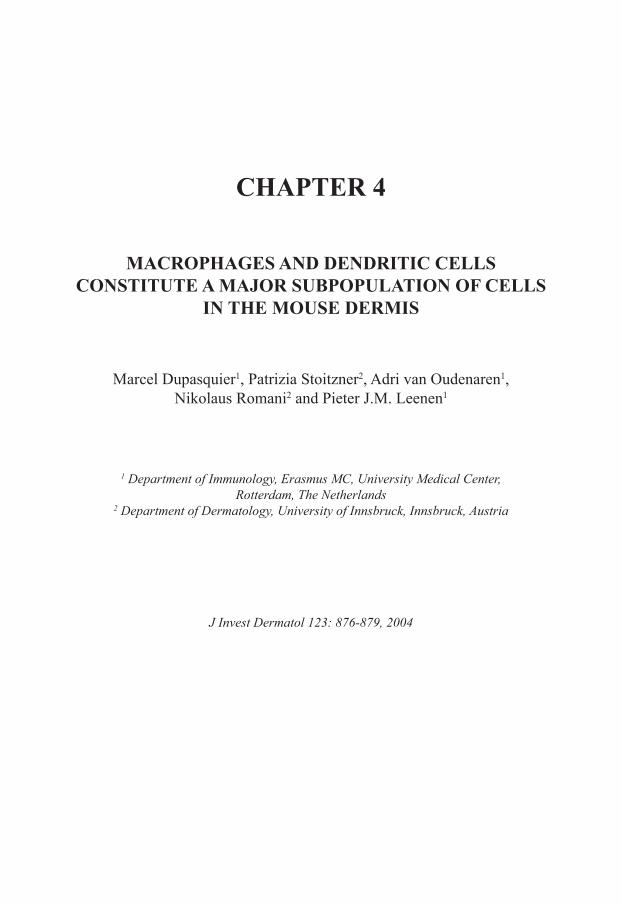
CHAPTER 4
MACROPHAGES AND DENDRITIC CELLS CONSTITUTE A MAJOR SUBPOPULATION OF CELLS
IN THE MOUSE DERMIS
Marcel Dupasquier1, Patrizia Stoitzner2, Adri van Oudenaren1,Nikolaus Romani2 and Pieter J.M. Leenen1
1 Department of Immunology, Erasmus MC, University Medical Center, Rotterdam, The Netherlands
2 Department of Dermatology, University of Innsbruck, Innsbruck, Austria
J Invest Dermatol 123: 876-879, 2004

Chapter 4
70
ABSTRACT
Macrophages and dendritic cells in tissues with close contact to the environment are of essential importance in host defense and are therefore present in sizeable numbers. Therefore, it is surprising that mononuclear phagocyte populations of the dermis have rarely been investigated in a quantitative manner. In this study, we examined mouse dermal skin immunophenotypically and related the observed numbers of observed cells to the total number of nucleated cells. These analyses show that about 70% of all dermal cells represent CD45+ leukocytes. The vast majority of these cells (~60% of total) expresses the mononuclear phagocyte markers mMGL (ER-MP23), F4/80 and CD11b. In addition, these cells show avid phagocytic capacity and thus are identified as dermal macrophages. Different subpopulations can be defined using markers such as sialoadhesin, ER-HR3 and mSIGN-R1 (ER-TR9). Interestingly, MHC class II expression differs significantly between dermal cells from ear vs. back skin. Moreover, we have identified small populations of dermal dendritic cells and migrating Langerhans cells (together ~10% of total). In summary, our findings show that mononuclear phagocyte populations form the majority of dermal cells and thus have been clearly underestimated so far.
INTRODUCTION
Mononuclear phagocytes, i.e. macrophages and dendritic cells (DC), are crucial initiators and regulators of both innate and adaptive host defense responses. As a consequence, especially tissues in close contact with the environment possess sizeable populations of mononuclear phagocytes. In the skin, different populations of such sentinel cells have been described: in the suprabasal layer of the epidermis, where Langerhans cells (LC) build up a network with their dendrites, and in the dermis, where interstitial dermal dendritic cells and dermal macrophages reside between extracellular matrix components. Dermal mononuclear phagocytes appear to be important for skin immunity, as they are highly competent in inducing an immune response without involvement of epidermal LC1,2. Surprisingly enough, dermal mononuclear phagocyte populations have never been investigated in a quantitative manner compared to the total amount of dermal cells. Therefore, we performed immunofluorescence double stainings on mouse ear and back skin sections using different macrophage and DC markers. To be able to compare the amount of stained cells to the total amount of dermal cells, we counterstained the sections with DAPI to identify all nucleated cells. Thereafter, we assessed the phenotype of 200 – 400 dermal nucleated cells, excluding cells in skin appendages.
MATERIALS AND METHODS
MiceFemale C57BL/6 mice were obtained from Harlan (Horst, The Netherlands), kept at the
animal care facility of the Erasmus MC Rotterdam and used at 10-14 weeks of age under institutional guidelines.

71
Macrophages and DC in the mouse dermis
Antibodies and conjugates The following antibodies were used as undiluted hybridoma culture supernatants: 30G12
(CD45), ER-HR3, ER-MP23 (mMGL / DC-ASGPR), ER-TR3 (MHC class II molecules Iak,b,d,q,r), ER-TR9 (mSIGN-R1), F4/80, M1/70 (CD11b) and MOMA-1 (sialoadhesin, SER). Anti-Langerin antibody (clone 929F3) was kindly provided by dr. Sem Saeland, Schering Plough, Dardilly, France. Biotinylated ER-MP23 was prepared in our laboratory. Biotinylated CD11c (clone HL3) was obtained from BD Pharmingen, San Diego, CA. Texas-Red-conjugated streptavidin was from Caltag, San Francisco, CA; FITC-conjugated rabbit-anti-rat immunoglobulins were from Dako, Glostrup, Denmark.
Immunofluorescence double staining procedureImmunofluorescence double stainings on mouse (C57BL/6) ear and back skin sections
have been performed essentially as described3. In short, steady-state ears and back skin were isolated from euthanized mice, frozen in Tissue-Tek O.C.T. Compound (Sakura Finetek, Zoeterwoude, The Netherlands) and cut into 6 µm thick sections on a cryostat (Leitz, Wetzlar, Germany). Sections were air-dried and stored at -20ºC until further use. Before staining, sections were thawed and fixed for 4 minutes in acetone. Thereafter, sections were sequentially rehydrated in phosphate-buffered saline pH 7.8 (PBS) plus 0.05% Tween-20 (Fluka, Buchs, Switzerland), incubated with avidin and biotin to block endogenous biotin (Avidin/Biotin Blocking Kit, Vector labs, Burlingame, CA, USA), blocked with 10% normal rabbit serum and incubated with hybridoma culture supernatant or irrelevant isotype-matched control antibody. Antibody binding was visualized with FITC-labeled anti-rat immunoglobulins. Sections were blocked with 5% normal rat serum and incubated with a second, biotinylated antibody or control antibody. After visualization with Texas-Red-labeled streptavidin, sections were embedded with Vectashield that contained DAPI to counterstain nuclei (Vector labs). Incubation steps were all performed in the dark at room temperature for 30 minutes; sections were washed twice between incubations with PBS supplemented with 0.05% Tween-20.
Quantification of dermal cellsSections were examined using a Zeiss Axioplan 2 imaging fluorescence microscope
(Zeiss, Göttingen, Germany). For quantitative analysis, the marker expression of 200 to 400 nucleated, DAPI+ dermal interstitial cells per section was determined, excluding cells in epidermal appendages such as hair follicles or sebaceous glands. Countings were performed on at least three sections that originated from different mice and were stained in independent experiments.
Determination of dermal cell phagocytic capacityDiI-labeled liposomes in PBS were provided by Dr. Nico van Rooijen, Vrije Universiteit,
Amsterdam, The Netherlands. A volume of 0.05 ml was injected with a Monoject syringe, 28 gauge into the murine ear dermis. Four hours afterwards, mice were killed, their ears dissected, frozen in Tissue-Tek and processed as described above, fixing the sections in acetone for 10 seconds.
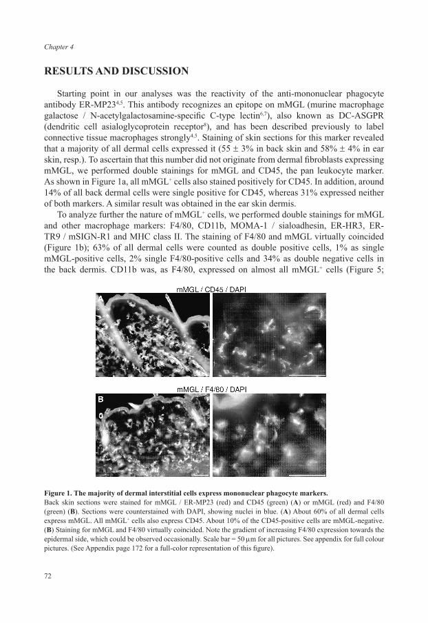
Chapter 4
72
RESULTS AND DISCUSSION
Starting point in our analyses was the reactivity of the anti-mononuclear phagocyte antibody ER-MP234,5. This antibody recognizes an epitope on mMGL (murine macrophage galactose / N-acetylgalactosamine-specific C-type lectin6,7), also known as DC-ASGPR (dendritic cell asialoglycoprotein receptor8), and has been described previously to label connective tissue macrophages strongly4,5. Staining of skin sections for this marker revealed that a majority of all dermal cells expressed it (55 ± 3% in back skin and 58% ± 4% in ear skin, resp.). To ascertain that this number did not originate from dermal fibroblasts expressing mMGL, we performed double stainings for mMGL and CD45, the pan leukocyte marker. As shown in Figure 1a, all mMGL+ cells also stained positively for CD45. In addition, around 14% of all back dermal cells were single positive for CD45, whereas 31% expressed neither of both markers. A similar result was obtained in the ear skin dermis.
To analyze further the nature of mMGL+ cells, we performed double stainings for mMGL and other macrophage markers: F4/80, CD11b, MOMA-1 / sialoadhesin, ER-HR3, ER-TR9 / mSIGN-R1 and MHC class II. The staining of F4/80 and mMGL virtually coincided (Figure 1b); 63% of all dermal cells were counted as double positive cells, 1% as single mMGL-positive cells, 2% single F4/80-positive cells and 34% as double negative cells in the back dermis. CD11b was, as F4/80, expressed on almost all mMGL+ cells (Figure 5;
Figure 1. The majority of dermal interstitial cells express mononuclear phagocyte markers.Back skin sections were stained for mMGL / ER-MP23 (red) and CD45 (green) (A) or mMGL (red) and F4/80 (green) (B). Sections were counterstained with DAPI, showing nuclei in blue. (A) About 60% of all dermal cells express mMGL. All mMGL+ cells also express CD45. About 10% of the CD45-positive cells are mMGL-negative. (B) Staining for mMGL and F4/80 virtually coincided. Note the gradient of increasing F4/80 expression towards the epidermal side, which could be observed occasionally. Scale bar = 50 µm for all pictures. See appendix for full colour pictures. (See Appendix page 172 for a full-color representation of this figure).

73
Macrophages and DC in the mouse dermis
55% double positive, 1% single mMGL-positive, 4% single CD11b-positive and 40% double negative cells). The 4% single CD11b-positive cells presumably represent dermal mast cells. MOMA-1 was found to bind a subpopulation of dermal phagocytes, in particular those cells laying deeper in the dermis (Figure 5; 23% double positive, 39% single mMGL-positive, 2% single MOMA-1-positive, 37% double negative). ER-HR3, with still unknown binding epitope, likewise stained a significant subpopulation of mMGL+ cells throughout the dermis (Figure 5; 42% double positive, 18% single mMGL-positive, 5% single ER-HR3-positive, 35% double negative). ER-TR9, which recognizes mSIGN-R19, has been found to stain a small percentage of cells in the dermis (6-8%; Figure 5). For all these markers, similar numbers have been obtained comparing back skin to ear skin dermis. When we compared the staining of dermal cells of the two sites for MHC class II, we found that virtually all mMGL-positive cells in the back dermis expressed this marker (58% double positive, 3% single mMGL-positive, 4% single MHC class II-positive, 35% double negative; Figure 2a). In marked contrast, only a minor subpopulation of all mMGL-positive cells expressed MHC class II in the ear dermis (20% double positive, 40% single mMGL-positive, 2% single MHC class II-positive, 38% double negative; Figure 2b). To explain this divergence, we speculate that differences in the structure of the dermis at different locations (subdermal adipose tissue vs. cartilage) create different microenvironments that differentially influence the expression of the MHC class II molecules.
Figure 2. Dermal macrophages from different sites show differential MHC class II expression.Back skin sections (A) and ear skin sections (B) were stained for mMGL (red) and for MHC class II (green). Sections were counterstained with DAPI. (A) In the back skin, virtually all mMGL-positive cells coexpress MHC class II. (B) Similar to the back skin, the ear dermis contains about 60% interstitial cells expressing mMGL. However, only about one third of these cells coexpresses MHC class II molecules. These mMGL / MHC class II double-positive cells are arranged in clusters. Scale bar = 50 µm for all pictures. (See Appendix page 173 for a full-color representation of this figure).

Chapter 4
74
To seek further confirmation regarding the nature of mMGL-positive dermal cells, we made use of the fact that macrophages are the only connective tissue cells showing avid phagocytic capacities. Therefore we injected DiI-labeled liposomes, which previously have been shown to target phagocytic cells exclusively10, into the ear dermis. Thereafter we killed the mice, made sections of the treated ears and stained for mMGL. As shown in Figure 3, we indeed found that the vast majority of mMGL-positive cells had taken up DiI-labeled liposomes. However, a few mMGL-positive cells as well as all mMGL-negative cells remained free of DiI-fluorescence, indicating the specificity of this procedure. Therefore, we conclude that mMGL-positive cells not only express mononuclear phagocyte markers but also possess phagocytic capacity, thus identifying them as dermal macrophages unequivocally.
To evaluate the presence of DC in the dermis, we stained back and ear sections for CD11c. To distinguish dermal DC from migrating LC that are on their way from the epidermis to skin-draining lymph nodes, we performed a double labeling for Langerin, which specifically stains LC11. As shown in Figure 4, we indeed found a subpopulation of CD11c+ Langerin– dermal DC (7%), while even fewer cells represented migrating LC which all expressed CD11c (4% CD11c+ Langerin+). DC-SIGN is a specific marker for dermal DC in the human situation12. Therefore, we wondered whether ER-TR9 (mSIGN-R1) would recognize dermal DC specifically in the mouse. Double stainings for ER-TR9 and CD11c revealed that ER-TR9 recognized a small subset of dermal APC (6-8%), but that it did not uniquely stain CD11c+ cells (Figure 5, and data not shown).
Figure 3. mMGL-positive dermal cells are phagocytic.DiI-labeled liposomes (red label) were injected into ear dermis. Four hours later, animals were killed, ears cut off, frozen, sectioned and stained for mMGL (green). Note that the vast majority, but not all mMGL-positive cells, has taken up liposomes, whereas mMGL-negative cells and keratinocytes remained free of labeling. Scale bar = 50 µm for all pictures. (See Appendix page 174 for a full-color representation of this figure).

75
Macrophages and DC in the mouse dermis
To summarize, we have shown that mononuclear phagocytes constitute the major population of nucleated cells in the mouse dermis where they amount to about 60% of all dermal cells (Table 1). Previous studies have quantified dermal mononuclear phagocyte subsets for mice13 and for humans14 by flowcytometric analyses of dermal cell suspensions. In contrast to our findings, these authors concluded that dermal macrophages and dermal DC only composed around 5% of all dermal cells. The discrepancy between their findings and ours lies most probably in the difference of the used methods, as we stained and counted cells in situ in the non-denaturated dermis. Clearly, during the digestion procedure needed to obtain a dermal cell suspension, markers either may have been destroyed by enzymatic digestion, as is known to happen with the CD1 molecules, or the procedure may have released fibroblasts preferentially. We further tried to strengthen our findings by staining and counting fibroblasts specifically, but were not successful due to the unavailability of suitable markers. However, the concept that most dermal interstitial cells are indeed mononuclear phagocytes is well supported as we found similar percentages for dermal mononuclear phagocytes using several markers (F4/80, CD11b, mMGL, CD68 (data not shown) and MHC class II in the back dermis). Furthermore, this high frequency is confirmed by the CD45 pan-leukocyte labeling. Taken together, we feel that the size of the dermal mononuclear phagocyte population has been clearly underestimated so far. These cells probably play crucial roles in skin inflammatory and immune responses and they should be taken into account when the role of the dermis is studied in steady state and inflammatory conditions.
Figure 4. Dermal DC constitute a small, distinct mononuclear phagocyte population.Back skin sections were stained for CD11c (red) and Langerin (green) (A) or CD11c (red) and mMGL (green) (B). Sections were counterstained with DAPI. (A) About 7% of all dermal cells express CD11c but does not express Langerin. These cells thus represent genuine DDC (marked with thin arrows). Note that the CD11c staining is too weak to let CD11c / Langerin-double-positive mLC (marked with thick arrows) appear yellow. (B) Double-staining for CD11c and mMGL shows that most CD11c-positive cells coexpress mMGL (arrow). Thus, most if not all DDC express mMGL. The red labeled cell marked with an asterisk is part of a sebaceous gland and displays autofluorescence rather than specific labeling. Scale bar = 50 µm for all pictures. (See Appendix page 175 for a full-color representation of this figure).

Chapter 4
76
Figure 5. Dermal macrophages show different degrees of heterogeneity.Back skin sections were double-stained for mMGL (red) and other macrophage markers in green: CD11b (A), MOMA-1 (B), ER-HR3 (C), mSIGN-R1 / ER-TR9 (D). Sections were counterstained with DAPI. (A) All mMGL-positive cells coexpress CD11b. Separate CD11b single-positive cells may represent dermal mast cells, or occasional monocytes, granulocytes or activated T cells. (B) Staining for MOMA-1 divides DMφ into two subpopulations: cells lying deeper in the dermis express MOMA-1, whereas cells directly underneath the epidermis do not. (C) ER-HR3 also divides DMφ into two subpopulations, but with no clear localization pattern. A small number of ER-HR3 single-positive cells presumably represent mLC. (D) mSIGN-R1 / ER-TR9 labels a small subpopulation of DMφ and DDC. The two arrows mark two mMGL / mSIGN-R1 double-positive cells. Scale bar = 50 µm for all pictures. (See Appendix page 176 for a full-color representation of this figure).

77
Macrophages and DC in the mouse dermis
Table 1. Frequencies of different cell populations in the mouse dermis, related to total nucleated cells. The quantification is derived from findings in both ear and back skin.
Cell type Mean percentage in the dermis CD45 Langerin CD11c ER-MP23
Dermal macrophages (DMφ) 50% + – – +
Dermal DC (DDC) 7% + – + 6-7%
Migrating LC (mLC) 4% + + + 1-2%
Non-BM-derived interstitial cells 29% – – – –
Other BM-derived cells (mast cells, T cells) 10% + – – –
ACKNOWLEDGEMENTS
We thank Drs. Errol Prens, Leslie van der Fits and Jon Laman for stimulating discussions, Dr. Sem Saeland (Schering Plough, Dardilly, France) for the anti-Langerin antibody, Dr. Nico van Rooijen (Free University Amsterdam, The Netherlands) for DiI-labeled liposomes and Tar van Os for preparing the figures. This study was supported by the Interuniversitary Research Institute for Radiopathology and Radioprotection (IRS), Leiden, The Netherlands.
REFERENCES
1. Allan, R.S., Smith, C.M., Belz, G.T., van Lint, A.L., Wakim, L.M., Heath, W.R. & Carbone, F.R. Epidermal viral immunity induced by CD8alpha+ dendritic cells but not by Langerhans cells. Science 301, 1925-1928 (2003).
2. Sreilein, J.W. Antigen-presenting cells in the induction of contact hypersensitivity in mice: evidence that Langerhans cells are sufficient but not required. J Invest Dermatol 93, 443-448 (1989).
3. Stoitzner, P., Holzmann, S., McLellan, A.D., Ivarsson, L., Stossel, H., Kapp, M., Kammerer, U., Douillard, P., Kampgen, E., Koch, F., Saeland, S. & Romani, N. Visualization and characterization of migratory Langerhans cells in murine skin and lymph nodes by antibodies against Langerin/CD207. J Invest Dermatol 120, 266-274 (2003).
4. Jansen, A., Homo-Delarche, F., Hooijkaas, H., Leenen, P.J., Dardenne, M. & Drexhage, H.A. Immunohistochemical characterization of monocytes-macrophages and dendritic cells involved in the initiation of the insulitis and beta-cell destruction in NOD mice. Diabetes 43, 667-675 (1994).
5. Leenen, P.J., de Bruijn, M.F., Voerman, J.S., Campbell, P.A. & van Ewijk, W. Markers of mouse macrophage development detected by monoclonal antibodies. J Immunol Methods 174, 5-19 (1994).
6. Oda, S., Sato, M., Toyoshima, S. & Osawa, T. Purification and characterization of a lectin-like molecule specific for galactose/N-acetyl-galactosamine from tumoricidal macrophages. J Biochem 104, 600-605 (1988).
7. McKnight, A.J. & Gordon, S. Membrane molecules as differentiation antigens of murine macrophages. Adv Immunol 68, 271-314 (1998).
8. Valladeau, J., Duvert-Frances, V., Pin, J.J., Kleijmeer, M.J., Ait-Yahia, S., Ravel, O., Vincent, C., Vega, F., Jr., Helms, A., Gorman, D., Zurawski, S.M., Zurawski, G., Ford, J. & Saeland, S. Immature human dendritic cells express asialoglycoprotein receptor isoforms for efficient receptor-mediated endocytosis. J Immunol 167, 5767-5774 (2001).

Chapter 4
78
9. Kang, Y.S., Yamazaki, S., Iyoda, T., Pack, M., Bruening, S.A., Kim, J.Y., Takahara, K., Inaba, K., Steinman, R.M. & Park, C.G. SIGN-R1, a novel C-type lectin expressed by marginal zone macrophages in spleen, mediates uptake of the polysaccharide dextran. Int Immunol 15, 177-186 (2003).
10. Buiting, A.M., de Rover, Z., Claassen, E. & van Rooijen, N. In vivo distribution of particulate antigens and liposomes in murine spleen. A possible role in the humoral immune response. Immunobiology 188, 13-22 (1993).
11. Valladeau, J., Clair-Moninot, V., Dezutter-Dambuyant, C., Pin, J.J., Kissenpfennig, A., Mattei, M.G., Ait-Yahia, S., Bates, E.E., Malissen, B., Koch, F., Fossiez, F., Romani, N., Lebecque, S. & Saeland, S. Identification of mouse langerin/CD207 in Langerhans cells and some dendritic cells of lymphoid tissues. J Immunol 168, 782-792 (2002).
12. Turville, S.G., Cameron, P.U., Handley, A., Lin, G., Pohlmann, S., Doms, R.W. & Cunningham, A.L. Diversity of receptors binding HIV on dendritic cell subsets. Nat Immunol 3, 975-983 (2002).
13. Duraiswamy, N., Tse, Y., Hammerberg, C., Kang, S. & Cooper, K.D. Distinction of class II MHC+ Langerhans cell-like interstitial dendritic antigen-presenting cells in murine dermis from dermal macrophages. J Invest Dermatol 103, 678-683 (1994).
14. Meunier, L., Gonzalez-Ramos, A. & Cooper, K.D. Heterogeneous populations of class II MHC+ cells in human dermal cell suspensions. Identification of a small subset responsible for potent dermal antigen-presenting cell activity with features analogous to Langerhans cells. J Immunol 151, 4067-4080 (1993).

CHAPTER 5
THE DERMAL MICROENVIRONMENT INDUCESTHE EXPRESSION OF THE ALTERNATIVE
ACTIVATION MARKER CD301/MMGL IN MONONUCLEAR PHAGOCYTES INDEPENDENT
OF IL-4/IL-13 SIGNALING
Marcel Dupasquier*, Patrizia Stoitzner†, Hui Wan*, Denise Cerqueira*, Adri van Oudenaren*, Jane S.A. Voerman*, Kaori Denda-Nagai‡,
Tatsuro Irimura‡, Geert Raes§, Nikolaus Romani†, and Pieter J.M. Leenen*
* Department of Immunology, Erasmus MC, University Medical Center, Rotterdam, The Netherlands
† Department of Dermatology, Innsbruck Medical University, Innsbruck, Austria‡ Laboratory of Cancer Biology and Molecular Immunology, Graduate School of
Pharmaceutical Sciences, University of Tokyo, Japan§ Laboratory of Cellular and Molecular Immunology, Department of Molecular and
Cellular Interactions, Vlaams Interuniversitair Instituut voor Biotechnologie,Vrije Universiteit Brussel, Brussels, Belgium
J Leukoc Biol 80: 838-849, 2006

Chapter 5
80
ABSTRACT
Recently we have shown that mononuclear phagocytes comprise the majority of interstitial cells in the mouse dermis, as indicated by their phenotypic and functional characteristics. In particular, these cells express the mouse macrophage galactose-/N-acetylgalactosamine-specific lectin (mMGL)/CD301, identified by the monoclonal antibody ER-MP23, as well as other macrophage markers. Since expression of mMGL is induced by IL-4 or IL-13 and is therefore a marker of alternatively activated macrophages, we asked whether dermal mononuclear phagocytes are genuinely alternatively activated. We observed that these cells expressed, next to mMGL, two other alternative activation markers, namely the mannose receptor/CD206 and Dectin-1. Yet, as this expression profile was very similar in IL-4Rα knock-out mice, neither IL-4 nor IL-13 signaling appeared to be required for this phenotype. We also found that Langerhans cells (LC), which showed only a low level of mMGL in the epidermis, upregulated mMGL expression upon migration through the dermis, allowing these cells to internalize limited amounts of mMGL ligands. LC isolated from epidermal preparations did not show this upregulation when cultured in standard medium, but whole skin-conditioned medium did stimulate mMGL expression by LC. The vast majority of mMGL molecules were present in the cytoplasm, however. LC that arrived in skin-draining lymph nodes quickly downregulated mMGL expression, while dermally derived cells retained significant mMGL levels. Taken together, these data suggest that the dermal microenvironment induces mononuclear phagocyte subpopulations to express mMGL and possibly other markers of alternatively activated macrophages independent of IL-4/IL-13 signaling.
INTRODUCTION
Mononuclear phagocytes are functionally versatile cells that perform key roles in the immune system. They are traditionally separated into macrophages, the professional phagocytic cells, and dendritic cells (DC), the professional antigen-presenting cells. Nevertheless, it is becoming increasingly clear that mononuclear phagocytes perform many more functions than just taking up pathogens and presenting peptides to T cells1. They orchestrate inflammatory and immune responses and are intimately involved in maintaining tissue homeostasis by cell-cell interactions and by the production of myriads of mediators and effector molecules. As a consequence, tissue damage is often connected with this function. On the other hand, mononuclear phagocytes also contribute to tissue repair by breaking down granulation tissue and stimulating the production of new extracellular matrix or by producing such components themselves2. As these functions contradict each other, they need to be tightly controlled. Environmental signals, such as cytokines, are now recognized to play pivotal roles in imposing specific phenotypes and functions in mononuclear phagocytes3,4. Specifically, a dichotomy of M1 and M2 macrophages, modeled after the Th1 and Th2 paradigm of T helper cells, was suggested after the discovery that IL-4 stimulation drives macrophages into an alternatively activated state that is connected with immunosuppression and tissue repair5. This state contrasts the classically activated state that is obtained after stimulation with IFN-γ and associates with microbicidal activity and tissue degradation.

81
mMGL as dermis-induced mononuclear phagocyte marker
However, distinguishing just two activation states is clearly an oversimplification3,4,6. Exposure of macrophages to IL-10, TGF-β or corticosteroids, for example, leads to a state that is connected with immunosuppression, comparable with alternatively activated macrophages, but those macrophages express phenotypes and functions that differ clearly from IL-4-treated macrophages7,8. As a consequence, it has been suggested that only macrophages stimulated with IL-4 or IL-13, which share a receptor chain and lead to very similarly activated cells, should be called alternatively activated6.
In vivo, it is difficult to demonstrate the functions of individual, differently activated macrophage populations. Consequently, identification of specific phenotypic markers that distinguish between differentially stimulated macrophages has been a major goal in the field. The mannose receptor (MR)/CD206 was initially found to serve this purpose for identification of alternatively activated macrophages5. More recently, other cell surface molecules, in particular the β-glucan receptor/Dectin-19 and the mouse macrophage galactose/N-acetylgalactosamine-specific C-type lectins 1 and 2 (mMGL1 and mMGL2)/CD301a and CD301b10 have been described as alternative activation markers of macrophages. Interestingly, we previously found that mMGL/CD301 expression typifies mononuclear phagocytes associated with connective tissue environments, such as the dermis11 or the pancreatic septa12. Therefore, we asked whether these cells might represent a population of alternatively activated macrophages in the tissue. We show in this report that, although dermal mononuclear phagocytes do express mMGL/CD301, MR/CD206 and Dectin-1, they are not alternatively activated macrophages in the strict sense, as they also express these receptors in IL-4Rα knock-out mice, in which cells are unable to respond to IL-4 and IL-13. Moreover, we demonstrate that also epidermal LC upregulate the expression of mMGL in response to dermal factors. Together, these results suggest that the dermal microenvironment stimulates mononuclear phagocytes to express markers of alternative activation, independent of IL-4/IL-13 signaling.
MATERIALS AND METHODS
AnimalsFemale C57BL/6J mice were obtained from Harlan (Horst, The Netherlands) and used
between 10-16 weeks of age. IL-4Rα knock-out mice were generated as described13 and backcrossed for 9 generations onto the C57BL/6 background.
Antibodies, conjugates and other reagentsThe following antibodies have been used in this study: rat anti-mMGL1 and mMGL2
clone ER-MP23, IgG2a, as hybridoma culture supernatant or biotinylated, and rat control IgG2a clone PH2-99, were generated in our lab. Rat anti-mouse Langerin (clone 929F3, IgG1, purified or Alexa Fluor 488-labeled) was kindly provided by Dr. Sem Saeland, INSERM U 503/IFR, Lyon, France; purified rat anti-MR (clone MR5D3, IgG2a) and rat anti-Dectin-1 (clone 2A11, IgG2b) were from Hycult Biotechnology, Uden, The Netherlands; PE-labeled rat anti-I-A/I-E (clone M5/114.15.2, IgG2b), PE-labeled rat anti-CD86 (clone GL1, IgG2a), APC-labeled rat anti-CD11b (clone M1/70, IgG2b), and APC-labeled streptavidin were from BD Pharmingen, San Diego, CA, USA. FITC-labeled and HRP- (horse radish peroxidase) labeled

Chapter 5
82
rabbit anti-rat immunoglobulins were from Dako, Glostrup, Denmark, and Texas Red-labeled streptavidin and PE-labeled goat anti-rat immunoglobulins were from Caltag, San Francisco, CA, USA. Culture medium was RPMI1640 (Cambrex, Verviers, Belgium), supplemented with 10% fetal calf serum (FCS; HyClone, Kansas City, MO, USA), 100 U/ml penicillin and 100 µg/ml streptomycin (both from Cambrex). FITC was from Sigma, whereas biotinylated α-N-acetylgalactosaminide (α-GalNAc), β-N-acetylgalactosaminide (β-GalNAc), β-N-acetylglucosaminide (β-GlcNAc) and LewisX (LeX) conjugated polyacrylamide carriers were obtained from GlycoTech, Rockville, MD, USA.
Solid Phase Binding AssayAdsorption of the purified soluble recombinant mMGL114 or mMGL215 onto enzyme-
linked immunosorbent assay plates (655061; Greiner, Kremsmünster Austria) was carried out by adding 100 µl of protein solution (2.5 µg/ml in DPBS) to each well and incubating the plates for 18h at 4°C. After blocking non-specific binding using 3% BSA in DPBS for 2h at room temperature, a dilution series of ER-MP23 hybridoma culture supernatant or purified control antibodies (stock concentration: 10 µg/ml) in DPBS plus 1% BSA was added. After incubation for 2h at room temperature, wells were washed three times with DPBS to remove unbound materials, and then 100 µl of HRP-conjugated goat anti-rat IgG solution (0.75 µg/ml in DPBS) was added to detect bound materials. After incubation for 1h at room temperature, the wells were washed three times with DPBS. Subsequently, 100 µl of 1 mM 2,2’-amino-bis(3-ethylbenzthiazoline-6-sulfonic acid) (ABTS) solution containing 0.34% H2O2 in 0.1 M sodium citrate buffer (pH 4.3) was added, and the absorbance was measured at 405 nm on a microplate reader (Model550, Bio-Rad, Hercules, CA, USA).
To detect blocking ability of the ER-MP23 antibody, 300 ng of recombinant mMGL1 or mMGL2 were adsorbed per well onto plates. Inhibition of non-specific binding and subsequent washing was done as described before. Thereafter, increasing concentrations of ER-MP23 or a rat control antibody were added to the wells and incubated for 2h at room temperature. Subsequently, biotinylated LeX -polymers (for mMGL1) or β-GalNAc-polymers (for mMGL2) were added to the wells and incubated for 1.5h at 4°C. Bound polymers were detected by incubation with 100 µl of HRP-conjugated streptavidin solution (1.25 µg/ml in DPBS) for 0.5h at 4°C and visualized with ABTS as before.
Binding of desialylated erythrocytes to macrophage cell linesMacrophage cell lines RAW309Cr.1 (expressing significant levels of mMGL) and
RAW264.7 (mMGL–/low), grown in a 96-well culture plate (Nunc, Roskilde, Denmark), were preincubated with 50 µg/ml purified ER-MP23, 200 mM galactose or 200 mM mannose for 15’ at room temperature. Thereafter, human erythrocytes, desialylated by neuraminidase treatment as described16, were added and incubated for 30’ at 37°C. Subsequently, plates were washed five times with PBS plus Ca2+ and Mg2+, remaining erythrocytes were lysed with 10 mg/ml Na2CO3
and hemoglobin absorbance was measured at 414 nm on a Titertek Multiscan (Flow Labs, Redwood City, CA, USA).
Staining of tissue sectionsBack skin or skin-draining lymph nodes were frozen in Tissue-Tek O.C.T. Compound

83
mMGL as dermis-induced mononuclear phagocyte marker
(Sakura Finetek, Zoeterwoude, The Netherlands) and cut into 6 µm thick sections on a cryostat (Leitz, Wetzlar, Germany). Subsequently, sections were fixed for 4 minutes (skin) or 10 seconds (lymph nodes) in acetone, rehydrated in PBS pH 7.8 plus 0.05% Tween-20 (Fluka, Buchs, Switzerland), incubated with Avidin/Biotin Blocking Kit (Vector labs, Burlingame, CA, USA), blocked with 10% normal rabbit serum and incubated with purified antibodies. Thereafter, antibody binding was detected with FITC-labeled (for Langerin) or HRP-labeled anti-rat immunoglobulins (for mMGL, MR and Dectin-1). HRP-labeled antibodies were visualized by incubating slides with nickel-3,3’-diaminobenzidine tetrahydrochloride (Ni-DAB; Sigma, St. Louis, MO, USA) plus 1% H2O2 for three minutes. Slides were subsequently embedded in Entellan mounting media (Merck, Darmstadt, Germany). For fluorescence stainings, sections were subsequently blocked with 5% normal rat serum and incubated with biotinylated ER-MP23 antibodies. After visualization with Texas-Red-labeled streptavidin, sections were embedded with Vectashield mounting media that contained DAPI to counterstain nuclei (Vector labs). Incubation steps were all performed in the dark at room temperature for 30 minutes; sections were washed twice between incubations with PBS supplemented with 0.05% Tween-20. Thereafter, sections were examined using a Zeiss Axioscop or a Zeiss Axioplan 2 fluorescence microscope (Zeiss, Göttingen, Germany).
Epidermal cell and lymph node cell suspensionsEars of euthanized mice were cleaned with 70% ethanol and cut off at the base. Thereafter,
they were split in dorsal and ventral halves by means of strong forceps; both halves were incubated at 37°C in 0.8% trypsin (Merck) in PBS for 25 minutes (cartilage-free half) or 45 minutes. Epidermis was peeled off, shaken in a waterbath for 30 minutes and cell suspension was filtered through a 70 µm nylon cell strainer (BD Falcon, Bedford, MA, USA). The resulting epidermal cell suspensions contained 1-3% Langerhans cells. Epidermal cells were either directly stained for flowcytometry or cultured for 1 day in culture medium, supplemented with 4 ng/ml murine GM-CSF (BioSource, Camarillo, CA, USA). In some experiments, epidermal cells were cultured for 1 day in conditioned medium obtained from a day 3 skin explant culture (see below), which also had been supplemented with GM-CSF. Cells were either stained for ER-MP23-bio, followed by SA-APC and I-A/I-E-PE to detect extracellular mMGL or only for I-A/I-E-PE. Thereafter, cells were fixed with 2% PFA (Merck), permeabilized with 0.25% saponin (Sigma) and subsequently stained with anti-Langerin-Alexa Fluor 488 antibodies, or, to detect intracellular mMGL, with anti-Langerin and ER-MP23-bio antibodies, followed by SA-APC.
Axillary, brachial and inguinal skin-draining lymph nodes were collected form untreated mice or from mice, which were treated with 250 µl 1% FITC in 1:1 acetone:dibutylphtalate (Sigma) on their shaved back skin 24h before they were euthanized. Skin-draining lymph nodes were cut into small pieces and subsequently filtered through a 70 µm nylon cell strainer. Cells were fixed and permeabilized as described above. Thereafter, they were stained with purified anti-Langerin antibodies, which were visualized with PE-labeled anti-rat immunoglobulins. Cells were subsequently stained with ER-MP23-bio and SA-APC. Fluorescence of all cells was measured on a FACSCalibur (BD) and analyzed using WinMDI 2.8 software.
Chapter 5
84
Skin explant culturesDorsal and ventral ear halves were cultured on 1 ml culture medium in 24 well plates for
one, two, three or four days. Cells that had emigrated out from the explants were collected afterwards and stained for flowcytometry as described for epidermal cell suspensions. For analysis of emigrant cells by confocal microscopy, cells were allowed to adhere for 6h on Lab-Tek II Chamber Slides (Nunc). Thereafter, they were fixed and permeabilizied with PFA and saponin and stained for Langerin-AF488 and ER-MP23-bio, followed by SA-TxR as described for tissue slides. Slides were analyzed with a Zeiss LSM 510 Meta confocal microscope. For the endocytosis experiments, 10 µg/ml biotinylated α-GalNAc, β-GalNAc or β-GlcNAc polymers were added to the cultured medium as indicated during the two days culture period. In preliminary experiments, we determined that both matured LC and mMGL+ dermal-derived cells expressed similar high levels of CD86 (data not shown). Therefore, collected emigrated cells were fixed, permeabilized and stained with anti-Langerin-Alexa Fluor 488, CD86-PE and SA-APC, to detect the endocytosed sugar-polymers. In some wells, 50 to 100 µg/ml purified ER-MP23 anti-mMGL antibodies were added during the culture period to block binding of the glycosylated polymers (no difference in blocking efficiency was observed between the two concentrations). For the emigration experiments, 100 µg/ml purified ER-MP23 antibodies or control rat IgG2a antibodies were added to the medium during the two days culture period. Thereafter, cells were fixed, permeabilized and stained with anti-Langerin-Alexa Fluor 488 and CD11b-APC. Cells were analyzed by flowcytometry as indicated above.
RESULTS
ER-MP23 binds both mMGL1 and mMGL2The previously described anti-mononuclear phagocyte mAb ER-MP2312,17 appears to
recognize the mouse macrophage galactose-/N-acetylgalactosamine- specific lectin (mMGL)/CD301. This notion is based on the following observations. In preliminary experiments, we found that ER-MP23 precipitated a 43-kDa antigen from a J774-1.6 macrophage cell line homogenate. Protein sequencing revealed the non-blocked N-terminus MEYENLQNIRIE, which corresponds to the described N-terminus of the mouse macrophage galactose/ N-acetylgalactosamine-specific C-type lectin 1 (mMGL1)18, with the exception of two amino acids (underscored). Recently, mMGL2 has been discovered, which shows 79.0% nucleotide identity and 91.5% amino acid identity to mMGL115. As shown in Figure 1A and 1B, we found now by ELISA assays with recombinant mMGL1 and mMGL2 that ER-MP23 bound to both extracellular domains of mMGL1 and mMGL2. We confirmed this finding by positive flowcytometry stainings of CHO cell clones that had been transfected with full-length mMGL1 or mMGL2 (data not shown). To investigate whether ER-MP23 interferes with the carbohydrate ligand binding function of the lectins, we incubated immobilized recombinant mMGL1 and mMGL2 with increasing concentrations ER-MP23. Thereafter, we assessed the remaining sugar binding capacity of the two recombinant lectins. As shown in Figure 1C and 1D, ER-MP23 could indeed inhibit the binding of ligands to both mMGL1 and mMGL2, although the inhibition of mMGL1 ligand binding is possibly more efficient. To confirm that this blocking
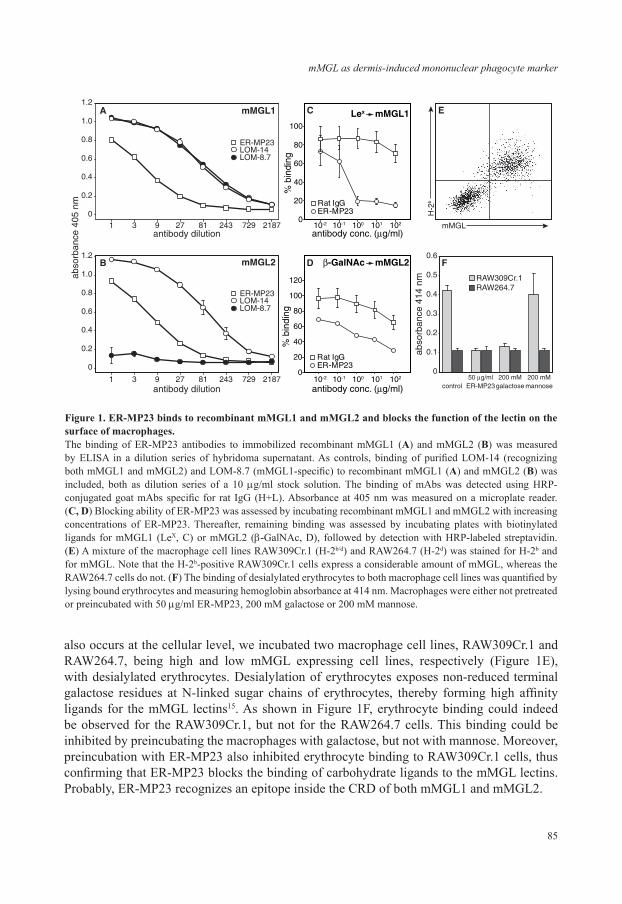
85
mMGL as dermis-induced mononuclear phagocyte marker
also occurs at the cellular level, we incubated two macrophage cell lines, RAW309Cr.1 and RAW264.7, being high and low mMGL expressing cell lines, respectively (Figure 1E), with desialylated erythrocytes. Desialylation of erythrocytes exposes non-reduced terminal galactose residues at N-linked sugar chains of erythrocytes, thereby forming high affinity ligands for the mMGL lectins15. As shown in Figure 1F, erythrocyte binding could indeed be observed for the RAW309Cr.1, but not for the RAW264.7 cells. This binding could be inhibited by preincubating the macrophages with galactose, but not with mannose. Moreover, preincubation with ER-MP23 also inhibited erythrocyte binding to RAW309Cr.1 cells, thus confirming that ER-MP23 blocks the binding of carbohydrate ligands to the mMGL lectins. Probably, ER-MP23 recognizes an epitope inside the CRD of both mMGL1 and mMGL2.
0
0.2
0.4
0.6
0.8
1.0
1.2
antibody dilution
B mMGL2
1 3 9 27 81 243 729 2187
1 3 9 27 81 243 729 2187
0
0.2
0.4
0.6
0.8
1.0
1.2
abso
rban
ce 4
05 n
m
antibody dilution
A mMGL1
ER-MP23 LOM-14 LOM-8.7
ER-MP23 LOM-14 LOM-8.7
0.6
C Lex mMGL1
0
20
40
60
80
100
antibody conc. (�g/ml)
control ER-MP23galactose mannose50 �g/ml
RAW309Cr.1RAW264.7
200 mM 200 mM0
0.1
0.2
0.3
0.4
0.5
abso
rban
ce 4
14 n
m
F
E
H-2
b
mMGL
% b
indi
ng
Rat IgGER-MP23
10210110010-110-2
D
antibody conc. (�g/ml)
Rat IgGER-MP23
10210110010-110-2
�-GalNAc mMGL2
0
20
40
60
80
100
120%
bin
ding
Figure 1. ER-MP23 binds to recombinant mMGL1 and mMGL2 and blocks the function of the lectin on the surface of macrophages.The binding of ER-MP23 antibodies to immobilized recombinant mMGL1 (A) and mMGL2 (B) was measured by ELISA in a dilution series of hybridoma supernatant. As controls, binding of purified LOM-14 (recognizing both mMGL1 and mMGL2) and LOM-8.7 (mMGL1-specific) to recombinant mMGL1 (A) and mMGL2 (B) was included, both as dilution series of a 10 µg/ml stock solution. The binding of mAbs was detected using HRP-conjugated goat mAbs specific for rat IgG (H+L). Absorbance at 405 nm was measured on a microplate reader. (C, D) Blocking ability of ER-MP23 was assessed by incubating recombinant mMGL1 and mMGL2 with increasing concentrations of ER-MP23. Thereafter, remaining binding was assessed by incubating plates with biotinylated ligands for mMGL1 (LeX, C) or mMGL2 (β-GalNAc, D), followed by detection with HRP-labeled streptavidin. (E) A mixture of the macrophage cell lines RAW309Cr.1 (H-2b/d) and RAW264.7 (H-2d) was stained for H-2b and for mMGL. Note that the H-2b-positive RAW309Cr.1 cells express a considerable amount of mMGL, whereas the RAW264.7 cells do not. (F) The binding of desialylated erythrocytes to both macrophage cell lines was quantified by lysing bound erythrocytes and measuring hemoglobin absorbance at 414 nm. Macrophages were either not pretreated or preincubated with 50 µg/ml ER-MP23, 200 mM galactose or 200 mM mannose.

Chapter 5
86
Dermal mononuclear phagocytes express markers of alternatively activated macrophages
Previously, we characterized antigen presenting cell (APC) populations in the mouse dermis and observed that the majority of all dermal cells represent mononuclear phagocytes expressing mMGL11,15,19. As the mMGL lectins have been identified as markers for alternatively activated macrophages10, we approached the question whether resident dermal mononuclear phagocytes might represent alternatively activated macrophages. To that end, we stained skin sections for two other well established markers of alternatively activated macrophages, the mannose receptor (MR)/CD2065,20 and the β-glucan receptor/Dectin-19. As shown in Figure 2, we found that a similarly large population of dermal cells also expressed MR (Figure 2B). These cells were recently identified as mononuclear phagocytes by Linehan21. Dectin-1 staining showed a less uniform profile indicating that this marker was expressed at a high level only by clusters of cells in the dermis (Figure 2C), reminiscent of the expression of MHC class II in the ear dermis11. Nevertheless, all cells were observed to express at least a low level of Dectin-1. In the ear dermis, we found that MHC class II and high Dectin-1 expression were indeed co-localized on the same cells (data not shown). Therefore, we conclude that dermal mononuclear phagocytes indeed express multiple markers of alternatively activated macrophages, a phenotype that can be induced by IL-4 or IL-13 signaling6. Macrophages that lack the IL-4/IL-13 common receptor α chain (IL-4Rα) cannot develop into alternatively activated macrophages22. To assess whether IL-4 or IL-13 is required in the induction of the phenotype of the resident dermal macrophages, we stained skin sections of IL-4Rα-/- mice13 for the same markers. As shown in Figure 2, we found that dermal macrophages in IL-4Rα-/- mice still expressed all these markers in similar patterns. Therefore, the expression of alternative activation markers on dermal macrophages does not depend on IL-4 or IL-13 signaling.
LC migrating through the dermis express mMGLIn our analyses of dermal mononuclear phagocyte populations, we also identified LC in
the dermis on the basis of their expression of Langerin/CD207 as a unique marker for LC. They most probably represent migrating cells on their way to skin-draining lymph nodes23. Quantitative determinations revealed that around 6% of all interstitial cells in the C57BL/6J mouse dermis expressed Langerin11. LC were previously found to be negative for mMGL19,24, and in agreement with these reports, we found no mMGL expression on epidermal LC in our in situ stainings (Figure 3A). In contrast, around 2% of all dermal cells, corresponding to about one third of all dermal LC, co-expressed mMGL and Langerin (Figure 3B), while the other dermal LC were mMGL-negative by immunofluorescence. When we studied dermal LC in the IL-4Rα-/- mouse dermis, we found a comparable subpopulation of these cells expressing mMGL (Figure 3C). Therefore, mMGL expression appears to be induced on at least a subset of migrating LC located in the dermis, which is, similar to other resident dermal mononuclear phagocytes, independent of IL-4 or IL-13 signaling.
mMGL induction on LC is not just a consequence of maturationUpon migration through the dermis, LC mature, which is reflected in the upregulation
of markers such as CD40, CD80, CD86, CCR7/CD197, CD205 MHC class I and MHC class II25. Hence, expression of mMGL by LC might be merely a consequence of LC

87
mMGL as dermis-induced mononuclear phagocyte marker
maturation. To investigate this possibility, we determined the immunophenotype of freshly prepared and 24h cultured epidermal single cell suspensions by flowcytometry. As shown in Figure 4C and E, we found that immature LC in fresh epidermal cell suspension expressed already a low level of mMGL, detectable both on their surface and after permeabilization of the cells. This is in contrast to our in situ findings, where we found no mMGL expression on LC (Figure 3A), and most probably reflects the higher sensitivity level of flowcytometry. After one day of culture, LC expressed a much higher level of MHC class II molecules,
A
B
C
mM
GL
MR
Dec
tin-1
IL-4R� knock outC57BL/6J
50 �m
Figure 2. Dermal mononuclear phagocytes express mMGL, MR and Dectin-1, independent of IL-4-/IL-13 signaling. C57BL/6J mice (left column) or IL-4Rα knock-out mice (right column) steady-state back skin sections were stained for mMGL (A), MR (B) or Dectin-1 (C). Antibody staining was visualized with nickel-3,3’-diaminobenzidine tetrahydrochloride (Ni-DAB). Whereas all dermal mononuclear phagocytes were observed to express mMGL and MR homogenously, different subpopulations were observed to express different levels of Dectin-1. Cells expressing a high Dectin-1 level were observed to be organized in clusters (half filled arrow), while the remaining dermal mononuclear phagocytes were found to express only lower levels of Dectin-1 (filled arrow).

Chapter 5
88
indicating that they indeed possessed a more mature phenotype (Figure 4B). Whereas intracellular mMGL detectability did not change during the one-day culture period (Figure 4F), we found that the cell surface mMGL expression by LC was completely downregulated (Figure 4D). Therefore, mMGL expression by in vitro maturing LC appears to be downregulated as is the case for bone marrow-derived DC26 and as has been shown for other endocytic receptors. The observed increased mMGL expression on LC in the dermis is thus not simply induced by LC maturation, but possibly relates to the dermal microenvironment and might represent a model for regulation of alternative activation marker expression by dermal mononuclear phagocytes in general. Therefore, we decided to investigate this in more detail.
LC that have migrated through the dermis express a high intracellular level of mMGL
To assess whether the dermal microenvironment induces mMGL expression on LC, we performed ear skin explant cultures for one, two, three or four days. During this procedure, LC will emigrate from the epidermis, interact with the dermal microenvironment and end up in the culture medium, where they can easily be collected for further analyses23,27. Whereas we found reasonable amounts of dermal emigrants already after 24h, only few LC were found at this early time point. This is most probably explained by the notion that they
overlay mMGL Langerin DAPI
A
B
C
Figure 3. LC migrating through the dermis show an upregulated expression of mMGL.Steady-state back skin sections from C57BL/6J mice (A, B) or IL-4Rα knock-out mice (C) were stained for Langerin (green) and mMGL (red) and counterstained for DAPI (blue). (A) LC in the epidermis do not express mMGL at detectable level. Note the high background fluorescence of the keratin layers. (B) In contrast, around one third of all dermal LC, thus LC that are migrating to skin-draining lymph nodes, were found to be mMGL-positive. (C) Comparable to dermal macrophages in IL-4Rα knock-out mice, also dermal LC express mMGL. (See Appendix page 177 for a full-color representation of this figure).

89
mMGL as dermis-induced mononuclear phagocyte marker
have to leave the epidermal environment and cross the basal membrane before accessing the dermis. After 48 h, however, LC could be efficiently recovered from the medium, and their number did not increase significantly anymore thereafter. Analysis of emigrated LC from all time points by flowcytometry indicated that these cells did hardly show mMGL on their surface
Lang
erin
MHC class II
fresh LC
extracellular staining
intracellular staining
day 1 cultured LCA B
C D
mMGL
E F
Figure 4. In vitro matured LC do not upregulate mMGL expression.LC that had been obtained by trypsinization of the epidermis were either processed freshly (A, C, E) or after one day of culture in culture medium (B, D, F). Cells were stained intracellularly for Langerin, extracellularly for MHC class II and extracellularly (C, D) or intracellularly (E, F) for mMGL. Whereas fresh LC show a low level of extracellular and intracellular mMGL expression, one day matured LC remain only intracellularly positive at a low level for mMGL staining. Gray histogram = isotype control, bold lined histogram = ER-MP23.
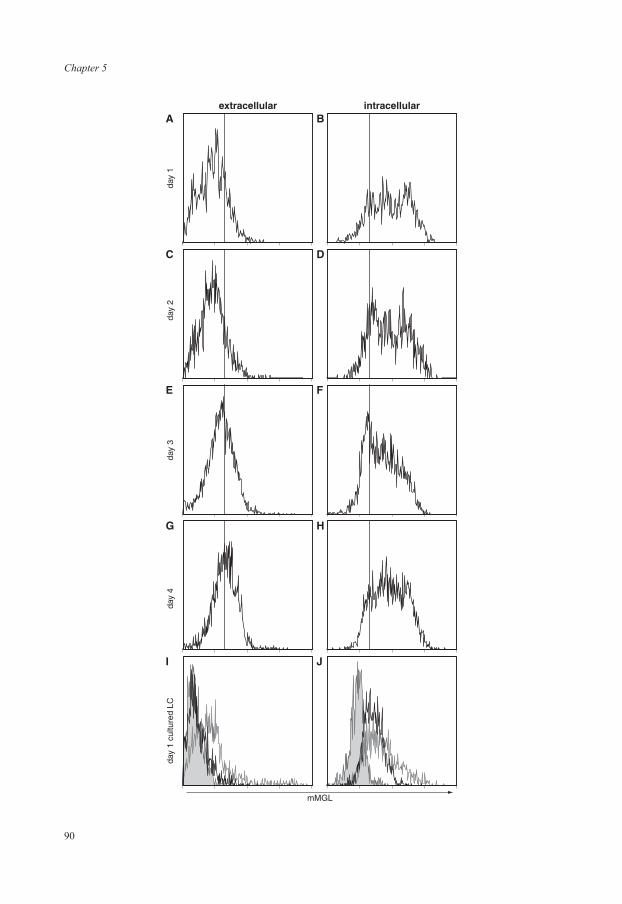
Chapter 5
90
extracellular
day
1da
y 2
day
3da
y 4
day
1 cu
lture
d LC
intracellularA B
mMGL
C D
E F
I J
G H

91
mMGL as dermis-induced mononuclear phagocyte marker
(Figure 5A, C, E, G). Yet, they expressed a significantly increased level of intracellular mMGL, compared to uncultured cells or cells matured in vitro (Figure 5B, D, E, H; Figure 4E, F). In line with the in situ profile in the steady state dermis, we found that about half of the LC would express a high level of intracellular mMGL, while the other half showed only a low level (Figure 5B, D, E, H). This expression pattern did not change essentially during the culture (Figure 5). We also analyzed emigrant cells with a confocal microscope to confirm the intracellular expression of mMGL by LC (Figure 6). These analyses revealed that mMGL is mainly, but not fully colocalized with Langerin in the cytoplasm of mature LC. Langerin previously has been shown to be internalized upon LC maturation28. Therefore, these findings suggest that the dermal microenvironment indeed induces (intracellular) mMGL expression in LC.
To determine whether a soluble factor derived from the dermis induces mMGL expression in LC, we cultured epidermal cell suspensions in whole skin-conditioned medium. In these cultures, we found that isolated LC cultured in skin-conditioned medium showed an intracellular mMGL expression pattern comparable to that of whole-skin emigrants, with a subpopulation of cells expressing higher mMGL levels than uncultured cells or cells cultured in standard culture medium (Figure 5J). Moreover, LC that had been cultured in skin-conditioned medium expressed mMGL on their surface (Figure 5I). Therefore, we conclude that a soluble factor
Figure 5. mMGL expression by LC is associated with exposure to dermal factors.Ear halves were cultured for one (A, B), two (C, D), three (E, F) or four days (G, H) in culture medium. Thereafter, cells that had migrated out were collected and stained intracellularly for Langerin, extracellularly for MHC class II and extracellularly (A, C, E, G) or intracellularly (B, D, F, H) for mMGL. A limited upregulation of mMGL was visible extracellularly, while emigrated LC showed clearly upregulated intracellular mMGL expression levels. The vertical line represents the maximum staining of isotype control antibodies. (I, J) To assess the influence of soluble dermis-derived factors, epidermal cell suspensions were cultured either one day in regular culture medium (bold line) or in medium obtained after a three day skin explant culture (thin line, open histogram). Filled histograms show isotype control profiles. LC that had been cultured in skin explant medium showed an upregulated intracellular mMGL expression level, comparable to whole skin emigrants, and retained (or upregulated) extracellular mMGL expression. Cells were gated as shown in Figure 4.
Langerin mMGL
Figure 6. mMGL colocalizes significantly with Langerin inside mature LC. Whole-skin emigrants were allowed to adhere on slides. Thereafter, they were fixed and permeabilized, stained for mMGL and Langerin and analyzed by confocal microscope. Note the colocalization of Langerin and mMGL in the cytoplasm of LC. (See Appendix page 178 for a full-color representation of this figure).

Chapter 5
92
from the dermis is indeed important in the induction of mMGL expression in and on LC. As we routinely added GM-CSF to the culture media for epidermal cell suspensions to ensure LC survival, this factor apparently is not instrumental for mMGL induction, even though it does induce Dectin-1, another marker of alternative activation9.
Only recently immigrating LC in skin-draining lymph nodes are mMGL+
After migration through the dermis and transport via afferent lymph, LC reach their destination in vivo, i.e. the skin-draining lymph nodes. To investigate the mMGL expression profile during this development, we obtained lymph node single cell suspensions and analyzed Langerin-positive cells for the detectability of intracellular mMGL. As represented in Figure 7C, we found that only a minor fraction of Langerin-positive cells in the steady-state skin-draining lymph nodes showed intracellular mMGL, suggesting that mMGL is downregulated again in LC once these cells have arrived in lymph nodes. Labeling steady state skin-draining lymph node tissue sections for Langerin and mMGL by immunofluorescence confirmed this notion (Figure 7E). Whereas we identified mMGLhigh-expressing cells scattered throughout the paracortex, which are presumably dermis-derived mononuclear phagocytes29, we did not find coexpression of mMGL on Langerin-positive cells. To study the mMGL phenotype of recently immigrating LC, we painted FITC onto shaved back skin and analyzed prior (FITC–) and recent immigrant (FITC+) LC in skin-draining LN 24h later. As depicted in Figure 7D, we found that FITC– LC were again virtually mMGL negative, whereas FITC+ LC showed the same mMGL profile as LC in skin explant cultures: one subpopulation of high mMGL expressing cells and one of low mMGL expressing cells. These results confirm the notion that LC that freshly arrive in LN still have detectable mMGL levels, which are downregulated once they have arrived in the skin-draining lymph nodes.
LC use mMGL only as a minor receptor to take up ligands As the dermal microenvironment induced mMGL expression on the surface of LC, we
wondered whether LC would use the lectins as endocytic receptors to take up antigens. To approach this question, we performed skin explant cultures with soluble mMGL ligands present in the medium (polyacrylamide conjugated to α-N-acetylgalactosaminides, α-GalNAc, or to the β-enantiomer, β-GalNAc) or a control glycosylated polymer that is not a ligand for the mMGL lectins (β-N-acetylglucosaminides-conjugated polyacrylamide, β-GlcNAc). It has been shown previously that the two GalNAc glycoconjugates are preferentially bound by mMGL2 and can be taken up by immature bone marrow-derived DC and that this ingestion can be blocked by adding mMGL-blocking antibodies26. After two days of culture, we harvested the skin emigrant cells in the medium and analyzed whether LC had taken up the ligands and whether ER-MP23 antibodies in the medium blocked it. As shown in Figure 8, we found that LC endocytosed α-GalNAc and β-GlcNAc, but barely β-GalNAc. Only the α-GalNAc uptake by LC could be inhibited to some degree by ER-MP23 antibodies, suggesting that a small amount of this ligand was taken up via mMGL2. In comparison, dermal cells endocytosed α-GalNAc and β-GalNAc to a much larger extent than LC, and this ingestion could be efficiently decreased by ER-MP23 antibodies. Therefore, we conclude that LC use mMGL only as minor receptors to take up antigens.
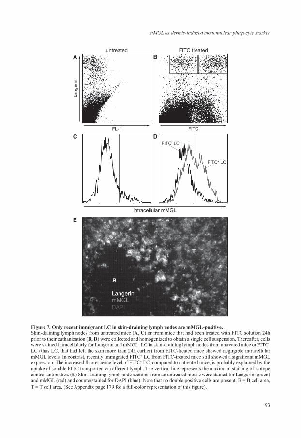
93
mMGL as dermis-induced mononuclear phagocyte marker
Lang
erin
FL-1 FITC
FITC+ LC
FITC- LC
untreated
intracellular mMGL
FITC treatedA B
B
T
C D
E
LangerinmMGLDAPI
Figure 7. Only recent immigrant LC in skin-draining lymph nodes are mMGL-positive.Skin-draining lymph nodes from untreated mice (A, C) or from mice that had been treated with FITC solution 24h prior to their euthanization (B, D) were collected and homogenized to obtain a single cell suspension. Thereafter, cells were stained intracellularly for Langerin and mMGL. LC in skin-draining lymph nodes from untreated mice or FITC– LC (thus LC, that had left the skin more than 24h earlier) from FITC-treated mice showed negligible intracellular mMGL levels. In contrast, recently immigrated FITC+ LC from FITC-treated mice still showed a significant mMGL expression. The increased fluorescence level of FITC– LC, compared to untreated mice, is probably explained by the uptake of soluble FITC transported via afferent lymph. The vertical line represents the maximum staining of isotype control antibodies. (E) Skin-draining lymph node sections from an untreated mouse were stained for Langerin (green) and mMGL (red) and counterstained for DAPI (blue). Note that no double positive cells are present. B = B cell area, T = T cell area. (See Appendix page 179 for a full-color representation of this figure).
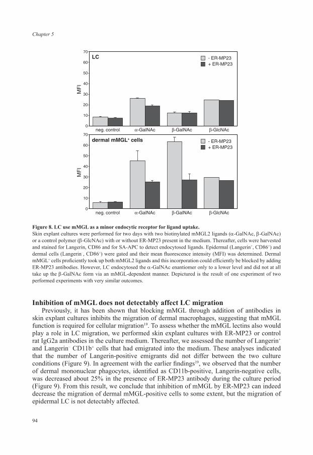
Chapter 5
94
Inhibition of mMGL does not detectably affect LC migrationPreviously, it has been shown that blocking mMGL through addition of antibodies in
skin explant cultures inhibits the migration of dermal macrophages, suggesting that mMGL function is required for cellular migration19. To assess whether the mMGL lectins also would play a role in LC migration, we performed skin explant cultures with ER-MP23 or control rat IgG2a antibodies in the culture medium. Thereafter, we assessed the number of Langerin+ and Langerin– CD11b+ cells that had emigrated into the medium. These analyses indicated that the number of Langerin-positive emigrants did not differ between the two culture conditions (Figure 9). In agreement with the earlier findings19, we observed that the number of dermal mononuclear phagocytes, identified as CD11b-positive, Langerin-negative cells, was decreased about 25% in the presence of ER-MP23 antibody during the culture period (Figure 9). From this result, we conclude that inhibition of mMGL by ER-MP23 can indeed decrease the migration of dermal mMGL-positive cells to some extent, but the migration of epidermal LC is not detectably affected.
neg. control
dermal mMGL+ cells
LC - ER-MP23+ ER-MP23
�-GalNAc �-GalNAc �-GlcNAc
neg. control �-GalNAc �-GalNAc �-GlcNAc0
10
20
30
40
50
60
70
MF
I
0
10
20
30
40
50
60
70
MF
I
- ER-MP23+ ER-MP23
Figure 8. LC use mMGL as a minor endocytic receptor for ligand uptake.Skin explant cultures were performed for two days with two biotinylated mMGL2 ligands (α-GalNAc, β-GalNAc) or a control polymer (β-GlcNAc) with or without ER-MP23 present in the medium. Thereafter, cells were harvested and stained for Langerin, CD86 and for SA-APC to detect endocytosed ligands. Epidermal (Langerin+, CD86+) and dermal cells (Langerin–, CD86+) were gated and their mean fluorescence intensity (MFI) was determined. Dermal mMGL+ cells proficiently took up both mMGL2 ligands and this incorporation could efficiently be blocked by adding ER-MP23 antibodies. However, LC endocytosed the α-GalNAc enantiomer only to a lower level and did not at all take up the β-GalNAc form via an mMGL-dependent manner. Depictured is the result of one experiment of two performed experiments with very similar outcomes.

95
mMGL as dermis-induced mononuclear phagocyte marker
DISCUSSION
We have shown here that resident mononuclear phagocytes in the steady-state dermis express a phenotype that is strongly reminiscent of alternatively activated macrophages: They express mMGL/CD301, MR/CD206 and Dectin-1, three surface lectins that are known to be induced by IL-4/IL-13 stimulation of macrophages and DC and considered to be markers of alternative activation5,9,10,20. Nevertheless, dermal mononuclear phagocytes express a similar phenotype in IL-4Rα knock-out mice, in which cells cannot respond to IL-4 or IL-13. Therefore, dermal mononuclear phagocytes do not represent alternatively activated macrophages in the strict sense, implying that the phenotype is a result of IL-4 or IL-13 signaling6. In this respect our data agree with the results found by Linehan et al. who showed that MR expression in IL-4Rα knock-out mice was not affected in resident tissue macrophages30. Granuloma macrophages induced by schistosome eggs, however, were MR-negative in IL-4Rα knock-out mice, demonstrating that these cells represent true alternatively activated macrophages in wild-type mice30. Nevertheless, we consider it likely that the gene expression profile of dermal and possibly also other connective tissue macrophages will show similarity with truly alternatively activated macrophages, as we will discuss below. Epidermal LC, which express a low level of extra- and intracellular mMGL in their immature state, strongly upregulate intracellular mMGL when they migrate through the dermis. Maturation of LC in vitro, in the absence of dermal factors, did not cause such an upregulation, therefore LC maturation in itself is not sufficient to increase mMGL levels. Once LC have left the dermis and reach skin-draining lymph nodes, mMGL is quickly downregulated again in LC. Therefore, dermal factors appear to be responsible for the induction of mMGL expression in LC.
all events Langerin+ Langerin-
CD11b+
0
20
40
60
80
100
rela
tive
cell
num
ber
IgG2aER-MP23
Figure 9. mMGL is not involved in LC migration.Skin explant cultures were performed for two days with ER-MP23 or an isotype control antibody in the medium. Thereafter, emigrated cells were harvested, stained for Langerin and CD11b and quantified by flowcytometry. Total acquired events in the control medium were set to 100%, the amount of total events in the ER-MP23-containing medium, as well as the amount of the gated cells were thereafter related to this number. Shown are the average cell numbers of four experiments. Addition of ER-MP23 could inhibit the emigration of about 25% of dermal cells, but did not affect emigration of LC.
Chapter 5
96
Previously, mMGL expression has been used to discriminate between dermal macrophages and LC11,29, as LC seemed to be mMGL-negative24. Our current findings show, however, that immature LC in the epidermis do express mMGL, although at levels that are too low to be detected by immunofluorescence on tissue sections. Migrating through the dermis, about a third to half of the LC express higher mMGL levels that allow detection by this method. Once these cells reach skin-draining lymph nodes, mMGL levels are downregulated again so that LC appear negative for mMGL on stained sections. Therefore, mMGL remains useful to discriminate LC from dermally derived mononuclear phagocytes using immunofluorescence or immunohistochemistry on steady state lymph nodes, as cells originating from the dermis retain mMGL-positivity29. This quantitative difference in mMGL levels also enables the flowcytometric distinction between the two lymph node antigen presenting cell populations (unpublished results). However, this difference in mMGL expression may not be sufficient to distinguish between LC and dermally derived cells in conditions where dermal factors profoundly influence LC. Therefore, mMGL expression is not a universal marker to distinguish between epidermal LC and dermally derived mononuclear phagocytes, but is useful in conditions where mMGL expression profiles have been defined, such as described here.
The transient induction of mMGL in LC raises the question whether this lectin serves particular functions in LC. For dermal macrophages several mMGL functions have been established. For instance, this lectin functions as endocytic receptor [this study; and ref. 6] and as signaling receptor involved in cellular migration induced by cytokine production [this study; and ref. 18]. However, mMGL seems to play only a minor role, if any, in endocytosis and migration by LC. At least two reasons can be envisaged that explain this difference. First, it might be that we used an ineffective ligand to test lectin-mediated endocytosis. The applied single residue-conjugated polymers have a higher affinity for mMGL2, compared to mMGL1; the latter lectin preferentially recognizes galactose and N-acetylgalactosamine residues in more complex structures such as the terminal Lewisx antigen15. It has been shown that dermal macrophages express both mMGL1 and mMGL215, but we cannot exclude that LC only express mMGL1. Therefore, conclusions can be drawn only to a limited extent, and testing LC with mMGL1- and mMGL2-specific antibodies will be necessary to resolve the issue whether LC express mMGL1, mMGL2 or both. Secondly, the two mMGL proteins might execute other functions specifically inside LC, as we found LC-associated mMGL to be expressed predominately intracellularly. It has been shown for two lectin families, galectins and annexins, that they perform intracellular functions31. Other C-type lectins that have been shown to disappear from the surface of mature DC, but that have been found to persist intracellularly include Langerin [this study; and ref. 28] and the MR [unpublished observation], thus two other well-known endocytic receptors. Hypothetically, these lectins might assist in accumulating and storing antigens in retention compartments32, in order (i) to break them down later and present them in MHC class II-context, (ii) to transfer them into the cytoplasm to cross-present them in MHC class I-context, or (iii) to transfer them to other DC subpopulations. In this respect, it is conceivable that the mMGL lectins are only briefly expressed on the LC surface, are quickly internalized again and delivered then to retention compartments. Our findings of extracellular mMGL expression on in vitro matured LC in skin-conditioned medium would fit into this picture. Future determinations of other mMGL functions might shed more light on the functions of mMGL expression in LC in particular and in mononuclear phagocytes in general.
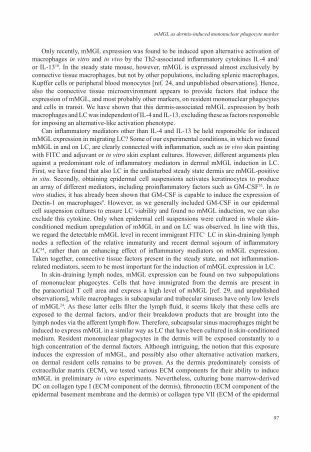
97
mMGL as dermis-induced mononuclear phagocyte marker
Only recently, mMGL expression was found to be induced upon alternative activation of macrophages in vitro and in vivo by the Th2-associated inflammatory cytokines IL-4 and/or IL-1310. In the steady state mouse, however, mMGL is expressed almost exclusively by connective tissue macrophages, but not by other populations, including splenic macrophages, Kupffer cells or peripheral blood monocytes [ref. 24, and unpublished observations]. Hence, also the connective tissue microenvironment appears to provide factors that induce the expression of mMGL, and most probably other markers, on resident mononuclear phagocytes and cells in transit. We have shown that this dermis-associated mMGL expression by both macrophages and LC was independent of IL-4 and IL-13, excluding these as factors responsible for imposing an alternative-like activation phenotype.
Can inflammatory mediators other than IL-4 and IL-13 be held responsible for induced mMGL expression in migrating LC? Some of our experimental conditions, in which we found mMGL in and on LC, are clearly connected with inflammation, such as in vivo skin painting with FITC and adjuvant or in vitro skin explant cultures. However, different arguments plea against a predominant role of inflammatory mediators in dermal mMGL induction in LC. First, we have found that also LC in the undisturbed steady state dermis are mMGL-positive in situ. Secondly, obtaining epidermal cell suspensions activates keratinocytes to produce an array of different mediators, including proinflammatory factors such as GM-CSF33. In in vitro studies, it has already been shown that GM-CSF is capable to induce the expression of Dectin-1 on macrophages9. However, as we generally included GM-CSF in our epidermal cell suspension cultures to ensure LC viability and found no mMGL induction, we can also exclude this cytokine. Only when epidermal cell suspensions were cultured in whole skin-conditioned medium upregulation of mMGL in and on LC was observed. In line with this, we regard the detectable mMGL level in recent immigrant FITC+ LC in skin-draining lymph nodes a reflection of the relative immaturity and recent dermal sojourn of inflammatory LC34, rather than an enhancing effect of inflammatory mediators on mMGL expression. Taken together, connective tissue factors present in the steady state, and not inflammation-related mediators, seem to be most important for the induction of mMGL expression in LC.
In skin-draining lymph nodes, mMGL expression can be found on two subpopulations of mononuclear phagocytes. Cells that have immigrated from the dermis are present in the paracortical T cell area and express a high level of mMGL [ref. 29, and unpublished observations], while macrophages in subcapsular and trabecular sinuses have only low levels of mMGL24. As these latter cells filter the lymph fluid, it seems likely that these cells are exposed to the dermal factors, and/or their breakdown products that are brought into the lymph nodes via the afferent lymph flow. Therefore, subcapsular sinus macrophages might be induced to express mMGL in a similar way as LC that have been cultured in skin-conditioned medium. Resident mononuclear phagocytes in the dermis will be exposed constantly to a high concentration of the dermal factors. Although intriguing, the notion that this exposure induces the expression of mMGL, and possibly also other alternative activation markers, on dermal resident cells remains to be proven. As the dermis predominately consists of extracellular matrix (ECM), we tested various ECM components for their ability to induce mMGL in preliminary in vitro experiments. Nevertheless, culturing bone marrow-derived DC on collagen type I (ECM component of the dermis), fibronectin (ECM component of the epidermal basement membrane and the dermis) or collagen type VII (ECM of the epidermal

Chapter 5
98
basement membrane) did not induce an increased mMGL expression in these cells, whereas IL-4 as positive control treatment did (unpublished observations). Therefore, the identity of the dermal factors that induce the expression of mMGL, and possibly other alternative activation markers on mononuclear phagocytes in the steady state dermis and lymph nodes, remains unknown.
Alternatively activated macrophages are often associated with tissue remodeling or tissue repair, thus with the breakdown and de novo synthesis of ECM components6,7. Despite the fact that the dermal mononuclear phagocytes are not IL-4/IL-13-activated, a major role of these cells in connective tissue maintenance is likely. We have recently shown that the majority of dermal interstitial cells are macrophages and not fibroblasts, as widely believed. Synthesis of ECM components by macrophages is well documented2 and as such, the notion that the majority of interstitial cells synthesize the ECM of the dermis might nevertheless turn out to be true. Alternatively activated macrophages are also associated with tolerance and suppression of immune responses. The dermis and other connective tissues might thus be places where an immunosuppressive microenvironment is supported. Tumor-associated macrophages, which mostly reside in and regulate supporting connective tissue35 consistently have been shown to express mMGL36-38. It is feasible that the extracellular matrix production stimulated by the tumor generates a microenvironment that activates tumor-infiltrating macrophages along an alternative route with related connective tissue maintaining and immune suppressive function. Identifying the factors that induce macrophages to adopt phenotypes similar to alternatively activated macrophages, and counteracting these, might thus also benefit cancer treatments.
To summarize, we have shown that resident dermal mononuclear phagocyte subpopulations express several markers of alternatively activated macrophages, for which IL-4 or IL-13 signalling is not required. Additionally, LC migrating through the dermis in vivo or cultured in vitro in medium from skin explant cultures contain higher mMGL levels than immature, epidermal LC or LC cultured in standard medium. LC that freshly arrive in skin-draining lymph nodes still show intracellular mMGL, whereas LC that have remained for a prolonged time in the lymph nodes did not. Therefore, the expression of mMGL in LC was closely linked to the exposure to the dermal microenvironment and factors derived thereof.
ACKNOWLEDGEMENTS
We wish to express our appreciation for the contribution of the following colleagues to the realization of this paper: Dr. Timo van den Berg for his help in the erythrocyte binding studies, Dr. Sem Saeland for making the anti-Langerin antibodies available, Dr. Frank Brombacher for the permission to use material from IL-4Rα KO mice, Dr. Gert van Cappellen for help with the confocal microscope and Tar van Os for preparation of the (photo)graphs in this work.
This study was supported by the J.A. Cohen Institute/Interuniversitary Research Institute for Radiopathology and Radioprotection (IRS), Leiden, The Netherlands. P. S. and N. R. were supported by grant P-14949 of the Austrian Science Fund, G. R. was supported by a grant from the “Institute for Promotion of Innovation by Science and Technology in Flanders” (IWT-Vlaanderen) for “Generisch Basisonderzoek aan de Universiteiten” (IWT-GBOU).

99
mMGL as dermis-induced mononuclear phagocyte marker
REFERENCES
1. Nikolic, T. & Leenen, P.J. Histiocyte function and development in the normal immune system, in Histiocytic Disorders of Children and Adults. (eds. S. Weitzman & R.M. Egeler) 40-65 (Cambridge University Press, Cambridge, UK. ; 2005).
2. Lindblad, W.J. Collagen expression by novel cell populations in the dermal wound environment. Wound Repair Regen. 6 186-193 (1998).
3. Hume, D.A., Ross, I.L., Himes, S.R., Sasmono, R.T., Wells, C.A. & Ravasi, T. The mononuclear phagocyte system revisited. J Leukoc Biol 72, 621-627 (2002).
4. Stout, R.D. & Suttles, J. Functional plasticity of macrophages: reversible adaptation to changing microenvironments. J Leukoc Biol 76, 509-513 (2004).
5. Stein, M., Keshav, S., Harris, N. & Gordon, S. Interleukin 4 potently enhances murine macrophage mannose receptor activity: a marker of alternative immunologic macrophage activation. J Exp Med 176, 287-292 (1992).
6. Gordon, S. Alternative activation of macrophages. Nat Rev Immunol 3, 23-35 (2003).7. Gratchev, A., Kzhyshkowska, J., Utikal, J. & Goerdt, S. Interleukin-4 and dexamethasone counterregulate
extracellular matrix remodelling and phagocytosis in type-2 macrophages. Scand J Immunol 61, 10-17 (2005).8. Porcheray, F., Viaud, S., Rimaniol, A.C., Leone, C., Samah, B., Dereuddre-Bosquet, N., Dormont, D. & Gras, G.
Macrophage activation switching: an asset for the resolution of inflammation. Clin Exp Immunol 142, 481-489 (2005).
9. Willment, J.A., Lin, H.H., Reid, D.M., Taylor, P.R., Williams, D.L., Wong, S.Y., Gordon, S. & Brown, G.D. Dectin-1 expression and function are enhanced on alternatively activated and GM-CSF-treated macrophages and are negatively regulated by IL-10, dexamethasone, and lipopolysaccharide. J Immunol 171, 4569-4573 (2003).
10. Raes, G., Brys, L., Dahal, B.K., Brandt, J., Grooten, J., Brombacher, F., Vanham, G., Noel, W., Bogaert, P., Boonefaes, T., Kindt, A., Van den Bergh, R., Leenen, P.J., De Baetselier, P. & Ghassabeh, G.H. Macrophage galactose-type C-type lectins as novel markers for alternatively activated macrophages elicited by parasitic infections and allergic airway inflammation. J Leukoc Biol 77, 321-327 (2005).
11. Dupasquier, M., Stoitzner, P., van Oudenaren, A., Romani, N. & Leenen, P.J. Macrophages and dendritic cells constitute a major subpopulation of cells in the mouse dermis. J Invest Dermatol 123, 876-879 (2004).
12. Geutskens, S.B., Otonkoski, T., Pulkkinen, M.A., Drexhage, H.A. & Leenen, P.J. Macrophages in the murine pancreas and their involvement in fetal endocrine development in vitro. J Leukoc Biol 78, 845-852 (2005).
13. Barner, M., Mohrs, M., Brombacher, F. & Kopf, M. Differences between IL-4R alpha-deficient and IL-4-deficient mice reveal a role for IL-13 in the regulation of Th2 responses. Curr Biol 8, 669-672 (1998).
14. Yamamoto, K., Ishida, C., Shinohara, Y., Hasegawa, Y., Konami, Y., Osawa, T. & Irimura, T. Interaction of immobilized recombinant mouse C-type macrophage lectin with glycopeptides and oligosaccharides. Biochemistry 33, 8159-8166 (1994).
15. Tsuiji, M., Fujimori, M., Ohashi, Y., Higashi, N., Onami, T.M., Hedrick, S.M. & Irimura, T. Molecular cloning and characterization of a novel mouse macrophage C-type lectin, mMGL2, which has a distinct carbohydrate specificity from mMGL1. J Biol Chem 277, 28892-28901 (2002).
16. Fearon, D.T. Regulation by membrane sialic acid of beta1H-dependent decay-dissociation of amplification C3 convertase of the alternative complement pathway. Proc Natl Acad Sci U S A 75, 1971-1975 (1978).
17. Jansen, A., Homo-Delarche, F., Hooijkaas, H., Leenen, P.J., Dardenne, M. & Drexhage, H.A. Immunohistochemical characterization of monocytes-macrophages and dendritic cells involved in the initiation of the insulitis and beta-cell destruction in NOD mice. Diabetes 43, 667-675 (1994).
18. Sato, M., Kawakami, K., Osawa, T. & Toyoshima, S. Molecular cloning and expression of cDNA encoding a galactose/N-acetylgalactosamine-specific lectin on mouse tumoricidal macrophages. J Biochem 111, 331-336 (1992).
19. Chun, K.H., Imai, Y., Higashi, N. & Irimura, T. Migration of dermal cells expressing a macrophage C-type lectin during the sensitization phase of delayed-type hypersensitivity. J Leukoc Biol 68, 471-478 (2000).
20. Martinez-Pomares, L., Reid, D.M., Brown, G.D., Taylor, P.R., Stillion, R.J., Linehan, S.A., Zamze, S., Gordon, S. & Wong, S.Y. Analysis of mannose receptor regulation by IL-4, IL-10, and proteolytic processing using novel monoclonal antibodies. J Leukoc Biol 73, 604-613 (2003).

Chapter 5
100
21. Linehan, S.A. The mannose receptor is expressed by subsets of APC in non-lymphoid organs. BMC Immunol 6, 4 (2005).
22. Herbert, D.R., Holscher, C., Mohrs, M., Arendse, B., Schwegmann, A., Radwanska, M., Leeto, M., Kirsch, R., Hall, P., Mossmann, H., Claussen, B., Forster, I. & Brombacher, F. Alternative macrophage activation is essential for survival during schistosomiasis and downmodulates T helper 1 responses and immunopathology. Immunity 20, 623-635 (2004).
23. Stoitzner, P., Holzmann, S., McLellan, A.D., Ivarsson, L., Stossel, H., Kapp, M., Kammerer, U., Douillard, P., Kampgen, E., Koch, F., Saeland, S. & Romani, N. Visualization and characterization of migratory Langerhans cells in murine skin and lymph nodes by antibodies against Langerin/CD207. J Invest Dermatol 120, 266-274 (2003).
24. Mizuochi, S., Akimoto, Y., Imai, Y., Hirano, H. & Irimura, T. Unique tissue distribution of a mouse macrophage C-type lectin. Glycobiology 7, 137-146 (1997).
25. Romani, N., Holzmann, S., Tripp, C.H., Koch, F. & Stoitzner, P. Langerhans cells - dendritic cells of the epidermis. APMIS 111, 725-740 (2003).
26. Denda-Nagai, K., Kubota, N., Tsuiji, M., Kamata, M. & Irimura, T. Macrophage C-type lectin on bone marrow-derived immature dendritic cells is involved in the internalization of glycosylated antigens. Glycobiology 12, 443-450 (2002).
27. Larsen, C.P., Steinman, R.M., Witmer-Pack, M., Hankins, D.F., Morris, P.J. & Austyn, J.M. Migration and maturation of Langerhans cells in skin transplants and explants. J Exp Med 172, 1483-1493 (1990).
28. Douillard, P., Stoitzner, P., Tripp, C.H., Clair-Moninot, V., Ait-Yahia, S., McLellan, A.D., Eggert, A., Romani, N. & Saeland, S. Mouse lymphoid tissue contains distinct subsets of langerin/CD207 dendritic cells, only one of which represents epidermal-derived Langerhans cells. J Invest Dermatol 125, 983-994 (2005).
29. Sato, K., Imai, Y. & Irimura, T. Contribution of dermal macrophage trafficking in the sensitization phase of contact hypersensitivity. J Immunol 161, 6835-6844 (1998).
30. Linehan, S.A., Coulson, P.S., Wilson, R.A., Mountford, A.P., Brombacher, F., Martinez-Pomares, L. & Gordon, S. IL-4 receptor signaling is required for mannose receptor expression by macrophages recruited to granulomata but not resident cells in mice infected with Schistosoma mansoni. Lab Invest 83, 1223-1231 (2003).
31. Wang, J.L., Gray, R.M., Haudek, K.C. & Patterson, R.J. Nucleocytoplasmic lectins. Biochim Biophys Acta 1673, 75-93 (2004).
32. Lutz, M.B., Rovere, P., Kleijmeer, M.J., Rescigno, M., Assmann, C.U., Oorschot, V.M., Geuze, H.J., Trucy, J., Demandolx, D., Davoust, J. & Ricciardi-Castagnoli, P. Intracellular routes and selective retention of antigens in mildly acidic cathepsin D/lysosome-associated membrane protein-1/MHC class II-positive vesicles in immature dendritic cells. J Immunol 159, 3707-3716 (1997).
33. Mann, A., Breuhahn, K., Schirmacher, P. & Blessing, M. Keratinocyte-derived granulocyte-macrophage colony stimulating factor accelerates wound healing: Stimulation of keratinocyte proliferation, granulation tissue formation, and vascularization. J Invest Dermatol 117, 1382-1390 (2001).
34. Geissmann, F., Dieu-Nosjean, M.C., Dezutter, C., Valladeau, J., Kayal, S., Leborgne, M., Brousse, N., Saeland, S. & Davoust, J. Accumulation of immature Langerhans cells in human lymph nodes draining chronically inflamed skin. J Exp Med 196, 417-430 (2002).
35. Mantovani, A., Allavena, P. & Sica, A. Tumour-associated macrophages as a prototypic type II polarised phagocyte population: role in tumour progression. Eur J Cancer 40, 1660-1667 (2004).
36. Biswas, S.K., Gangi, L., Paul, S., Schioppa, T., Saccani, A., Sironi, M., Bottazzi, B., Doni, A., Vincenzo, B., Pasqualini, F., Vago, L., Nebuloni, M., Mantovani, A. & Sica, A. A distinct and unique transcriptional program expressed by tumor-associated macrophages (defective NF-kappaB and enhanced IRF-3/STAT1 activation). Blood 107, 2112-2122 (2006).
37. Imai, Y., Akimoto, Y., Mizuochi, S., Kimura, T., Hirano, H. & Irimura, T. Restricted expression of galactose/N-acetylgalactosamine-specific macrophage C-type lectin to connective tissue and to metastatic lesions in mouse lung. Immunology 86, 591-598 (1995).
38. Oda, S., Sato, M., Toyoshima, S. & Osawa, T. Purification and characterization of a lectin-like molecule specific for galactose/N-acetyl-galactosamine from tumoricidal macrophages. J Biochem 104, 600-605 (1988).

CHAPTER 6
DERMAL MACROPHAGES DEVELOP INTOGENUINE LYMPH NODE DENDRITIC CELLS
UPON EMIGRATION FROM THE SKIN
Marcel Dupasquier*, Joey Riepsaame*, Hui Wan*, Adri van Oudenaren*, Patrizia Stoitzner†, Sem Saeland‡, and Pieter J.M. Leenen*
*Department of Immunology, Erasmus MC, University Medical Center, Rotterdam, The Netherlands
†Department of Dermatology, Medical University, Innsbruck, Austria‡DermImmune, Lyon, France
Submitted for publication
Chapter 6
102
ABSTRACT
In a recent characterization of the mouse dermis we observed that ~60% of the interstitial cells represent macrophages as they avidly ingest intradermally injected particles and express at high levels typical macrophage markers but mostly lack dendritic cell (DC-)associated molecules. Previously, it has been shown that dermal macrophages emigrate from the dermis upon activation and contribute to the induction of contact hypersensitivity. Here, we investigate the relationship between dermal macrophages and skin-derived DC in vitro and in vivo. We report that the majority of dermal macrophages, upon skin emigration in vitro, develop into phenotypically mature DC, with expression levels of MHC class II and costimulatory molecules comparable to co-migrating Langerhans cells (LC). Ex vivo, skin-derived cells were identified in draining lymph nodes by intracellular expression of the dermal marker macrophage galactose/N-acetylgalactosamine-specific lectin (mMGL/CD301) and these cells displayed a similar mature DC phenotype. To assess whether these cells originated from the dermis, we applied FITC onto the skin and traced marked cells in draining lymph nodes. We observed that mMGL+ cells, distinct from LC, represented the majority of FITC+ cells in the draining lymph nodes. Functionally, dermal emigrants and LC possessed similar capacities to stimulate naïve alloantigen-specific T cells. Therefore, we conclude that dermal macrophages represent a sizeable population of DC precursors in the skin. These cells thus constitute a previously underestimated antigen presenting cell population that will play a major role in skin-related immune reactions.
INTRODUCTION
Dendritic cells (DC) are the sentinels of the immune system. Apart from being crucial constituents of secondary lymphoid organs, they also reside as immature cells in virtually all non-lymphoid organs where their main function is to sample the environment by endocytosis and to sense invading pathogens1,2. Upon stimulation, however, their state will change dramatically as they emigrate from peripheral tissues and mature into potent antigen presenting cells. They do so by upregulating essential T cell stimulating and co-stimulating factors, such as MHC class I and class II, CD40, CD80 and CD86 molecules, while downregulating proteins of the antigen uptake machinery. Consequently, once DC arrive in draining lymph nodes (LN), they appear fundamentally different compared to their immature precursors in the tissues.
In spite of the fact that immature DC populations have been observed in multiple peripheral tissues, only few of those have been characterized in detail so far. Peripheral DC are often described using two paradigms: Langerhans cells (LC) as DC subpopulation of skin and mucosal epithelial surfaces and dermal DC as DC subpopulation of interstitial connective tissues2-4. Due to the limitation of obtaining sufficient numbers of these cells from either human or mouse tissues, in vitro representatives derived from precursor cells have been used frequently to investigate these two subpopulations. Culturing human cord blood hematopoietic progenitor cells with the appropriate cytokines leads to a concurrent accumulation of CD14+ and CD1a+ DC subpopulations, resembling dermal DC and LC, respectively5.
103
Dermal macrophages as DC precursors
Similarly, human monocytes, stimulated with GM-CSF and IL-4 without or with additional TGF-β, develop into CD14+ DC or CD1a+ LC-like cells, respectively6,7.
When considering the in situ counterparts of the two paradigms of peripheral DC subpopulations, it is noticeable that LC have been characterized in much more detail than dermal DC. This is most probably due to the fact that LC constitute a subpopulation of cells in the epidermis that can be identified easily by staining for example for Langerin, MHC class II or ATPase8. Moreover, these cells can be isolated in relatively large numbers by obtaining epidermal single cell suspensions. In comparison, dermal DC are much more difficult to acquire as they are strongly attached to the extracellular matrix. Enzymatic digestion of dermal tissue yields a heterogeneous population of cells that barely reflects the actual composition of the interstitium (cf. refs. 9,10). Therefore, the identity of the immature dermal DC in the tissue remains unclear. In particular, the distinction of dermal DC from macrophages that are also present in the dermis in large numbers11 remains an unsolved issue. Adding to the complexity is the fact that there are no phenotypic or functional markers that unequivocally distinguish immature DC from macrophages12,13.
Recently, we have investigated the phenotype of mononuclear phagocytes in the mouse dermis, and observed that these cells represent the majority of interstitial cells in the steady-state10. Moreover, we found that the vast majority of these cells express macrophage markers at high levels, such as F4/80, CD68, CD11b, the murine macrophage galactose/ N-acetylgalactosamine-specific C-type lectin (mMGL/CD301), and the mannose receptor (MR/CD206). Additionally, we demonstrated that these cells are highly phagocytic. Conversely, only a small fraction of the dermal interstitial cells express CD11c, a marker that is relatively specific for DC in mice. When we analyzed the expression of MHC class II molecules, we found that all mononuclear phagocytes in the dermis of the back skin but only about one third of these cells in the ear dermis are MHC class II+. Notably, no additional phenotypic differences were observed between cells in these locations. Therefore, we concluded that the vast majority of dermal mononuclear phagocytes represent cells with a macrophage rather than a DC phenotype10. Since the dermal microenvironment induces expression of mMGL in both macrophages and DC, but not in other cell types, this apparently is a useful marker for the identification of dermis-derived mononuclear phagocytes14.
Previously, it was described that the majority of mMGL+ cells emigrate from the dermis after the application of an adjuvant onto the skin, thus after the initiation of an inflammatory response15,16. These cells migrate into skin-draining lymph nodes and accumulate in paracortical T cell areas, a feature that is highly reminiscent of DC. Additionally, these mMGL+ cells contribute to the induction of a contact hypersensitivity (CHS) response, an immune reaction that is highly dependent on the activation of hapten-specific T cells15. Taken together, these findings suggest that mMGL+ dermal cells, exhibiting a macrophage phenotype in situ, adopt a localization and accessory function reminiscent of DC upon stimulation.
Therefore, we hypothesized that dermal macrophages might actually represent precursors of DC. To characterize these cells further, we performed explant cultures of mouse skin, where we could analyze emigrating cells phenotypically and functionally. Taking advantage of the fact that also LC, as unequivocal DC, emigrate from the tissue in this system, we show that the majority of dermal emigrants indeed acquires a mature DC phenotype. Moreover, dermal emigrants appear to be as efficient as LC in stimulating a mixed leukocyte

Chapter 6
104
reaction. Using FITC labeling to track the migration of epidermal and dermal cells into the skin-draining lymph nodes in vivo, we show that the majority of FITC-positive cells are dermally derived. Therefore, we conclude that dermal macrophages represent precursors of a sizeable subpopulation of DC in skin-draining lymph nodes.
MATERIALS AND METHODS
AnimalsSpecific pathogen-free female C57BL/6J and C3H/FeJ mice were obtained from Harlan
(Horst, The Netherlands) and housed in the Erasmus MC Animal Center with free access to food and water. Housing, care and experimental procedures were performed in accordance with Dutch legal regulations, including approval by the ethical committee. Mice were used between 10-16 weeks of age.
Antibodies and reagentsThe following antibodies were used in this study: purified and Alexa Fluor 488-labeled rat
anti-mouse Langerin (CD207, clone 929F3, IgG1, Dendritics, Dardilly, France); biotinylated rat anti-mMGL1 and mMGL2 (CD301; clone ER-MP23, IgG2a;14, biotinylated rat anti-CD71 (clone ER-MP21, IgG2a) and supernatants of rat anti-F4/80 (clone CI:A3-1, IgG2b), CD205 (clone NLDC-145, rat IgG2a) and CD8α (clone 53-6.7, rat IgG2a) were generated in our lab; purified anti-MR (clone MR5D3, rat IgG2a) and anti-Dectin-1 (clone 2A11, rat IgG2b) were from Hycult Biotechnology, Uden, The Netherlands; FITC-labeled CD4 (clone H129.19, rat IgG2a) and PE-labeled CD3 (clone 145-2C11, Armenian hamster IgG1), CD11b (clone M1/70, rat IgG2b), CD11c (clone HL3, Armenian hamster IgG1), CD40 (clone 3/23, rat IgG2a), CD80 (clone 16-10A1, Armenian hamster IgG2), CD86 (clone GL1, rat IgG2a) and anti-I-A/I-E (clone M5/114.15.2, rat IgG2b) were from BD Pharmingen, San Diego, CA, USA, whereas PE-labeled rat-anti mouse CCR7/CD197 (clone 4B12, IgG2a) was from eBioscience (San Diego, CA, USA). FITC-labeled and PE-labeled goat anti-rat immunoglobulins were obtained from Caltag (San Francisco, CA, USA), allophycocyanin-labeled streptavidin (SA-APC) from BD Pharmingen and Texas Red-labeled streptavidin (SA-TR) from Caltag. Culture medium was RPMI-1640 (Cambrex, Verviers, Belgium), supplemented with 10% fetal calf serum (FCS; HyClone, Kansas City, MO, USA), 100 U/ml penicillin and 100 µg/ml streptomycin (both from Cambrex). FITC was from Sigma, whereas FITC-labeled β-N-acetylgalactosaminide-PAA-polymers (β-GalNAc) were obtained from GlycoTech (Rockville, MD, USA).
Skin tissue section stainingsImmunofluorescence staining of ear tissue sections was performed as described14.
In short, uncultured and cultured ear halves were frozen in Tissue-Tek O.C.T. Compound (Sakura Finetek, Zoeterwoude, The Netherlands) and 6 µm thick cryostat sections were cut. Subsequently, sections were fixed for 4 minutes in acetone, rehydrated in PBS pH 7.8 plus 0.05% Tween-20 (Fluka, Buchs, Switzerland), incubated with avidin/biotin blocking kit (Vector labs, Burlingame, CA, USA), blocked with 10% normal rabbit serum and incubated

105
Dermal macrophages as DC precursors
with biotinylated ER-MP23 (ER-MP23-bio) antibodies that were visualized with Texas-Red-labeled streptavidin. Thereafter, sections were embedded in DAPI-containing Vectashield mounting media (Vector labs). Incubation steps were all performed in the dark at room temperature for 30 minutes; sections were washed twice between incubations with PBS supplemented with 0.05% Tween-20. Subsequently, sections were examined using a Zeiss Axioplan 2 fluorescence microscope (Zeiss, Göttingen, Germany). At least 20 microscopic fields of three independently stained sections were assessed.
Skin explant culturesWhole skin explant cultures were performed as described14. In short, ears were split and
both halves were cultured for two days. Thereafter, emigrant cells were harvested from the culture medium, washed, fixed with 2% PFA (Merck) for 10 minutes at RT and permeabilized with 0.25% saponin (Sigma) for 15 minutes at RT. Subsequently, cells were stained for Langerin, mMGL and a third marker using PE-labeled antibodies, followed by SA-APC. In some indicated experiments, cells were incubated with unlabeled antibodies, followed by FITC-labeled anti-rat immunoglobulins before fixation and permeabilization of the cells to detect expression of markers only at the cell surface. Alternatively, cells were first fixed and permeabilized and labeled thereafter with unlabeled antibodies, followed by FITC-labeled anti-rat immunoglobulins to detect intracellular (and extracellular) presence of detected markers. Next, these cells were labeled for CD86 and for mMGL. Fluorescent labeling of cells was subsequently measured on a FACSCalibur (BD Biosciences, San Jose, CA, USA) and analyzed using WinMDI 2.8 software.
In vivo FITC labeling of cutaneous cells and analysis of lymph nodesTwenty four hours prior to skin painting, mouse backs were shaved. Then, 250 µl 1%
FITC in 1:1 acetone:dibutylphtalate (Sigma) was applied onto the hairless skin. 4h, 24h or 48h later, mice were euthanized and their axillary, brachial and inguinal skin-draining lymph nodes were removed. Lymph nodes were subsequently cut into small pieces and suspended through a 70 µm nylon cell strainer. Cells were fixed and permeabilized as described above. Thereafter, they were stained with anti-Langerin antibodies, which were visualized with PE-labeled anti-rat immunoglobulins. Cells were subsequently blocked with 3% normal rat serum, stained for ER-MP23-bio and SA-APC and analyzed by flowcytometry as described above. To determine the phenotype of steady-state lymph node DC, lymph node cell suspensions were obtained as described above without prior treatment. Then, cells were fixed, permeabilized and stained for ER-MP23-bio, followed by SA-APC and the indicated markers.
Separation of epidermal and dermal antigen presenting cells and functional analysis in mixed leukocyte reactions
To distinguish between epidermally and dermally derived DC, skin explant cultures were performed for two days with 10 µg/ml FITC-labeled β-GalNAc-polymers added to the culture medium. These mMGL ligands are avidly ingested by dermal cells, but barely by LC14. This difference is based on the differential surface expression of mMGL. Consequently, by identifying mature DC by their high MHC class II expression, we could separate skin emigrants into MHC class IIhigh FITClow LC and MHC class IIhigh FITChigh dermally derived DC by cell

Chapter 6
106
sorting using a FACS Vantage (BD Biosciences). In addition, surface mMGL+ cells were sorted. For comparison, DC were generated in vitro by stimulating bone marrow precursors for 7 days (BMDC) with GM-CSF, essentially as described before17. Increasing numbers of these antigen-presenting cells were incubated in 96-well plates with 100,000 C3H/FeJ T cells, which had been obtained by depleting splenic cell suspensions for CD11b-, CD45R- (B220-) and MHC class II-expressing cells by MACS. DC and T cells were co-cultured for three days. Then, 0.5 µCi 3H-labeled thymidine was added to the cultures, which were incubated for another 16h. Subsequently, cells were harvested and 3H-thymidine incorporation was measured using a scintillation counter. Alternatively, cells were harvested after the three day culture period and stained for CD3, CD4 and the activation marker CD71. Thereafter, cells were analyzed by flowcytometry as described above, and the fractions of activated CD4+ T cells (CD4+ CD71+) and activated CD8+ T cells (CD3+ CD4– CD71+) were determined.
RESULTS
The majority of mMGL+ macrophages emigrates from the dermis in skin explant culture
In our previous characterization of dermal mononuclear phagocytes, we observed that mMGL can be regarded as a universal marker for these cells, while only a small subpopulation of mMGL+ cells in the steady-state dermis expresses CD11c10. As the potent adjuvant activity ascribed to migrating mMGL+ cells15,16 might theoretically be mediated by only this small subpopulation, representing genuine dermal DC, we investigated the migration and developmental fate of all dermal mMGL+ cells upon culture. Therefore, we performed skin explant cultures of ear halves for one or two days and subsequently stained sections for mMGL to determine the number of mMGL+ cells that remained in the dermis. In agreement with Chun et al.16, we observed that dermal cells retain mMGL in culture (see below). As shown in Figure 1, we found that already after one day of culture, around two thirds of all mMGL+ cells had emigrated from the dermis. This number did not change significantly at later time points. This is in contrast to LC, of which only small numbers could be found in the culture medium after one day, but which numbers increased significantly upon prolonged culture14. Therefore, we conclude that indeed the majority of dermal mMGL+ cells, not only the CD11c+ subset, emigrates from the dermis upon stimulation induced by the culture conditions.
Dermal emigrants show a DC phenotype similar to co-migrated LCNext, we determined the immunophenotype of the dermal emigrants obtained from skin-
explant cultures of ear halves after two or three days of culture. By flowcytometry, we identified epidermal LC and dermal mononuclear phagocytes on the basis of differential Langerin and mMGL expression. As some LC co-express both markers14, we first gated Langerin+ cells prior to selecting those expressing mMGL (Figure 2). We found that not only the epidermal LC but also the majority of dermal emigrants expressed high levels of MHC class II, CD80, CD86 and CD40. Moreover, both emigrant subpopulations expressed the same (low) level of CD11c, whereas dermal emigrants showed a lower, nevertheless still positive expression of CD205. CD11b and F4/80 were expressed by both cell populations, with most dermis-

107
Dermal macrophages as DC precursors
derived cells expressing a higher level of CD11b but a lower level of F4/80. We found CD8α not present on the vast majority of cells from either subpopulation, whereas both uniformly expressed CCR7 at considerable levels, making it highly likely that both cell types migrate in vivo towards skin-draining lymph nodes.
0
10
20
30
fresh 24h 48h0
10
# E
R-M
P23
+ c
ells
per
fiel
d
20
30
fresh 24h 48h
C
B
A
Figure 1. The majority of mMGL+ cells emigrate from ear dermis in culture. Fresh ear halves (A) or ear halves cultured for 24h (not shown) or 48h (B) were sectioned and stained for mMGL. Thereafter, mMGL+ cells were counted in at least 20 microscopic fields of three independently stained slides (C). Note that about 2/3 of the cells emigrate within the first 24h. Red fluorescence signals were recorded and converted to B/W images using standard imaging software. Background staining of the keratin layer indicates the skin border. These data represent 3 separate experiments and quantitative determinations.
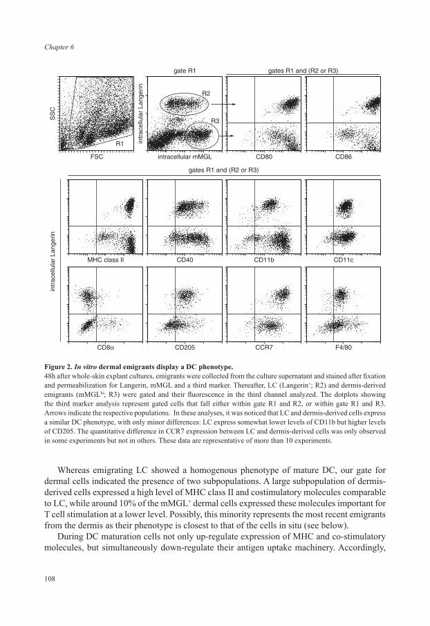
Chapter 6
108
Whereas emigrating LC showed a homogenous phenotype of mature DC, our gate for dermal cells indicated the presence of two subpopulations. A large subpopulation of dermis-derived cells expressed a high level of MHC class II and costimulatory molecules comparable to LC, while around 10% of the mMGL+ dermal cells expressed these molecules important for T cell stimulation at a lower level. Possibly, this minority represents the most recent emigrants from the dermis as their phenotype is closest to that of the cells in situ (see below).
During DC maturation cells not only up-regulate expression of MHC and co-stimulatory molecules, but simultaneously down-regulate their antigen uptake machinery. Accordingly,
MHC class II CD40 CD11b
intr
acel
lula
r La
nger
in
gates R1 and (R2 or R3)
gate R1
R2
R1
R3
gates R1 and (R2 or R3)
CD11c
CD8� CD205 CCR7 F4/80
FSC
SS
C
intracellular mMGL
intr
acel
lula
r La
nger
in
CD80 CD86
Figure 2. In vitro dermal emigrants display a DC phenotype.48h after whole-skin explant cultures, emigrants were collected from the culture supernatant and stained after fixation and permeabilization for Langerin, mMGL and a third marker. Thereafter, LC (Langerin+; R2) and dermis-derived emigrants (mMGLhi; R3) were gated and their fluorescence in the third channel analyzed. The dotplots showing the third marker analysis represent gated cells that fall either within gate R1 and R2, or within gate R1 and R3. Arrows indicate the respective populations. In these analyses, it was noticed that LC and dermis-derived cells express a similar DC phenotype, with only minor differences: LC express somewhat lower levels of CD11b but higher levels of CD205. The quantitative difference in CCR7 expression between LC and dermis-derived cells was only observed in some experiments but not in others. These data are representative of more than 10 experiments.
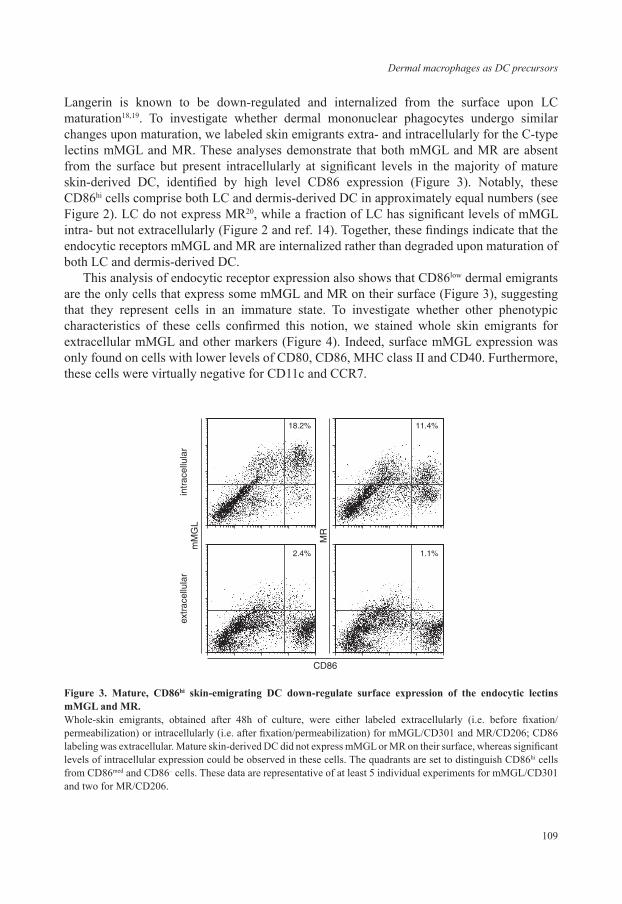
109
Dermal macrophages as DC precursors
Langerin is known to be down-regulated and internalized from the surface upon LC maturation18,19. To investigate whether dermal mononuclear phagocytes undergo similar changes upon maturation, we labeled skin emigrants extra- and intracellularly for the C-type lectins mMGL and MR. These analyses demonstrate that both mMGL and MR are absent from the surface but present intracellularly at significant levels in the majority of mature skin-derived DC, identified by high level CD86 expression (Figure 3). Notably, these CD86hi cells comprise both LC and dermis-derived DC in approximately equal numbers (see Figure 2). LC do not express MR20, while a fraction of LC has significant levels of mMGL intra- but not extracellularly (Figure 2 and ref. 14). Together, these findings indicate that the endocytic receptors mMGL and MR are internalized rather than degraded upon maturation of both LC and dermis-derived DC.
This analysis of endocytic receptor expression also shows that CD86low dermal emigrants are the only cells that express some mMGL and MR on their surface (Figure 3), suggesting that they represent cells in an immature state. To investigate whether other phenotypic characteristics of these cells confirmed this notion, we stained whole skin emigrants for extracellular mMGL and other markers (Figure 4). Indeed, surface mMGL expression was only found on cells with lower levels of CD80, CD86, MHC class II and CD40. Furthermore, these cells were virtually negative for CD11c and CCR7.
mM
GL
extr
acel
lula
rin
trac
ellu
lar
CD86
18.2% 11.4%
2.4% 1.1%
MR
Figure 3. Mature, CD86hi skin-emigrating DC down-regulate surface expression of the endocytic lectins mMGL and MR.Whole-skin emigrants, obtained after 48h of culture, were either labeled extracellularly (i.e. before fixation/ permeabilization) or intracellularly (i.e. after fixation/permeabilization) for mMGL/CD301 and MR/CD206; CD86 labeling was extracellular. Mature skin-derived DC did not express mMGL or MR on their surface, whereas significant levels of intracellular expression could be observed in these cells. The quadrants are set to distinguish CD86hi cells from CD86med and CD86– cells. These data are representative of at least 5 individual experiments for mMGL/CD301 and two for MR/CD206.

Chapter 6
110
DC derived from dermal macrophages transfer FITC into skin-draining lymph nodes after epicutaneous application
To investigate whether dermal mononuclear phagocytes might also acquire a DC phenotype in vivo upon emigration, we analyzed mMGL+ cells in skin-draining lymph nodes. Figure 5A shows that lymph node cells with high levels of intracellular mMGL are CD80+, CD86+, CD11b+, CD11c+ or hi, CD40hi and MHC class IIhi. This phenotype strongly resembles the in vitro skin emigrants originating from the dermis (cf. Figure 2) and lymph node DC identified previously (recent reviews refs. 3, 4). In contrast, the population of intracellular mMGLlo cells predominately consists of CD11clo, CD11b–, CD80–, CD86–, CD40– and MHC class II– cells with low forward and side scatter (data not shown). These characteristics match those of plasmacytoid DC precursors21, but this identification awaits final verification.
Previously, we have shown that mMGLhi cells localize to paracortical T cell areas in skin-draining lymph nodes, comparable to LC14,18. To analyze a putative local interaction
extr
acel
lula
r m
MG
L
CD80 CD86
MHC class II CD40
CD11c CCR7
Figure 4. Surface mMGL-positive skin-emigrating cells display an immature DC phenotype.Whole skin-emigrants were obtained after 48h of culture and labeled extracellularly for mMGL and for other DC markers. An immature subpopulation was observed that expressed mMGL on the cell surface and further showed intermediate levels of CD80, a low level of CD86, CD40 and MHC class II, but virtually no CD11c or CCR7. In contrast, mature DC, comprising LC and dermis-derived DC, express higher levels of all these markers and no mMGL on their surface.

111
Dermal macrophages as DC precursors
FIT
CF
ITC
1 day after FITCgate FITC+ cells
gate FITC+ cells2 days after FITC
R2
gate R2
intracellular Langerin
2.96% 0.44%
0.18%
1.23% 0.26%
0.19%
0.34% 1.14%
0.74%
0.73% 2.66%
0.58%
5.7% 7.8%
78.6%7.9%
11.7% 5.2%
71.5%11.5%
intracellular Langerin
intracellular mMGL
intracellular mMGL
intracellular mMGL
intracellular mMGL
intracellular mMGL
neg
CD80
mMGL CD3 DAPI overlay
CD
86CD11c
CD
11b
FIT
C
intr
acel
lula
r La
nger
in
FIT
C
intr
acel
lula
r La
nger
in
CD40
MH
C c
lass
II
A
B
C D
0.0%
0h
4h
24h
48h
2.5%
percentage mMGL+ cellsin the total LN suspension
5.0% 7.5%
FITC+
FITC-
Figure 5. Dermis-derived mMGL+ DC are present in skin-draining lymph nodes, cluster with lymphocytes and are major hapten-transporting cells.(A) Skin-draining lymph node single cell suspensions were prepared from untreated mice and labeled for intracellular mMGL and for other DC markers. mMGLhi cells were observed and express a similar mature DC phenotype as did in vitro skin emigrants. (B) Cytospins of non-enzymatically treated lymph node single cell suspensions were prepared and stained for mMGL (red) and for CD3 (green), with DAPI counterstaining (blue). mMGL+ cells were often discovered in close interaction with CD3+ T cells but also with CD3– B cells. The selected cluster shows a representative mMGLhi cell surrounded by lymphocytes. (C) 24h and 48h after application of FITC in adjuvant onto the back skin, skin-draining lymph node single cell suspensions were obtained and stained for Langerin and mMGL. Note that the vast majority of FITC+ cells, even after 48h express intracellular mMGL (78% of all FITC+ cells after 24h, 77% after 48h), whereas only a minority of them express Langerin (13% after 24h, 17% after 48h). This experiment was performed twice with virtually identical results. (D) Percentages of mMGL+ FITC+ and mMGL+ FITC– cells in the total lymph node single cell suspension before, and 4h, 24h and 48h after FITC application. Note that 4h after FITC application, only few mMGL+ cells have taken up soluble FITC that has diffused into the lymph nodes with incoming lymph.(See Appendix page 180 for a full-color representation of this figure).

Chapter 6
112
with T cells, we stained cytospin preparations of lymph node cell suspensions that had been obtained without proteolytic digestion in order to maintain clusters of associated cells. Indeed, mMGL+ cells are found to be in intimate contact with T and some non-T, presumably B cells (Figure 5B).
To show unequivocally that these mMGLhi cells originated from the dermis, we painted FITC onto the backs of mice. By applying this tracer in relatively large quantity, the dermal population of antigen-presenting cells is targeted as well as the epidermal LC22. Flowcytometric analysis of skin-draining lymph nodes of mice that had been treated with FITC indicated that more than 80% of all mMGL+ cells were FITC+ 24h after skin painting, indicating their recent skin derivation (Figure 5C). This frequency decreased to about 60% after 48h, suggesting a high turn over of the cells. In comparison, about 70% and 60% of all Langerin+ cells were FITC+ at these time points, respectively. It is interesting to note that dermally derived cells comprise the vast majority of cells carrying hapten from the skin into the draining lymph nodes as mMGL+ Langerin– cells represent more than 70% of all FITC+ cells at both 1 and 2 days after FITC application, whereas LC account for only 13-17%. To investigate the putative contribution of uptake of diffusing, soluble FITC by lymph node-resident mMGL+ cells, we analyzed lymph node cells from mice 4h after of FITC application. This was based on the previously reported finding that skin-applied molecules reach lymph node-resident DC via lymph and lymph node conduit as soon as 30 minutes after application and peak at 3hrs23. In contrast, skin-derived DC do not arrive in the lymph node before 12-18hr. Our early time point analysis showed that only very few mMGL+ cells (Figure 5D) and hardly any Langerin+ cells (data not shown) were FITC+ after 4h.
To summarize, these experiments indicate that, also in vivo, dermis-derived macrophages acquire a DC phenotype upon migration to skin-draining lymph nodes. They are the main cells transferring skin-applied haptens early on, and their close physical contact to lymphocytes suggests that they may have an important role in antigen presentation.
Dermis- and epidermis-derived DC possess similar capacities to stimulate allogeneic T cells
To investigate the antigen-presenting capacities of the different populations of skin-derived mononuclear phagocytes, we aimed to separate these cells from skin explant cultures. However, their similarity in DC phenotype and internalization of endocytic receptors, including Langerin, precluded their isolation based on differential expression of cell surface markers. As an alternative, we took advantage of the fact that these cells express different endocytic receptors in the tissue before they emigrate and mature. Dermal mononuclear phagocytes efficiently take up mMGL ligands such as α- or β-N-acetylgalactosamine-polyacylamide polymers when these are added to the medium during the explant cultures14. LC, on the other hand, barely absorb these glycosyl-polymers. Therefore, we performed skin explant cultures with FITC-labeled β-GalNac polymers added to the culture medium from the start of the experiment. After two days, emigrants were collected and sorted according to their FITC fluorescence into MHC class IIhigh, FITChigh dermis-derived DC and MHC class IIhigh, FITClow LC (Figure 6). In a subsequent experiment, a population of immature surface mMGL+ cells was obtained and compared with the mature DC (LC + DdDC). Sorted subpopulations were then co-cultured with allogeneic T cells to test their capacities to stimulate T cell proliferation.
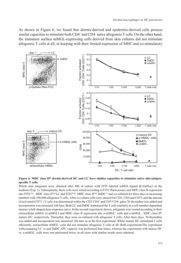
113
Dermal macrophages as DC precursors
As shown in Figure 6, we found that dermis-derived and epidermis-derived cells possess similar capacities to stimulate both CD4+ and CD4– naïve allogeneic T cells. On the other hand, the immature surface mMGL-expressing cells derived from skin cultures did not stimulate allogeneic T cells at all, in keeping with their limited expression of MHC and co-stimulatory
LCDdDCunstim.
1/10
0.6
0.5
0.4
0.3
Frac
tion
activ
ated
CD
3+ C
D4+
Tce
lls (
CD
71+)
0.2
0.1
01/33 1/100 1/333 T cells only
LCDdDCunstim.
1/10
0.3
0.25
0.2
0.15
Frac
tion
activ
ated
CD
3+ C
D4- T
cells
(C
D71
+)
0.1
0.05
01/33 1/100 1/333 T cells only
mature DCe-mMGL+ cellsBMDCunstim.
1/25
50,000
40,000
30,000
CP
M
20,000
10,000
0
1/50 1/100
DC / T cell ratio
1/200 T cells only
�-GalNAc-PAA-FITC
exp. 1 LC
DdDC
MH
C c
lass
II
extracellular mMGL
MH
C c
lass
II
exp. 2 mature DC
e-mMGL cells
Figure 6. MHC class IIhi dermis-derived DC and LC have similar capacities to stimulate naïve allo-antigen-specific T cells.Whole skin emigrants were obtained after 48h of culture with FITC-labeled mMGL-ligand (β-GalNac) in the medium (Exp. 1). Subsequently, these cells were sorted according to FITC fluorescence and MHC class II expression into FITClow, MHC class IIhigh LC and FITChigh, MHC class IIhigh DdDC14 and co-cultured for three days in increasing numbers with 100,000 allogeneic T cells. After co-culture cells were stained for CD3, CD4 and CD71 and the amount of activated (CD71+) T cells was determined within the CD3+CD4+ and CD3+CD4– gates 3H-thymidine was added and incorporation was measured 16h later. Both LC and DdDC stimulated the T cells similarly in a cell-number dependent manner a bell-shaped dose-response curve. In the second experiment shown, emigrants were sorted according to their extracellular mMGL (e-mMGL) and MHC class II expression into e-mMGL+ cells and e-mMGL–, MHC class IIhi mature DC, respectively. Thereafter, they were co-cultured with allogeneic T cells. After three days, 3H-thymidine was added and incorporation was measured 16h later as in the first experiment. While mature DC stimulated T cells efficiently, extracellular mMGL+ cells did not stimulate allogeneic T cells at all. Both experimentsThe experiment withcomparing LC vs.and DdDC APC capacity was performed four times, whereas the experiment with mature DC vs. e-mMGL+ cells were was performed twice; in all cases with similar results were obtained.

Chapter 6
114
molecules. Taken together, we conclude that DC deriving from dermal macrophages possess similar capacities to stimulate naïve T cells compared to LC-derived DC.
DISCUSSION
Previously, we have shown that mononuclear phagocytes constitute the majority of interstitial cells in the mouse dermis10. We have classified these cells as macrophages10,14 according to the following criteria: (i) they express typical macrophage markers at high levels, such as F4/80, CD11b, CD68, mMGL/CD301, MR/CD206 and Dectin-1, (ii) they largely lack the expression of typical DC markers, such as CD11c, CD80, CD86 and CD40, (iii) they partially lack the expression of MHC class II in the ear dermis (in contrast to the dermis of the back skin where all mononuclear phagocytes express MHC class II molecules) and (iv) they avidly phagocytose intradermally injected particles. Therefore, we consider these cells to represent typical connective tissue macrophages or histiocytes as described by Aschoff and Kiyone already in 191324.
In this report, we show that these resident dermal tissue macrophages are precursors of DC in vitro and in vivo. We base this conclusion on the following findings: (i) dermal emigrants in skin explant cultures acquire a DC phenotype as indicated by up-regulated expression of MHC class II and costimulatory molecules CD40, CD80, CD86 as well as CD11c and CCR7 to a similar extent as matured LC, (ii) in vitro dermal emigrants stimulate naïve T cells as efficiently as mature LC, (iii) similar to LC, in vitro dermal emigrants internalize their endocytic receptors, in particular mMGL/CD301 and MR/CD206, (iv) in vivo, dermis-derived cells, identified by intracellular mMGL/CD301, constitute the major subpopulation to transfer FITC into skin-draining lymph nodes after epicutaneous application, (v) dermis-derived mMGL+ cells in lymph nodes display a DC phenotype after hapten application and in steady state (this study) and locate in the paracortical T cell area14. Our current findings are in full agreement with previous reports indicating that mMGL/CD301+ dermal cells accumulate in the paracortex of skin-draining lymph nodes after adjuvant application in vivo15, and emigrate in majority from skin explants in vitro16. We presently extend these studies by demonstrating that these dermal macrophages phenotypically and functionally convert into genuine DC.
We realize that a proper definition of the involved cell types, i.e. macrophages vs. DC, is important for the relevance of our findings. It is generally accepted that DC are the prime cells in initiation and regulation of adaptive immune responses; they do so in particular in secondary lymphoid organs25,26. Conversely, macrophages are considered to be universal scavengers in peripheral tissues, which function as effector cells against foreign invaders, but also perform homeostatic functions such as the orchestration of inflammatory responses from initiation until resolution and final tissue restoration27. When the immunophenotype of DC in the lymphoid environment was initially described, it was found to be distinctive from macrophages with respect to several markers, and therefore DC were considered to represent a separate cell lineage28. However, the finding that monocytes, as universal macrophage precursors, also have the capacity to develop into DC questioned this presumption (reviewed in 29). A current view on this issue is that DC and macrophages share a common bone marrow progenitor, prior

115
Dermal macrophages as DC precursors
to the monocytic stage, and that subsets of monocytes give rise to DC only under specific conditions30-32. Epidermal LC, as unequivocal immature DC, provide a good example of this notion as it was recently shown that LC can develop from inflammatory monocytes after skin injury33. The repopulated LC appear to be long-lived and indistinguishable from those that are normally generated during fetal development34. Together, these findings suggest that DC and macrophage lineages separate ultimately at the monocyte stage, and then only under non-steady state conditions.
Our present identification of dermal macrophages as immature DC contrasts with the view that DC and macrophages represent distinct lineages beyond the precursor level. Nevertheless, macrophage - DC transitions have been shown for other macrophages as well, including human monocyte culture-derived macrophages35 and isolated mouse macrophages, such as resident peritoneal and Peyer’s patch macrophages36-38. However, these developmental changes have been observed after in vitro stimulation, in particular with GM-CSF and IL-4, while our study provides evidence that dermal macrophages undergo this transition to DC also in vivo. In essence, also epidermal LC have multiple characteristic features of macrophages. Most notable is the dependence on M-CSF as an essential factor in their development33. But also their high level expression of macrophage markers such as F4/80 and CD11b, limited surface expression of MHC class II, lack of costimulatory molecules, and local maintenance under steady-state conditions, independent from circulating precursors, are characteristics that LC share with many macrophage populations8,39,40. Thus, it could be argued that LC, while being recognized precursors of lymph node DC, might represent epidermal macrophages rather than DC.
Taking into account their close functional and phenotypic relationship, extensive heterogeneity as well as absence of a clear developmental distinction, we propose that macrophages and dendritic cells should be considered a continuum of cells, rather than separate entities. In a recent review, Hume similarly expressed the view that DC represent a specialized adaptive state of mononuclear phagocyte development41. The end stages of the continuum are represented on the one side by cells which main function is to present antigens, thus mature lymph node DC, and on the other side by cells that primarily scavenge and digest exogenous material, e.g. osteoclasts. These cells could be called DC and macrophages, respectively, but only the ends of the spectrum can be easily distinguished. We would argue that most mononuclear phagocytes display functional and developmental flexibility in either direction. Probably, the degree of stimulation that is required to increase antigen presenting or endocytic function will be dictated by the microenvironment in which they are localized. As we observed, the egress from the skin dermis appears to be sufficient to induce a shift towards DC.
Using human skin explant cultures, the importance of the local micro-environmental conditions in the interchangeability of dermal DC and macrophages was shown recently42. In this study, de Gruijl and colleagues injected different cytokines intradermally and followed the phenotypic and functional development of emigrating skin mononuclear phagocytes. At 2 days after culture initiation, the majority of cells emigrating from unconditioned skin expressed a mature, CD83+ DC phenotype, similar to our findings in mouse explant cultures. However, initial intradermal application of IL-10 accelerated a switch of these DC upon prolonged culture towards CD14+ CD83– macrophage-like cells with poor immuno-
Chapter 6
116
stimulating function. Since no direct relationship was made with the dermal cells in situ, it remains to be established whether a prior macrophage - DC switch occurred directly upon emigration, as we observed. This is possible, since the human dermis contains a sizeable CD14+ population, which is approximately twice as large as the CD14– MHC class IIhi dermal DC population43 and which emigrates like DC do44. Therefore, these recent findings in human skin explant cultures strengthen the notion that mononuclear phagocytes derived from the dermis may easily switch their phenotype and function between DC and macrophage-like cells, depending on changing micro-environmental conditions. In this respect, it is interesting to note that CD301/MGL, which was initially characterized as macrophage-specific marker, is also expressed by immature DC in both humans and mice45,46.
Other studies using human whole-skin explants have described three emigrating subpopulations: two CD14– subpopulations, being CD1a+ and CD1a–, and a CD14+ CD1a– subpopulation44,47,48. Both the CD14– CD1a+ and the CD14– CD1a– subpopulations consist of cells with a mature DC phenotype that efficiently stimulate T cells but do not phagocytose. In contrast, the CD14+ CD1a– subpopulation shows a less mature phenotype, does not efficiently activate T cells but shows a significant degree of phagocytosis. When we compare these subpopulations with our emigrants from mouse skin, the corresponding counterparts are probably the following: the CD14– CD1a+ subpopulation comprises mature LC, in our case identified by Langerin, whereas the CD14– CD1a– subpopulation consists of mature dermis-derived DC, identified in the mouse by high level intracellular, but not extracellular mMGL/CD301. Both of these DC subsets in human and mouse have matured under the applied culture conditions and therefore are potent stimulators of T cells. The third subset of immature CD14+ cells obtained in human cultures may correspond to the mouse cells with remaining surface mMGL/CD301 expression, as both express low but detectable levels of MHC class II and costimulatory molecules and exhibit poor T cell stimulating capabilities. Interestingly, this limited APC functionality might relate to the presence of MGL/CD301 at the surface of the cells, as van Vliet and collaborators recently observed that, on human cells, this lectin interacts specifically with CD45 isoforms on effector T cells, thereby down-regulating T cell proliferation and cytokine production49.
Recently, dermal Langerin+ DC, unrelated to epidermal Langerin+ LC emigrating through the dermis, have been described constituting a unique antigen-presenting cell subpopulation in the mouse dermis. These cells are thought to migrate from the blood through the dermis to skin-draining lymph nodes, continuously surveying the dermis50-52. Although these cells, like the dermal mMGL/CD301+ cells have been shown to transport antigen into skin-draining lymph-nodes, unique or redundant roles of these cells and the mMGL/CD301+ subpopulation remain to be elucidated, especially in view of the relative abundance of mMGL/301+ cells compared to Langerin+ cells in the mouse dermis10. In this respect, Wang and collaborators have recently observed that elimination of Langerin-expressing cells impacts on the induction of skin immune responses only under defined conditions, in line with a potentially important role for dermis-derived DC53. In our analysis, this new subpopulation of dermis-restricted Langerin+ DC was included in the LC gate, and not in the dermal cell gate. Consequently, we can conclude that the emigrated counterparts of these cells represent mature DC that cannot be distinguished phenotypically from mature LC (Figure 2).

117
Dermal macrophages as DC precursors
To summarize, we have shown in this report that mouse dermal mMGL/CD301+ cells, possessing a typical macrophage phenotype and function in situ, emigrate from the dermis in vitro as well as in vivo. Upon emigration, the majority undergoes a transition into cells with a mature DC phenotype and T cell-stimulating function, comparable to mature LC. Upon excess epicutaneous FITC application, the mMGL/CD301+ dermal population is the major population transferring FITC into the lymph nodes. Therefore, our results show that dermal macrophages constitute a major population of lymph node DC precursors.
ACKNOWLEDGMENTS
We would like to thank Dennis Tax for assistance in some of the experiments and Tar van Os for preparing the figures for this manuscript. This study was supported by the J.A. Cohen Institute / Interuniversitary Research Institute for Radiopathology and Radioprotection (IRS), Leiden, The Netherlands.
DISCLOSURES
The authors declare no conflict of interest with regard to the content of this study.
REFERENCES
1. Banchereau, J. & Steinman, R.M. Dendritic cells and the control of immunity. Nature 392, 245-252 (1998).2. Rossi, M. & Young, J.W. Human dendritic cells: potent antigen-presenting cells at the crossroads of innate and
adaptive immunity. J Immunol 175, 1373-1381 (2005).3. Valladeau, J. & Saeland, S. Cutaneous dendritic cells. Semin Immunol 17, 273-283 (2005).4. Villadangos, J.A. & Heath, W.R. Life cycle, migration and antigen presenting functions of spleen and lymph
node dendritic cells: limitations of the Langerhans cells paradigm. Semin Immunol 17, 262-272 (2005).5. Caux, C., Vanbervliet, B., Massacrier, C., Dezutter-Dambuyant, C., de Saint-Vis, B., Jacquet, C., Yoneda,
K., Imamura, S., Schmitt, D. & Banchereau, J. CD34+ hematopoietic progenitors from human cord blood differentiate along two independent dendritic cell pathways in response to GM-CSF+TNF alpha. J Exp Med
184, 695-706 (1996).6. Sallusto, F. & Lanzavecchia, A. Efficient presentation of soluble antigen by cultured human dendritic cells
is maintained by granulocyte/macrophage colony-stimulating factor plus interleukin 4 and downregulated by tumor necrosis factor alpha. J Exp Med 179, 1109-1118 (1994).
7. Geissmann, F., Prost, C., Monnet, J.P., Dy, M., Brousse, N. & Hermine, O. Transforming growth factor beta1, in the presence of granulocyte/macrophage colony-stimulating factor and interleukin 4, induces differentiation of human peripheral blood monocytes into dendritic Langerhans cells. J Exp Med 187, 961-966 (1998).
8. Romani, N., Holzmann, S., Tripp, C.H., Koch, F. & Stoitzner, P. Langerhans cells - dendritic cells of the epidermis. APMIS 111, 725-740 (2003).
9. Duraiswamy, N., Tse, Y., Hammerberg, C., Kang, S. & Cooper, K.D. Distinction of class II MHC+ Langerhans cell-like interstitial dendritic antigen-presenting cells in murine dermis from dermal macrophages. J Invest Dermatol 103, 678-683 (1994).
10. Dupasquier, M., Stoitzner, P., Oudenaren, A., Romani, N. & Leenen, P.J. Macrophages and dendritic cells constitute a major subpopulation of cells in the mouse dermis. J Invest Dermatol 123, 876-879 (2004).
Chapter 6
118
11. Sontheimer, R.D. Perivascular dendritic macrophages as immunobiological constituents of the human dermal microvascular unit. J Invest Dermatol 93, 96S-101S (1989).
12. Nestle, F.O. & Nickoloff, B.J. Dermal dendritic cells are important members of the skin immune system, in Dendritic Cells in Fundamental and Clinical Immunology, Vol. 2. (eds. J. Banchereau & D. Schmitt) 111-116 (Plenum Press, New York; 1995).
13. Nikolic, T. & Leenen, P.J.M. Histiocyte function and development in the normal immune system. Chapter 3., in Histiocytic Disorders of Children and Adults. (eds. S. Weitzman & R.M. Egeler) 40-65 (Cambridge University Press, Cambridge, UK; 2005).
14. Dupasquier, M., Stoitzner, P., Wan, H., Cerqueira, D., van Oudenaren, A., Voerman, J.S., Denda-Nagai, K., Irimura, T., Raes, G., Romani, N. & Leenen, P.J. The dermal microenvironment induces the expression of the alternative activation marker CD301/mMGL in mononuclear phagocytes, independent of IL-4/IL-13 signaling. J Leukoc Biol 80, 838-849 (2006).
15. Sato, K., Imai, Y. & Irimura, T. Contribution of dermal macrophage trafficking in the sensitization phase of contact hypersensitivity. J Immunol 161, 6835-6844 (1998).
16. Chun, K.H., Imai, Y., Higashi, N. & Irimura, T. Migration of dermal cells expressing a macrophage C-type lectin during the sensitization phase of delayed-type hypersensitivity. J Leukoc Biol 68, 471-478. (2000).
17. Nikolic, T., Bunk, M., Drexhage, H.A. & Leenen, P.J. Bone marrow precursors of nonobese diabetic mice develop into defective macrophage-like dendritic cells in vitro. J Immunol 173, 4342-4351 (2004).
18. Stoitzner, P., Holzmann, S., McLellan, A.D., Ivarsson, L., Stossel, H., Kapp, M., Kammerer, U., Douillard, P., Kampgen, E., Koch, F., Saeland, S. & Romani, N. Visualization and characterization of migratory Langerhans cells in murine skin and lymph nodes by antibodies against Langerin/CD207. J Invest Dermatol 120, 266-274 (2003).
19. Douillard, P., Stoitzner, P., Tripp, C.H., Clair-Moninot, V., Ait-Yahia, S., McLellan, A.D., Eggert, A., Romani, N. & Saeland, S. Mouse lymphoid tissue contains distinct subsets of langerin/CD207 dendritic cells, only one of which represents epidermal-derived Langerhans cells. J Invest Dermatol 125, 983-994 (2005).
20. Turville, S.G., Cameron, P.U., Handley, A., Lin, G., Pohlmann, S., Doms, R.W. & Cunningham, A.L. Diversity of receptors binding HIV on dendritic cell subsets. Nat Immunol 3, 975-983 (2002).
21. Barchet, W., Cella, M. & Colonna, M. Plasmacytoid dendritic cells--virus experts of innate immunity. Semin Immunol 17, 253-261 (2005).
22. Bacci, S., Alard, P., Dai, R., Nakamura, T. & Streilein, J.W. High and low doses of haptens dictate whether dermal or epidermal antigen-presenting cells promote contact hypersensitivity. Eur J Immunol 27, 442-448 (1997).
23. Itano, A.A. & Jenkins, M.K. Antigen presentation to naive CD4 T cells in the lymph node. Nat Immunol 4, 733-739 (2003).
24. Aschoff, L. & Kiyono, K. Zur Frage der grossen Mononukleären. Folia Haematol (Leipzig) 15, 383-390 (1913).
25. Steinman, R.M. The dendritic cell system and its role in immunogenicity. Ann Rev Immunol. 9, 271-296 (1991).
26. Banchereau, J. & Palucka, A.K. Dendritic cells as therapeutic vaccines against cancer. Nat Rev Immunol 5, 296-306 (2005).
27. Gordon, S. Alternative activation of macrophages. Nat Rev Immunol 3, 23-35 (2003).28. Agger, R., Crowley, M.T. & Witmer-Pack, M.D. The surface of dendritic cells in the mouse as studied with
monoclonal antibodies. Int Rev Immunol. 6, 89-101 (1990).29. Peters, J., Gieseler, R., Thiele, B. & Steinbach, F. Dendritic cells: from ontogenic orphans to myelomonocytic
descendants. Immunol Today 17, 273-278 (1996).30. Qu, C., Edwards, E.W., Tacke, F., Angeli, V., Llodra, J., Sanchez-Schmitz, G., Garin, A., Haque, N.S., Peters,
W., van Rooijen, N., Sanchez-Torres, C., Bromberg, J., Charo, I.F., Jung, S., Lira, S.A. & Randolph, G.J. Role of CCR8 and Other Chemokine Pathways in the Migration of Monocyte-derived Dendritic Cells to Lymph Nodes. J Exp Med 200, 1231-1241 (2004).
31. Fogg, D.K., Sibon, C., Miled, C., Jung, S., Aucouturier, P., Littman, D.R., Cumano, A. & Geissmann, F. A clonogenic bone marrow progenitor specific for macrophages and dendritic cells. Science 311, 83-87 (2006).

119
Dermal macrophages as DC precursors
32. Naik, S.H., Metcalf, D., van Nieuwenhuijze, A., Wicks, I., Wu, L., O’Keeffe, M. & Shortman, K. Intrasplenic steady-state dendritic cell precursors that are distinct from monocytes. Nat Immunol 7, 663-671 (2006).
33. Ginhoux, F., Tacke, F., Angeli, V., Bogunovic, M., Loubeau, M., Dai, X.M., Stanley, E.R., Randolph, G.J. & Merad, M. Langerhans cells arise from monocytes in vivo. Nat Immunol 7, 265-273 (2006).
34. Mende, I., Karsunky, H., Weissman, I.L., Engleman, E.G. & Merad, M. Flk2+ myeloid progenitors are the main source of Langerhans cells. Blood 107, 1383-1390 (2006).
35. Palucka, K.A., Taquet, N., Sanchez-Chapuis, F. & Gluckman, J.C. Dendritic cells as the terminal stage of monocyte differentiation. J Immunol 160, 4587-4595 (1998).
36. Rezzani, R., Rodella, L., Zauli, G., Caimi, L. & Vitale, M. Mouse peritoneal cells as a reservoir of late dendritic cell progenitors. Br J Haematol 104, 111-118 (1999).
37. Makala, L.H., Nishikawa, Y., Kamada, T., Xuan, X. & Nagasawa, H. Antigen presentation by murine peritoneal cavity macrophage-derived dendritic cells. Pathobiology 69, 104-112 (2001).
38. Makala, L.H., Reyes, J.C., Nishikawa, Y., Tsushima, Y., Xuan, X., Huang, X. & Nagasawa, H. A comparison of the phenotype of dendritic cells derived from discrete Peyer’s patch macrophages of non-infected and Toxoplasma gondii infected mice. J Vet Med Sci 65, 591-597 (2003).
39. Takahashi, K. Development and differentiation of macrophages and related cells :historical review and current concepts. J Clin Exp Hematopathol 41, 1-33 (2001).
40. Merad, M., Manz, M.G., Karsunky, H., Wagers, A., Peters, W., Charo, I., Weissman, I.L., Cyster, J.G. & Engleman, E.G. Langerhans cells renew in the skin throughout life under steady-state conditions. Nat Immunol 3, 1135-1141 (2002).
41. Hume, D.A. The mononuclear phagocyte system. Curr Opin Immunol 18, 49-53 (2006).42. de Gruijl, T.D., Sombroek, C.C., Lougheed, S.M., Oosterhoff, D., Buter, J., van den Eertwegh, A.J., Scheper, R.J.
& Pinedo, H.M. A postmigrational switch among skin-derived dendritic cells to a macrophage-like phenotype is predetermined by the intracutaneous cytokine balance. J Immunol 176, 7232-7242 (2006).
43. McLellan, A.D., Heiser, A., Sorg, R.V., Fearnley, D.B. & Hart, D.N. Dermal dendritic cells associated with T lymphocytes in normal human skin display an activated phenotype. J Invest Dermatol 111, 841-849 (1998).
44. Larregina, A.T., Morelli, A.E., Spencer, L.A., Logar, A.J., Watkins, S.C., Thomson, A.W. & Falo, L.D., Jr. Dermal-resident CD14+ cells differentiate into Langerhans cells. Nat Immunol 2, 1151-1158. (2001).
45. Valladeau, J., Duvert-Frances, V., Pin, J.J., Kleijmeer, M.J., Ait-Yahia, S., Ravel, O., Vincent, C., Vega, F., Jr., Helms, A., Gorman, D., Zurawski, S.M., Zurawski, G., Ford, J. & Saeland, S. Immature human dendritic cells express asialoglycoprotein receptor isoforms for efficient receptor-mediated endocytosis. J Immunol 167, 5767-5774. (2001).
46. Denda-Nagai, K., Kubota, N., Tsuiji, M., Kamata, M. & Irimura, T. Macrophage C-type lectin on bone marrow-derived immature dendritic cells is involved in the internalization of glycosylated antigens. Glycobiology 12, 443-450 (2002).
47. Nestle, F.O., Zheng, X.G., Thompson, C.B., Turka, L.A. & Nickoloff, B.J. Characterization of dermal dendritic cells obtained from normal human skin reveals phenotypic and functionally distinctive subsets. J Immunol 151, 6535-6545 (1993).
48. Morelli, A.E., Rubin, J.P., Erdos, G., Tkacheva, O.A., Mathers, A.R., Zahorchak, A.F., Thomson, A.W., Falo, L.D., Jr. & Larregina, A.T. CD4+ T cell responses elicited by different subsets of human skin migratory dendritic cells. J Immunol 175, 7905-7915 (2005).
49. van Vliet, S.J., Gringhuis, S.I., Geijtenbeek, T.B. & van Kooyk, Y. Regulation of effector T cells by antigen-presenting cells via interaction of the C-type lectin MGL with CD45. Nat Immunol 7, 1200-1208 (2006).
50. Bursch, L.S., Wang, L., Igyarto, B., Kissenpfennig, A., Malissen, B., Kaplan, D.H. & Hogquist, K.A. Identification of a novel population of Langerin+ dendritic cells. J Exp Med 204, 3147-3156 (2007).
51. Ginhoux, F., Collin, M.P., Bogunovic, M., Abel, M., Leboeuf, M., Helft, J., Ochando, J., Kissenpfennig, A., Malissen, B., Grisotto, M., Snoeck, H., Randolph, G. & Merad, M. Blood-derived dermal langerin+ dendritic cells survey the skin in the steady state. J Exp Med 204, 3133-3146 (2007).
52. Poulin, L.F., Henri, S., de Bovis, B., Devilard, E., Kissenpfennig, A. & Malissen, B. The dermis contains langerin+ dendritic cells that develop and function independently of epidermal Langerhans cells. J Exp Med 204, 3119-3131 (2007).
53. Wang, L., Bursch, L.S., Kissenpfennig, A., Malissen, B., Jameson, S.C. & Hogquist, K.A. Langerin expressing cells promote skin immune responses under defined conditions. J Immunol 180, 4722-4727 (2008).


CHAPTER 7
MINIMAL EFFECTS OF SINGLE ERYTHEMALUV-IRRADIATION ON ANTIGEN-PRESENTING
CELL SUBSETS IN THE MOUSE SKIN
Marcel Dupasquier, Dennis Tax, Adri van Oudenaren and Pieter J.M. Leenen
Department of Immunology, Erasmus MC, University Medical Center, Rotterdam, The Netherlands

Chapter 7
122
ABSTRACT
UV-irradiation has harmful consequences for the body. Besides inducing DNA-damage and thereby causing skin cancer, it can also suppress immune responses. This is thought on the one hand to occur by the release of a multitude of immunosuppressive mediators from the irradiated skin cells, such as keratinocytes, that affect different antigen-presenting cell subpopulations. On the other hand, single low-dose UV-irradiation is portrayed also to affect directly the cutaneous antigen-presenting cell subpopulations. As a consequence, they are impaired in their function and induce immune tolerance once they have migrated to skin-draining lymph nodes. We have recently shown that mononuclear phagocytes constitute the majority of interstitial cells in the mouse dermis. Moreover, these cells emigrate from the dermis upon activation, such as in skin-explant cultures, and mature into potent antigen-presenting cells. To gain more insight into the effects of a single low-dose UV-irradiation regimen onto these dermal mononuclear phagocytes, we irradiated mouse ear skin, either in vivo or in vitro, and directly afterwards let them mature and emigrate from the dermis by performing skin-explant cultures. Thereafter, we analyzed their phenotype and function. At the same time, we analyzed Langerhans cells (LC) that simultaneously emigrated from the epidermis. These experiments revealed that a single-dose UV-irradiation regimen decreased the number of epidermis-derived LC emigrants significantly after in vitro, but not in vivo irradiation, while the effect on dermis-derived DC was less. Moreover, single low-dose UV-irradiation diminished the allogeneic T cell stimulation capacity of skin-derived APC. Single low-dose UV-irradiation did not change the phenotype of the emigrants, however. No major changes in cytokine production between irradiated and control explants were found besides a lower level of the DC-produced IL-12p40; in both cases high levels of pro-inflammatory cytokines were produced. Therefore, we conclude that single low-dose UV-irradiation represents only one participant in the subtle balance between immunity and tolerance, and that the activation caused by the tissue isolation and subsequent culture probably causes a dominant stimulation that could not be counteracted efficiently by the a single low-dose UV-irradiation regimen. In line with this, we observed no inhibition of the induction of a contact hypersensivity reaction by a single-dose UV-irradiation regimen.
INTRODUCTION
UV-irradiation has been investigated now for more than thirty years for its immunosuppressive and tolerogenic capacity, since it was recognized that UV-irradiation not only initiates the development of skin cancer, but also promotes the growth of these cancer cells. UV-induced cancer cells have been demonstrated to be highly antigenic and to become readily rejected in normal immunocompetent mice1,2. Apparently, UV-irradiation suppresses the normal immunity against these cancers. Subsequent progress in method development enabled to abandon the relatively time-consuming read-out method of transplanted tumor growth and to change to the relatively easy and fast assessment of UV-induced inhibition of initiation of the cutaneous contact hypersensivity (CHS) reaction. This process is similarly suppressed by UV-irradiation3.

123
Effects of single dose UV on cutaneous APC
UV-irradiation is nowadays considered to suppress the immune system via two different mechanisms that to a certain degree may share mediators and cell populations: whereas low erythemal UV-irradiation dosages lead to a local immunosuppression, higher supraerythemal dosages cause systemic immunosuppression4,5. Local immunosuppression is believed to occur when skin-resident antigen-presenting cells (APC) at the site of irradiation are compromised in their function, so that they cannot induce appropriate immune effector cell responses anymore. Either the skin-resident APC can not react to cutaneously applied antigens at all, or they may even induce regulatory T cells, leading to long-term tolerance6. Systemic immunosuppression, on the other hand, is considered to be caused by high levels of immunosuppressive mediators that are released from the irradiated cells, in particular keratinocytes, after UV-irradiation. Such mediators are transported via the lymph and the blood into the rest of the body where they affect APC systemically, thus also splenic DC4,5. Consequently, under these conditions, application of antigens onto different, non-irradiated parts of the body does not lead to an appropriate immune response as well.
Local UV-induced immunosuppression is thus connected with APC that experience the impact of the UV-irradiation or its aftereffects directly in the affected skin. Insights into the mechanism of local UV-induced immunosuppression are still incomplete, and discrepancies exist regarding the identity and contribution of distinct APC subpopulations to immunosuppression and tolerance. The lack of induction of an efficient immune response hints to a functional alteration of the local-resident APC subsets. Whether or not they can also induce tolerance in this situation remains controversial. It has been described that local skin-resident APC subsets cannot induce tolerance after UV-irradiation7. In this situation, it has been postulated that APC subsets that are recruited into the UV-irradiated skin are the cells that induce local UV-mediated tolerance. Other reports have demonstrated, however, that local skin-resident APC do induce tolerance after UV-irradiation8,9. Therefore, the contribution of skin-resident APC subsets and the recruited APC in the induction of tolerance after UV-irradiation is unclear.
Until now, the effects of UV-irradiation on Langerhans cells (LC), the APC subset of the epidermis, have been investigated most extensively10. We have recently shown that mononuclear phagocytes represent the majority of interstitial cells in the mouse dermis11 (Chapter 4) and that these cells possess considerable antigen-presenting capacities (Chapter 6). However, their role in UV-mediated immunosuppression and tolerance has never been investigated so far. UV-A radiation has a much higher skin penetrating ability than UV-B, with most UV-B being absorbed in the epidermis, whereas a significant amount of UV-A radiation reaches the dermis12. It has been calculated that 100 times more UV-A than UV-B radiation reaches the dermis13. Recently, the importance of UV-A radiation in the process of UV-induced immunosuppression has been recognized13. We hypothesized therefore that UV radiation also affects the dermal APC subpopulation to induce immunosuppression and tolerance.
In order to study the effect of UV-irradiation on skin APC populations, we used our skin explant culture system (Chapter 6) to obtain large quantities of epidermally and dermally derived APC. By irradiating ear halves, either in vitro or in vivo before the onset of culture, we were able to assess the effects of UV-irradiation on these cells. We analyzed irradiated and non-irradiated skin emigrants quantively, phenotypically and functionally.

Chapter 7
124
These analyses revealed that significantly less LC and somewhat less dermally-derived DC could be obtained emigrating from UV-irradiated skin. The skin emigrants after UV-irradiation stimulated allogeneic T cells less well. This impaired performance in the mixed leukocyte reaction (MLR) could not be explained by an altered phenotype of the skin-derived APC after UV-irradiation. Cytokine analyses in bulk skin explants showed only a difference in IL-12p40 production, which is produced specifically by DC. To gain an insight into the effects of our UV-irradiation regimen in vivo, we irradiated mice with similar, erythemal dosages and induced a CHS response. In this experimental setting, no inhibition of the CHS response was noticed. Therefore, our results indicate that UV-irradiation of skin-resident cells plays only a subtle role in the balance between local immunity and tolerance.
MATERIALS AND METHODS
AnimalsFemale C57BL/6J mice were obtained from Harlan (Horst, The Netherlands) and used
between 10-16 weeks of age.
UV-irradiationUV-irradiation was applied on mouse ear halves in vitro, on whole ears in vivo and on
mice onto their backs. For this purpose, a Waldmann medical home UV-irradiation device UV236B (Waldmann Medizinaltechnik, Villingen-Schwenningen, Germany) was used. This device was equipped with two UV6 fluorescent light tubes, emitting a broadband UV-B spectrum (280-320 nm), containing about an equal amounts of UV-A irradiation (320-400 nm; Figure 1). For in vitro irradiations, ears were removed and split as described14 (Chapter 6; see below). Thereafter, they were layered onto PBS and irradiated with the indicated doses.
2400
20
40
60
rela
tive
spec
tral
rad
iatio
n di
strib
utio
n [%
]
wavelength [nm]
80
100
260 280 300 320 340 360 380 400 420 440
Figure 1. Spectral output of the Waldman UV6 broad band UV sources in the UV-C (240-280 nm), in the UV-B (280-320 nm) and in the UV-A (320-400 nm) range.Adapted from the Waldman homepage (http://www.waldmann-medizintechnik.com/).

125
Effects of single dose UV on cutaneous APC
Subsequently, they were layered onto culture medium and cultured for the indicated time periods (see below). For in vivo irradiation of ears, mice were sedated by injecting i.p. 1.25 µl ketamine and 0.4 µg medetomidine in physiological salt per 10g. Thereafter, they were laid under the UV source and irradiated with the indicated doses, whereby only the dorsal side received the full irradiation. The effects of in vivo irradiation on the dorsal sides and the ventral sides of the ears, in which the latter received only penetrating radiation, were analyzed separately, and only the results of the dorsal sides are given. Within ten minutes after irradiation, mice were euthanized and their ears processed (see below). For in vivo irradiation of mice for the assessment of local immunosuppression, mouse backs were shaved and irradiated once with the indicated doses while allowing them to move freely below the UV source. After the indicated time periods, they were treated with hapten onto their shaved, irradiated backs (see below).
Ear explant cultures to obtain skin-derived antigen-presenting cellsEar explant cultures were performed essentially as described14 (Chapter 6). In short,
mice were sacrificed, their ears cleaned with 70% ethanol, cut off, and split by means of two forceps. Both dorsal and ventral halves were either irradiated in vitro (see above) or cultured directly on 1 ml RPMI-1640 (Cambrex, Verviers, Belgium) plus 10% fetal calf serum (FCS; HyClone, Kansas City, MO, USA) in 24 well plates for the indicated times. Thereafter, cells that had emigrated from the explants were harvested and either stained extracellularly for phycoerythrin-labeled CD86 (CD86-PE; BD Pharmingen, San Diego, CA, USA) and biotinylated CD301 (ER-MP23-bio; generated in the lab), detected by allophycocyanin-labeled streptavidin (SA-APC; BD Pharmingen) for cell sorting, or intracellularly for FITC-labeled anti-Langerin (kind gift of Dr. S. Saeland), ER-MP23-bio and MHC class II-PE, CD11c-PE, CD40-PE, CD80-PE, CD86-PE (all from BD Pharmingen) or CCR7-PE (eBioscience, San Diego, CA, USA) for the detection of the phenotype of emigrants by flowcytometry.
Staining of skin tissue sections and quantitative analyses of remaining cellsImmunofluorescence staining of ear tissue sections was performed essentially as
described11,14 (Chapter 6). In short, cultured ear halves were frozen in Tissue-Tek O.C.T. Compound (Sakura Finetek, Zoeterwoude, The Netherlands) and 6 µm thick cryostat sections were cut. Subsequently, sections were fixed for 4 minutes in acetone, rehydrated in PBS pH 7.8 plus 0.05% Tween-20 (Fluka, Buchs, Switzerland), incubated with avidin/biotin blocking kit (Vector labs, Burlingame, CA, USA), blocked with 10% normal rabbit serum and incubated with unlabeled Langerin, MHC Class II (ER-TR3; generated in the lab) or biotinylated ER-MP23 antibodies that were visualized with FITC-labeled goat anti-rat-antibodies or Texas-Red-labeled streptavidin (both from Caltag, San Francisco, CA, USA), respectively. Thereafter, sections were embedded in DAPI-containing Vectashield mounting medium (Vector labs, Burlingame, CA, USA). Incubation steps were all performed in the dark at room temperature for 30 minutes; between incubations sections were washed twice with PBS plus 0.05% Tween-20. Subsequently, sections were examined using a Zeiss Axioplan 2 fluorescence microscope (Zeiss, Göttingen, Germany). At least 20 microscopic fields of three independently stained sections were counted.

Chapter 7
126
Measurement of cytokines in skin explant culture supernatantsCytokine concentrations (TNF-α, IL-1β, IL-6, IL-10, IL-12p40 and IL-12p70) in ear skin-
explant supernatants were measured with enzyme-linked immunosorbent assay (ELISA) kits according to the manufacturer’s instructions (TNF-α, IL-12p40, IL-12p70: R&D Systems, Minneapolis, MN; IL-1β, IL-6, IL-10; BioSource, Camarillo, CA, USA). The lower detection limits of all these assays were about 20 pg/ml.
CHS responseFor induction of the CHS response, irradiated or sham-irradiated mice were anesthetized
by putting them into a glass jar that contained saturated Isoflurane vapor. Thereafter, 150 µl of 5% picryl chloride (TNCB; 2-chloro-1,3,5-trinitrobenzene; Fluka, Buchs, Switzerland) in 1:3 acetone:ethanol was applied onto their back. Five days later, they received 40 µl 1.6% picryl chloride in olive oil onto their ears, 10 µl per ear half, to evoke an effector cell immune response. Ear swelling was assessed 24h later using an engineer’s micrometer (Mitutoyo, Veenendaal, The Netherlands).
RESULTS
UV-irradiation decreases the number of emigrating antigen-presenting cells in skin explant cultures after in vitro, but not after in vivo irradiation
We have previously shown that the majority of resident mononuclear phagocytes present in the dermis emigrate in skin organ culture as do epidermal LC (Chapter 6). Moreover, we have demonstrated that these dermal emigrants possess strong alloantigen-presenting capacities, comparable to co-emigrating LC. Therefore, we investigated dermal mononuclear phagocytes as potential targets of UV-irradiation, of which at least the UV-A component efficiently reaches the dermis and which dominates the sun radiation that reaches the earth surface. For that reason, we cultured ear halves that had been irradiated before in vitro with different doses of UV-irradiation and analyzed after two days of culture the cells that had emigrated. These analyses revealed that UV-irradiation decreased the number of emigrating cells (Figure 2A). While the numbers of LC, identified by Langerin expression (Chapter 6), that could
Figure 2. Quantification of cutaneous emigrants in skin explant cultures from in vitro and in vivo irradiated skin.Cutaneous emigrants obtained after 48h of culture of explants of irradiated ears were stained intracellularly for Langerin and mMGL and percentages of LC (Langerin+) and dermally derived DC (Langerin− mMGLhi) were determined (cf. Figure 2, Chapter 6). Percentages are given in the left columns. Thereafter, these percentages were multiplied with the total number of cells that emigrated and that were determined using an unstained sample in order to avoid varying cell loses during the staining procedure. These numbers are given in the right columns per ear half. (A) Quantification of emigrants 48h after irradiation in vitro with the indicated UV-irradiation doses up to 1 MED. Note that the numbers of LC dropped significantly after 0.5 MED, whereas the decrease in dermal emigrants reached only statistical significance at 0.75 MED in the absolute number of cells. (B) Quantification of emigrants 48h after irradiation in vivo with the indicated doses up to 4 MED. For LC, no decrease was found, whereas after 0.5 MED even an increase could be observed. Dermis-derived DC behaved similarly, where the drop at 4 MED and the increase at 0.5 MED reached significance in the percentages. A much higher variation was observed after in vivo irradiation, represented in the high standard deviations. * p<0.05

127
Effects of single dose UV on cutaneous APC
��
�
0 MED0
5
10
15
% o
f epi
derm
al L
C in
the
tota
l em
igra
nts in vitro UV-irradiation
abso
lute
num
bers
of
epid
erm
al L
C e
mig
rant
s
20
25
30
0.25 MED 0.5 MED 0.75 MED 1 MED
��
�
0 MED0
1000
2000
3000
4000
5000
6000
0.25 MED 0.5 MED 0.75 MED 1 MED
0 MED0
5
10
15
% o
f der
mal
DC
in th
e to
tal e
mig
rant
s
abso
lute
num
bers
of
derm
al D
C e
mig
rant
s20
25
30
0.25 MED 0.5 MED 0.75 MED 1 MED
�
0 MED0
1000
2000
3000
4000
5000
6000
0.25 MED 0.5 MED 0.75 MED 1 MED
�
�
0 MED0
5
10
15
% o
f epi
derm
al L
C in
the
tota
l em
igra
nts
abso
lute
num
bers
of
epid
erm
al L
C e
mig
rant
s
20
25
0
5
10
15
20
25
0.5 MED 1 MED 2 MED 4 MED 0 MED0
1000
2000
3000
4000
5000
6000
7000
8000
0
1000
2000
3000
4000
5000
6000
7000
8000
0.5 MED 1 MED 2 MED 4 MED
0 MED
% o
f der
mal
DC
in th
e to
tal e
mig
rant
s
abso
lute
num
bers
of
derm
al D
C e
mig
rant
s
0.5 MED 1 MED 2 MED 4 MED 0 MED 0.5 MED 1 MED 2 MED 4 MED
in vivo UV-irradiation

Chapter 7
128
be recovered form the culture medium dropped significantly after UV-irradiation in a dose-dependent manner, reaching statistical significance at 0.5 MED, the numbers of emigrating Langerin– mMGL+ dermal cells decreased only slightly. UV-irradiation has been demonstrated to cause the emigration of LC and dermal cells from the skin3,15-17. These observations were made in vivo in animals, however, where quiescent cutaneous APC subpopulations were induced to emigrate to the draining-lymph nodes. The UV-irradiation thus was the only stimulus for emigration. In our model, however, cutaneous APC were induced to migrate already by the tissue preparation and culture condition18 (Chapter 6).
In an attempt to use a more physiological model, we next analyzed cells from skin explants, where ears were first irradiated in vivo on the living mouse, and only then harvested and cultured. With this modification, we aimed to address two draw-backs of our model at the same time. Firstly, we now studied the direct effects of UV-irradiation on cutaneous APC that were subsequently stimulated to emigrate by the tissue isolation and culture condition. And secondly, we included mediators important in the effects of UV-irradiation that need the interaction of multiple systems in the living mouse, such as for instance neuropeptides19-21 and hormones22-25. Like before, we thus cultured ears for two days that had been irradiated in vivo with different UV-irradiation doses and analyzed the numbers of emigrants. In this setting, however, we could not detect effects of the UV-irradiation on the emigration of cutaneous APC (Figure 2B). About 2000 LC and 4000 dermal cells emigrants were obtained in the culture medium even after an in vivo irradiation with 4 MED. As our in vitro UV-irradiation led to much more consistent results (cf. errors bars between Figure 2A and 2B), we performed our subsequent analyses with in vitro irradiated cells.
Fate of cutaneous cells after in vitro UV-irradiationTo gain further insight into the fate of those APC that stayed behind in the in vitro irradiated
skin, we analyzed their numbers by staining LC (Langerin, MHC class II) and dermal cells (mMGL, MHC class II) in cross-sections of skin tissues after culture. These analyses revealed that similar numbers of cells could be found in cultured tissue sections with or without UV-irradiation (Figure 3A). This is probably due to the fact that the used cross-sections, due to the much thicker dermis than epidermis, contain many more dermal APC than epidermal LC11. Consequently, differences in dermal cells can be picked up with more accurately. Yet, as we observed only a small difference in emigrating dermal cells, compared to the pronounced alteration in LC numbers, we may have missed an increased number of cells being retained in the UV-irradiated epidermis.
In order to determine whether APC emigration after in vitro UV-irradiation was merely delayed, and would reach similar levels after longer culture periods, we performed skin-explant cultures for up to four days. In these analyses, we found that LC in the unirradiated skin kept emigrating for up to four days of culture, whereas in the irradiated skin, LC emigration reached its zenith after one to two days of culture and did not increase anymore thereafter (Figure 3B). Moreover, dermal cell emigration reached its zenith after one to two days independently of UV-irradiation (Figure 3B). Therefore, the lack of LC accumulation after in vitro UV-irradiation is not just due to their delayed emigration and represents an aberrant capacity of these cells to emigrate.

129
Effects of single dose UV on cutaneous APC
A
B
00 MED
% Langerin+ cells of total DAPI+ cells
0.5 MED 1 MED
5
10
15
20
% p
ositi
ve-s
tain
ed c
ells
of a
ll D
AP
I+ c
ells
in th
e sk
in ti
ssue
afte
r ex
plan
t cul
ture
s
25
30
35
% mMGL+ cells of total DAPI+ cells
% MHC class II+ cells of total DAPI+ cells
�
day 1 day 2 day 3 day 40
5
10
15
% o
f epi
derm
al L
C in
the
tota
l em
igra
nts
% o
f der
mal
DC
in th
e to
tal e
mig
rant
s
20
25
30
day 1 day 2 day 3 day 40
5
10
15
20
25
30
day 1 day 2 day 3 day 40
10000
abso
lute
num
bers
of
epid
erm
al L
C e
mig
rant
s
20000
30000
40000
day 1 day 2 day 3 day 40
10000
abso
lute
num
bers
of d
erm
al D
C
20000
30000
40000
Figure 3. Quantification of tissue-remaining APC after UV-irradiation and skin-explant culture and kinetic of cutaneous APC emigration.(A) Quantification of the Langerin+, mMGL+ and MHC class II+ cells that remained in the dermis after UV-irradiation and culture. Tissue cross-sections were made from cultured ear skin explants 48h after irradiation in vitro with the indicated doses and were stained for Langerin, mMGL or MHC class II. Percentages of total nucleated (DAPI+) cells were determined in ten randomly chosen fields of vision. Percentages of positively stained cells are given. Using this method, no increased retention of cells could be observed in the dermis in skin-explant cultures after irradiation. (B) Quantification of emigrants at indicated time points after irradiation in vitro with 1 MED UV-irradiation. Without irradiation, LC continued to accumulate in the culture medium up to four days after culture, whereas after irradiation, they ceased to accumulate after one day. Dermal cells emigrated essentially after one day. Fewer cells emigrated after irradiation, and a small increase was observed between one and two days. * p<0.05

Chapter 7
130
In vitro UV-irradiation abrogates the alloantigen-presenting capacity of cutaneous emigrants
In vitro UV-irradiation caused thus a decreased emigration of cutaneous APC after explant culture. As a next step, we assessed whether this quantitative drop was associated with a qualitative difference as well. Therefore, we approached our emigrants functionally. Immunosuppressive effects of UV-irradiation on APC preparations have been studied before by investigating their T cell simulating capacities. These investigations showed that UV-irradiation abrogated the antigen-presenting capacitiy of isolated DC populations, such as purified LC or bone marrow-derived dendritic cells (BMDC)26-32. To investigate whether UV-irradiation also impaired the alloantigen-presenting capacity of our skin emigrant APC, of which dermal DC constitute the vast majority (Chapter 6), we collected these cells after two days of explant culture without or with one MED irradiation in vitro prior to culture and sorted them thereafter according to their high MHC class II and extracellular mMGL expression into mature and immature cutaneous emigrants, respectively (Chapter 6). Subsequently, we cultured the isolated APC populations together with allogeneic T cells to assess the proliferative responses of the T cells. As shown in Figure 4, we indeed found that the alloantigen-presenting capacity of our mature emigrants was significantly impaired after UV-irradiation. At the highest ratio of DC to T cells, 1:50, we observed that the 3H-tritium uptake of the stimulated T cells was diminished up to 50%, compared to the stimulation with unirradiated mature DC. With decreasing DC to T cells ratio, this difference decreased, however, so that at the lowest DC to T cell ratio, 1:200, the alloantigen-presenting capacities of irradiated and unirradiated DC were equal. In vitro UV-irradiation is therefore indeed connected with functional consequences for emigrating APC as they cannot stimulate allogeneic T cells as efficiently as unirradiated APC.
mature DC - UVB
immature DC - UVB
mature DC + UVB
immature DC + UVB
T cells only
01/50 1/100
DC / T cell ratio1/200
4000
8000
12000
cpm
16000
20000
Figure 4. Alloantigen-presenting capacity of dermal emigrants after UV-irradiation.Dermal emigrants were sorted according to their high CD86 expresssion (mature DC) or their extracellular mMGL expression (immature DC) (cf. Figure 2, Chapter 6) and added in increasing numbers to 100,000 allogeneic T cells. After three days, 3H-thymidine was added and the cells cultured for an additional day. 3H-thymdine incorporation was determined as measurement for T cell proliferation. At the highest DC to T cell ratio, T cell stimulation by UV-irradiated APC was inhibited compared to APC from unirradiated skin. This difference disappeared at lower DC to T cell ratios. Moreover, immature APC generally presented to a much lower extent.

131
Effects of single dose UV on cutaneous APC
UV-irradiation does not cause phenotypical alterations in emigrating cellsUV-irradiation has been reported to cause inhibition of upregulation of costimulatory-
molecules such as CD54/ICAM-1, CD80 and CD86 on different APC populations29,33-37. As we had found that our skin emigrant APC showed functional aberrancies after in vitro UV-irradiation, we studied whether UV-irradiation inhibited the phenotypic maturation of epidermal and dermal emigrants. Therefore, we performed skin-explant cultures after in vitro UV-irradiation and determined thereafter the phenotype of the emigrants. These analyses revealed that skin emigrants, either derived from the epidermis or from the dermis, demonstrated a normal mature LC and DC phenotype, respectively, with high levels of MHC class II, CD80, CD86, CD40, and CCR7 molecules and positive CD11c expression (Figure 5). The phenotypes of LC and DC, originating either from non-irradiated or irradiated skin, appeared to be indistinguishable. Therefore, the observed functional changes induced by
0 MED 1 MED 0 MED
Lang
erin
1 MED
MHC class II CD40
CD80 CCR7
CD86 CD11c
Figure 5. Phenotypes of skin explant emigrants after in vitro UV-irradiation and subsequent culture.Cutaneous emigrants from irradiated (1 MED) and non-irradiated skin were isolated after 48h of culture and stained intracellularly for Langerin and mMGL to distinguish LC (Langerin+) and dermally derived DC (mMGL+) by gating. Thereafter, the indicated markers were analyzed in a third fluorescence channel (cf. Figure 2, Chapter 6). Besides the decreased cell numbers, especially noticeable for LC, no difference in expression of these antigen presentation-related markers was observed.
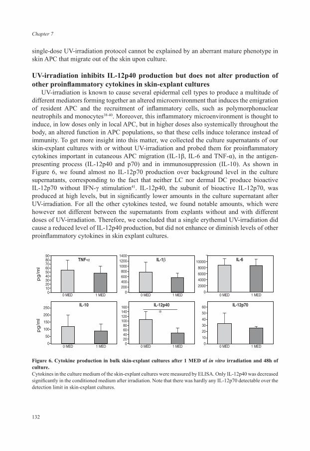
Chapter 7
132
single-dose UV-irradiation protocol cannot be explained by an aberrant mature phenotype in skin APC that migrate out of the skin upon culture.
UV-irradiation inhibits IL-12p40 production but does not alter production of other proinflammatory cytokines in skin-explant cultures
UV-irradiation is known to cause several epidermal cell types to produce a multitude of different mediators forming together an altered microenvironment that induces the emigration of resident APC and the recruitment of inflammatory cells, such as polymorphonuclear neutrophils and monocytes38-40. Moreover, this inflammatory microenvironment is thought to induce, in low doses only in local APC, but in higher doses also systemically throughout the body, an altered function in APC populations, so that these cells induce tolerance instead of immunity. To get more insight into this matter, we collected the culture supernatants of our skin-explant cultures with or without UV-irradiation and probed them for proinflammatory cytokines important in cutaneous APC migration (IL-1β, IL-6 and TNF-α), in the antigen-presenting process (IL-12p40 and p70) and in immunosuppression (IL-10). As shown in Figure 6, we found almost no IL-12p70 production over background level in the culture supernatants, corresponding to the fact that neither LC nor dermal DC produce bioactive IL-12p70 without IFN-γ stimulation41. IL-12p40, the subunit of bioactive IL-12p70, was produced at high levels, but in significantly lower amounts in the culture supernatant after UV-irradiation. For all the other cytokines tested, we found notable amounts, which were however not different between the supernatants from explants without and with different doses of UV-irradiation. Therefore, we concluded that a single erythemal UV-irradiation did cause a reduced level of IL-12p40 production, but did not enhance or diminish levels of other proinflammatory cytokines in skin explant cultures.
TNF-�
0102030405060708090
0 MED 1 MED
0 MED 1 MED
0 MED 1 MED 0 MED 1 MED
0 MED 1 MED 0 MED 1 MED
pg
/ml
IL-1�
0200400600800
100012001400
IL-6
0
2000
4000
6000
8000
IL-10
0
50
100
150
200
250
pg
/ml
IL-12p70
0
10
20
30
40
50
60IL-12p40
020406080
100120140160
�
10000
Figure 6. Cytokine production in bulk skin-explant cultures after 1 MED of in vitro irradiation and 48h of culture.Cytokines in the culture medium of the skin-explant cultures were measured by ELISA. Only IL-12p40 was decreased significantly in the conditioned medium after irradiation. Note that there was hardly any IL-12p70 detectable over the detection limit in skin-explant cultures.

133
Effects of single dose UV on cutaneous APC
Single-dose UV-irradiation does not lead to an impairment of CHS inductionSo far, we have seen contradictory effects of UV-irradiation on cutaneous APC populations
with respect to their functional and phenotypic changes. To gain insight into the in vivo consequences of our single-dose erythemal UV-irradiation regimen, we induced a CHS response in the presence or absence of prior irradiation. Therefore, we applied 2-3-5-trinitro-1-chlorobenzene (TNCB; picrylchloride) in adjuvant onto the back skin of mice that had been irradiated before on this side with one or two MED doses of UV-irradiation, or not. Thereafter, we challenged these mice five days later onto their ears, and read out one day later the ear thickness. This correlated directly to the induction of the CHS response against TNCB, or the lack thereof. Against our expectations, we did not observe a state of immunosuppression even at two MED of UV-irradiation, independently of whether we applied the TNCB directly, or one day or two days after UV-irradiation (representative experiment shown in Figure 7). On the opposite, we rather found a slight stimulation of the immune response, manifested in the higher CHS response than could be measured when TNCB was applied one day after UV-irradiation. Therefore, we concluded that our single-dose UV-irradiation regime did not cause any local immunosuppression in the living mouse, even when we irradiated mice with two MED.
DISCUSSION
In this study, we assessed the effects of single-dose erythemal UV-irradiation on cutaneous APC subpopulations. For this, we irradiated mouse skin in vivo or ex vivo in the structurally intact skin. To enable their subsequent isolation as single cells, we irradiated whole ear halves and cultured them thereafter. We have shown previously that during these
CHS response
0
10
20
30
40
50
60
70
80
90
%in
crea
seea
rth
ickn
ess
no UV control
2 MED day-1
2 MED day-2
Figure 7. Lack of inhibition of contact hypersensitivity response initiation by two MED UV irradiation in vivo.Mice were irradiated with 2 MED of UV irradiation. Directly thereafter, one or two days later, mice were sensitized with TNCB on their irradiated back. Five days thereafter, mice were challenged on their ears with TNCB and one day later, ear swelling was determined. Note that there was no inhibition of the CHS response initiation by two MED UV-irradiation, independently whether the UV irradiation was applied directly before, or one day or two days before sensitization.

Chapter 7
134
skin-explant cultures, dermal and epidermal mononuclear phagocytes become activated and emigrate from the skin in large numbers. Moreover, the cells also mature during these explant cultures, so that mature APC derived from the dermis and epidermis can be collected from the medium (Chapter 6). Comparing skin-explant cultures from irradiated and unirradiated skin, we found that lower numbers of cells emigrated from in vitro UV-irradiated skin. While epidermal LC were decreased significantly, the reduction in dermal cell numbers was only marginal. In contrast, in vivo irradiation of ears and subsequent explant cultures showed no decrease of either emigrating population. Functionally, we found that emigrants from in vitro UV-irradiated skin stimulated allogeneic T cells less efficiently than from unirradiated skin. Phenotypically, however, we did not find any difference between cells emigrated from irradiated or unirradiated skin with respect to markers essential for their antigen-presenting function such as MHC and costimulatory molecules. Levels of IL-12p40 production in bulk skin explant cultures from UV-irradiated skin tissues were decreased, while there was no difference in other proinflammatory cytokines. Finally, assessing the suppressive potential of our single-dose UV-irradiation regimen in vivo in living mice, we were unable to determine any immunosuppressive effects on the CHS response induction to 2-3-5-trinitro-1-chlorobenzene (TNCB; picrylchloride) with up to two MED of UV-irradiation.
At first sight, it is contradictory to find fewer APC emigrating out of UV-irradiated skin. UV-irradiation consistently has been shown to induce the emigration of these cells, rather than inhibiting it3,15-17. The technical set-up of the experiments in the mentioned studies, however, differed significantly from our approach. We applied both the stimulus of UV-irradiation as well as the procedure of the skin-explant culture, which already provides a strong trigger for the majority of both epidermal and dermal mononuclear phagocytes to emigrate42 (Chapter 6). This probably is the reason that no additional effect of UV-irradiation was observed. Finding fewer cells emigrating from explant cultures after UV-irradiation has been reported previously from similar experimental settings43,44. In these studies, however, only bulk skin emigrants have been analyzed. A new finding of our experiments is that in particular epidermal LC were inhibited to emigrate.
An explanation for this decreased emigration of epidermal LC might be that cells after UV-irradiation can undergo apoptosis and are therefore not available for emigration. However, no increased percentages of apoptotic cells have been observed in the two mentioned papers. In these studies, like in our, single, emigrated cells have been investigated. These cells therefore are probably not the ones that will undergo apoptosis. Determination of apoptosis in the UV-irradiated skin itself should result in further insight in this respect. Since the inhibition of migration is significantly stronger for LC than for dermal mononuclear phagocytes, it is likely that this is directly related to the amount of cellular damage induced by UV-irradiation. However, we did not observe a delay in emigration, required for repair of DNA damage, as we did not find increasing numbers of emigrants after up to four days of culture. Thus, the levels of applied UV-irradiation either caused increased apoptosis, or left more cells fixed in the tissue for reasons unknown so far. Although we quantified the remaining cells by immuno-histochemistry, this method is probably too inaccurate to detect numerical differences between the different experimental groups. Notably, we did not observe this inhibition of emigration when we irradiated skin first in vivo and just then initiated skin-explant cultures. Therefore, this result suggests that factors in the intact, UV-irradiated skin in the living mouse, such

135
Effects of single dose UV on cutaneous APC
as hormones22-25 or neurotransmitters19-21 might counteract the inhibition of migration, for instance by stimulating DNA damage repair. This has been demonstrated before for IL-12p704 and IL-1845, although we found that the former factor is virtually absent in explant cultures.
At the moment, it is not clear why not all cutaneous mononuclear phagocytes emigrate upon culture. Similarly, it is unknown which discriminating factors exist to make cells stay behind. Our analysis of dermal mononuclear phagocytes in the steady-state ear dermis revealed that some cells expressed CD11c, whereas another subpopulation exhibited MHC class II expression11 (chapter 4). Nevertheless, analyzing ear dermis after culture did not reveal any of these subpopulations remaining preferentially as indicated by a heterogeneous expression profile for CD11c and MHC class II (unpublished observations). Interestingly, remaining cells were found predominately in clusters in the vicinity of hair follicles, and as hair follicles are known to harbor several cutaneous precursor populations, it might be that these cells have just developed from a precursor cell population, and are therefore still too immature to emigrate. Alternatively, hair follicles might create a distinct dermal microenvironment with lower levels of proinflammatory cytokines than are required for emigration42.
A simple explanation for the decrease of IL-12p40 release in bulk skin explant cultures from UV-irradiated skin tissues might be provided by the fewer cells found in the explant cultures to produce IL-12p40, as IL-12p40 is specifically produced by mononuclear phagocytes46. Estimating the amount of IL-12p40 produced per DC in the supernatant, there was no difference between the unirradiated and the irradiated samples.
Quite similar experiments to the ones we report here have been performed using human skin explants47. In these experiments, human skin was irradiated in vitro before skin-explant culture. Thereafter, emigrants where investigated phenotypically and their cytokine production and alloantigen presentation capacities were assessed. UV-irradiation caused the appearance of two different APC subpopulations: one subset consisted of small cells that did not possess a mature phenotype, that did not stimulate T cells efficiently and that stained positive for annexin V, suggesting that these cells represented apoptotic cells. The other subset were larger cells with a normal, mature phenotype that could efficiently present alloantigen47. Moreover, the latter cells produced higher cytokine levels than cells from control skin explants, hinting to the fact that they were even more activated.
Also in other studies contradictory effects of UV-irradiation have been observed. In one report, blisters were generated in in vivo irradiated skin, enabling the determination of differences in cytokine production between irradiated and unirradiated skin48. Thereby, it was noticed that the irradiation hardly raised the levels of cytokines recovered from the blister fluids. Only IL-6 could be recovered in increased amounts from these fluids48. Another report is quite instructive about the effects of UV-irradiation. In this report, Laihia and Jansen (2000) studied the influence of UV-irradiation on costimulatory molecule expression by LC. As they had found previously that UV-irradiation induced the expression of CD80 and CD86 on LC in vivo49, they tried to assess why other reports showed that UV-irradiation does not induce or even inhibits this upregulation. To gain insight into this issue, they compared LC that had been irradiated either in vivo, or ex vivo, thus in skin biopsies that were subsequently cultured, or in vitro, thus in epidermal cell suspensions. Comparable to other reports, they found that ex vivo and in vitro irradiation would inhibit, or at least not induce, the upregulation of CD80 and CD86. In agreement with their earlier results, they observed, however, that UV-irradiation upregulated CD80 and CD86 expression in in vivo irradiated LC.
Chapter 7
136
Therefore, we can conclude that cutaneous UV-irradiation has two different outcomes besides immunosuppression. Firstly, UV-irradiation can have seemingly no effects. This is most probably a result of the settings that are used to study the effects of UV-irradiation. One thing that all the mentioned studies have in common to our examination is that they estimate the effects of UV-irradiation in environments that involve additional inflammatory triggers caused by tissue injury and that stimulated the cutaneous immune system independently of the UV-irradiation. Factors that are produced by UV-irradiated keratinocytes, such as IL-1β or TNF-α39, are also induced during skin injuries and stimulate the emigration of epidermal and dermal APC42,50,51. In these situations, where the skin immune system is triggered independently of UV-irradiation, UV-irradiation turned out to be unable to counteract efficient immune responses.
Secondly, UV-irradiation can also be seen to activate the immune system. This seems to be a real effect of UV-irradiation and can be observed at dosages that lie deeper than the dosages that are used to obtain immunosuppression. In this respect, activation of UV-irradiated APC has been shown as well for another model APC subset, for bone marrow-derived DC (BMDC)52. UV-irradiated BMDC showed higher CD86 and MHC class II levels, produced more cytokines and possessed a higher alloantigen presenting capacity than unirradiated DC. Additionally, in an in vivo model, UV-irradiation has been shown to boost the immune response against Leishmania amazonensis53.
In view of the findings reported by others, we are puzzled by our inability to obtain immunosuppression in vivo using a single-dose irradiation regimen with up to two MED or about 5000 J/m2 and hapten applied up to 48h after irradiation. There are clear indications in the literature that single erythemal or even suberythemal doses of UV-B irradiation can induce immunosuppression and tolerance (Hammerberg et al., who used 1380 J/m2 or 1.1 MED, delivered by FS-40 lamps on C3H/HeN mice7; Vink et al., 5000 J/m2 or 2 MED with FS-40 lamps and C3H/HeNCr(MTV–) mice8; Kurimoto et al., 400 J/m2 with FS-20 lamps and C57BL/6 mice9). While the first two reports reported tolerance only when antigen was applied two to three days after UV-irradiation, the third paper demonstrated that UV-induced tolerance can also be obtained directly after UV-irradiation. The differences in kinetics for tolerance induction between these studies might lie in the dose of hapten used54 or to the strength of the adjuvant that was co-applied55. These differences are thought to target different layers of the skin to become affected in the CHS response and in the tolerance induction54,55. The contribution of recruited inflammatory cells in the induction of tolerance after UV-irradiation remains controversial. On the one hand, Hammerberg et al. suggested their crucial contribution7, but Vink et al. showed that repairing DNA damage in cells obtained from the dermis could abrogate their capacity to induce tolerance8. Nevertheless, what is clear from these studies is that tolerance can be obtained by applying hapten in adjuvant onto skin after a single-dose of UV-irradiation.
We used a UV-irradiation dose comparable to the ones used by Hammerberg et al., and Vink et al. Consequently, we can only speculate why we did not observe immunosuppression up to two days after UV-irradiation with up to two MED UV-irradiation. Reasons that might apply are the mouse strain used, the hapten used, the amount of hapten applied, the kind of adjuvant used to dissolve the hapten, the kind of UV-lamp used to irradiate mice and, last but not least, the microbiological state of the mice. They might in theory all affect the

137
Effects of single dose UV on cutaneous APC
way how UV-irradiation affects resident epidermal, dermal and recruited inflammatory cells. Different lamps may possess different outputs in the low percentage of UV-C irradiation, which has nevertheless been shown to contribute disproportionately to erythema and edema56. Therefore, minute differences in UV-C radiation might significantly affect the induction of immunosuppression as the UV dose is applied in erythemal (or edemal) doses, thus related to the biological response of the mouse strain to the UV source used. Also the amount of UV-A irradiation may affect the result of the irradiation protocol. UV-A irradiation, which energy is 20 times more abundant in the ambient sunlight13, was negligibly low in the used UV-B source. These lamps emitted photons in the UV-A range in equal amounts than in the UV-B range, with UV-A photons carrying much lower energies. Inclusion of UV-A radiation into the immunosuppressive protocol can add to the immunosuppressive properties of UV-B, or it can, depending on dosage and irradiation regimen, protect from subsequent UV-B induced immunosuppression57. Therefore, different dosages of included UV-A radiation might alter the outcome of the overall irradiation. To explain our inability to induce immune suppression using the applied regimen, we can speculate that we applied a too low dose of UV-irradiation in connection with a too high dose of hapten. Therefore, comparable to our in vitro experiments, the inflammation caused by our hapten/adjuvant application may have been too heavy/large to be counter-regulated by the UV-irradiation.
Finally, we also have to take a look at the cell types involved in the UV-induced immunosuppression and tolerance. Having analyzed APC in this study, it remains a possibility that DC are not the major players in the induction of immunosuppresion. There are two recent reports that analyzed DC subpopulations in skin-draining lymph nodes after high doses of UV-irradiation, which caused systemic immunosuppression58,59. In both reports, it was shown that DC isolated from skin-draining lymph nodes after the irradiation showed a normal phenotype, a normal cytokine production profile and a normal T cell stimulation capacity, indicating that DC in skin-draining lymph nodes might not be involved in the process of immunosuppression. The study by Byrne and Halliday suggested that B cells might suppress CHS response induction after UV-irradiation58. Therefore, it might be that the cytokine environment in the skin-draining lymph node is important for the impairment of DC function, and that this impairment is lost once these cells are isolated from the lymph nodes and cultured in medium. On the other hand, it might be that the reasons indicated above apply as well in the process of systemic immunosuppression and that different cell types may be involved in UV-induced tolerance, depending on different irradiation regimens and read-out mechanisms. Reports of DC aberrancies in systemic UV-induced immunosuppression at least hint in this direction60,61. Future research will hopefully shed more light onto the different cell populations of the innate and the adaptive immune system and their roles in UV-irradiation induced immunosuppression.
Therefore, although UV-irradiation is known for its immunosuppressive capacities, it seems that a strong immune response will counterbalance a weak UV-induced immunosuppression, as will a strong UV-induced immunosuppression counterbalance a weak immune response. Consequently, UV-irradiation plays a delicate role in the subtle balance between immunity and tolerance. As immune responses against cancers are mostly weak responses, it is well conceivable that UV-irradiation impairs their elimination, especially considering that chronic UV-irradiation is needed to induce skin cancers.

Chapter 7
138
REFERENCES
1. Kripke, M.L. Antigenicity of murine skin tumors induced by ultraviolet light. J Natl Cancer Inst 53, 1333-1336 (1974).
2. Kripke, M.L. & Fisher, M.S. Immunologic parameters of ultraviolet carcinogenesis. J Natl Cancer Inst 57, 211-215 (1976).
3. Toews, G.B., Bergstresser, P.R. & Streilein, J.W. Epidermal Langerhans cell density determines whether contact hypersensitivity or unresponsiveness follows skin painting with DNFB. J Immunol 124, 445-453 (1980).
4. Schwarz, T. Photoimmunosuppression. Photodermatol Photoimmunol Photomed 18, 141-145 (2002).5. Ullrich, S.E. Mechanisms underlying UV-induced immune suppression. Mutat Res 571, 185-205 (2005).6. Schwarz, T. 25 years of UV-induced immunosuppression mediated by T cells-from disregarded T suppressor
cells to highly respected regulatory T cells. Photochem Photobiol 84, 10-18 (2008).7. Hammerberg, C., Duraiswamy, N. & Cooper, K.D. Temporal correlation between UV radiation locally-
inducible tolerance and the sequential appearance of dermal, then epidermal, class II MHC+CD11b+ monocytic/macrophagic cells. J Invest Dermatol 107, 755-763 (1996).
8. Vink, A.A., Strickland, F.M., Bucana, C., Cox, P.A., Roza, L., Yarosh, D.B. & Kripke, M.L. Localization of DNA damage and its role in altered antigen-presenting cell function in ultraviolet-irradiated mice. J Exp Med 183, 1491-1500 (1996).
9. Kurimoto, I., Kitazawa, T. & Streilein, J.W. Suprathreshold doses of hapten are required to induce both contact hypersensitivity and tolerance. J Dermatol Sci 24, 48-59 (2000).
10. Meunier, L. Ultraviolet light and dendritic cells. Eur J Dermatol 9, 269-275 (1999).11. Dupasquier, M., Stoitzner, P., van Oudenaren, A., Romani, N. & Leenen, P.J. Macrophages and dendritic cells
constitute a major subpopulation of cells in the mouse dermis. J Invest Dermatol 123, 876-879 (2004).12. Bruls, W.A., Slaper, H., van der Leun, J.C. & Berrens, L. Transmission of human epidermis and stratum
corneum as a function of thickness in the ultraviolet and visible wavelengths. Photochem Photobiol 40, 485-494 (1984).
13. Halliday, G.M., Byrne, S.N., Kuchel, J.M., Poon, T.S. & Barnetson, R.S. The suppression of immunity by ultraviolet radiation: UVA, nitric oxide and DNA damage. Photochem Photobiol Sci 3, 736-740 (2004).
14. Dupasquier, M., Stoitzner, P., Wan, H., Cerqueira, D., van Oudenaren, A., Voerman, J.S., Denda-Nagai, K., Irimura, T., Raes, G., Romani, N. & Leenen, P.J. The dermal microenvironment induces the expression of the alternative activation marker CD301/mMGL in mononuclear phagocytes, independent of IL-4/IL-13 signaling. J Leukoc Biol 80, 838-849 (2006).
15. Zelickson, A.S. & Mottaz, J. The effect of sunlight on human epidermis. A quantitative electron microscopic study of dendritic cells. Arch Dermatol 101, 312-315 (1970).
16. Aberer, W., Schuler, G., Stingl, G., Honigsmann, H. & Wolff, K. Ultraviolet light depletes surface markers of Langerhans cells. J Invest Dermatol 76, 202-210 (1981).
17. Meunier, L., Bata-Csorgo, Z. & Cooper, K.D. In human dermis, ultraviolet radiation induces expansion of a CD36+ CD11b+ CD1- macrophage subset by infiltration and proliferation; CD1+ Langerhans-like dendritic antigen-presenting cells are concomitantly depleted. J Invest Dermatol 105, 782-788 (1995).
18. Larsen, C.P., Steinman, R.M., Witmer-Pack, M., Hankins, D.F., Morris, P.J. & Austyn, J.M. Migration and maturation of Langerhans cells in skin transplants and explants. J Exp Med 172, 1483-1493 (1990).
19. Garssen, J., Buckley, T.L. & Van Loveren, H. A role for neuropeptides in UVB-induced systemic immunosuppression. Photochem Photobiol 68, 205-210 (1998).
20. Kitazawa, T. & Streilein, J.W. Hapten-specific tolerance promoted by calcitonin gene-related peptide. J Invest Dermatol 115, 942-948 (2000).
21. Legat, F.J., Jaiani, L.T., Wolf, P., Wang, M., Lang, R., Abraham, T., Solomon, A.R., Armstrong, C.A., Glass, J.D. & Ansel, J.C. The role of calcitonin gene-related peptide in cutaneous immunosuppression induced by repeated subinflammatory ultraviolet irradiation exposure. Exp Dermatol 13, 242-250 (2004).
22. Ross, D.J., Keynes, H.L. & McDonald, J.C. SWORD ‘97: surveillance of work-related and occupational respiratory disease in the UK. Occup Med (Lond) 48, 481-485 (1998).
23. Hiramoto, K., Tanaka, H., Yanagihara, N., Sato, E.F. & Inoue, M. Effect of 17beta-estradiol on immunosuppression induced by ultraviolet B irradiation. Arch Dermatol Res 295, 307-311 (2004).

139
Effects of single dose UV on cutaneous APC
24. Howes, R.A., Halliday, G.M. & Damian, D.L. Effect of topical melatonin on ultraviolet radiation-induced suppression of Mantoux reactions in humans. Photodermatol Photoimmunol Photomed 22, 267-269 (2006).
25. Redondo, P., Garcia-Foncillas, J., Okroujnov, I. & Bandres, E. Alpha-MSH regulates interleukin-10 expression by human keratinocytes. Arch Dermatol Res 290, 425-428 (1998).
26. Denfeld, R.W., Hara, H., Tesmann, J.P., Martin, S. & Simon, J.C. UVB-irradiated dendritic cells are impaired in their APC function and tolerize primed Th1 cells but not naive CD4+ T cells. J Leukoc Biol 69, 548-554 (2001).
27. Dittmar, H.C., Weiss, J.M., Termeer, C.C., Denfeld, R.W., Wanner, M.B., Skov, L., Barker, J.N., Schopf, E., Baadsgaard, O. & Simon, J.C. In vivo UVA-1 and UVB irradiation differentially perturbs the antigen-presenting function of human epidermal Langerhans cells. J Invest Dermatol 112, 322-325 (1999).
28. Goettsch, W., Hurks, H.M., Garssen, J., Mommaas, A.M., Slob, W., Hoekman, J., Pierik, F., Roholl, P.J. & Van Loveren, H. Comparative immunotoxicology of ultraviolet B exposure I. Effects of in vitro and in situ ultraviolet B exposure on the functional activity and morphology of Langerhans cells in the skin of different species. Br J Dermatol 139, 230-238 (1998).
29. Mizuno, K., Okamoto, H. & Horio, T. Ultraviolet B radiation suppresses endocytosis, subsequent maturation, and migration activity of langerhans cell-like dendritic cells. J Invest Dermatol 122, 300-306 (2004).
30. Rattis, F.M., Peguet-Navarro, J., Courtellemont, P., Redziniak, G. & Schmitt, D. In vitro effects of ultraviolet B radiation on human Langerhans cell antigen-presenting function. Cell Immunol 164, 65-72 (1995).
31. Simon, J.C., Tigelaar, R.E., Bergstresser, P.R., Edelbaum, D. & Cruz, P.D., Jr. Ultraviolet B radiation converts Langerhans cells from immunogenic to tolerogenic antigen-presenting cells. Induction of specific clonal anergy in CD4+ T helper 1 cells. J Immunol 146, 485-491 (1991).
32. Wachter, T., Averbeck, M., Hara, H., Tesmann, J.P., Simon, J.C., Termeer, C.C. & Denfeld, R.W. Induction of CD4+ T cell apoptosis as a consequence of impaired cytoskeletal rearrangement in UVB-irradiated dendritic cells. J Immunol 171, 776-782 (2003).
33. Denfeld, R.W., Tesmann, J.P., Dittmar, H., Weiss, J.M., Schopf, E., Weltzien, H.U. & Simon, J.C. Further characterization of UVB radiation effects on Langerhans cells: altered expression of the costimulatory molecules B7-1 and B7-2. Photochem Photobiol 67, 554-560 (1998).
34. Rattis, F.M., Concha, M., Dalbiez-Gauthier, C., Courtellemont, P., Schmitt, D. & Peguet-Navarro, J. Effects of ultraviolet B radiation on human Langerhans cells: functional alteration of CD86 upregulation and induction of apoptotic cell death. J Invest Dermatol 111, 373-379 (1998).
35. Tang, A. & Udey, M.C. Effects of ultraviolet radiation on murine epidermal Langerhans cells: doses of ultraviolet radiation that modulate ICAM-1 (CD54) expression and inhibit Langerhans cell function cause delayed cytotoxicity in vitro. J Invest Dermatol 99, 83-89 (1992).
36. Weiss, J.M., Renkl, A.C., Denfeld, R.W., de Roche, R., Spitzlei, M., Schopf, E. & Simon, J.C. Low-dose UVB radiation perturbs the functional expression of B7.1 and B7.2 co-stimulatory molecules on human Langerhans cells. Eur J Immunol 25, 2858-2862 (1995).
37. Mittelbrunn, M., Tejedor, R., de la Fuente, H., Garcia-Lopez, M.A., Ursa, A., Penas, P.F., Garcia-Diez, A., Alonso-Lebrero, J.L., Pivel, J.P., Gonzalez, S., Gonzalez-Amaro, R. & Sanchez-Madrid, F. Solar-simulated ultraviolet radiation induces abnormal maturation and defective chemotaxis of dendritic cells. J Invest Dermatol 125, 334-342 (2005).
38. Kang, K., Hammerberg, C., Meunier, L. & Cooper, K.D. CD11b+ macrophages that infiltrate human epidermis after in vivo ultraviolet exposure potently produce IL-10 and represent the major secretory source of epidermal IL-10 protein. J Immunol 153, 5256-5264 (1994).
39. Takashima, A. & Bergstresser, P.R. Impact of UVB radiation on the epidermal cytokine network. Photochem Photobiol 63, 397-400 (1996).
40. Kolgen, W., van Meurs, M., Jongsma, M., van Weelden, H., Bruijnzeel-Koomen, C.A., Knol, E.F., van Vloten, W.A., Laman, J. & de Gruijl, F.R. Differential expression of cytokines in UV-B-exposed skin of patients with polymorphous light eruption: correlation with Langerhans cell migration and immunosuppression. Arch Dermatol 140, 295-302 (2004).
41. Morelli, A.E., Rubin, J.P., Erdos, G., Tkacheva, O.A., Mathers, A.R., Zahorchak, A.F., Thomson, A.W., Falo, L.D., Jr. & Larregina, A.T. CD4+ T cell responses elicited by different subsets of human skin migratory dendritic cells. J Immunol 175, 7905-7915 (2005).

Chapter 7
140
42. Chun, K.H., Imai, Y., Higashi, N. & Irimura, T. Involvement of cytokines in the skin-to-lymph node trafficking of cells of the monocyte-macrophage lineage expressing a C-type lectin. Int Immunol 12, 1695-1703 (2000).
43. Richters, C.D., Reits, E.A., Van Pelt, A.M., Hoekstra, M.J., Van Baare, J., Du Pont, J.S. & Kamperdijk, E.W. Effect of low dose UVB irradiation on the migratory properties and functional capacities of human skin dendritic cells. Clin Exp Immunol 104, 191-197 (1996).
44. Bacci, S., Alard, P. & Streilein, J.W. Evidence that ultraviolet B radiation transiently inhibits emigration of Langerhans cells from exposed epidermis, thwarting contact hypersensitivity induction. Eur J Immunol 31, 3588-3594 (2001).
45. Schwarz, A., Maeda, A., Stander, S., van Steeg, H. & Schwarz, T. IL-18 reduces ultraviolet radiation-induced DNA damage and thereby affects photoimmunosuppression. J Immunol 176, 2896-2901 (2006).
46. Guery, J.C., Ria, F., Galbiati, F. & Adorini, L. Normal B cells fail to secrete interleukin-12. Eur J Immunol 27, 1632-1639 (1997).
47. Nakagawa, S., Koomen, C.W., Bos, J.D. & Teunissen, M.B. Differential modulation of human epidermal Langerhans cell maturation by ultraviolet B radiation. J Immunol 163, 5192-5200 (1999).
48. Kuhn, M., Wolber, R., Kolbe, L., Schnorr, O. & Sies, H. Solar-simulated radiation induces secretion of IL-6 and production of isoprostanes in human skin in vivo. Arch Dermatol Res 297, 477-479 (2006).
49. Laihia, J.K. & Jansen, C.T. Solar-simulating irradiation of the skin of human subjects in vivo produces Langerhans cell responses distinct from irradiation ex vivo and in vitro. Exp Dermatol 9, 240-247 (2000).
50. Cumberbatch, M. & Kimber, I. Dermal tumour necrosis factor-alpha induces dendritic cell migration to draining lymph nodes, and possibly provides one stimulus for Langerhans’ cell migration. Immunology 75, 257-263 (1992).
51. Enk, A.H., Angeloni, V.L., Udey, M.C. & Katz, S.I. An essential role for Langerhans cell-derived IL-1 beta in the initiation of primary immune responses in skin. J Immunol 150, 3698-3704 (1993).
52. Nakagawa, S., Ohtani, T., Mizuashi, M., Mollah, Z.U., Ito, Y., Tagami, H. & Aiba, S. p38 Mitogen-Activated protein kinase mediates dual role of ultraviolet B radiation in induction of maturation and apoptosis of monocyte-derived dendritic cells. J Invest Dermatol 123, 361-370 (2004).
53. Khaskhely, N.M., Maruno, M., Uezato, H., Takamiyagi, A., Ramzi, S.T., Al-Kasem, K.M., Kariya, K., Toda, T., Hashiguchi, Y., Gomez Landires, E.A. & Nonaka, S. Low-dose UVB contributes to host resistance against Leishmania amazonensis infection in mice through induction of gamma interferon and tumor necrosis factor alpha cytokines. Clin Diagn Lab Immunol 9, 677-686 (2002).
54. Bacci, S., Alard, P., Dai, R., Nakamura, T. & Streilein, J.W. High and low doses of haptens dictate whether dermal or epidermal antigen-presenting cells promote contact hypersensitivity. Eur J Immunol 27, 442-448 (1997).
55. Sato, K., Imai, Y. & Irimura, T. Contribution of dermal macrophage trafficking in the sensitization phase of contact hypersensitivity. J Immunol 161, 6835-6844 (1998).
56. Learn, D.B., Beard, J. & Moloney, S.J. The ultraviolet C energy emitted from FS lamps contributes significantly to the induction of human erythema and murine ear edema. Photodermatol Photoimmunol Photomed 9, 147-153 (1993).
57. Byrne, S.N., Spinks, N. & Halliday, G.M. Ultraviolet a irradiation of C57BL/6 mice suppresses systemic contact hypersensitivity or enhances secondary immunity depending on dose. J Invest Dermatol 119, 858-864 (2002).
58. Byrne, S.N. & Halliday, G.M. B cells activated in lymph nodes in response to ultraviolet irradiation or by interleukin-10 inhibit dendritic cell induction of immunity. J Invest Dermatol 124, 570-578 (2005).
59. Gorman, S., Tan, J.W., Thomas, J.A., Townley, S.L., Stumbles, P.A., Finlay-Jones, J.J. & Hart, P.H. Primary defect in UVB-induced systemic immunomodulation does not relate to immature or functionally impaired APCs in regional lymph nodes. J Immunol 174, 6677-6685 (2005).
60. Kitazawa, T. & Streilein, J.W. Studies on delayed systemic effects of ultraviolet B radiation on the induction of contact hypersensitivity, 3. Dendritic cells from secondary lymphoid organs are deficient in interleukin-12 production and capacity to promote activation and differentiation of T helper type 1 cells. Immunology 99, 296-304 (2000).
61. Boonstra, A., van Oudenaren, A., Barendregt, B., An, L., Leenen, P.J. & Savelkoul, H.F. UVB irradiation modulates systemic immune responses by affecting cytokine production of antigen-presenting cells. Int Immunol 12, 1531-1538 (2000).

CHAPTER 8
GENERAL DISCUSSION

Chapter 8
142
Connective tissues build up many different structures in the body. Despite their highly diverse appearance, they are all composed of cells, fibers, ground substance and tissue fluid. The non-cellular part of connective tissue is generally the largest, such in contrast to other tissues. Embedded in between the collagenous and elastic fibers and ground substance of the connective tissue proper are interstitial cells that are involved in the various functions and the maintenance of the connective tissue. For loose connective tissues, the cells that produce and maintain the components of the extracellular matrix are fibroblasts. Additionally, other cells can be found in connective tissues, such as cells of the immune system, like macrophages, dendritic cells (DC), mast cells and T cells, but also others, such as nerve cells and endothelial cells of blood and lymph vessels. There is still a lack of insight into the cellular composition of the various connective tissues.
Interstitial cells generally are of mesenchymal origin and contribute to the extracellular matrix production. Moreover, as they are mainly characterized according to morphological criteria (see for example ref. 1), they are usually identified as fibroblasts2. Other cells are thought to contribute only marginally to the interstitial cell population.
THE DERMIS AND ITS MONONUCLEAR PHAGOCYTES
The dermis is a prototypical example of a loose connective tissue. As such, it forms a strong, resilient layer underneath the epidermis, the cornified, stratified epithelium of the skin. Thereby, it anchors the epidermis onto the body, forming together a firm and tough, yet still elastic and resilient layer towards the outside.
As for other connective tissues, fibroblasts have been regarded as the major cellular constituents of the dermis, and were reported to form over 90% of all dermal interstitial cells3,4. Consequently, the steady-state dermis has received less attention compared to the epidermis when cutaneous immune responses were investigated. The epidermis and its major immunological players, the Langerhans cells (LC) and epidermal T cells, were thought to outweigh by far any contribution from the dermis. The dermis, so it was thought, consists mainly of extracellular matrix, and a few cells, mainly, fibroblasts, that will behave immunologically “inert”, apart from some cytokine production. However, initial findings from our and some other laboratories questioned this cellular composition of the dermis, suggesting a significantly larger contribution of macrophages and related cells5,6.
The scope of this thesis therefore was the investigation of the cells of the immune system in the dermis. Particularly, mononuclear phagocytes, i.e. macrophages and dendritic cells, were studied, considering their strong antigen-presentation potential and their bridging function between the innate and the adaptive immune responses. These cells were on the one hand analyzed quantitatively and phenotypically in the intact skin in vivo in tissue sections (Chapter 4). On the other hand, the cells were studied after emigration from the dermis in vitro (Chapter 6). Although the cells were activated during this process, this isolation step enabled thereafter the analysis of their functional potential. The characterization of dermal mononuclear phagocytes led to the description of immunologically versatile cells that might contribute significantly to cutaneous immune responses.

143
General discussion
MONONUCLEAR PHAGOCYTES CONSTITUTE THE MAJORITY OF INTERSTITIAL CELLS IN THE MOUSE DERMIS
In chapter 4, we show that the number of mononuclear phagocytes in the mouse dermis has been largely underestimated so far. Mononuclear phagocytes, thus macrophages and DC, appeared to constitute the majority of interstitial cells in the mouse dermis, according to their expression of CD45, F4/80, CD11b and the C-type lectins MR/CD206 and mMGL/CD301. In line with their expression of typical macrophage markers, such as F4/80 and CD11b, their lack of expression of typical DC markers, such as CD40, CD80, CD86, and in most cases CD11c, and their avid phagocytosis, we consider these cells as typical tissue macrophages (Chapter 4). Moreover, their expression of the two C-type lectins MR/CD206 and mMGL/CD301 suggest that these cells are alternatively activated macrophages7,8, although we have shown as well that they do not represent alternatively activated macrophages in the strict sense as their phenotype is not dependent on IL-4 or IL-13 signaling (Chapter 5). What might this phenotype of alternative activation suggest for the function of dermal macrophages under steady-state conditions?
DERMAL MACROPHAGES AS REPARATIVE MACROPHAGES IN THE STEADY-STATE DERMIS
In general, alternative activation of macrophages has been associated with connective tissue repair and fibrogenesis9,10. The first stage of wound healing consists of an inflammatory reaction, where the initially infiltrating polymorphonuclear neutrophils (PMN) are followed after two to three days by infiltrating monocytes. These monocytes originally have been thought to develop into classically activated macrophages, producing multiple proinflammatory cytokines but also other mediators, such as NO11,12. These mediators help destroy invaders and sterilize the wound but at the same time also inflict tissue damage. Later, it has been recognized, however, that recruited monocytes rather develop into alternatively activated macrophages, contributing to the wound healing process13. Therefore, these cells can be considered as reparative macrophages.
Alternatively activated macrophages contribute to tissue repair via the activation of fibroblasts: TGF-β produced by infiltrating monocytes support the wound healing process by inducing fibroblasts to express and secrete collagens and to express α-smooth muscle actin, enabling them to contract the healing granulation tissue14. In this respect, the phagocytosis of apoptotic PMN is an important signal that induces the monocytes to promote wound healing14,15. Additionally, they have been shown to produce an abundance of signaling molecules that induce tissue repair, such as the insulin-like growth factor-1, IGF-113,16. IGF-1 itself also induces α-smooth muscle actin and collagen in fibroblasts and fibroblast mitogenesis17. Therefore, by the expression of soluble mediators, infiltrating monocytes stimulate fibroblasts to close the wound.
Infiltrating monocytes also have the potential to play a much more direct role in the wound healing process. Cells in the peripheral blood, unfortunately termed fibrocytes, thus confusing them with resting fibroblasts, express markers of fibroblasts (procollagen I, III and fibronectin)

Chapter 8
144
as well as mononuclear phagocytes (CD45, MHC class II) and develop from monocytes after IL-4 or IL-13 stimulation18. As IL-4 and IL-13 are stimulating the genuine alternatively activated macrophages9, it comes as no surprise that alternatively activated macrophages appear to express extracellular matrix proteins, such as βIG-H3, tenascin-C10, fibronectin10,19,20 and collagens21-24. The collagen production is mediated via autocrine induction by TGF-β23, which is by itself induced by IL-1325. Additionally, tissue transglutaminase (tTG)10 and the coagulation factor XIII-A (FXIIIa), an intracellular transglutaminase26, are expressed by alternatively activated macrophages. These enzymes possess a crucial function in crosslinking the newly translated extracellular matrix components.
Although we have shown that the dermal resident macrophages do not represent IL-4-/IL-13-stimulated alternatively activated macrophages in the strict sense, we have revealed that their phenotype and probably also their function overlaps considerably with truly alternatively activated macrophages (Chapter 5). In line with this, one of the markers of dermal macrophages, at least in humans, is their expression of FXIIIa (Chapter 2). These cells have been demonstrated to produce collagen27. Consequently, dermal macrophages clearly contribute to the maintenance of the dermal extracellular matrix, not only in wound healing, but also under steady-state conditions. On the one hand, they produce multiple mediators that control fibroblasts and therewith the homeostasis of the dermal extracellular matrix28, such as TGF-β29. On the other hand, they directly maintain the extracellular matrix by producing and degrading extracellular matrix components. A schematic representation of the putative interactions between macrophages and fibroblasts in the steady-state dermis is shown in Figure 1. As a consequence of their functional overlap, the differences between macrophages and fibroblasts are not as strict as previously thought30. Classically, these two cell types have been thought to differentiate from different stem cells, fibroblasts from mesenchymal stem cells in the tissue and macrophages from hematopoetic stem cells in the bone marrow. This view, is challenged by the finding that dermal DC can derive in the steady-
collagens�-SMA
apoptoticPMN
IL-13,IL-4
TGF-�IGF-1…
fibronectincollagenstransglutaminases
+
+
M� Fibroblast
Figure 1. The putative role of resident dermal macrophages in the steady-state dermis.Refer to the text for details. Abbreviations: α-SMA: α-smooth muscle actin; IGF-1: Insulin-like growth factor-1; IL-4: Interleukin-4; IL-13: Interleukin-13; Mφ: macrophage; PMN: polymorphonuclear neutrophils; TGF-β: Transforming growth factor-β.
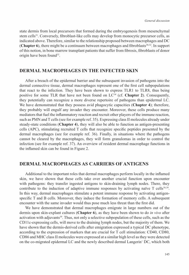
145
General discussion
state dermis from local precursors that formed during the embryogenesis from mesenchymal stem cells31. Conversely, fibroblast-like cells may develop from monocytic precursor cells, as indicated above. Therefore, similar to the relationship proposed between macrophages and DC (Chapter 6), there might be a continuum between macrophages and fibroblasts30,32. In support of this notion, in bone marrow transplant patients that suffer from fibrosis, fibroblasts of donor origin have been found33.
DERMAL MACROPHAGES IN THE INFECTED SKIN
After a breach of the epidermal barrier and the subsequent invasion of pathogens into the dermal connective tissue, dermal macrophages represent one of the first cell subpopulations that react to the infection. They have been shown to express TLR1 to TLR8, thus being positive for some TLR that have not been found on LC34 (cf. Chapter 2). Consequently, they potentially can recognize a more diverse repertoire of pathogens than epidermal LC. We have demonstrated that they possess avid phagocytic capacities (Chapter 4); therefore, they probably will engulf any invader they encounter. Moreover, these cells produce many mediators that fuel the inflammatory reaction and recruit other players of the immune reaction, such as PMN and T cells (see for example ref. 35). Expressing class II molecules already under steady-state conditions (Chapter 4), they will also be able to function as antigen-presenting cells (APC), stimulating recruited T cells that recognize specific peptides presented by the dermal macrophages (see for example ref. 36). Finally, in situations where the pathogens cannot be cleared by the macrophages, they will form granulomas in order to control the infection (see for example ref. 37). An overview of resident dermal macrophage functions in the inflamed skin can be found in Figure 2.
DERMAL MACROPHAGES AS CARRIERS OF ANTIGENS
Additional to the important roles that dermal macrophages perform locally in the inflamed skin, we have shown that these cells take over another crucial function upon encounter with pathogens: they transfer ingested antigens to skin-draining lymph nodes. There, they contribute to the induction of adaptive immune responses by activating naïve T cells38,39. In this way, dermal macrophages stimulate a potent immune response by activating antigen-specific T and B cells. Moreover, they induce the formation of memory cells. A subsequent encounter with the same invader would thus pose much less threat than the first did.
We have demonstrated that dermal macrophages emigrate in large numbers out of the dermis upon skin-explant cultures (Chapter 6), as they have been shown to do in vivo after activation with adjuvants39. Thus, not only a selective subpopulation of these cells, such as the CD11c-expressing cells, will move to the draining lymph nodes, but the majority of them. We have shown that the dermis-derived cells after emigration expressed a typical DC phenotype, according to the expression of markers that are crucial for T cell stimulation: CD40, CD80, CD86 and MHC class II molecules were expressed at a similar high level as they were detected on the co-migrated epidermal LC and the newly described dermal Langerin+ DC, which both

Chapter 8
146
occur in the Langerin+ gate (Chapter 6). CD11c and CCR7 were expressed by all dermis-derived DC, the latter receptor being important for their migration towards the draining lymph node. In skin-draining lymph nodes, we have described an mMGL+ cell type that expressed a similar mature DC phenotype. Moreover, we have confirmed that they originated from the skin, as they were they main carrier of FITC into the draining lymph nodes one and two days after FITC application. Finally, we have shown that the dermis-derived emigrants stimulated naïve T cells as efficient as did epidermis-derived LC, confirming their function as mature DC (Chapter 6). Therefore, we conclude that dermal macrophages represent precursor cells of lymph node DC and designated these activated, mature dermal macrophages as dermally derived DC (DdDC), in order to distinguish them from dermal DC, which show the DC phenotype and function already in the dermis. A graphic representation of the maturation of dermal macrophages into lymph node DC is shown in Figure 3.
Considering the thickness of the mouse dermis, compared to the epidermis, and the relatively high density of the mononuclear phagocytes in this connective tissue, amounting to about 60% of all interstitial cells (Chapter 4), it is apparent that the dermis harbors a vast antigen-presenting potential. This clearly contributes to the barrier function of the dermis as the second layer of defense of the body to the environment. Recent studies approaching the specific role of LC in skin immunity have confirmed that the contribution of LC is mostly redundant, implying that dermis-derived APC are in general more important than LC40-43. Interestingly, recruited wound-repair macrophages comparably have been shown to emigrate to skin-draining lymph nodes after resolution of the inflammation44,45. This hints to the notion that one function of alternatively activated macrophages might be to present antigen.
Figure 2. Resident dermal macrophage in the infected dermis. Refer to the text for details. Abbreviations: IL-1β: Interleukin-1β; IL-6: Interleukin-6; Mφ: macrophage; mMGL1 and 2: macrophage specific galactose/N-acetylgalactosamine-specific C-type lectin 1 and 2, respectively; MR: mannose receptor; TCR: T cell receptor.
mMGL2
mMGL1
MR
TNF-�IL-1�IL-6…
MHC class II
TCR
M�
T cellsGiant multi-nucleated cells
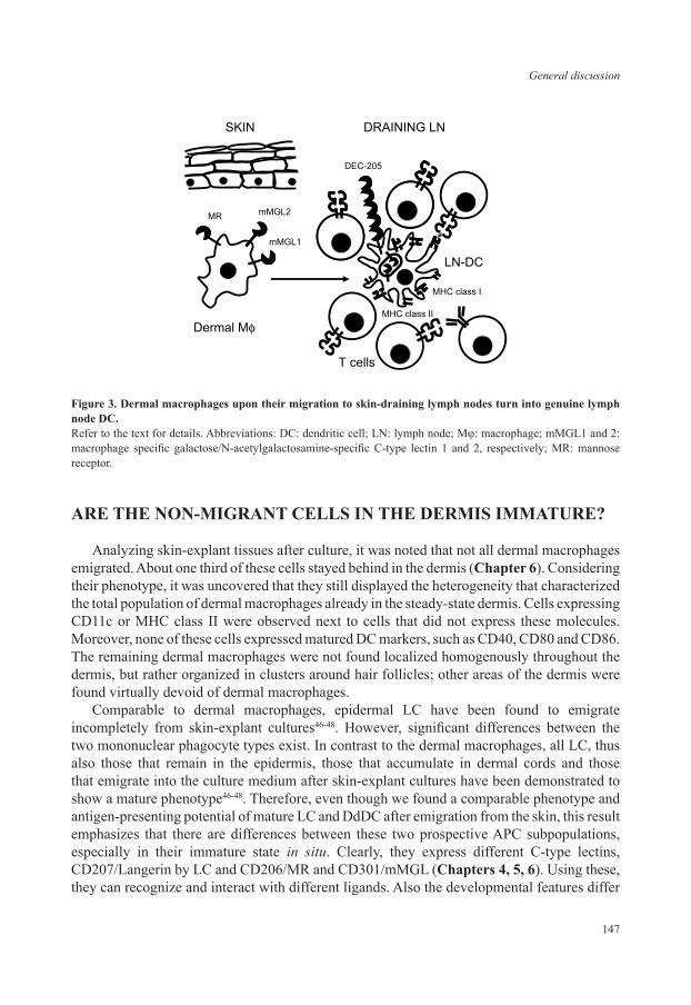
147
General discussion
ARE THE NON-MIGRANT CELLS IN THE DERMIS IMMATURE?
Analyzing skin-explant tissues after culture, it was noted that not all dermal macrophages emigrated. About one third of these cells stayed behind in the dermis (Chapter 6). Considering their phenotype, it was uncovered that they still displayed the heterogeneity that characterized the total population of dermal macrophages already in the steady-state dermis. Cells expressing CD11c or MHC class II were observed next to cells that did not express these molecules. Moreover, none of these cells expressed matured DC markers, such as CD40, CD80 and CD86. The remaining dermal macrophages were not found localized homogenously throughout the dermis, but rather organized in clusters around hair follicles; other areas of the dermis were found virtually devoid of dermal macrophages.
Comparable to dermal macrophages, epidermal LC have been found to emigrate incompletely from skin-explant cultures46-48. However, significant differences between the two mononuclear phagocyte types exist. In contrast to the dermal macrophages, all LC, thus also those that remain in the epidermis, those that accumulate in dermal cords and those that emigrate into the culture medium after skin-explant cultures have been demonstrated to show a mature phenotype46-48. Therefore, even though we found a comparable phenotype and antigen-presenting potential of mature LC and DdDC after emigration from the skin, this result emphasizes that there are differences between these two prospective APC subpopulations, especially in their immature state in situ. Clearly, they express different C-type lectins, CD207/Langerin by LC and CD206/MR and CD301/mMGL (Chapters 4, 5, 6). Using these, they can recognize and interact with different ligands. Also the developmental features differ
Figure 3. Dermal macrophages upon their migration to skin-draining lymph nodes turn into genuine lymph node DC.Refer to the text for details. Abbreviations: DC: dendritic cell; LN: lymph node; Mφ: macrophage; mMGL1 and 2: macrophage specific galactose/N-acetylgalactosamine-specific C-type lectin 1 and 2, respectively; MR: mannose receptor.
mMGL2
mMGL1
MR
MHC class II
MHC class I
DEC-205
T cells
Dermal M�
SKIN DRAINING LN
LN-DC

Chapter 8
148
between them, with LC being radio-resistant and self renewing in the epidermis49, whereas dermal macrophages are radio-sensitive and most likely dependent on bone marrow-derived precursors to replace emigrated cells50,51. This is in line with the scenario we have drawn before (Chapter 2) that dermal mononuclear phagocyte precursors immigrate into the dermis via blood, migrate upwards through the dermis surveying it, mature during this journey and emigrate at the top of the dermis via lymphatic vessels. This course of development remains to be proven. Alternatively, it might be that LC, being located at the outermost layer of the skin, acquired during the evolution the capability to deal efficiently with environmentally induced DNA damage, whereas dermal macrophages repair DNA damage less efficiently. Therefore, in experiments with the purpose to generate bone marrow chimeric mice, the latter cells might undergo apoptosis during the whole-body γ-irradiation whereas LC might repair their damage. LC would thus remain host-derived, whereas dermal macrophages would have to be replaced by donor-derived precursor cells. Last but not least, the two cell populations reside in different tissues; LC in the epithelium, the dermal macrophages in the connective tissue. Consequently, the cells interact with a different outside world; LC bind to and interact with keratinocytes, whereas dermal macrophages interact mainly with the extracellular matrix and not with cells. In this regard, LC and dermal macrophages are different cell subpopulations that perform different functions in an immature state in situ. As part of this different function, LC seem to be more prone to mature into potent APC. Nevertheless, once both subsets have emigrated from their tissue into draining lymph nodes, they display a similar phenotype and function. Consequently, in the lymph nodes they are difficult to distinguish.
Dermal macrophages that stayed behind in the dermis after explant culture were not found scattered throughout the dermis, but rather organized in clusters, especially in the vicinity of hair follicles. Hair follicles are special structures for skin homeostasis that have been shown to contain stem cell populations, which can give rise to epidermal stem cells regenerating the whole epidermal architecture52, but also to melanocytes, mast cells53 and Langerhans cells54. Moreover, the dermal CD207/Langerin+ DC have been described to be localized adjacent to hair follicles55. Although the significance of these dermal CD207/Langerin+ DC and their developmental relationship to other cutaneous cells remains to be elucidated, it might be that dermal macrophage precursors are not derived from the blood but localize somewhere near the hair follicles. These cells might replenish emigrating dermal macrophages from there. Those macrophages that are found close to the hair follicles might thus be more immature than other cells in the rest of the dermis, although there were no specific phenotypic indications to support this. Comparably, a distinction between more and less mature LC in the epidermis has been reported56. It might be the consequence of this immaturity that a part of the residing cells do not emigrate from the dermis in skin-explant cultures. Further characterization of the dermal macrophages and their developmental stages and potentials is thus warranted to address this issue.
ULTRAVIOLET (UV)-IRRADIATION OF THE SKIN LEADS TO A TOLEROGENIC INFLAMMATION
After having characterized dermal macrophages in the steady-state skin in situ and after emigration from cultured skin explants, we were interested in studying their contribution
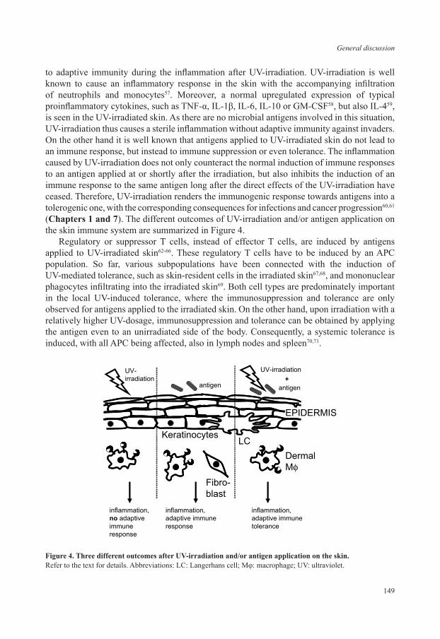
149
General discussion
to adaptive immunity during the inflammation after UV-irradiation. UV-irradiation is well known to cause an inflammatory response in the skin with the accompanying infiltration of neutrophils and monocytes57. Moreover, a normal upregulated expression of typical proinflammatory cytokines, such as TNF-α, IL-1β, IL-6, IL-10 or GM-CSF58, but also IL-459, is seen in the UV-irradiated skin. As there are no microbial antigens involved in this situation, UV-irradiation thus causes a sterile inflammation without adaptive immunity against invaders. On the other hand it is well known that antigens applied to UV-irradiated skin do not lead to an immune response, but instead to immune suppression or even tolerance. The inflammation caused by UV-irradiation does not only counteract the normal induction of immune responses to an antigen applied at or shortly after the irradiation, but also inhibits the induction of an immune response to the same antigen long after the direct effects of the UV-irradiation have ceased. Therefore, UV-irradiation renders the immunogenic response towards antigens into a tolerogenic one, with the corresponding consequences for infections and cancer progression60,61 (Chapters 1 and 7). The different outcomes of UV-irradiation and/or antigen application on the skin immune system are summarized in Figure 4.
Regulatory or suppressor T cells, instead of effector T cells, are induced by antigens applied to UV-irradiated skin62-66. These regulatory T cells have to be induced by an APC population. So far, various subpopulations have been connected with the induction of UV-mediated tolerance, such as skin-resident cells in the irradiated skin67,68, and mononuclear phagocytes infiltrating into the irradiated skin69. Both cell types are predominately important in the local UV-induced tolerance, where the immunosuppression and tolerance are only observed for antigens applied to the irradiated skin. On the other hand, upon irradiation with a relatively higher UV-dosage, immunosuppression and tolerance can be obtained by applying the antigen even to an unirradiated side of the body. Consequently, a systemic tolerance is induced, with all APC being affected, also in lymph nodes and spleen70,71.
Figure 4. Three different outcomes after UV-irradiation and/or antigen application on the skin. Refer to the text for details. Abbreviations: LC: Langerhans cell; Mφ: macrophage; UV: ultraviolet.
UV-irradiationUV-irradiation
antigen antigen
inflammation,no adaptiveimmuneresponse
+
inflammation,adaptive immuneresponse
inflammation,adaptive immunetolerance
DermalM�
LC
Fibro-blast
Keratinocytes
EPIDERMIS

Chapter 8
150
THE FUNCTION OF DERMAL MACROPHAGES IN THE INDUCTION OF UV-B MEDIATED TOLERANCE
We proposed that also dermal macrophages with their potent antigen-presenting capacities are involved in the induction of local UV-induced tolerance, as (i) UV-irradiation, in particular UV-A reaches the dermis72, (ii) UV-irradiation consequently also causes DNA damage in dermal cells67,73, (iii) UV-irradiation causes a higher level of oxidative stress in the dermis than in the epidermis74, (iv) human dermal CD1c+ cells have been described to be depleted from UV-irradiated dermis, as are LC from irradiated epidermis75, and (v) UV-irradiation has already been shown to impair the phenotypic and functional maturation of human dermal CD1c+ cells76. Consequently, we aimed to assess the effect of UV-irradiation on the function of dermal macrophages in inducing tolerance.
In the literature, two different tolerance-inducing UV-irradiation regimes have been described, either using a single-dose irradiation protocol, or performing daily irradiations on three to five subsequent days. As we wanted to study the effects of UV-irradiation on the resident dermal macrophages, we collected these cells before PMN and other mononuclear phagocyte subpopulations infiltrated the irradiated skin. Moreover, we wanted to avoid that these infiltrating cells themselves became irradiated and underwent functional alterations or apoptosis, which would render the process even more complicated and hamper the delineation of the contribution of individual cell types. Therefore we have used a single-dose UV-irradiation protocol, which had been shown previously by other research groups to interfere with the induction of contact hypersensivity (CHS) reactions already at moderate UV-irradiation doses, i.e. below one minimal erythemal dose (MED)67-69.
We irradiated ear halves with various irradiation dosages up to four MED and directly cultured them to obtain irradiated resident cells activated to emigrate by the irradiation and by the culture conditions. We noted that from in vitro irradiated ear cultures, dermally derived cells, and especially LC accumulated in significantly decreased numbers in the culture medium (Chapter 7). This in contrast to the literature, showing that UV-irradiation induces the emigration of epidermal and dermal APC75,77,78. The lack of emigration induction is most probably caused by the culture process, which by itself constitutes a strong emigration signal for epidermal LC and dermal macrophages (Chapter 6). UV-irradiation apparently does not induce additional emigration. The reduction of emigration that we observed, which correlated with the amount of UV-irradiation the cells were exposed to, can be explained by the cells being arrested in their maturation and emigration until they have dealt with their DNA damage. Alternatively, they may react differently to the culture process due to the UV-irradiation. Or third, a significant number of cells might undergo apoptosis due to the UV-irradiation. Considering the phenotype of the dermal and epidermal emigrants, we noted no difference in the expression of different DC maturation markers. Comparably, we observed hardly any difference in cytokine production between irradiated and unirradiated skin explants: only IL-12p40 was significantly lower in the UV-irradiated skin explant cultures. IL-12p40 is a cytokine typically produced only by DC. Normalizing the amount of IL-12p40 with the number of DC that have emigrated from epidermis and dermis corrected for the decrease in IL-12p40 production. Thus with or without UV-irradiation, the amount of IL-12p40 produced per DC did not differ.

151
General discussion
Functionally, the UV-treated skin-derived mature DC (CD86high) were less potent in stimulating the proliferation of allogeneic T cells: at the highest ratio of DC/T cells (1:50), the stimulation by UV-treated DC was only half compared to control DC. This is comparable to findings reported in the literature79,80. In comparison to immature, non-irradiated skin emigrants (surface mMGL+), the UV-treated DC displayed a higher stimulation capacity, in accordance with the higher expression of costimulatory molecules. Therefore, our findings do not support the notion that UV-irradiation inhibits the maturation of cutaneous DC-precursors. In accordance with our lack of effects of UV-irradiation on cutaneous DC, in an in vivo immunosuppression approach, we did not observe any interference with the induction of a CHS response by a single-dose UV-irradiation regimen up to two MED (Chapter 7).
UV-IRRADIATION CONTRIBUTES TO THE SUBTLE BALANCE BETWEEN IMMUNE SUPPRESSION AND IMMUNE ACTIVATION
Our lack of effect of UV-irradiation on immunosuppression or tolerance induction might have been caused by several confounding influences. These include the methods applied to assess the immunosuppression, such as the choice of antigen and adjuvant, the strain and condition of mice used or the applied UV-light source. We have used several techniques to assess an immunosuppressed state: phenotypical maturation of the emigrating APC, their cytokine production, functional performance in T cell stimulation and in vivo assessment of a CHS response induction. We found only minor suppressive influences of UV-irradiation in all these assays at the UV-doses used. Instead, in several models even stimulating effects of UV-irradiation were found, making it unlikely that we missed the immunosuppression. We used C57BL/6 mice. While different mouse strains have been used in different studies, BL/6 mice have been shown to be relatively susceptible to UV-induced immunosuppression. Therefore this strain is used regularly to study UV-induced immunosuppression81,82. Also the microbiological state of the mice may influence their immunological response. It has been noted, for instance, with autoimmune-prone non-obese diabetic (NOD) mice that the diabetes incidence in the colony increases significantly with a decreasing pathogenic load of the mice83. Generalizing this observation, it might be that improving hygienic conditions for the mice correlates with a decreasing capacity to induce tolerance. This would represent a mouse correlate of the hygiene theory, which has been proposed to explain the increasing frequency of allergic and autoimmune diseases observed among humans in the western world over the last decades84,85. Finally, the UV-source might have affected the outcome. UV-C light, which is not found in the sun light that reaches the earth but that nevertheless can be emitted by UV-sources, has been found to affect erythema and edema disproportionally86. On the other hand, UV-A, depending on protocol and dosage, has been shown to induce immunosuppression or to protect from UV-B-induced immunosuppression87,88. Therefore, depending on the amount of UV-A and UV-C light in the UV-source, the MED might be underestimated (in the presence of UV-C) or the effects by UV-B might be counteracted (by simultaneously applied UV-A). Although the spectrum of the UV-source we have used (Waldmann UV 6 lamp) does not differ significantly from the generally used FS20 and FS40 lamps (see Figure 5), dose rates and duration might nevertheless influence the effects of UV-B irradiation on skin cells.

Chapter 8
152
Reflecting on all the factors that play a role in the cutaneous response to UV-irradiation, we conclude that the UV-irradiation itself represents only one factor contributing to the subtle balance between immunity and tolerance. A strong UV-irradiation regimen, however, applied over several days, does interfere with the induction of weak immune responses as several studies89, including our own (e.g. ref. 90), have shown. On the other hand, strong immunoactivators, such as a strong antigen/adjuvant formulation or the procedure of skin tissue isolation and ex vivo culture, can overcome the inhibiting effects of a weak UV-irradiation. Therefore, UV-irradiation, although well known for its carcinogenic and immunosuppressive effects, does not necessarily have to be harmful for the body’s host defense. On the contrary, low-dose UV-irradiation seems to be even beneficial. It has been demonstrated that low-dose UV-irradiation regimens activate rather than inhibit the immune response against Leishmania amazonensis in BALB/c mice91. Additionally, low dose UV-irradiation is necessary for efficient vitamin D3 production. As a direct consequence, low-dose UV-irradiation inhibits cancer growth. This inhibition is both due to the increased vitamin D3 level92,93 as well as to the immunoactivating effects of low-dose UV irradiation. The body seems thus to have adopted to absorb, deal with and even use some ambient UV-irradiation, reflected by the decreasing body pigmentation with increasing latitude. Complete avoidance of sunlight is consequently as unhealthy as is exaggerated sun light consumption for tanning purposes. A reasonable UV consumption dosage requires that benefits and disadvantages balance each other.
UNRAVELING THE ROLE OF DERMAL MACROPHAGES IN UV-INDUCED IMMUNOSUPPRESSION
The aim of our UV study was to investigate the role of, in particular, dermal macrophages in the elicitation of UV-induced immunosuppression using single UV exposure in vivo or in vitro.
Figure 5. The spectral output of the FS20 lamp and the Waldmann UV 6 lamp. The output of the FS20 lamp was taken from Clingen et al.94, the output of the UV 6 lamp was obtained from the Waldman homepage (http://www.waldmann-medizintechnik.com/).
0.0001
0.001
0.01
0.1
1
10
Relativespectraloutput
280
300
320
340
360
380
400
Wavelength (nm)
FS20 (solid black line)
Waldmann UV 6(dotted gray line)
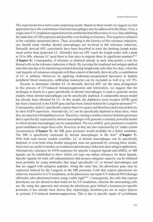
153
General discussion
The experiments have led to some surprising results. Based on these results we suggest several approaches how the contribution of dermal macrophages may be addressed in the future. First, a single-dose UV-irradiation regimen has to be established that affects mice in vivo; thus inhibiting the induction of CHS responses and possibly even leading to tolerance. This requires evaluation of the variables mentioned above. Then, according to the kinetics of this tolerance induction, one should study whether dermal macrophages are involved in this tolerance induction. Dermally derived APC consistently have been described to enter the draining lymph nodes much earlier than epidermal LC. Dermally derived APC reach the lymph nodes with a peak at 24 to 48h, whereas LC need three to four days to migrate there in significant numbers40,41 (Chapter 6). Consequently, if tolerance is obtained already at early time-points, a role for dermal cells in the tolerance induction is likely. By excising the irradiated and antigen-applied side after one day or by transferring isolated draining lymph node cells after two days, when the vast majority of cutaneous emigrants will thus consist of dermally derived cells, a contribution of LC is unlikely. Moreover, by applying clodronate-encapsulated liposomes to deplete peripheral blood monocytes, infiltrating monocytes can be excluded as well (e.g. ref. 95). Second, to determine whether LC or dermally derived APC are the main protagonists in this process of UV-induced immunosuppression and tolerization, we suggest that the technique to knock-in a gene specifically in dermal macrophages is used to generate mouse models where dermal macrophages can be specifically depleted. A comparable mouse model has already been obtained for LC. In this model, the diphtheria toxin receptor (DTR) gene has been connected to the EGFP gene and has been cloned behind the Langerin-promoter40,96. Consequently, such LC specifically express these two genes and therefore can be traced directly by their EGFP expression. Alternatively, LC can be specifically depleted in these mice, when they are injected with diphtheria toxin. Therefore, cloning a similar construct behind a promoter that is specifically expressed by dermal macrophages will generate a similarly powerful model in which dermal macrophages can be manipulated. The two mMGL gene promoters would be good candidates to target these cells. However, as they are also expressed by LC under certain circumstances (Chapter 5), the MR gene promoter would probably be a better candidate. The MR is specifically expressed by dermal macrophages in the skin97 (Chapter 5). With both such mouse models available, LC or dermal macrophages can be specifically depleted, or even both when double transgenic mice are generated by crossing these strains. Such mice are useful in studies on irradiation and tolerance induction upon antigen application. Alternatively, tolerance for DTH responses for specific Langerin ligands and mMGL or MR ligands can be established to show which cell type can induce tolerance for these ligands. Specific ligands for both cell subpopulations that possess antigenic capacity can be obtained most probably by using antibodies that target specifically LC or dermal macrophages and that are tagged with antigenic peptides. Along the same line, the photolyase gene may be expressed induced by the Langerin or the MR promoter. Cells that express photolyase are relatively insensitive to UV-irradiation, as the photolyase can repair UV-induced DNA damage efficiently after photoreactivation using visible light90,98. Consequently, the cells that express photolyase are not altered in their behavior by UV-irradiation, whereas the surrounding cells are. By using this approach and cloning the photolyase gene behind a keratinocyte-specific promoter, it has already been shown that, surprisingly, keratinocytes are no major players in systemic UV-induced immunosuppression. This is due to specific repair of cyclobutane

Chapter 8
154
pyrimidine dimers in these cells which inhibit acute sunburn effects and cancer induction but not systemic immunosuppression90. Therefore, making LC or dermal macrophages insensitive to UV-irradiation should in a similar manner allow studies on the contribution of LC and dermally derived APC in UV-induced immunosuppression and tolerance.
CONCLUDING REMARKS ON UV-MEDIATED IMMUNE SUPPRESSION
UV-irradiation can lead to the induction of regulatory T cells. APC that are affected by the UV-irradiation transfer antigens to the draining lymph nodes to induce the activation of these T cells. The induction of regulatory T cells has been assigned so far to different APC subsets, such as the skin-resident DC precursors and the infiltrating monocytes. In systemic tolerance, generally all DC populations, thus also those resident in secondary lymphoid organs are affected by the UV-irradiation, even though cutaneous DC subpopulations transfer the skin-derived antigens to the lymph nodes. Despite the focus on the classic APC, such as DC, in tolerance induction, other cell types may have crucial contributions as well. Literature of recent years suggests the involvement of an unusual immune cell that is normally not considered in adaptive immune responses: dermal mast cells appear to be crucial for immune suppression after UV-irradiation in mice99,100 and man101,102. They have been shown to emigrate after UV-irradiation into skin-draining lymph nodes103,104 where they induce suppressor B cells105,106. Their contribution has been revealed to be restricted to systemic UV-induced immunosuppression107. Whether mast cells function in skin-draining lymph nodes as APC 108,109 or influence B cells as prime APC by their vast array of pre-produced and stored soluble mediators, remains to be determined. Therefore, research into the protagonists and mediators of UV-induced immunosuppression may still result in surprises. As concepts of UV-induced immune suppression later on often have been found applicable in other aspects of immunity, for example the occurrence of suppressor T cells62 or the role of IL-10 in the suppression process110, it is conceivable that findings on UV-mediated immune suppression might also apply to a broader context of immunity.
REFERENCES
1. Miller, C.C., Godeau, G., Lebreton-DeCoster, C., Desmouliere, A., Pellat, B., Dubertret, L. & Coulomb, B. Validation of a morphometric method for evaluating fibroblast numbers in normal and pathologic tissues. Exp Dermatol 12, 403-411 (2003).
2. Quatresooz, P. & Pierard, G.E. Imiquimod-responsive basal cell carcinomas and factor XIIIa-enriched dendrocytes. Clin Exp Dermatol 28 Suppl 1, 27-29 (2003).
3. Meunier, L., Gonzalez-Ramos, A. & Cooper, K.D. Heterogeneous populations of class II MHC+ cells in human dermal cell suspensions. Identification of a small subset responsible for potent dermal antigen-presenting cell activity with features analogous to Langerhans cells. J Immunol 151, 4067-4080 (1993).
4. Duraiswamy, N., Tse, Y., Hammerberg, C., Kang, S. & Cooper, K.D. Distinction of class II MHC+ Langerhans cell-like interstitial dendritic antigen-presenting cells in murine dermis from dermal macrophages. J Invest Dermatol 103, 678-683 (1994).

155
General discussion
5. Hume, D.A., Robinson, A.P., MacPherson, G.G. & Gordon, S. The mononuclear phagocyte system of the mouse defined by immunohistochemical localization of antigen F4/80. Relationship between macrophages, Langerhans cells, reticular cells, and dendritic cells in lymphoid and hematopoietic organs. J Exp Med 158, 1522-1536 (1983).
6. Leenen, P.J., de Bruijn, M.F., Voerman, J.S., Campbell, P.A. & van Ewijk, W. Markers of mouse macrophage development detected by monoclonal antibodies. J Immunol Methods 174, 5-19 (1994).
7. Stein, M., Keshav, S., Harris, N. & Gordon, S. Interleukin 4 potently enhances murine macrophage mannose receptor activity: a marker of alternative immunologic macrophage activation. J Exp Med 176, 287-292 (1992).
8. Raes, G., Brys, L., Dahal, B.K., Brandt, J., Grooten, J., Brombacher, F., Vanham, G., Noel, W., Bogaert, P., Boonefaes, T., Kindt, A., Van den Bergh, R., Leenen, P.J., De Baetselier, P. & Ghassabeh, G.H. Macrophage galactose-type C-type lectins as novel markers for alternatively activated macrophages elicited by parasitic infections and allergic airway inflammation. J Leukoc Biol 77, 321-327 (2005).
9. Gordon, S. Alternative activation of macrophages. Nat Rev Immunol 3, 23-35 (2003).10. Gratchev, A., Kzhyshkowska, J., Utikal, J. & Goerdt, S. Interleukin-4 and dexamethasone counterregulate
extracellular matrix remodelling and phagocytosis in type-2 macrophages. Scand J Immunol 61, 10-17 (2005).11. Dannenberg, A.M., Jr., Burstone, M.S., Walter, P.C. & Kinsley, J.W. A histochemical study of phagocytic and
enzymatic functions of rabbit mononuclear and polymorphonuclear exudate cells and alveolar macrophages. I. Survey and quantitation of enzymes, and states of cellular activation. J Cell Biol 17, 465-486 (1963).
12. Oren, R., Farnham, A.E., Saito, K., Milofsky, E. & Karnovsky, M.L. Metabolic patterns in three types of phagocytizing cells. J Cell Biol 17, 487-501 (1963).
13. Noble, P.W., Henson, P.M. & Riches, D.W. Insulin-like growth factor-1 (IGF-1) mRNA expression in bone marrow derived macrophages is stimulated by chrysotile asbestos and bleomycin. A potential marker for a reparative macrophage phenotype. Chest 99, 79S-80S (1991).
14. Peters, T., Sindrilaru, A., Hinz, B., Hinrichs, R., Menke, A., Al-Azzeh, E.A., Holzwarth, K., Oreshkova, T., Wang, H., Kess, D., Walzog, B., Sulyok, S., Sunderkotter, C., Friedrich, W., Wlaschek, M., Krieg, T. & Scharffetter-Kochanek, K. Wound-healing defect of CD18(-/-) mice due to a decrease in TGF-beta1 and myofibroblast differentiation. Embo J 24, 3400-3410 (2005).
15. Serhan, C.N. & Savill, J. Resolution of inflammation: the beginning programs the end. Nat Immunol 6, 1191-1197 (2005).
16. Kodelja, V., Muller, C., Tenorio, S., Schebesch, C., Orfanos, C.E. & Goerdt, S. Differences in angiogenic potential of classically vs alternatively activated macrophages. Immunobiology 197, 478-493 (1997).
17. Chetty, A., Cao, G.J. & Nielsen, H.C. Insulin-like growth factor-I signaling mechanisms, type I collagen and alpha smooth muscle actin in human fetal lung fibroblasts. Pediatr Res 60, 389-394 (2006).
18. Shao, D.D., Suresh, R., Vakil, V., Gomer, R.H. & Pilling, D. Pivotal Advance: Th-1 cytokines inhibit, and Th-2 cytokines promote fibrocyte differentiation. J Leukoc Biol 83, 1323-1333 (2008).
19. Alitalo, K., Hovi, T. & Vaheri, A. Fibronectin is produced by human macrophages. J Exp Med 151, 602-613 (1980).
20. Gratchev, A., Guillot, P., Hakiy, N., Politz, O., Orfanos, C.E., Schledzewski, K. & Goerdt, S. Alternatively activated macrophages differentially express fibronectin and its splice variants and the extracellular matrix protein betaIG-H3. Scand J Immunol 53, 386-392 (2001).
21. Lindblad, W.J. Perspective article: collagen expression by novel cell populations in the dermal wound environment. Wound Repair Regen 6, 186-193 (1998).
22. Kuwana, M., Okazaki, Y., Kodama, H., Izumi, K., Yasuoka, H., Ogawa, Y., Kawakami, Y. & Ikeda, Y. Human circulating CD14+ monocytes as a source of progenitors that exhibit mesenchymal cell differentiation. J Leukoc Biol 74, 833-845 (2003).
23. Sakai, N., Wada, T., Furuichi, K., Shimizu, K., Kokubo, S., Hara, A., Yamahana, J., Okumura, T., Matsushima, K., Yokoyama, H. & Kaneko, S. MCP-1/CCR2-dependent loop for fibrogenesis in human peripheral CD14-positive monocytes. J Leukoc Biol 79, 555-563 (2006).
24. Schnoor, M., Cullen, P., Lorkowski, J., Stolle, K., Robenek, H., Troyer, D., Rauterberg, J. & Lorkowski, S. Production of type VI collagen by human macrophages: a new dimension in macrophage functional heterogeneity. J Immunol 180, 5707-5719 (2008).

Chapter 8
156
25. Fichtner-Feigl, S., Strober, W., Kawakami, K., Puri, R.K. & Kitani, A. IL-13 signaling through the IL-13alpha2 receptor is involved in induction of TGF-beta1 production and fibrosis. Nat Med 12, 99-106 (2006).
26. Torocsik, D., Bardos, H., Nagy, L. & Adany, R. Identification of factor XIII-A as a marker of alternative macrophage activation. Cell Mol Life Sci 62, 2132-2139 (2005).
27. Akagi, A., Tajima, S., Ishibashi, A., Matsubara, Y., Takehana, M., Kobayashi, S. & Yamaguchi, N. Type XVI collagen is expressed in factor XIIIa+ monocyte-derived dermal dendrocytes and constitutes a potential substrate for factor XIIIa. J Invest Dermatol 118, 267-274 (2002).
28. Gonzalez-Ramos, A., Cooper, K.D. & Hammerberg, C. Identification of a human dermal macrophage population responsible for constitutive restraint of primary dermal fibroblast proliferation. J Invest Dermatol 106, 305-311 (1996).
29. Bhowmick, N.A., Chytil, A., Plieth, D., Gorska, A.E., Dumont, N., Shappell, S., Washington, M.K., Neilson, E.G. & Moses, H.L. TGF-beta signaling in fibroblasts modulates the oncogenic potential of adjacent epithelia. Science 303, 848-851 (2004).
30. Postlethwaite, A.E., Shigemitsu, H. & Kanangat, S. Cellular origins of fibroblasts: possible implications for organ fibrosis in systemic sclerosis. Curr Opin Rheumatol 16, 733-738 (2004).
31. Bogunovic, M., Ginhoux, F., Wagers, A., Loubeau, M., Isola, L.M., Lubrano, L., Najfeld, V., Phelps, R.G., Grosskreutz, C., Scigliano, E., Frenette, P.S. & Merad, M. Identification of a radio-resistant and cycling dermal dendritic cell population in mice and men. J Exp Med 203, 2627-2638 (2006).
32. Arlein, W.J., Shearer, J.D. & Caldwell, M.D. Continuity between wound macrophage and fibroblast phenotype: analysis of wound fibroblast phagocytosis. Am J Physiol 275, R1041-1048 (1998).
33. Forbes, S.J., Russo, F.P., Rey, V., Burra, P., Rugge, M., Wright, N.A. & Alison, M.R. A significant proportion of myofibroblasts are of bone marrow origin in human liver fibrosis. Gastroenterology 126, 955-963 (2004).
34. van der Aar, A.M., Sylva-Steenland, R.M., Bos, J.D., Kapsenberg, M.L., de Jong, E.C. & Teunissen, M.B. Loss of TLR2, TLR4, and TLR5 on Langerhans cells abolishes bacterial recognition. J Immunol 178, 1986-1990 (2007).
35. Cailhier, J.F., Partolina, M., Vuthoori, S., Wu, S., Ko, K., Watson, S., Savill, J., Hughes, J. & Lang, R.A. Conditional macrophage ablation demonstrates that resident macrophages initiate acute peritoneal inflammation. J Immunol 174, 2336-2342 (2005).
36. van Lent, P.L., Figdor, C.G., Barrera, P., van Ginkel, K., Sloetjes, A., van den Berg, W.B. & Torensma, R. Expression of the dendritic cell-associated C-type lectin DC-SIGN by inflammatory matrix metalloproteinase-producing macrophages in rheumatoid arthritis synovium and interaction with intercellular adhesion molecule 3-positive T cells. Arthritis Rheum 48, 360-369 (2003).
37. Saunders, B.M. & Cooper, A.M. Restraining mycobacteria: role of granulomas in mycobacterial infections. Immunol Cell Biol 78, 334-341 (2000).
38. Sato, K., Imai, Y. & Irimura, T. Contribution of dermal macrophage trafficking in the sensitization phase of contact hypersensitivity. J Immunol 161, 6835-6844 (1998).
39. Chun, K.H., Imai, Y., Higashi, N. & Irimura, T. Migration of dermal cells expressing a macrophage C-type lectin during the sensitization phase of delayed-type hypersensitivity. J Leukoc Biol 68, 471-478 (2000).
40. Kissenpfennig, A., Ait-Yahia, S., Clair-Moninot, V., Stossel, H., Badell, E., Bordat, Y., Pooley, J.L., Lang, T., Prina, E., Coste, I., Gresser, O., Renno, T., Winter, N., Milon, G., Shortman, K., Romani, N., Lebecque, S., Malissen, B., Saeland, S. & Douillard, P. Disruption of the langerin/CD207 gene abolishes Birbeck granules without a marked loss of Langerhans cell function. Mol Cell Biol 25, 88-99 (2005).
41. Allan, R.S., Waithman, J., Bedoui, S., Jones, C.M., Villadangos, J.A., Zhan, Y., Lew, A.M., Shortman, K., Heath, W.R. & Carbone, F.R. Migratory dendritic cells transfer antigen to a lymph node-resident dendritic cell population for efficient CTL priming. Immunity 25, 153-162 (2006).
42. Bennett, C.L. & Clausen, B.E. DC ablation in mice: promises, pitfalls, and challenges. Trends Immunol 28, 525-531 (2007).
43. Fukunaga, A., Khaskhely, N.M., Sreevidya, C.S., Byrne, S.N. & Ullrich, S.E. Dermal dendritic cells, and not Langerhans cells, play an essential role in inducing an immune response. J Immunol 180, 3057-3064 (2008).
44. Bellingan, G.J., Caldwell, H., Howie, S.E., Dransfield, I. & Haslett, C. In vivo fate of the inflammatory macrophage during the resolution of inflammation: inflammatory macrophages do not die locally, but emigrate to the draining lymph nodes. J Immunol 157, 2577-2585 (1996).

157
General discussion
45. Bellingan, G.J., Xu, P., Cooksley, H., Cauldwell, H., Shock, A., Bottoms, S., Haslett, C., Mutsaers, S.E. & Laurent, G.J. Adhesion molecule-dependent mechanisms regulate the rate of macrophage clearance during the resolution of peritoneal inflammation. J Exp Med 196, 1515-1521 (2002).
46. Larsen, C.P., Ritchie, S.C., Hendrix, R., Linsley, P.S., Hathcock, K.S., Hodes, R.J., Lowry, R.P. & Pearson, T.C. Regulation of immunostimulatory function and costimulatory molecule (B7-1 and B7-2) expression on murine dendritic cells. J Immunol 152, 5208-5219 (1994).
47. Larsen, C.P., Steinman, R.M., Witmer-Pack, M., Hankins, D.F., Morris, P.J. & Austyn, J.M. Migration and maturation of Langerhans cells in skin transplants and explants. J Exp Med 172, 1483-1493 (1990).
48. Weinlich, G., Heine, M., Stossel, H., Zanella, M., Stoitzner, P., Ortner, U., Smolle, J., Koch, F., Sepp, N.T., Schuler, G. & Romani, N. Entry into afferent lymphatics and maturation in situ of migrating murine cutaneous dendritic cells. J Invest Dermatol 110, 441-448 (1998).
49. Merad, M., Manz, M.G., Karsunky, H., Wagers, A., Peters, W., Charo, I., Weissman, I.L., Cyster, J.G. & Engleman, E.G. Langerhans cells renew in the skin throughout life under steady-state conditions. Nat Immunol 3, 1135-1141 (2002).
50. Waithman, J., Allan, R.S., Kosaka, H., Azukizawa, H., Shortman, K., Lutz, M.B., Heath, W.R., Carbone, F.R. & Belz, G.T. Skin-derived dendritic cells can mediate deletional tolerance of class I-restricted self-reactive T cells. J Immunol 179, 4535-4541 (2007).
51. Iijima, N., Linehan, M.M., Saeland, S. & Iwasaki, A. Vaginal epithelial dendritic cells renew from bone marrow precursors. Proc Natl Acad Sci U S A 104, 19061-19066 (2007).
52. Ito, M., Liu, Y., Yang, Z., Nguyen, J., Liang, F., Morris, R.J. & Cotsarelis, G. Stem cells in the hair follicle bulge contribute to wound repair but not to homeostasis of the epidermis. Nat Med 11, 1351-1354 (2005).
53. Kumamoto, T., Shalhevet, D., Matsue, H., Mummert, M.E., Ward, B.R., Jester, J.V. & Takashima, A. Hair follicles serve as local reservoirs of skin mast cell precursors. Blood 102, 1654-1660 (2003).
54. Gilliam, A.C., Kremer, I.B., Yoshida, Y., Stevens, S.R., Tootell, E., Teunissen, M.B., Hammerberg, C. & Cooper, K.D. The human hair follicle: a reservoir of CD40+ B7-deficient Langerhans cells that repopulate epidermis after UVB exposure. J Invest Dermatol 110, 422-427 (1998).
55. Bursch, L.S., Wang, L., Igyarto, B., Kissenpfennig, A., Malissen, B., Kaplan, D.H. & Hogquist, K.A. Identification of a novel population of Langerin+ dendritic cells. J Exp Med 204, 3147-3156 (2007).
56. Shibaki, A., Meunier, L., Ra, C., Shimada, S., Ohkawara, A. & Cooper, K.D. Differential responsiveness of Langerhans cell subsets of varying phenotypic states in normal human epidermis. J Invest Dermatol 104, 42-46 (1995).
57. Cooper, K.D., Duraiswamy, N., Hammerberg, C., Allen, E., Kimbrough-Green, C., Dillon, W. & Thomas, D. Neutrophils, differentiated macrophages, and monocyte/macrophage antigen presenting cells infiltrate murine epidermis after UV injury. J Invest Dermatol 101, 155-163 (1993).
58. Takashima, A. & Bergstresser, P.R. Impact of UVB radiation on the epidermal cytokine network. Photochem Photobiol 63, 397-400 (1996).
59. Teunissen, M.B., Piskin, G., di Nuzzo, S., Sylva-Steenland, R.M., de Rie, M.A. & Bos, J.D. Ultraviolet B radiation induces a transient appearance of IL-4+ neutrophils, which support the development of Th2 responses. J Immunol 168, 3732-3739 (2002).
60. Schwarz, T. Photoimmunosuppression. Photodermatol Photoimmunol Photomed 18, 141-145 (2002).61. Ullrich, S.E. Mechanisms underlying UV-induced immune suppression. Mutat Res 571, 185-205 (2005).62. Fisher, M.S. & Kripke, M.L. Suppressor T lymphocytes control the development of primary skin cancers in
ultraviolet-irradiated mice. Science 216, 1133-1134 (1982).63. Shreedhar, V.K., Pride, M.W., Sun, Y., Kripke, M.L. & Strickland, F.M. Origin and characteristics of ultraviolet-
B radiation-induced suppressor T lymphocytes. J Immunol 161, 1327-1335 (1998).64. Moodycliffe, A.M., Nghiem, D., Clydesdale, G. & Ullrich, S.E. Immune suppression and skin cancer
development: regulation by NKT cells. Nat Immunol 1, 521-525 (2000).65. Schwarz, A., Beissert, S., Grosse-Heitmeyer, K., Gunzer, M., Bluestone, J.A., Grabbe, S. & Schwarz, T.
Evidence for functional relevance of CTLA-4 in ultraviolet-radiation-induced tolerance. J Immunol 165, 1824-1831 (2000).
66. Schwarz, T. 25 years of UV-induced immunosuppression mediated by T cells-from disregarded T suppressor cells to highly respected regulatory T cells. Photochem Photobiol 84, 10-18 (2008).

Chapter 8
158
67. Vink, A.A., Strickland, F.M., Bucana, C., Cox, P.A., Roza, L., Yarosh, D.B. & Kripke, M.L. Localization of DNA damage and its role in altered antigen-presenting cell function in ultraviolet-irradiated mice. J Exp Med 183, 1491-1500 (1996).
68. Kurimoto, I., Kitazawa, T. & Streilein, J.W. Suprathreshold doses of hapten are required to induce both contact hypersensitivity and tolerance. J Dermatol Sci 24, 48-59 (2000).
69. Hammerberg, C., Duraiswamy, N. & Cooper, K.D. Temporal correlation between UV radiation locally-inducible tolerance and the sequential appearance of dermal, then epidermal, class II MHC+CD11b+ monocytic/macrophagic cells. J Invest Dermatol 107, 755-763 (1996).
70. Kitazawa, T. & Streilein, J.W. Studies on delayed systemic effects of ultraviolet B radiation on the induction of contact hypersensitivity, 3. Dendritic cells from secondary lymphoid organs are deficient in interleukin-12 production and capacity to promote activation and differentiation of T helper type 1 cells. Immunology 99, 296-304 (2000).
71. Boonstra, A., van Oudenaren, A., Barendregt, B., An, L., Leenen, P.J. & Savelkoul, H.F. UVB irradiation modulates systemic immune responses by affecting cytokine production of antigen-presenting cells. Int Immunol 12, 1531-1538 (2000).
72. Bruls, W.A., Slaper, H., van der Leun, J.C. & Berrens, L. Transmission of human epidermis and stratum corneum as a function of thickness in the ultraviolet and visible wavelengths. Photochem Photobiol 40, 485-494 (1984).
73. Katiyar, S.K., Matsui, M.S. & Mukhtar, H. Kinetics of UV light-induced cyclobutane pyrimidine dimers in human skin in vivo: an immunohistochemical analysis of both epidermis and dermis. Photochem Photobiol 72, 788-793 (2000).
74. He, G., Kutala, V.K., Kuppusamy, P. & Zweier, J.L. In vivo measurement and mapping of skin redox stress induced by ultraviolet light exposure. Free Radic Biol Med 36, 665-672 (2004).
75. Meunier, L., Bata-Csorgo, Z. & Cooper, K.D. In human dermis, ultraviolet radiation induces expansion of a CD36+ CD11b+ CD1- macrophage subset by infiltration and proliferation; CD1+ Langerhans-like dendritic antigen-presenting cells are concomitantly depleted. J Invest Dermatol 105, 782-788 (1995).
76. Furio, L., Berthier-Vergnes, O., Ducarre, B., Schmitt, D. & Peguet-Navarro, J. UVA radiation impairs phenotypic and functional maturation of human dermal dendritic cells. J Invest Dermatol 125, 1032-1038 (2005).
77. Toews, G.B., Bergstresser, P.R. & Streilein, J.W. Epidermal Langerhans cell density determines whether contact hypersensitivity or unresponsiveness follows skin painting with DNFB. J Immunol 124, 445-453 (1980).
78. Aberer, W., Schuler, G., Stingl, G., Honigsmann, H. & Wolff, K. Ultraviolet light depletes surface markers of Langerhans cells. J Invest Dermatol 76, 202-210 (1981).
79. Denfeld, R.W., Hara, H., Tesmann, J.P., Martin, S. & Simon, J.C. UVB-irradiated dendritic cells are impaired in their APC function and tolerize primed Th1 cells but not naive CD4+ T cells. J Leukoc Biol 69, 548-554 (2001).
80. Dittmar, H.C., Weiss, J.M., Termeer, C.C., Denfeld, R.W., Wanner, M.B., Skov, L., Barker, J.N., Schopf, E., Baadsgaard, O. & Simon, J.C. In vivo UVA-1 and UVB irradiation differentially perturbs the antigen-presenting function of human epidermal Langerhans cells. J Invest Dermatol 112, 322-325 (1999).
81. Yoshikawa, T. & Streilein, J.W. Genetic basis of the effects of ultraviolet light B on cutaneous immunity. Evidence that polymorphism at the Tnfa and Lps loci governs susceptibility. Immunogenetics 32, 398-405 (1990).
82. Noonan, F.P. & Hoffman, H.A. Susceptibility to immunosuppression by ultraviolet B radiation in the mouse. Immunogenetics 39, 29-39 (1994).
83. David, T., Thomas, C., Zaccone, P., Dunne, D.W. & Cooke, A. The impact of infection on the incidence of autoimmune disease. Curr Top Med Chem 4, 521-529 (2004).
84. Liu, A.H. Hygiene theory and allergy and asthma prevention. Paediatr Perinat Epidemiol 21 Suppl 3, 2-7 (2007).
85. Ram, M., Anaya, J.M., Barzilai, O., Izhaky, D., Porat Katz, B.S., Blank, M., Juan-Carlos, R. & Shoenfeld, Y. The putative protective role of hepatitis B virus (HBV) infection against autoimmune disorders. Autoimmun Rev (2008).
86. Learn, D.B., Beard, J. & Moloney, S.J. The ultraviolet C energy emitted from FS lamps contributes significantly to the induction of human erythema and murine ear edema. Photodermatol Photoimmunol Photomed 9, 147-153 (1993).

159
General discussion
87. Byrne, S.N., Spinks, N. & Halliday, G.M. Ultraviolet a irradiation of C57BL/6 mice suppresses systemic contact hypersensitivity or enhances secondary immunity depending on dose. J Invest Dermatol 119, 858-864 (2002).
88. Reeve, V.E., Domanski, D. & Slater, M. Radiation sources providing increased UVA/UVB ratios induce photoprotection dependent on the UVA dose in hairless mice. Photochem Photobiol 82, 406-411 (2006).
89. Noonan, F.P. & De Fabo, E.C. Ultraviolet-B dose-response curves for local and systemic immunosuppression are identical. Photochem Photobiol 52, 801-810 (1990).
90. Jans, J., Garinis, G.A., Schul, W., van Oudenaren, A., Moorhouse, M., Smid, M., Sert, Y.G., van der Velde, A., Rijksen, Y., de Gruijl, F.R., van der Spek, P.J., Yasui, A., Hoeijmakers, J.H., Leenen, P.J. & van der Horst, G.T. Differential role of basal keratinocytes in UV-induced immunosuppression and skin cancer. Mol Cell Biol 26, 8515-8526 (2006).
91. Khaskhely, N.M., Maruno, M., Uezato, H., Takamiyagi, A., Ramzi, S.T., Al-Kasem, K.M., Kariya, K., Toda, T., Hashiguchi, Y., Gomez Landires, E.A. & Nonaka, S. Low-dose UVB contributes to host resistance against Leishmania amazonensis infection in mice through induction of gamma interferon and tumor necrosis factor alpha cytokines. Clin Diagn Lab Immunol 9, 677-686 (2002).
92. Grant, W. Insufficient sunlight may kill 45 000 Americans each year from internal cancer. J Cosmet Dermatol 3, 176-178 (2004).
93. Moon, S.J., Fryer, A.A. & Strange, R.C. Ultraviolet radiation: effects on risks of prostate cancer and other internal cancers. Mutat Res 571, 207-219 (2005).
94. Clingen, P.H., Berneburg, M., Petit-Frere, C., Woollons, A., Lowe, J.E., Arlett, C.F. & Green, M.H. Contrasting effects of an ultraviolet B and an ultraviolet A tanning lamp on interleukin-6, tumour necrosis factor-alpha and intercellular adhesion molecule-1 expression. Br J Dermatol 145, 54-62 (2001).
95. Sunderkotter, C., Nikolic, T., Dillon, M.J., Van Rooijen, N., Stehling, M., Drevets, D.A. & Leenen, P.J. Subpopulations of mouse blood monocytes differ in maturation stage and inflammatory response. J Immunol 172, 4410-4417 (2004).
96. Bennett, C.L., van Rijn, E., Jung, S., Inaba, K., Steinman, R.M., Kapsenberg, M.L. & Clausen, B.E. Inducible ablation of mouse Langerhans cells diminishes but fails to abrogate contact hypersensitivity. J Cell Biol 169, 569-576 (2005).
97. McKenzie, E.J., Taylor, P.R., Stillion, R.J., Lucas, A.D., Harris, J., Gordon, S. & Martinez-Pomares, L. Mannose receptor expression and function define a new population of murine dendritic cells. J Immunol 178, 4975-4983 (2007).
98. Garinis, G.A., Jans, J. & van der Horst, G.T. Photolyases: capturing the light to battle skin cancer. Future Oncol 2, 191-199 (2006).
99. Hart, P.H., Grimbaldeston, M.A., Swift, G.J., Jaksic, A., Noonan, F.P. & Finlay-Jones, J.J. Dermal mast cells determine susceptibility to ultraviolet B-induced systemic suppression of contact hypersensitivity responses in mice. J Exp Med 187, 2045-2053 (1998).
100. Alard, P., Niizeki, H., Hanninen, L. & Streilein, J.W. Local ultraviolet B irradiation impairs contact hypersensitivity induction by triggering release of tumor necrosis factor-alpha from mast cells. Involvement of mast cells and Langerhans cells in susceptibility to ultraviolet B. J Invest Dermatol 113, 983-990 (1999).
101. Grimbaldeston, M.A., Pearce, A.L., Robertson, B.O., Coventry, B.J., Marshman, G., Finlay-Jones, J.J. & Hart, P.H. Association between melanoma and dermal mast cell prevalence in sun-unexposed skin. Br J Dermatol 150, 895-903 (2004).
102. Grimbaldeston, M.A., Skov, L., Baadsgaard, O., Skov, B.G., Marshman, G., Finlay-Jones, J.J. & Hart, P.H. Communications: high dermal mast cell prevalence is a predisposing factor for basal cell carcinoma in humans. J Invest Dermatol 115, 317-320 (2000).
103. Wang, H.W., Tedla, N., Lloyd, A.R., Wakefield, D. & McNeil, P.H. Mast cell activation and migration to lymph nodes during induction of an immune response in mice. J Clin Invest 102, 1617-1626 (1998).
104. Byrne, S.N., Limon-Flores, A.Y. & Ullrich, S.E. Mast cell migration from the skin to the draining lymph nodes upon ultraviolet irradiation represents a key step in the induction of immune suppression. J Immunol 180, 4648-4655 (2008).
105. Byrne, S.N. & Halliday, G.M. B cells activated in lymph nodes in response to ultraviolet irradiation or by interleukin-10 inhibit dendritic cell induction of immunity. J Invest Dermatol 124, 570-578 (2005).
106. Matsumura, Y., Byrne, S.N., Nghiem, D.X., Miyahara, Y. & Ullrich, S.E. A role for inflammatory mediators in the induction of immunoregulatory B cells. J Immunol 177, 4810-4817 (2006).

Chapter 8
160
107. Hart, P.H., Grimbaldeston, M.A., Jaksic, A., Tan, J.E., Swift, G.J., Hosszu, E.K., Halliday, G.M. & Finlay-Jones, J.J. Ultraviolet B-induced suppression of immune responses in interleukin-4-/- mice: relationship to dermal mast cells. J Invest Dermatol 114, 508-513 (2000).
108. Frandji, P., Tkaczyk, C., Oskeritzian, C., David, B., Desaymard, C. & Mecheri, S. Exogenous and endogenous antigens are differentially presented by mast cells to CD4+ T lymphocytes. Eur J Immunol 26, 2517-2528 (1996).
109. Poncet, P., Arock, M. & David, B. MHC class II-dependent activation of CD4+ T cell hybridomas by human mast cells through superantigen presentation. J Leukoc Biol 66, 105-112 (1999).
110. Rivas, J.M. & Ullrich, S.E. Systemic suppression of delayed-type hypersensitivity by supernatants from UV-irradiated keratinocytes. An essential role for keratinocyte-derived IL-10. J Immunol 149, 3865-3871 (1992).
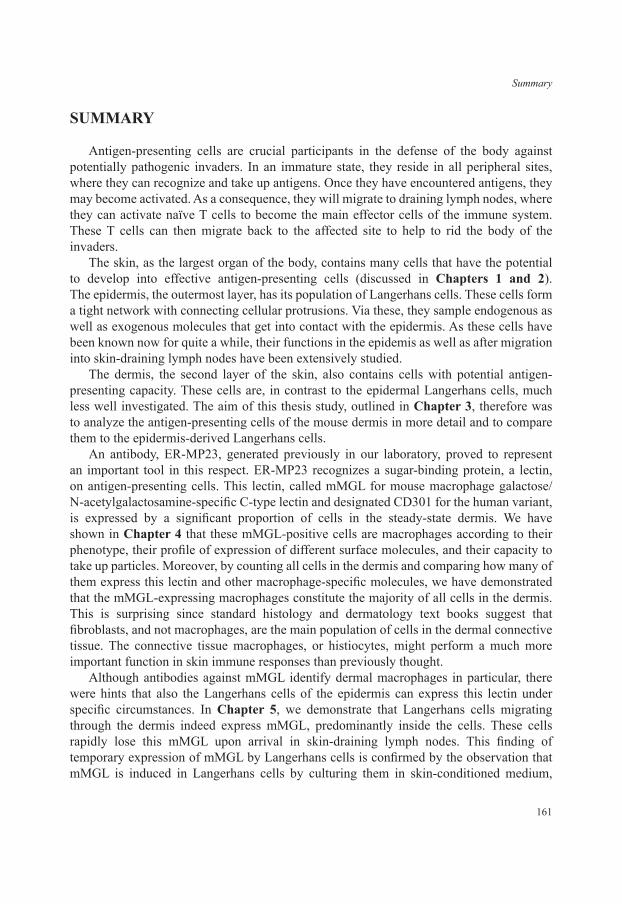
Summary
161
SUMMARY
Antigen-presenting cells are crucial participants in the defense of the body against potentially pathogenic invaders. In an immature state, they reside in all peripheral sites, where they can recognize and take up antigens. Once they have encountered antigens, they may become activated. As a consequence, they will migrate to draining lymph nodes, where they can activate naïve T cells to become the main effector cells of the immune system. These T cells can then migrate back to the affected site to help to rid the body of the invaders.
The skin, as the largest organ of the body, contains many cells that have the potential to develop into effective antigen-presenting cells (discussed in Chapters 1 and 2). The epidermis, the outermost layer, has its population of Langerhans cells. These cells form a tight network with connecting cellular protrusions. Via these, they sample endogenous as well as exogenous molecules that get into contact with the epidermis. As these cells have been known now for quite a while, their functions in the epidemis as well as after migration into skin-draining lymph nodes have been extensively studied.
The dermis, the second layer of the skin, also contains cells with potential antigen-presenting capacity. These cells are, in contrast to the epidermal Langerhans cells, much less well investigated. The aim of this thesis study, outlined in Chapter 3, therefore was to analyze the antigen-presenting cells of the mouse dermis in more detail and to compare them to the epidermis-derived Langerhans cells.
An antibody, ER-MP23, generated previously in our laboratory, proved to represent an important tool in this respect. ER-MP23 recognizes a sugar-binding protein, a lectin, on antigen-presenting cells. This lectin, called mMGL for mouse macrophage galactose/ N-acetylgalactosamine-specific C-type lectin and designated CD301 for the human variant, is expressed by a significant proportion of cells in the steady-state dermis. We have shown in Chapter 4 that these mMGL-positive cells are macrophages according to their phenotype, their profile of expression of different surface molecules, and their capacity to take up particles. Moreover, by counting all cells in the dermis and comparing how many of them express this lectin and other macrophage-specific molecules, we have demonstrated that the mMGL-expressing macrophages constitute the majority of all cells in the dermis. This is surprising since standard histology and dermatology text books suggest that fibroblasts, and not macrophages, are the main population of cells in the dermal connective tissue. The connective tissue macrophages, or histiocytes, might perform a much more important function in skin immune responses than previously thought.
Although antibodies against mMGL identify dermal macrophages in particular, there were hints that also the Langerhans cells of the epidermis can express this lectin under specific circumstances. In Chapter 5, we demonstrate that Langerhans cells migrating through the dermis indeed express mMGL, predominantly inside the cells. These cells rapidly lose this mMGL upon arrival in skin-draining lymph nodes. This finding of temporary expression of mMGL by Langerhans cells is confirmed by the observation that mMGL is induced in Langerhans cells by culturing them in skin-conditioned medium,
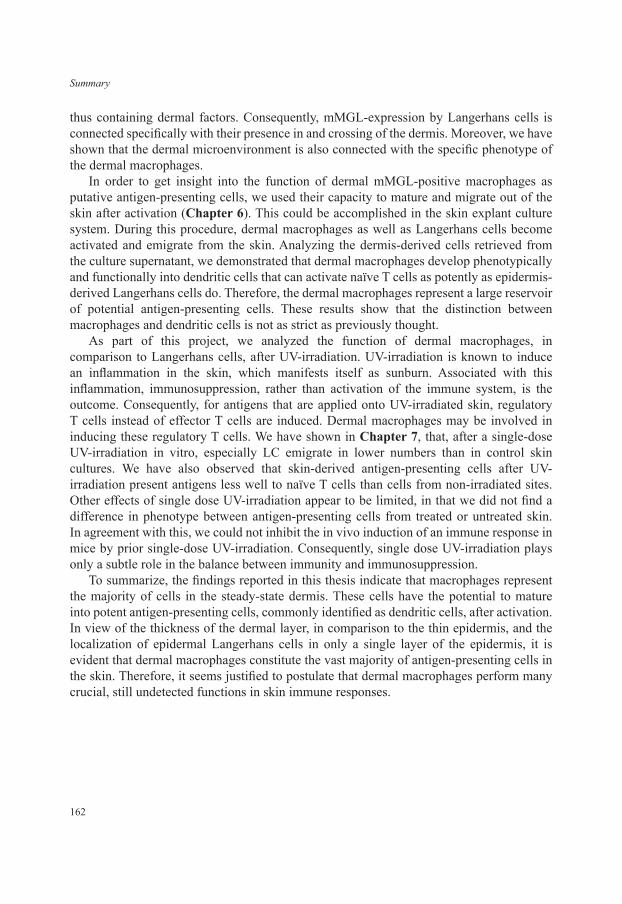
Summary
162
thus containing dermal factors. Consequently, mMGL-expression by Langerhans cells is connected specifically with their presence in and crossing of the dermis. Moreover, we have shown that the dermal microenvironment is also connected with the specific phenotype of the dermal macrophages.
In order to get insight into the function of dermal mMGL-positive macrophages as putative antigen-presenting cells, we used their capacity to mature and migrate out of the skin after activation (Chapter 6). This could be accomplished in the skin explant culture system. During this procedure, dermal macrophages as well as Langerhans cells become activated and emigrate from the skin. Analyzing the dermis-derived cells retrieved from the culture supernatant, we demonstrated that dermal macrophages develop phenotypically and functionally into dendritic cells that can activate naïve T cells as potently as epidermis-derived Langerhans cells do. Therefore, the dermal macrophages represent a large reservoir of potential antigen-presenting cells. These results show that the distinction between macrophages and dendritic cells is not as strict as previously thought.
As part of this project, we analyzed the function of dermal macrophages, in comparison to Langerhans cells, after UV-irradiation. UV-irradiation is known to induce an inflammation in the skin, which manifests itself as sunburn. Associated with this inflammation, immunosuppression, rather than activation of the immune system, is the outcome. Consequently, for antigens that are applied onto UV-irradiated skin, regulatory T cells instead of effector T cells are induced. Dermal macrophages may be involved in inducing these regulatory T cells. We have shown in Chapter 7, that, after a single-dose UV-irradiation in vitro, especially LC emigrate in lower numbers than in control skin cultures. We have also observed that skin-derived antigen-presenting cells after UV-irradiation present antigens less well to naïve T cells than cells from non-irradiated sites. Other effects of single dose UV-irradiation appear to be limited, in that we did not find a difference in phenotype between antigen-presenting cells from treated or untreated skin. In agreement with this, we could not inhibit the in vivo induction of an immune response in mice by prior single-dose UV-irradiation. Consequently, single dose UV-irradiation plays only a subtle role in the balance between immunity and immunosuppression.
To summarize, the findings reported in this thesis indicate that macrophages represent the majority of cells in the steady-state dermis. These cells have the potential to mature into potent antigen-presenting cells, commonly identified as dendritic cells, after activation. In view of the thickness of the dermal layer, in comparison to the thin epidermis, and the localization of epidermal Langerhans cells in only a single layer of the epidermis, it is evident that dermal macrophages constitute the vast majority of antigen-presenting cells in the skin. Therefore, it seems justified to postulate that dermal macrophages perform many crucial, still undetected functions in skin immune responses.

Samenvatting
163
SAMENVATTING
Antigeen-presenterende cellen zijn cruciale cellen in de afweerreactie van het lichaam tegen microbiële pathogenen. Deze cellen bevinden zich in alle perifere organen, waar ze als onrijpe cellen antigeen herkennen en kunnen opnemen. Als ze met antigeen in contact komen, kunnen ze hierdoor geactiveerd raken. Daarop migreren ze naar drainerende lymfeklieren, waar ze naïeve T cellen kunnen activeren tot belangrijke effector cellen van het immuunsysteem. Deze T cellen kunnen remigreren naar de locatie van infectie, waar ze kunnen helpen het antigeen omschadelijk te maken en te helpen opruimen.
De huid, als het grootste orgaan van het lichaam, bevat veel cellen die zich kunnen ontwikkelen tot effectieve antigeen-presenterende cellen (beschreven in hoofdstuk 1 en 2). De opperhuid, of epidermis, als de buitenste laag van de huid, bevat als antigeen-presenterende cellen Langerhanscellen. Deze vormen een dicht netwerk van cellen die via uitlopers met elkaar in verbinding staan. Hiermee verzamelen Langerhanscellen zowel endogene als exogene moleculen waarmee de epidermis in contact komt. Aangezien deze cellen al geruime tijd bekend zijn, zijn hun functies in de epidermis en hun migratie naar huiddrainerende lymfeklieren inmiddels uitgebreid bestudeerd.
De dermis, of lederhuid, als tweede laag van de huid, bevat een eigen populatie cellen die antigeen kunnen presenteren. Over deze cellen is veel minder bekend. Het doel van het onderzoek voor dit proefschrift, beschreven in hoofdstuk 3, was de antigeen-presenterende cellen in de dermis van muizen nader te analyseren en te vergelijken met de Langerhanscellen uit de epidermis.
Een monoclonaal antilichaam, ER-MP23, dat eerder in ons laboratorium was gegenereerd, bleek een belangrijk hulpmiddel in deze studies. ER-MP23 herkent een suiker-bindend eiwit, het lectine mMGL, wat staat voor muis macrofaag galactose/N-acetylgalactosamine-specifieke C-type lectine. De humane variant is ook bekend als CD301. Dit mMGL wordt door veel cellen in de dermis van de gezonde huid tot expressie gebracht. In hoofdstuk 4 worden deze mMGL-positieve cellen op basis van hun immunofenotype geïdentificeerd als macrofagen, dus op basis van hun profiel van oppervlakte-eiwitten, en hun capaciteit om partikels op te nemen. Bovendien hebben wij vastgesteld, door alle cellen in de dermis de tellen, en te bepalen hoeveel hiervan deze en andere karakteristieke macrofaagmerkers tot expressie brengen, dat de mMGL-positieve macrofagen de meerderheid van cellen in de dermis uitmaken. Dit is opmerkelijk, omdat algemeen wordt aangenomen dat de meerderheid van de cellen in het dermale bindweefsel fibroblasten, en niet macrofagen, zijn, zoals ook beschreven wordt in hedendaagse histologie- en dermatologieboeken. Waarschijnlijk spelen bindweefsel macrofagen een belangrijker rol in immuunresponsen in de huid dan tot nu toe is gedacht.
Hoewel het antilichaam tegen mMGL specifiek dermale macrofagen herkent, waren er aanwijzingen dat ook epidermale Langerhans cells dit lectine onder bepaalde omstandigheden tot expressie brengen. In het hoofdstuk 5 hebben wij daarom naar de mMGL expressie door Langerhanscellen gekeken. Daarbij bleek dat Langerhanscellen, met name bij hun migratie door de dermis, mMGL tot expressie brengen, en dit dan voornamelijk binnen de cel.

Samenvatting
164
Ze verliezen deze expressie weer snel na aankomst in de huid-drainerende lymfeklieren. Deze tijdelijke expressie van mMGL door Langerhanscellen hebben wij bevestigd in in vitro experimenten waarbij mMGL expressie werd geïnduceerd door de cellen te kweken in huid-geconditioneerd medium dus in medium, dat een grote hoeveelheid uit de dermis afkomstige factoren bevat. Op basis van deze bevindingen concluderen wij dat mMGL expressie door Langerhanscellen direct gerelateerd is aan hun aanwezigheid in en migratie door de dermis. Verder hebben wij aangetoond dat het specifieke fenotype van de dermale macrofagen te maken heeft met het locale micromilieu.
Om inzicht te krijgen in de functie van dermale macrofagen als potentiële antigeen-presenterende cellen, hebben wij gebruik gemaakt van hun vermogen om na activering uit te rijpen en uit de huid te migreren (hoofdstuk 6). Door huidbiopten in kweek te brengen, krijgen zowel dermale macrofagen als Langerhans-cellen een sterke activeringsprikkel waardoor ze uit de gekweekte huid emigreren. Door de dermale cellen uit het kweekmedium te oogsten en ze met Langerhanscellen afkomstig uit de epidermis te vergelijken, hebben wij kunnen aantonen dat dermale macrofagen uitrijpen tot fenotypisch en functioneel potente dendritische cellen die een vergelijkbare capaciteit hebben als Langerhanscellen om naïeve T cellen te activeren. Hieruit concluderen wij dat de dermale macrofagen een groot reservoir van antigeen-presenterende cellen vormen. Bovendien blijkt uit deze studies dat het onderscheid tussen macrofagen en dendritische cellen niet zo strikt is als algemeen wordt aangenomen.
Als onderdeel van ons onderzoek hebben wij een vergelijkende studie gedaan naar de invloed van UV bestraling op de functie van dermale macrofagen en epidermale Langerhans cellen. Van UV straling is bekend dat het een ontstekingsreactie in de huid induceert die als zonnebrand waarneembaar is. Het bijzondere van deze ontsteking is dat deze niet een activatie van het adaptieve immuunsysteem teweegbrengt, maar juist een suppressie ervan. Als een antigeen wordt aangebracht op de ontstoken huid na UV bestraling, dan worden er geen effector T cellen geactiveerd, maar regulatoire T cellen. Dermale macrofagen zouden in het actveren van deze regulatoire T cellen een rol kunnen spelen. In hoofdstuk 7 laten wij zien dat na één enkele UV bestraling in vitro minder cellen, en dan met name Langerhanscellen, uit gekweekte huidbiopten migreren dan uit biopten die niet bestraald zijn. Bovendien zijn antigeen-presenterende cellen uit UV bestraalde huid minder goed in staat om naïeve T cellen te activeren. Andere effecten van de enkelvoudige UV bestraling bleken tamelijk beperkt te zijn. Zo hebben wij geen verschil in fenotype kunnen aantonen tussen cellen uit kweken van bestraalde en onbestraalde huid. In overeenstemming met dit resultaat bleek ook in muizen de inductie van een immuunrespons niet te worden onderdrukt door één enkele UV bestraling. Hieruit hebben wij geconcludeerd dat enkelvoudige UV bestraling slechts een beperkte rol speelt in de balans tussen immuunactivering en immuunsuppressie.
De bevindingen in dit proefschrift samenvattend kunnen wij zeggen dat macrofagen de meerderheid van de cellen in de gezonde dermis uitmaken. Deze cellen hebben de mogelijkheid om na activatie uit te rijpen tot potente antigeen-presenterende cellen, met het fenotype en de functie van dendritische cellen. Als wij de dikte van de dermale laag

Samenvatting
165
in ogenschouw nemen, in vergelijking met de dunne epidermis, en daarbij betrekken dat epidermale Langerhanscellen slechts in één laag van de epidermis voorkomen, dan is evident dat dermale macrofagen een veel grotere populatie potentiële antigeen-presenterende cellen in de huid vormen. Daarom is het aannemelijk dat dermale macrofagen een veel belangrijker functie hebben in huid-gerelateerde immuunresponsen dan tot dusver wordt gedacht.

Abbreviations
166
ABBREVIATIONS
APC antigen-presenting cellsBMDC bone marrow-derived dendritic cellsCHS contact hypersensitivitycis-UCA cis-urocanic acidCLE cutaneous lupus erythematosusCR cysteine-richCRD carbohydrate recognition domainCTLD C-type lectin-like carbohydrate recognition domainDC dendritic cellsDdDC dermally derived DCDETC dendritic epidermal T cellsDTR diphtheria-toxin receptorEGF epidermal growth factorFcR receptor for constant part of Ig moleculeFNII fibronectin type twoFXIIIa coagulation factor XIII-AGa1NAc N-acetylgalactosamineGFP green-fluorescent proteinGlcNAc N-acetylglucosaminehMGL human macrophage galactose-/N-acetylgalactosamine-specific C-type lectinLC Langerhans cellsMGL macrophage galactose-/N-acetylgalactosamine-specific C-type lectinmMGL mouse macrophage galactose-/N-acetylgalactosamine-specific C-type lectinMPS mononuclear phagocyte system MR mannose receptorNO nitric oxidePAF platelet-activating factorPBMC peripheral blood mononuclear cellsPGE2 prostaglandin E2PLE polymorphic light eruptionPMN polymorphonuclear neutrophilsRES reticuloendothelial systemSSR solar-simulated UV radiationT4N5 T4 endonucleaseTLR Toll-like receptorsTM transmembranetTG tissue transglutaminasevit D3 vitamin D3

167
Acknowledgements
ACKNOWLEDGEMENTS
I always thought that writing these acknowledgements would be the most difficult part of this thesis. Now that I’m actually writing them in a later stage of the correction phase, I recognize that they flow quite naturally out of my hands.
First of all, I would like to thank Pieter Leenen for supervising me during the four and a half years of practical and writing work, and during the subsequent two and a half years of correction work, where most of the time I was out of the lab. Pieter, I know it wasn’t always easy with me, with my lack of patience, and my complicated way to see the world, but finally we managed to finish this thesis, and I know it’s as much a result of your efforts as of mine. Helemaal bedankt ervoor.
Rob Benner, I would like to thank you for giving me the opportunity to work in this department, and consequently to go in the opposite direction of Desiderius Erasmus, who died and is buried in Basel, Switzerland.
Next, I would also like to thank the two other group leaders of the unit, Hemmo Drexhage and Marjan Versnel, for your input and discussions during the countless unit meetings where I was allowed to present. Starting from the first unit meeting, which took place in the small library on the 9th floor, where nowadays about ten -80º freezers stand, and where I had to lift my hand as only participant of a non-Dutch speaking background, to the last one, where the discussions after my presentations mostly turned about the exact difference between macrophages and dendritic cells, these meetings helped me to sharpen my vision of the world of immunology, and the troubles one faces exploring it.
I would also like to thank our collaborators in Innsbruck, Patti Stoitzner and Niki Romani, for their contributions.
Moreover, I would like to thank the countless people in the lab over the years, starting with Tanja, Anouk, Marjolijn, Berlinda, Adri, a.k.a. A3, Katarina, Annabrita, Femke, Rogier, working first in that part of the lab until changing afterwards to the other side, Rudi, Sabine and Gemma in 2001, to the composition of the lab when I left in 2006, still with A3 and Berlinda, but now with Dennis and Joey, Rebecca, Manon, Jojanneke, Lonneke, Corine, Harm and Roos. I don’t even want to try to put now all the people that worked there in between, and beyond, as I am sure I would forget a lot of them. Nevertheless, I want to thank all of you for your diverse input. I especially want to mention A3, for all the help with handling mice, especially irradiating them, and Dennis Tax, who, under A3’s guidance, did the work of almost a whole chapter in this thesis all by himself.
I owe also thanks to the people that contributed in the generation of this thesis with their infrastructural support; Tar van Os, for all the nice pictures and illustrations, and the secretaries, Daniëlle Korpershoek, Marcia IJdo-Reintjes and Wendy Netten for the lay-out and all the other work.
Next, I want to thank those people that made my stay especially at the beginning of the studies a little bit more bearable, Dariusz Stępniak, for the countless Chicken Curry meals, Onyebuchi Arah for the plentiful philosophical talks, Johannes Meerman, for the help with the numerous moving efforts, of which even once a car became victim, Ronald Bakker, for showing me Willemstad, even though it was not the one in Curaçao, and Etienne Bucher and Danijela Vrbnjak for sharing with me the Swiss heritage.

Acknowledgements
168
I would also like to thank Diederik Deursen for often going to lunch with me in the later stages of the studies.
Beyond, I have to thank people in later stages of my career for their support, Georges Lahoud and Cuiping Liu in Philadelphia, and first Daniel Rhymer and later Shiva Tyagarajan, Himanish Ghosh and Amrita Madhusudan in Zürich.
And last but not least, there are two people that merit special acknowledgements. Hui Wan, for contributing to this thesis, not the least of it setting most of the references into Endnote. And my mother, Anna Dupasquier for her continuous support, and often letting me use her computer on which, among others, also these lines were written. Thank you all!

169
Curriculum vitae
CURRICULUM VITAE
Marcel Dupasquier Date & Place of Birth: February, 25th 1977 in Basel, Switzerland
EDUCATION and WORK EXPERIENCE 1984 - 1988 Primary School at Christoph Merian Schulhaus, Basel, Switzerland 1988 - 1990 Secondary School at Hohlbein Gymnasium, Basel, Switzerland 1990 - 1996Secondary School at Mathematisches-naturwissenschaftliches Gymnasium (MNG), Basel, Switzerland 1996 - 2001 Universität Basel: Study of Biology II, specialization in biochemistry 1999 - 2001 Diploma work at the Friedrich Miescher Institute (FMI), Basel, SwitzerlandResearch group Prof. Witold Filipowicz.Diploma thesis: “Characterization of UBA2, a plant hnRNP-like protein” July 2001 - April 2006PhD Student at the Department of Immunology, Erasmus Universiteit Rotterdam (EUR), Rotterdam, The NetherlandsResearch group Dr. Pieter J.M. Leenen Research topic: “Role of dermal mononuclear phagocytes in the induction of UV-mediated immunosuppression” April 2006 - August 2007Postdoctoral fellow at the Department of Biochemistry and Molecular Biology, ThomasJefferson University (TJU), Philadelphia, USAResearch group Prof. Ya-Ming Hou Research topic: “Kinetic characterization of the E. coli Class II CCA-adding enzyme”
November 2007 - June 2008Postdoctoral fellow at the Institute of Physiology, Universität Zürich, SwitzerlandResearch group Prof. Thierry HennetResearch topic: “Glycosylation by virus-encoded glycosyltransferases”

Curriculum vitae
170
July 2008 - presentResearch assistant at the Institute of Pharmacology and Toxicology, Universität Zürich, Zürich, Switzerland Research group Dr. Shiva Tyagarajan, Prof. Jean-Marc FritschyResearch topic: “The role of posttranslational modifications in the aggregation of gephyrin ininhibitory synapses”

171
List of publications
LIST OF PUBLICATIONS
Lambermon MH, Fu Y, Wieczorek Kirk DA, Dupasquier M, Filipowicz W and Lorković ZJ. UBA1 and UBA2, two proteins that interact with UBP1, a multifunctional effector of pre-mRNA maturation in plants. Mol Cell Biol. Jun 2002, 22(12), 4346-4357.
Dupasquier M, Stoitzner P, van Oudenaren A, Romani N and Leenen PJM. Macrophages and dendritic cells constitute a major subpopulation of cells in the mouse dermis. J Invest Dermatol. Nov 2004, 123(5), 876-879. Wan H and Dupasquier M. Dendritic cells in vivo and in vitro. Cell Mol Immunol. Feb 2005, 2(1), 28-35.
Dupasquier M, Stoitzner P, Wan H, Cerqueira D, van Oudenaren A, Voerman JS, Denda-Nagai K, Irimura T, Raes G, Romani N and Leenen PJM. The dermal microenvironment induces the expression of the alternative activation marker CD301/mMGL in mononuclear phagocytes, independent of IL-4/IL-13 signaling. J Leukoc Biol. Oct 2006, 80(4), 838-849.
Dupasquier M, Kim S, Halkidis K, Gamper H and Hou Y-M. tRNA integrity is a prerequisite for rapid CCA addition: implication for quality control. J Mol Biol. Jun 2008, 379(3), 579-588.
Dupasquier M, Prens EP and Leenen PJM. Dermal mononuclear phagocytes. In: Recent Advances in Skin Immunology, Saeland S. ed., Research Signpost Publisher, 2008, Chapter 3, 75-104.

Appendix
172
APPENDIX
Chapter 4: Figure 1
Figure 1. The majority of dermal interstitial cells express mononuclear phagocyte markers.Back skin sections were stained for mMGL / ER-MP23 (red) and CD45 (green) (A) or mMGL (red) and F4/80 (green) (B). Sections were counterstained with DAPI, showing nuclei in blue. (A) About 60% of all dermal cells express mMGL. All mMGL+ cells also express CD45. About 10% of the CD45-positive cells are mMGL-negative. (B) Staining for mMGL and F4/80 virtually coincided. Note the gradient of increasing F4/80 expression towards the epidermal side, which could be observed occasionally. Scale bar = 50 µm for all pictures. See appendix for full colour pictures.

173
Appendix
Figure 2. Dermal macrophages from different sites show differential MHC class II expression.Back skin sections (A) and ear skin sections (B) were stained for mMGL (red) and for MHC class II (green). Sections were counterstained with DAPI. (A) In the back skin, virtually all mMGL-positive cells coexpress MHC class II. (B) Similar to the back skin, the ear dermis contains about 60% interstitial cells expressing mMGL. However, only about one third of these cells coexpresses MHC class II molecules. These mMGL / MHC class II double-positive cells are arranged in clusters. Scale bar = 50 µm for all pictures.
Chapter 4: Figure 2

Appendix
174
Figure 3. mMGL-positive dermal cells are phagocytic.DiI-labeled liposomes (red label) were injected into ear dermis. Four hours later, animals were killed, ears cut off, frozen, sectioned and stained for mMGL (green). Note that the vast majority, but not all mMGL-positive cells, has taken up liposomes, whereas mMGL-negative cells and keratinocytes remained free of labeling. Scale bar = 50 µm for all pictures.
Chapter 4: Figure 3

175
Appendix
Figure 4. Dermal DC constitute a small, distinct mononuclear phagocyte population.Back skin sections were stained for CD11c (red) and Langerin (green) (A) or CD11c (red) and mMGL (green) (B). Sections were counterstained with DAPI. (A) About 7% of all dermal cells express CD11c but does not express Langerin. These cells thus represent genuine DDC (marked with thin arrows). Note that the CD11c staining is too weak to let CD11c / Langerin-double-positive mLC (marked with thick arrows) appear yellow. (B) Double-staining for CD11c and mMGL shows that most CD11c-positive cells coexpress mMGL (arrow). Thus, most if not all DDC express mMGL. The red labeled cell marked with an asterisk is part of a sebaceous gland and displays autofluorescence rather than specific labeling. Scale bar = 50 µm for all pictures.
Chapter 4: Figure 4

Appendix
176
Figure 5. Dermal macrophages show different degrees of heterogeneity.Back skin sections were double-stained for mMGL (red) and other macrophage markers in green: CD11b (A), MOMA-1 (B), ER-HR3 (C), mSIGN-R1 / ER-TR9 (D). Sections were counterstained with DAPI. (A) All mMGL-positive cells coexpress CD11b. Separate CD11b single-positive cells may represent dermal mast cells, or occasional monocytes, granulocytes or activated T cells. (B) Staining for MOMA-1 divides DMφ into two subpopulations: cells lying deeper in the dermis express MOMA-1, whereas cells directly underneath the epidermis do not. (C) ER-HR3 also divides DMφ into two subpopulations, but with no clear localization pattern. A small number of ER-HR3 single-positive cells presumably represent mLC. (D) mSIGN-R1 / ER-TR9 labels a small subpopulation of DMφ and DDC. The two arrows mark two mMGL / mSIGN-R1 double-positive cells. Scale bar = 50 µm for all pictures.
Chapter 4: Figure 5

177
Appendix
overlay mMGL Langerin DAPI
A
B
C
Figure 3. LC migrating through the dermis show an upregulated expression of mMGL.Steady-state back skin sections from C57BL/6J mice (A, B) or IL-4Rα knock-out mice (C) were stained for Langerin (green) and mMGL (red) and counterstained for DAPI (blue). (A) LC in the epidermis do not express mMGL at detectable level. Note the high background fluorescence of the keratin layers. (B) In contrast, around one third of all dermal LC, thus LC that are migrating to skin-draining lymph nodes, were found to be mMGL-positive. (C) Comparable to dermal macrophages in IL-4Rα knock-out mice, also dermal LC express mMGL.
Chapter 5: Figure 3

Appendix
178
Langerin mMGL
Figure 6. mMGL colocalizes significantly with Langerin inside mature LC. Whole-skin emigrants were allowed to adhere on slides. Thereafter, they were fixed and permeabilized, stained for mMGL and Langerin and analyzed by confocal microscope. Note the colocalization of Langerin and mMGL in the cytoplasm of LC.
Chapter 5: Figure 6

179
Appendix
Lang
erin
FL-1 FITC
FITC+ LC
FITC- LC
untreated
intracellular mMGL
FITC treatedA B
B
T
C D
E
LangerinmMGLDAPI
Figure 7. Only recent immigrant LC in skin-draining lymph nodes are mMGL-positive.Skin-draining lymph nodes from untreated mice (A, C) or from mice that had been treated with FITC solution 24h prior to their euthanization (B, D) were collected and homogenized to obtain a single cell suspension. Thereafter, cells were stained intracellularly for Langerin and mMGL. LC in skin-draining lymph nodes from untreated mice or FITC– LC (thus LC, that had left the skin more than 24h earlier) from FITC-treated mice showed negligible intracellular mMGL levels. In contrast, recently immigrated FITC+ LC from FITC-treated mice still showed a significant mMGL expression. The increased fluorescence level of FITC– LC, compared to untreated mice, is probably explained by the uptake of soluble FITC transported via afferent lymph. The vertical line represents the maximum staining of isotype control antibodies. (E) Skin-draining lymph node sections from an untreated mouse were stained for Langerin (green) and mMGL (red) and counterstained for DAPI (blue). Note that no double positive cells are present. B = B cell area, T = T cell area.
Chapter 5: Figure 7

Appendix
180
FIT
CF
ITC
1 day after FITCgate FITC+ cells
gate FITC+ cells2 days after FITC
R2
gate R2
intracellular Langerin
2.96% 0.44%
0.18%
1.23% 0.26%
0.19%
0.34% 1.14%
0.74%
0.73% 2.66%
0.58%
5.7% 7.8%
78.6%7.9%
11.7% 5.2%
71.5%11.5%
intracellular Langerin
intracellular mMGL
intracellular mMGL
intracellular mMGL
intracellular mMGL
intracellular mMGL
neg
CD80
mMGL CD3 DAPI overlay
CD
86
CD11c
CD
11b
FIT
C
intr
acel
lula
r La
nger
in
FIT
C
intr
acel
lula
r La
nger
in
CD40
MH
C c
lass
II
A
B
C D
0.0%
0h
4h
24h
48h
2.5%
percentage mMGL+ cellsin the total LN suspension
5.0% 7.5%
FITC+
FITC-
Figure 5. Dermis-derived mMGL+ DC are present in skin-draining lymph nodes, cluster with lymphocytes and are major hapten-transporting cells.(A) Skin-draining lymph node single cell suspensions were prepared from untreated mice and labeled for intracellular mMGL and for other DC markers. mMGLhi cells were observed and express a similar mature DC phenotype as did in vitro skin emigrants. (B) Cytospins of non-enzymatically treated lymph node single cell suspensions were prepared and stained for mMGL (red) and for CD3 (green), with DAPI counterstaining (blue). mMGL+ cells were often discovered in close interaction with CD3+ T cells but also with CD3– B cells. The selected cluster shows a representative mMGLhi cell surrounded by lymphocytes. (C) 24h and 48h after application of FITC in adjuvant onto the back skin, skin-draining lymph node single cell suspensions were obtained and stained for Langerin and mMGL. Note that the vast majority of FITC+ cells, even after 48h express intracellular mMGL (78% of all FITC+ cells after 24h, 77% after 48h), whereas only a minority of them express Langerin (13% after 24h, 17% after 48h). This experiment was performed twice with virtually identical results. (D) Percentages of mMGL+ FITC+ and mMGL+ FITC– cells in the total lymph node single cell suspension before, and 4h, 24h and 48h after FITC application. Note that 4h after FITC application, only few mMGL+ cells have taken up soluble FITC that has diffused into the lymph nodes with incoming lymph.
Chapter 6: Figure 5