Development and Characterization of Somatic Hybrids of Ulva reticulata Forsskål (×)...
15
ORIGINAL RESEARCH ARTICLE published: 29 January 2015 doi: 10.3389/fpls.2015.00003 Development and Characterization of Somatic Hybrids of Ulva reticulata Forsskål (×) Monostroma oxyspermum (Kutz.)Doty Vishal Gupta , Puja Kumari and CRK Reddy* Seaweed Biology and Cultivation Group, Discipline of Marine Biotechnology and Ecology, CSIR-Central Salt and Marine Chemicals Research Institute, Bhavnagar, India Edited by: Benedicte Charrier, Centre National de la Recherche Scientifique, France Reviewed by: Thomas Wichard, Friedrich Schiller University Jena, Germany Wen-Wu Guo, Huazhong Agricultural University, China *Correspondence: CRK Reddy, Seaweed Biology and Cultivation Group, Discipline of Marine Biotechnology and Ecology, CSIR-Central Salt and Marine Chemicals Research Institute, GB Marg, Bhavnagar 364002, India e-mail: [email protected] Ulvophycean species with diverse trait characteristics provide an opportunity to create novel allelic recombinant variants. The present study reports the development of seaweed variants with improved agronomic traits through protoplast fusion between Monostroma oxyspermum (Kutz.) Doty and Ulva reticulata Forsskål. A total of 12 putative hybrids were screened based on the variations in morphology and total DNA content over the fusion partners. DNA-fingerprinting by inter simple sequence repeat (ISSR) and amplified fragment length polymorphism (AFLP) analysis confirmed genomic introgression in the hybrids. The DNA fingerprint revealed sharing of parental alleles in regenerated hybrids and a few alleles that were unique to hybrids. The epigenetic variations in hybrids estimated in terms of DNA methylation polymorphism also revealed sharing of methylation loci with both the fusion partners. The functional trait analysis for growth showed a hybrid with heterotic trait (DGR% = 36.7 ± 1.55%) over the fusion partners U. reticulata (33.2 ± 2.6%) and M. oxyspermum (17.8 ± 1.77%), while others were superior to the mid-parental value (25.2 ± 2.2%) (p < 0.05). The fatty acid (FA) analysis of hybrids showed notable variations over fusion partners. Most hybrids showed increased polyunsaturated FAs (PUFAs) compared to saturated FAs (SFAs) and mainly includes the nutritionally important linoleic acid, α-linolenic acid, oleic acid, stearidonic acid, and docosahexaenoic acid. The other differences observed include superior cellulose content and antioxidative potential in hybrids over fusion partners. The hybrid varieties with superior traits developed in this study unequivocally demonstrate the significance of protoplast fusion technique in developing improved varients of macroalgae. Keywords: heterosis, hybrid, Monostroma, protoplast, Ulva INTRODUCTION Marine macroalgae (seaweeds) consist of an assemblage of geneti- cally, morphologically and functionally diverse photoautotrophic species that carry out key ecosystem functions in the marine envi- ronment. Seaweeds are historically consumed as human food in several Asian Countries and as raw material for extraction of industrially import phycocolloids (agar, carrageenan and algi- nate) and recently as phycosupplements, nutraceutical, cosmetics, bioactive substances etc. (Holdt and Kraan, 2011). The benefi- cial utilization of seaweed resources for humankind has provided impetus for domestication of economically important seaweeds leading to their commercial cultivation. There has been a substan- tial growth in the production of seaweeds from 3.8 million fresh tons per annum in 1998 to about 25 million fresh tons in 2012 (FAO, 2014). A significant proportion of seaweed produced, rep- resenting about 76% of the tonnage of total harvest (88% of the value of entire seaweed industry) is consumed as human food or animal feed (FAO, 2012). The emergence of seaweed aquaculture as a promising seaweed industry has opened up avenues for devel- oping varieties with agronomically improved traits for higher economic gains. However, genetic improvement programmes are seriously lacking in seaweeds and conventional practices were largely resorted to selection of clones with useful traits from wild populations followed by development of homogenous lines (Robinson et al., 2013). The continuous rigorous selection from the same progeny on long run may deplete genetic diversity caus- ing a gradual decline in adaptability to changing environments and eventually affecting the vigor of economic traits. Selective breeding, though applied for developing elite vari- eties, is largely restricted to a few seaweed genera such as Porphyra and Laminaria. For example, only 30 cultivars of Porphyra have so far been developed ever since its life cycle and control of repro- duction became known in 1949 (Robinson et al., 2013). Similarly, a few cultivars of Laminaria such as Danhai No. 1 (Ou et al., 1983), Danza No. 10 (Fang et al., 1985), Danhai No. 901 (Zhang et al., 2001), and recently the Dongfang No. 2 (Li et al., 2007) have been developed since the first gametophyte cloning and gameto- phyte clone hybridizing methods were developed in 1970 (Fang et al., 1978; Zhou and Wu, 1998). Nevertheless, selective breed- ing has remained inconclusive for other seaweed groups such www.frontiersin.org January 2015 | Volume 6 | Article 3 | 1
Transcript of Development and Characterization of Somatic Hybrids of Ulva reticulata Forsskål (×)...

ORIGINAL RESEARCH ARTICLEpublished: 29 January 2015
doi: 10.3389/fpls.2015.00003
Development and Characterization of Somatic Hybrids ofUlva reticulata Forsskål (×) Monostroma oxyspermum(Kutz.)DotyVishal Gupta , Puja Kumari and CRK Reddy*
Seaweed Biology and Cultivation Group, Discipline of Marine Biotechnology and Ecology, CSIR-Central Salt and Marine Chemicals Research Institute, Bhavnagar,India
Edited by:
Benedicte Charrier, Centre Nationalde la Recherche Scientifique, France
Reviewed by:
Thomas Wichard, Friedrich SchillerUniversity Jena, GermanyWen-Wu Guo, Huazhong AgriculturalUniversity, China
*Correspondence:
CRK Reddy, Seaweed Biology andCultivation Group, Discipline ofMarine Biotechnology and Ecology,CSIR-Central Salt and MarineChemicals Research Institute,GB Marg, Bhavnagar 364002, Indiae-mail: [email protected]
Ulvophycean species with diverse trait characteristics provide an opportunity to createnovel allelic recombinant variants. The present study reports the development of seaweedvariants with improved agronomic traits through protoplast fusion between Monostromaoxyspermum (Kutz.) Doty and Ulva reticulata Forsskål. A total of 12 putative hybridswere screened based on the variations in morphology and total DNA content over thefusion partners. DNA-fingerprinting by inter simple sequence repeat (ISSR) and amplifiedfragment length polymorphism (AFLP) analysis confirmed genomic introgression in thehybrids. The DNA fingerprint revealed sharing of parental alleles in regenerated hybrids anda few alleles that were unique to hybrids. The epigenetic variations in hybrids estimatedin terms of DNA methylation polymorphism also revealed sharing of methylation loci withboth the fusion partners. The functional trait analysis for growth showed a hybrid withheterotic trait (DGR% = 36.7 ± 1.55%) over the fusion partners U. reticulata (33.2 ±2.6%) and M. oxyspermum (17.8 ± 1.77%), while others were superior to the mid-parentalvalue (25.2 ± 2.2%) (p < 0.05). The fatty acid (FA) analysis of hybrids showed notablevariations over fusion partners. Most hybrids showed increased polyunsaturated FAs(PUFAs) compared to saturated FAs (SFAs) and mainly includes the nutritionally importantlinoleic acid, α-linolenic acid, oleic acid, stearidonic acid, and docosahexaenoic acid. Theother differences observed include superior cellulose content and antioxidative potentialin hybrids over fusion partners. The hybrid varieties with superior traits developed inthis study unequivocally demonstrate the significance of protoplast fusion technique indeveloping improved varients of macroalgae.
Keywords: heterosis, hybrid, Monostroma, protoplast, Ulva
INTRODUCTIONMarine macroalgae (seaweeds) consist of an assemblage of geneti-cally, morphologically and functionally diverse photoautotrophicspecies that carry out key ecosystem functions in the marine envi-ronment. Seaweeds are historically consumed as human food inseveral Asian Countries and as raw material for extraction ofindustrially import phycocolloids (agar, carrageenan and algi-nate) and recently as phycosupplements, nutraceutical, cosmetics,bioactive substances etc. (Holdt and Kraan, 2011). The benefi-cial utilization of seaweed resources for humankind has providedimpetus for domestication of economically important seaweedsleading to their commercial cultivation. There has been a substan-tial growth in the production of seaweeds from 3.8 million freshtons per annum in 1998 to about 25 million fresh tons in 2012(FAO, 2014). A significant proportion of seaweed produced, rep-resenting about 76% of the tonnage of total harvest (88% of thevalue of entire seaweed industry) is consumed as human food oranimal feed (FAO, 2012). The emergence of seaweed aquacultureas a promising seaweed industry has opened up avenues for devel-oping varieties with agronomically improved traits for higher
economic gains. However, genetic improvement programmes areseriously lacking in seaweeds and conventional practices werelargely resorted to selection of clones with useful traits fromwild populations followed by development of homogenous lines(Robinson et al., 2013). The continuous rigorous selection fromthe same progeny on long run may deplete genetic diversity caus-ing a gradual decline in adaptability to changing environmentsand eventually affecting the vigor of economic traits.
Selective breeding, though applied for developing elite vari-eties, is largely restricted to a few seaweed genera such as Porphyraand Laminaria. For example, only 30 cultivars of Porphyra haveso far been developed ever since its life cycle and control of repro-duction became known in 1949 (Robinson et al., 2013). Similarly,a few cultivars of Laminaria such as Danhai No. 1 (Ou et al.,1983), Danza No. 10 (Fang et al., 1985), Danhai No. 901 (Zhanget al., 2001), and recently the Dongfang No. 2 (Li et al., 2007) havebeen developed since the first gametophyte cloning and gameto-phyte clone hybridizing methods were developed in 1970 (Fanget al., 1978; Zhou and Wu, 1998). Nevertheless, selective breed-ing has remained inconclusive for other seaweed groups such
www.frontiersin.org January 2015 | Volume 6 | Article 3 | 1

Gupta et al. Development of seaweed hybrids
as agarophytes, carragenophytes and green seaweeds. Attemptsto cross different Gracilaria species (Bird and McLachlan,1982; Plastino and De Oliveira, 1988) and Ulva species(Hiraoka et al., 2003, 2011) failed due to sexual incompatibilitybarriers.
Somatic hybridization through protoplast fusion is a usefultechnique that facilitates recombination of economically impor-tant traits by harnessing the natural genetic diversity from phylo-genetically distant species (Hodge et al., 2010). This technique hasbeen extensively applied in terrestrial plants and showed unprece-dented success in developing numerous agricultural varietiesimproved for either productivity or resistance to biotic/abioticfactors (Davey et al., 2005; Li et al., 2014; review Liu and Xia,2014). However, this area of research for seaweeds lags far behindthat of land plants despite considerable success in protoplast iso-lation and regeneration in numerous seaweed species (reviewedin Reddy et al., 2008). In the past, a few attempts have been madeto develop somatic hybrids of Porphyra but most failed to regen-erate hybrid plants. Nevertheless, a few succeeded in developingchimeric thalli with no significant benefits. Most importantly,these studies lacked the characterization of regenerated hybrids ata genetic level to confirm the genetic recombination (reviewed inReddy et al., 2008). Thus, this field remained largely inconclusiveto date.
This study aimed at development of hybrid varieties withimproved functional traits through intergeneric protoplast fusionbetween Monostroma oxyspermum (Kutz.) Doty and Ulva retic-ulata Forsskål. These Ulvophycean species are cultivated at acommercial scale in East Asian countries for utilization in variousfood preparations. In recent times, western countries have alsoaccepted the utility of these species in seafood cuisines (Mouritsenet al., 2013). Most recently, the genus Ulva has also been stronglyadvocated as potential source of non-lignocellulosic feedstockfor production of renewable fuels along with other associatedhigh value chemicals (Trivedi et al., 2013). Therefore, it is ofimmense interest to highly desirable to generate genetic variantsof Ulvophycean species by harnessing the prevalent vast naturalgenetic diversity.
MATERIALS AND METHODSSELECTION OF SEAWEED SPECIESThe species Monostroma oxyspermum (Kützing) Doty and Ulvareticulata Forsskål were selected for development of somatichybrids through protoplast fusion. Henceforth, these two speciesare mentioned as “fusion partners” in this manuscript. Thesespecies were selected based on the higher growth rate of U. reticu-lata and nutritional values of M. oxyspermum. The thalli of M.oxyspermum were collected from mangrove swamps at Achra,central west coast of India (16◦ 11′N, 73◦ 26′E) and U. reticu-lata was collected from Kalubhar Island, north western coast ofIndia (22◦ 29′N, 69◦ 37′E). The thalli were brought to the lab-oratory under cool conditions in a thermos flask. Young thalli ofeach species were cleaned thoroughly with autoclaved seawater forremoval of dirt and adhering particles. The cleaned material ofeach species was maintained in unialgal culture in the laboratory.The unialgal culture was established by growing the algal thalli insterile enriched seawater media (Provasoli, 1968) supplementedwith GeO2(10 mgL−1) for a week under white fluorescent lamps
of irradiance intensity about 15 μmole photon m−2 s−1 with a12: 12 h light:dark photoperiod. During this period, the culturemedia was changed every 2 days.
PROTOPLAST ISOLATION AND FUSIONAxenic culture of algal thalli for both the fusion partner speciesand subsequent protoplast isolation from them was carried outfollowing the protocol described by Krishnakumar et al. (1999)and Reddy et al. (2006). Protoplasts from both species were sus-pended in seawater in a 1:1 ratio (1.0 × 10⁵ cellsml−1) and werewashed twice with electro-fusion buffer [0.8 M mannitol, 0.2 mMtris (hydroxymethyl) amino-methane, 1 mM CaCl2 and 1 mMMgCI2 in distilled water, adjusted to pH 7.5]. An aliquot of 100 μlprotoplast mix was placed between the two electrodes (1 mmspacing) in a fusion chamber (FTC-2, Shimadzu Co, Japan) andallowed to settle for few minutes prior to the start of the elec-trofusion. The electric field-mediated protoplast fusion was per-formed using a Somatic hybridizer SSH-2 (Shimadzu Co., Japan).Protoplasts were initially aligned into short chains, preferablypairs of 2 to4 protoplasts in an alternating current (AC) field andsubsequently fused by the application of a single short-durationdirect current (DC) pulse. The alignment of the protoplasts onapplication of AC voltage was optimized by varying the voltagefrom 10 to 40 V for different durations, from 10 to 25 s. The fusionfrequency was also optimized by determining the optimum DCpulse voltage and pulse width. For this, the DC pulse voltage wasvaried from 100 to 350 V and pulse width was varied from 10to 60 μs.
CULTURE OF HETEROPLASMIC FUSION PRODUCTSThe heteroplasmic fusion products were identified based on thepresence of two morphologically distinct chloroplasts in a singlecell and the larger size of the cell compared to unfused protoplasts.Presumptive hybrid products were immediately transferred to aseparate Petri-dish using glass pipette and cultured in seawatercontaining 0.4 M mannitol in dim light of irradiance 10 μmolphoton m−2 s−1 using day light fluorescent lamps with 12:12L:D photoperiod at 20 ± 1◦C for 2 days and later transferredto increased irradiance of 25 μmol photon m−2 s−1. After settle-ment of presumptive hybrid products, the medium was replacedwith sterile seawater. The cellular division and regeneration wasmonitored at regular time intervals. After 15 days of culture, whenpresumptive hybrids had developed to many-celled stages, seawa-ter was supplemented with PES medium (Provasoli, 1968). Afterattainment of thalli around 1 cm in length, plantlets were trans-ferred to round flat bottom aerated flasks and cultured under thesame conditions. The culture medium was replenished frequentlyto sustain cell division and growth.
CHARACTERIZATION OF THE FUSION PRODUCTSThe fusion products obtained were first screened based on theirmorphological distinctiveness compared to fusion partners, fol-lowed by ploidy analysis and molecular level characterization.
PLOIDY ANALYSIS USING CONFOCAL MICROSCOPYThalli of each regenerant were sectioned and processed for ploidyanalysis using confocal microscopy (Shimizu et al., 2008; Zittaet al., 2012). Briefly, the sections of thalli were incubated with 4,6-diamidino-2-phenylindole (DAPI) at a concentration of 0.5 μg
Frontiers in Plant Science | Plant Evolution and Development January 2015 | Volume 6 | Article 3 | 2

Gupta et al. Development of seaweed hybrids
mL−1 in sea water for 1 h (Sheahan et al., 2004). The materialwas mounted on slides and observed with a confocal micro-scope (Carl Zeiss). DAPI-stained nuclei were observed at 405 nm(blue) laser wavelength excitation with emission spectrum from510 to 566 nm. Chloroplast auto- fluorescence was observed at488 nm (violet) laser wavelength excitation with emission spec-trum from 639 to 701 nm. Bright-field images were also captured,as a reference.
Images of nuclear fluorescence were digitized and reduced to300 kB with 8 bits to facilitate the quantification of fluorescentdots. Images were captured in only one plane to avoid duplica-tion of nuclear fluorescence. The intensity of pixels in these areaswas calculated with the inbuilt software (Carl Zeiss). For eachgenotype, 100 nuclear areas were randomly selected and analyzed.Among the fusion partners, the one that showed the highest fluo-rescence intensity was defined as 100% and fluorescence intensityfor rest of the genotypes was estimated relative to this.
MOLECULAR CHARACTERIZATIONThe molecular characterization of putative hybrids includedmulti-locus genotyping by inter simple sequence repeat (ISSR),and amplified fragment length polymorphism (AFLP) markers.In addition to these markers, epigenetic variations among theparental genotypes and their transmission to hybrid progeny wasdetermined by a methylation sensitive amplification polymor-phism (MSAP) assay.
GENOMIC DNA EXTRACTIONGenomic DNA from the putative hybrids and parental thalli-was extracted by modified cetyltrimethyl ammonium bromide(CTAB) method. Thalli were blotted with tissue paper, homog-enized in liquid nitrogen and mixed with preheated (65◦C) CTABbuffer consisting of [2% CTAB (w/v), 1.4 M NaCl, 100 mM Tris–HCl (pH 8.0), 50 mM ethylene diamine tetra acetate (EDTA 2Na),50 mM sodium sulphite and 1% PVP] and incubated at 65◦C ina water bath for 1 h with occasional gentle mixing. The cooledmixture was extracted twice with an equal volume of chloroform:isoamyl alcohol (24:1), centrifuged at 12,000 × g for 5 min andthe upper aqueous layer was collected. The recovered aqueouslayer was treated with RNase A (10 μg) for 30 min at 37◦C. DNAwas precipitated with isopropanol and pelleted by centrifugationat 12,000 × g. The DNA pellet was washed with a mixture ofethanol (76%) and sodium acetate (0.2 M). A second wash wasperformed with mixture of pure ethanol and 10 mM ammoniumacetate and the resulting pellet was air dried and finally rinsedwith pure ethanol. The final DNA pellet obtained was dissolvedin Tris-EDTA (10 mM, pH 8.0). The purity of the extracted DNAwas checked by measuring the OD260/OD280 ratio and by agarosegel electrophoresis.
ISSR ANALYSISA total of 20 ISSR primers were screened for amplification ofwhich 8 gave a highly reproducible band profile (SupplementaryTable 1). Amplification was carried out in a Bio-Rad thermalcycler (USA). The ISSR programming involved an initial denatu-ration step of 3 min at 94◦C followed by 45 cycles at 94◦C for 40 s,respective primer annealing temperature for 45 s and 72◦C for
1.5 min, with final extension step at 72◦C for 5 min. The ampli-cons were size fractionated by electrophoresis on 3% agarose geland bands were visualized under UV light after ethidium bromidestaining.
AFLP ANALYSISAFLP analysis was carried out using a Life Technologies (USA)commercial kit following the instructions given in the manualsupplied with the kit. Purified genomic DNA from the parentand the putative hybrids were restriction digested with EcoRIand MseI endonucleases for 5–6 h at 37◦C followed by denat-uration for 1 h at 65◦C. Two adapters were then ligated to thesticky ends of the digested DNA. The ligation reaction was car-ried out for 16–18 h at 20◦C. The resultant ligated products werediluted and were pre-amplified using adapter-specific primers.Resulting PCR products were diluted and were amplified using12 pairs of randomly selected primers from the commercial kit(Life Technologies, USA). The final PCR products were visual-ized on 6% polyacrylamide gel by silver staining (Bassam et al.,1991).
ISSR AND AFLP DATA ANALYSISBoth these experiments were carried out in duplicate to ensure forassuring the consistency of the bands. The fractionated ISSR andAFLP profiles were scored for each genotype as discrete charactersbased on the presence or absence of amplified bands (Ruas et al.,2003; Toppino et al., 2008). Each locus was then analyzed for thepresence or absence of the band. The band profile was convertedinto a binary matrix of 1 or 0 depending on the presence andabsence of band respectively. The frequency of shared and poly-morphic bands between hybrids and fusion partners were thendetermined.
MSAP ANALYSISMSAP analysis was performed according to the method describedby Gupta et al. (2012). Briefly, genomic DNA from each thallustype was cleaved with EcoRI and either HpaII or MspI restrictionendonucleases. The restricted fragments were ligated with EcoRIand HpaII/MspI adapters (Supplementary Table 2) using T4 DNAligase. Primary PCR amplification was carried out using primerscomplementary to the EcoRI and HpaII/MspI adapters with oneadditional selective nucleotide at the 3′ end (SupplementaryTable 3) where ligated DNA fragments served as templates. PCRproducts thus obtained were diluted and used as templates forsecondary selective amplification with combinations of primerscomplementary to the EcoRI and one of the HpaII/MspI adaptors,but this time with two or three selective nucleotides, respectively,at the 3′ end (Supplementary Table 4). The final PCR prod-ucts were visualized on 6% polyacrylamide gel by silver staining(Bassam et al., 1991).
MSAP DATA ANALYSISMSAP data originated from the electrophoresis of PCR productswere converted into a binary matrix of 1 and 0 based on thepresence or absence of bands. An amplification pattern of thetype 11 corresponded to samples displaying bands in both theMspI and HpaII lanes and was defined as an unmethylated site.Amplification patterns of the type 01 corresponded to samples
www.frontiersin.org January 2015 | Volume 6 | Article 3 | 3
Gupta et al. Development of seaweed hybrids
showing an amplified band after restriction with MspI but notafter restriction with HpaII (internal cytosine fully-methylatedsite). Pattern 10 corresponded to samples displaying an amplifiedband after restriction with HpaII but not after restriction withMspI (external cytosine hemi-methylated site). Pattern 00 indi-cated no band amplified after restriction with either isoschizomer,revealing either full methylation at the locus in both cytosines, orfull-methylation of the external cytosine. Two replicates of DNAextraction were performed from two different sets of culturedprotoplasts. MSAP was also performed in duplicate to assess thereproducibility and consistency of amplification profile.
All the aforementioned analyses were carried out in replicatesof two to confirm the reproducibility of the results. The distri-bution matrix for all genotypes, i.e., parent and putative hybridsbased on all the analyses was analyzed using the software packagemsapver 1.1.0 (Pérez-Figueroa, 2013).
FUNCTIONAL TRAITS ANALYSISThe traits analyzed for characterization of the putative hybridswere: growth rate, thermal tolerance and proximate composi-tion analysis including lipid/fatty acid composition, total proteincontent, total carbohydrate, cellulose content and C, H, and Nanalysis. Before analysing these functional traits, seaweed thalliwere mass propagated by vegetative fragmentation in sterile sea-water supplemented with PES medium enrichment. The biomassthereby obtained was made axenic and cultured for 1 week beforeanalyzing these traits.
DETERMINATION OF GROWTH RATEThe growth rate of selected genotypes was determined as ameasure of daily growth rate or DGR (%). For the DGR (%) cal-culation, thalli of each genotype weighing 100 mg were culturedin 500 mL round flat bottom aerated flasks containing 400 mLsterile seawater supplemented with PES medium for 15 days attemperature 25◦C under white cool fluorescent light of irradi-ance 50 μmol photons m−2 s−1 under a day-night photoperiod oflight: dark 12: 12 h. During this culture duration the medium waschanged every third day. The DGR (%) was measured for eachgenotype by estimating the weight of each thallus at every thirdday. The DGR (%) was calculated according to the equation DGR(%) = (Wt/W0)1/t - 1 × 100 where Wt is the weight after time tand W0 is the initial weight at time t = 0.
PROXIMATE COMPOSITION ANALYSISThe proximate composition including protein, carbohydrate-and lipid content was determined following the methods ofLowry et al. (1951), Dubois et al. (1956) and Bligh and Dyer(1959). Cellulose profiling was carried out following the methoddescribed by Siddhanta et al. (2009). For lipid extraction, thalliwere first washed with distilled water to remove surface salts andthen blotted with paper towels. Lipids were extracted followingthe method of Bligh and Dyer (1959). The samples were groundto a fine powder in liquid N2 and extracted with chloroform:methanol (1: 2, v/v). The residue obtained was re-extracted threetimes with small amount of chloroform: methanol (1: 1, v/v).Extracts were filtered and mixed with chloroform and water forphase separation. The organic phase was collected and evaporated
to dryness in vacuo to obtain total lipids. For fatty acid profil-ing, fatty acids from lipids were converted into their respectivemethyl esters by trans-methylation using 1% NaOH in methanoland heated for 15 min at 55◦C, followed by the addition of5% methanolicHCl and again heated for 15 min at 55◦C. Fattyacid methyl esters (FAMEs) were extracted in hexane and ana-lyzed by GC-2010 coupled with GCMS-QP2010. FAMEs peakswere identified by comparing their retention time with those ofthe standard mixture (FAME mix C4-C24) by GCMS post-runanalysis and quantified by area normalization.
The total content of phenolic compounds was determinedspectrophotometrically using Folin-Ciocalteu reagent followingLim et al. (2002). In brief, extracts at a concentration of 1 mgmL−1 were first diluted with methanol, and then an aliquot of0.1 mL from the diluted extracts was added into the test tubes.To this, 2.9 mL of distilled water and 0.5 mL of Folin-Ciocalteu’sreagent were added and mixed thoroughly. After 10 min, 1.5 mLof 20% sodium carbonate solution was added, and the mixturewas mixed thoroughly and allowed to stand at room temperaturein the dark for 1 h. Absorbance was measured at 725 nm, and thetotal content of phenolic compounds was calculated based on astandard curve of phloroglucinol.
Total antioxidant activity (TAC) was determined according toPrieto et al. (1999). In brief, the methanolic extract at concen-tration of 1 mg mL−1 was mixed with 3.0 mL of reagent solutioncontaining 0.6 M H2SO4, 28 mM sodium phosphate, and 4 mMammonium molybdate and incubated at 95◦C for 90 min in waterbath. The absorbance was measured at 695 nm, using ascorbicacid as a standard.
For C, H, N analysis, thalli of parent and putative hybrids wereshed dried at a constant temperature of 35 ± 1◦C until constantweight was achieved. 100 mg of the dried biomass was taken andcarbon, hydrogen and nitrogen contents were determined usingCHN analyser (Vario micro cube, Elementar, Germany).
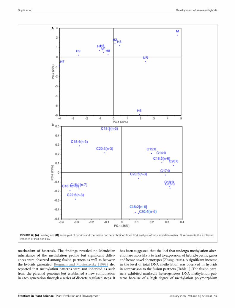
STATISTICAL ANALYSESAll the values for daily growth rate, carbohydrate, lipids, protein,cellulose, Fas and TAC for hybrids and their parents are reportedas means ± SD (n = 2). Further the significant differences amongthe mean values were evaluated by Tukey’s HSD test after per-forming One-Way analysis of variance (ANOVA) using statisticalsoftware, JMP 11.0 (trial version), values significant at p < 0.05.The distribution of hybrids and the fusion partners based ontheir fatty acid composition was analyzed by Principal componentanalysis (PCA). For PCA analysis data was mean centered andaccomplished using Unscrambler 9.8 software package (CAMOAS, Trondheim, Norway).
RESULTSThe protoplasts isolated from M. oxyspermum and U. reticu-lata were fused using the electrofusion method under an electricfield (Figure 1A). The optimization of electrofusion conditionsshowed that the application of an AC voltage of 40 V for 25–30 syielded alignment of about 70–80% of protoplasts into shortchains of 3–7 protoplasts (Figure 1B). Subsequently, applica-tion of a single higher-voltage DC pulse of 300 V for 20 μsduration induced about 4–6% fusions in pre-aligned protoplasts
Frontiers in Plant Science | Plant Evolution and Development January 2015 | Volume 6 | Article 3 | 4

Gupta et al. Development of seaweed hybrids
FIGURE 1 | Protoplast fusion between the selected species M.
oxyspermum and U. reticulata. (A) Mixture of protoplasts isolated fromthe selected species, (B,C) protoplasts alignment and pearl-chain formationafter applying AC pulse, (D,E) protoplast fusion after applying DC pulseshowing two chloroplasts in a single cell. Scale bar = 50 μm.
(Figure 1C). The fusion products were identified based on thelarger size of the cell and presence of two morphologically dis-tinct chloroplasts in a fused cell compared to unfused protoplasts(Figures 1D,E). On subsequent culturing of fused cells, it wasobserved that many cells were unable to regenerate even the cellwall. A total of 22 fusion products showed cell wall formation in3 days and underwent repeated cell division and differentiationgiving rise to plantlets after 4 weeks of culture. The regeneratedplantlets were then transferred to aerated culture flasks whereregenerants attained full thallus morphology after 4–6 weeks ofculture. Of the 22 plantlets screened, a total of 12 germlings werefinally selected as putative hybrids based on their distinct thallusmorphology compared to both fusion partners. The thallus mor-phology of putative hybrids and the fusion partners is shown inFigure 2. The thallus architecture of regenerated putative hybridswas distinct from that of both fusion partners. Of the fusion part-ners, M. oxyspermum had a flat monostromatic sheet-like thalluswhile U. reticulata had a typical distromatic, reticulated thallus.However, all the regenerated putative hybrids were distromaticbut diverged from wild type morphology of U. reticulata and weredistinct from each other (Figure 2).
PLOIDY ANALYSIS USING CONFOCAL MICROSCOPYFollowing treatment with DAPI, blue fluorescence was observedin stained nuclei. Both the nuclei and chloroplast were clearlydistinguished when the slides were observed in bright field with
superposition of the nuclear fluorescence and chloroplast autoflu-orescence (Supplementary Figure 1). The fusion partners and theregenerated putative hybrids showed clear differences in nuclearfluorescence intensity. The relative difference in the fluorescenceintensity among the fusion partners and the putative hybrids wasused as a measure for analysing the ploidy. Among the fusionpartners, M. oxyspermum showed the highest nuclear fluorescenceintensity and the nuclear fluorescence intensity for rest of thegenotypes was estimated relative to it. The fluorescence inten-sity shown by other fusion partner, i.e., U. reticulata was 60% ofM. oxyspermum. The putative hybrids showed fluorescence inten-sity ranging between 72 and 92% (Figure 3). In addition, the totalgenomic DNA showed differences in migration rate when runon an agarose gel, revealing variation in genome sizes (Figure 3).These results showed that the regenerated hybrids contained DNAcontent (ploidy) greater than that of the fusion partner U. retic-ulata but less than that of the other partner M. oxyspermumsuggesting that they are asymmetric hybrids.
MOLECULAR ANALYSIS OF PUTATIVE HYBRIDSThe molecular characterization of regenerated hybrids was car-ried out using ISSR, AFLP and MSAP analysis.
ISSR PROFILINGThe hybrid nature of the regenerants was further confirmed byISSR profiling. A total of 8 primers gave highly reproducible poly-morphic bands in the somatic hybrids and their fusion partners.A total of 428 bands were scored of which 348 were polymorphicamong the fusion partners. The ISSR profile revealed the species-specific bands of both fusion partners in the somatic hybrids,which confirmed their hybrid nature. The ISSR profile is shownin Supplementary Figures 2A,B, in which transfer of the fusionpartner -specific band in the regenerated hybrids is shown. TheISSR analysis of hybrids (1–12) revealed a genetic similarity of62, 67, 58, 62, 56, 64, 68, 68, 46, 62, 54, 56% respectively withU. reticulata and 42, 44, 52, 46, 58, 56, 42, 44, 63, 58, 68, 72%respectively with M. oxyspermum. These findings confirmed theintrogression of genomes of both the fusion partners into theregenerated hybrids.
AFLP ANALYSISAFLP profiles for three putative hybrids, i.e., H3, H7, H12 werenot obtained in a reproducible manner so were excluded fromsubsequent studies. As a result of the AFLP analysis, a total of324 bands were scored. The AFLP profile of the two fusion part-ners showed distinct band profiles, with 286 polymorphic bands(Figure 4A). The regenerated hybrids showed bands common toboth the partners. The band-sharing frequency analysis revealeda higher match for hybrids 1–5 with U. reticulata, while theother hybrids (6–9) were more similar to M. oxyspermum. Inaddition to sharing common bands with fusion partners, a fewadditional bands that were not present in either fusion partnerwere also observed in hybrids. However, the frequency of suchadditional bands was less than 1% of the total bands. The dis-tribution plot based on band-sharing is shown in Figure 4B. Theprincipal co-ordinate distribution clearly showed that both fusionpartners were at the extreme ends and the regenerated hybrids
www.frontiersin.org January 2015 | Volume 6 | Article 3 | 5

Gupta et al. Development of seaweed hybrids
FIGURE 2 | The morphology of fusion partners and putative hybrids generated after protoplast fusion. (A) M. oxyspermum and (B) U. reticulata, (C–N)
putative hybrids. The age of the hybrids represented in the figure is 4 months.
FIGURE 3 | Relative fluorescence intensity of the regenerated hybrids and the fusion partners determined from confocal microscopy. The gel imageabove the graph showed the migration of total genomic DNA of hybrids and fusion partners. UR, U. reticulata; M, M. oxyspermum; H1–12, Putative hybrids.
Frontiers in Plant Science | Plant Evolution and Development January 2015 | Volume 6 | Article 3 | 6

Gupta et al. Development of seaweed hybrids
FIGURE 4 | Characterization of regenerated hybrids based on AFLP
analysis. (A) AFLP gel wherein arrowheads showed the bands shared byhybrids with either of the fusion partner (B) AFLP based clustering of
regenerated hybrids and fusion partners. % represents the explainedvariance at PC1 and PC2. UR, U. reticulata; M, M. oxyspermum; H1–9,Hybrids.
remained between the partners with proximity to either parentaccording to their band-sharing frequency. The AFLP analysiscoincides well with the inferences made from the ISSR and ploidyanalyses. Combining all the results, i.e., morphology, ploidy anal-ysis, and molecular characterization, has confirmed the geneticintrogressions in the regenerated hybrids.
MSAP ANALYSISA total of 324 methylation loci were scored for MSAP analy-sis. The loci were classified as unmethylated, hemi-methylated atthe external cytosine, fully methylated at the internal cytosine,and fully methylated at the external cytosine or both the internaland external cytosines (Supplementary Figures 3A,B). Both theparent plants showed distinct methylation profiles. M. oxysper-mum showed significantly higher DNA unmethylation (38%) andhemimethylation (24%) rates compared to U. reticulata with cor-responding values of 19 and 11% respectively. The methylationfrequency of the internal cytosine and full methylation estimatedfor U. reticulata (39 and 31% respectively) was significantly higherthan that of M. oxyspermum (26% and 12%). The frequency oftotal methylation was 88% in M. oxyspermum and 69% in U.reticulata. The fusion partners exhibited markedly heterogeneousDNA methylation patterns because of high degrees of methylationpolymorphism, as 76.24%. Comparison of DNA methylation pat-terns with those of the regenerated hybrids revealed no large-scalealterations in DNA methylation compared to the fusion part-ners methylation profile. An increase in total methylation wasobserved for all the hybrids in comparison to U. reticulata whiletotal methylation of the hybrids was lower than in M. oxysper-mum. The increase in total methylation frequency was due torandom increases in either of the methylated loci in differentgenotype (Table 1).
IMPROVEMENT IN FUNCTIONAL TRAITS OF REGENERATED HYBRIDSGrowth, fatty acids and proximate composition were analyzedfor the regenerated hybrids and were compared with the fusionpartners’ traits. The growth rate analysis revealed significant dif-ferences between the DGR (%) of both the fusion partners.
U. reticulata showed DGR of 33.2 ± 2.6% which is about 2-foldhigher than that of M. oxyspermum (17.8 ± 1.77%), (p < 0.05).Among all the hybrids generated, Hybrid 9 showed DGR% of36.7 ± 1.55% which is the highest among all the hybrids as wellas greater than the fusion partner U. reticulata (p < 0.05). Otherhybrids showed DGR higher than the mid-parent value, i.e.,25.5 ± 2.2% except for hybrid 2, 7, and 8 (p < 0.05) (Figure 5).
The other characteristics investigated, including proximatecomposition of regenerated hybrids and the fusion partnersare summarized in Table 2. The total carbohydrate content was51.11 ± 2.2% for U. reticulata and 20.85 ± 1.12% for M. oxysper-mum and ranged between 36.8 ± 1.8% and 52.5 ± 2.2% in thehybrids. Hybrid 9 had a total carbohydrate content higher thanthe fusion partners (thus a superior trait), while all other hybridshad a carbohydrate content greater than that of the average ofboth the fusion partners (32.5 ± 2.2%) (p < 0.05). The cellulosecontent was higher in U. reticulata (11 ± 1.75%) in comparisonto M. oxyspermum (6.5 ± 0.22%) and the mid-parent value forthis trait was 8.75 ± 0.98%. The cellulose content in the hybridsranged between 6.1 ± 1.2% and 14.2 ± 2.2%. Similarly to thetotal carbohydrate content, hybrid 9 showed cellulose contenthigher than that of the fusion partners, indicating a superior trait(p < 0.05). The total protein content was higher in M. oxysper-mum (11.75 ± 2.6%) than U. reticulata (6.8 ± 2.12%) and themid-parent value for this trait was 9.27 ± 2.36%. The content oftotal protein in the fusion partner was in reverse order in com-parison to the content of carbohydrate. The protein content ofthe hybrids was found to be in the range from 7.1 ± 1.5% to9.2 ± 1.8%. The protein content of hybrid 1 was higher thanthe mid-parent value while others had lower protein content thanthe mid-parent value but higher than the parent having this asthe inferior trait (U. reticulata) (p < 0.05). The total antioxi-dant capacity (TAC) was estimated as 0.64 ± 0.08 (mg AAE/gextract) and 0.44 ± 0.1 (mg AAE/g extract) for M. oxyspermumand U. reticulata respectively while the mid-parent value for thistrait was 0.54 ± 0.09 (mg AAE/g extract). The estimated TACin the hybrids was ranged from 0.33 ± 0.16 (mg AAE/g extract)to 0.72 ± 0.92 (mg AAE/g extract). Among the hybrids, hybrid
www.frontiersin.org January 2015 | Volume 6 | Article 3 | 7

Gupta et al. Development of seaweed hybrids
Table 1 | Comparison of DNA methylation levels between fusion partners M. oxyspermum and U. reticulata and the somatic hybrids
determined by methylation-sensitive amplified polymorphism (MSAP) assay.
Genotypes
M. oxyspermum H1 H2 H3 H4 H5 H6 H7 H8 H9 U. reticulata
DN
Am
eth
yla
tio
n(%
)
u 38 21 28 16 14 40 20 40 18 18 19
h 24 30 18 14 34 10 20 14 22 18 11
i 26 35 30 48 40 20 40 28 31 29 39
f 12 14 24 25 17 30 20 21 29 25 31
Total methylation 88 86 76 78 88 70 80 82 71 65 69
FIGURE 5 | Growth rate (DGR %) of the regenerated hybrids and the fusion partners. H1–9, hybrids. Bars with different letters represent meanssignificantly different from each other at p < 0.05.
9 showed a TAC (0.72 ± 0.92 mg AAE/g extract) higher thanthat of the fusion partner with higher trait value (p < 0.05). Theother hybrids, hybrid 2, 3, 7, and 8 showed a TAC value betterthan U. reticulata as well as higher or equal to the mid-parentvalue, while hybrid 1, 4, and 6 showed a TAC less than the mid-parent value but similar to U. reticulata (p < 0.05). The fatty acid(FA) compositions for all the genotypes are shown in Table 3.PCA of the FA data matrix provided a global statistical distinc-tion between the fusion partners and the hybrids (Figures 6A,B).The FA profile investigated for the genotypes explained a totalvariation of 65% (PC1-39% and PC2-26%) revealing a distinctdistribution of hybrids and the fusion partners. Both the fusionpartners and the generated hybrids showed characteristic fattyacid profile with the richness of ω3 and content of C18 PUFAs> C20 PUFAs. The PUFA content varied from 37 to 55% amongthe fusion partners and the hybrids generated. However, thesum of the content of arachidonic acid (C20:4 n-6) and eicos-apentaenioc acid (C20:5 n-3) accounts for only ≤1.4% amongthe genotypes studied. The total saturated and unsaturated fattyacid content was analogous in the fusion partners; the formerwas higher in M. oxyspermum while the latter was higher in U.reticulata. The FA profile revealed an increase in unsaturation(1.1–1.6-fold) in nearly all the hybrids, (the only exception was
hybrid 6), and a decrease in saturation for all the hybrids com-pared to the fusion partners. The increase in unsaturation wasdue to an increase in the contents of both PUFAs by 1.2–1.5-fold,mainly C18 PUFAs and MUFAs by 1.1–3.7-fold (except hybrid 3and 5). The content of C18 PUFAs in the hybrids was higher thanin both the fusion partners (1.1–1.5-fold) with the only excep-tion being hybrid 6 that exhibited a different FA profile from restof the hybrids. Interestingly, the FAs profile revealed that all thehybrids except hybrid 6 have an elevated PUFA content (includ-ing n-3 PUFAs), compared to the average of both the fusionpartners (Table 3). Hybrid 6 had a higher saturated FA content(SFA/UFA = 1.1), mainly due to higher stearic acid (C18:0), andthe lowest PUFA due to a decrease in C18 PUFA content, espe-cially C18:3 (n-3) and C18:4 (n-3). Despite the decrease in totalPUFAs, the content of C20 PUFAs increased in hybrid 6 by 1.9–3.1-fold due to increase in arachidonic acid content (C20:4, n-6).The other hybrids showed a predominance of palmitoleic (C16:1,n-7) and oleic acid (C18:1, n-9) among MUFAs. The content ofoleic acid was 1.1–5.2-fold higher in all the hybrids compared tofusion partners (p < 0.05). The content of two essential FAs, ALA(C18:3, n-3) and LA (C18:2, n-6) differed among the fusion part-ners: the former was higher in M. oxyspermum while the latterwas higher in U. reticulata. Interestingly the hybrids showed ALA
Frontiers in Plant Science | Plant Evolution and Development January 2015 | Volume 6 | Article 3 | 8

Gupta et al. Development of seaweed hybrids
Tab
le2
|C
om
pari
so
no
fth
ep
roxim
ate
co
mp
osit
ion
of
reg
en
era
ted
hyb
rid
san
dth
efu
sio
np
art
ners
.
Co
mp
on
en
ts(%
on
dry
wt
ba
sis
)
M.o
xysp
erm
um
Hyb
rid
1H
yb
rid
2H
yb
rid
3H
yb
rid
4H
yb
rid
5H
yb
rid
6H
yb
rid
7H
yb
rid
8H
yb
rid
9U
.re
ticu
lata
Car
bohy
drat
e20
.85
±0.
91d
37.8
7±
1.62
bc44
.55
±1.
90ab
41.2
±3.
53b
42.2
±1.
97b
37.1
±2.
40bc
38.5
±1.
69bc
36.8
±1.
71bc
37.1
±1.
83bc
32.5
±1.
8c51
.11
±0.
43a
Mid
-par
ent
valu
e35
.98
±0.
67
Prot
ein
11.7
5±
2.68
a9.
2±
1.55
ab8.
85±
2.33
ab8.
8±
2.26
ab7.
1±
1.13
b8.
4±
0.98
ab7.
1±
0.56
b7.
1±
1.71
b7.
4±
0.70
b7.
8±
0.84
b6.
8±
1.83
b
Mid
-par
ent
valu
e9.
27±
2.25
Lipi
don
Fwt
basi
s1.
73±
0.29
ab0.
91±
0.36
cd0.
42±
0.15
de0.
49±
0.07
de0.
56±
0.19
de0.
43±
0.26
de0.
9±
0.21
cde
0.34
±0.
15e
0.54
±0.
14de
1.42
±0.
45bc
2.03
±0.
25a
Mid
-par
ent
valu
e1.
88±
0.27
Cel
lulo
se6.
5±
0.38
de
6.5
±1.
01de
8.2
±1.
97bc
de9.
8±
1.97
bc6.
1±
1.27
e9.
4±
0.98
bcd
7.1
±0.
42cd
e7.
8±
1.41
cde
7.8
±0.
56cd
e14
.2±
1.97
a11
±1.
69b
Mid
-par
ent
valu
e8.
75±
1.03
TAC
(mg
AA
E/g
extr
act)
0.64
±0.
11ab
0.43
±0.
07ab
c0.
6±
0.16
abc
0.53
±0.
08ab
c0.
41±
0.11
bc0.
33±
0.15
c0.
48±
0.19
abc
0.58
±0.
08ab
c0.
55±
0.21
abc
0.72
±0.
05a
0.44
±0.
11ab
c
Mid
-par
ent
valu
e0.
54±
0.11
Valu
esw
ithdi
ffere
ntle
tter
sab
ove
repr
esen
tm
eans
sign
ifica
ntly
diffe
rent
from
each
othe
rat
p<
0.05
(mea
n±
SD
).
content similar to M. oxyspermum, except for hybrids 6 and 7,while LA content was similar to U. reticulata except for hybrids3 and 4 (p < 0.05). The content of palmitoleic acid (C16:1) washigher in hybrids 4, 7, and 9 while others showed an increase inoleic acid (C18:1, n-9) content with a 1.1–4.5-fold increase com-pared to M. oxyspermum and a 1.1–2.6-fold decrease comparedto the content in U. reticulata. Among MUFAs, both heptade-cenoic acid (C17:1, n-7) and oleic acid (C18:1, n-9) showed thetraits of heterosis (superior hybrids compared to both the fusionpartners) while C16:1 (n-9) showed heterosis in three hybrids(4, 6, and 9) while three hybrids (1, 2, and 8) had the traits ofmid-parent heterosis. Similarly, among PUFAs, stearidonic acid(C18:4, n-3) (except hybrid 6) and docosahexanoic acid (C22:6,n-3) showed higher contents than both the fusion partners. Allthese contents showed improvement in traits, therefore indicat-ing the potential of this technique to develop elite germplasm.All these results summarized that hybrid 9 showed improvedfunctional traits for growth, carbohydrate and cellulose content,anti-oxidative potential and fatty acid profile compared to thefusion partners.
DISCUSSIONPlant selection and breeding techniques have been extensivelyapplied for genetic improvements in agriculture crops, butremained underutilized for seaweeds despite their proven eco-nomic and commercial values. This study demonstrated thedevelopment of seaweed hybrid varieties with improved func-tional traits through protoplast fusion between species of thegenus Monostroma and Ulva. Monostroma and Ulva species arewell known for their nutritional values and both have character-istic growth and biochemical composition. The hybrids generatedin this study showed heterozygous vigor (heterosis) displayingsome traits superior to both the fusion partners. Earlier reportson the somatic hybridization of seaweeds have largely dealt withmorphology and pigmentation, albeit showing a few improvedtraits such as growth (Reddy et al., 1992) and fatty acid com-position (Reddy and Fujita, 1989) but lacked the evidence forthe molecular level of recombination. Also, most of the stud-ies reported the development of chimeric thalli after protoplastfusion indicating independent segregation of the two genomeswithout recombination. The most recent report of the devel-opment of a hybrid variety of the seaweed was that of Kitoet al. (1998), between Monostroma nitidum and Porphyra yezoen-sis, which showed development of chimeric thalli with distictmorphology as string- and bud-like thallus development, andconfirmed genetic introgression based on RAPD marker analysis.This study also showed fatty acid profiles characteristic of bothred and green seaweeds in regenerated hybrids. In the presentstudy, evidence of recombination between the two genomes of thefusion partners in hybrid progeny was confirmed by independentDNA fingerprinting techniques (ISSR and AFLP) coupled withcritical morphological observations of hybrids over fusion part-ners. In addition, for the first time provided additional insightsinto the transmission of epigenetic traits in hybrids based onthe analysis of methylation sensitive loci. The shown morpho-logical variations by hybrids might be a result of protoplast-differentiation under axenic condition. The previous studies have
www.frontiersin.org January 2015 | Volume 6 | Article 3 | 9

Gupta et al. Development of seaweed hybrids
Tab
le3
|Fatt
yacid
co
mp
osit
ion
(giv
en
in%
of
tota
lfa
tty
acid
meth
yl
este
rs)
of
fusio
np
art
ners
an
dth
ere
gen
era
ted
hyb
rid
s(m
ean
±S
D).
FA
sM
.o
xysp
erm
um
Hyb
rid
1H
yb
rid
2H
yb
rid
3H
yb
rid
4H
yb
rid
5H
yb
rid
6H
yb
rid
7H
yb
rid
8H
yb
rid
9U
.re
ticu
lata
C12
:01.
15±
0.05
a0.
4±
0.18
cdnd
0.4
±0.
11cd
0.2
±0.
02de
0.7
±0.
02b
nd0.
1±
0.02
end
0.3
±0.
02de
0.6
±0.
07bc
C14
:04.
37±
0.51
ab2.
8±
0.56
cd4.
1±
0.5ab
2.3
±0.
3de3.
2±
0.28
cd1.
6±
0.28
bc1.
8±
0.24
ef0.
7±
0.26
fg4.
8±
1.4a
0.4
±0.
11g
3.8
±0.
56ab
c
C15
:00.
6±
0.28
a0.
4±
0.28
ab0.
6±
0.28
a0.
4±
0.28
ab0.
3±
0.02
ab0.
3±
0.02
ab0.
3±
0.03
ab0.
2±
0.05
b0.
2±
0.03
b0.
3±
0.02
ab0.
2±
0.02
b
C16
:032
.86
±3.
1a29
.7±
2.8ab
26.3
±0.
7bc26
±1.
7bc26
±0.
3bc30
±1.
7ab33
.4±
1.8a
25.9
±2.
01bc
25.9
±0.
84bc
24.3
±1.
55c
33.9
±3.
01a
C17
:00.
52±
0.04
a0.
2±
0.02
cd0.
4±
0.03
ab0.
5±
0.11
a0.
4±
0.07
a0.
2±
0.03
cd0.
5±
0.07
a0.
1±
0.01
d0.
3±
0.02
bcN
d0.
2±
0.11
cd
C18
:014
.87
±0.
77a
5.2
±0.
84c
4.3
±0.
7c9.
3±
2.6b
5.4
±0.
56c
3.3
±0.
7c14
±0.
28a
3.5
±0.
28c
4.8
±0.
84c
7.9
±1.
1b7.
9±
0.98
b
C20
:00.
65±
0.14
a0.
2±
0.02
b0.
1±
0.02
b0.
2±
0.02
b0.
2±
0.03
b0.
3±
0.05
b0.
2±
0.16
bnd
0.1
±0.
05b
nd0.
7±
0.3a
C22
:01.
53±
0.02
abc
1.7
±0.
14ab
1±
0.14
c1.
6±
0.28
ab1.
9±
0.42
a1.
6±
0.28
ab1.
9±
0.28
a1.
7±
0.28
ab1.
5±
0.28
abc
1.4
±0.
28ab
c1.
3±
0.28
bc
C24
:01.
85±
0.35
a0.
3±
0.28
b0.
2±
0.02
b0.
2±
0.05
b0.
2±
0.02
bnd
0.5
±0.
35b
0.2
±0.
05b
0.1
±0.
02b
0.3
±0.
2bnd
C16
:1(n
-7)
0.97
±0.
18bc
2.3
±0.
28bc
d2.
2±
0.7bc
d1.
1±
0.6d
3.1
±0.
56ab
1.7
±0.
84cd
2.6
±0.
28bc
4.4
±0.
7a2.
1±
0.21
bcd
3.4
±0.
56d
2.9
±1.
13ab
C17
:1(n
-7)
0.09
±0.
04f
0.3
±0.
03de
0.2
±0.
03ef
0.3
±0.
02de
0.6
±0.
11ab
0.3
±0.
05de
nd0.
7±
0.11
a0.
5±
0.02
bc0.
4±
0.05
cd0.
2±
0.02
ef
C18
:1(n
-9)
1.7
±0.
31g
5±
0.21
bcd
4.7
±0.
35cd
e3.
1±
0.45
f4.
7±
0.56
cde
4.4
±0.
3de5.
3±
0.28
bc8.
9±
0.42
a4.
6±
0.28
cde
5.7
±0.
3b4
±0.
5f
C18
:2(n
-6)
4.04
±0.
21c
5.8
±0.
6bc6.
8±
0.56
b3.
8±
0.56
c3.
8±
0.52
c5.
8±
1.4bc
16.6
±2.
12a
5.6
±0.
6bc7.
2±
0.56
b5.
5±
0.42
bc7.
3±
0.7b
C18
:3(n
-6)
0.89
±0.
43a
0.4
±0.
11b
0.5
±0.
03b
0.4
±0.
02b
0.5
±0.
11b
0.5
±0.
11b
0.4
±0.
11b
0.5
±0.
07b
0.4
±0.
07b
0.4.
03b
1±
0.28
a
C18
:3(n
-3)
21.3
3±
2.78
a19
±3.
11a
21±
2.96
a20
.1±
3.01
a19
.3±
3.1a
20.2
±2.
96a
10±
2.4b
18.6
±1.
41a
19.1
±2.
4a21
±1.
83a
17.3
±1.
55a
C18
:4(n
-3)
7.46
±0.
61c
20.4
±2.
12a
18.9
±3.
4ab23
±2.
54a
23.3
±1.
83a
22.7
±2.
68a
0.7
±0.
17d
19.2
±2.
82ab
20.7
±2.
4a22
.1±
1.41
a14
.7±
1.55
b
C20
:3(n
-3)
1.73
±0.
12a
1±
0.14
b0.
6±
0.28
bc0.
6±
0.14
bc1
±0.
07b
1±
0.02
b0.
2±
0.01
cd1.
7±
0.6a
1±
0.22
b1
±0.
26b
nd
C20
:3(n
-6)
0.09
±0.
01c
0.1
±0.
01c
Nd
nd0.
1±
0.01
cN
d0.
6±
0.2b
0.2
±0.
03c
ndnd
0.9
±0.
07a
C20
:4(n
-6)
0.43
±0.
07b
0.5
±0.
05b
0.2
±0.
05b
0.7
±0.
28b
0.6
±0.
11b
0.3
±0.
16b
4.5
±0.
98a
0.6
±0.
03b
0.8
±0.
17b
0.6
±0.
11b
0.9
±0.
11b
C20
:5(n
-3)
0.73
±0.
14bc
0.2
±0.
05c
0.2
±0.
02c
0.2
±0.
04c
0.3
±0.
09c
0.3
±0.
09c
0.6
±0.
16bc
0.5
±0.
08bc
1.7
±0.
56a
0.3
±0.
14c
1±
0.56
b
C22
:6(n
-3)
1.2
±0.
56e
4.3
±1.
13bc
d3.
4±
0.7cd
3.9
±1.
13cd
4.8
±0.
98ab
c4.
7±
0.70
abc
6±
0.84
ab6.
4±
0.98
a4.
1±
0.98
cd4.
5±
0.56
bc2.
6±
0.14
de
∑S
FA58
.4±
2.33
40.9
±3.
6737
.1±
1.71
40.8
±2.
6137
.9±
1.41
37.9
±2.
7352
.5±
4.64
32.6
±1.
4537
.8±
2.83
35.0
±2.
6548
.8±
2.61
∑U
FA41
.6±
2.25
59.3
±4.
2462
.8±
4.63
59.2
±5.
4162
.0±
6.64
62.0
±4.
6747
.4±
2.63
67.3
±3.
4862
.1±
5.69
64.9
±4.
4851
.8±
2.59
∑M
UFA
3.8
±0.
567.
6±
0.67
11.2
±0.
296.
4±
0.75
8.4
±0.
726.
5±
0.53
7.8
±0.
5214
.0±
1.62
7.2
±0.
799.
6±
0.57
7.1
±0.
65∑
PU
FA37
.9±
2.73
51.7
±4.
3351
.6±
3.24
52.7
±3.
4653
.6±
2.48
55.5
±4.
8339
.6±
149
53.3
±3.
7255
.0±
3.69
55.3
±1.
5344
.8±
1.53
∑C
18P
UFA
33.7
±3.
8245
.6±
2.25
47.2
±2.
8347
.3±
2.61
46.9
±2.
5849
.2±
4.49
27.7
±1.
6743
.8±
3.68
47.4
±2.
7648
.9±
1.48
40.3
±3.
48∑
C20
PU
FA3.
0±
0.28
1.8
±0.
561.
0±
0.24
1.5
±0.
541.
9±
0.54
1.5
±0.
685.
9±
0.54
3.0
±0.
7335
.0±
2.45
1.9
±0.
721.
9±
0 .63
∑n3
PU
FA32
.4±
3.36
44.9
±2.
3544
.0±
2.29
47.8
±2.
6348
.6±
2.45
48.9
±3.
7817
.5±
1.48
46.4
±3.
5246
.6±
2.72
48.9
±1.
5135
.6±
1.52
∑n6
PU
FA5.
4±
0.45
6.8
±0.
377.
6±
0.23
5.0
±0.
575.
0±
0.38
6.6
±0.
5922
.1±
1.63
6.9
±0.
638.
4±
0.57
6.4
±0.
439.
2±
0.48
n6/n
30.
2±
0.03
0.2
±0.
410.
2±
0.11
0.1
±0.
070.
1±
0.02
0.1
±0.
071.
3±
0.68
0.1
±0.
010.
2±
0.06
0.1
±0.
030.
3±
0.11
Valu
esin
the
sam
ero
ww
ithdi
ffere
ntsu
pers
crip
tsle
tter
sar
esi
gnifi
cant
lydi
ffere
ntat
p<
0.05
.
nd,n
otde
tect
able
.
Frontiers in Plant Science | Plant Evolution and Development January 2015 | Volume 6 | Article 3 | 10

Gupta et al. Development of seaweed hybrids
shown that the microflora associated with the seaweeds regu-lates growth and morphogenetic ability of seaweeds (Marshallet al., 2006; Singh et al., 2011). Wichard and Oertel (2010)showed that seaweed associated bacteria secrete morphogeneticfactors that control the thallus morphogenesis in Ulva. Due to thelack of PCR analyses we cannot rule out, that the cultures pre-sented in Figure 2 contain bacteria, which are, e.g., not cultivable.Therefore, present bacteria might still have an impact on the mor-phogenesis of the hybrids. Nevertheless, the molecular analysisof somatic hybrids carried out in this study showed the genomecomplement of both the fusion partners and thus confirms unam-biguously the hybrid nature of the somatic hybrids.
The hybrids regenerated in this study were asymmetric sincethe ploidy content was not additive of both the genomes of thefusion partners. The previous study on protoplast fusion betweenU. pertusa and E. prolifera resulted in hybrids with no changein chromosome number (Reddy et al., 1992) whereas Kapraun(1989) reported parasexual fusion products of Enteromorpha withdiploid and tetraploid levels. The occurrence of a non-additivechromosome count is a common phenomenon and reported inseveral studies of somatic hybridization in land plants (Chenet al., 2004; Minquin et al., 2008; Tu et al., 2009; Wang et al.,2011) but this study is the first in macroalgae. Partial introgres-sion of chromosomes from one fusion partner to the other hasbeen described in developing the monosomic lines of a wheat-rye combination (Fu et al., 2013). In the same study, eliminationof chromosomes was also illustrated. The chromosome elimina-tion was mainly attributed to differences in the cell cycle. Thedifferences in the growth rates between fusion partners in thisstudy could perhaps result in chromosome elimination in somatichybrids due to variation in cell cycles. Addition and introgressionof either full or part chromosomes of one parent partner intoanother has also been reported in hybrids of Brassica rapa (×)Isatis indigotica (Tu et al., 2009).
The varieties developed through protoplast fusion displayedeither heterosis or mid-parent heterosis for the various traitsinvestigated. With the advent of random genetic recombination,the expression of various functional traits was found to be vari-able among the somatic hybrids. The contents of carbohydrateand cellulose in the somatic hybrids were found to be higher thanthat of fusion partner M. oxyspermum (p < 0.05), while the con-tents of protein and total antioxidant potential were better thanthat of the other fusion partner U. reticulata, and the growth ratewas improved in comparison to M. oxypermum with one hybridhaving growth rate even higher than U. reticulata. Both the fusionpartners and the generated hybrids showed characteristic fattyacid profile with the richness of ω3 and content of C18 PUFAs >
C20 PUFAs. This FA profile is in agreement with recent studies(Khotimchenko et al., 2002; Pereira et al., 2012; Kumari et al.,2013; Alsufyani et al., 2014). The PUFA content varied from 37 to55% among the fusion partners and hybrids. However, the sum ofthe content of arachidonic acid (C20:4 n-6) and eicosapentaeniocacid (C20:5 n-3) accounts for only ≤1.4% among the genotypesstudied. Recently, Alsufyani et al. (2014) reported polyunsatu-rated aldehydes (PUAs) production in Ulva due to woundinginduced chemical transformation of PUFAs mainly the C20:4 n-6and C20:5 n-3. The lower content of both these FAs in the present
study might be due to the chemical transformation of theseFAs into PUAs. However, the PUAs production is highly speciesdependent and reported to vary significantly among Ulva species(Alsufyani et al., 2014) and diatoms (Wichard et al., 2005). Thefatty acid profile of the somatic hybrids revealed a mixed profilefrom both the fusion partners. The hybrids showed a decline insaturated fatty acid content with concomitant increase in unsat-urated fatty acids. Among the unsaturated fatty acids, PUFAscontributed more to the increase in unsaturation. Interestingly,the content of two indispensable fatty acids LA and ALA, whichcannot be synthesized by human body, were in opposing levelsamong the fusion partners. The former was higher in U. reticulatawhile latter was higher in M. oxyspermum. The hybrids showedboth LA and ALA content from the fusion partner having thistrait superior. This further confirms the genetic recombination inhybrids expressing the superior traits from the fusion partners.The content of DHA was found to be higher in all the hybrids incomparison to their fusion partners while the content of EPA wasinferior in all the hybrids compared to the fusion partners. Thiscould be due to the higher transcriptional activity of downstreamelongase and saturase enzymes. The higher content of C18 PUFAs(LA, ALA, STA) is the characteristic feature of Chlorophyceanspecies belonging to genus Ulva (Kumari et al., 2013). On thecontrary, Monostroma showed a higher content of saturated fattyacid and lower content of PUFAs. The n6/n3 ratio for all thehybrids were recorded in the nutritionally beneficial range (0.1:1to 1.3:1) and was improved compared to the fusion partners. Theincrease in n6/n3 ratio could further aid in decreasing low-densitylipoproteins, cholesterol levels, preventing inflammatory, cardio-vascular and nervous system disorders. Therefore, these hybridvarieties may be a better source of n3 PUFAs for the developmentof healthier food formulations. The PUFA/SFA ratio, which deter-mines the nutritional value of lipids, is in accordance with thenutritional guidelines (≥0.4). Interestingly this ratio was found tobe higher in hybrids in comparison to both the fusion partners.
Though heterosis has been known since Darwin, the underly-ing molecular mechanisms are little known (Miller et al., 2012;Hopkins, 2013). The current hypothesis is that the genetic inputfrom each parental line serves to mask the deleterious alleles ofthe other, resulting in hybrid plants that are superior to either par-ent. It is presumed that changes in gene expression and regulatorynetworks are responsible for heterosis in hybrids (Birchler et al.,2003). Epigenetic regulation, as a change in DNA methylation, isgaining prominence nowadays as a means to establish the molec-ular basis for heterosis (Moghaddam et al., 2010). The changesin the cytosine methylation pattern are the result of eliminationand/or introgression of donor chromatin from fusion partners(Cai et al., 2007). Also, a change in DNA methylation controlsthe expression of genes (Cai et al., 2007). Greaves et al. (2012)while analysing the DNA methylomes of two Arabidopsis ecotypesand their reciprocal hybrids revealed the altered methylomes inhybrids offspring in the form of non-additive methylation pat-terns in hybrids. This study also revealed the correlation betweenthe DNA methylation changes in hybrids and the altered expres-sion of the genes. In the present study an attempt was made todetermine the transmission of polymorphic DNA methylationloci in the generated hybrids to highlight the possible underlying
www.frontiersin.org January 2015 | Volume 6 | Article 3 | 11
Gupta et al. Development of seaweed hybrids
FIGURE 6 | (A) Loading and (B) score plot of hybrids and the fusion partners obtained from PCA analysis of fatty acid data matrix. % represents the explainedvariance at PC1 and PC2.
mechanism of heterosis. The findings revealed no Mendelianinheritance of the methylation profile but significant differ-ences were observed among fusion partners as well as betweenthe hybrids generated. Bergman and Mostoslavsky (1998) alsoreported that methylation patterns were not inherited as suchfrom the parental genomes but established a new combinationin each generation through a series of discrete regulated steps. It
has been suggested that the loci that undergo methylation alter-ation are more likely to lead to expression of hybrid-specific genesand hence novel phenotypes (Zhang, 2008). A significant increasein the level of total DNA methylation was observed in hybridsin comparison to the fusion partners (Table 1). The fusion part-ners exhibited markedly heterogeneous DNA methylation pat-terns because of a high degree of methylation polymorphism
Frontiers in Plant Science | Plant Evolution and Development January 2015 | Volume 6 | Article 3 | 12

Gupta et al. Development of seaweed hybrids
(76.24%). However, comparison of fusion partner DNA methy-lation patterns with those of the regenerated hybrids revealed nolarge-scale alteration in DNA methylation. An increase in totalmethylation was observed for all the hybrids in comparison toU. reticulata while the same was lower than M. oxyspermum. Theincrease in total methylation frequency was due to a randomincrease in either of the methylated loci in different genotypes(Table 1). Similarly, no gross alteration in DNA methylation,histone modifications or transcript levels was observed for inter-specific hybrids of Arabidopsis thaliana (Moghaddam et al., 2010).Hypermethylation is known to cause transcriptional inactivationof CG/CNG sites (Diequez et al., 1998) and has been highlyassociated with the formation of heterochromatin-mediated genesilencing (Klein and Coasta, 1997). The predominance of hyper-methylation may lead to repression of spurious global tran-scription, in order to achieve efficient transcription or completeexpression of desirable loci in hybrids. The observed significantdifference in methylation profile is consistent with the hypothesisthat the epigenetic changes are more pronounced when there ismerger of two different genomes rather than by genome doubling(Madlung et al., 2002).
Among the hybrids generated, hybrid 9 showed the preva-lence of superior traits over the fusion partners. Also, the hybrid9 showed the biomass characteristics equivalent to most Ulvaspecies with an additional complementation of fatty acid pro-file of M. oxyspermum. Moreover, the growth rate shown byhybrid 9 is superior to the growth rate of most Ulva species(Bruhn et al., 2011; Trivedi et al., 2013). Hybrid 9 showed theinheritance of complementary characteristics of both the fusionpartners for example the higher growth rate from U. reticulataand nutritionally important FAs from M. oxyspermum. In addi-tion to these traits, hybrid 9 showed cellulose content superiorto the fusion partners. Hybrid 9 with nutritional characteristicsof M. oxyspermum and ∼double the growth rate of M. oxysper-mum is of interest for cultivation. The vegetative propagation ofsuch biomass offers the most important advantage of the reduc-tion in harvest time which in turn may increase the total cropproductivity and better economic gains. Moreover, the develop-ment of biomass with higher cellulose content along with bettergrowth and nutritional richness is sought after for developmentof biorefinery for food, fuel and commodity products.
In conclusion, the present study demonstrates successful pro-toplast fusion and regeneration of somatic hybrids with improvedtraits of agronomical importance over their fusion partners. Thegenetic relatedness as well as distinctness of hybrids comparedto fusion partners was confirmed by cytological and molecularevidences. The traits transferred to hybrids include the nutri-tional richness of M. oxyspermum and higher growth and cellulosecontent of U. reticulata. The inheritance of epigenetic variationsas a change in methylation enabled us to understand the possi-ble underlying mechanism pertaining to activation or expressionof gene machinery, possibly explaining the observed heterosis ofhybrids. Such hybrid material is of particular interest for genome-wide analysis studies to identify and characterize the backgroundgenetic loci regulating the functional traits. The developedsomatic hybrids with improved nutritional value, growth rate andcellulose content unequivocally demonstrate the significance of
the protoplast fusion technique for developing improved variantsof economically important marine macroalgae.
ACKNOWLEDGMENTSThe financial support received from DBT Indo-UK collabora-tive project (SuBBSea) under grant agreement no. BT/IN/Indo-UK/SuBB/22/AML/2013 is gratefully acknowledged. Dr. Juliet C.Coates, Birmingham University, UK is acknowledged for Englishcorrections. We are thankful to the anonymous reviewers for theirvaluable comments.
SUPPLEMENTARY MATERIALThe Supplementary Material for this article can be foundonline at: http://www.frontiersin.org/journal/10.3389/fpls.2015.
00003/abstract
REFERENCESAlsufyani, T., Engelen, A. H., Diekmann, O. E., Kuegler, S., and Wichard, T. (2014).
Prevalence and mechanism of polyunsaturated aldehydes production in thegreen tide forming macroalgal genus Ulva (Ulvales, Chlorophyta). Chem. Phys.Lipids 183, 100–109. doi: 10.1016/j.chemphyslip.2014.05.008
Bassam, B. J., Caetano-Anollés, G., and Gresshoff, P. M. (1991). Fast and sensitivesilver staining of DNA in polyacrylamide gels. Anal. Biochem. 196, 80–83. doi:10.1016/0003-2697(91)90120-I
Bergman, Y., and Mostoslavsky, R. (1998). DNA demethylation: turning genes on.Biol. Chem. 379, 401–407.
Birchler, J. A., Auger, D. L., and Riddle, N. C. (2003). In search of the molecularbasis of heterosis. Plant Cell 15, 2236–2239. doi: 10.1105/tpc.151030
Bird, C. J., and McLachlan, J. (1982). Some Underutilized Taxonomic Criteriain Gracilaria (Rhodophyta, Gigartinales). Bot. Mar. 25, 557–562. doi:10.1515/botm.1982.25.12.557
Bligh, E. G., and Dyer, W. J. (1959). A rapid method of total lipid extraction andpurification. Can. J. Biochem. Biophysiol. 37, 911–915.
Bruhn, A., Dahl, J., Nielsen, H. B., Nikolaisen, L., Rasmussen, M. B., Markager,S., et al. (2011). Bioenergy potential of Ulva lactuca: biomass yield,methane production and combustion. Bioresour. Technol. 102, 2595–2604. doi:10.1016/j.biortech.2010.10.010
Cai, Y., Xiang, F., Zhi, D., Liu, H., and Xia, G. (2007). Genotyping of somatichybrids between Festuca arundinacea Schreb. and Triticum aestivum L. Plant CellRep. 26, 1809–1819. doi: 10.1007/s00299-007-0397-5
Chen, C. L., Guo, W. W., Yi, H. L., and Deng, X. X. (2004). Cytogenetic analysis oftwo interspecific Citrus allotetraploid somatic hybrids and their diploid fusionparents. Plant Breeding. 123, 332–337. doi: 10.1111/j.1439-0523.2004.00972.x
Davey, M. R., Anthony, P., Power, J. B., and Lowe, K. C. (2005). Plant proto-plasts: status and biotechnological perspectives. Biotechnol. Adv. 23, 131–171.doi: 10.1016/j.biotechadv.2004.09.008
Diequez, M. J., Vaucheret, H., Paszkowski, J., and Scheid, O. M. (1998). Cytosinemethylation at CG and CNG sites is not a prerequisite for the initiation of tran-scriptional gene silencing in plants, but it is required for its maintenance. Mol.Gen. Genet. 259, 207–215. doi: 10.1007/s004380050806
Dubois, M., Giles, K. A., Hamilton, K. S., Rebers, P. A., and Smith, F. (1956).Colorimetric method for the determination of sugar and related substances.Anal. Chem. 18, 350–356. doi: 10.1021/ac60111a017
Fang, Z., Ou, Y., and Cui, J. (1985). Breeding of hybrid Laminaria “Danza No.10”:an application of the Laminarian haploid cell clones (in Chinese with Englishabstract). J. Shangdong. Col. Oceanol. 15, 64–72.
Fang, Z., Ou, Y., Cui, J., and Dai, J. (1978). Success in culturing clones of the game-tophytes of Laminaria japonica (in Chinese with English abstract). Transact. Sci.2, 115–116.
Food and Agriculture Organisation of the United Nations. (2012). The State ofWorld Fisheries and Aquaculture. Rome: FAO.
Food and Agriculture Organisation of the United Nations. (2014). The State ofWorld Fisheries and Aquaculture. Rome: FAO.
Fu, S., Yang, M., Fei, Y., Tan, F., Ren, Z., Yan, B., et al. (2013). Alteration and abnor-mal mitosis of wheat chromosomes induced by wheat-rye monosomic additionlines. PLoS ONE 8:e70483. doi: 10.1371/journal.pone.0070483
www.frontiersin.org January 2015 | Volume 6 | Article 3 | 13
Gupta et al. Development of seaweed hybrids
Greaves, I. K., Groszmann, M., Ying, H., Taylor, J. M., Peacock, W. J., and Dennis,E. S. (2012). Trans chromosomal methylation in Arabidopsis hybrids. Proc. Natl.Acad. Sci. U.S.A. 109, 3570e3575. doi: 10.1073/pnas.1201043109
Gupta,V., Bijo, A. J., Kumar, M., Reddy, C. R. K., and Jha, B. (2012). Detectionof epigenetic variations in the protoplast-derived germlings of Ulva reticu-lata using methylation sensitive amplification polymorphism (MSAP). Mar.Biotechnol. 14, 692–700. doi: 10.1007/s10126-012-9434-7
Hiraoka, M., Dan, A., Shimada, S., Hagihira, M., Migita, M., and Ohno, M.(2003). Different life histories of Enteromorpha prolifera (Ulvales, Chlorophyta)from four rivers on Shikoku Island, Japan. Phycologia 42, 275–284. doi:10.2216/i0031-8884-42-3-275.1
Hiraoka, M., Ichihara, K., Zhu, W., Ma, J., and Shimada, S. (2011). Cultureand hybridization experiments on an Ulva clade including the Qingdaostrain blooming in the Yellow sea. PLoS ONE 6:e19371. doi: 10.1371/jour-nal.pone.0019371
Hodge, F. J., Buchanan, J., and Zuccarello, G. C. (2010). Hybridization betweenthe endemic brown algae Carpophyllum maschalocarpum and Carpophyllumangustifolium (Fucales): Genetic and morphological evidence. Phycol. Res. 58,239–247. doi: 10.1111/j.1440-1835.2010.00583.x
Holdt, S. L., and Kraan, S. (2011). Bioactive compounds in seaweed: func-tional food applications and legislation. J. Appl. Phycol. 23, 543–597. doi:10.1007/s10811-010-9632-5
Hopkins, R. (2013). Reinforcement in plants. New Phytol. 197, 1095–1103. doi:10.1111/nph.12119
Kapraun, D. F. (1989). Recovery and development of parasexual fusion products inEnteromorpha linza (L.) J Ag (Ulvales, Chlorophyta). J. App. Phycol. 1, 239–246.doi: 10.1007/BF00003649
Khotimchenko, S. V., Vaskovsky, V. E., and Titlyanova, T. V. (2002). Fatty acids ofmarine algae from the Pacific Coast of North California. Bot. Mar. 45, 17–22.doi: 10.1515/BOT.2002.003
Kito, H., Kunimoto, M., Kamanishi, Y., and Mizukami, Y. (1998). Protoplasts fusionbetween Monostroma nitidum and Porphyra yezoensis and subsequent growth ofhybrid plants. J. Appl. Phycol. 10, 15–21. doi: 10.1023/A:1008063415548
Klein, B. C., and Coasta, M. (1997). DNA methylation, heterochromatinand epigenetic carcinogens. Mutat. Res. 386, 163–180. doi: 10.1016/S1383-5742(96)00052-X
Krishnakumar, G. R., Addepalli, M. K., and Reddy, C. R. K. (1999). Regenerationof the thallus of Monostroma oxyspermum (Chlorophyta) from proto-plasts in axenic culture. Phycologia 38, 503–507. doi: 10.2216/i0031-8884-38-6-503.1
Kumari, P., Bijo, A. J., Mantri, V. A., Reddy, C. R. K., and Jha, B. (2013).Fatty acid profiling of tropical marine macroalgae: an analysis fromchemotaxonomic and nutritional perspective. Phytochemistry 86, 44–56. doi:10.1016/j.phytochem.2012.10.015
Li, C. L., Cheng, A. X., Wang, M. Q., and Xia, G. M. (2014). Fertile introgressionproducts generated via somatic hybridization between wheat and Thinopyrumintermedium. Plant Cell Rep. 33, 633–641. doi: 10.1007/s00299-013-1553-8
Li, X. J., Cong, Y. Z., Yang, G. P., Qu, S. C., Li, Z. L., Wang, G. W., et al. (2007).Trait evaluation and trial cultivation of Dongfang No.2, the hybrid of a malegametophyte clone of Laminaria longissima (Laminariales, Phaeophyta) and afemale one of L. japonica. J. Appl. Phycol. 19, 139–151. doi: 10.1007/s10811-006-9120-0
Lim, S. N., Cheung, P. C. K., Ooi, V. E. C., and Ang, P. O. (2002). Evaluation ofantioxidative activity of extracts from brown seaweed, Sargassum siliquastrum.J. Agric. Food. Chem. 50, 3862–3866. doi: 10.1021/jf020096b
Liu, S. W., and Xia, G. M. (2014). The place of asymmetric somatic hybridizationin wheat breeding. Plant Cell Rep. 33, 595–603. doi: 10.1007/s00299-013-1552-9
Lowry, O. H., Rosebrough, N. J., Farr, A. L., and Randall, R. J. (1951). Proteinmeasurement with the folin phenol reagent. J. Biol. Chem. 193, 265–275.
Madlung, A., Masuelli, R. W., Watson, B., Reynolds, S. H., and Davison, J.(2002). Remodeling of DNA methylation and phenotypic and transcriptionalchanges in synthetic Arabidopsis allotetraploids. Plant Physiol. 129, 733–746. doi:10.1104/pp.003095
Marshall, K., Joint, I., Callow, M. E., and Callow, J. A. (2006). Effect of marinebacterial isolates on the growth and morphology of axenic plantlets of the greenalga Ulva linza. Microb. Ecol. 52, 302–310. doi: 10.1007/s00248-006-9060-x
Miller, M., Zhang, C., and Chen, Z. J. (2012). Ploidy and hybridity effects on growthvigor and gene expression in Arabidopsis thaliana hybrids and their parents. G32, 505–513 doi: 10.1534/g3.112.002162
Minquin, W., Junsheng, Z., Zhenying, P., Wei, G., Yun, W., Le, W., et al. (2008).Chromosomes are eliminated in the symmetric fusion between Arabidopsisthaliana L. and Bupleurum scorzonerifolium Wild. Plant Cell Tiss. Organ. Cult.92, 121–130. doi: 10.1007/s11240-007-9308-2
Moghaddam, A. M. B., Fuchs, J., Czauderna, T., Houben, A., and Mette, M. F.(2010). Intraspecific hybrids of Arabidopsis thaliana revealed no gross alter-ations in endopolyploidy, DNA methylation, histone modifications and tran-script levels. Theor. Appl. Genet. 120, 215–226. doi: 10.1007/s00122-009-1127-x
Mouritsen, O. G., Dawczynski, C., Duelund, L., Jahreis, G., Vetter, W., andSchröder, M. (2013). On the human consumption of the red seaweed dulse(Palmaria palmate (L.) Weber & Mohr). J. Appl. Phycol. 25, 1777–1791. doi:10.1007/s10811-013-0014-7
Ou, Y., Cui, J., and Fang, Z. (1983). Experiment of Laminaria heterosis (in Chinesewith English abstract). Mar. Sci. Bull. 2, 57–61.
Pereira, H., Barreira, L., Figueiredo, F., Custodio, L., Vizetto-Duarte, C., Polo, C.,et al. (2012). Polyunsaturated fatty acids of marine macroalgae: potential fornutritional and pharmaceutical applications. Mar. Drugs 10, 1920–1935. doi:10.3390/md10091920
Pérez-Figueroa, A. (2013). msap: a tool for the statistical analysis of methylation-sensitive amplified polymorphism data. Mol. Ecol. Resour. 13, 522–527. doi:10.1111/1755-0998.12064
Plastino, E. M., and De Oliveira, E. C. F. (1988). Sterility barriers among speciesof Gracilaria (Rhodophyta, Gigartinales) from the Sao Paulo Littoral, Brazil. Br.Phycol. J. 23, 267–271. doi: 10.1080/00071618800650301
Prieto, P., Pineda, M., and Aguilar, M. (1999). Spectrophotometric quantitation ofantioxidant capacity through the formation of a phosphor molybdenum com-plex: specific application to the determination of vitamin E. Anal. Biochem. 269,337–341. doi: 10.1006/abio.1999.4019
Provasoli, L. (1968). “Media and products for the cultivation of marine algae,” inCulture and Collection of Algae. The Japanese Society of Plant Physiologists, eds A.Watanabe and A. Hattori (Tokyo: The Japanese Society of Plant Physiologists),63–75.
Reddy, C. R. K., Dipakkore, S., Kumar, R., Jha, B., Cheney, D. P., and Fujita,Y. (2006). An improved enzyme preparation for rapid mass production ofprotoplast as seed stock for aquaculture of macrophytic marine green algae.Aquaculture 260, 290–297. doi: 10.1016/j.aquaculture.2006.06.034
Reddy, C. R. K., and Fujita, Y. (1989). “Protoplasts isolation and fusion ofUlvapertusa and U conglobata,” in Current Topics in Marine Biotechnology. TheJapan Society for Marine Biotechnology, eds S. Miyachi, I. Karube I, and Y. Ishida(Tokyo: The Japanese Society for Marine Biotechnology), 235–238.
Reddy, C. R. K., Gupta, M. K., Mantri, V. A., and Jha, B. (2008). Seaweed pro-toplasts: status, biotechnological perspectives and needs. J. Appl. Phycol. 20,619–632. doi: 10.1007/s10811-007-9237-9
Reddy, C. R. K., Iima, M., and Fujita, Y. (1992). Induction of fast growing andmorphologically different strains through intergeneric protoplasts fusion ofUlva and Enteromorpha (Ulvales, chlorophyta). J. Appl. Phycol. 4, 57–65. doi:10.1007/BF00003961
Robinson, N., Winberg, P., and Kirkendale, L. (2013). Genetic improvement ofmacroalgae: status to date and needs for the future. J. Appl. Phycol. 25, 703–716.doi: 10.1007/s10811-012-9950-x
Ruas, P. M., Ruas, C. F., Rampim, L., and Carvalho, V. P. (2003). Genetic rela-tionship in Coffea species and parentage determination of interspecific hybridsusing ISSR (Inter-Simple Sequence Repeat) markers. Genet. Mol. Biol. 26,319–327. doi: 10.1590/S1415-47572003000300017
Sheahan, M. B., Staiger, C. J., Rose, R. J., and McCurdy, D. W. (2004). A greenfluorescent protein fusion to actin-binding domain 2 of Arabidopsis fimbrinhighlights new features of a dynamic actin cytoskeleton in live plant cells. PlantPhysiol. 136, 3968–3978. doi: 10.1104/pp.104.049411
Shimizu, A., Morishima, K., Masahiro, K., Kunimoto, M., and Nakayama, I. (2008).Identification of Porphyra yezoensis (Rhodophyta) meiosis by DNA quantifica-tion using confocal laser scanning microscopy. J. Appl. Phycol. 20, 83–88. doi:10.1007/s10811-007-9184-5
Siddhanta, A. K., Prasad, K., Meena, R., Prasad, G., Mehta, G. K., Chhatbar, M. U.,et al. (2009). Profiling of cellulose content in Indian seaweed species. Bioresour.Technol. 100, 6669–6673. doi: 10.1016/j.biortech.2009.07.047
Singh, R. P., Mantri, V. A., Reddy, C. R. K., and Jha, B. (2011). Isolation ofseaweed associated bacteria and their morphogenesis inducing capability inaxenic cultures of the green alga Ulva fasciata. Aquat. Biol. 12, 13–21. doi:10.3354/ab00312
Frontiers in Plant Science | Plant Evolution and Development January 2015 | Volume 6 | Article 3 | 14

Gupta et al. Development of seaweed hybrids
Toppino, L., Menella, G., Rizza, F., D’Alessandro, A., Sihachakr, D., and Rotino,G. L. (2008). ISSR and isozyme characterization of androgenetic dihaploidsreveals tetrasomic inheritance in tetraploid somatic hybrids between Solanummelongena and Solanum aethiopicum Group Gilo. J. Hered. 99, 304–315. doi:10.1093/jhered/esm122
Trivedi, N., Gupta, V., Reddy, C. R. K., and Jha, B. (2013). Enzymatic hydrolysis andproduction of bioethanol from common macrophytic green alga Ulva fasciataDelile. Bioresour. Technol. 150, 106–112. doi: 10.1016/j.biortech.2013.09.103
Tu, Y. Q., Sun, J., Ge, X. H., and Li, Z. Y. (2009). Chromosome elimi-nation, addition and introgression in intertribal partial hybrids betweenBrassica rapa and Isatisindigotica. Ann. Bot. 103, 1039–1048. doi: 10.1093/aob/mcp045
Wang, J., Zhao, C., Liu, C., Xia, G., and Xiang, F. (2011). Introgression of Swertiamussotii into Bupleurum scorzonerifolium via somatic hybridization. BMC PlantBiol. 11:71. doi: 10.1186/1471-2229-11-71
Wichard, T., and Oertel, W. (2010). Gametogenesis and gamete release of Ulvamutabilis and Ulva lactuca (Chlorophyta): Regulatory effects and chemicalcharacterization of the “Swarming Inhibitor.” J. Phycol. 46, 248–259. doi:10.1111/j.1529-8817.2010.00816.x
Wichard, T., Poulet, S. A., Halsband-Lenk, C., Albaina, A., Harris, R., Liu, D. Y.,et al. (2005). Survey of the chemical defence potential of diatoms: screening offifty one species for α, β, δ, γ -unsaturated aldehydes. J. Chem. Ecol. 31, 949–958.doi: 10.1007/s10886-005-3615-z
Zhang, Q., Liu, S., Qu, S., Tian, Z., Guo, Z., and Cong, Y., et al. (2001). Studieson rearing new variety of kelp “901” (in Chinese with English abstract). Trans.Oceanol. Limnol. 2, 46–53.
Zhang, X. (2008). The epigenetic landscape of plants. Science 320, 489e492.Zhou, Z., and Wu, C. (1998). Clone culture of Laminaria japonica and induction
of its sporophytes (in Chinese with English abstract). Chinese J. Biotechnol. 14,109–111.
Zitta, C. S., Oliveira, E. M., Bouzon, Z. L., and Hayashi, L. (2012). Ploidy deter-mination of three Kappaphycus alvarezii strains (Rhodophyta, Gigartinales)by confocal fluorescence microscopy. J. Appl. Phycol. 24, 495–499. doi:10.1007/s10811-011-9704-1
Conflict of Interest Statement: The authors declare that the research was con-ducted in the absence of any commercial or financial relationships that could beconstrued as a potential conflict of interest.
Received: 10 November 2014; accepted: 05 January 2015; published online: 29 January2015.Citation: Gupta V, Kumari P and Reddy CRK (2015) Development andCharacterization of Somatic Hybrids of Ulva reticulata Forsskål (×) Monostromaoxyspermum (Kutz.)Doty. Front. Plant Sci. 6:3. doi: 10.3389/fpls.2015.00003This article was submitted to Plant Evolution and Development, a section of thejournal Frontiers in Plant Science.Copyright © 2015 Gupta, Kumari and Reddy. This is an open-access article dis-tributed under the terms of the Creative Commons Attribution License (CC BY). Theuse, distribution or reproduction in other forums is permitted, provided the originalauthor(s) or licensor are credited and that the original publication in this jour-nal is cited, in accordance with accepted academic practice. No use, distribution orreproduction is permitted which does not comply with these terms.
www.frontiersin.org January 2015 | Volume 6 | Article 3 | 15