Optomotor test for wavelength sensitivity in guppyfish (< i> Poecilia reticulata)
9
Pergamon PII: S0042-6989(97)00159-4 Vision Res., Vol.38, No. 1, pp. 45-53, 1998 © 1997ElsevierScience Ltd.All rights reserved Printedin GreatBritain 0042-6989/98 $19.00+ 0.00 Optomotor Test for Wavelength Sensitivity in Guppyfish (Poecilia reticulata) STUART ANSTIS,*§ PATRICIA HUTAHAJAN,t PATRICK CAVANAGH:~ Received 23 December 1996; in revised form 30 May 1997 Wavelength sensitivity was measured in the guppyflsh by means of optomotor responses to a special apparent-motion display. A set of red and green bars appeared to humans to move to the left if red was darker than green, but to the right if red was lighter than green. At equiluminance there was no apparent motion. By noting the direction in which the fish swam to follow the stripes we were able to record equiluminance points for red, green and blue. Store-bought guppies (Poecilia reticulata) were mildly protan compared with humans, and wild-strain guppies were strongly protan, being 50% more sensitive to short wavelengths and 67% more sensitive to medium wavelengths than human observers. We also measured optomotor responses to achromatic Michelson contrast: responses were maximum if the contrast exceeded 0.3. Finally, the optomotor threshold (signal/ noise ratio) for motion coherence was 20% for line dots and 40% for coarse dots. These stimuli should be easy to use on any non-verbal species. © 1997 Elsevier Science Ltd INTRODUCTION Fishes have excellent color vision (Wheeler, 1982). The photoreceptors have evolved to match the life style and environment of each species (Levine & MacNichol, 1982). Water absorbs short wavelengths so that green or yellow-green lights dominate at depth. Rods and cones have evolved in each species of fish to match the light available at the depth at which the fish lives. The broadest range of visual pigments is found in species that live near the surface where the light is bright. Species that are nocturnal or that live in the deep sea seldom encounter enough light to stimulate cone cells, so they often have all-rod retinae. Psychophysical methods of studying fish vision have been reviewed by Northmore & Yager (1975). Behavior- al studies of fish color vision often rely upon operant conditioning (e.g. Yager, 1968b: Cameron, 1982) or cardiac conditioning (e.g. Anthony & Hawkins, 1983). Some studies exploit responses that lie within the species' behavioral repertoire. For instance, Shao, Lin, & Chen (1981) exposed amphioxus lancelets to mono- chromatic light, observed their photokinetic reactions, and found them to be most sensitive to medium wavelengths. They also measured wavelength effective- ness by placing ten fishes in an aquarium illuminated with *Departmentof Psychology, UC San Diego,La Jolla, CA 92093-0109, U.S.A. tDepartment of Psychology,York University, 4700 Keele St., North York, Ontario, CanadaM3J 1P3. :~Department of Psychology, Harvard University, Cambridge, MA 02138, U.S.A. §To whom all correspondence shouldbe addressed [Tel: +1 619 534 5456; Fax: +1 619 534 7190; Email: [email protected]]. red light on one side and with white, blue, green or yellow on the other, and measured the relative activity of fish on the two sides. Silver (1974) measured the photopic spectral sensitivity of neon tetras using a sideways tilt, due to the dorsal light reaction, as an index of the imbalance of the white and monochromatic light stimuli on opposite sides of the fish. The fish acted as a null detector to perform photometric matching between the two lights, and the relative radiances of different colored lights required to keep the fish upright were measured to give light-adapted spectral sensitivity curves. Powers & Easter (1978a,b) measured absolute visual sensitivity in the goldfish and also wavelength discrimination near its absolute visual threshold. Yager (1968a) measured spectral sensitivity in the goldfish by an operant technique. The fish viewed two stimulus patches, each about 1 deg diameter and about 13 deg apart. Only one was illuminated with dim monochromatic light. The fish swam forward and was rewarded if it pushed the illuminated patch. Sensitivity was maximum near 450 nm, there was a rapid falloff in sensitivity above 650 nm, and there were suggestions of secondary peaks in the region of 535 or 554 and 625 or 653 nm. Data on spectral saturation functions were also collected, and these suggested that the goldfish possesses trichromatic vision. In a control experiment a human subject viewed the stimuli from where the fish had been and moved her eyes back and forth as the fish had been free to do so. Thus, she viewed the stimuli both with her fovea and with other regions of the retina. Her spectral sensitivity curve was similar to that of the goldfish, and very different from the human foveal spectral sensitivity curve as normally measured with strict fixation (Yager, 1970). Presumably Yager's method measured some 45
-
Upload
independent -
Category
Documents
-
view
3 -
download
0
Transcript of Optomotor test for wavelength sensitivity in guppyfish (< i> Poecilia reticulata)
Pergamon PII: S0042-6989(97)00159-4
Vision Res., Vol. 38, No. 1, pp. 45-53, 1998 © 1997 Elsevier Science Ltd. All rights reserved
Printed in Great Britain 0042-6989/98 $19.00 + 0.00
Optomotor Test for Wavelength Sensitivity in Guppyfish (Poecilia reticulata) STUART ANSTIS,*§ PATRICIA HUTAHAJAN,t PATRICK CAVANAGH:~
Received 23 December 1996; in revised form 30 May 1997
Wavelength sensitivity was measured in the guppyflsh by means of optomotor responses to a special apparent-motion display. A set of red and green bars appeared to humans to move to the left if red was darker than green, but to the right if red was lighter than green. At equiluminance there was no apparent motion. By noting the direction in which the fish swam to follow the stripes we were able to record equiluminance points for red, green and blue. Store-bought guppies (Poecilia reticulata) were mildly protan compared with humans, and wild-strain guppies were strongly protan, being 50% more sensitive to short wavelengths and 67% more sensitive to medium wavelengths than human observers. We also measured optomotor responses to achromatic Michelson contrast: responses were maximum if the contrast exceeded 0.3. Finally, the optomotor threshold (signal/ noise ratio) for motion coherence was 20% for line dots and 40% for coarse dots. These stimuli should be easy to use on any non-verbal species. © 1997 Elsevier Science Ltd
INTRODUCTION
Fishes have excellent color vision (Wheeler, 1982). The photoreceptors have evolved to match the life style and environment of each species (Levine & MacNichol, 1982). Water absorbs short wavelengths so that green or yellow-green lights dominate at depth. Rods and cones have evolved in each species of fish to match the light available at the depth at which the fish lives. The broadest range of visual pigments is found in species that live near the surface where the light is bright. Species that are nocturnal or that live in the deep sea seldom encounter enough light to stimulate cone cells, so they often have all-rod retinae.
Psychophysical methods of studying fish vision have been reviewed by Northmore & Yager (1975). Behavior- al studies of fish color vision often rely upon operant conditioning (e.g. Yager, 1968b: Cameron, 1982) or cardiac conditioning (e.g. Anthony & Hawkins, 1983). Some studies exploit responses that lie within the species' behavioral repertoire. For instance, Shao, Lin, & Chen (1981) exposed amphioxus lancelets to mono- chromatic light, observed their photokinetic reactions, and found them to be most sensitive to medium wavelengths. They also measured wavelength effective- ness by placing ten fishes in an aquarium illuminated with
*Department of Psychology, UC San Diego, La Jolla, CA 92093-0109, U.S.A.
tDepartment of Psychology, York University, 4700 Keele St., North York, Ontario, Canada M3J 1P3.
:~Department of Psychology, Harvard University, Cambridge, MA 02138, U.S.A.
§To whom all correspondence should be addressed [Tel: +1 619 534 5456; Fax: +1 619 534 7190; Email: [email protected]].
red light on one side and with white, blue, green or yellow on the other, and measured the relative activity of fish on the two sides. Silver (1974) measured the photopic spectral sensitivity of neon tetras using a sideways tilt, due to the dorsal light reaction, as an index of the imbalance of the white and monochromatic light stimuli on opposite sides of the fish. The fish acted as a null detector to perform photometric matching between the two lights, and the relative radiances of different colored lights required to keep the fish upright were measured to give light-adapted spectral sensitivity curves. Powers & Easter (1978a,b) measured absolute visual sensitivity in the goldfish and also wavelength discrimination near its absolute visual threshold.
Yager (1968a) measured spectral sensitivity in the goldfish by an operant technique. The fish viewed two stimulus patches, each about 1 deg diameter and about 13 deg apart. Only one was illuminated with dim monochromatic light. The fish swam forward and was rewarded if it pushed the illuminated patch. Sensitivity was maximum near 450 nm, there was a rapid falloff in sensitivity above 650 nm, and there were suggestions of secondary peaks in the region of 535 or 554 and 625 or 653 nm. Data on spectral saturation functions were also collected, and these suggested that the goldfish possesses trichromatic vision. In a control experiment a human subject viewed the stimuli from where the fish had been and moved her eyes back and forth as the fish had been free to do so. Thus, she viewed the stimuli both with her fovea and with other regions of the retina. Her spectral sensitivity curve was similar to that of the goldfish, and very different from the human foveal spectral sensitivity curve as normally measured with strict fixation (Yager, 1970). Presumably Yager's method measured some
45

46 S. ANSTIS et al.
averaged response from different regions of the goldfish retina. This limitation applies to most behavioral measure of fish vision, including, of course, the optomotor measurements described in the present paper.
The guppy (Poec i l ia re t iculata) generally lives just below the surface in clear freshwater streams. Its vision is adapted to its habitat and to its behaviour patterns; Levine & MacNichol (1982) point out that the guppy's lower retina, which receives light from the upper visual field, is lined with cells that all contain the same green-sensitive pigment. This lower retina is best suited to finding dark bits of food silhouetted against the predominantly green sky light. The upper retina, however, which receives light from below and in front of the guppy, is a mosaic of cone cells sensitive to three widely spaced spectral regions; it is welt designed to make color discriminations. In mating displays the male guppy takes up a position below or in front of the female so that his colors will be registered on the most color-sensitive area of the female's retina (Endler, 1983).
We propose a new test that exploits the optomotor response. In the optomotor response, fishes swim along to follow a pattern of moving stripes. This allows them to remain stationary with respect to the bed of a flowing river, instead of being carried downstream by the current. This optomotor behaviour is innate (Rock, Tauber, & Heller, 1964). Pelagic schooling fish such as anchovies and pickerel and rheophilic fish such as minnows give good optomotor responses but bottom dwellers do so much less (Protasov, 1970, p. 128). Optomotor responses have been used to measure spectral sensitivity curves in goldfish (Cronly-Dillon & Muntz, 1965) and in stickle- backs (Cronly-Dillon & Sharma, 1968) by rotating a black and white striped cylinder around a fish contained in a stationary glass cylinder. The stripes were illumi- nated with monochromatic light and the lowest light level that just gave an optomotor response was taken as a measure of spectral sensitivity. The photopic curve of the goldfish showed three humps, which could be separated out to a large extent by suitable background illumina- tions. One hump was maximal at about 610nm, the classical position for cone sensitivity in freshwater fish. A second hump was maximal at about 530 nm and could be removed by a narrow-band green adapting light at 538 nm. This hump was attributed to the rods, which were thought to be active in photopic as well as in scotopic vision. The third hump was maximal at about 450 nm in the blue. The authors concluded that the goldfish has three receptors, in what humans would label the blue, the green, and the orange, and all three are capable of functioning under photopic conditions.
All our stimuli filled the lower half of the guppy's visual field, stimulating the upper retina that contains three cone types. We used an optomotor method to assess behavioral response to color. Unlike earlier optomotor methods, in which the stimulus strength is reduced until the response disappears, our method essentially pits red stripes moving to the left against green stripes moving to the right, so that as one passes through the equiluminance
Time 1
Time 2
Time 3
~/,//,~ %.\ ~///2 %' - , \ R G R G
It 91 G R G R
It 91 Time 4 ~ ~ 3
R G R G R (Time 1) ~ ~ 6 / ~ " ~ , . , ~
FIGURE I. Diagram of the stimulus. Red and green bars at time 1 were replaced by light and dark yellow bars at time 2, shifted sideways by half a bar width. Direction of apparent motion depended upon the relative lightness of red and green. If the red bars were darker than the green they appeared to jump left (dark arrows) to the nearest dark yellow bar. If they were lighter than the green they appeared to jump right (light arrows) to the nearest light yellow bar. At equiluminance no
motion was seen.
point the optomotor response reverses instead of disappearing (Anstis & Cavanagh, 1983). This makes the measurements easier and more reliable.
METHOD
We measured the relative sensitivity of guppies to red, green and blue by observing the optomotor following responses that the fish made to apparent motion in a special computer-generated display. In the case of red vs green, the direction of apparent movement depended on whether the red stripes appeared lighter or darker than the green stripes. A colored square-wave grating of red and green stripes was presented briefly and then replaced by an overlapping grating of light and dark yellow stripes displaced sideways by half a bar width (Fig. 1). Adding two more gratings produced a continuous four-stroke cycle, like a movie loop four frames long, which was displayed on a computer-controlled TV screen. Frames T1 and T3 were gratings of red and green stripes, and frames T2 and T4 were gratings of light and dark yellow stripes. Human observers who view this stimulus report apparent motion in a direction that depends on the relative luminance (not the hue) of the red and green stripes. If the red stripes appear darker than the green stripes, the red stripes are seen jumping to the left into the succeeding dark yellow stripes (dark arrows in Fig. 1). If the red stripes appear lighter than the green stripes, they are seen as jumping to the right into the succeeding light yellow stripes (white arrows in Fig. 1). If the red and green stripes are of equal luminance, then no motion is seen. This "minimum motion" technique has been successfully used to measure the luminous efficiency of red and green in human adults (Anstis & Cavanagh, 1983; Anstis, Cavanagh, Maurer, Lewis, Macleod, & Mather, 1986; Anstis, Cavanagh, Maurer, & Lewis, 1987), to screen adults for defective color vision (Cavanagh, Anstis, & Mather, 1984a) and to investigate the role of

OPTOMOTOR TEST FOR WAVELENGTH SENSITIVITY IN GUPPYFISH 47
the chromatic pathways in motion perception (Cavanagh & Anstis, 1991). The minimum motion stimuli give the same results as conventional heterochromatic photometry methods such as minimum flicker and minimally distinct border (Wagner & Boynton, 1972), but since they can elicit following eye movements they can be applied, as these other methods cannot, to non-verbal subjects, and they have been used to ,assess color vision in monkeys (Logothetis & Charles.. 1990) and to explore the contribution of the retinal cone classes to the luminance pathways during the early visual development of human infants (Maurer, Lewis.. Cavanagh, & Anstis, 1989; Lindsey & Teller, 1990; Teller & Lindsey, 1993a,b).
In practice, the stripes were 64 radial sectors arranged in a disk, like the rotating spokes of a wheel. The pattern of radial sectors rotated at 14 rpm and was projected vertically upward from a Kodak liquid crystal video projector, via a mirror, on to the lower half of a stationary spherical opal glass lampshade, 10 cm in diameter, that was used as the experimental fish tank (Fig. 2). The rotating pattern covered the whole "southern hemi- sphere" and the stripes lay along great circles of longitude and rotated about the "south pole". Inside the sphere was a guppy, swimming in 1-2 cm of water. The rotating back-projected pattem was clearly visible from inside the sphere, and it looked (to the experimenter looking in through the top, and presumably to the fish inside) as though the whole bottom half of the sphere were rotating about its vertical axis.
The stimulus picture was generated with a Commodore Amiga 500 computer (Anstis, 1986; Anstis & Paradiso, 1989). A central 3 cm disk was perforce left black at the "south pole" of the sphere, where the TV pixels were too coarse to render the rotation. The projected picture was blurred slightly to get rid of artifactual luminance steps at pixel boundaries.
Two color-conditions were run: red/green and red/ blue. The colors were calibrated by illuminating the globe with uniform fields of red, green or blue of different intensities and measuring the luminance and chromi- nance with a Minolta Chromameter II clamped immedi- ately above the globe and looking down into it. The CIE coordinates of the stimuli were: red x = 0.6, y = 0.36; green x = 0.33, y = 0.58; and blue x = 0.18, y = 0.20.
In the red/green condition the colored disk had 32 red sectors and 32 green sectors. A method of constant stimuli was used in which the red sectors were held constant at 2.48 cd.m -2, while the green sectors were varied in 22 steps over a range from 0.64 to 7.29 cd.m -2. To produce the light yellow/dark yellow gratings, light yellow was first set to a mixture of equal parts of red and green (CIE x = 0.387, y = 0.369, lumi- nance=0.32cd .m -2) luminance and then the dark yellow was set to CIE x=0.359, y=-0.339, lumi- nance = 0.25 cd.m -2. The yellow chroma became slightly greener as green was made more luminous. The purpose of making the yellows float up and down in step with the green in this way was to reduce flicker and hold the chromatic adaptation of the eye constant. The eight
TV monitor
. t V' .~leo Computer ~ '-1 PrOjectOr H
FIGURE 2. A computer generated a coloured sectored pattern like a rotating wheel. The colors were as in Fig. 1. For the experimenter 's benefit this pattern went to a TV monitor, and for the fish's benefit it went to a Kodak liquid-crystal video projector which projected it, via a mirror inclined at 45 deg, onto the bottom of a frosted stationary globe with the fish inside. Result: The fish swam in circles in step with the
rotating pattern.
luminance values for green were presented in pseudo- random order. In practice, every pattern was presented twice on successive trials, non-reversed on one trial and reversed left-to-right on the next trial, to counterbalance the effects of any clockwise or counterclockwise response bias in the fish. Without this precaution, a motor tendency to turn clockwise might be confounded with an increased visual sensitivity to some color. One erect-image and one reversed-image reading were taken in each condition. In presenting the results we shall ignore this mirror-image precaution and we shall assume for simplicity that the pattern appeared to rotate to the left (anticlockwise) when green was darker than red, and to the right (clockwise) when green was lighter than red.
In the red/blue condition, the disk had 32 red sectors and 32 blue sectors. The red sectors were held constant at 2.48 cd.m -2, while the blue sectors were varied in 22 steps over a range from 0.73 to 1.88 cd.m -2. To produce the light magenta/dark magenta gratings, light magenta was first set to a mixture of equal parts of red and blue (CIE x = 0.300, y = 0.295, luminance = 0.445 cd.m -2) luminance and then the dark magenta was set to CIE x = 0.297, y = 0.324, luminance = 0.382 cd.m -2.
Scoring the responses
The experimenter viewed the fish through the aperture at the top of the globe and observed the fish's responses. These responses could include: optomotor circling, swimming along to follow the stripes like an athlete running around a circular track: compass reaction, in which the fish's body rotated about its own midpoint; nystagmic reaction, in which the fish turned its head slowly to follow the stripes and then snapped it back; and zero motion, when the fish swam seemingly at random or
48 S. ANSTIS et al.
did not move at all. The overall response on each trial was scored as 2 (strong response), 1 (weak response) or zero (no response). Plus scores were used for clockwise responses and minus scores for counterclockwise re- sponses.
Subjects
The subjects were four groups of guppies (Poecilia reticulata). We used four groups to look for any differences between wild types and store-bought fish. Groups 1 and 2 were groups of respectively eight and ten "feeder guppies" purchased from two different local pet stores. Henceforward Groups 1 and 2 will be arbitrarily described as the "pet guppies" and the "feeder guppies", respectively. Groups 3 and 4 were lab-born guppies descended from wild stock collected by Helen Rodd from two streams in Trinidad and kindly loaned to us by her. Group 3 were 17 E1 Cedro downstream fishes, and Group 4 were ten Aripo tributary fishes. These two groups coexisted in the wild with two different predators (Crenicichla alta and Rivulus harti, respectively) and it was thought that selective pressure from these predators, and also from sexual selection, might have led to different color vision in the two groups (Endler, 1983). As we shall see, Group 4 did not give useable results.
Individual, group and species differences in responsive- ness
Apart from differences in wavelength sensitivity, we noticed marked individual differences in responsiveness between different fishes. Some fish took to the swimming task like ducks to water, responding to our stimuli with alacrity, whilst others responded sluggishly or not at all. We formed some qualitative anecdotal impressions while working with the fishes. Within groups, medium sized fishes (1-2 cm) seemed to respond most briskly: younger ( < 1 cm) and older fishes (>2cm) responded less reliably. Between groups, pet, feeder and E1 Cedro guppies responded well, but Aripo guppies, neon tetras and goldfish responded less well.
We noted marked differences in responsiveness between the petshop guppies and the two Trinidadian wild strains: the petshop guppies were ready to respond immediately as soon as they were put into the testing globe, but the E1 Cedro fishes seemed to need 5 or 10 min to recover from being transported from the aquarium to the testing globe in order to acclimatise to the globe. Then they were easy to work with and readily "co- operated" (unknowingly) with the experimenter. They seemed to do best during the first half of each run, slowing down after about 20 trials and requiring 2-3- times as much waiting time after that. An E1 Cedro fish could typically run through 42 trials in about 45 rain.
The Aripo fishes were more temperamental. When put into the testing globe they would often swim around randomly at great speed as if agitated, and would then remain absolutely still and not follow the moving stripes at all. It could take up to 2 hr to run 42 trials, and out of 15 fishes tested only three gave reasonable results.
We also found that other species such as neon tetras (Paracheirodon innesi), zebra Danios (Danio aequipin- natus) and goldfish were reluctant to follow the moving colored patterns. Tetras were passive and immobile, whilst zebra Danios were hyperactive and swam around at great speed but apparently randomly. Goldfish too were stirred into activity when the colored patterns were switched on, and would often make digging motions with their snouts at the stimuli, but they would not swim around clockwise or anticlockwise to follow them.
RESULTS
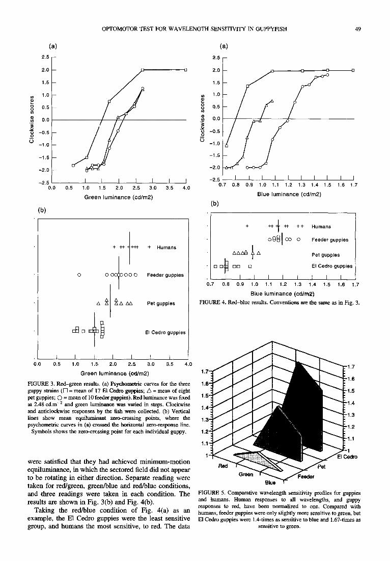
Results are shown in Figs 3-5. Each figure is a psychometric curve in which one color (red in Fig. 3) was held constant across trials while the luminance of the second color (green in Fig. 3) was varied in steps, as shown on the x-axis. The y-axis shows the strength and direction of the fish's turning responses, ranging from - 2 (strong counterclockwise turning) to +2 (strong clock- wise turning). Group mean response curves are shown for the E1 Cedro, pet and feeder guppies. The zero-crossing point, that is the x luminance at which the fishes showed zero net response, is taken as the equiluminance point.
Figure 3(a) shows that in the red/green condition, the green luminance selected as a match for the fixed red luminance of 2.48 cd.m -2 was 1.5 cd.m -2 for the E1 Cedro guppies, 1.87 cd.m -2 for the pet guppies, and 2.12 cd.m -2 for the feeder guppies. Thus, since the E1 Cedro group needed the least green to match the fixed red, they were relatively the most sensitive to green. Figure 3(b) shows the equiluminant zero-crossing points for each individual guppy, and the vertical lines show the mean zero-crossing points, taken from Fig. 3(a).
Figure 4(a) shows that in the red/blue condition, the blue luminance selected as a match for the fixed red luminance of 2.48 cd.m -2 was 0.8 cd.m -2 for the E1 Cedro guppies, 0.99 cd.m -2 for the pet guppies, and 1.2 cd.m -2 for the feeder guppies. Thus, the E1 Cedro group was most sensitive to blue. Figure 4(b) shows the equiluminant zero-crossing points for each individual guppy. These response functions are quite steep, with most of the change in response taking place within 0.2 log units, a value comparable to halfway between infant and adult human subjects.
Comparison with human vision
In order to compare the wavelength sensitivity of guppies with that of humans, psychophysical data were collected from nine humans with normal color vision by a method of adjustment, in conditions that approximated to those used for the fishes. The observer looked down into the globe from above, at a viewing distance of 45 cm. The neck of the globe defined a circular region, 9.5 deg in diameter, of the interior bottom of the globe on which the rotating patterns were projected. It was explained to the subjects that turning the potentiometer clockwise (or anticlockwise) made the perceived motion tend to be more clockwise (or anticlockwise). Subjects were asked to adjust the red, green or blue potentiometer until they
OPTOMOTOR TEST FOR WAVELENGTH SENSITIVITY IN GUPPYFISH 49
( a )
2 . 5 - -
( a )
2.5 -
P 0 0
(I)
0 0
2 . 0 - -
1 . 5 - -
1.0 -
0.5 -
0.0
-0 .5 -
- 1 . 0 -
- 1 . 5 -
-2 .0 -
-2 .5 0.0
(b)
7 /
[ I I I I [ I I 0.5 1.0 1.5 2.0 2,5 3.0 3.5 4.0
G r e e n l u m i n a n c e ( cd /m2)
0 0 0 ~ 0 0 0
+ Humans
Feeder guppies
Pet guppies
El Cedro guppies
0 0
0
0 O
2.0 - [3 []
1.5
1.0
0.5
0.0
-0 .5
-1 .0
-1 .5
-2 .0
- 2 . 5 I I I I I I I I 0.7 0.8 0.9 1.0 1.1 1.2 1.3 1.4 1.5
B lue l u m i n a n c e (cd/m2)
(b )
I I 1 . 6 1 . 7
++ 4 -H- + + Humans
O ~ l O0 0 Feeder guppies
zxzxz~ ~ zx L
[] D(~ DD D !
I I [ I I I 0.7 0.8 0.9 1.0 1,1 1.2 1.3
Pet guppies
El Cedro guppies
I I I 1.4 1.5 1.6 1.7
Blue luminance (ca/m2)
FIGURE 4. Red-blue results. Conventions are the same as in Fig. 3.
I I I I I I I 0.0 0.5 1.0 1.5 2.0 2.5 3.0 3.5 4.0
G r e e n l u m i n a n c e (cd /m2) 1"7 t
1 FIGURE 3. Red-green results. (a) Psychometric curves for the three 1.6 guppy strains (I-I = mean o f 17 El Cedro guppies; /k = mean o f eight pet guppies; C) = mean of 10 feeder guppies). Red luminance was fixed 1.5 at 2.48 cd.m -2 and green luminance was varied in steps. Clockwise 1.4-," and anticlockwise responses by the fish were collected. (b) Vertical lines show mean equilumhaant zero-crossing points, where the 1.3' psychometric curves in (a) crossed the horizontal zero-response line.
Symbols shows the zero-crossing point for each individual guppy. 1.2'
1.1'
1' were satisfied that they had achieved minimum-motion equiluminance, in which the sectored field did not appear to be rotating in either direction. Separate reading were taken for red/green, green/blue and red/blue conditions, and three readings were taken in each condition. The results are shown in Fig. 3(b) and Fig. 4(b).
Taking the red/blue condition of Fig. 4(a) as an example, the E1 Cedro guppies were the least sensitive group, and humans the most sensitive, to red. The data
"1.7
• 1 . 6
" 1 . 5
" 1 . 4
" 1 . 3
- 1 . 2
• 1.1
"1 ;edm
FIGURE 5. Comparative wavelength sensitivity profiles for guppies and humans. Human responses to all wavelengths, and guppy responses to red, have been normalized to one. Compared with humans, feeder guppies were only slightly more sensitive to green, but E1 Cedro guppies were 1.4-times as sensitive to blue and 1.67-times as
sensitive to green.

50 S. ANSTIS et al.
from Figs 3 and 4 are replotted in Fig. 5 to show the ratio 30- of guppy to human wavelength sensitivities, with the data normalized for red. Compared with humans, the guppies 25 were strongly protan; the feeder guppies were 9% more sensitive to green light than humans, whereas the pet guppies were 20% more sensitive to blue light and 39% ~ 20 more sensitive to green light, and the E1 Cedro guppies were 50% more sensitive to blue light and 67% more ~ t5 sensitive to green light. This is consistent with Levine .~ and MacNichols' assertion (Levine & MacNichol, 1982) _~ that guppies are sensitive primarily to greens and blues. ~_ to
Transitivity
Our data suggest that the apparent-motion paradigm taps a single mechanism in each potential direction of motion, and the responses in this mechanism are nulled at the "isoluminance" point. However, that does not specifically address interactions (possibly among multi- ple color-vision channels) that may occur upstream of the site where the motion null is computed. A good example of this is the influence of rods on trichromatic human vision. Another example is some failures of transitivity that we found in our fishes. An example of transitivity is that if A = B and B = C, then A = C. If transitivity held in our experiments, as it should do, then when red was matched separately to green and to blue, the chosen green should match the chosen blue when compared directly. We tested for transitivity by using the results from the red/green and red/blue conditions to predict a match for green and blue, and then testing this prediction with a third, green/blue condition in which the disk had 32 green sectors and 32 blue sectors. The green sectors were held constant at the luminance value previously selected by the fish as a match to the standard red, while the blue sectors were varied in 22 steps over a range from 0.73 to 1.88 cd.m -2. The cyan sectors in this condition were: light cyan (x = 0.303, y = 0.303, 0.445 cd.m-2), and dark cyan (x = 0.249, y = 0.307, 0.382 cd.m-2).
Results for this green/blue test of transitivity showed an unexplained failure of transitivity, in which the predicted blue matches were higher than those actually made. Fishes selected a lower level of blue when selecting a direct match with green than would be predicted by calculation from their red/green and red/blue matches, and their mean transitivity, defined as (actual blue luminance selected as direct match)/(blue luminance predicted from red/green and red/blue matches), was 90.6% +0.38% instead of the ideal 100%. Transitivity for the human group was 103.3%--very close to perfect.
Incidentally, an observer, whether human or fish, would select a green or blue luminance of 2.48 cd.m -2 as a match for red 2.48 cd.m -2 if, and only if, his sensitivity were just the same as that of the Minolta Chromameter, which is somewhat likely for humans (the spectral sensitivity of the Chromameter is designed to mimic the human response) but not so likely for fish.
Contrast dependence of the optomotor response
We measured the optomotor response of guppies as a
0 0.0
j i i i i
0.1 0.2 0.3 0.4 0.5 0.6 0.7 0.8 0.9 1.0
Contrast
FIGURE 6. Optomotor responses of pet guppies (IlL goldfish (z~), black oscars (O) and zebra Danios (l-q) to the Michelson contrast of achromatic stimuli. Responses were maximal for contrasts above 0.3.
function of the Michelson contrast of moving achromatic luminance patterns. For comparison purposes we also measured the responses of goldfish, black oscar and zebra Danio fish. Rotating disks with 64 sharp-edged sectors were used as before, but now the sectors were set to two shades of gray, Lmax and Lmin. The contrast, which is defined as (Lmax - Lmin)/(Lmax+Lmin), was set on different trials to values of 0.1, 0.3, 0.5, 0.7 and 0.9. These were presented in random order for periods of 30 sec each. Note that these achromatic stimuli, unlike the colored patterns that we used hitherto, rotated unambiguously in one direction and did not contain a mixture of clockwise and counterclockwise energy.
The experimenter timed with a computerized stop- watch program the portion of the 30 sec exposure time for which the fish swam clockwise, counterclockwise, or in neither direction. Results are shown in Fig. 6. The optomotor response saturated at a contrast of 0.3, in other words provided that the contrast of the achromatic patterns was greater than 0.3, the fish started turning almost immediately and continued to follow the moving pattern for almost 100% of the time. At lower contrasts the optomotor response fell off markedly. We found that there was a latent period at the beginning of each run before the fish started turning, but once it started turning it continued to do so for the remainder of the 30 sec. This latent period was minimal at high Michelson contrasts and was longer for contrasts below 0.3. Results were very similar for all four species of fish tested.
Signal~noise ratio and the motion coherence threshold
We noticed informally that if any conspicuous stationary objects were visible to the fish it would stop following the moving display. Accordingly we measured the sensitivity of the guppy optomotor response to the signal/noise ratio of the motion stimulus. Instead of using

OPTOMOTOR TEST FOR WAVELENGTH SENSITIVITY IN GUPPYFISH 51
stationary objects as noise, we introduced a percentage of twinkling, dynamic noise; dots which did not controvert the motion signal by providing stationary landmarks but did weaken the motion by adding noise. A field of drifting random dots gives a strong motion signal. The motion signal is weaker if some percentage of the dots is made to twinkle or flicker (dynarnic visual noise), and the signal/ noise ratio can be regarded as an index of the strength of the motion stimulus (Williams & Sekuler, 1984; Van de Grind, Koenderink, & van Doom, 1987; Newsome & Parr, 1988).
A field of black and white random dots rotated at 9.25 rpm. The dots were either fine (2 mm wide) or coarse ( 4 m m wide). On different trials, different percentages of dots were made to flicker, alternating between black and white. This percentage varied from zero up to 100% in steps of 6.8% (coarse dots) or 10% (fine dots). The stimuli were presented in random order. When none of the dots were twinkling, the signal/noise ratio was 100% and a solid field of drifting random dots was seen, and when all of dots were twinkling the signal/ noise ratio was 0% and no motion was seen. As before, the swimming of the fishes was timed over a 30-sec period in each condition.
Results are shown in Fig. 7. As in the contrast experiment, there was a latent period at the beginning of each run which was )onger at very low signal/noise ratios, but provided that the signal/noise ratio of the achromatic patterns was greater than 20% for the fine dots or 40% for the co~a'se dots, the fish followed the moving pattern for almost 100% of the time. At lower signal/noise ratios the latency increased and the opto-
motor response fell off. The results for signal/noise ratio were similar to those for Michelson contrast.
DISCUSSION
Note that the apparent motion in our colored stimuli depends upon the luminous efficiency of the different colors. This means that we are measuring wavelength sensitivity, not "color vision"; our results tell us about the contribution made by the fishes' retinal receptors to the luminance pathways, not to the opponent-hue pathways (Fig. 8). Stated differently, a color TV camera pointed at a colored scene will render the hues of the scene, while a monochrome ("black and white") TV camera will not render the hues but will convert them into different shades of gray. In principle, one style of monochrome camera might be more sensitive to long wavelengths, rendering red as light gray and blue as dark gray, while another style might be more sensitive to short wave- lengths, rendering blue as a lighter gray than red. Our test, if applied to cameras, would distinguish between these different styles of monochrome camera, but it could never tell whether a given camera were color or monochrome. There is plenty of evidence, reviewed in our Introduction, to show that fishes have excellent color vision, but our results are consistent with the possibility that optomotor responses are driven by a "gray-scale" picture, which is the output from the luminance path- ways, not by a "colored picture", which is output from the opponent-hue pathways. We have described elsewhere some variations of our method which do measure the contribution of opponent-hue pathways to motion (Anstis
30
25
~ 2 0
E . m
.c: E 15 E 3= t / )
~- 10 i
! 1
| i i i i 1
0 10 20 30 40 50 60 70
% Motion
8
i i
80 90 100
FIGURE 7. Guppy optomotor responses as a function of the signal/noise ratio of the motion stimulus. Responses were maximal for signal/noise ratios exceeding 20% for fine dots (O) or 40% for coarse dots ([5]).

52 S. ANSTIS et al.
~ Cones I Luminance I Io""xnen' I
FIGURE 8. Our experiment measured the luminous efficiency of the contributions of long, medium and short wavelength receptors as they combine additively into the luminance pathways to drive optomotor responses (black arrow). It did not measure any motion produced by subtractive combinations from receptors into the opponent-hue
pathways (gray arrow).
et al., 1986; Cavanagh & Anstis, 1991). Doubtless these opponent-hue tests could be administered to fishes.
Our results are consistent with Schaerer and Neu- meyer 's findings (Schaerer & Neumeyer, in press) that the optomotor response in goldfish is "color blind". These authors found that in the dark adapted fish, the action spectrum showed a single maximum in the range of 500- 520nm, and resembled the rod spectral sensitivity function. Surprisingly, the action spectrum in the light adapted fish also showed a single maximum, this time between 620 and 660 nm, consistent with the spectral sensitivity of the L-cones. They demonstrated the "color- blindness" of the optomotor response with a rotating drum of red and green stripes whose intensities could be varied independently: the optomotor response disap- peared when the red and green stripes were made "equiluminous". In this condition, calculations showed that the L-cones were only slightly modulated by a red- green stimulus, although the M-cones were modulated strongly. Neumeyer & Schaerer (1992) found the same photopic action spectrum when they trained goldfish to discriminate between steady and flickering light: the discrimination disappeared at "equiluminance", so it seems probable that motion and flicker are processed by the same neural pathways, as has been suggested for humans (Anstis & Cavanagh, 1983). Srinivasan (1985) offered a theoretical model for the color blindness of motion perception.
In humans, matters are more complex. Motion perception disappears for equiluminous colored moving patterns, and this has led to the proposal of separate pathways for color and motion (Anstis, 1970; Ramachan- dran & Gregory, 1978; Cavanagh, Tyler, & Favreau, 1984b; Livingstone & Hubel, 1987; Logothetis, 1991). However, Gegenfurtner, Hawken, & Tang (1994) and Gegenfurtner & Hawken (1995, 1996) measured the perceived velocity of moving luminous and chromatic targets, and found that slow moving stimuli defined by
chromaticity or by second-order statistics were processed in a different manner from luminance defined stimuli. They identified two mechanisms contributing to motion: a low-temporal-pass, color-opponent mechanism and a high-temporal-pass luminance mechanism, and they concluded that motion perception is the result of the interplay between a number of independent mechanisms that process motion targets. Note that the apparently greater complexity of motion perception in humans compared with fishes might simply be a by-product of the superior psychophysical methods available for humans.
We found that the video projector was an essential element in providing a "wrap-around" stimulus to drive optomotor responses. In pilot work we put the fishes on, near, or between fiat television screens but we could never obtain following responses until we purchased the video projector. At present, the technique is limited to the colors produced by television phosphors. It would be a considerable improvement if one could incorporate true monochromatic hues generated optically.
In summary, the method described in this paper suggests different degrees of green- and blue-sensitivity in store-bought vs wild-strain guppyfish. This method, which has already proved useful for human adults and babies (Anstis et al., op. cit) and for monkeys (Logothetis & Charles, 1990) has the advantage of providing a reversal in direction of swimming, rather than a mere cessation of response at the equiluminous point, and it should be readily applicable to other species to allow direct comparisons of wavelength sensitivity across species in similar experimental conditions.
REFERENCES
Anstis, S. M. (1970). Phi movement as a subtraction process. Vision Research, 10, 1411-1430.
Anstis, S. M. (1986). Visual stimuli on the Commodore Amiga: A tutorial. Behavioral Research Methods, Instrumentation, & Computers, 18, 535-541.
Anstis, S. M. & Cavanagh, P. (1983). A minimum-motion technique for judging equiluminance In Mollon, J. & Sharpe, L. T. (Eds), Color vision: physiology and psychophysics. London: Academic Press.
Anstis, S. M. & Paradiso, M. (1989). Programs for visual psycho- physics on the Amiga: A tutorial. Behavioral Research Methods, Instrumentation, & Computers, 21,548-563.
Anstis, S. M., Cavanagh, P., Maurer, D. & Lewis, T. (1987). Optokinetic technique measures infants' responses to color. Applied Optics, 26, 1510-1516.
Anstis, S. M., Cavanagh, P., Maurer, D., Lewis, T., Macleod, D. I. A. & Mather, G. (1986). Computer-generated screening test for colorblindness. Color Research and Application, 11, 63-66.
Anthony, P. D. & Hawkins, A. D. (1983). Spectral sensitivity of the cod Gadus morhua. Marine Behavior and Physiology, 10, 145-166.
Cameron, N. E. (1982). The photopic spectral sensitivity of a dichromatic teleost fish (Perca fluviatilis). Vision Research, 22, 1341-1348.
Cavanagh, P. & Anstis, S. M. (1991). The contribution of color to motion in normal and color-deficient observers. Vision Research, 31, 2109-2148.
Cavanagh, P., Anstis, S. M. & Mather, G. (1984b). Screening for colour blindness using optokinetic nystagmus. Investigative Ophthalmology & Visual Science, 25, 463--466.
Cavanagh, P., Tyler, C. W. & Favreau, O. E. (1984b) Perceived

OPTOMOTOR TEST FOR WAVELENGTH SENSITIVITY IN GUPPYFISH 53
velocity of moving chromatic gratings. Journal of the Optical Society of America A, 1, 893-899.
Cronly-Dillon, J. R. & Muntz, W. R. A. (1965). The spectral sensitivity of the goldfish and the clawed tadpole under photopic conditions. Journal of Experimental Biology, 42, 481--493.
Cronly-Dillon, J. R. & Sharma, S. C. (1968). Effect of season and sex on the photopic spectral sensitivity of the three-spined stickleback. Journal of Experimental Biology, 49, 679-687.
Endler, J. A. (1983). Natural and sexual selection on color patterns in poeciliid fishes. Environmental Biology of Fishes, 9, 173-190.
Gegenfurtner, K. R. & Hawken.~ M. J. (1995). Temporal and chromatic properties of motion mechanisms. Vision Research, 35, 1547-1563.
Gegenfurtner, K. R. & Hawken, M. J. (1996). Perceived velocity of luminance, chromatic and non-Fourier stimuli: influence of contrast and temporal frequency. Vision Research, 36, 1281-1290.
Gegenfurtner, K. R., Hawken.. M. J. & Tang, C. (1994). Contrast dependence of colour and lmninance motion mechanisms in human vision. Nature, 367, 268-27(I.
Levine, J. S. & MacNichol, E. G. (1982). Color vision in fishes. Scientific American, 246, 140-149.
Lindsey, D. T. & Teller, D. Y. (1990). Motion at isoluminance: discrimination/detection ratios for moving isoluminant gratings. Special Issue: Optics, physiology and vision. Vision Research, 30, 1751-1761.
Livingstone, M. S. & Hubel, 1-). H. (1987). Psychophysical evidence for separate channels for the perception of form, movement, and depth. Journal of Neuroscience, 7, 3416-3468.
Logothetis, N. K. (1991). Is movement perception color blind?. Current Biology, 1, 298-300.
Logothetis, N. K. & Charles, E. R. (1990). The minimum motion technique applied to determine isoluminance in psychophysical experiments with monkeys. Vision Research, 30, 829-838.
Maurer, D., Lewis, T., Cavanagh, P. & Anstis, S. M. (1989). A new test of luminous efficiency in babies. Investigative Ophthalmology and Visual Science, 30, 297-303.
Neumeyer, C. & Schaerer, S. (1992). Two separate pathways in the processing of L-cone type information in goldfish: color and acuity vs brightness and flicker, hwestigative Ophthalmology & Visual Science, 33, 703.
Newsome, W. T. & ParE, J. B. (1988). A selective impairment of motion perception following lesions of the middle temporal area (MT). Journal of Neuroscience, 8, 2201-2211.
Northmore, D. P. M. & Yager, D. (1975). Psychophysical methods for investigations of vision in fi,;hes. In Ali, M. (Ed.), Vision in fishes. New York: Plenum Press.
Powers, M. K. & Easter, S. S. (1978a) Absolute visual sensitivity of the goldfish. Vision Research, 18, 1137-1148.
Powers, M. K. & Easter, S. S. (1978b) Wavelength discrimination by the goldfish near absolute visual threshold. Vision Research, 18, 1149-1154.
Protasov, V. R. (1970). Vision and near orientation offish. Translated from the Russian by M. Hadeh. Jerusalem: Israel Program for Scientific Translation.
Ramachandran, V. S. & Gregory, R. L. (1978). Does colour provide an input into human motion perception?. Nature, 275, 55-56.
Rock, I., Tauber, E. S. & Heller, D. P. (1964). Perception of stroboscopic movement: evidence for its innate basis. Science, 147, 1050-1052.
Shao, J., Lin, G. & Chen, S. (1981). The photo-kinetic reaction of amphioxus (Branchiostoma belchari). Acta Psychologica Sinica, 13, 358-363.
Silver, P. H. (1974). Photopic spectral sensitivity of the neon tetra (Paracheirodon innesi (Myers)) found by the use of a dorsal light reaction. Vision Research, 14, 329-334.
Srinivasan, M. V. (1985). Shouldn't directional movement detection necessarily be "colour-blind"?. Vision Research, 25, 997-1000.
Teller, D. Y. & Lindsey, D. T. (1993a). Motion nulling techniques and infant color vision. In Granrud, C. (Ed.), Visual perception and cognition in infancy (pp. 47-73). Carnegie Mellon symposia on cognition. Hillsdale, NJ: Lawrence Erlbaum.
Teller, D. Y. & Lindsey, D. T. (1993b). Infant color vision: OKN techniques and null plane analysis. In: Simons, K. (Ed.), Early visual development: normal and abnormal (pp. 143-162). National Research Council. New York, NY: Oxford University Press.
Van de Grind, A. A., Koenderink, J. J. & van Doom, A. J. (1987). Influence of contrast on foveal and peripheral detection of coherent motion in moving random dot patterns. Journal of the Optical Society of America A, 4, 1643-1652.
Wagner, G. & Boynton, R. M. (1972). Comparison of four methods of heterochromatic photometry. Journal of the Optical Society of America, 62, 1508-1515.
Wheeler, T. G. (1982). Color vision and retinal chromatic information processing in teleosts: a review. Brain Research Reviews, 4, 177- 235.
Williams, D. W. & Sekuler, R. (1984). Coherent global motion percepts from stochastic local motions. Vision Research, 24, 55-62.
Yager, D. (1968a) Behavioral measure of the spectral sensitivity of the dark-adapted goldfish. Nature, 220, 1052-1053.
Yager, D. (1968b) Behavioral measures and theoretical analysis of spectral sensitivity and spectral saturation in the goldfish. Vision Research, 7, 707-727.
Yager, D. (1970). Spectral sensitivity with the freely moving eye. Vision Research, 10, 521-523.
Acknowledgements--This research was supported by Grant A0260 from NSERC (Canada) and EY-10241 from NEI to SMA, and by Grant EY-09258 from NEI to PC. We are grateful to Helen Rodd and Maria Sokolovska for the loan of their E1 Cedro and Aripo guppyfish.