SPECIATION OF SELENIUM IN Pleurotus ostreatus AND Lentinula edodes MUSHROOMS
Upload
khangminh22Category
view
1download
0
RESEARCH ARTICLE
Comparative efficacy of selenate and
selenium nanoparticles for improving growth,
productivity, fruit quality, and postharvest
longevity through modifying nutrition,
metabolism, and gene expression in tomato;
potential benefits and risk assessment
Maryam Neysanian1, Alireza IranbakhshID1*, Rahim Ahmadvand2, Zahra Oraghi Ardebili3,
Mostafa Ebadi4
1 Department of Biology, Science and Research Branch, Islamic Azad University, Tehran, Iran,
2 Department of Seed and Plant Research Improvement Institute, Karaj, Iran, 3 Department of Biology,
Garmsar Branch, Islamic Azad University, Garmsar, Iran, 4 Department of Biology, Damghan Branch,
Islamic Azad University, Damghan, Iran
Abstract
This study attempted to address molecular, developmental, and physiological responses
of tomato plants to foliar applications of selenium nanoparticles (nSe) at 0, 3, and 10 mgl-1
or corresponding doses of sodium selenate (BSe). The BSe/nSe treatment at 3 mgl-1
increased shoot and root biomass, while at 10 mgl-1 moderately reduced biomass accumu-
lation. Foliar application of BSe/nSe, especially the latter, at the lower dose enhanced fruit
production, and postharvest longevity, while at the higher dose induced moderate toxicity
and restricted fruit production. In leaves, the BSe/nSe treatments transcriptionally upregu-
lated miR172 (mean = 3.5-folds). The Se treatments stimulated the expression of the bZIP
transcription factor (mean = 9.7-folds). Carotene isomerase (CRTISO) gene was tran-
scriptionally induced in both leaves and fruits of the nSe-treated seedlings by an average
of 5.5 folds. Both BSe or nSe at the higher concentration increased proline concentrations,
H2O2 accumulation, and lipid peroxidation levels, suggesting oxidative stress and impaired
membrane integrity. Both BSe or nSe treatments also led to the induction of enzymatic
antioxidants (catalase and peroxidase), an increase in concentrations of ascorbate, non-
protein thiols, and soluble phenols, as well as a rise in the activity of phenylalanine ammo-
nia-lyase enzyme. Supplementation at 3 mgl-1 improved the concentration of mineral
nutrients (Mg, Fe, and Zn) in fruits. The bioaccumulated Se contents in the nSe-treated
plants were much higher than the corresponding concentration of selenate, implying a
higher efficacy of the nanoform towards biofortification programs. Se at 10 mgl-1, espe-
cially in selenate form, reduced both size and density of pollen grains, indicating its poten-
tial toxicity at the higher doses. This study provides novel molecular and physiological
PLOS ONE
PLOS ONE | https://doi.org/10.1371/journal.pone.0244207 December 18, 2020 1 / 17
a1111111111
a1111111111
a1111111111
a1111111111
a1111111111
OPEN ACCESS
Citation: Neysanian M, Iranbakhsh A, Ahmadvand
R, Oraghi Ardebili Z, Ebadi M (2020) Comparative
efficacy of selenate and selenium nanoparticles for
improving growth, productivity, fruit quality, and
postharvest longevity through modifying nutrition,
metabolism, and gene expression in tomato;
potential benefits and risk assessment. PLoS ONE
15(12): e0244207. https://doi.org/10.1371/journal.
pone.0244207
Editor: Tamil Selvan Anthonymuthu, University of
Pittsburgh, UNITED STATES
Received: July 17, 2020
Accepted: December 4, 2020
Published: December 18, 2020
Copyright: © 2020 Neysanian et al. This is an open
access article distributed under the terms of the
Creative Commons Attribution License, which
permits unrestricted use, distribution, and
reproduction in any medium, provided the original
author and source are credited.
Data Availability Statement: All relevant data are
within the paper and its Supporting Information
files.
Funding: The authors received no specific funding
for this work.
Competing interests: The authors have declared
that no competing interests exist.
insights into the nSe efficacy for improving plant productivity, fruit quality, and fruit post-
harvest longevity.
Introduction
Nowadays, diverse attempts have been made to improve productivity, stress tolerance, and dis-
ease management in crops as well as to produce biofortified seeds or fruits containing minerals
essential for humans. Zinc (Zn), iron (Fe), and selenium (Se) are the most important mineral
nutrients that are often considered in biofortification programs. In this regard, the daily nutri-
tional requirement of Se is estimated to be 55 micrograms. Taking nano-fertilizers and nano-
pesticides into account, nanotechnology has provided a great opportunity to improve crop
productivity and crop protection and has solved some of the challenges that we face today in
agriculture. It has been highlighted in recent studies that bioavailability and function of Se in
plant growth and metabolism were significantly more efficient in form of nanoparticles (nSe)
compared to other natural Se forms such as selenate and selenite [1–6]. On the other hand,
there is evidence that nSe use could potentially compromise plant growth and development [3,
5, 6]. However, the potential benefit or phytotoxicity of nSe is still controversial. Therefore,
more detailed investigations are necessary to fill the knowledge gaps.
In plants, microRNAs (miRNAs) are transcriptionally modulated by endogenous and sur-
rounding environmental cues [7]. Most miRNAs are efficiently involved in adjusting the percep-
tion/signaling of phytohormones, as well as regulating plant growth and development through
transcriptional and posttranscriptional regulations of a multitude of target genes [8]. Some miR-
NAs play a significant role in adapting plants to different types of environmental stresses [7, 8].
Moreover, the flowering phase and fruit development are rigorously controlled through orches-
trated molecular strategies in which miRNAs play critical roles at transcriptional and post-tran-
scriptional levels [7]. In this regard, microRNA172 (miR172) is one of the most important
miRNAs playing critical roles in controlling developmental programs, particularly at the flowering
phase, as well as in conferring stress tolerance [8, 9]. To the best of our best knowledge, there has
been no study investigating the role of miRNAs during plant responses to nanoparticles. In this
study, we attempted to monitor transcriptional rates of miR172 following the nSe applications.
Carotenoid pigments are natural organic substances derived from an isoprene precursor.
These pigments are involved in plant protection against oxidative stress and the photoinhibition
process. Furthermore, these antioxidant metabolites provide numerous advantages to the health
maintenance of humans owing to their great antioxidant characteristics. Therefore, investigating
the effects of environmental cues, chemicals, and fertilizers on the concentrations of carotenoids
is of great importance. In carotenoid production, the carotene isomerase (CRTISO) is one of the
most important genes involved in the biosynthesis of carotenoids through mediating the conver-
sion of z carotene to trans-lycopene [10–12]. CRTISO is widely considered in plant biotechnology
programs to improve fruit quality and plant resistance to stresses [10–12]. Hence, the concentra-
tion of carotenoids in tomato fruit is considered an important aspect of fruit quality. Therefore,
we aimed to highlight the potential alterations in fruit production, transcription of the CRTISOgene, and fruit longevity following foliar applications of nSe or selenate as a bulk (BSe).
The basic leucine zipper (bZIP) transcription factors are involved in a plethora of plant bio-
logical processes and responses, including signal transduction [6, 13], tissue differentiation
[13], hormone signaling [14], nutrition [15], and stress tolerance [6, 16]. For this reason, evalu-
ating the potential nSe-associated alteration in the bZIP transcription factor was also one of
the aims of this study.
PLOS ONE Comparative efficacy of selenate and selenium nanoparticles
PLOS ONE | https://doi.org/10.1371/journal.pone.0244207 December 18, 2020 2 / 17
Within this framework, this study was carried out to address the possible responses of tomato
seedlings to foliar application of BSe or nSe at different concentrations. In this manuscript, for the
first time, we aimed to address transcriptional responses of miR172, CRTISO, and bZIP genes to
foliar application of bulk Se (selenate) or nano counterpart (nSe) at the same concentration.
Moreover, this study provides anatomical evidence highlighting the potential risk of Se or nSe on
the development of pollen grains for the first time. Taking knowledge gaps into account, we
attempt to present comparative comprehensive data on the Se- or nSe-mediated changes in plant
growth, physiology, metabolism, the transcriptional program of genes, crop productivity, fruit
quality, and fruit postharvest longevity to gain new insights into the benefits or the risk associated
with Se function in agriculture. The main aims of this experiment are documenting the nSe-asso-
ciated changes in (I) plant growth and flowering time (II) fruit production, quality, and posthar-
vest life, (III) development of pollens in stamen, (IV) transcriptional responses of miR172,
CRTISO, and bZIP genes, (V) enzymatic (catalase and peroxidase) and non-enzymatic (ascorbate
and thiols) antioxidants, (VI) H2O2 concentration and membrane stability, (VII) proline concen-
tration (a multifunctional protective amino acid), and (VIII) nutritional status in fruits.
Material and methods
Experimental conditions
Red elemental nSe (1000 mgl−1 containing polyvinylpyrrolidone (PVP) as a stabilizer;
CAS#7446-08-4; APS:10–40 nm; true density: 3.89 gcm−3; morphology: near-spherical) was
supplied from NanoSany Corporation, Mashhad, Iran. A sodium selenate (Na2SeO4) was sup-
plied from TitraChem, Tehran, Iran.
In soilless conditions, seeds were grown in pots containing perlite and vermiculite (3:1) and
irrigated with a Hoagland nutrient solution. The 37-day-old seedlings in the treatment groups
were sprayed with different doses of nSe (0, 3, and 10 mgl-1) dissolved in deionized water and
those in the control groups with different doses of sodium selenate (bulk Se (BSe); 0, 3, and 10
mgl-1). Seedlings were treated six times within 42 days, with one-week intervals between treat-
ments. Growth-related traits (shoot fresh mass and root fresh mass), flowering time, number
of fruits, and fruit postharvest longevity were recorded. Furthermore, molecular and physio-
logical responses were monitored in leaves and fruits.
Transcriptions of miR172, CRTISO, and bZIP genes
Total RNA was extracted using an RNA extraction kit (Promega-GRE#AS1500) from both
leaves and fruits. The forward and reverse sequences (5’-3’) of primers for miR172, bZIP,
CRTISO, and GAPDH genes are displayed in Table 1. Quantitative polymerase chain reaction
(qPCR) was performed using mi-1STEP RT-qPCR kit (Metabione, Co-GRE#mi-E8105), the
transcription level of target genes were quantified using a PCR instrument (QIAGEN’s real-
time PCR cycler, Rotor-Gene Q) under the program specified by the manufacturer (reverse
transcription in 50˚C for 10–15 min; initial denaturation in 95˚C for 5 min; denaturation in
95˚C for 15 sec; annealing and elongation in 60–65˚C for 1 min). Transcription rate was calcu-
lated using the equation 2-ΔΔCT in which ΔCT was calculated by subtracting the internal con-
trol CT value from the CT amount of each target gene.
Hydrogen peroxide (H2O2) concentration and lipid peroxidation level
H2O2 concentrations in leaves of BSe/nSe-treated plants were measured according to the pro-
cedure described by Sheteiwy et al. [17]. Briefly, 0.1% trichloroacetic acid (TCA) was applied
to prepare leaf extract. After that, the resulting extract was centrifuged and the supernatant
PLOS ONE Comparative efficacy of selenate and selenium nanoparticles
PLOS ONE | https://doi.org/10.1371/journal.pone.0244207 December 18, 2020 3 / 17
was separated. The reaction mixture consisted of 1000μL KI (1 mM), 500μL of potassium
phosphate buffer (10 mM, pH 7.0), and 500 μL supernatant. Next, the absorbance was spectro-
photometrically recorded at 390 nm. H2O2 concentration was calculated according to the
equation of a standard curve. Membrane integrity was also evaluated by quantifying Malon-
dialdehyde (MDA) contents in leaves according to the protocol described by Salah et al. [18]
with a small modification. Briefly, fresh leaf tissue was grounded in 5 ml of 0.25% thiobarbitu-
ric acid (TBA) in trichloroacetic acid (TCA) (0.25 g TBA was dissolved in 100 ml of 10%
TCA). The resulting homogenate was then placed at 95˚ for 20 minutes. Next, the samples
were quickly cooled in an ice bath and centrifuged. The supernatant absorption was spectro-
photometrically measured at 600 and 532 nm. Calculation of MDA was performed using the
extinction coefficient of 155 mM-1cm and expressed in micromol per gram fresh weight (μMg-
1fw).
Activities of peroxidase, catalase, and phenylalanine ammonia-lyase (PAL)
enzymes
In order to extract enzymes, phosphate buffer (0.1 M; pH of 7.2) was applied to the liquid
nitrogen-grounded leaves. Next, the homogenate was centrifuged at 4˚C and the resulting
supernatant was stored at −80˚C until enzyme analysis. The nSe-mediated variations in
defense-related enzymes, including peroxidase [19], catalase [18], and PAL [20] were quanti-
fied in leaves. The peroxidase reaction was triggered by the addition of 100 μl enzyme extract
to the reaction medium containing 100 μl guaiacol, 100 μl H2O2, and 2700 μl phosphate buffer
(25 mM, pH 7.1). To estimate enzyme activity, the absorbance difference was monitored at
470 nm. Finally, the peroxidase activity was expressed in terms of Unit enzyme per gram fresh
weight (UnitEg-1fw). To determine catalase activity, the reaction mixture containing 200 μl
H2O2, 200 μL enzyme extract, and 2600 μl phosphate buffer (25 mM, pH 7.1) were prepared.
In this assessment, a decrease in absorbance at 240 nm was recorded following the addition of
H2O2. To quantify PAL activity in leaves, 200 μl enzyme extract was added to the reaction mix-
ture containing 6 μM phenylalanine and Tris-HCl buffer (500 mM, pH8). After that, the reac-
tion mixture was incubated at 37˚C for 1 hour. Then, the PAL reaction was stopped by the
addition of 50 μl HCl (5 N). Finally, the PAL activity was determined according to the cinna-
mate standard curve and calculated in terms of microgram cinnamate per hour per gram fresh
weight (μgCin.h−1g−1fw).
Characterization of Mg, Fe, Zn, and Se concentrations
Ash solution was prepared by thermal decomposition in a furnace (650˚C) and subsequently
dissolving in nitric acid and hydrochloric acid (2:1). Mg, Zn, and Fe contents were assessed
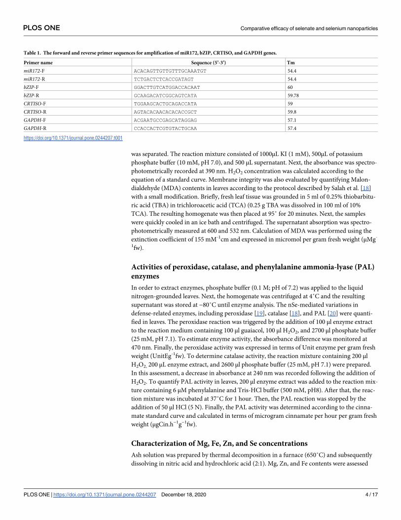
Table 1. The forward and reverse primer sequences for amplification of miR172, bZIP, CRTISO, and GAPDH genes.
Primer name Sequence (5’-3’) Tm
miR172-F ACACAGTTGTTGTTTGCAAATGT 54.4
miR172-R TCTGACTCTCACCGATAGT 54.4
bZIP-F GGACTTGTCATGGACCACAAT 60
bZIP-R GCAAGACATCGGCAGTCATA 59.78
CRTISO-F TGGAAGCACTGCAGACCATA 59
CRTISO-R AGTACACAACACACACCGCT 59.8
GAPDH-F ACGAATGCCGAGCATAGGAG 57.1
GAPDH-R CCACCACTCGTGTACTGCAA 57.4
https://doi.org/10.1371/journal.pone.0244207.t001
PLOS ONE Comparative efficacy of selenate and selenium nanoparticles
PLOS ONE | https://doi.org/10.1371/journal.pone.0244207 December 18, 2020 4 / 17
using Atomic Absorption Spectroscopy (AAS; Varian, Spectr AA.200). Furthermore, Se con-
centration was measured using a fully automatic Flame/Graphite Furnace AASAA.200;
YOUNGLIN AAS 8020).
Ascorbate, non-protein thiols, proline, and soluble phenols
The concentrations of non-protein thiols were quantified in leaves according to the protocol
described by Del Longo et al. [21]. The leaves were homogenized in a 5% (w/v) solution of sul-
fosalicylic acid. The assay reaction of non-protein thiols consisted of 500μl DTNB (1 mM),
500μl phosphate buffer (100 mM, pH 7.1), 100μl of leaf extract, and 500 μM EDTA. After incu-
bation for 10 minutes, the absorbance amount was recorded at 412 nm. The ascorbate contents
were measured in leaves according to the protocol described by De Pinto et al. [22]. Briefly,
metaphosphoric acid (5%) was used to prepare the leaf extract. Then, the resulting extract was
centrifuged and the supernatant was separated. The reaction mixture was a 100 μl leaf extract,
250 μl phosphate buffer (50 mM, pH 7.2) supplemented with 50μl dithiothreitol (DTT; 10
mM), and 5 mM EDTA. After that, this mixture was incubated at room temperature for 10
minutes. After incubation for 10 min at room temperature, the process was followed by the
addition of 150 μl N-ethylmaleimide (0.5%). Next, 200 μl dipyridyl of 4% in 70% ethanol,
200 μl orthophosphoric acid (44%), 200μl trichloroacetic acid (TCA; 10%), and 0.3% (w/v)
FeCI3 were added and the mixture was vortexed. After incubation at 40˚C for 40 minutes, the
absorbance of each sample was recorded at 525 nm. Ascorbate concentration was calculated
based on the standard curve equation of ascorbic acid. The concentration of proline amino
acid in leaves was determined following the protocol described by Bates et al., [23]. To prepare
leaf extract for proline assay, sulfa salicylic acid (3% w/v) was utilized. The ninhydrin reagent
was prepared by mixing 30 ml acetic acid and 20 ml phosphoric acid (6 M). Next, 2 ml leaf
extract was added to the reaction mixture containing 2 ml ninhydrin reagent and 2ml acetic
acid (glacial). This mixture was heated at a boiling water bath for one hour. Then, this reaction
mixture was immediately cooled down at an ice bath. After that, the reaction was followed by
the addition of toluene solvent and vigorously shaking using a vortex. Then, the absorbance of
the toluene phase was spectrophotometrically recorded at 520 nm. Proline concentration was
estimated by using the equation of the standard curve of proline amino acid. Finally, Folin-
Ciocalteu reagent was utilized to measure soluble phenols in leaves [24]. Briefly, total soluble
phenols were extracted by homogenizing leaves in an ethanol solvent of 80% (v/v) and incu-
bating in a boiling water bath. The reaction was started by the addition of 1 ml leaf ethanolic
extract to a mixture containing 1 ml Folin-Ciocalteu reagent and saturated Sodium carbonate
(21%). After 10 minutes and the development of a blue color, the mixture was centrifuged. The
absorbance of the resulting supernatant was read at 760 nm. The concentration of total soluble
phenols was quantified based on the equation of the standard curve of tannic acid.
Histological study
The flower buds at the same developmental age were fixed in FAA fixator [6]. Longitudinal
sections were made by a microtome and were stained using Hematoxylin/Eosin staining pro-
tocol. Stained sections were observed at different magnification levels and photographed using
a light microscope [6].
Statistical analysis
The experimental design was completely randomized. Every experiment was independently
performed in the replication of three. All data were subjected to analysis of variance (ANOVA)
PLOS ONE Comparative efficacy of selenate and selenium nanoparticles
PLOS ONE | https://doi.org/10.1371/journal.pone.0244207 December 18, 2020 5 / 17
using GraphPad software. The mean values of the three independent replications were com-
pared using Tukey’s range test.
Results
Growth, biomass, productivity, and fruit postharvest longevity
To understand whether the nSe effectiveness in inducing growth, flowering, and yield changes
are different from selenate, several traits, including shoot biomass, yield, fruit quality, and fruit
postharvest life were evaluated. Compared with controls, both BSe and nSe treatments at 3
mgl-1 significantly increased fresh biomass in the shoot by 23% and 35%, respectively (Fig 1A).
While BSe and nSe treatments at 10 mgl-1 led to a significant decrease in fresh biomass in the
shoot, 31% and 20%, respectively, when compared with controls (Fig 1A). Similarly, treatment
of seedlings with both BSe or nSe at 3 mgl-1 significantly improved fresh biomass in roots, by
an average of 20.75%, whereas treatments at 10 mgl-1 adversely influenced root biomass by an
average of 23% relative to the control (Fig 1B). However, all applied treatments accelerated the
vegetative/reproductive stage switch and reduced flowering time (Fig 1C). The numbers of
produced fruits were increased in response to foliar application of either BSe or nSe (especially
the latter) at 3 mgl-1 by an average of 25.3% compared to the control (Fig 1D). On the other
hand, the BSe or nSe treatment at 10 mgl-1 was associated with toxicity and restricted fruit pro-
duction by a mean of 39.5% when compared to the control (Fig 1D). The fruit fresh mass in
the seedlings treated with BSe3 or nSe at 3 mgl-1 was also higher than the control by an average
of 18% (Fig 1E). The supplements at the high dose (10 mgl-1) not only reduced fruit produc-
tion but also significantly reduced fruit fresh weight (Fig 1E). Finally, treatments at all applied
concentrations, significantly improved fruit postharvest longevity by an average of 38% (Fig
1F).
Transcriptional responses of genes miR172, bZIP transcription factor, and
CRTISOIn addition to the growth-related responses, BSe or nSe-mediated changes in the expression of
target genes were investigated to provide comparative molecular evidence, for the first time. In
leaves, the BSe3, nSe3, BSe10, and nSe10 treatments transcriptionally upregulated miR172 by
2.4, 4.6, 3.2, and 3.9 folds, respectively (Fig 2A). likewise, the BSe or nSe treatments at the
lower concentration (3 mgl-1) moderately stimulated the expression of the bZIP gene
(mean = 5.7 folds) (Fig 2B); while, the supplements at the higher dose (10 mgl-1), mediated
drastic transcriptional upregulation in the bZIP gene (mean = 13.7 folds) (Fig 2B). Similarly,
the transcription of the CRTISO gene was upregulated in leaves of the seedlings treated with
BSe or nSe (mean = 4.5 folds); the highest expression was observed in seedlings treated with
BSe or nSe at 10 mgl-1 (Fig 2C). In fruit, the CRTISO gene was also found to be transcription-
ally upregulated in response to the supplements by an average of 5.9 folds (Fig 2D).
Physiological responses
To evaluate the occurrence of oxidative stress, activation of the defense system, and changes in
metabolism in response to BSe or nSe utilization, several physiological traits were measured.
In this regard, H2O2 concentration and MDA content (membrane integrity index) were moni-
tored to estimate the BSe- or nSe-associated risk of oxidative stress. To monitor the activation
of the defense system, the BSe- or nSe-mediated changes in enzymatic antioxidants (catalase
and peroxidase), non-enzymatic antioxidants (ascorbate and non-protein thiols), and proline
concentration were assayed. PAL activity and concentration of total soluble phenols were
PLOS ONE Comparative efficacy of selenate and selenium nanoparticles
PLOS ONE | https://doi.org/10.1371/journal.pone.0244207 December 18, 2020 6 / 17
PLOS ONE Comparative efficacy of selenate and selenium nanoparticles
PLOS ONE | https://doi.org/10.1371/journal.pone.0244207 December 18, 2020 7 / 17
Fig 1. Variations in shoot and root biomass (a, b), flowering time (c), numbers of produced fruits (d), fruit biomass (e),
and fruit postharvest longevity (f) in response to foliar application of BSe or nSe. Data are Mean ± standard deviation (SD)
of three independent replications (n = 3). Treatment groups: C- Control; BSe3- Bulk Se at 3 mgl-1; nSe3- nSe at 3 mgl-1;
BSe10- Bulk Se at 10 mgl-1; nSe10- nSe at 10 mgl-1; Different letters on top of columns refer statistically significant
difference according to the Tukey’s multiple comparisons test. ns: non-significant; �: 0.01<p�0.05; ��: 0.001<p�0.01; ���:
0.0001<p�0.001; ����: p�0.0001. The asterisk on each column indicates a p-value level for comparing the mean of each
group with the control group. Additionally, a comparison between the two Se groups at the same concentration is
displayed by placing an asterisk (�) on the line.
https://doi.org/10.1371/journal.pone.0244207.g001
Fig 2. BSe or nSe-mediated transcriptional changes in leaf miR172 (a), leaf bZIP transcription factor (b), leaf CRTISO (c), and fruit CRTISO (d); Data are
Mean ± standard deviation (SD) of three independent replications (n = 3). Treatment groups: C- Control; BSe3- Bulk Se at 3 mgl-1; nSe3- nSe at 3 mgl-1;
BSe10- Bulk Se at 10 mgl-1; nSe10- nSe at 10 mgl-1; Different letters on top of columns refer statistically significant difference according to the Tukey’s
multiple comparisons test. ns: non-significant; �: 0.01<p�0.05; ��: 0.001<p�0.01; ���: 0.0001<p�0.001; ����: p�0.0001. The asterisk on each column
indicates a p-value level for comparing the mean of each group with the control group. Additionally, a comparison between the two Se groups at the same
concentration is displayed by placing an asterisk (�) on the line.
https://doi.org/10.1371/journal.pone.0244207.g002
PLOS ONE Comparative efficacy of selenate and selenium nanoparticles
PLOS ONE | https://doi.org/10.1371/journal.pone.0244207 December 18, 2020 8 / 17
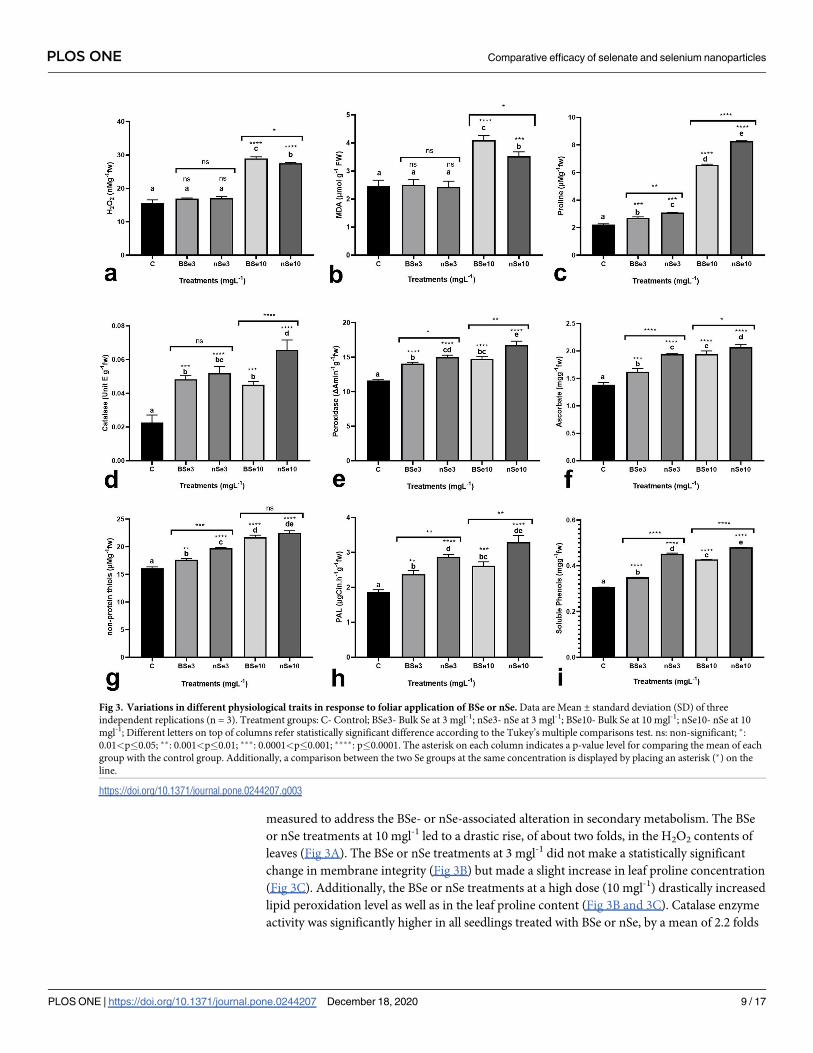
measured to address the BSe- or nSe-associated alteration in secondary metabolism. The BSe
or nSe treatments at 10 mgl-1 led to a drastic rise, of about two folds, in the H2O2 contents of
leaves (Fig 3A). The BSe or nSe treatments at 3 mgl-1 did not make a statistically significant
change in membrane integrity (Fig 3B) but made a slight increase in leaf proline concentration
(Fig 3C). Additionally, the BSe or nSe treatments at a high dose (10 mgl-1) drastically increased
lipid peroxidation level as well as in the leaf proline content (Fig 3B and 3C). Catalase enzyme
activity was significantly higher in all seedlings treated with BSe or nSe, by a mean of 2.2 folds
Fig 3. Variations in different physiological traits in response to foliar application of BSe or nSe. Data are Mean ± standard deviation (SD) of three
independent replications (n = 3). Treatment groups: C- Control; BSe3- Bulk Se at 3 mgl-1; nSe3- nSe at 3 mgl-1; BSe10- Bulk Se at 10 mgl-1; nSe10- nSe at 10
mgl-1; Different letters on top of columns refer statistically significant difference according to the Tukey’s multiple comparisons test. ns: non-significant; �:
0.01<p�0.05; ��: 0.001<p�0.01; ���: 0.0001<p�0.001; ����: p�0.0001. The asterisk on each column indicates a p-value level for comparing the mean of each
group with the control group. Additionally, a comparison between the two Se groups at the same concentration is displayed by placing an asterisk (�) on the
line.
https://doi.org/10.1371/journal.pone.0244207.g003
PLOS ONE Comparative efficacy of selenate and selenium nanoparticles
PLOS ONE | https://doi.org/10.1371/journal.pone.0244207 December 18, 2020 9 / 17
when compared to the controls at all applied concentrations (Fig 3D); similarly, peroxidase
activity and ascorbate concentration were higher in leaves, by an average of 30% and 36%,
respectively (Fig 3E and 3F). With increasing the BSe or nSe concentration, the non-protein
thiols were also increased linearly by an average of 27% (Fig 3G). Furthermore, treatments
with the BSe3 (26.8%), nSe3 (52%), BSe10 (39.6%), and nSe10 (75.3%) treatments significantly
stimulated the PAL activity in leaves (Fig 3H). Likewise, Se treatment, particularly the nSe
treatment, significantly increased concentrations of the soluble phenols by an average of 39%
(Fig 3I).
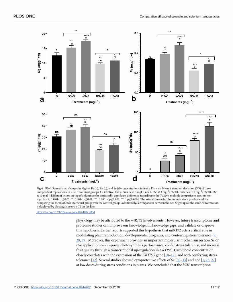
Nutritional status in fruit
Fruit quality is an important determining factor in terms of fruit longevity and human nutri-
tion. In this regard, the BSe or nSe-mediated changes in concentration of several important
minerals were monitored. The Se supplementation altered concentrations of minerals in fruits
in a dose-dependent manner (Fig 4A–4C). At 3 mgl-1, the BSe or nSe, especially the latter, sig-
nificantly increased Mg, Fe, and Zn concentrations in fruits by averages of 29.8%, 27.6%, and
21%, respectively, when compared to the control (Fig 4A–4C); while supplementation at high
dose (10 mgl-1) moderately decreased Mg, Fe, and Zn contents in comparison to the control
(Fig 4A–4C). Furthermore, the foliar applications of both BSe or nSe led to Se-bioaccumula-
tion in tomato fruits (Fig 4D); with nSe treatments leading to a higher amount of bioaccumu-
lated Se in fruits compared with the bulk treatments (Fig 4D).
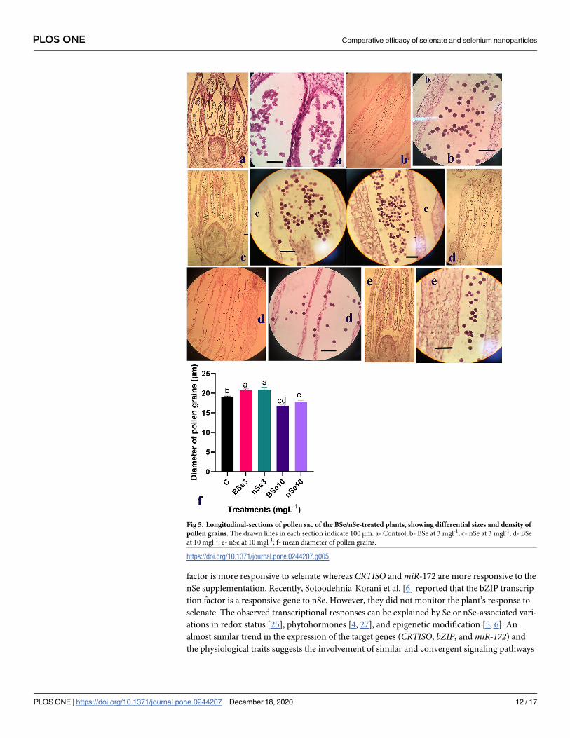
The density of pollen grains in the pollen sac
As mentioned above, our results showed that a high concentration of BSe or nSe significantly
reduced crop yield. Hence, the BSe- or nSe-associated toxicity on the pollen grain development
was evaluated for the first time. Assessment of the longitudinal sections of the flower buds
indicated that the size and density of pollen grains were affected by the BSe/nSe treatments in
a dose- and material type-dependent manners (Fig 5A–5F). The BSe or nSe treatments at all
concentrations were associated with a decreased density of pollen grains in pollen sacs. How-
ever, the size of pollen grains increased with supplementation at 3 mgl-1. It is important to
note that Se treatment at 10 mgl-1, especially in form of BSe, reduced both the size and the den-
sity of pollen grains, highlighting its potential toxicity at the higher dose (Fig 5D–5F).
Discussion
According to our findings, the potential benefits of nSe utilization toward improving plant
growth, biomass accumulation, yield, nutrition, flowering time, fruit quality, and postharvest
longevity were much higher than the use of selenate, and at the same time, the potential nSe-
associated risks are fewer. These results are consistent with several recent reports [5, 6, 25, 26].
The following mechanisms appear to mediate partly differential responses to nSe relative to
the selenate; (I) variations in uptake kinetics (nSe inactive influx through aquaporins vs sec-
ondary active influx mechanism of selenate through symporter) [2], (II) differences in their
interactions with biomolecules and metabolism into the organic forms [2, 26], (III) differential
physicochemical properties of nanoparticles by which a nSe interaction with biomolecules can
be different from that of selenate [2, 5, 6, 26], (IV) variation in a nSe-mediated change in phy-
tohormones [3, 4, 27], and (V) epigenetic modification [5, 6]. However, the involved mecha-
nisms are still poorly characterized which further emphasizes the need for further studies.
This study provides the first comparative evidence on the Se- or nSe-mediated variations in
miRNAs as key regulatory checkpoints. The observed selenate/nSe-associated changes in
developmental vegetative/reproductive switch, fruit-related characteristics, and plant
PLOS ONE Comparative efficacy of selenate and selenium nanoparticles
PLOS ONE | https://doi.org/10.1371/journal.pone.0244207 December 18, 2020 10 / 17
physiology may be attributed to the miR172 involvements. However, future transcriptome and
proteome studies can improve our knowledge, fill knowledge gaps, and validate or disprove
this hypothesis. Earlier reports suggested this hypothesis that miR172 acts a critical role in
modulating plant reproduction, developmental programs, and conferring stress tolerance [9,
28, 29]. Moreover, this experiment provides an important molecular mechanism on how Se or
nSe application can improve photosynthesis performance, confer stress tolerance, and increase
fruit quality through a transcriptional up-regulation in CRTISO. Carotenoid concentration
closely correlates with the expression of the CRTISO gene [10–12], and with conferring stress
tolerance [12]. Several studies showed cytoprotective effects of Se [30–33] and nSe [1, 25, 27]
at low doses during stress conditions in plants. We concluded that the bZIP transcription
Fig 4. BSe/nSe-mediated changes in Mg (a), Fe (b), Zn (c), and Se (d) concentrations in fruits. Data are Mean ± standard deviation (SD) of three
independent replications (n = 3). Treatment groups: C- Control; BSe3- Bulk Se at 3 mgl-1; nSe3- nSe at 3 mgl-1; BSe10- Bulk Se at 10 mgl-1; nSe10- nSe
at 10 mgl-1; Different letters on top of columns refer statistically significant difference according to the Tukey’s multiple comparisons test. ns: non-
significant; �: 0.01<p�0.05; ��: 0.001<p�0.01; ���: 0.0001<p�0.001; ����: p�0.0001. The asterisk on each column indicates a p-value level for
comparing the mean of each individual group with the control group. Additionally, a comparison between the two Se groups at the same concentration
is displayed by placing an asterisk (�) on the line.
https://doi.org/10.1371/journal.pone.0244207.g004
PLOS ONE Comparative efficacy of selenate and selenium nanoparticles
PLOS ONE | https://doi.org/10.1371/journal.pone.0244207 December 18, 2020 11 / 17
factor is more responsive to selenate whereas CRTISO and miR-172 are more responsive to the
nSe supplementation. Recently, Sotoodehnia-Korani et al. [6] reported that the bZIP transcrip-
tion factor is a responsive gene to nSe. However, they did not monitor the plant’s response to
selenate. The observed transcriptional responses can be explained by Se or nSe-associated vari-
ations in redox status [25], phytohormones [4, 27], and epigenetic modification [5, 6]. An
almost similar trend in the expression of the target genes (CRTISO, bZIP, and miR-172) and
the physiological traits suggests the involvement of similar and convergent signaling pathways
Fig 5. Longitudinal-sections of pollen sac of the BSe/nSe-treated plants, showing differential sizes and density of
pollen grains. The drawn lines in each section indicate 100 μm. a- Control; b- BSe at 3 mgl-1; c- nSe at 3 mgl-1; d- BSe
at 10 mgl-1; e- nSe at 10 mgl-1; f- mean diameter of pollen grains.
https://doi.org/10.1371/journal.pone.0244207.g005
PLOS ONE Comparative efficacy of selenate and selenium nanoparticles
PLOS ONE | https://doi.org/10.1371/journal.pone.0244207 December 18, 2020 12 / 17
in response to selenate and nSe. However, significant differences in terms of transcription
rates in the nSe-treated plants relative to selenate can be a good sign of partly differential signal
perception, transduction, and signaling cascades. Several recent studies have addressed tran-
scriptional responses to nSe, namely, HSFA4A in wheat [34], RAS and HPPR in Melissa offici-nalis [3], a bZIP transcription factor in pepper [6], DREB1A, PAL, HCT1, and HQT1 genes in
chicory [25], and WRKY1, PAL, and 4CL in bitter melon [5]. However, these studies have only
reported a response to the nano form. This research can, therefore, provide a new perspective
in terms of comparing molecular responses to selenate and nSe.
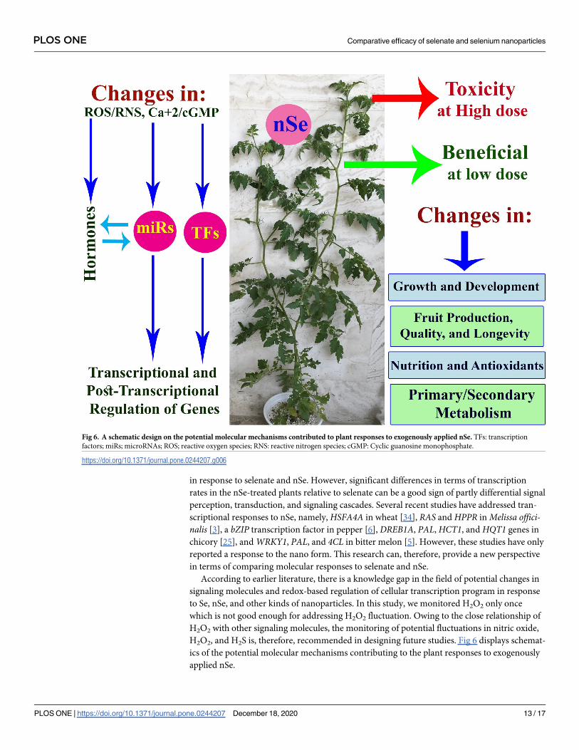
According to earlier literature, there is a knowledge gap in the field of potential changes in
signaling molecules and redox-based regulation of cellular transcription program in response
to Se, nSe, and other kinds of nanoparticles. In this study, we monitored H2O2 only once
which is not good enough for addressing H2O2 fluctuation. Owing to the close relationship of
H2O2 with other signaling molecules, the monitoring of potential fluctuations in nitric oxide,
H2O2, and H2S is, therefore, recommended in designing future studies. Fig 6 displays schemat-
ics of the potential molecular mechanisms contributing to the plant responses to exogenously
applied nSe.
Fig 6. A schematic design on the potential molecular mechanisms contributed to plant responses to exogenously applied nSe. TFs: transcription
factors; miRs; microRNAs; ROS; reactive oxygen species; RNS: reactive nitrogen species; cGMP: Cyclic guanosine monophosphate.
https://doi.org/10.1371/journal.pone.0244207.g006
PLOS ONE Comparative efficacy of selenate and selenium nanoparticles
PLOS ONE | https://doi.org/10.1371/journal.pone.0244207 December 18, 2020 13 / 17
This study provides the first comparative data in terms of physiological responses to sele-
nate and nSe. According to the H2O2 accumulation and MDA level, Se or nSe at the low dose
did not mediate oxidative stress. It seems that Se or nSe signaling at low concentration occurs
through a slight transient increase in the H2O2 concentration which is subsequently detoxified
by an increase in enzymatic antioxidants. H2O2 signaling triggers the transcriptional changes
in downstream genes and activation of the defense system, including antioxidants and second-
ary metabolism. These physiological responses along with the observed molecular changes
suggest mechanisms by which Se or nSe utilization, especially the nanoform, can improve crop
tolerance to environmental stresses. However, it should be warned that these supplements at
the high dose were associated with induction in the oxidative burst, impaired membrane integ-
rity, disrupted metabolism, and growth disorders. The lower concentration of H2O2 and MDA
along with higher content of proline in the nSe10-treated group is another reason for the
lower toxicity of the nano form relative to the selenate counterpart. The nSe-mediated changes
in proline, a multifunctional protective amino acid, have been attributed to the modified nitro-
gen assimilation metabolism [24], and activation of the defense system [5, 6]. Se or nSe supple-
mentation, especially the latter, stimulated enzymatic antioxidants, non-enzymatic antioxidant
compounds, and secondary metabolism. In agreement with our results, the nSe treatment
improved enzymatic antioxidants [5, 6, 25], non-enzymatic antioxidants [25], and accumula-
tion of secondary metabolites [3]. However, previous reports have only reported physiological
responses to nSe. To the best of our knowledge, this study, for the first time, showed that nSe is
more efficient than selenate in activating the antioxidant system and stimulating secondary
metabolism.
The results confirmed a higher efficacy of the nano type for application in biofortification
programs. Our results on Se-associated modification in nutritional status are in line with the
findings of Amirabad et al., [33] in radish, Babajani et al. [3] in lemon balm, Alam et al. [32] in
mung bean, and Zahedi et al. [4] in strawberry. Along with these reports, the nSe-associated
modifications in vascular conducting tissues (xylem and phloem) [5, 6] may be responsible for
the alterations in plant nutritional status. The higher fruit longevity, an economically impor-
tant index, in the nSe-supplemented plants can be explained by a higher efficiency of nSe in
enhancing the antioxidant system, improving minerals, and the antimicrobial properties of
nSe when compared to that of selenate.
The results highlight how Se or nSe-mediated changes in vegetative growth and metabolism
are coordinated with alteration in plant productivity at a reproductive stage. Herein, the pro-
vided anatomical evidence, for the first time, underlines the risk of Se at high concentration in
restricting crop fertility through disrupting the development of pollen grains. It has been
recently reported in pepper [6] and bitter melon [5] plants that in vitro toxicity of nSe is associ-
ated with impaired tissue differentiation and abnormalities in stem’s apical meristem. Further
anatomical and developmental investigations of Phyto-toxicological concerns of nanomater-
ials are required to improve our knowledge of plant responses to nanoproducts, especially
nano-pesticides and nano-fertilizers [5, 35–37].
Conclusion
Taken together, this study highlights the considerable efficacy of Se or nSe, especially the nano
form, for modifying growth, yield, primary and secondary metabolism, nutrition, defense sys-
tem, and transcription program. The Se- or nSe-associated transcriptional changes in miR172,
CRTISO, and bZIP are introduced as key potential molecular mechanisms through which Se
application may confer stress tolerance. Moreover, this study provided anatomical evidence
displaying potential phytotoxicity of Se or nSe in terms of disrupting the development of
PLOS ONE Comparative efficacy of selenate and selenium nanoparticles
PLOS ONE | https://doi.org/10.1371/journal.pone.0244207 December 18, 2020 14 / 17
pollen grains. Taking knowledge gaps into account, these comparative comprehensive data
can be helpful to gain novel insights into the benefits or the risk associated with Se or nSe func-
tion in agriculture. This study underlines the necessity of including anatomical, developmen-
tal, and molecular assessments in future phyto-toxicological investigations of nanomaterials.
CRediT authorship contribution statement
Maryam Neysanian: Resources, methodology, review, and editing; Alireza Iranbakhsh:
Conceptualization, visualization, investigation, formal analysis, writing-original draft, review,
and editing; Rahim Ahmadvand: Conceptualization, visualization, investigation, software
analysis, review, and editing; Zahra Oraghi Ardebili: Conceptualization, Investigation, review,
and editing. Mostafa Ebadi: Conceptualization, visualization, investigation, formal analysis,
writing, review, and editing. All authors have contributed, seen, and approved the manuscript.
Supporting information
S1 Data.
(XLSX)
Acknowledgments
The authors would like to thank Dr. N. Oraghi Ardebili and Dr. Sara Beiggi for their benevo-
lent and professional collaborations in the research procedure and language editing.
Author Contributions
Conceptualization: Alireza Iranbakhsh, Rahim Ahmadvand, Zahra Oraghi Ardebili, Mostafa
Ebadi.
Data curation: Zahra Oraghi Ardebili.
Formal analysis: Maryam Neysanian, Alireza Iranbakhsh, Zahra Oraghi Ardebili.
Investigation: Maryam Neysanian, Alireza Iranbakhsh, Rahim Ahmadvand, Zahra Oraghi
Ardebili, Mostafa Ebadi.
Methodology: Maryam Neysanian, Alireza Iranbakhsh, Rahim Ahmadvand, Zahra Oraghi
Ardebili, Mostafa Ebadi.
Resources: Maryam Neysanian.
Software: Maryam Neysanian, Alireza Iranbakhsh, Zahra Oraghi Ardebili, Mostafa Ebadi.
Supervision: Alireza Iranbakhsh.
Validation: Alireza Iranbakhsh, Rahim Ahmadvand, Zahra Oraghi Ardebili, Mostafa Ebadi.
Visualization: Rahim Ahmadvand.
Writing – original draft: Alireza Iranbakhsh.
Writing – review & editing: Maryam Neysanian, Alireza Iranbakhsh, Rahim Ahmadvand,
Zahra Oraghi Ardebili, Mostafa Ebadi.
References1. Djanaguiraman M, Belliraj N, Bossmann S, Prasad PV. High temperature stress alleviation by selenium
nanoparticle treatment in grain sorghum. ACS Omega. 2018; 3(3): 2479–2491. https://doi.org/10.1021/
acsomega.7b01934 PMID: 31458542
PLOS ONE Comparative efficacy of selenate and selenium nanoparticles
PLOS ONE | https://doi.org/10.1371/journal.pone.0244207 December 18, 2020 15 / 17
2. Hu T, Li H, Li J, Zhao G, Wu W, Liu L, et al. Absorption and bio-transformation of Selenium nanoparti-
cles by wheat seedlings (Triticum aestivum L). Front Plant Sci. 2018; 9: 597 https://doi.org/10.3389/
fpls.2018.00597 PMID: 29868060
3. Babajani A, Iranbakhsh A, Ardebili ZO and Eslami B. Differential growth, nutrition, physiology, and gene
expression in Melissa officinalis mediated by zinc oxide and elemental selenium nanoparticles. Environ
Sci Poll Res. 2019; 26(24): 24430–24444.
4. Zahedi SM, Abdelrahman M, Hosseini MS, Hoveizeh NF and Tran LSP. Alleviation of the effect of salin-
ity on growth and yield of strawberry by foliar spray of selenium-nanoparticles. Environ Pollut. 2019;
253:246–258. https://doi.org/10.1016/j.envpol.2019.04.078 PMID: 31319241
5. Rajaee Behbahani S, Iranbakhsh A, Ebadi M, Majd A and Ardebili ZO. Red elemental selenium nano-
particles mediated substantial variations in growth, tissue differentiation, metabolism, gene transcrip-
tion, epigenetic cytosine DNA methylation, and callogenesis in bittermelon (Momordica charantia); an in
vitro experiment. PloS one. 2020; 15(7): 0235556.
6. Sotoodehnia-Korani S, Iranbakhsh A, Ebadi M, Majd A and Ardebili ZO. Selenium nanoparticles
induced variations in growth, morphology, anatomy, biochemistry, gene expression, and epigenetic
DNA methylation in Capsicum annuum; an in vitro study. Environ Pollut. 2020; p114727. https://doi.org/
10.1016/j.envpol.2020.114727 PMID: 32806441
7. Farinati S, Forestan C, Canton M, Varotto S and Bonghi C. microRNA Regulation of Fruit Development.
In Plant microRNAs. Springer Cham.2020; 75–98.
8. Singh D, Sinha V, Kumar A and Teotia S. Small RNAs and cold stress tolerance. In Plant Small RNA.
Academic Press. 2020; 209–230.
9. Balanzà V, Martınez-Fernandez I, Sato S, Yanofsky MF and Ferrandiz C. Inflorescence Meristem fate
is dependent on seed development and FRUITFULL in Arabidopsis thaliana. Front Plant Sci. 2019; 10:
p1622. https://doi.org/10.3389/fpls.2019.01622 PMID: 31921264
10. Wisutiamonkul A, Ampomah-Dwamena C, Allan AC and Ketsa S. Carotenoid accumulation and gene
expression during durian (Durio zibethinus) fruit growth and ripening. Sci Hortic. 2017; 220: 233–242.
11. Liang M, Su X, Yang Z, Deng H, Yang Z, Liang R et al. Carotenoid composition and expression of caro-
tenogenic genes in the peel and pulp of commercial mango fruit cultivars. Sci Hortic. 2020; 263:
p109072.
12. Li C, Ji J, Wang G, Li Z, Wang Y and Fan Y. Over-Expression of LcPDS, LcZDS, and LcCRTISO,
Genes from Wolfberry for Carotenoid Biosynthesis, Enhanced Carotenoid Accumulation, and Salt Tol-
erance in Tobacco. Front Plant Sci. 2020; 11: p119.
13. Khan SA, Li MZ, Wang SM and Yin HJ. Revisiting the role of plant transcription factors in the battle
against abiotic stress. Int J Mol Sci. 2018; 19(6): p1634. https://doi.org/10.3390/ijms19061634 PMID:
29857524
14. Joo H, Lim CW and Lee SC. Roles of pepper bZIP transcription factor Ca ATBZ 1 and its interacting
partner RING-type E3 ligase Ca ASRF 1 in modulation of ABA signalling and drought tolerance. Plant J.
2019; 100(2): 399–410. https://doi.org/10.1111/tpj.14451 PMID: 31278798
15. Lilay GH, Castro PH, Guedes JG, Almeida DM, Campilho A, Azevedo H, et al. Rice F-bZIP transcription
factors regulate the zinc deficiency response. J Exp Bot. 2020; 71(12): 3664–3677. https://doi.org/10.
1093/jxb/eraa115 PMID: 32133499
16. Das P, Lakra N, Nutan KK, Singla-Pareek SL and Pareek A. A unique bZIP transcription factor imparting
multiple stress tolerance in Rice. Rice. 2019; 12(1): p58. https://doi.org/10.1186/s12284-019-0316-8
PMID: 31375941
17. Sheteiwy MS, Fu Y, Hu Q, Nawaz A, Guan Y, Li Z, et al. Seed priming with polyethylene glycol induces
antioxidative defense and metabolic regulation of rice under nano-ZnO stress. Environ Sci Pollut Res.
2016; 23(19): 19989–20002. https://doi.org/10.1007/s11356-016-7170-7 PMID: 27438877
18. Salah SM, Yajing G, Dongdong C, Jie L, Aamir N, Qijuan H, et al. Seed priming with polyethylene glycol
regulating the physiological and molecular mechanism in rice (Oryza sativa L.) under nano-ZnO stress.
Sci Rep. 2015; 5: p14278. https://doi.org/10.1038/srep14278 PMID: 26419216
19. Babajani A, Iranbakhsh A, Ardebili ZO and Eslami B. Seed priming with non-thermal plasma modified
plant reactions to selenium or zinc oxide nanoparticles: cold plasma as a novel emerging tool for plant
science. Plasma Chem Plasma Process. 2019; 39(1): 21–34.
20. Beaudoin-Eagan LD, Thorpe T. Tyrosine and phenylalanine ammonia lyase activities during shoot initi-
ation in tobacco callus cultures. Plant Physiol. 1985; 78:438–441. https://doi.org/10.1104/pp.78.3.438
PMID: 16664262
21. Del Longo OT, Gonzalez CA, Pastori GM, Trippi VS. Antioxidant defenses under hyperoxygenic and
hyperosmotic conditions in leaves of two lines of maize with differential sensitivity to drought. Plant Cell
Physiol. 1993; 34: 1023–1028.
PLOS ONE Comparative efficacy of selenate and selenium nanoparticles
PLOS ONE | https://doi.org/10.1371/journal.pone.0244207 December 18, 2020 16 / 17
22. De Pinto MC, Francis D and De Gara L. The redox state of the ascorbate-dehydroascorbate pair as a
specific sensor of cell division in tobacco BY-2 cells. Protoplasma. 1999; 209(1–2): 90–97. https://doi.
org/10.1007/BF01415704 PMID: 18987797
23. Bates L, Waldren R, and Teare I. Rapid determination of free proline for water-stress studies. Plant Soil.
1973; 39: 205–207. https://doi.org/10.1007/BF00018060
24. Nazerieh H, Ardebili ZO, Iranbakhsh A. Potential benefits and toxicity of nanoselenium and nitric oxide
in peppermint. Acta Agric. Slov. 2018; 111(2): 357–368.
25. Abedi S, Iranbakhsh A, Oraghi Ardebili Z, Ebadi M. Nitric oxide and selenium nanoparticles confer
changes in growth, metabolism, antioxidant machinery, gene expression, and flowering in chicory
(Cichorium intybus L.): potential benefits and risk assessment. Environ Sci Pollut Res. 2020. https://doi.
org/10.1007/s11356-020-10706-2 PMID: 32902749
26. Li Y, Zhu N, Liang X, Zheng L, Zhang C, Li YF, et al. A comparative study on the accumulation, translo-
cation and transformation of selenite, selenate, and SeNPs in a hydroponic-plant system. Ecotox Envi-
ron Safe. 2020; 189: p.109955.
27. Soleymanzadeh R, Iranbakhsh A, Habibi G and Oraghi Ardebili Z. Selenium nanoparticle protected
strawberry against salt stress through modifications in salicylic acid, ion homeostasis, antioxidant
machinery, and photosynthesis performance. Acta Biol Cracovien Ser. Bot. 2020; 62: 33–42. https://
doi.org/10.24425/abcsb.2019.127751
28. Wang R, da Rocha Tavano EC, Lammers M, Martinelli AP, Angenent GC and de Maagd RA. Re-evalu-
ation of transcription factor function in tomato fruit development and ripening with CRISPR/Cas9-muta-
genesis. Sci Rep. 2019; 9(1): 1–10. https://doi.org/10.1038/s41598-018-37186-2 PMID: 30626917
29. Li XY, Guo F, Ma SY, Zhu MY, Pan WH and Bian HW. Regulation of flowering time via miR172-medi-
ated APETALA2-like expression in ornamental gloxinia (Sinningia speciosa). Journal of Zhejiang Uni-
versity-SCIENCE B. 2019; 20(4): 322–331. https://doi.org/10.1631/jzus.B1800003 PMID: 30932377
30. Elkelish AA, Soliman MH, Alhaithloul HA and El-Esawi MA. Selenium protects wheat seedlings against
salt stress-mediated oxidative damage by up-regulating antioxidants and osmolytes metabolism. Plant
Physiol Biochem. 2019; 137: 144–153. https://doi.org/10.1016/j.plaphy.2019.02.004 PMID: 30784986
31. Yin H, Qi Z, Li M, Ahammed GJ, Chu X and Zhou J. Selenium forms and methods of application differ-
entially modulate plant growth, photosynthesis, stress tolerance, selenium content and speciation in
Oryza sativa L. Ecotox Environ Safe. 2019; 169: 911–917. https://doi.org/10.1016/j.ecoenv.2018.11.
080 PMID: 30597791
32. Alam MZ, McGee R, Hoque MA, Ahammed GJ and Carpenter-Boggs L. Effect of Arbuscular Mycor-
rhizal Fungi, Selenium and Biochar on Photosynthetic Pigments and Antioxidant Enzyme Activity Under
Arsenic Stress in Mung Bean (Vigna radiata). Front Physiol. 2019;10. https://doi.org/10.3389/fphys.
2019.00010 PMID: 30740057
33. Amirabad SA, Behtash F and Vafaee Y. Selenium mitigates cadmium toxicity by preventing oxidative
stress and enhancing photosynthesis and micronutrient availability on radish (Raphanus sativus L.) cv.
Cherry Belle. Environ Sci Pollut Res. 2020; 27:12476–12490. https://doi.org/10.1007/s11356-020-
07751-2.
34. Safari M, Ardebili ZO and Iranbakhsh A. Selenium nano-particle induced alterations in expression pat-
terns of heat shock factor A4A (HSFA4A), and high molecular weight glutenin subunit 1Bx (Glu-1Bx)
and enhanced nitrate reductase activity in wheat (Triticum aestivum L.). Acta Physiol Plant. 2018; 40
(6): p117.
35. Moghanloo M, Iranbakhsh A, Ebadi M, Ardebili ZO. Differential physiology and expression of phenylala-
nine ammonia lyase (PAL) and universal stress protein (USP) in the endangered species Astragalus fri-
dae following seed priming with cold plasma and manipulation of culture medium with silica
nanoparticles. 3Biotech. 2019; 9(7): p288.
36. Seddighinia FS, Iranbakhsh A, Ardebili ZO, Satari TN and Soleimanpour S. Seed priming with cold
plasma and multi-walled carbon nanotubes modified growth, tissue differentiation, anatomy, and yield
in bitter melon (Momordica charantia). J. Plant Growth Regul. 2020; 39: 87–98. https://doi.org/10.1007/
s00344-019-09965-2
37. Moghanloo M, Iranbakhsh A, Ebadi M, Satari TN, Ardebili ZO. Seed priming with cold plasma and sup-
plementation of culture medium with silicon nanoparticle modified growth, physiology, and anatomy in
Astragalus fridae as an endangered species. Acta Physiol Plant. 2019; 41(4): p54.
PLOS ONE Comparative efficacy of selenate and selenium nanoparticles
PLOS ONE | https://doi.org/10.1371/journal.pone.0244207 December 18, 2020 17 / 17
Copyright © 2022 FDOKUMEN