Tumor microenvironment and neurofibromatosis type I - Nature
8
REVIEW Tumor microenvironment and neurofibromatosis type I: connecting the GAPs LQ Le 1,2,3 and LF Parada 1 1 Department of Developmental Biology and Kent Waldrep Foundation Center for Basic Research on Nerve Growth and Regeneration, The University of Texas Southwestern Medical Center, Dallas, TX, USA; 2 Department of Dermatology, The University of Texas Southwestern Medical Center, Dallas, TX, USA and 3 Physician Scientist Training Program, The University of Texas Southwestern Medical Center, Dallas, TX, USA The human disease von Recklinghausen’s neurofibroma- tosis (Nf1) is one of the most common genetic disorders. It is caused by mutations in the NF1 tumor suppressor gene, which encodes a GTPase activating protein (GAP) that negatively regulates p21-RAS signaling. Dermal and plexiform neurofibromas as well as malignant peripheral nerve sheath tumors and other malignant tumors, are significant complications in Nf1. Neurofibromas are complex tumors and composed mainly of abnormal local cells including Schwann cells, endothelial cells, fibroblasts and additionally a large number of infiltrating inflamma- tory mast cells. Recent work has indicated a role for the microenvironment in plexiform neurofibroma genesis. The emerging evidence points to mast cells as crucial contributors to neurofibroma tumorigenesis. Therefore, further understanding of the molecular interactions between Schwann cells and their environment will provide tools to develop new therapies aimed at delaying or preventing tumor formation in Nf1 patients. Oncogene (2007) 26, 4609–4616; doi:10.1038/sj.onc.1210261; published online 12 February 2007 Keywords: neurofibromin; tumor microenvironment; mast cell; neurofibroma; NF1; GAP von Recklinghausen’s Neurofibromatosis Type I Neurofibromatosis type I (Nf1) was first recorded by Tilesius in 1793 and described in organized detail by the German pathologist Friedrich Daniel von Recklinghau- sen in 1882. Fifty years later, the Viennese ophthalmol- ogist Lisch described the presence of iris nodules, which are now an important criterion for the clinical diagnosis of Nf1 (Riccardi, 1992). It is one of the most common human genetic diseases. It has a de novo incidence of one in 3000–4000 individuals and affects male and female subjects equally in all races (Szudek et al., 2000; Trovo- Marqui and Tajara, 2006). Although it is inherited as an autosomal-dominant trait with essentially 100% pene- trance, spontaneous mutations occur in 50% of cases (Yohay, 2006). This means the disease will likely remain in the human population and will have increasing incidence over time. Nf1 patients have defects in neural crest-derived tissues, leading to a wide spectrum of clinical presentations, including developmental, pigment and neoplastic aberrations (Cichowski and Jacks, 2001; Zhu et al., 2001). The cardinal features of Nf1 are cafe´ au lait macules, axillary and groin freckling, combined with multiple peripheral and central nerve tumors. They also exhibit a less penetrant variety of additional pathologies of the skin, nervous system, bones, endo- crine organs, blood vessels and the eyes (Ward and Gutmann, 2005) (Table 1). Biology of Neurofibromin The NF1 gene is located on human chromosome 17q11.2 and was identified by positional cloning in 1990 (Ballester et al., 1990; Xu et al., 1990). It spans over 350 kb of genomic DNA, and has at least 60 exons (Jentarra et al., 2006). It encodes a tumor suppressor, known as Neurofibromin (NF1), which is ubiquitously expressed but most abundant in neurons, Schwann cells, astrocytes, oligodendrocytes and leukocytes (Gutmann et al., 1991; Daston et al., 1992). Despite its large size, very little is known about its function. Previous studies have shown a variety of human mutations associated with Nf1 across the entire gene (Castle et al., 2003; Jentarra et al., 2006). The majority of NF1 mutations predict truncations in the protein, and there is no clear correlation between specific mutation and clinical presentations (Castle et al., 2003). To date, it is known to have two functional domains, Sec14 and RasGAP (Trovo-Marqui and Tajara, 2006) (Figure 1). Sec14-interactive domain is located between amino acids 1545–1816 and is homologous to the yeast Sec14p, which is known to regulate intracellular proteins and lipid trafficking in yeast (Mousley et al., 2006). However, the biological role of Sec14 domain in NF1 is currently unknown. Received 1 December 2006; accepted 7 December 2006; published online 12 February 2007 Correspondence: Dr LF Parada or LQ Le, Center for Developmental Biology, The University of Texas Southwestern Medical Center at Dallas, 5323 Harry Hines Blvd, Dallas, TX 75390-9133, USA. E-mails: [email protected], or Lu.Le@UT Southwestern.edu Oncogene (2007) 26, 4609–4616 & 2007 Nature Publishing Group All rights reserved 0950-9232/07 $30.00 www.nature.com/onc
-
Upload
khangminh22 -
Category
Documents
-
view
0 -
download
0
Transcript of Tumor microenvironment and neurofibromatosis type I - Nature
REVIEW
Tumor microenvironment and neurofibromatosis type I: connecting
the GAPs
LQ Le1,2,3 and LF Parada1
1Department of Developmental Biology and Kent Waldrep Foundation Center for Basic Research on Nerve Growth and Regeneration,The University of Texas Southwestern Medical Center, Dallas, TX, USA; 2Department of Dermatology, The University of TexasSouthwestern Medical Center, Dallas, TX, USA and 3Physician Scientist Training Program, The University of Texas SouthwesternMedical Center, Dallas, TX, USA
The human disease von Recklinghausen’s neurofibroma-tosis (Nf1) is one of the most common genetic disorders. Itis caused by mutations in the NF1 tumor suppressor gene,which encodes a GTPase activating protein (GAP) thatnegatively regulates p21-RAS signaling. Dermal andplexiform neurofibromas as well as malignant peripheralnerve sheath tumors and other malignant tumors, aresignificant complications in Nf1. Neurofibromas arecomplex tumors and composed mainly of abnormal localcells including Schwann cells, endothelial cells, fibroblastsand additionally a large number of infiltrating inflamma-tory mast cells. Recent work has indicated a role forthe microenvironment in plexiform neurofibroma genesis.The emerging evidence points to mast cells as crucialcontributors to neurofibroma tumorigenesis. Therefore,further understanding of the molecular interactionsbetween Schwann cells and their environment will providetools to develop new therapies aimed at delaying orpreventing tumor formation in Nf1 patients.Oncogene (2007) 26, 4609–4616; doi:10.1038/sj.onc.1210261;published online 12 February 2007
Keywords: neurofibromin; tumor microenvironment;mast cell; neurofibroma; NF1; GAP
von Recklinghausen’s Neurofibromatosis Type I
Neurofibromatosis type I (Nf1) was first recorded byTilesius in 1793 and described in organized detail by theGerman pathologist Friedrich Daniel von Recklinghau-sen in 1882. Fifty years later, the Viennese ophthalmol-ogist Lisch described the presence of iris nodules, whichare now an important criterion for the clinical diagnosisof Nf1 (Riccardi, 1992). It is one of the most commonhuman genetic diseases. It has a de novo incidence of one
in 3000–4000 individuals and affects male and femalesubjects equally in all races (Szudek et al., 2000; Trovo-Marqui and Tajara, 2006). Although it is inherited as anautosomal-dominant trait with essentially 100% pene-trance, spontaneous mutations occur in 50% of cases(Yohay, 2006). This means the disease will likely remainin the human population and will have increasingincidence over time. Nf1 patients have defects in neuralcrest-derived tissues, leading to a wide spectrum ofclinical presentations, including developmental, pigmentand neoplastic aberrations (Cichowski and Jacks, 2001;Zhu et al., 2001). The cardinal features of Nf1 are cafeau lait macules, axillary and groin freckling, combinedwith multiple peripheral and central nerve tumors. Theyalso exhibit a less penetrant variety of additionalpathologies of the skin, nervous system, bones, endo-crine organs, blood vessels and the eyes (Ward andGutmann, 2005) (Table 1).
Biology of Neurofibromin
The NF1 gene is located on human chromosome 17q11.2and was identified by positional cloning in 1990 (Ballesteret al., 1990; Xu et al., 1990). It spans over 350kb ofgenomic DNA, and has at least 60 exons (Jentarra et al.,2006). It encodes a tumor suppressor, known asNeurofibromin (NF1), which is ubiquitously expressedbut most abundant in neurons, Schwann cells, astrocytes,oligodendrocytes and leukocytes (Gutmann et al., 1991;Daston et al., 1992). Despite its large size, very little isknown about its function. Previous studies have shown avariety of human mutations associated with Nf1 acrossthe entire gene (Castle et al., 2003; Jentarra et al., 2006).The majority ofNF1mutations predict truncations in theprotein, and there is no clear correlation between specificmutation and clinical presentations (Castle et al., 2003).To date, it is known to have two functional domains,Sec14 and RasGAP (Trovo-Marqui and Tajara, 2006)(Figure 1). Sec14-interactive domain is located betweenamino acids 1545–1816 and is homologous to the yeastSec14p, which is known to regulate intracellular proteinsand lipid trafficking in yeast (Mousley et al., 2006).However, the biological role of Sec14 domain in NF1 iscurrently unknown.
Received 1 December 2006; accepted 7 December 2006; published online12 February 2007
Correspondence: Dr LF Parada or LQ Le, Center for DevelopmentalBiology, The University of Texas Southwestern Medical Center atDallas, 5323 Harry Hines Blvd, Dallas, TX 75390-9133, USA.E-mails: [email protected], or [email protected]
Oncogene (2007) 26, 4609–4616& 2007 Nature Publishing Group All rights reserved 0950-9232/07 $30.00
www.nature.com/onc
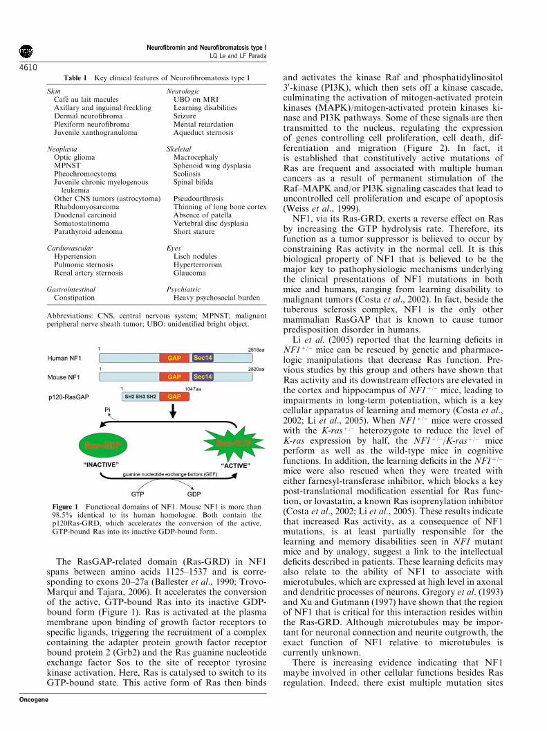
The RasGAP-related domain (Ras-GRD) in NF1spans between amino acids 1125–1537 and is corre-sponding to exons 20–27a (Ballester et al., 1990; Trovo-Marqui and Tajara, 2006). It accelerates the conversionof the active, GTP-bound Ras into its inactive GDP-bound form (Figure 1). Ras is activated at the plasmamembrane upon binding of growth factor receptors tospecific ligands, triggering the recruitment of a complexcontaining the adapter protein growth factor receptorbound protein 2 (Grb2) and the Ras guanine nucleotideexchange factor Sos to the site of receptor tyrosinekinase activation. Here, Ras is catalysed to switch to itsGTP-bound state. This active form of Ras then binds
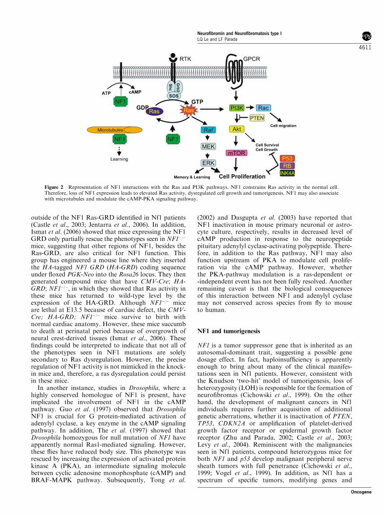
and activates the kinase Raf and phosphatidylinositol30-kinase (PI3K), which then sets off a kinase cascade,culminating the activation of mitogen-activated proteinkinases (MAPK)/mitogen-activated protein kinases ki-nase and PI3K pathways. Some of these signals are thentransmitted to the nucleus, regulating the expressionof genes controlling cell proliferation, cell death, dif-ferentiation and migration (Figure 2). In fact, itis established that constitutively active mutations ofRas are frequent and associated with multiple humancancers as a result of permanent stimulation of theRaf–MAPK and/or PI3K signaling cascades that lead touncontrolled cell proliferation and escape of apoptosis(Weiss et al., 1999).NF1, via its Ras-GRD, exerts a reverse effect on Ras
by increasing the GTP hydrolysis rate. Therefore, itsfunction as a tumor suppressor is believed to occur byconstraining Ras activity in the normal cell. It is thisbiological property of NF1 that is believed to be themajor key to pathophysiologic mechanisms underlyingthe clinical presentations of NF1 mutations in bothmice and humans, ranging from learning disability tomalignant tumors (Costa et al., 2002). In fact, beside thetuberous sclerosis complex, NF1 is the only othermammalian RasGAP that is known to cause tumorpredisposition disorder in humans.Li et al. (2005) reported that the learning deficits in
NF1þ /� mice can be rescued by genetic and pharmaco-logic manipulations that decrease Ras function. Pre-vious studies by this group and others have shown thatRas activity and its downstream effectors are elevated inthe cortex and hippocampus of NF1þ /� mice, leading toimpairments in long-term potentiation, which is a keycellular apparatus of learning and memory (Costa et al.,2002; Li et al., 2005). When NF1þ /� mice were crossedwith the K-rasþ /� heterozygote to reduce the level ofK-ras expression by half, the NF1þ /�/K-rasþ /� miceperform as well as the wild-type mice in cognitivefunctions. In addition, the learning deficits in the NF1þ /�
mice were also rescued when they were treated witheither farnesyl-transferase inhibitor, which blocks a keypost-translational modification essential for Ras func-tion, or lovastatin, a known Ras isoprenylation inhibitor(Costa et al., 2002; Li et al., 2005). These results indicatethat increased Ras activity, as a consequence of NF1mutations, is at least partially responsible for thelearning and memory disabilities seen in NF1 mutantmice and by analogy, suggest a link to the intellectualdeficits described in patients. These learning deficits mayalso relate to the ability of NF1 to associate withmicrotubules, which are expressed at high level in axonaland dendritic processes of neurons. Gregory et al. (1993)and Xu and Gutmann (1997) have shown that the regionof NF1 that is critical for this interaction resides withinthe Ras-GRD. Although microtubules may be impor-tant for neuronal connection and neurite outgrowth, theexact function of NF1 relative to microtubules iscurrently unknown.There is increasing evidence indicating that NF1
maybe involved in other cellular functions besides Rasregulation. Indeed, there exist multiple mutation sites
Table 1 Key clinical features of Neurofibromatosis type I
Skin NeurologicCafe au lait macules UBO on MRIAxillary and inguinal freckling Learning disabilitiesDermal neurofibroma SeizurePlexiform neurofibroma Mental retardationJuvenile xanthogranuloma Aqueduct sternosis
Neoplasia SkeletalOptic glioma MacrocephalyMPNST Sphenoid wing dysplasiaPheochromocytoma ScoliosisJuvenile chronic myelogenousleukemia
Spinal bifida
Other CNS tumors (astrocytoma) PseudoarthrosisRhabdomyosarcoma Thinning of long bone cortexDuodenal carcinoid Absence of patellaSomatostatinoma Vertebral disc dysplasiaParathyroid adenoma Short stature
Cardiovascular EyesHypertension Lisch nodulesPulmonic sternosis HyperterrorismRenal artery sternosis Glaucoma
Gastrointestinal PsychiatricConstipation Heavy psychosocial burden
Abbreviations: CNS, central nervous system; MPNST; malignantperipheral nerve sheath tumor; UBO: unidentified bright object.
Figure 1 Functional domains of NF1. Mouse NF1 is more than98.5% identical to its human homologue. Both contain thep120Ras-GRD, which accelerates the conversion of the active,GTP-bound Ras into its inactive GDP-bound form.
Neurofibromin and Neurofibromatosis type ILQ Le and LF Parada
4610
Oncogene
outside of the NF1 Ras-GRD identified in Nf1 patients(Castle et al., 2003; Jentarra et al., 2006). In addition,Ismat et al. (2006) showed that mice expressing the NF1GRD only partially rescue the phenotypes seen in NF1�/�
mice, suggesting that other regions of NF1, besides theRas-GRD, are also critical for NF1 function. Thisgroup has engineered a mouse line where they insertedthe HA-tagged NF1 GRD (HA-GRD) coding sequenceunder floxed PGK-Neo into the Rosa26 locus. They thengenerated compound mice that have CMV-Cre; HA-GRD; NF1�/�, in which they showed that Ras activity inthese mice has returned to wild-type level by theexpression of the HA-GRD. Although NF1�/� miceare lethal at E13.5 because of cardiac defect, the CMV-Cre; HA-GRD; NF1�/� mice survive to birth withnormal cardiac anatomy. However, these mice succumbto death at perinatal period because of overgrowth ofneural crest-derived tissues (Ismat et al., 2006). Thesefindings could be interpreted to indicate that not all ofthe phenotypes seen in NF1 mutations are solelysecondary to Ras dysregulation. However, the preciseregulation of NF1 activity is not mimicked in the knock-in mice and, therefore, a ras dysregulation could persistin these mice.In another instance, studies in Drosophila, where a
highly conserved homologue of NF1 is present, haveimplicated the involvement of NF1 in the cAMPpathway. Guo et al. (1997) observed that DrosophilaNF1 is crucial for G protein-mediated activation ofadenylyl cyclase, a key enzyme in the cAMP signalingpathway. In addition, The et al. (1997) showed thatDrosophila homozygous for null mutation of NF1 haveapparently normal Ras1-mediated signaling. However,these flies have reduced body size. This phenotype wasrescued by increasing the expression of activated proteinkinase A (PKA), an intermediate signaling moleculebetween cyclic adenosine monophosphate (cAMP) andBRAF-MAPK pathway. Subsequently, Tong et al.
(2002) and Dasgupta et al. (2003) have reported thatNF1 inactivation in mouse primary neuronal or astro-cyte culture, respectively, results in decreased level ofcAMP production in response to the neuropeptidepituitary adenylyl cyclase-activating polypeptide. There-fore, in addition to the Ras pathway, NF1 may alsofunction upstream of PKA to modulate cell prolife-ration via the cAMP pathway. However, whetherthe PKA-pathway modulation is a ras-dependent or-independent event has not been fully resolved. Anotherremaining caveat is that the biological consequencesof this interaction between NF1 and adenylyl cyclasemay not conserved across species from fly to mouseto human.
NF1 and tumorigenesis
NF1 is a tumor suppressor gene that is inherited as anautosomal-dominant trait, suggesting a possible genedosage effect. In fact, haploinsufficiency is apparentlyenough to bring about many of the clinical manifes-tations seen in Nf1 patients. However, consistent withthe Knudson ‘two-hit’ model of tumorigenesis, loss ofheterozygosity (LOH) is responsible for the formation ofneurofibromas (Cichowski et al., 1999). On the otherhand, the development of malignant cancers in Nf1individuals requires further acquisition of additionalgenetic aberrations, whether it is inactivation of PTEN,TP53, CDKN2A or amplification of platelet-derivedgrowth factor receptor or epidermal growth factorreceptor (Zhu and Parada, 2002; Castle et al., 2003;Levy et al., 2004). Reminiscent with the malignanciesseen in Nf1 patients, compound heterozygous mice forboth NF1 and p53 develop malignant peripheral nervesheath tumors with full penetrance (Cichowski et al.,1999; Vogel et al., 1999). In addition, as Nf1 has aspectrum of specific tumors, modifying genes and
Figure 2 Representation of NF1 interactions with the Ras and PI3K pathways. NF1 constrains Ras activity in the normal cell.Therefore, loss of NF1 expression leads to elevated Ras activity, dysregulated cell growth and tumorigenesis. NF1 may also associatewith microtubules and modulate the cAMP-PKA signaling pathway.
Neurofibromin and Neurofibromatosis type ILQ Le and LF Parada
4611
Oncogene

epigenetic phenomena have been shown to play a role inmodulating Nf1-associated tumor susceptibility (Eastonet al., 1993; Reilly et al., 2004).
Neurofibromas and malignant peripheral nervesheath tumorsNeurofibromas are the most common tumor in Nf1 andclassified into three subtypes. Dermal and subcutaneousforms appear at puberty and increase in number withage and during pregnancy. They occur as a result ofproliferation of all supporting elements of the nervefibers, including Schwann cells, perineurial cells, fibro-blasts, blood vessels, as well as infiltration of mast cells.Plexiform neurofibromas occur in about 30% of Nf1individuals and are virtually pathognomonic of thedisease. They are congenital and progressively enlargethroughout life. Although dermal neurofibromas aremost frequently benign, patients with plexiform neurofi-bromas have a 10% lifetime risk of developing malignantperipheral nerve sheath tumors (MPNST), which canmetastases widely and are often signaling a fatal outcome(Lakkis and Tennekoon, 2000; Ferner, 2006). In addition,owing to their unusual capacity for growth, plexiformneurofibromas can be life threatening by their physicalimpairment of organ or neural function.Research over the past decade using mouse models
has greatly enhanced our knowledge of neurofibromadevelopment and malignant progression in Nf1. Micehomozygous for NF1 mutation are embryonic lethal atE13.5 secondary to defective heart and malformation ofthe major cardiac outflow tracts. They also exhibithyperplasia of neural crest-derived sympathetic ganglia(Brannan et al., 1994; Jacks et al., 1994). To thecontrary, heterozygous NF1 mutant mice are viableand only have increased incidence of pheochromocyto-mas and myeloid leukemias beyond 10- to 12-month old(Parada, 2000). Puzzlingly, in contrast to the human
condition, NF1þ /� mice do not develop neurofibromas(Brannan et al., 1994; Jacks et al., 1994) (Figure 3). Onepossible explanation for this observation is that LOHnecessary for neurofibroma development is impaired inmice. Perhaps, given the short time of gestation andlifespan and a smaller neural crest compartmentcompared with human, the NF1þ /� mice do not havethe necessary window of opportunity to undergoeffective LOH in target cells to initiate neurofibromaformation. Cichowski et al. (1999) addressed this issueelegantly when they created chimeric mice by injectingLacZ-positive NF1�/� ES cells into wild-type C57BL/6blastocysts. These mice developed microscopic neurofi-bromas derived from the injected ES cells, demonstrat-ing the requirement of NF1 homozygosity for tumorformation. However, the degree of chimerism in thesemice occurs randomly and cannot be controlledgenetically. As a result, it is difficult to establish thetarget cell or whether other cell types contribute to thetumorigenesis.To circumvent this problem and to develop a more
precise mouse model for evaluating the evolution ofNf1-associated tumors, Cre/loxP technology that allowstissue-specific ablation of NF1 function has beenadopted. Schwann cell-specific NF1-deficient mice werederived by crossing NF1flox/flox mice with the Krox20-cretransgenic mice, an embryonic Schwann cell-specificpromoter (Zhu et al., 2002). Mice with a conditionalknockout (KO) of NF1 only in embryonic Schwanncells, but wild-type in all other cell lineages (NF1flox/flox;Krox20-Cre), exhibit microscopic hyperplasia in sensoryganglia but do not develop neurofibromas. However,when mice homozygous for NF1 mutation (NF1�/�) inSchwann cells but heterozygous for NF1 (NF1þ /�) in allother somatic cells (NF1flox/�; Krox20-Cre) were gene-rated, these mice developed multiple classic plexiformneurofibromas with a massive degranulating mast cellinfiltration, modeling human neurofibroma (Zhu et al.,
Figure 3 Schwann cell origin and the role of tumor microenvironment in neurofibroma formation. Both nullizygosity at NF1 locus inSchwann cells and haploinsufficiency of NF1 in the somatic tissue are required for neurofibroma tumorigenesis. In addition, the NF1-heterozygous mast cells infiltration into nerve tissues precedes development of NF1flox/�;Krox20-Cre plexiform neurofibromas, implyinga unique affinity between NF1�/� Schwann cells and NF1þ /� mast cells and a causal role for mast cells in tumor initiation.
Neurofibromin and Neurofibromatosis type ILQ Le and LF Parada
4612
Oncogene
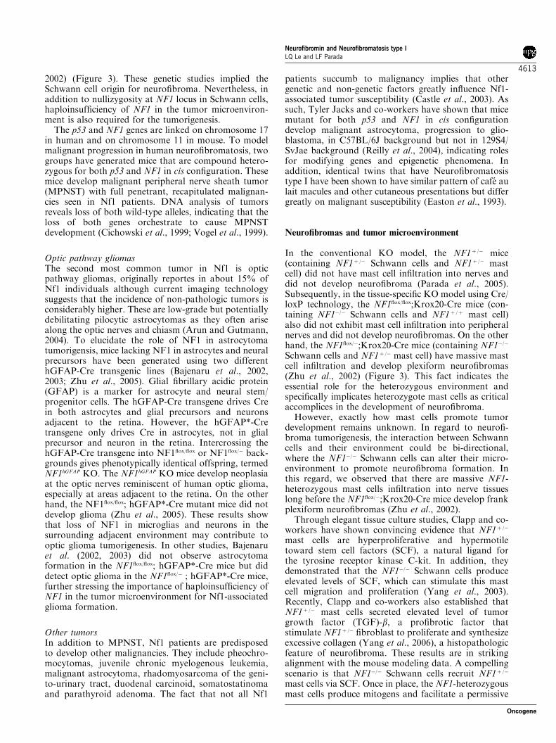
2002) (Figure 3). These genetic studies implied theSchwann cell origin for neurofibroma. Nevertheless, inaddition to nullizygosity at NF1 locus in Schwann cells,haploinsufficiency of NF1 in the tumor microenviron-ment is also required for the tumorigenesis.The p53 and NF1 genes are linked on chromosome 17
in human and on chromosome 11 in mouse. To modelmalignant progression in human neurofibromatosis, twogroups have generated mice that are compound hetero-zygous for both p53 and NF1 in cis configuration. Thesemice develop malignant peripheral nerve sheath tumor(MPNST) with full penetrant, recapitulated malignan-cies seen in Nf1 patients. DNA analysis of tumorsreveals loss of both wild-type alleles, indicating that theloss of both genes orchestrate to cause MPNSTdevelopment (Cichowski et al., 1999; Vogel et al., 1999).
Optic pathway gliomasThe second most common tumor in Nf1 is opticpathway gliomas, originally reportes in about 15% ofNf1 individuals although current imaging technologysuggests that the incidence of non-pathologic tumors isconsiderably higher. These are low-grade but potentiallydebilitating pilocytic astrocytomas as they often arisealong the optic nerves and chiasm (Arun and Gutmann,2004). To elucidate the role of NF1 in astrocytomatumorigensis, mice lacking NF1 in astrocytes and neuralprecursors have been generated using two differenthGFAP-Cre transgenic lines (Bajenaru et al., 2002,2003; Zhu et al., 2005). Glial fibrillary acidic protein(GFAP) is a marker for astrocyte and neural stem/progenitor cells. The hGFAP-Cre transgene drives Crein both astrocytes and glial precursors and neuronsadjacent to the retina. However, the hGFAP*-Cretransgene only drives Cre in astrocytes, not in glialprecursor and neuron in the retina. Intercrossing thehGFAP-Cre transgene into NF1flox/flox or NF1flox/� back-grounds gives phenotypically identical offspring, termedNF1hGFAP KO. The NF1hGFAP KO mice develop neoplasiaat the optic nerves reminiscent of human optic glioma,especially at areas adjacent to the retina. On the otherhand, the NF1flox/flox; hGFAP*-Cre mutant mice did notdevelop glioma (Zhu et al., 2005). These results showthat loss of NF1 in microglias and neurons in thesurrounding adjacent environment may contribute tooptic glioma tumorigenesis. In other studies, Bajenaruet al. (2002, 2003) did not observe astrocytomaformation in the NF1flox/flox; hGFAP*-Cre mice but diddetect optic glioma in the NF1flox/� ; hGFAP*-Cre mice,further stressing the importance of haploinsufficiency ofNF1 in the tumor microenvironment for Nf1-associatedglioma formation.
Other tumorsIn addition to MPNST, Nf1 patients are predisposedto develop other malignancies. They include pheochro-mocytomas, juvenile chronic myelogenous leukemia,malignant astrocytoma, rhadomyosarcoma of the geni-to-urinary tract, duodenal carcinoid, somatostatinomaand parathyroid adenoma. The fact that not all Nf1
patients succumb to malignancy implies that othergenetic and non-genetic factors greatly influence Nf1-associated tumor susceptibility (Castle et al., 2003). Assuch, Tyler Jacks and co-workers have shown that micemutant for both p53 and NF1 in cis configurationdevelop malignant astrocytoma, progression to glio-blastoma, in C57BL/6J background but not in 129S4/SvJae background (Reilly et al., 2004), indicating rolesfor modifying genes and epigenetic phenomena. Inaddition, identical twins that have Neurofibromatosistype I have been shown to have similar pattern of cafe aulait macules and other cutaneous presentations but differgreatly on malignant susceptibility (Easton et al., 1993).
Neurofibromas and tumor microenvironment
In the conventional KO model, the NF1þ /� mice(containing NF1þ /� Schwann cells and NF1þ /� mastcell) did not have mast cell infiltration into nerves anddid not develop neurofibroma (Parada et al., 2005).Subsequently, in the tissue-specific KO model using Cre/loxP technology, the NF1flox/flox;Krox20-Cre mice (con-taining NF1�/� Schwann cells and NF1þ /þ mast cell)also did not exhibit mast cell infiltration into peripheralnerves and did not develop neurofibromas. On the otherhand, the NF1flox/�;Krox20-Cre mice (containing NF1�/�
Schwann cells and NF1þ /� mast cell) have massive mastcell infiltration and develop plexiform neurofibromas(Zhu et al., 2002) (Figure 3). This fact indicates theessential role for the heterozygous environment andspecifically implicates heterozygote mast cells as criticalaccomplices in the development of neurofibroma.However, exactly how mast cells promote tumor
development remains unknown. In regard to neurofi-broma tumorigenesis, the interaction between Schwanncells and their environment could be bi-directional,where the NF1�/� Schwann cells can alter their micro-environment to promote neurofibroma formation. Inthis regard, we observed that there are massive NF1-heterozygous mast cells infiltration into nerve tissueslong before the NF1flox/�;Krox20-Cre mice develop frankplexiform neurofibromas (Zhu et al., 2002).Through elegant tissue culture studies, Clapp and co-
workers have shown convincing evidence that NF1þ /�
mast cells are hyperproliferative and hypermotiletoward stem cell factors (SCF), a natural ligand forthe tyrosine receptor kinase C-kit. In addition, theydemonstrated that the NF1�/� Schwann cells produceelevated levels of SCF, which can stimulate this mastcell migration and proliferation (Yang et al., 2003).Recently, Clapp and co-workers also established thatNF1þ /� mast cells secreted elevated level of tumorgrowth factor (TGF)-b, a profibrotic factor thatstimulate NF1þ /� fibroblast to proliferate and synthesizeexcessive collagen (Yang et al., 2006), a histopathologicfeature of neurofibroma. These results are in strikingalignment with the mouse modeling data. A compellingscenario is that NF1�/� Schwann cells recruit NF1þ /�
mast cells via SCF. Once in place, the NF1-heterozygousmast cells produce mitogens and facilitate a permissive
Neurofibromin and Neurofibromatosis type ILQ Le and LF Parada
4613
Oncogene
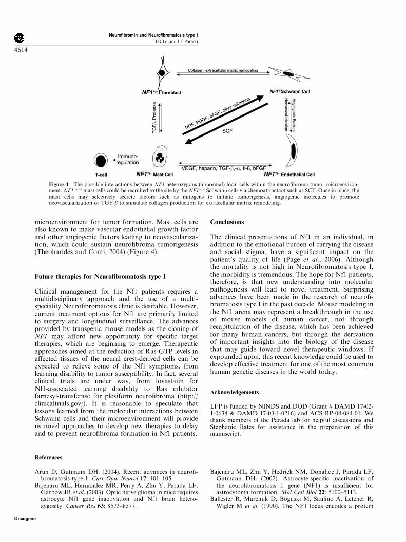
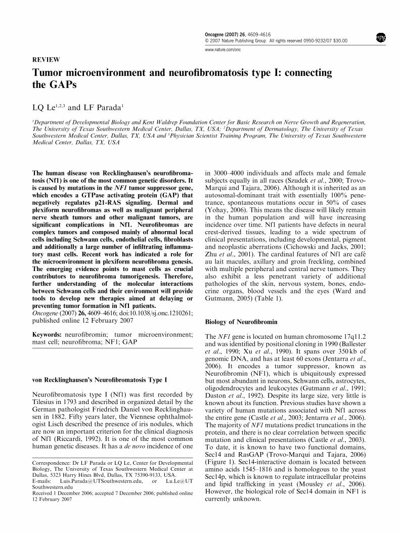
microenvironment for tumor formation. Mast cells arealso known to make vascular endothelial growth factorand other angiogenic factors leading to neovasculariza-tion, which could sustain neurofibroma tumorigenesis(Theoharides and Conti, 2004) (Figure 4).
Future therapies for Neurofibromatosis type I
Clinical management for the Nf1 patients requires amultidisciplinary approach and the use of a multi-speciality Neurofibromatosis clinic is desirable. However,current treatment options for Nf1 are primarily limitedto surgery and longitudinal surveillance. The advancesprovided by transgenic mouse models as the cloning ofNF1 may afford new opportunity for specific targettherapies, which are beginning to emerge. Therapeuticapproaches aimed at the reduction of Ras-GTP levels inaffected tissues of the neural crest-derived cells can beexpected to relieve some of the Nf1 symptoms, fromlearning disability to tumor susceptibility. In fact, severalclinical trials are under way, from lovastatin forNf1-associated learning disability to Ras inhibitorfarnesyl-transferase for plexiform neurofibroma (http://clinicaltrials.gov/). It is reasonable to speculate thatlessons learned from the molecular interactions betweenSchwann cells and their microenvironment will provideus novel approaches to develop new therapies to delayand to prevent neurofibroma formation in Nf1 patients.
Conclusions
The clinical presentations of Nf1 in an individual, inaddition to the emotional burden of carrying the diseaseand social stigma, have a significant impact on thepatient’s quality of life (Page et al., 2006). Althoughthe mortality is not high in Neurofibromatosis type I,the morbidity is tremendous. The hope for Nf1 patients,therefore, is that new understanding into molecularpathogenesis will lead to novel treatment. Surprisingadvances have been made in the research of neurofi-bromatosis type I in the past decade. Mouse modeling inthe Nf1 arena may represent a breakthrough in the useof mouse models of human cancer, not throughrecapitulation of the disease, which has been achievedfor many human cancers, but through the derivationof important insights into the biology of the diseasethat may guide toward novel therapeutic windows. Ifexpounded upon, this recent knowledge could be used todevelop effective treatment for one of the most commonhuman genetic diseases in the world today.
Acknowledgements
LFP is funded by NINDS and DOD (Grant # DAMD 17-02-1-0638 & DAMD 17-03-1-0216) and ACS RP-04-084-01. Wethank members of the Parada lab for helpful discussions andStephanie Bates for assistance in the preparation of thismanuscript.
References
Arun D, Gutmann DH. (2004). Recent advances in neurofi-bromatosis type 1. Curr Opin Neurol 17: 101–105.
Bajenaru ML, Hernandez MR, Perry A, Zhu Y, Parada LF,Garbow JR et al. (2003). Optic nerve glioma in mice requiresastrocyte Nf1 gene inactivation and Nf1 brain hetero-zygosity. Cancer Res 63: 8573–8577.
Bajenaru ML, Zhu Y, Hedrick NM, Donahoe J, Parada LF,Gutmann DH. (2002). Astrocyte-specific inactivation ofthe neurofibromatosis 1 gene (NF1) is insufficient forastrocytoma formation. Mol Cell Biol 22: 5100–5113.
Ballester R, Marchuk D, Boguski M, Saulino A, Letcher R,Wigler M et al. (1990). The NF1 locus encodes a protein
Figure 4 The possible interactions between NF1 heterozygous (abnormal) local cells within the neurofibroma tumor microenviron-ment. NF1 þ /� mast cells could be recruited to the site by the NF1�/� Schwann cells via chemoattractant such as SCF. Once in place, themast cells may selectively secrete factors such as mitogens to initiate tumorigenesis, angiogenic molecules to promoteneovascularization or TGF-b to stimulate collagen production for extracellular matrix remodeling.
Neurofibromin and Neurofibromatosis type ILQ Le and LF Parada
4614
Oncogene
functionally related to mammalian GAP and yeast IRAproteins. Cell 63: 851–859.
Brannan CI, Perkins AS, Vogel KS, Ratner N, Nordlund ML,Reid SW et al. (1994). Targeted disruption of the neuro-fibromatosis type-1 gene leads to developmental abnor-malities in heart and various neural crest-derived tissues.Genes Dev 8: 1019–1029.
Castle B, Baser ME, Huson SM, Cooper DN, Upadhyaya M.(2003). Evaluation of genotype-phenotype correlations inneurofibromatosis type 1. J Med Genet 40: e109.
Cichowski K, Jacks T. (2001). NF1 tumor suppressor genefunction: narrowing the GAP. Cell 104: 593–604.
Cichowski K, Shih TS, Schmitt E, Santiago S, Reilly K,McLaughlin ME et al. (1999). Mouse models of tumordevelopment in neurofibromatosis type 1. Science 286:2172–2176.
Costa RM, Federov NB, Kogan JH, Murphy GG, Stern J,Ohno M et al. (2002). Mechanism for the learning deficits ina mouse model of neurofibromatosis type 1. Nature 415:526–530.
Dasgupta B, Dugan LL, Gutmann DH. (2003). The neurofi-bromatosis 1 gene product neurofibromin regulates pituitaryadenylate cyclase-activating polypeptide-mediated signalingin astrocytes. J Neurosci 23: 8949–8954.
Daston MM, Scrable H, Nordlund M, Sturbaum AK,Nissen LM, Ratner N. (1992). The protein product of theneurofibromatosis type 1 gene is expressed at highestabundance in neurons, Schwann cells, and oligodendrocytes.Neuron 8: 415–428.
Easton DF, Ponder MA, Huson SM, Ponder BA. (1993). Ananalysis of variation in expression of neurofibromatosis(NF) type 1 (NF1): evidence for modifying genes. Am J HumGenet 53: 305–313.
Ferner RE. (2006). Neurofibromatosis 1. Eur J Hum Genet[Epub ahead of print].
Gregory PE, Gutmann DH, Mitchell A, Park S, Boguski M,Jacks T et al. (1993). Neurofibromatosis type 1 gene product(neurofibromin) associates with microtubules. Somat CellMol Genet 19: 265–274.
Guo HF, The I, Hannan F, Bernards A, Zhong Y. (1997).Requirement of Drosophila NF1 for activation of adenylylcyclase by PACAP38-like neuropeptides. Science 276: 795–798.
Gutmann DH, Wood DL, Collins FS. (1991). Identification ofthe neurofibromatosis type 1 gene product. Proc Natl AcadSci USA 88: 9658–9662.
Ismat FA, Xu J, Lu MM, Epstein JA. (2006). Theneurofibromin GAP-related domain rescues endothelialbut not neural crest development in Nf1 mice. J Clin Invest116: 2378–2384.
Jacks T, Shih TS, Schmitt EM, Bronson RT, Bernards A,Weinberg RA. (1994). Tumour predisposition in miceheterozygous for a targeted mutation in Nf1. Nat Genet 7:353–361.
Jentarra G, Snyder SL, Narayanan V. (2006). Genetic aspects ofneurocutaneous disorders. Semin Pediatr Neurol 13: 43–47.
Lakkis MM, Tennekoon GI. (2000). Neurofibromatosis type1. I. General overview. J Neurosci Res 62: 755–763.
Levy P, Vidaud D, Leroy K, Laurendeau I, Wechsler J,Bolasco G et al. (2004). Molecular profiling of malignantperipheral nerve sheath tumors associated with neurofibro-matosis type 1, based on large-scale real-time RT-PCR. MolCancer 3: 20.
Li W, Cui Y, Kushner SA, Brown RA, Jentsch JD, FranklandPW et al. (2005). The HMG-CoA reductase inhibitorlovastatin reverses the learning and attention deficits in amouse model of neurofibromatosis type 1. Curr Biol 15:1961–1967.
Mousley CJ, Tyeryar KR, Ryan MM, Bankaitis VA. (2006).Sec14p-like proteins regulate phosphoinositide homoeosta-sis and intracellular protein and lipid trafficking in yeast.Biochem Soc Trans 34: 346–350.
Page PZ, Page GP, Ecosse E, Korf BR, Leplege A,Wolkenstein P. (2006). Impact of neurofibromatosis 1 onquality of life: a cross-sectional study of 176 American cases.Am J Med Genet A 140: 1893–1898.
Parada LF. (2000). Neurofibromatosis type 1. Biochim BiophysActa 1471: M13–9.
Parada LF, Kwon CH, Zhu Y. (2005). Modeling neurofibro-matosis type 1 tumors in the mouse for therapeuticintervention. Cold Spring Harb Symp Quant Biol 70:173–176.
Reilly KM, Tuskan RG, Christy E, Loisel DA, Ledger J,Bronson RT et al. (2004). Susceptibility to astrocytoma inmice mutant for Nf1 and Trp53 is linked to chromosome 11and subject to epigenetic effects. Proc Natl Acad Sci USA101: 13008–13013.
Riccardi VM. (1992). Neurofibromatosis phenotype, naturalhistory and pathogenesis. The Johns Hopkins UniversityPress: Baltimore.
Szudek J, Birch P, Riccardi VM, Evans DG, Friedman JM.(2000). Associations of clinical features in neurofibromatosis1 (NF1). Genet Epidemiol 19: 429–439.
The I, Hannigan GE, Cowley GS, Reginald S, Zhong Y,Gusella JF et al. (1997). Rescue of a Drosophila NF1mutant phenotype by protein kinase A. Science 276:791–794.
Theoharides TC, Conti P. (2004). Mast cells: the Jekylland Hyde of tumor growth. Trends Immunol 25:235–241.
Tong J, Hannan F, Zhu Y, Bernards A, Zhong Y. (2002).Neurofibromin regulates G protein-stimulated adenylylcyclase activity. Nat Neurosci 5: 95–96.
Trovo-Marqui AB, Tajara EH. (2006). Neurofibromin: ageneral outlook. Clin Genet 70: 1–13.
Vogel KS, Klesse LJ, Velasco-Miguel S, Meyers K, Rushing EJ,Parada LF. (1999). Mouse tumor model for neurofibroma-tosis type 1. Science 286: 2176–2179.
Ward BA, Gutmann DH. (2005). Neurofibromatosis 1: fromlab bench to clinic. Pediatr Neurol 32: 221–228.
Weiss B, Bollag G, Shannon K. (1999). Hyperactive Ras as atherapeutic target in neurofibromatosis type 1. Am J MedGenet 89: 14–22.
Xu GF, O’Connell P, Viskochil D, Cawthon R, Robertson M,Culver M et al. (1990). The neurofibromatosis type 1gene encodes a protein related to GAP. Cell 62:599–608.
Xu H, Gutmann DH. (1997). Mutations in the GAP-relateddomain impair the ability of neurofibromin to associate withmicrotubules. Brain Res 759: 149–152.
Yang FC, Chen S, Clegg T, Li X, Morgan T, Estwick SA et al.(2006). Nf1+/� mast cells induce neurofibroma likephenotypes through secreted TGF-beta signaling. HumMol Genet 15: 2421–2437.
Yang FC, Ingram DA, Chen S, Hingtgen CM, Ratner N,Monk KR et al. (2003). Neurofibromin-deficient Schwanncells secrete a potent migratory stimulus for Nf1+/� mastcells. J Clin Invest 112: 1851–1861.
Yohay KH. (2006). The genetic and molecular pathogenesis ofNF1 and NF2. Semin Pediatr Neurol 13: 21–26.
Zhu Y, Ghosh P, Charnay P, Burns DK, Parada LF. (2002).Neurofibromas in NF1: Schwann cell origin and role oftumor environment. Science 296: 920–922.
Zhu Y, Harada T, Liu L, Lush ME, Guignard F, Harada Cet al. (2005). Inactivation of NF1 in CNS causes increased
Neurofibromin and Neurofibromatosis type ILQ Le and LF Parada
4615
Oncogene
glial progenitor proliferation and optic glioma formation.Development 132: 5577–5588.
Zhu Y, Parada LF. (2002). The molecular and genetic basis ofneurological tumours. Nat Rev Cancer 2: 616–626.
Zhu Y, Romero MI, Ghosh P, Ye Z, Charnay P, Rushing EJet al. (2001). Ablation of NF1 function in neurons inducesabnormal development of cerebral cortex and reactivegliosis in the brain. Genes Dev 15: 859–876.
Neurofibromin and Neurofibromatosis type ILQ Le and LF Parada
4616
Oncogene