S100A4 Expression in Xenograft Tumors of Human Carcinoma Cell Lines Is Induced by the Tumor...
8
Tumorigenesis and Neoplastic Progression S100A4 Expression in Xenograft Tumors of Human Carcinoma Cell Lines Is Induced by the Tumor Microenvironment Hilde Ljones Wetting,* Elin Hadler-Olsen, † Synnøve Magnussen, † Oddveig Rikardsen, †‡ Sonja E. Steigen, § Elisabeth Sundkvist,* Thrina Loennechen,* Premasany Kanapathippillai, † Hanne Kildalsen,* Jan-Olof Winberg, † Lars Uhlin-Hansen, †§ and Gunbjørg Svineng † From the Departments of Pharmacy,* and Medical Biology, † Faculty of Health Sciences, University of Tromsø, Tromsø; and the Departments of Ear, Nose, and Throat, ‡ and Pathology, § University Hospital of North Norway, Tromsø, Norway Increased expression of the invasion- and metastasis- associated protein S100A4 is found in many types of cancer, but the regulation of S100A4 expression is poorly understood. The microenvironment sur- rounding tumors has a significant effect on tumor progression, and in the present study, we investigated the role of the microenvironment in the expression of S100A4. Tumors of three different human carci- noma cell lines were established in the tongue or skin of mice, and S100A4 expression was assessed by quantitative RT-PCR, Western blotting, and immuno- histochemical analysis in tumors and stromal tissue and in cancer cells grown in vitro. Tongue tumors of the oral squamous cell carcinoma cell line HSC-4 showed a pronounced increase in S100A4 expres- sion during tumor growth, whereas only a minor increase was detected in skin tumors of the same cell line. The S100A4 expression correlated with the methylation status of cytosine-guanine sites in the first intron of the gene. For all cell lines, S100A4 expression in the tumor stroma was related to the presence of inflammatory cells rather than to the level of S100A4 in the tumor cells. (Am J Pathol 2011, 178:2389 –2396; DOI: 10.1016/j.ajpath.2011.01.022) In a tumor, extensive cross talk occurs between the ma- lignant cancer cells and the “nonmalignant” stroma. Nu- merous studies have shown that tumor-associated stro- mal cells are important suppliers of cytokines, growth factors, and proteases that can facilitate tumor progres- sion, invasion, and metastasis. 1,2 The metastasis-promoting protein S100A4 belongs to the S100 family of calcium-binding proteins. Many stud- ies have verified S100A4 as an important player in the metastatic process, and increased expression of the pro- tein has been associated with poor prognosis in various human cancer types. 3,4 The mechanism of the metasta- sis-promoting function of S100A4 is, however, not well- defined. The protein seems to have multiple intracellular and extracellular functions that may contribute to its pro- metastatic effects (reviewed in several papers 5,6 ). S100A4 has been shown to be expressed in cancer cells 7,8 and in several types of stromal cells, eg, fibro- blasts, lymphocytes, and macrophages. 9,10 The protein is also secreted from cell lines in vitro 8,10,11 and can be detected in the tumor interstitial fluid, 10,11 suggesting a role for S100A4 in the tumor-stroma interplay. Squamous cell carcinomas (SCCs) can arise at several locations in the body. In the oral cavity, SCCs are char- acterized by an aggressive behavior, with frequent lymph node metastasis and poor prognosis. 12 Skin SCCs usu- ally have a much more benign course; they seldom me- tastasize, and very few patients die of the disease. 13,14 This difference may be due to inherent properties of the cancer cells, but it may also be influenced by local fac- tors in the oral cavity and the skin. To study the stromal effect on cancer growth and progression, we established a xenograft model where we inject human cancer cells into the tongue or skin of BALB/c nude mice. We previ- Supported by grants from The Norwegian Cancer Society, The North Norwegian Regional Health Authorities, The Erna and Olav Aakre Foun- dation for Cancer Research, and The University of Tromsø. Accepted for publication January 18, 2011. Supplemental material for this article can be found at http://ajp. amjpathol.org or at doi: 10.1016/j.ajpath.2011.01.022. Address reprint requests to Elin Hadler-Olsen, Ph.D., Tumor Biology Research Group, Department of Medical Biology, Faculty of Health Sciences, University of Tromsø, N-9037 Tromsø, Norway; or Hilde Ljones Wetting, Department of Pharmacy, Faculty of Health Sciences, University of Tromsø, N-9037 Tromsø, Norway. E-mail: elin.hadler-olsen@ uit.no or [email protected]. The American Journal of Pathology, Vol. 178, No. 5, May 2011 Copyright © 2011 American Society for Investigative Pathology. Published by Elsevier Inc. All rights reserved. DOI: 10.1016/j.ajpath.2011.01.022 2389
Transcript of S100A4 Expression in Xenograft Tumors of Human Carcinoma Cell Lines Is Induced by the Tumor...

The American Journal of Pathology, Vol. 178, No. 5, May 2011
Copyright © 2011 American Society for Investigative Pathology.
Published by Elsevier Inc. All rights reserved.
DOI: 10.1016/j.ajpath.2011.01.022
Tumorigenesis and Neoplastic Progression
S100A4 Expression in Xenograft Tumors of HumanCarcinoma Cell Lines Is Induced by the Tumor
MicroenvironmentHilde Ljones Wetting,* Elin Hadler-Olsen,†
Synnøve Magnussen,† Oddveig Rikardsen,†‡
Sonja E. Steigen,§ Elisabeth Sundkvist,*Thrina Loennechen,* Premasany Kanapathippillai,†
Hanne Kildalsen,* Jan-Olof Winberg,†
Lars Uhlin-Hansen,†§ and Gunbjørg Svineng†
From the Departments of Pharmacy,* and Medical Biology,†
Faculty of Health Sciences, University of Tromsø, Tromsø; and the
Departments of Ear, Nose, and Throat,‡ and Pathology,§
University Hospital of North Norway, Tromsø, Norway
Increased expression of the invasion- and metastasis-associated protein S100A4 is found in many types ofcancer, but the regulation of S100A4 expressionis poorly understood. The microenvironment sur-rounding tumors has a significant effect on tumorprogression, and in the present study, we investigatedthe role of the microenvironment in the expressionof S100A4. Tumors of three different human carci-noma cell lines were established in the tongue or skinof mice, and S100A4 expression was assessed byquantitative RT-PCR, Western blotting, and immuno-histochemical analysis in tumors and stromal tissueand in cancer cells grown in vitro. Tongue tumors ofthe oral squamous cell carcinoma cell line HSC-4showed a pronounced increase in S100A4 expres-sion during tumor growth, whereas only a minorincrease was detected in skin tumors of the samecell line. The S100A4 expression correlated with themethylation status of cytosine-guanine sites in thefirst intron of the gene. For all cell lines, S100A4expression in the tumor stroma was related to thepresence of inflammatory cells rather than to thelevel of S100A4 in the tumor cells. (Am J Pathol 2011,178:2389–2396; DOI: 10.1016/j.ajpath.2011.01.022)
In a tumor, extensive cross talk occurs between the ma-lignant cancer cells and the “nonmalignant” stroma. Nu-merous studies have shown that tumor-associated stro-
mal cells are important suppliers of cytokines, growthfactors, and proteases that can facilitate tumor progres-sion, invasion, and metastasis.1,2
The metastasis-promoting protein S100A4 belongs tothe S100 family of calcium-binding proteins. Many stud-ies have verified S100A4 as an important player in themetastatic process, and increased expression of the pro-tein has been associated with poor prognosis in varioushuman cancer types.3,4 The mechanism of the metasta-sis-promoting function of S100A4 is, however, not well-defined. The protein seems to have multiple intracellularand extracellular functions that may contribute to its pro-metastatic effects (reviewed in several papers5,6).S100A4 has been shown to be expressed in cancercells7,8 and in several types of stromal cells, eg, fibro-blasts, lymphocytes, and macrophages.9,10 The proteinis also secreted from cell lines in vitro8,10,11 and can bedetected in the tumor interstitial fluid,10,11 suggesting arole for S100A4 in the tumor-stroma interplay.
Squamous cell carcinomas (SCCs) can arise at severallocations in the body. In the oral cavity, SCCs are char-acterized by an aggressive behavior, with frequent lymphnode metastasis and poor prognosis.12 Skin SCCs usu-ally have a much more benign course; they seldom me-tastasize, and very few patients die of the disease.13,14
This difference may be due to inherent properties of thecancer cells, but it may also be influenced by local fac-tors in the oral cavity and the skin. To study the stromaleffect on cancer growth and progression, we establisheda xenograft model where we inject human cancer cellsinto the tongue or skin of BALB/c nude mice. We previ-
Supported by grants from The Norwegian Cancer Society, The NorthNorwegian Regional Health Authorities, The Erna and Olav Aakre Foun-dation for Cancer Research, and The University of Tromsø.
Accepted for publication January 18, 2011.
Supplemental material for this article can be found at http://ajp.amjpathol.org or at doi: 10.1016/j.ajpath.2011.01.022.
Address reprint requests to Elin Hadler-Olsen, Ph.D., Tumor BiologyResearch Group, Department of Medical Biology, Faculty of HealthSciences, University of Tromsø, N-9037 Tromsø, Norway; or HildeLjones Wetting, Department of Pharmacy, Faculty of Health Sciences,University of Tromsø, N-9037 Tromsø, Norway. E-mail: elin.hadler-olsen@
uit.no or [email protected].2389
2390 Ljones Wetting et alAJP May 2011, Vol. 178, No. 5
ously found that the various cell lines formed larger tu-mors and behaved more aggressively when they grew intongue compared with in skin, irrespective of organ oforigin of the cancer cells.15 Tongue tumors showed, incontrast to skin tumors, an infiltrative growth pattern ac-companied by increased proteolytic activity toward theinvasive front.16 Lymph node metastases were detectedfrom some of the tongue tumors but not from any of theskin tumors.15 Because S100A4 is reported to affect ex-pression and activation of matrix metalloproteinases17
and to promote invasion and metastasis of cancer cells,we hypothesized that this protein could be differentiallyexpressed in tongue and skin tumors in our xenograftmodel. In the present study, we investigated the role ofthe tumor microenvironment for expression of S100A4 intumor cells and the surrounding stroma.
Materials and Methods
Cell Lines and Xenograft Tumors
The SCC9 cell line was purchased from American TypeCulture Collection (Rockville, MD). Invitrogen’s Flp-InTechnology (Invitrogen, Carlsbad, CA) was used toestablish SCC9 cells stably overexpressing S100A4(SCC9/A4) or enhanced green fluorescent protein(SCC9/EGFP). HSC-4 was a kind gift from Prof. M.Yanagishita, Tokyo Medical and Dental University, To-kyo, Japan. The Ishikawa cell line was purchased fromSigma-Aldrich (St. Louis, MO), and UT-SCC-12A wasgenerously provided by Prof. Reidar Grénman (Universityof Turku, Turku, Finland) and established as described.18
Cell lines and culture media are listed in SupplementalTable S1 (available at http://ajp.amjpathol.org). Xenografttumors of the various cell lines were established in thetongue or skin of BALB/c nude mice as previously de-scribed.15 The mice were sacrificed after 5 to 28 days oftumor growth, and tumor tissue was either submerged inRNAlater (Sigma-Aldrich) or fixed in Zn-based fixative.19
Tissue/tumor samples stored in RNAlater were dissectedunder a dissecting microscope and were divided into threedifferent fractions: i) tumor, ii) stroma close to tumor (�0.5mm from the tumor), and iii) stroma further away from thetumor (�0.5 mm from the tumor).
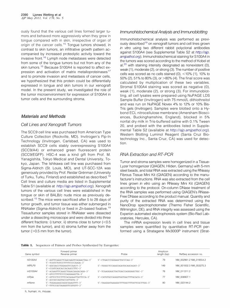
Table 1. Sequences of Primers and Probes Synthesized by Eurog
Gene symbolForward primerReverse primer
hS100A4 5=-AGTTCAAGCTCAACAAGTCAGAACTAA-3=5=-TCATCTGTCCTTTTCCCCAAGA-3=
5=-CTGACCC
hRPLP0 5=-GGCGACCTGGAAGTCCAACT-3=5=-CCATCAGCACCACAGCCTTC-3=
5=-TGGGCTC
mS100A4 5=-ACAAGTTCAAGCTGAACAAGACAGA-3=5=-GTCCTTTTCCCCAGGAAGCTA-3=
5=-TCAAGGA
mPpia 5=-GTTCCTCCTTTCACAGAATTATTCCA-3=5=-GCCGCCAGTGCCATTATG-3=
5=-CATGTGC
mNono 5=-TGGAAAAGCAGGCGAAGTTT-3=5=-TTTCCGCTAGGGTTCGTGTT-3=
5=-TAAGGAT
h, human; m, mouse.
Immunohistochemical Analysis and Immunoblotting
Immunohistochemical analysis was performed as previ-ously described19 on xenograft tumors and cell lines grownin vitro using two different rabbit polyclonal antibodiesagainst S100A4 (see Supplemental Table S2 at http://ajp.amjpathol.org). Immunohistochemical staining for S100A4 inthe tumors was scored according to the method of Kobel etal,20 with staining intensity designated as nonexistent (0),weak (1), moderate (2), or strong (3). The number of positivecells was scored as no cells stained (0), �10% (1), 10% to50% (2), 51% to 80% (3), or �80% (4). The final score wascalculated by multiplication of these two variables.Stromal S100A4 staining was scored as negative (0),weak (1), moderate (2), or strong (3). For immunoblot-ting, all cell lysates were prepared using NuPAGE LDSSample Buffer (Invitrogen) with 75 mmol/L dithiothreitoland was run on NuPAGE Novex 4% to 12% or 10% Bis-Tris gels (Invitrogen). Samples were blotted onto a Hy-bond ECL nitrocellulose membrane (Amersham Biosci-ences, Buckinghamshire, England), blocked in 5%nonfat dry milk in Tris-buffered saline with 0.1% Tween20, and probed with the antibodies listed in Supple-mental Table S2 (available at http://ajp.amjpathol.org).Western Blotting Luminol Reagent (Santa Cruz Bio-technology Inc., Santa Cruz, CA) was used for detec-tion.
RNA Extraction and RT-PCR
Tumor and stroma samples were homogenized in a Tissue-Lyzer homogenizer (QIAGEN, Hilden, Germany) with 5-mmsteel beads, and total RNA was extracted using the RNeasyFibrous Tissue Mini Kit (QIAGEN) according to the manu-facturer’s instructions. RNA was also extracted from the celllines grown in vitro using an RNeasy Mini Kit (QIAGEN)according to the protocol. On-column DNase treatment ofthe RNA samples was performed using QIAGEN’s RNase-Free DNase according to the product manual. Quantity andpurity of the extracted RNA was determined using theNanoDrop spectrophotometer (Thermo Fisher Scientific,Wilmington, DE), and RNA integrity was assessed using theExperion automated electrophoresis system (Bio-Rad Lab-oratories, Hercules, CA).
The mRNA expression levels in cell lines and tissuesamples were quantified by quantitative RT-PCR per-formed using a Stratagene Mx3000P instrument (Strat-
ProbeAmplicon
length (bp) RefSeq accession no.
GCCCAG-3= 79 NM_002961.2 NM_019554.2
ATGCAGCAGAT-3= 149 NM_001002.3 NM_053275.3
ACCAGGGAGCTGC-3= 76 NM_011311.2
GTGACTTTACACG-3= 77 NM_008907.1
TTGGCTTTATTCGCTTGG-3= 80 NM_023144.2
entec
GGGAGCT
CAAGCAG
GCTACTG
CAGGGTG
AAAGGCT

S100A4 in Tumor and Surrounding Stroma 2391AJP May 2011, Vol. 178, No. 5
agene, La Jolla, CA). Reverse transcription of total RNAwas performed using a Reverse Transcriptase Core Kit(Eurogentec SA, Searing, Belgium) with random nonamerprimers according to the manufacturer’s recommenda-tions using 100 ng of RNA per 10 �L of cDNA reaction.
For the analysis of S100A4, hydrolysis probe–basedassays were used. Primer pairs and FAM/Dark quenc-her–labeled hydrolysis probes were designed usingPrimer Express v.2.0 (Applied Biosystems, Foster City,CA) and were synthesized by Eurogentec (EurogentecSA). All the sequences are listed in Table 1. cDNA cor-responding to 10 ng of RNA was amplified for 40 cyclesin a 25-�L PCR mix (qPCR Mastermix Plus Low ROX;Eurogentec SA) containing a final concentration of 5mmol/L MgCl2, 100 nmol/L probe, and 400 nmol/L ofeach primer. Cycling conditions: 95°C for 10 minutes, 40cycles at 95°C for 30 seconds, and 60°C for 1 minute.
A SYBR Green (Applied Biosystems)–based methodwas used for analysis of human DNMT3b [DNA (cytosine-5)methyltransferase-3b]. Primers were purchased fromQIAGEN (assay name: Hs_DNMT3B_1_SG; catalog num-ber QT00032067). cDNA corresponding to 10 ng of RNAwas amplified for 40 cycles in a 25-�L PCR mix (RT2
Real-Time SYBR Green/ROX PCR Master Mix (SA Bio-sciences, Frederick, MD). Cycling conditions: 95°C for10 minutes, 40 cycles at 95°C for 30 seconds, 60°C for1 minute, and 72°C for 1 minute.
Duplicate reverse transcription reactions were per-formed for each RNA sample, and duplicate PCR analy-ses were performed on each cDNA sample. Primer spec-ificities and absence of primer dimers were determinedby SYBR Green melting curve analysis and agarose gelelectrophoresis. The absence of genomic DNA was con-firmed by performing a no–reverse transcription control,and absence of contaminations was assessed by includ-ing a no-template control in every run.
The expression of 12 human and mouse candidate ref-erence genes was tested using human and mouse house-keeping genes RT2 Profiler PCR Array (SA Biosciences),respectively. From this, hRPLP0 was chosen as the refer-ence gene for the samples derived from human cell lines,and mPpia and mNono were chosen for the stromal sam-ples. The ��Cq method21 was used to determine the rela-tive amount of target mRNA in the different samples.
Methylation Analysis
To study the effect of DNA methylation on S100A4 ex-pression in vitro, HSC-4, Ishikawa, and UT-SCC-12A cellswere treated for 4 days with 1 �mol/L of the DNA meth-yltransferase inhibitor 5-Aza-2=-deoxycytidine (Sigma-Al-drich). Fresh medium was added daily. For sequencingstudies, genomic DNA was isolated from HSC-4 cellsgrown in vitro and from HSC-4 skin and tongue tumortissue using the QIAmp DNA Mini Kit (QIAGEN) accord-ing to the protocol. DNA was bisulfite modified using theEpiTect Bisulfite Kit from QIAGEN following the manufac-turer’s recommendations. A set of previously publishedprimers (S100A4-F: 5=-TGTTTTTGAGATGTGGGTTTG-3=and S100A4-R: 5=-CACAATTACCTTCTACCTTTC-3=)22 was
used to amplify a human-specific region in the first intron ofthe S100A4 gene, which encompasses three cytosine-gua-nine (CpG) sites whose methylation status has been foundto be associated with S100A4 expression.22,23 PCR prod-ucts were purified using a QIAquick PCR Purification Kit(QIAGEN) according to the instructions, and purified prod-ucts were sequenced in both directions using the BigDyev.3.1 Terminator Cycle Sequencing Kit and the AppliedBiosystems 3130xl Genetic Analyzer (both from AppliedBiosystems, Warrington, England).
Statistical Analysis
Two-tailed t-tests were used to evaluate differences inS100A4 expression. A P � 0.05 was accepted as statis-tically significant.
Results
S100A4 Expression Was Up-Regulated inTongue Tumors
To study the effect of the tumor microenvironment onS100A4 expression in tumor cells, we analyzed threedifferent cell lines when grown in vitro and as xenografttumors in tongue and skin. The cell lines originate frompatients with tongue SCC (HSC-4), endometrial adeno-carcinoma (Ishikawa), or skin SCC (UT-SCC-12A), thusenabling growth of orthotopic and heterotopic tumors.
Immunohistochemical staining of the HSC-4, Ishikawa,and UT-SCC-12A cell lines grown in vitro were found to benegative for S100A4 (Figure 1, A–C), whereas immuno-blotting showed that all cell lines expressed low to mod-erate levels of S100A4 (Figure 1J).
Skin and tongue tumors established from the three celllines were also subjected to immunohistochemical stainingfor S100A4. The staining pattern and intensity were similarfor the two S100A4 antibodies used, and Table 2 providesthe immunohistochemical scores of the tumors. The skintumors of the HSC-4 cell line showed focal weak S100A4staining (Figure 1D). In contrast, tongue tumors of the samecell line had moderate to strong cytoplasmic staining inalmost all tumor cells; in addition, nuclear staining was seenin approximately 20% of tumor cells (Figure 1G). Skin tu-mors of the Ishikawa cell line were negative for S100A4(Figure 1E), whereas expression was induced in the tonguetumors, where cytoplasmic staining was seen toward theinvasive front of the tumors (Figure 1H). In contrast to thetwo former cell lines, no significant difference in S100A4expression was detected between tongue and skin tumorsof the UT-SCC-12A cell line (Figure 1, F and I).
The relative amount of S100A4 mRNA in tongue andskin tumors was analyzed using RT-PCR. For the HSC-4cell line, S100A4 mRNA levels in tongue tumors wereup-regulated approximately twofold compared with thosein skin tumors and approximately sixfold compared withthose in cells grown in vitro (Figure 1K). For the Ishikawacell line, mRNA levels were only slightly up-regulated intongue compared with in vitro levels (Figure 1L), whereas
tumors of the UT-SCC-12A cells had no increase in
2392 Ljones Wetting et alAJP May 2011, Vol. 178, No. 5
S100A4 mRNA levels compared with cells grown in vitro(Figure 1M).
S100A4 Expression Was Gradually IncreasedDuring Tumor Growth
Tongue tumors of the HSC-4 cell line obtained after 5, 10,15, and 28 days of tumor growth were immunohisto-chemically stained for S100A4 to study the onset andprogression of S100A4 protein expression. After 5 days invivo, all the tumor cells were negative (Figure 2A), andsome cells showed weak positive staining after 10 days(Figure 2B). The number of positive cells gradually in-creased, and at day 15, more than half of the tumor cellsshowed weak S100A4 staining (Figure 2C). After 28 days,most tumor cells revealed moderate to strong positivestaining (Figure 2D). The staining of the tumors was
Table 2. Quantification of S100A4 Expression Based on ImmunoUT-SCC-12A (15 Days) Tongue and Skin Tumors
Tumor ce
Cell line Tongue
HSC-4 (n � 4) 6.0 � 0.0Ishikawa (n � 6) 1.8 � 0.5UT-SCC-12A (n � 5) 1.8 � 1.1
Pooled tongue and skin tumors (n � 15) 2.9 � 0.6 0.7 �scored (Figure 2E), and the increase in staining from day5 to days 10, 15, and 28 was significant (P � 0.038, P �0.004 and P � 0.003, respectively).
Hypomethylation of CpG Sites in the S100A4Gene Increased Expression
S100A4 expression has previously been shown to bedependent on the methylation status of CpG sites in thefirst intron of the gene.22,24–26 To determine whethermethylation status affected S100A4 expression in thecells used in the present study, cells were cultured in vitroin the presence of the DNA methyltransferase inhibitor5-Aza-2=-deoxycytidine. RT-PCR analyses showed thatthe relative amount of S100A4 mRNA increased approx-imately twofold in treated samples compared with in con-trol samples of all three cell lines (data not shown).
Figure 1. Increased S100A4 protein and mRNA ex-pression levels in tumors compared with cells grownin vitro. Immunohistochemical S100A4 staining of theparaffin-embedded HSC-4 (A), Ishikawa (B), and UT-SCC-12A (C) cell lines grown in vitro and of skin (D, E,and F) and tongue (G, H, and I) tumors of the samecell lines. Representative areas of four to six paralleltumors are presented. S100A4 staining is shown inbrown, and nuclei are stained blue. t, tumor area; ands, stromal area. Nuclear localization of S100A4 in anHSC-4 tongue tumor is indicated with an arrowheadin G. Tumors of the HSC-4 and Ishikawa cell lines wereanalyzed after 28 days of tumor growth, and tumors ofthe UT-SCC-12A cell line were analyzed after 15 days.J: Immunoblots of HSC-4, Ishikawa (ISH), and UT-SCC12A cells grown in vitro probed with anti-S100A4and anti–�-actin. S100A4 mRNA levels in HSC-4 (K),Ishikawa (L), and UT-SCC-12A (M) analyzed by RT-PCR. Expression levels in tumors from skin (gray bars� skin tumor; n � 3) and tongue (black bars � tonguetumor; n � 3) were quantified relative to the expres-sion levels in cells grown in vitro (white bars � cellculture; n � 1) Error bars represent SEM. All expres-sion levels were normalized to expression of the ref-erence gene RPLP0. RNA was isolated from tumorsharvested after 15 days of tumor growth.
emical Staining of HSC-4 and Ishikawa (28 Days) and
an � SEM) Stroma (mean � SEM)
P value Tongue Skin P value
0.6 0.003 1.0 � 0.4 0.0 � 0.0 0.0500.0 0.012 1.0 � 0.4 0.7 � 0.2 0.4480.2 0.724 1.8 � 0.4 1.2 � 0.2 0.195
histoch
lls (me
Skin
1.0 �0.0 �1.4 �
0.2 0.004 1.3 � 0.2 0.7 � 0.2 0.041

S100A4 in Tumor and Surrounding Stroma 2393AJP May 2011, Vol. 178, No. 5
Immunoblotting revealed increased S100A4 protein ex-pression in treated cells compared with control samples(Figure 2F), demonstrating the relevance of CpG methyl-ation for S100A4 expression in the HSC-4, Ishikawa, andUT-SCC-12A cell lines.
To study whether hypomethylation was involved inthe observed induction of S100A4 expression duringtumor growth, the methylation status of three CpG sitesin the first intron of the S100A4 gene was investigatedin HSC-4 tongue and skin tumors and in HSC-4 cellsgrown in vitro. Bisulphite sequencing showed thatmethylation status correlated well with the observedexpression patterns of S100A4; the highest methylationlevel was found in cells grown in vitro, and all threeCpG sites were unmethylated in HSC-4 tongue tumors(Figure 2G).
The methylation of DNA is catalyzed by DNA methyl-
Figure 2. S100A4 expression is gradually increased during tumor growthand is dependent on hypomethylation of the gene. ImmunohistochemicalS100A4 staining of HSC-4 tongue tumors after 5 days (A), 10 days (B), 15 days(C), and 28 days (D) of tumor growth. Representative areas of three paralleltumors are presented. S100A4 staining is shown in brown, and nuclei arestained blue. E: Graph shows quantification of S100A4 staining at 5, 10, 15,and 28 days of tumor growth. n � 3 for all time points. Error bars representSEM. F: Immunoblots of HSC-4, Ishikawa (ISH), and UT-SCC-12A cells cul-tured in the absence (control; C) or presence (treated; T) of 1 �mol/L5-Aza-2=deoxycytidine, probed with anti-S100A4 and anti–�-actin. G: Meth-ylation status of three CpG residues in the first intron of S100A4 in HSC-4 skin(n � 3) and tongue (n � 3) tumors and in cells grown in vitro determinedby bisulphite sequencing and estimation of relative peak heights. Filledcircles, �75% methylation; half-filled circles, 25% to 75% methylation; andempty circles, �25% methylation.
transferases (DNMTs), and DNMT3B is reported to be
one of the main enzymes responsible for the establish-ment of new methylation patterns of DNA.27,28 To deter-mine whether the expression level of this DNMT couldexplain the observed differences in methylation statusbetween HSC-4 tongue and skin tumors, quantitative RT-PCR was used to quantify the relative amount of DNMT3BmRNA in the tumors. No significant difference in expres-sion was found between tongue and skin tumors (datanot shown), suggesting that other DNMTs or other mech-anisms were involved in modification of the methylationpattern in the tumors.
S100A4 Expression in Tumor Stroma Was NotCorrelated with S100A4 Levels in Tumor Cellsbut Rather with the Degree of Inflammation
Because stromal S100A4 also has been reported toaffect the invasive and metastatic potential of cancercells,11 stromal S100A4 expression was assessed inthe xenograft model (Table 2). Prominent immunohis-tochemical S100A4 staining was seen in specific cellsand in the extracellular matrix of the subepithelial con-nective tissue in normal tongue (Figure 3A) and skin(Figure 3B) of BALB/c nude mice. In general, stainingwas stronger in normal skin tissue compared with innormal tongue tissue.
RT-PCR was used to compare the S100A4 mRNA ex-pression levels in the stromal tissue close to and furtheraway (�0.5 mm) from the tumors. S100A4 expressionwas approximately twice as high in skin as in tonguefurther away from the tumors (data not shown); however,the stromal cells close to the tumors showed a largerincrease in S100A4 mRNA expression in tongue than inskin (Figure 3C). Similarly, immunohistochemical S100A4staining was stronger in stroma close to tongue and skintumors of all the cell lines compared with that seen innormal tongue and skin tissues. The increase was slightlyhigher in stroma surrounding tongue tumors comparedwith stroma surrounding skin tumors; see the immunohis-tochemical scores in Table 2. The intensity of stromalstaining varied considerably between the parallel tumorsof each cell line and did not correlate with the stainingintensity of the cancer cells but rather with the level ofinflammation. In a previous study using the same xeno-graft tumors, we found that the inflammatory infiltrate wasmore pronounced in tongue tumors compared with in thecorresponding skin tumors for all three cell lines. Neutro-phils were the predominant cell type in the inflammatoryinfiltrate,15 and in the present study, stromal S100A4staining was mainly seen in and surrounding cells withmorphologic features similar to those of neutrophils andmacrophages (data not shown).
Previous studies have shown that some cell linesrelease S100A4 into the growth medium in vitro. Theprotein can also be detected in tumor interstitialfluid,10,29 but the source of extracellular S100A4 in thetumor is unknown. From the tongue SCC cell lineSCC9, we successfully established a cell line stablyoverexpressing S100A4 (SCC9/A4) (Figure 3D). To
study whether S100A4 in tumor cells affected stromal
rs are pr
2394 Ljones Wetting et alAJP May 2011, Vol. 178, No. 5
S100A4 levels, by either secreting the protein or induc-ing S100A4 expression in stromal cells, SCC9/A4 cellswere used to establish tongue tumors in the xenograftmodel. Cells overexpressing EGFP (SCC9/EGFP) wereused as a control. Very faint S100A4 bands were de-tected on immunoblots of conditioned media from theS100A4-overexpressing cells, indicating some releasefrom the SCC9/A4 cell line in vitro (data not shown).Tumors of the SCC9/A4 cell line showed intenseS100A4 staining in the cancer cells (Figure 3, E and F).However, only focal stromal staining was seen in areaswith sparse inflammation (Figure 3E), whereas muchmore staining was seen in areas with a more pro-nounced inflammatory reaction (Figure 3F). Tumors ofthe parental SCC9 cell line showed weak S100A4 stain-ing in the cancer cells, and the stromal staining wasstrong in areas with a pronounced inflammatory reac-tion (Figure 3G). Tumors of the control cell line SCC9/EGFP showed the same staining pattern as the SCC9tumors (data not shown). Thus, S100A4 expression inthe tumor-associated stromal cells was unaffected byS100A4 expression levels in the cancer cells but rather
Figure 3. S100A4 expression is increased in tumor-associated stroma but isand skin (B) tissue from BALB/C nude mice were immunohistochemically stais shown in brown, and nuclei are stained blue. C: S100A4 mRNA levels in tumin stroma close to the tumors of HSC-4, Ishikawa (ISH), and UT-SCC-12A (baway (�0.5 mm) from the tumors (white bars; n � 3). All expression levelsrepresent SEM. D: Immunoblots of cell lysates from SCC9, SCC9/A4, andS100A4 staining of tongue tumors of the SCC9/A4 cell line with sparse stromwith strong stromal staining (G). Representative areas of three parallel tumotumor area; and s, stromal area.
was correlated with the degree of inflammation.
Discussion
Tumor-stroma interactions are known to be important forcancer progression. Using a xenograft model where thesame cancer cell lines were used to establish tumors intongue and skin, we previously found that the cells showremarkably different growth patterns in the two environ-ments.15,16 In the present study, we investigated the role ofthe tumor microenvironment in regulation of the metastasis-associated protein S100A4. We found increased expres-sion of S100A4 in tongue tumors of the oral SCC cell lineHSC-4 and the endometrial adenocarcinoma cell lineIshikawa compared with the corresponding skin tumors. Incontrast, tumors of the skin SCC cell line UT-SCC-12Ashowed similar S100A4 expression in tongue and skin.
The signaling events regulating S100A4 expressionare not well characterized, but several studies haveshown that S100A4 expression is regulated epigeneti-cally by methylation of CpG dinucleotides in the firstintron of the gene.23,25,30 Hypomethylation of these cyto-sine residues is thought to be involved in cancer-associ-ated up-regulation of S100A4 expression.22,23,25,30 In the
related with S100A4 levels in the tumor cells. Sections of normal tongue (A)S100A4. Representative areas of the samples are presented. S100A4 staining
ciated stroma in tongue and skin were analyzed by RT-PCR. Expression levelss; n � 3) were quantified relative to the expression levels in stroma furtherrmalized to the expression of the reference genes Ppia and Nono. Error barsFP cells probed with anti-S100A4 and anti–�-actin. Immunohistochemical
ing (E) and more pronounced stromal staining (F) and of the SCC9 cell lineesented; S100A4 staining is shown in brown, and nuclei are stained blue. t,
not corined foror-assolack barwere noSCC9/EGal stain
present study, we found increased expression of S100A4

S100A4 in Tumor and Surrounding Stroma 2395AJP May 2011, Vol. 178, No. 5
in the cell lines after treatment with a DNA methyltrans-ferase inhibitor in vitro. When examining the intronic se-quence in DNA from HSC-4 tongue and skin tumors andHSC-4 cells grown in vitro, we also found a correlationbetween methylation status and S100A4 expression,showing that hypomethylation was involved in the ob-served induction of S100A4 expression in the tumors.Several studies have demonstrated DNA hypermethyl-ation of cancer cells grown in vitro,31,32 and oral SCC celllines have shown a higher propensity to become hyper-methylated than have cell lines isolated from other cancertypes.32 This may explain the observation that the oralSCC cell line HSC-4 showed the strongest induction ofS100A4 in vivo. S100A4 was recently shown to be down-regulated in epidermal cancers compared with expres-sion in normal epidermal tissue.33 DNA sequencing ofcancer cells isolated from epidermal cancers revealedmethylation of the regulatory CpG dinucleotides in thefirst intron of the S100A4 gene. This epigenetic silencingof S100A4 expression in skin cancers may contribute totheir generally low metastatic potential. The report is inaccordance with our observation that xenograft skin tu-mors expressed low levels of S100A4, although normalskin tissue of the mice showed stronger S100A4 expres-sion than did normal tongue tissue. The results indicatethat factors in the tumor microenvironment affect the DNAmethylation status of the cancer cells. These findings alsohave clinical implications because DNA methyltrans-ferase inhibitors have been recognized as promisingcandidate anticancer drugs because of their ability toreactivate expression of tumor suppressor genes si-lenced by aberrant methylation.28,34 The present findingsdemonstrate that inhibition of methyltransferases can ac-tivate prometastatic genes, raising concerns that thetreatment could actually promote metastasis.
S100A4 expression has also been shown to be in-duced by some extracellular growth factors, eg, epider-mal growth factor, basic fibroblast growth factor, andtransforming growth factor-�.35,36 Proteolytic enzymes,including plasmin and matrix metalloproteinases, can re-lease and activate growth factors stored in the extracel-lular matrix, such as latent transforming growth factor-�and basic fibroblast growth factor.37,38 In a previousstudy using the same xenograft tumors, we found in-creased proteolytic activity in tongue tumors comparedwith skin tumors.16 This may yield higher levels of biolog-ically active transforming growth factor-� and basic fibro-blast growth factor with a subsequent induction ofS100A4 expression in the tongue tumors. BecauseS100A4 is known to induce increased expression andactivation of several matrix metalloproteinases,17 a posi-tive feedback loop may exist.
S100A4 expression in tumor stroma is also shown tocontribute to increased invasion and metastasis of can-cer cells. This was demonstrated in a study where tumorsof highly metastatic carcinoma cells did not metastasizein S100A4 knockout mice, but the metastatic phenotypewas partly restored by co-injecting the cancer cells withS100A4-expressing stromal cells.11 In the xenograftmodel, tongue tumors of the UT-SCC-12A cell line were
the most aggressive, with lymph node metastases de-tected in half of the mice.15 Still, S100A4 staining of thesetumors was not prominent and did not differ significantlyfrom staining of the noninvasive skin tumors of the samecell line. However, UT-SCC-12A tongue tumors provokeda pronounced inflammatory reaction, and this was ac-companied by marked stromal S100A4 expression. Thisindicates that S100A4 expression in the tumor stromamay be as important for metastases as S100A4 expres-sion in the tumor cells.
In the present study, the staining intensity of S100A4 intumor and stroma did not correlate. Instead, stromalS100A4 staining reflected the degree of inflammation andwas seen within and outside tumor-associated inflamma-tory cells. Although immunodeficient mice (Charles RiverLaboratories, Wilmington, MA) were used in the presentexperiments, they do have B cells, natural killer cells, andall the cells of the innate immune system. High numbersof tumor-associated inflammatory cells, such as macro-phages and neutrophils, are associated with poor prog-nosis in several cancer types. Because many inflamma-tory cells are reported to express S100A4,10,39 a role assuppliers of stromal S100A4 may contribute to their can-cer-promoting effect.
The present findings show that the microenvironmentcan affect S100A4 expression in cancer cells by regulat-ing the methylation of CpG units in the gene. The ability oftongue stroma to increase S100A4 expression in cancercells, and the increased number of tumor-associated,S100A4-expressing inflammatory cells, may contribute tothe aggressive behavior of oral SCCs. This study, there-fore, merits further investigation of the significance ofS100A4 expression in patients with oral cancer.
Acknowledgments
We thank Dr. Kjetil Boye, Ph.D., for critical reading andDr. Peter McCourt, Ph.D., for critical reading and linguis-tic revision of the manuscript.
References
1. Mueller MM, Fusenig NE: Friends or foes: bipolar effects of the tumourstroma in cancer. Nat Rev Cancer 2004, 4:839–849
2. Kalluri R, Zeisberg M: Fibroblasts in cancer. Nat Rev Cancer 2006,6:392–401
3. Rudland PS, Platt-Higgins A, Renshaw C, West CR, Winstanley JH,Robertson L, Barraclough R: Prognostic significance of the metasta-sis-inducing protein S100A4 (p9Ka) in human breast cancer. CancerRes 2000, 60:1595–1603
4. Gongoll S, Peters G, Mengel M, Piso P, Klempnauer J, Kreipe H, vonWasielewski R: Prognostic significance of calcium-binding proteinS100A4 in colorectal cancer. Gastroenterology 2002, 123:1478–1484
5. Garrett SC, Varney KM, Weber DJ, Bresnick AR: S100A4, a mediatorof metastasis. J Biol Chem 2006, 281:677–680
6. Sherbet GV: Metastasis promoter S100A4 is a potentially valuablemolecular target for cancer therapy. Cancer Lett 2009, 280:15–30
7. Ebralidze A, Tulchinsky E, Grigorian M, Afanasyeva A, Senin V, Re-vazova E, Lukanidin E: Isolation and characterization of a gene spe-cifically expressed in different metastatic cells and whose deducedgene product has a high degree of homology to a Ca2�-bindingprotein family. Genes Dev 1989, 3:1086–1093
8. Kikuchi N, Horiuchi A, Osada R, Imai T, Wang C, Chen X, Konishi I:Nuclear expression of S100A4 is associated with aggressive behavior

2396 Ljones Wetting et alAJP May 2011, Vol. 178, No. 5
of epithelial ovarian carcinoma: an important autocrine/paracrinefactor in tumor progression. Cancer Sci 2006, 97:1061–1069
9. Schmidt-Hansen B, Klingelhofer J, Grum-Schwensen B, Chris-tensen A, Andresen S, Kruse C, Hansen T, Ambartsumian N,Lukanidin E, Grigorian M: Functional significance of metastasis-inducing S100A4(Mts1) in tumor-stroma interplay. J Biol Chem2004, 279:24498 –24504
10. Cabezon T, Celis JE, Skibshoj I, Klingelhofer J, Grigorian M, GromovP, Rank F, Myklebust JH, Maelandsmo GM, Lukanidin E, Ambartsum-ian N: Expression of S100A4 by a variety of cell types present in thetumor microenvironment of human breast cancer. Int J Cancer 2007,121:1433–1444
11. Grum-Schwensen B, Klingelhofer J, Berg CH, El-Naaman C, Grigo-rian M, Lukanidin E, Ambartsumian N: Suppression of tumor devel-opment and metastasis formation in mice lacking the S100A4(mts1)gene. Cancer Res 2005, 65:3772–3780
12. Funk GF, Karnell LH, Robinson RA, Zhen WK, Trask DK, Hoffman HT:Presentation, treatment, and outcome of oral cavity cancer: a NationalCancer Data Base report. Head Neck 2002, 24:165–180
13. Miller DL, Weinstock MA: Nonmelanoma skin cancer in the UnitedStates: incidence. J Am Acad Dermatol 1994, 30:774–778
14. McGuire JF, Ge NN, Dyson S: Nonmelanoma skin cancer of the headand neck, I: histopathology and clinical behavior. Am J Otolaryngol2009, 30:121–133
15. Hadler-Olsen E, Wetting HL, Rikardsen O, Steigen SE, Kanapathip-pillai P, Grenman R, Winberg JO, Svineng G, Uhlin-Hansen L: Stromalimpact on tumor growth and lymphangiogenesis in human carcinomaxenografts. Virchows Arch 2010, 457:677–692
16. Hadler-Olsen E, Wetting HL, Ravuri C, Omair A, Rikardsen O, SvinengG, Kanapathippillai P, Winberg JO, Uhlin-Hansen L: Organ specificregulation of tumour invasiveness and gelatinolytic activity at theinvasive front. Eur J Cancer 2011, 47:305–315
17. Elenjord R, Ljones H, Sundkvist E, Loennechen T, Winberg JO: Dys-regulation of matrix metalloproteinases and their tissue inhibitors byS100A4. Connect Tissue Res 2008, 49:185–188
18. Grenman R, Pekkola-Heino K, Joensuu H, Aitasalo K, Klemi P,Lakkala T: UT-MUC-1, a new mucoepidermoid carcinoma cell line,and its radiosensitivity. Arch Otolaryngol Head Neck Surg 1992,118:542–547
19. Hadler-Olsen E, Kanapathippillai P, Berg E, Svineng G, Winberg JO,Uhlin-Hansen L: Gelatin in situ zymography on fixed, paraffin-embed-ded tissue: zinc and ethanol fixation preserve enzyme activity. J His-tochem Cytochem 2010, 58:29–39
20. Kobel M, Weichert W, Cruwell K, Schmitt WD, Lautenschlager C,Hauptmann S: Epithelial hyaluronic acid and CD44v6 are mutuallyinvolved in invasion of colorectal adenocarcinomas and linked topatient prognosis. Virchows Arch 2004, 445:456–464
21. Livak KJ, Schmittgen TD: Analysis of relative gene expression datausing real-time quantitative PCR and the 2(-��C(T)) method. Meth-ods 2001, 25:402–408
22. Rosty C, Ueki T, Argani P, Jansen M, Yeo CJ, Cameron JL, HrubanRH, Goggins M: Overexpression of S100A4 in pancreatic ductal
adenocarcinomas is associated with poor differentiation and DNAhypomethylation. Am J Pathol 2002, 160:45–5023. Tulchinsky E, Grigorian M, Tkatch T, Georgiev G, Lukanidin E: Tran-scriptional regulation of the mts1 gene in human lymphoma cells: therole of DNA-methylation. Biochim Biophys Acta 1995, 1261:243–248
24. Rehman I, Goodarzi A, Cross SS, Leiblich A, Catto JW, Phillips JT,Hamdy FC: DNA methylation and immunohistochemical analysis ofthe S100A4 calcium binding protein in human prostate cancer. Pros-tate 2007, 67:341–347
25. Xie R, Loose DS, Shipley GL, Xie S, Bassett RL Jr., Broaddus RR:Hypomethylation-induced expression of S100A4 in endometrial car-cinoma. Mod Pathol 2007, 20:1045–1054
26. Dokun OY, Florl AR, Seifert HH, Wolff I, Schulz WA: Relationship ofSNCG, S100A4, S100A9 and LCN2 gene expression and DNA meth-ylation in bladder cancer. Int J Cancer 2008, 123:2798–2807
27. Luczak MW, Jagodzinski PP: The role of DNA methylation in cancerdevelopment. Folia Histochem Cytobiol 2006, 44:143–154
28. Kelly TK, De Carvalho DD, Jones PA: Epigenetic modifications astherapeutic targets. Nat Biotechnol 2010, 28:1069–1078
29. Pedersen KB, Andersen K, Fodstad O, Maelandsmo GM: Sensitiza-tion of interferon-� induced apoptosis in human osteosarcoma cellsby extracellular S100A4. BMC Cancer 2004, 4:52
30. Lindsey JC, Lusher ME, Anderton JA, Gilbertson RJ, Ellison DW,Clifford SC: Epigenetic deregulation of multiple S100 gene familymembers by differential hypomethylation and hypermethylationevents in medulloblastoma. Br J Cancer 2007, 97:267–274
31. Antequera F, Boyes J, Bird A: High levels of de novo methylation andaltered chromatin structure at CpG islands in cell lines. Cell 1990,62:503–514
32. Smiraglia DJ, Rush LJ, Fruhwald MC, Dai Z, Held WA, Costello JF,Lang JC, Eng C, Li B, Wright FA, Caligiuri MA, Plass C: ExcessiveCpG island hypermethylation in cancer cell lines versus primaryhuman malignancies. Hum Mol Genet 2001, 10:1413–1419
33. Li Y, Liu ZL, Zhang KL, Chen XY, Kong QY, Wu ML, Sun Y, Liu J, LiH: Methylation-associated silencing of S100A4 expression in humanepidermal cancers. Exp Dermatol 2009, 18:842–848
34. Issa JP, Kantarjian HM: Targeting DNA methylation. Clin Cancer Res2009, 15:3938–3946
35. Okada H, Danoff TM, Kalluri R, Neilson EG: Early role of Fsp1 inepithelial-mesenchymal transformation. Am J Physiol 1997, 273:F563–F574
36. Strutz F, Zeisberg M, Ziyadeh FN, Yang CQ, Kalluri R, Muller GA,Neilson EG: Role of basic fibroblast growth factor-2 in epithelial-mesenchymal transformation. Kidney Int 2002, 61:1714–1728
37. Yu Q, Stamenkovic I: Cell surface-localized matrix metalloprotei-nase-9 proteolytically activates TGF-� and promotes tumor invasionand angiogenesis. Genes Dev 2000, 14:163–176
38. Folgueras AR, Pendas AM, Sanchez LM, Lopez-Otin C: Matrix met-alloproteinases in cancer: from new functions to improved inhibitionstrategies. Int J Dev Biol 2004, 48:411–424
39. Takenaga K, Nakamura Y, Sakiyama S: Expression of a calciumbinding protein pEL98 (mts1) during differentiation of human promy-elocytic leukemia HL-60 cells. Biochem Biophys Res Commun 1994,
202:94–101