
Map the tumor microenvironment - AMP 2018 - Association for ...
Upload
independentCategory
view
1download
0
Interactions between microenvironment and cancer cells
in two animal models of bone metastasis.
Stephane Blouin, Michel Felix Basle, Daniel Chappard
To cite this version:
Stephane Blouin, Michel Felix Basle, Daniel Chappard. Interactions between microenvironmentand cancer cells in two animal models of bone metastasis.. British Journal of Cancer, CancerResearch UK, 2008, 98 (4), pp.809-15. <10.1038/sj.bjc.6604238>. <inserm-00259380>
HAL Id: inserm-00259380
http://www.hal.inserm.fr/inserm-00259380
Submitted on 4 Aug 2008
HAL is a multi-disciplinary open accessarchive for the deposit and dissemination of sci-entific research documents, whether they are pub-lished or not. The documents may come fromteaching and research institutions in France orabroad, or from public or private research centers.
L’archive ouverte pluridisciplinaire HAL, estdestinee au depot et a la diffusion de documentsscientifiques de niveau recherche, publies ou non,emanant des etablissements d’enseignement et derecherche francais ou etrangers, des laboratoirespublics ou prives.

1
Interactions between microenvironment and cancer cells in two animal models of bone metastasis
Running title: Bone microenvironment and cancer cells
S. BLOUIN a, M.F. BASLÉ a, D. CHAPPARD a.
a INSERM, EMI 0335 – LHEA, Faculté de Médecine, 49045 ANGERS Cedex -
FRANCE.
e-mail adress : S. Blouin [email protected]
D. Chappard [email protected]
Please send all correspondence to:
* Daniel CHAPPARD, M.D., Ph.D. Tel: (33) 241 73 58 65
INSERM, EMI 0335 – LHEA Fax: (33) 241 73 58 86
Faculté de Médecine, E-Mail: [email protected]
49045 ANGERS Cedex – FRANCE
HA
L author manuscript inserm
-00259380, version 1
HAL author manuscriptBritish Journal of Cancer 2008;98(4):809-15
2
ABSTRACT
The preferential proliferation of cancer cells in the bone microenvironment is poorly
characterized. Expression pattern of bone marrow and other organs microenvironment in
contact with osteolytic (Walker W256) and osteoblastic (MatLyLu MLL) metastases were
investigated.
Fisher and Copenhagen rats received respectively W256 and MLL cells injection. Bone and
soft tissues were analyzed by immunochemistry for DKK1, cathepsin K, RANKL, MCSF, or
IL6 expression. TRAcP+ cells were detected by a histoenzymatic technique.
In bone, expressions of MCSF and DKK1 were shown in stromal cells of the bone marrow, in
contact with metastatic foci of both tumors. Many stromal cells were found RANKL+ in the
vicinity of the tumors. Cells expressing cathepsin K and multinucleated TRAcP+ cells were
found in direct contact with trabeculae but also in bone marrow spaces near metastatic cells.
In extraosseous tumors, cells in contact with malignant cells did not expressed DKK1, MCSF,
cathepsin K, IL6. Some RANKL+ cells were found in the periphery of subcutaneous tumors
but may represent Langerhans cells. Abnormal presence of TRAcP+ cells was never observed
in the vicinity of malignant cells.
Interaction between stromal and cancer cells induces the expression on the formers of
characteristics leading to osteoclastogenesis only in the bone microenvironment.
Key words: Bone metastasis; osteolysis; osteoclastogenesis; osteoclast; microenvironment.
HA
L author manuscript inserm
-00259380, version 1
3
INTRODUCTION
Bone is a preferential site for metastasis in different types of cancer. Bone metastases induce
an increased morbidity (pain, fractures, nerve compression, hypercalcemia…) and
compromise the long-term survival. They occur in approximately 65-75 % of patients with an
advanced breast or prostate cancer and 15 to 40 % of patients with a carcinoma of the lung,
bladder, skin or kidney. The median survival after first recurrence of breast cancer in bone is
20 months (Coleman, 2001). Once a tumor has metastasized to bone, it is virtually incurable
and only palliative therapies can be proposed (Mundy, 1997).
Interactions between the bone marrow environment and cancer cells were first advocated by
Paget who described them as ‘the seed and the soil’ theory (cited by (Guise & Mundy, 1998)):
bone provides the ‘fertile soil’ in which certain cancer cell (‘seeds’) find favourable
conditions for growth. Mineralized bone matrix is known to represent a rich storehouse of
growth factors which are mobilized by osteoclastic resorption and become active in the local
microenvironment. The release of these growth factors constitutes one of the key mechanisms
in the bone remodeling allowing the physiological coupling between osteoblasts (the bone
forming cells) and the osteoclasts (bone resorbing cells) (Mundy et al., 2001). When localized
in the marrow spaces, tumor cells, in turn, secrete additional factors that interfere with bone
remodeling leading to tissular changes that characterize the osteolytic or osteoblastic
expression of the metastases. Thus, local interactions between tumor and bone cells form a
vicious cycle which underlies the development of skeletal metastases. During the last decade,
it has become evident that interactions between tumor and bone cells may change the
phenotype of both populations.
Breast and prostate cancer are the two tumor types that most commonly metastasize to bone.
On one hand, bone has characteristics that allow the selective homing of tumor cells: 1) a
release of attractive molecules for these particular types of cancer (stromal cell-derived factor-
1, epidermal growth factor, collagen degradation products, low glycosylated osteonectin…)
inducing specific chemotaxis (Cooper et al., 2003) 2) the expression of adhesion molecule on
the endothelial surface of bone marrow capillaries (due to a variety of integrins such as α4β1,
α5β1, αvβ3, αvβ5)(Roodman, 2003) 3) appropriate growth factors and extracellular matrix
proteins present in the bone marrow microenvironment (parathyroid hormone related protein
(PTHrP), tumor growth factor beta (TGF-β), vascular endothelium growth factor (VEGF)…)
(Guise & Mundy, 1998; Keller et al., 2001; Woodhouse et al., 1997). On the other hand, these
peculiar tumor cells express proteins which favor propensity to migrate and anchor in bone:
this includes chemotaxis (CXCR4) (Muller et al., 2001), extravasation and bone marrow
HA
L author manuscript inserm
-00259380, version 1
4
homing (integrins, E-cadherin) (Woodhouse et al., 1997), pericellular proteolysis and invasion
(MMPs, ADAMs) (Woodhouse et al., 1997; Zigrino et al., 2005), angiogenesis (Woodhouse
et al., 1997), osteoclastogenesis (Roodman, 2004), growth factor regulation (follistatin) (Reis
et al., 2004; Risbridger et al., 2001), and extracellular matrix alteration (proteoglycan-
1) (Timar et al., 2002). A multigenic study of breast cancer metastasis to bone has provided
evidences for a causal role of IL-11, connective tissue-derived growth factor (CTGF), and
CXCR4, along with osteopontin in osteolytic metastases. The combined overexpression of
these genes give to transfected cells a metastatic activity close to that of the highly aggressive
cell populations endogenously expressing the entire bone metastasis gene set (Kang et al.,
2003).
Bone metastases are commonly characterized as osteolytic or osteoblastic from a radiological
point of view. This represents two extremes of a continuum in which dysregulation of the
normal bone remodeling process occurs and alters bone mass and bone microarchitecture.
Breast cancer is most often associated with osteolytic metastases while osteoblastic
metastases are common in prostate cancer. Osteolytic lesions are due to a marked increase in
osteoclast number with a reduced osteoblastic activity. PTHrP is a major mediator between
tumor and bone cells and induces the osteolytic process through the RANKL pathway (Guise,
2000). On the contrary, osteoblastic metastases are characterized by a dramatic increase in
osteoformation but always possess a resorption component (Logothetis & Lin, 2005; Mundy,
2002).
Most researches have focused on the interactions between tumor cells and osteoclasts or
osteoblasts. However interactions with surroundings cells have been poorly investigated.
Because osteoblast precursors are stromal cells (present in the marrow spaces) that interact
with hematopoietic osteoclast precursors via the RANK/RANKL/osteoprotegerin system
(Thomas et al., 1999), the potential influence of stromal cells on metastatic cells needs to be
investigated.
The aim of this study was to induce metastases in the rat to investigate the expression pattern
of these key molecules in the bone microenvironment and also in the stroma of other organs
invaded by a metastasis. Two cell lines known to induce either osteolytic or osteoblastic
metastases were used.
HA
L author manuscript inserm
-00259380, version 1
5
MATERIALS AND METHODS
Cell lines and culture conditions
Two malignant cell lines capable of inducing bone metastases were used in the present study:
Walker 256/B cells. The Walker 256/B (W256) cells (issued of a rat mammary carcinoma) are
associated with osteolytic metastases (Blouin et al., 2005; Rizzoli & Fleisch, 1987; Wingen et
al., 1986). W256 cells, (kindly provided by Pr. R. Rizzoli, Geneva, Switzerland), were grown
in Dulbecco's modified Eagle's medium (DMEM - Eurobio, Les Ulis, France) containing 10%
heat inactivated fetal calf serum (FCS-Seromed Biochrom, Berlin, Deutschland), 100 UI/ml
of penicillin (Eurobio), 100µg/ml of streptomycin sulfate (Eurobio), 1% of Non Essential
Amino Acids (NEAA – Cambrex, Walkersville, MD, USA), and 1 mM of sodium pyruvate
(Eurobio). Cells were cultured at 37°C in a humidified incubator with 5% CO2. In order to
obtain cells with a bone trophicity, 107 cells were serially passaged intraperitoneally at 7 days
intervals in Fisher rats to obtain malignant ascite.
MatLyLu cells. The MatLyLu cell line is an androgen independent, highly metastatic, and
anaplastic tumor of rat origin, which spreads to the lymph nodes and invariably to the lung.
metastases are reported to be osteoblastic in the vertebrae (Vieweg et al., 1994). The
MatLyLu tumor cell line was obtained from ECCAC (European Collection of Animal Cell
Cultures, Salisbury, Wiltshire, UK). MatLyLu cells were grown in Roswell Park Memorial
Institute 1640 medium (RPMI 1640 - Eurobio) containing 10% heat inactivated fetal calf
serum (FCS-Seromed Biochrom, Berlin, Deutschland), 100 UI/ml of penicillin (Eurobio),
100µg/ml of streptomycin sulfate (Eurobio) and 250nM of dexamethasone (Sigma, Saint
Quentin Fallavier, France). Cells were cultured at 37°C in a humidified incubator with 5%
CO2.
Animals
The Animal Care and Use committee at the University of Angers approved all procedures.
Nine female Fisher F344/NHsd rats (Harlan, Gannat, France), 3 weeks old were maintained 6
months to the local vivarium conditions (24°C and a 12-h/12-h light/dark cycle). Nine female
Copenhagen born from COP/NCrl rats (Charles River, Sulzfeld, Germany) and bred in our
University animal facilities were maintained 6 months to the same conditions. Rats were
given a standard laboratory food (UAR, Villemoison sur Orge, France) and water ad libitum.
F344 rats were anesthetized with isoflurane (AErrane®, Baxter S.A, Belgium) and received
an intracardiac injection of 107 W256 cells in 0.25 ml saline. They also received
HA
L author manuscript inserm
-00259380, version 1
6
intramuscular (quadriceps femoris) and subcutaneous (base of the neck) injection with 107
W256 cells.
COP rats were anesthetized with isoflurane and received an intracardiac injection of 3.25x10⁴
MLL cells in 0.25 ml saline. They also received intramuscular, intraliver, intraspleen and
subcutaneous injections with 3.25x10⁴ MLL cells in the same locations than above. For both
types of rats, the cell number to be injected was determined as the most appropriate in
preliminary experiments. Data were compared with results obtained on control rats.
Rats were sacrificed by asphyxiation with CO2, 9 days (F344) or 15 days (COP) after the
intracardiac injection of tumor cells. These durations have been determined by preliminary
studies and were found sufficient to obtain bone metastases before animals die from other
tumor localization. The hindlimbs were carefully dissected; bones and other tissue samples
(liver, spleen, brain, subcutaneous and intramuscular nodules) were placed in a formalin-
alcohol based fixative at +4°C during 24 h.
Histological staining methods on paraffin sections
Paraffin sections (4µm) were deparaffined and stained by Hematoxylin Phloxine Saffron
(HPS) as a standard tissue stain. Immunohistochemistry was performed on femur and organs
of 6 F344 and 6 COP rats. Bone decalcification was done using a ready-to-use decalcifying
fluid according to the manufacturer’s recommendations (Surgipath®, Labonord, Templemars,
France). Bone and other tissues were then dehydrated, embedded in paraffin and sectioned at
4 μm. Antigen retrieval was performed by bain-marie heating in a citrate buffer (pH 6).
Immunochemistry was performed using DAKOCYTOMATION® procedure with the Dako
autostainer automat (Dako, Glostrup, Denmark). The following proteins were traced using
specific antibodies:
• Dickkopf-1 (Dkk-1) is an antagonist of the canonical Wnt signaling by binding
directly to the LDL receptor-related protein 5/6 (LRP5/6). The interaction between
DKK1 ant LRP5/6 induces an inhibition of bone formation (Johnson et al., 2004). The
reagent was a purified goat polyclonal antibody (Tebu-bio, sc-14949) used at a
dilution of 1:50. Positivity is characterized by a cell membrane labeling.
• Interleukin 6 (IL-6) is a pleitropic cytokine. Its production by bone marrow stromal
cells enhances osteoclastogenesis (with subsequent bone resorption) and tumor cell
growth (Roodman, 2003). We used a purified goat polyclonal antibody (Tebu-bio, sc-
1265) at a dilution of 1:50. Positivity is characterized by a cytoplasmic labeling.
HA
L author manuscript inserm
-00259380, version 1
7
• RANKL is produced by osteoblasts and stromal cells and induces osteoclastogenesis
by interacting with its receptor at the osteoclastic surface (Roodman, 2003). We used a
purified goat polyclonal antibody (Tebu-bio, sc-7628) at a dilution of 1:200. Positivity
is characterized by a cell membrane labeling.
• Cathepsin K is a cysteine protease highly expressed by osteoclasts and is implicated in
the bone matrix collagen breakdown (Yasuda et al., 2005). The purified goat
polyclonal antibody (Tebu-bio, sc-6507) was used at a dilution of 1:200. Positivity is
characterized by a cytoplasmic labeling.
• Macrophage-colony stimulating factor (M-CSF) is expressed by stromal cells and
osteoblasts. It is required together with RANKL to induce the development of mature
osteoclasts (Boyle et al., 2003). The purified goat polyclonal antibody (Tebu-bio, sc-
1324) was used at a dilution of 1:200. Positivity is characterized by a cytoplasmic
labeling.
Control positive tissues were obtained for each antibody. Sections were counterstained with
hematoxylin.
TRAcP cells detection
Tartrate Resistant Acid Phosphatase (TRAcP) positive cells were detected by a
histoenzymatic technique in 3 F344 and 3 COP rats. TRAcP is a key enzyme found in high
level in osteoclasts; it is involved in the intracellular degradation of collagen and non-
collagenous proteins of the bone matrix (Vaaraniemi et al., 2004). Tissues were embedded in
methylmethacrylate at 4°C to maintain enzyme activity. Bone samples were embedded
undecalcified. Sections (7µm thick) were cut dry on a heavy-duty microtome equipped with
50° tungsten carbide knives (Leica Polycut S, Rueil-Malmaison, France). TRAcP was
revealed by a simultaneous coupling reaction using 1-naphtyl phosphate (Sigma-Aldrich
Chemical, Illkirsh, France) as substrate and Fast Violet B as the diazonium salt. Sections were
counterstained with phosphomolybdic aniline blue (Chappard et al., 1983). TRAcP positive
cells appeared with a brownish tint. Collagen fibers densely packed in the form of bone
matrix or in the tissue stroma appear with a blue tint.
HA
L author manuscript inserm
-00259380, version 1
8
RESULTS
Tumor growth in animals
F344 rats developed tumors at subcutaneous and intramuscular site of injection. They also
developed pulmonary, liver, brain and bone tumors secondary to the intracardiac injection of
W256 cells. In the femur, osteolytic foci were preferentially localized below the growth
cartilage and formed a large and irregular band filled with malignant cells.
COP rats similarly developed tumors at subcutaneous, intramuscular, liver and spleen sites of
injection. They also developed pulmonary and bone tumors after the intracardiac injection. In
the femur, focal areas of osteolysis were disseminated all over the metaphysis. No
osteosclerotic lesions were found.
Histological and immunohistochemical characteristics of bone tumors
Tumor cells appeared most often as closely packed nodules ill-demarcated from the
hematopoeitic marrow. Some tumoral cells were also found isolated in bone marrow. For both
line cells, tumor cells were large with a high nuclear/cytoplasmic ratio, they exhibited
proeminent nucleoli and a basophilic cytoplasm without vacuoles. Numerous mitoses were
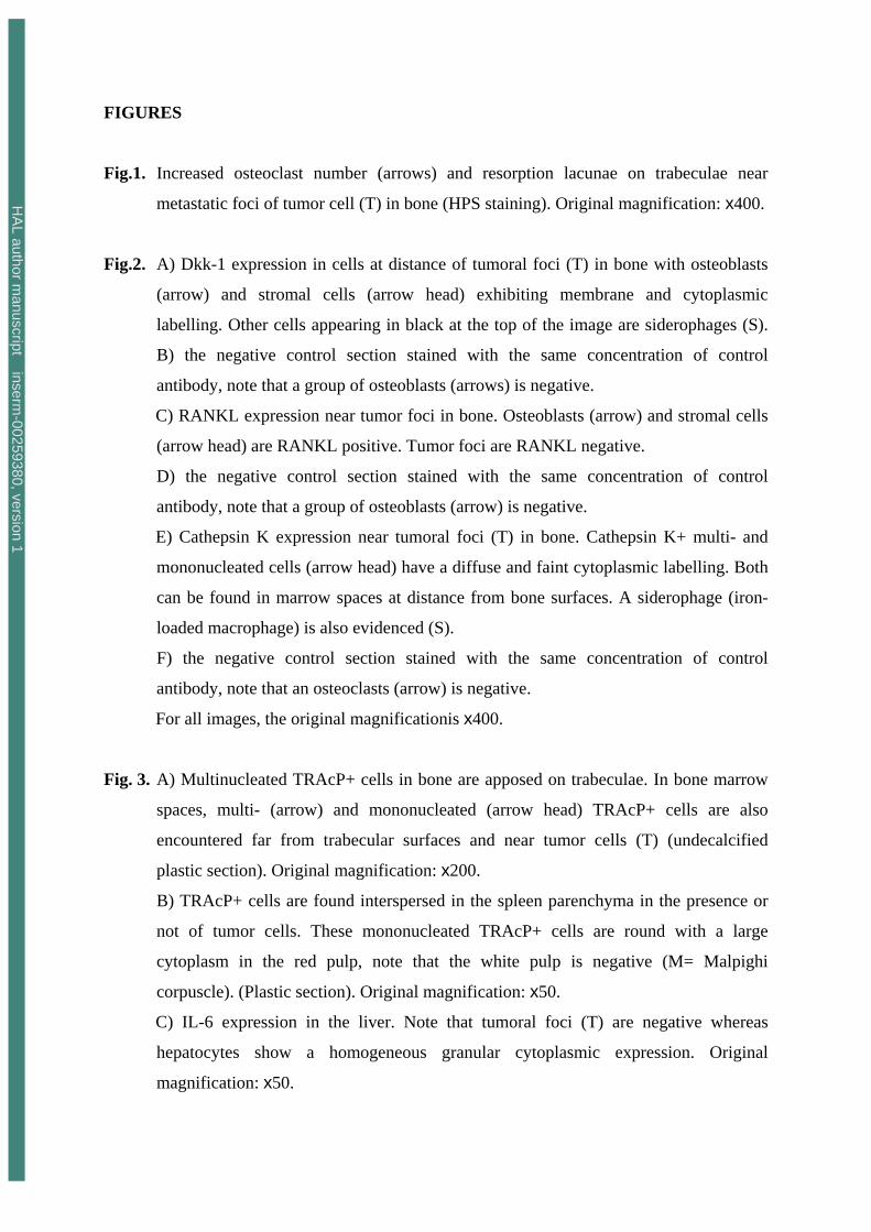
observed. An increased osteoclastogenesis was assessed in the immediate vicinity of the
tumor foci, leading to a dramatic increased of active erosion surfaces and the resorption of
complete bone trabeculae (Fig. 1). Siderophages (macrophages containing brownish
cytoplasmic particles) were frequently found, they reflect the necrotic and remodelling steps
occurring in the hematopoietic bone marrow secondary to tumor invasion (Weinberg, 1983).
These cells were positive with the Perls reaction for iron. They are readily distinguishable
from immunolabeled cells under the microscope, although they can appear with a similar
intensity on black and white microphotographs.
Both models exhibited the same immunohistochemical labeling pattern: tumor cells never
exhibited significant labeling for M-CSF, Dkk-1, RANKL, IL-6 or cathepsin K (Fig. 2). A
strong membrane expression of M-CSF was found in most bone marrow stromal cells in
contact with the metastatic foci. Dkk-1 was also expressed at high levels in stromal cells and
osteoblasts in contact with metastatic foci in both models (Fig.2 A). At distance, there was no
significant labeling for Dkk-1 in these cell populations. Many stromal cells showed RANKL
positivity in the vicinity of the tumor (Fig. 2C). RANKL labeling was also seen on osteoblasts
at distance of the tumor foci. These stromal cells (which were labelled by Dkk1, M-CSF and
RANKL) were found at distance from trabeculae: they were mononucleated with often a large
granular cytoplasm; some of them appeared spindle-shaped. The IL-6 detection was not
conclusive due to a non specific label induced by the prior decalcification.
HA
L author manuscript inserm
-00259380, version 1
9
Two types of cells were found to express a faint and homogenous cytoplasmic positivity for
cathepsin K: typical multinucleated osteoclasts at the surface of bone trabeculae and multi- or
mono-nucleated cells (either round or with cytoplasmic branches) in the marrow spaces.
These cells were mixed with haematopoietic cells and were at distance from bone surfaces
(Fig. 2E); they concentrate in the close vicinity of the tumor foci, on the contrary, they
appeared scantily distributed at distance.
Two types of cells exhibited TRAcP positivity (TRAcP+): cells apposed on trabeculae were
multinucleated; cells encountered in bone marrow spaces were either multi- or mono-
nucleated; they were situated far from trabecular surfaces and in the close vicinity of
metastatic cells, their density decreased at distance from tumoral foci (Fig. 3A).
Histological and immunohistochemical characteristics of extraosseous tumors
Tumor nodules of both types of carcinomas were dense and well-delimited by a fibrous
capsule. There were composed of a large majority of tumor cells mixed with less numerous
stromal cells. Tumor cells showed a different aspect according to the metastatic site: brain
metastases were composed of spindle-shaped cells whereas in other sites, they appeared round
or polygonal (especially with Walker cells). For both line cells, tumor cells were large with a
high nucleocytoplasmic ratio; prominent nucleoli were often present and the cytoplasms were
devoid of vacuoles. Numerous mitoses were observed but small foci of necrosis were also
noted inside nodules.
Both tumor models exhibited the same immunohistochemical labeling pattern: tumor cells
never exhibited any significant labeling for M-CSF, Dkk-1, RANKL, IL-6, cathepsin K.
Stromal cells in contact with malignant cells did not express Dkk-1, M-CSF, cathepsin K. At
distance from tumor foci, they also failed to express these markers. IL-6 expression was found
homogeneous in the liver parenchyma with diffused granular cytoplasmic expression in the
hepatocytes. The staining was found in all the liver parenchyma and not restricted to the areas
surrounding the tumor. Few stromal cells were located around the tumoral foci and did not
exhibit IL-6 expression (Fig. 3C). Some RANKL positive (RANKL+) stromal cells were
found at the periphery of subcutaneous tumors. These mononucleated cells were round or
stellate with an abundant cytoplasm. No RANKL+ cells were found in any other extraosseous
site.
TRAcP+ cells were found interspersed in the spleen parenchyma whether tumor cells were
present or not. These mononucleated TRAcP+ cells appeared round with a large granular
HA
L author manuscript inserm
-00259380, version 1
10
cytoplasm; the nucleus was ovoid or reniform (Fig. 3B). TRAcP+ cells were never observed
in the vicinity of malignant cells in any other extraosseous metastases.
DISCUSSION
It is an oversimplification to consider only osteoblasts and osteoclasts as the interacting
partners of tumors cells in the vicious cycle of cancer bone metastases. In addition to a variety
of matrix components, the tumor stroma contains a rich cell population, which includes
fibroblasts, smooth muscle cells, endothelial cells, dendritic cells, macrophages and other
inflammatory cells (Zigrino et al., 2005). This forms a network with a complex crosstalk
influencing cellular activities. For example, various actors of the tumor microenvironment
such as fibroblasts, immune cells and the extracellular matrix influence the ability of TGF-β
to promote or suppress carcinoma progression and metastasis (Bierie & Moses, 2006). In a
previous work, we have shown an influence of the microenvironment on tumor cell in the 5T2
multiple myeloma murine model since a high bone remodeling (induced by ovariectomy)
dramatically increased tumor growth (Libouban et al., 2003). This altered microenvironment
was able to select a highly aggressive myeloma cell line (Libouban et al., 2004). Others have
also reported that parathyroid hormone injection in athymic mice induced a preferential
localization of PC-3 prostate cancer cells in bones (Schneider et al., 2005).
Interactions of malignant cells with endothelial cells in bone metastases have been previously
investigated. Van der Pluijm et al. observed significantly elevated mRNA levels for VEGF-A
and -B in bone metastases compared to soft tissue metastases induced by the human breast
cancer cells MDA-MB-231 (van der Pluijm et al., 2001). Di Raimondo et al. similarly
detected higher concentrations of the angiogenic peptides (VEGF, bFGF (basic fibroblast
growth factor) and HGF (hepatocyte growth factor)) in bone marrow than in the peripheral
circulation of patients with multiple myeloma. This suggests the specific induction within the
bone microenvironment of angiogenic factors that allows the development of a neovascular
network which favours the tumor growth (Di Raimondo et al., 2000).
Interaction between tumor cells and other stromal components also plays a major role in
cancer progression. Cancer cells can alter the surrounding connective tissues and modulate the
metabolism of fibroblasts thus resulting in the production of a collagenous stroma that
supports the tumoral process. Fibroblast are a key component of tumors because they acquire
a modified phenotype similar to those associated with wound healing. Such “activated”
fibroblasts has been coined “carcinoma-associated fibroblasts” (CAFs) (Kalluri & Zeisberg,
2006). CAFs are implicated in extracellular matrix production which provides a lattice that
facilitates cancer progression; they are one of the main sources of VEGF, thus providing more
HA
L author manuscript inserm
-00259380, version 1
11
nutriment to the metastastic cells (Kalluri & Zeisberg, 2006). Shimamura et al. have
investigated the microenvironment of osteolytic metastases caused by MDA-MB 231 breast
carcinoma cells in nude mice. Two weeks after an intracardiac injection with MDA-MB 231
cells, foci of tumor cells were associated with osteoclastogenesis together with angiogensis
related to intense the expression of VEGF (Shimamura et al., 2005). In our study, we have
also found specific modifications in the bone microenvironment around the tumor foci with
expression of M-CSF, Dkk-1, and RANKL in non-osteoblastic cells. Moreover,
mononucleated TRAcP+ cells were also encountered in bone marrow spaces far from
trabecular surfaces. This denotes an increased induction of osteoclast precursors since
mononuclear TRAcP+ cells in the bone marrow are a source for mature multinucleated
osteoclasts (Baron et al., 1986). These mononucleated TRAcP+ cells expressed cathepsin K
and were found in medullar spaces at distance from bone surfaces. All these cells were found
with a density decreasing as the distance from tumor cells increased. It is probable that a
gradient of cytokines released from malignant cells can explain this finding. Tumor cells
produce a variety of soluble factors (PTHrP, TGF-β, IGF, Endothelin-1…) which are
employed as a paracrine communication system with surrounding cells or as an autocrine
regulation system (Guise & Mundy, 1998; Guise et al., 2003; Lee et al., 2003). W256 and
MatLyLu cells are unable to resorb bone by themselves and have been reported to produce
high levels of PTHrP which promotes osteoclastogenesis (Rabbani et al., 1999; Rizzoli &
Fleisch, 1987).
The stromal expression of important cytokines involved in the bone remodeling was found
altered only when metastatic cells were developing in a bony microenvironment. No
expression of these cytokines could be observed in the tumor stroma when metastastic cells
had developed in other organs. The few RANKL+ cells found at the periphery of
subcutaneous tumors were probably Langerhans cells. TRAcP+ cells encountered both in
invaded and non-invaded spleens probably corresponded to activated macrophages, a
condition previously noted by others (Hayman et al., 2000). TRAcP+ cells were previously
observed in the stroma of bone tumors (Yam et al., 1987). TRAcP+ osteoclast-like cells are
also observed in giant cells tumors which can develop in bone (and cause osteolytic lesions)
or in soft tissues (Teot et al., 1996). In this tumor, the malignant component is of fibroblastic
origin and express RANKL which provokes osteoclastogenesis (Murata et al., 2005). In our
models, stromal cells were able to express RANKL and to induce osteoclastogenesis only in
the bone compartment.
HA
L author manuscript inserm
-00259380, version 1
12
Tumor cells do not have the same cytokine pattern of expression according to their location
and can modulate it depending on the surrounding stromal cells. For example, breast cancer
cells have an increased expression of PTHrP in bone metastases when compared to other sites
(Mundy, 2002). As a consequence, the microenvironment can also influence the tumor cells:
Walker cells acquired a spindle-shape only when located in brain metastases, whereas they
appeared round or polygonal in other sites. The difference may also be supported by soft
tissue stromal cells which can have different surface receptors than bone stromal cells,
making them unable to response to tumor cell stimulation.
In conclusion, interaction between stromal and cancer cells induces the expression on the
formers of characteristics leading to osteoclastogenesis in the bone microenvironment. These
modifications allow the growth of the metastases in a convenient microenvironment. It thus
explains the preferential localization observed in our bone metastasis models. The major role
of osteoclasts and osteoblasts in bone metastasis formation has led to underestimate the
critical function of microenvironment in metastasis growth.
Acknowledgments
Authors are greatly indebted to P. Legras and J. Roux for their help with the animal care. This
work was supported by funds from “Pays de la Loire” – Axe Biomatériaux, la Ligue contre le
Cancer and INSERM.
HA
L author manuscript inserm
-00259380, version 1
13
REFERENCES
Baron, R., Neff, L., Tran Van, P., Nefussi, J.R. & Vignery, A. (1986). Kinetic and
cytochemical identification of osteoclast precursors and their differentiation into
multinucleated osteoclasts. American Journal of Pathology, 122, 363-78.
Bierie, B. & Moses, H.L. (2006). Tumour microenvironment: TGFbeta: the molecular Jekyll
and Hyde of cancer. Nature Reviews Cancer, 6, 506-20.
Blouin, S., Basle, M.F. & Chappard, D. (2005). Rat models of bone metastases. Clinical and
Experimental Metastasis, 22, 605-14.
Boyle, W.J., Simonet, W.S. & Lacey, D.L. (2003). Osteoclast differentiation and activation.
Nature, 423, 337-42.
Chappard, D., Alexandre, C. & Riffat, G. (1983). Histochemical identification of osteoclasts.
Review of current methods and reappraisal of a simple procedure for routine diagnosis
on undecalcified human iliac bone biopsies. Basic and Applied Histochemistry, 27, 75-
85.
Coleman, R.E. (2001). Metastatic bone disease: clinical features, pathophysiology and
treatment strategies. Cancer Treatment Reviews, 27, 165-76.
Cooper, C.R., Chay, C.H., Gendernalik, J.D., Lee, H.L., Bhatia, J., Taichman, R.S.,
McCauley, L.K., Keller, E.T. & Pienta, K.J. (2003). Stromal factors involved in
prostate carcinoma metastasis to bone. Cancer, 97, 739-47.
Di Raimondo, F., Azzaro, M.P., Palumbo, G., Bagnato, S., Giustolisi, G., Floridia, P., Sortino,
G. & Giustolisi, R. (2000). Angiogenic factors in multiple myeloma: higher levels in
bone marrow than in peripheral blood. Haematologica, 85, 800-5.
Guise, T.A. (2000). Molecular mechanisms of osteolytic bone metastases. Cancer, 88, 2892-
8.
Guise, T.A. & Mundy, G.R. (1998). Cancer and bone. Endocrine Reviews, 19, 18-54.
Guise, T.A., Yin, J.J. & Mohammad, K.S. (2003). Role of endothelin-1 in osteoblastic bone
metastases. Cancer, 97, 779-84.
Hayman, A.R., Bune, A.J., Bradley, J.R., Rashbass, J. & Cox, T.M. (2000). Osteoclastic
tartrate-resistant acid phosphatase (Acp 5): its localization to dendritic cells and
diverse murine tissues. Journal of Histochemistry and Cytochemistry, 48, 219-28.
Johnson, M.L., Harnish, K., Nusse, R. & Van Hul, W. (2004). LRP5 and Wnt signaling: a
union made for bone. Journal of Bone and Mineral Research, 19, 1749-57.
Kalluri, R. & Zeisberg, M. (2006). Fibroblasts in cancer. Nature Reviews Cancer, 6, 392-401.
HA
L author manuscript inserm
-00259380, version 1
14
Kang, Y., Siegel, P.M., Shu, W., Drobnjak, M., Kakonen, S.M., Cordon-Cardo, C., Guise,
T.A. & Massague, J. (2003). A multigenic program mediating breast cancer metastasis
to bone. Cancer Cell, 3, 537-49.
Keller, E.T., Zhang, J., Cooper, C.R., Smith, P.C., McCauley, L.K., Pienta, K.J. & Taichman,
R.S. (2001). Prostate carcinoma skeletal metastases: cross-talk between tumor and
bone. Cancer Metastasis Reviews, 20, 333-49.
Lee, Y., Schwarz, E., Davies, M., Jo, M., Gates, J., Wu, J., Zhang, X. & Lieberman, J.R.
(2003). Differences in the cytokine profiles associated with prostate cancer cell
induced osteoblastic and osteolytic lesions in bone. Journal of Orthopaedic Research,
21, 62-72.
Libouban, H., Moreau, M.F., Basle, M.F., Bataille, R. & Chappard, D. (2003). Increased bone
remodeling due to ovariectomy dramatically increases tumoral growth in the 5T2
multiple myeloma mouse model. Bone, 33, 283-92.
Libouban, H., Moreau, M.F., Basle, M.F., Bataille, R. & Chappard, D. (2004). Selection of a
highly aggressive myeloma cell line by an altered bone microenvironment in the
C57BL/KaLwRij mouse. Biochemical and Biophysical Research Communications,
316, 859-66.
Logothetis, C.J. & Lin, S.H. (2005). Osteoblasts in prostate cancer metastasis to bone. Nature
Reviews Cancer, 5, 21-8.
Muller, A., Homey, B., Soto, H., Ge, N., Catron, D., Buchanan, M.E., McClanahan, T.,
Murphy, E., Yuan, W., Wagner, S.N., Barrera, J.L., Mohar, A., Verastegui, E. &
Zlotnik, A. (2001). Involvement of chemokine receptors in breast cancer metastasis.
Nature, 410, 50-6.
Mundy, G.R. (1997). Mechanisms of bone metastasis. Cancer, 80, 1546-56.
Mundy, G.R. (2002). Metastasis to bone: causes, consequences and therapeutic opportunities.
Nature Reviews Cancer, 2, 584-93.
Mundy, G.R., Chen, D., Zhao, M., Dallas, S., Xu, C. & Harris, S. (2001). Growth regulatory
factors and bone. Reviews in Endocrine and Metabolic Disorders, 2, 105-15.
Murata, A., Fujita, T., Kawahara, N., Tsuchiya, H. & Tomita, K. (2005). Osteoblast lineage
properties in giant cell tumors of bone. Journal of Orthopaedic Science, 10, 581-8.
Rabbani, S.A., Gladu, J., Harakidas, P., Jamison, B. & Goltzman, D. (1999). Over-production
of parathyroid hormone-related peptide results in increased osteolytic skeletal
metastasis by prostate cancer cells in vivo. International Journal of Cancer, 80, 257-
64.
HA
L author manuscript inserm
-00259380, version 1
15
Reis, F.M., Luisi, S., Carneiro, M.M., Cobellis, L., Federico, M., Camargos, A.F. & Petraglia,
F. (2004). Activin, inhibin and the human breast. Molecular and Cellular
Endocrinology, 225, 77-82.
Risbridger, G.P., Mellor, S.L., McPherson, S.J. & Schmitt, J.F. (2001). The contribution of
inhibins and activins to malignant prostate disease. Molecular and Cellular
Endocrinology, 180, 149-53.
Rizzoli, R. & Fleisch, H. (1987). The Walker 256/B carcinosarcoma in
thyroparathyroidectomized rats: a model to evaluate inhibitors of bone resorption.
Calcified Tissue International, 41, 202-7.
Roodman, G.D. (2003). Role of stromal-derived cytokines and growth factors in bone
metastasis. Cancer, 97, 733-8.
Roodman, G.D. (2004). Mechanisms of bone metastasis. New England Journal of Medicine,
350, 1655-64.
Schneider, A., Kalikin, L.M., Mattos, A.C., Keller, E.T., Allen, M.J., Pienta, K.J. &
McCauley, L.K. (2005). Bone turnover mediates preferential localization of prostate
cancer in the skeleton. Endocrinology, 146, 1727-36.
Shimamura, T., Amizuka, N., Li, M., Freitas, P.H., White, J.H., Henderson, J.E., Shingaki, S.,
Nakajima, T. & Ozawa, H. (2005). Histological observations on the microenvironment
of osteolytic bone metastasis by breast carcinoma cell line. Biomedical Research, 26,
159-72.
Teot, L.A., O'Keefe, R.J., Rosier, R.N., O'Connell, J.X., Fox, E.J. & Hicks, D.G. (1996).
Extraosseous primary and recurrent giant cell tumors: transforming growth factor-
beta1 and -beta2 expression may explain metaplastic bone formation. Human
Pathology, 27, 625-32.
Thomas, R.J., Guise, T.A., Yin, J.J., Elliott, J., Horwood, N.J., Martin, T.J. & Gillespie, M.T.
(1999). Breast cancer cells interact with osteoblasts to support osteoclast formation.
Endocrinology, 140, 4451-8.
Timar, J., Lapis, K., Dudas, J., Sebestyen, A., Kopper, L. & Kovalszky, I. (2002).
Proteoglycans and tumor progression: Janus-faced molecules with contradictory
functions in cancer. Seminars in Cancer Biology, 12, 173-86.
Vaaraniemi, J., Halleen, J.M., Kaarlonen, K., Ylipahkala, H., Alatalo, S.L., Andersson, G.,
Kaija, H., Vihko, P. & Vaananen, H.K. (2004). Intracellular machinery for matrix
degradation in bone-resorbing osteoclasts. Journal of Bone and Mineral Research, 19,
1432-40.
HA
L author manuscript inserm
-00259380, version 1
16
van der Pluijm, G., Sijmons, B., Vloedgraven, H., Deckers, M., Papapoulos, S. & Lowik, C.
(2001). Monitoring metastatic behavior of human tumor cells in mice with species-
specific polymerase chain reaction: elevated expression of angiogenesis and bone
resorption stimulators by breast cancer in bone metastases. Journal of Bone and
Mineral Research, 16, 1077-91.
Vieweg, J., Rosenthal, F.M., Bannerji, R., Heston, W.D., Fair, W.R., Gansbacher, B. &
Gilboa, E. (1994). Immunotherapy of prostate cancer in the Dunning rat model: use of
cytokine gene modified tumor vaccines. Cancer Research, 54, 1760-5.
Weinberg, E.D. (1983). Iron in neoplastic disease. Nutrition and cancer, 4, 223-33.
Wingen, F., Eichmann, T., Manegold, C. & Krempien, B. (1986). Effects of new
bisphosphonic acids on tumor-induced bone destruction in the rat. Journal of Cancer
Research and Clinical Oncology, 111, 35-41.
Woodhouse, E.C., Chuaqui, R.F. & Liotta, L.A. (1997). General mechanisms of metastasis.
Cancer, 80, 1529-37.
Yam, L.T., Janckila, A.J., Li, C.Y. & Lam, W.K. (1987). Cytochemistry of tartrate-resistant
acid phosphatase: 15 years' experience. Leukemia, 1, 285-8.
Yasuda, Y., Kaleta, J. & Bromme, D. (2005). The role of cathepsins in osteoporosis and
arthritis: rationale for the design of new therapeutics. Advanced Drug Delivery
Reviews, 57, 973-93.
Zigrino, P., Loffek, S. & Mauch, C. (2005). Tumor-stroma interactions: their role in the
control of tumor cell invasion. Biochimie, 87, 321-8.
HA
L author manuscript inserm
-00259380, version 1
FIGURES
Fig.1. Increased osteoclast number (arrows) and resorption lacunae on trabeculae near
metastatic foci of tumor cell (T) in bone (HPS staining). Original magnification: x400.
Fig.2. A) Dkk-1 expression in cells at distance of tumoral foci (T) in bone with osteoblasts
(arrow) and stromal cells (arrow head) exhibiting membrane and cytoplasmic
labelling. Other cells appearing in black at the top of the image are siderophages (S).
B) the negative control section stained with the same concentration of control
antibody, note that a group of osteoblasts (arrows) is negative.
C) RANKL expression near tumor foci in bone. Osteoblasts (arrow) and stromal cells
(arrow head) are RANKL positive. Tumor foci are RANKL negative.
D) the negative control section stained with the same concentration of control
antibody, note that a group of osteoblasts (arrow) is negative.
E) Cathepsin K expression near tumoral foci (T) in bone. Cathepsin K+ multi- and
mononucleated cells (arrow head) have a diffuse and faint cytoplasmic labelling. Both
can be found in marrow spaces at distance from bone surfaces. A siderophage (iron-
loaded macrophage) is also evidenced (S).
F) the negative control section stained with the same concentration of control
antibody, note that an osteoclasts (arrow) is negative.
For all images, the original magnificationis x400.
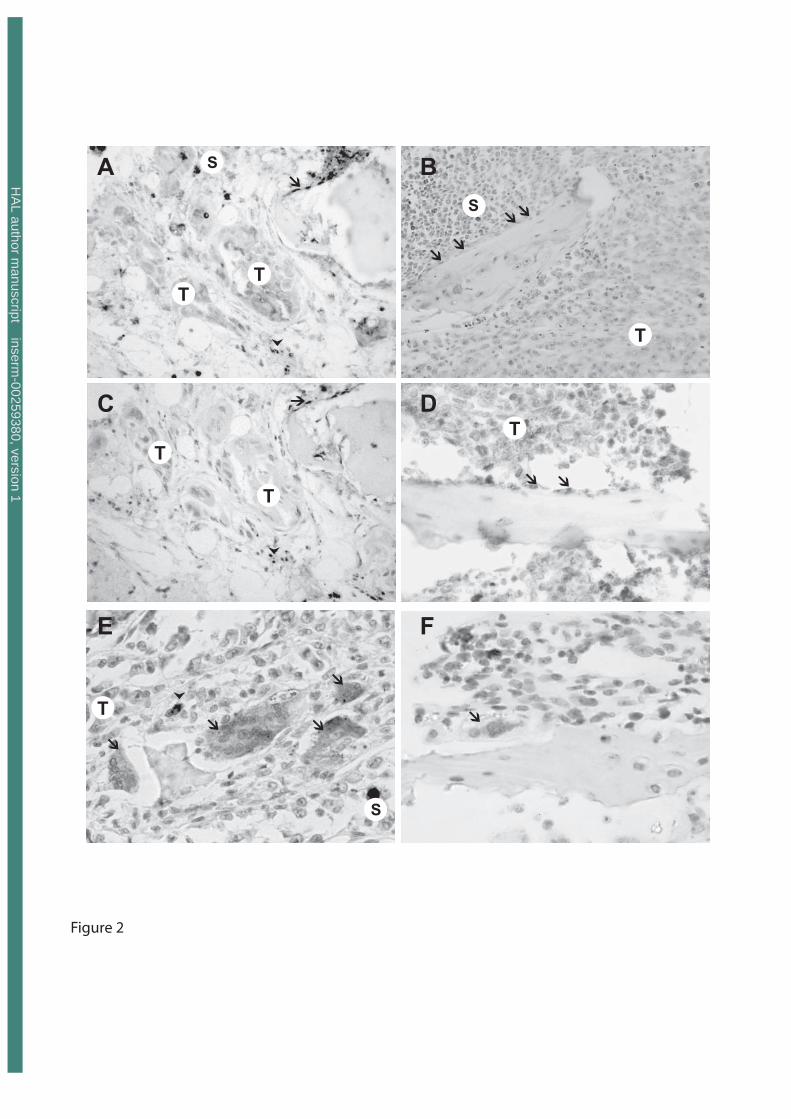
Fig. 3. A) Multinucleated TRAcP+ cells in bone are apposed on trabeculae. In bone marrow
spaces, multi- (arrow) and mononucleated (arrow head) TRAcP+ cells are also
encountered far from trabecular surfaces and near tumor cells (T) (undecalcified
plastic section). Original magnification: x200.
B) TRAcP+ cells are found interspersed in the spleen parenchyma in the presence or
not of tumor cells. These mononucleated TRAcP+ cells are round with a large
cytoplasm in the red pulp, note that the white pulp is negative (M= Malpighi
corpuscle). (Plastic section). Original magnification: x50.
C) IL-6 expression in the liver. Note that tumoral foci (T) are negative whereas
hepatocytes show a homogeneous granular cytoplasmic expression. Original
magnification: x50.
HA
L author manuscript inserm
-00259380, version 1

D) the negative control section stained with the same concentration of control
antibody, note that an osteoclasts (arrow) is negative. Original magnification: x50.
HA
L author manuscript inserm
-00259380, version 1
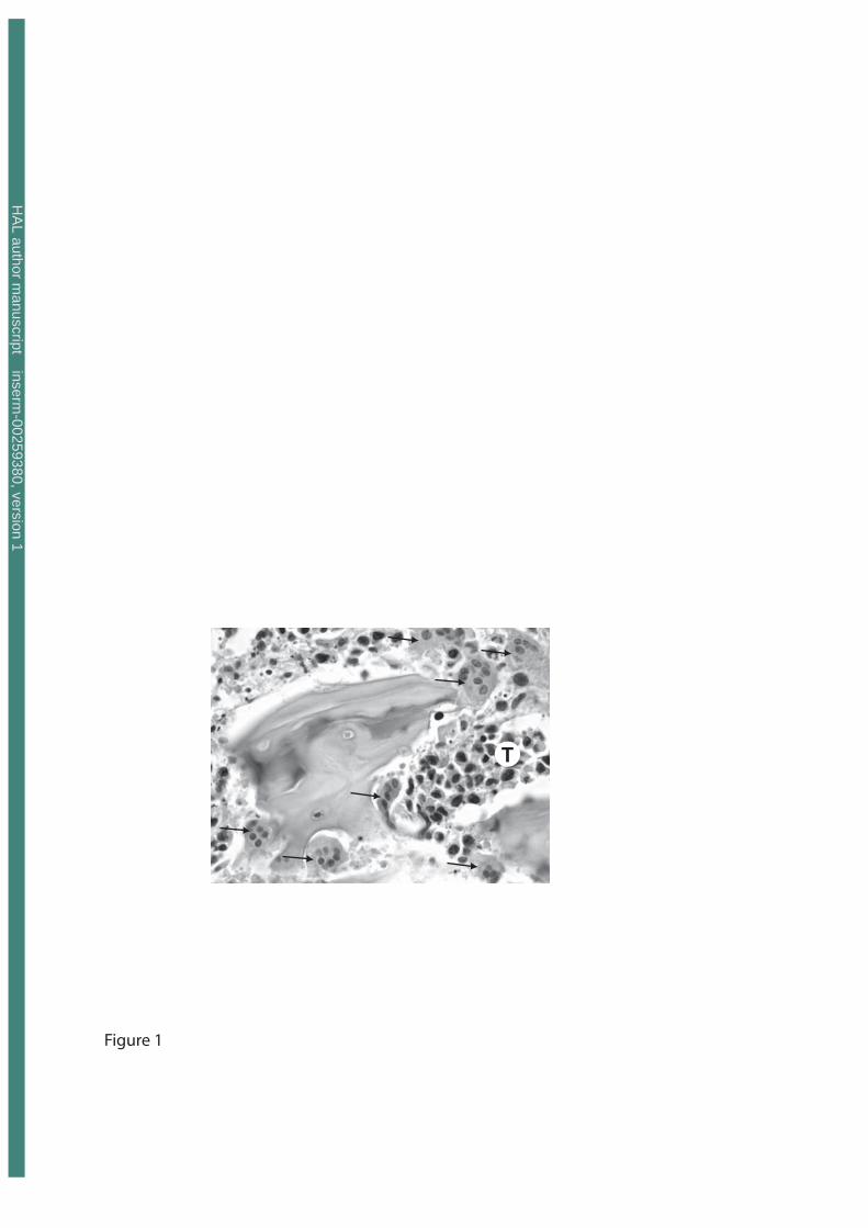
Figure 1
T
HA
L author manuscript inserm
-00259380, version 1
T�
�
�
�
�
S
Figure 2
�
�
TT
S
T
S
��
��
A B
�
�
T
T
C D
E F
�
�
T
�
HA
L author manuscript inserm
-00259380, version 1
Figure 3
�
�
� �
��
T
M
T
T
T
A B
C D
T
T
HA
L author manuscript inserm
-00259380, version 1
Copyright © 2022 FDOKUMEN