Coexpression and nuclear colocalization of metastasis-promoting protein S100A4 and p53 without...
10
ORIGINAL ARTICLE Coexpression and nuclear colocalization of metastasis-promoting protein S100A4 and p53 without mutual regulation in colorectal carcinoma Gisle Berge • Daniela Elena Costea • Marianne Berg • Heidi Rasmussen • Ida Grotterød • Ragnhild A. Lothe • Gunhild M. Mælandsmo • Kjersti Flatmark Received: 8 October 2009 / Accepted: 3 February 2010 Ó Springer-Verlag 2010 Abstract Nuclear localization of the metastasis-associ- ated protein S100A4 has been shown to correlate with advanced disease stage in primary colorectal carcinomas (CRC), but nuclear function and its relevance for the metastatic capacity of tumor cells is still unclear. Among several nuclear interacting protein partners suggested for S100A4, the tumor suppressor protein p53 has attracted particular interest, and previous studies suggest direct and indirect modes of interaction between the two proteins. The present study was undertaken to assess coexpression and potential interaction in CRC. TP53 mutational status and S100A4 expression were investigated in a selected series of primary CRC specimens (n = 40) and cell lines (n = 17) using DNA sequencing, western blot, and double immunostaining. Additionally, S100A4 and p53 were experimentally up- and down-regulated in vitro to assess reciprocal effects. For the first time, S100A4 and p53 coexpression was demonstrated in individual CRC cells, with nuclear colocalization as a particularly interesting feature. In contrast to previous studies, no correlation was observed between TP53 mutational status and S100A4 expression, and no evidence was obtained to support reci- procal regulation between the two molecules in the HCT116 isogenic cell line model. In conclusion, S100A4 and p53 were shown to be colocalized in individual nuclei of CRC cells, and it might be speculated whether the proteins interact in this subcellular compartment. Keywords Colorectal neoplasms S100A4 p53 TP53 Metastasis Introduction The S100A4 protein belongs to the S100 protein family, which in humans comprises at least 20 small, acidic Ca 2? - binding proteins, exhibiting distinct expression patterns, tissue distribution and biological properties (Donato 2003; Marenholz et al. 2004). The S100A4 gene product has been convincingly linked to the invasive and metastatic pheno- type of cancer cells through experimental data derived from transfections with expression constructs (Davies et al. 1993; Ford et al. 1995; Maelandsmo et al. 1996; Takenaga et al. 1997) and studies of transgenic animals (Ambartsu- mian et al. 1996; Davies et al. 1996). In addition, clinical evidence has indicated a correlation between augmented S100A4 expression and poor prognosis in several cancer types, including colorectal carcinoma (CRC) (Andersen et al. 2004; Gongoll et al. 2002; Rudland et al. 2000). The S100A4 protein has been shown to influence various Electronic supplementary material The online version of this article (doi:10.1007/s00726-010-0514-6) contains supplementary material, which is available to authorized users. G. Berge H. Rasmussen I. Grotterød G. M. Mælandsmo K. Flatmark (&) Department of Tumor Biology, Institute for Cancer Research, The Norwegian Radium Hospital, Oslo University Hospital, 0310 Oslo, Norway e-mail: kjersti.fl[email protected] D. E. Costea Section of Pathology, The Gade Institute, University of Bergen, 5021 Bergen, Norway M. Berg R. A. Lothe Department of Cancer Prevention, Institute for Cancer Research, The Norwegian Radium Hospital, Oslo University Hospital, 0310 Oslo, Norway M. Berg R. A. Lothe Centre for Cancer Biomedicine, University of Oslo, 0310 Oslo, Norway 123 Amino Acids DOI 10.1007/s00726-010-0514-6
-
Upload
independent -
Category
Documents
-
view
0 -
download
0
Transcript of Coexpression and nuclear colocalization of metastasis-promoting protein S100A4 and p53 without...

ORIGINAL ARTICLE
Coexpression and nuclear colocalization of metastasis-promotingprotein S100A4 and p53 without mutual regulation in colorectalcarcinoma
Gisle Berge • Daniela Elena Costea • Marianne Berg • Heidi Rasmussen •
Ida Grotterød • Ragnhild A. Lothe • Gunhild M. Mælandsmo •
Kjersti Flatmark
Received: 8 October 2009 / Accepted: 3 February 2010
� Springer-Verlag 2010
Abstract Nuclear localization of the metastasis-associ-
ated protein S100A4 has been shown to correlate with
advanced disease stage in primary colorectal carcinomas
(CRC), but nuclear function and its relevance for the
metastatic capacity of tumor cells is still unclear. Among
several nuclear interacting protein partners suggested for
S100A4, the tumor suppressor protein p53 has attracted
particular interest, and previous studies suggest direct
and indirect modes of interaction between the two proteins.
The present study was undertaken to assess coexpression
and potential interaction in CRC. TP53 mutational status
and S100A4 expression were investigated in a selected
series of primary CRC specimens (n = 40) and cell lines
(n = 17) using DNA sequencing, western blot, and double
immunostaining. Additionally, S100A4 and p53 were
experimentally up- and down-regulated in vitro to assess
reciprocal effects. For the first time, S100A4 and p53
coexpression was demonstrated in individual CRC cells,
with nuclear colocalization as a particularly interesting
feature. In contrast to previous studies, no correlation was
observed between TP53 mutational status and S100A4
expression, and no evidence was obtained to support reci-
procal regulation between the two molecules in the
HCT116 isogenic cell line model. In conclusion, S100A4
and p53 were shown to be colocalized in individual nuclei
of CRC cells, and it might be speculated whether the
proteins interact in this subcellular compartment.
Keywords Colorectal neoplasms � S100A4 � p53 �TP53 � Metastasis
Introduction
The S100A4 protein belongs to the S100 protein family,
which in humans comprises at least 20 small, acidic Ca2?-
binding proteins, exhibiting distinct expression patterns,
tissue distribution and biological properties (Donato 2003;
Marenholz et al. 2004). The S100A4 gene product has been
convincingly linked to the invasive and metastatic pheno-
type of cancer cells through experimental data derived
from transfections with expression constructs (Davies et al.
1993; Ford et al. 1995; Maelandsmo et al. 1996; Takenaga
et al. 1997) and studies of transgenic animals (Ambartsu-
mian et al. 1996; Davies et al. 1996). In addition, clinical
evidence has indicated a correlation between augmented
S100A4 expression and poor prognosis in several cancer
types, including colorectal carcinoma (CRC) (Andersen
et al. 2004; Gongoll et al. 2002; Rudland et al. 2000). The
S100A4 protein has been shown to influence various
Electronic supplementary material The online version of thisarticle (doi:10.1007/s00726-010-0514-6) contains supplementarymaterial, which is available to authorized users.
G. Berge � H. Rasmussen � I. Grotterød �G. M. Mælandsmo � K. Flatmark (&)
Department of Tumor Biology, Institute for Cancer Research,
The Norwegian Radium Hospital, Oslo University Hospital,
0310 Oslo, Norway
e-mail: [email protected]
D. E. Costea
Section of Pathology, The Gade Institute, University of Bergen,
5021 Bergen, Norway
M. Berg � R. A. Lothe
Department of Cancer Prevention, Institute for Cancer Research,
The Norwegian Radium Hospital, Oslo University Hospital,
0310 Oslo, Norway
M. Berg � R. A. Lothe
Centre for Cancer Biomedicine, University of Oslo,
0310 Oslo, Norway
123
Amino Acids
DOI 10.1007/s00726-010-0514-6

biological functions with potential implications for the
metastatic phenotype, such as invasion, cell motility and
angiogenesis (Garrett et al. 2006; Helfman et al. 2005;
Sherbet and Lakshmi 1998). However, the exact functions
of S100A4 and how it exerts its metastasis-promoting
effects remain incompletely understood.
The tumor suppressor protein p53 plays a pivotal role in
the maintenance and regulation of normal cellular func-
tions, and mutations in TP53 are observed in approximately
50% of human CRC (Iacopetta et al. 2006). Through steps
of stabilization, phosphorylation, and nuclear translocation,
p53 is activated in response to various cellular stress sig-
nals, forming a tetrameric transcription factor able to
modulate the expression of numerous downstream genes.
A large set of target genes have been identified, and the
corresponding proteins are involved in diverse functions,
such as cell cycle regulation, apoptosis, cellular signaling
and regulation of extracellular matrix [reviewed in Aylon
and Oren (2007), Levine (1997)]. Additionally, to mediate
its pleiotropic effects, p53 interacts with a number of
proteins, and has also recently been implicated in regula-
tion of angiogenic transcription factors and vascular
development, which are important features of metastasis
(Teodoro et al. 2007).
Certain key observations have instigated research
towards a possible physical and regulatory interaction
between S100A4 and p53. When S100A4 was transfected
into TP53 wt cell lines, apoptotic cell death was observed,
whereas stable clones were constructed successfully in
TP53 mutated cell lines (Grigorian et al. 2001). It was
hypothesized that coexistence of S100A4 and wt TP53
would be lethal to tumor cells, and that the implied
selection pressure towards an aggressive, TP53 mutated
and metastatic phenotype, in part, could explain the
metastasis-promoting effects of S100A4. Additionally,
interactions between p53 and two other S100 family
members, S100B and S100A2, have been demonstrated,
and both these S100 proteins may regulate p53 at the
transcriptional level and by way of protein–protein inter-
actions (Fernandez-Fernandez et al. 2005; Lin et al. 2001,
2004; Mueller et al. 2005). Since neither of these proteins
are expressed in colorectal epithelium or its malignant
derivatives (Bronckart et al. 2001), one might speculate
that S100A4 could be responsible for the execution of
‘‘S100-functions’’ with respect to interaction with p53 in
CRC. Substantial research has documented in vitro physi-
cal interactions between S100A4 and p53, and S100A4 has
been shown to influence TP53 transcriptional regulation in
experimental settings (Chen et al. 2001; Fernandez-Fer-
nandez et al. 2005; Grigorian et al. 2001). Finally, our
previous finding that nuclear localization of S100A4 cor-
related with tumor stage in CRC led us to consider whether
a potential S100A4-p53 interaction might be a nuclear
phenomenon (Flatmark et al. 2003). The objective of the
present study was to examine the correlation between
S100A4 expression and TP53 mutational status in primary
CRC and cell lines, and assess potential reciprocal influ-
ence between the two proteins in experimental model
systems.
Materials and methods
CRC samples
From a clinically representative series of primary CRC, 40
cases were selected, including ten tumors from each
Duke’s stage (Table 2). Within each group, five tumors
were negative and five were positive for nuclear S100A4,
according to previous immunohistochemical analysis
(Flatmark et al. 2003). All tumors with nuclear S100A4
and nine of the nucleus negative samples also expressed
cytoplasmic S100A4. Tumor tissue was collected at the
time of surgery, snap frozen in liquid nitrogen and stored at
-80�C until use. For each sample, the proportion of tumor
cells was assessed in frozen sections neighboring the tissue
used for DNA extraction. If possible, microscopic-guided
rough dissection was performed to remove normal tissue
prior to DNA extraction. The study was approved by the
Regional Committee for Medical Research Ethics Regional
Ethics Committee (Approval#S-98080) and informed
consent was obtained.
Cell lines
Co205, HCT15, Colo320DM, SW480, SW620, CaCo2,
HT29, RKO, SW48 and WiDr cell lines were purchased
from ATCC (Manassas, VA, USA). HCT116 cells were
purchased from ATCC; but when specified, two isogenic
variants obtained from Dr. Bert Vogelstein (The Sidney
Kimmel Comprehensive Cancer Centre, The Johns Hop-
kins Medical Institutions, Baltimore, MD, USA) were
used: HCT116 TP53 knock-out (TP53-/-) and HCT116
wild-type (TP53?/?) (Bunz et al. 1999). KM20L2 and
HCC2998 were kindly provided by Michael R. Boyd
(National Cancer Institute, Frederick, MD, USA). Co115
cells were obtained from B. Sordat (Epalinges, Lausanne,
Switzerland), whereas EB, LS174T and TC7 cell lines
were acquired from Richard Hamelin (INSERM, Paris,
France). Cells were cultivated in RPMI 1640 (BioWhit-
taker, Lonza Verviers, Belgium) containing 10% fetal
bovine serum (Hyclone, Logan, UT, USA), 20 mM Hepes
(BioWittaker) and 2 mM Glutamax (Gibco, Invitrogen,
Norway). All cell lines were negative for mycoplasma
infection. TP53 mutational status of cell lines is available
at the TP53 web site (http://p53.free.fr/index.html), except
G. Berge et al.
123

for cell lines KM20L2 and Co115 which were found to
harbor mutated (k273; CGT-CAT; Arg-His) and wt TP53,
respectively (for methods see below).
TP53 mutation analysis
TP53 mutation status within the evolutionary conserved
DNA binding domain, encoded by exons 5–8, was assessed
for each tumor sample. PCR products with aberrant
migrating bands detected by temporal temperature gradient
gel electrophoresis (TTGE) (Sorlie et al. 2005) were
indicative of a mutation. New PCR products from the same
DNA stock solution were then submitted to direct
sequencing of the targeted exon using dideoxy sequencing
(ABI PRISM TM373 DNA Sequencer, Applied Biosys-
tems) to identify the specific sequence change (mutation or
polymorphism). The four exons were amplified by PCR
using the standard protocol for HotStar DNA Polymerase
(Qiagen, GmbH, Hilden, Germany) followed by sequenc-
ing with primers and annealing temperatures as shown in
Supplementary table 1. In one sample, an aberrant band
was detected on TTGE, but the mutation could not be
confirmed by sequencing, and was thus interpreted as wt.
No wt conclusions were drawn from analysis of samples
containing less than 20% tumor cells.
Double immunohistochemistry (DIHC) and double
immunofluorescence (DIF)
Five micrometer formalin-fixed, paraffin-embedded
sections were deparaffinized, rehydrated and antigen
retrieved in Tris–EDTA buffer, pH 9.0 in Pascal pressure
chamber (DAKO) at 120–125�C, 18 psi (30 s) for cell
pellets, and by microwaving at 750 W (9 min) and
250 W (15 min) for tumor tissue. For DIHC, sections
were blocked with 10% goat serum in TBST (Sigma, St.
Louis, Missouri) (30 min) and afterwards with Dual
Endogenous Enzyme Block (DAKO) (10 min) prior to
the addition of primary antibody #1. After washing,
secondary antibody Polymer/HRP (EnVision G|2 Double
System Rabbit/Mouse DAB?/Permanent red kit, DAKO)
was added, followed by visualization with 3,30-diam-
inobenzidine (10 min). Doublestain block (DAKO) was
added (3 min) before primary antibody #2. To enhance
the second primary antibody signal, Link reagent
(EnVision G|2 Double System Kit) was added (30 min)
prior to secondary antibody Polymer/AP (EnVision G|2
Double System Kit), and visualization was performed
with liquid permanent red (10 ll/ml) (DAKO) diluted
with Levamisole (30 ll/ml) (DAKO) in substrate buffer
(8 min).
For DIF, after 30 min blocking with 10% goat serum,
primary antibodies were applied in mixture, followed by
washing and addition of fluorochrome conjugated second-
ary antibodies in mixture. Slides were mounted in DAPI-
Vectashield mounting medium (Vector Laboratories Ltd,
Peterborough, UK) and analyzed by confocal laser scan-
ning microscopy (Axiovert25 inverted microscope, Carl
Zeiss MicroImaging, Gottingen, Germany). Specimens
incubated with Antibody Diluent alone, or with single
primary antibody were used as negative controls (for
conditions see Table 1).
Table 1 Antibodies and conditions used for western blot (WB), double immunohistochemistry (DIHC) and double immunofluorescence (DIF)
Application Target M/P (species) Catalog/Clone Source Dilution Buffer Conditions
WB S100A4 P (rabbit) A5114 DAKO 1/300 TBST 1 h, RT
WB S100A4 M 22.3 In-housea 1/300 TBST 1 h, RT
WB p53 M SC-6243 Santa Cruz 1/3000 TBST 1 h, RT
WB a-tubulin M CP06 Calbiochem 1/300 TBST 1 h, RT
DIHC S100A4 M 20.1 In-housea 1/200 AD 1 h, RT
DIHC p53 M DO-7 DAKO 1/1000 AD 30 min, RT
DIF S100A4 P (rabbit) A5114 DAKO 1/50 TBS 1 h, RT
DIF p53 M DO-7 DAKO 1/10 TBS 1 h, RT
DIF Anti-mouse P (goat) Alexa Fluor 594 Invitrogen 1/200 AD 30 min, RT
DIF Anti-rabbit P (goat) Alexa Fluor 488 Invitrogen 1/200 AD 30 min, RT
DAKO, Glostrup, Denmark; Santa Cruz Biotechnology, Santa Cruz, CA, USA; Calbiochem, Merck Biosciences GmbH, Germany; Invitrogen,
Carlsbad, CA, USA
M monoclonal; P polyclonal; TBST tris buffered saline with 0.25% Tween 20 and 5% non-fat dry milk; TBS tris-buffered saline; AD antibody
diluent, DAKO; RT room temperaturea Flatmark et al. 2004
S100A4 and p53 in colorectal carcinoma
123

Transfection experiments
Transfections constructs for wt and mutated TP53 were a
gift from Dr. Bert Vogelstein (The Sidney Kimmel Com-
prehensive Cancer Centre, The Johns Hopkins Medical
Institutions, Baltimore, MD, USA) (Baker et al. 1990). The
S100A4 expression construct was made by PCR amplify-
ing the insert from S100A4/pBlusescript (kindly provided
by Dr. Heizmann, Department of Pediatrics, University of
Zurich, Zurich, Switzerland), and subsequently ligating the
product into pcDNA3.1 (Invitrogen). Transient transfec-
tions of S100A4 and TP53 wt and mutated constructs in
HCT116 were performed by electroporation, using 4 mm
cuvette gap, 240 V, 26 ms, 1 pulse exposure (BTX, ECM
830, San Diego, CA, USA). For transfection of 2 9 106
cells in RPMI/FBS, 2.5–10 lg pcDNA3.1/S100A4, the
corresponding pcDNA3.1 control vector, pcDNA3.1/wt
p53 or pcDNA3.1/mutated p53, were used. Following
electroporation, 1.5 9 106 cells were seeded in serum-
containing medium in T25 flasks.
Silencing of endogenous expression of S100A4 was per-
formed using siRNA duplexes; si-S100A4: 5-UGA-GCA-
AGU-UCA-AUA-AAG-A-3 (against position 481–499,
in the 30UTR area), and si-scrambled control: 5-CGC-
AUA-AGU-GAA-AUA-GAA-U-3 (Eurogentec, Belgium).
Transfections were performed with Lipofectamine 2000
(Invitrogen) according to the manufacturer’s instructions.
Briefly, 5 9 105 cells were seeded in T25 flasks and grown
overnight before adding 100 nM siRNA/5 ll Lipofect-
amine in RPMI/w/o FBS. After 6 h medium containing
serum was added.
Cellular responses to ultraviolet (UV) radiation
48 or 72 h after S100A4 or si-RNA transfection, respec-
tively, HCT116 cells were exposed to a fixed dose of UV
irradiation while in PBS (12 J/m2, 254 nm) in a GS gene
linker instrument (Bio-Rad, Hercules, CA, USA) and left
for 6 h in complete medium. Total cell lysates were har-
vested, or cell pellets were formalin fixed and paraffin
embedded for DIF or DIHC studies. For all experiments,
mock-irradiated control cultures were handled identically.
Cell pellets were sectioned and immunostained as descri-
bed above, and staining of tumor cells was quantitatively
evaluated by counting six fields for each sample at 209
magnification (median number of cells per field was 128,
range 87–235) (Table 3).
Cell lysis and western blot analysis
Total cell lysates were made by adding lysis buffer
(150 mM NaCl, 50 mM Tris–HCl pH 7.5, 0.1% NP-40,
10 lg/ml Leupeptide Hemisulfate, Aprotinin, Pepstatin A,
20 mM b-glycerolphosphate, 1 mM PMSF, 1 mM sodium
orthovanadate and 100 mM sodium fluoride) to dry cell
pellets, and following sonication and centrifugation,
supernatant was stored at -80�C. Lysates were separated
by SDS-PAGE (4–12% NuPAGE, Invitrogen), transferred
to 0.45 lM PVDF-membranes (Millipore, MA, USA) and
probed with antibodies against S100A4, p53 and a-tubulin
(as internal control) (for conditions see Table 1). Immune
complexes were detected with Super Signal chemilumi-
nescence detection system (Pierce, Rockford, IL, USA).
Scanning and densitometry analysis was performed using a
GS-800 Calibrated Densitometer and Quanti-One 4.6.6
software (Bio-Rad).
Statistics
All group comparisons were performed using the Mann–
Whitney U test (SPSS version 15.0; SPSS, Chicago, Ill.
USA), and P B 0.05 was considered statistically
significant.
Results
TP53 mutational status and S100A4 expression in CRC
Approximately half (19 of 40) of the CRC samples
exhibited TP53 mutations (Table 2), in agreement with
previous results (Borresen-Dale et al. 1998; Iacopetta et al.
2006). The 19 mutations are listed in Supplementary
table 2. Mutations were present in 43, 58 and 43% in
tumors from the rectum, left colon, and right colon,
respectively, in accordance with previous studies from us
and others (Borresen-Dale et al. 1998). In tumors that
expressed nuclear S100A4, mutated TP53 was present in
45% of the cases (9 of 20), whereas wild-type was found in
55% (11 of 20). Similarly, no difference in TP53 muta-
tional frequency could be detected between tumors with
cytoplasmic S100A4.
S100A4 expression in cell lines with wt and mutated
TP53
To assess whether S100A4 expression was dependent on
p53 mutational status in human CRC cell lines, protein
lysates from 7 wt and 10 mutated cell lines were analyzed
by Western blot. Variable amounts of S100A4 were
detected in most of the cell lines as shown in Fig. 1.
Although the four cell lines with the highest S100A4 levels
were in the TP53 mutated group (KM20L2, SW620,
SW480 and Co205), six of the seven wt lines had detect-
able S100A4 with expression levels similar to the
remaining mutated cell lines. Only two cell lines, RKO and
G. Berge et al.
123

HCT15, were entirely negative for S100A4, harboring wt
and mutated TP53, respectively.
Coexpression of S100A4 and p53 in HCT116 cells and
patient samples
The presence of S100A4 in cell lines with wt TP53 was
demonstrated by western blot, but to assess whether p53
and S100A4 were present in the same cells, cellular
imaging strategies were applied (Fig. 2). Co-immuno-
staining performed in HCT116 cells with or without
induction of p53 by UV irradiation revealed p53 expression
exclusively in the nuclei of HCT116 cells, whereas
S100A4 exhibited nuclear and cytoplasmic expression.
A sixfold increase in the fraction of cells expressing p53
was observed upon UV exposure, while S100A4 expres-
sion was essentially unchanged (Table 3). In unirradiated
cells, 1.5% of cells exhibited expression of both p53 and
S100A4, and the fraction of double-stained cells increased
to 4.6% upon UV exposure. The pattern of coexpression
varied, as co-immunostaining in nuclei was observed in
some cells, while concomitant presence of nuclear p53 and
cytoplasmic S100A4 was detected in other cells. Two
patient samples were similarly analyzed by DIHC, showing
variable nuclear expression of wt p53, and occasional cells
(\1%) were observed with simultaneous expression of
mainly nuclear S100A4.
Experimental regulation of S100A4 and p53
To assess potential reciprocal regulation between S100A4
and p53 in vitro, isogenic variants of the CRC cell line
HCT116 harboring wt (TP53?/?) and knock-out p53
(TP53-/-) were used. Protein levels of S100A4 or p53
were experimentally altered using transient transfection or
by exposing cells to genotoxic stress by UV radiation. In
accordance with previous results (Daoud et al. 2003), high
and very low basal levels of S100A4 were detected in wt
(TP53?/?) and knock-out (TP53-/-) cells, respectively
(Fig. 3a).
Restoring p53 expression in HCT116 TP53 knock-out
cells by transfection with an expression construct encoding
wt p53 did not alter S100A4 levels (Fig. 3b). Furthermore,
by performing the opposite experiment, transfecting
S100A4 into the TP53 wt cells, an anticipated up-regula-
tion of S100A4, but no changes in p53 protein levels
were observed (Fig. 4; Supplementary Fig. 1). Similarly,
siRNA-mediated down regulation of S100A4 did not affect
constitutive p53 expression in HCT116 wt cells (Fig. 5;
Supplementary Fig. 2).
Even if augmenting p53 expression by transfection did
not affect S100A4 expression levels, manipulation of
endogenous p53 might in theory induce biological effects
that could not be achieved with exogenously introduced
protein. Thus, UV irradiation of TP53 wt cells was per-
formed, resulting in increased amounts of p53, while
S100A4 expression remained unchanged (Fig. 4). To fur-
ther assess whether manipulating S100A4 levels could
influence the cells’ ability to activate p53 following
genotoxic stress, S100A4 was experimentally up- or down-
regulated in HCT116 TP53 wt cells by transfection strat-
egies prior to UV exposure. UV dependent increase of p53
expression was not influenced by altered S100A4
Table 2 TP53 mutational status according to tumor stage, localiza-
tion and nuclear and cytoplasmic expression of S100A4
TP53 mutational status
Mutated number (%) Wild-type number (%)
Dukes’ stage
A 6 (60) 4 (40)
B 5 (50) 5 (50)
C 6 (60) 4 (40)
D 2 (20) 8 (80)
Localization
Rectum 6 (43) 8 (57)
Left colon 7 (58) 5 (42)
Right colon 6 (43) 8 (57)
S100A4 expression
N S100A4? 9 (45) 11 (55)
N S100A4- 10 (50) 10 (50)
C S100A4? 15 (52) 14 (48)
C S100A4- 4 (36) 7 (64)
All samples 19 (48) 21 (52)
N nuclear, C cytoplasmic
Fig. 1 Western blot of SDS
PAGE separated protein lysates
from seven TP53 wt (asterisk)
and ten mutated human
colorectal carcinoma cell lines,
immunostained with a mAb
against S100A4. Staining for
a-tubulin was used as internal
standard
S100A4 and p53 in colorectal carcinoma
123

expression after transfection with the S100A4 expression
construct (Fig. 4) or with siRNA against S100A4 (Fig. 5).
Discussion
In the examined CRC samples, no association was observed
between nuclear or cytoplasmic S100A4 expression and
TP53 mutational status. Moreover, six of the seven TP53 wt
cell lines expressed S100A4, as assessed by immunoblot-
ting of total protein lysates. The idea that S100A4 expres-
sion could induce cell death in TP53 wt cells, thus
contributing to selection of a metastatic phenotype harbor-
ing mutated TP53 and S100A4 is appealing, but the
hypothesis has not been tested in clinical samples or on the
single cell level. According to this theory, one would expect
G. Berge et al.
123
the favored combinations in tumor samples to be wt TP53/
S100A4 negative and mutated TP53/S100A4 positive. In
the present study, expression of nuclear and cytoplasmic
S100A4 was equally frequent in TP53 wt and mutated
samples, and thus the results did not corroborate the
hypothesis that the presence of S100A4 and wt TP53 is
mutually exclusive.
Because of tumor heterogeneity, variable abundance of
normal cells in clinical samples and the possibility of
mutations existing outside the examined region of the p53
gene, there could be a risk that existing mutations might
not be detected. Conversely, since some mutations are
associated with production of functional p53 and the exact
effect of every mutation has not been clarified, detected
mutations might not necessarily indicate complete loss of
p53 function (Iacopetta et al. 2006). The TTGE screening
method has a limit of detection (LOD) of *5% tumor cells
present in the sample (Sorlie et al. 2005), while for the
direct sequencing method, LOD is *20% (unpublished
results; Dahl et al. 2006). Since no wt conclusion was
drawn from samples containing less that 20% tumor cells,
the chance of missing mutations in the examined region is
considered negligible. Moreover, more than 80% of all
mutations in TP53 are reported to be situated within exons
5 through 8, involving most of the DNA binding domain
which is crucial for the transactivating ability of p53
(Walker et al. 1999). Therefore, the chance of false wt
conclusions in tumor samples is low and the risk of missing
mutations relevant for p53 function should be even lower,
while some of the mutated samples might potentially retain
p53 function.
Using co-immunostaining strategies with antibodies
against S100A4 and p53, we were able, for the first time, to
demonstrate simultaneous presence of the two proteins in
individual CRC cells, and in particular, nuclear coexpres-
sion was noted (Fig. 2). We previously showed that nuclear
expression of S100A4 was correlated to advanced disease
stage in CRC (Flatmark et al. 2003), and preliminary
results indicate that nuclear S100A4 is an independent
predictor of adverse prognosis in the same patient cohort
(Boye, manuscript in preparation). Thus, the possibility of
molecular interaction between S100A4 and other nuclear
proteins, such as p53, is intriguing. While recently dis-
puted, a substantial amount of data from in vitro experi-
ments using different methods and cell systems supports a
direct physical interaction between p53 and S100A4,
involving binding of S100A4 to the C terminus of p53,
with potential implications for cellular localization, DNA
affinity, p53 phosphorylation by PKC as well as down-
stream target activation (Chen et al. 2001; Fernandez-
Fernandez et al. 2005; Grigorian et al. 2001; van Dieck
et al. 2009). The only in vivo finding suggesting direct
protein–protein interaction involved co-immunoprecipita-
tion from cells harboring mutant TP53 (Grigorian et al.
2001), while co-immunoprecipitation of S100A4 with wt
p53 has so far not been demonstrated. This could be
attributed to instability of wt p53 and technical issues, and
Table 3 Immunohistochemical analysis of p53 and S100A4 staining in HCT116 cells
Mean percentage positively stained HCT116 cells (SD)
p53 N S100A4 C S100A4 p53 and S100A4
Untreated cells 4.7 (1.0) 6.6 (2.5) 11.0 (3.9) 1.5 (0.9)
UV treated cells 31.7 (6.9) 7.5 (4.1) 13.9 (4.7) 4.6 (3.0)
Tumor cells were counted at 209 magnification, six fields for each sample (median number of cells per field was 128, range 87–235)
N nuclear, C cytoplasmic, SD standard deviation
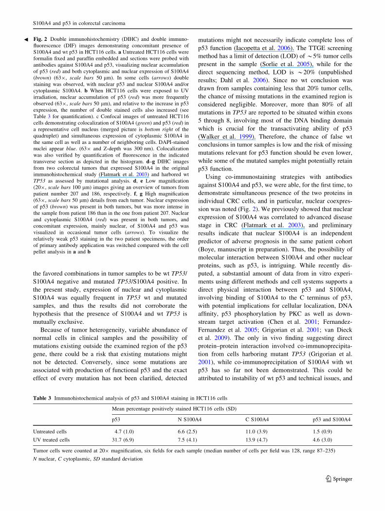
Fig. 2 Double immunohistochemistry (DIHC) and double immuno-
fluorescence (DIF) images demonstrating concomitant presence of
S100A4 and wt p53 in HCT116 cells. a Untreated HCT116 cells were
formalin fixed and paraffin embedded and sections were probed with
antibodies against S100A4 and p53, visualizing nuclear accumulation
of p53 (red) and both cytoplasmic and nuclear expression of S100A4
(brown) (639, scale bars 50 lm). In some cells (arrows) double
staining was observed, with nuclear p53 and nuclear S100A4 and/or
cytoplasmic S100A4. b When HCT116 cells were exposed to UV
irradiation, nuclear accumulation of p53 (red) was more frequently
observed (639, scale bars 50 lm), and relative to the increase in p53
expression, the number of double stained cells also increased (see
Table 3 for quantification). c Confocal images of untreated HCT116
cells demonstrating colocalization of S100A4 (green) and p53 (red) in
a representative cell nucleus (merged picture is bottom right of the
quadruplet) and simultaneous expression of cytoplasmic S100A4 in
the same cell as well as a number of neighboring cells. DAPI-stained
nuclei appear blue. (639 and Z-depth was 300 nm). Colocalization
was also verified by quantification of fluorescence in the indicated
transverse section as depicted in the histogram. d–g DIHC images
from two colorectal tumors that expressed S100A4 in the original
immunohistochemical study (Flatmark et al. 2003) and harbored wt
TP53 as assessed by mutational analysis. d, e Low magnification
(209, scale bars 100 lm) images giving an overview of tumors from
patient number 207 and 186, respectively. f, g High magnification
(639, scale bars 50 lm) details from each tumor. Nuclear expression
of p53 (brown) was present in both tumors, but was more intense in
the sample from patient 186 than in the one from patient 207. Nuclear
and cytoplasmic S100A4 (red) was present in both tumors, and
concomitant expression, mainly nuclear, of S100A4 and p53 was
visualized in occasional tumor cells (arrows). To visualize the
relatively weak p53 staining in the two patient specimens, the order
of primary antibody application was switched compared with the cell
pellet analysis in a and b
b
S100A4 and p53 in colorectal carcinoma
123

interaction between the two proteins could still take place
and be of biological relevance, for instance, in the cell
nucleus.
An alternative and more indirect possibility for molec-
ular interaction between S100A4 and p53 could be through
transcriptional regulation. However, results from in vitro
experiments indicated that although S100A4 and p53 were
indeed present in the same cells and even in the same nuclei,
no reciprocal regulation was observed. These findings are in
contrast to results from studies of other S100 proteins, such
as S100B and S100A2. Increased S100B levels in malignant
melanoma cell lines dose dependently down-regulated p53
and inhibited p53-mediated cellular effects, whereas p53
levels and function were restored when S100B was down-
regulated by antisense RNA. Additionally, over-expression
of S100B prevented induction of p53, p21 and MDM2 in
response to bleomycin-induced DNA damage. S100B
transcription was up-regulated by p53 through interaction
with consensus p53 binding sites in the S100B promoter,
and also, UV induction of p53 led to increased mRNA and
protein levels of S100B (Lin et al. 2001, 2004). The evi-
dence supporting mutual regulation of S100A2 and p53 is
less substantial, but in osteosarcoma cells, up-regulation of
S100A2 on the transcriptional level was described through
p53 binding sites in the promoter sequence (Tan et al.
1999). Since the S100A4 promoter also contains putative
p53 binding sites (Parker et al. 1994), up-regulation of p53
by transfection and UV irradiation could be expected to
influence S100A4 transcription, but no such effects were
observed. Hence, although comparable experimental con-
ditions were applied, we did not observe any of the pro-
found effects that would be expected if S100A4 had a
regulatory interaction with p53 comparable to that of S100B
or S100A2. However, although the isogenic HCT116 cell
Fig. 3 Western blot showing the expression of S100A4 and p53 in
the HCT116 isogenic cell model and the effects of transfecting wt or
mutated TP53 into HCT116 TP53-/- cells. a The TP53 wt cells
from the isogenic HCT116 cell model expressed high levels of
S100A4 and detectable p53, while the TP53 knock-out cells had very
low amounts of S100A4 and no p53 protein was detected. b Recon-
stituting p53 expression in the knock-out cells by transfection with wt
or mutated construct did not influence S100A4 expression levels
Fig. 4 The figure shows mean values (?SEM) for S100A4 and p53
expression relative to respective a-tubulin staining derived from
densitometry scans of western blots from three independent exper-
iments (ns not significant). a Transfection with S100A4 resulted in
significant (P B 0.05) up-regulation of S100A4 expression compared
to mock transfected cells, while UV irradiation did not influence
S100A4 expression. b UV exposure gave rise to p53 induction
(P B 0.05) regardless of S100A4 protein expression levels, while
S100A4 transfection did not influence constitutive or UV induced
amounts of p53
G. Berge et al.
123

model is an established and much-used tool for the study of
p53-specific interactions, results could be limited to the
experimental system and do not exclude interaction
between p53 and S100A4.
Since nuclear localization of the metastasis-related protein
S100A4 is clinically relevant in CRC, identification of puta-
tive nuclear interacting protein partners is of interest. In the
present study, nuclear and cytoplasmic S100A4 were
expressed equally frequently in TP53 wt and mutated tumors
and cell lines, and coexpression of the two proteins was
demonstrated in CRC cells and in clinical specimens.
Although experimental manipulation of S100A4 and p53
expression did not show evidence for mutual regulation in the
isogenic HCT116 cell lines, a role for direct or indirect
interaction between S100A4 and p53 cannot be excluded.
Simultaneous detection of S100A4 and p53 in the nucleus of
CRC cells lends support to our initial theory that interaction
between p53 and S100A4 could take place in the cell nucleus,
but potential biological implications remain obscure.
Acknowledgments We would like to thank Dr. Bert Vogelstein for
kindly making the isogenic HCT116 cell model and p53 constructs
available to us, Sigurd Bø for providing the siRNA constructs and
Gunnvor Øijordsbakken for excellent technical assistance. The pres-
ent work was supported by postdoctoral grants to K.F. (Norwegian
Research Council, Grant Number 160604/V50), G.B. (Norwegian
Cancer Society, Grant Number C99026) and D.E.C (Norwegian
Research Council, Grant Number 178601), and by a PhD grant to
M.B. (Norwegian Cancer Society, RAL: Grant Number A95068) and
project support from the Functional Genomics Program in the Nor-
wegian Research Council (Grant Number 152004/S10).
Conflict of interest statement The authors declare no conflict of
interest.
References
Ambartsumian NS, Grigorian MS, Larsen IF, Karlstrom O, Sidenius
N, Rygaard J, Georgiev G, Lukanidin E (1996) Metastasis of
mammary carcinomas in GRS/A hybrid mice transgenic for the
mts1 gene. Oncogene 13:1621–1630
Andersen K, Nesland JM, Holm R, Florenes VA, Fodstad O,
Maelandsmo GM (2004) Expression of S100A4 combined with
reduced E-cadherin expression predicts patient outcome in
malignant melanoma. Mod Pathol 17:990–997
Aylon Y, Oren M (2007) Living with p53, dying of p53. Cell
130:597–600
Baker SJ, Markowitz S, Fearon ER, Willson JK, Vogelstein B (1990)
Suppression of human colorectal carcinoma cell growth by wild-
type p53. Science 249:912–915
Borresen-Dale AL, Lothe RA, Meling GI, Hainaut P, Rognum TO,
Skovlund E (1998) TP53 and long-term prognosis in colorectal
cancer: mutations in the L3 zinc-binding domain predict poor
survival. Clin Cancer Res 4:203–210
Bronckart Y, Decaestecker C, Nagy N, Harper L, Schafer BW,
Salmon I, Pochet R, Kiss R, Heizman CW (2001) Development
and progression of malignancy in human colon tissues are
correlated with expression of specific Ca(2?)-binding S100
proteins. Histol Histopathol 16:707–712
Bunz F, Hwang PM, Torrance C, Waldman T, Zhang Y, Dillehay L,
Williams J, Lengauer C, Kinzler KW et al (1999) Disruption of
p53 in human cancer cells alters the responses to therapeutic
agents. J Clin Invest 104:263–269
Chen H, Fernig DG, Rudland PS, Sparks A, Wilkinson MC,
Barraclough R (2001) Binding to intracellular targets of the
metastasis-inducing protein, S100A4 (p9Ka). Biochem Biophys
Res Commun 286:1212–1217
Dahl C, Ralfkiaer U, Guldberg P (2006) Methods for detection of
subtle mutations in cancer genomes. Crit Rev Oncog 12:41–74
Daoud SS, Munson PJ, Reinhold W, Young L, Prabhu VV, Yu Q,
LaRose J, Kohn KW, Weinstein JN et al (2003) Impact of p53
knockout and topotecan treatment on gene expression profiles in
human colon carcinoma cells: a pharmacogenomic study. Cancer
Res 63:2782–2793
Davies BR, Davies MP, Gibbs FE, Barraclough R, Rudland PS (1993)
Induction of the metastatic phenotype by transfection of a benign
rat mammary epithelial cell line with the gene for p9Ka, a rat
calcium-binding protein, but not with the oncogene EJ-ras-1.
Oncogene 8:999–1008
Davies MP, Rudland PS, Robertson L, Parry EW, Jolicoeur P,
Barraclough R (1996) Expression of the calcium-binding protein
S100A4 (p9Ka) in MMTV-neu transgenic mice induces metas-
tasis of mammary tumours. Oncogene 13:1631–1637
Donato R (2003) Intracellular and extracellular roles of S100 proteins.
Microsc Res Tech 60:540–551
Fernandez-Fernandez MR, Veprintsev DB, Fersht AR (2005) Proteins
of the S100 family regulate the oligomerization of p53 tumor
suppressor. Proc Natl Acad Sci 102:4735–4740
Flatmark K, Pedersen KB, Nesland JM, Rasmussen H, Aamodt G,
Mikalsen SO, Bjornland K, Fodstad O, Maelandsmo GM (2003)
Nuclear localization of the metastasis-related protein S100A4
correlates with tumour stage in colorectal cancer. J Pathol
200:589–595
Flatmark K, Maelandsmo GM, Mikalsen SO, Nustad K, Varaas T,
Rasmussen H, Meling GI, Fodstad O, Paus E (2004) Immuno-
fluorometric assay for the metastasis-related protein S100A4:
release of S100A4 from normal blood cells prohibits the use of
S100A4 as a tumor marker in plasma and serum. Tumour Biol
25:31–40
Ford HL, Salim MM, Chakravarty R, Aluiddin V, Zain SB (1995)
Expression of Mts1, a metastasis-associated gene, increases
motility but not invasion of a nonmetastatic mouse mammary
adenocarcinoma cell line. Oncogene 11:2067–2075
Garrett SC, Varney KM, Weber DJ, Bresnick AR (2006) S100A4, a
mediator of metastasis. J Biol Chem 281:677–680
Fig. 5 Representative western blots showing expression of S100A4
and p53 in HCT116 cells upon transfection with siRNA against
S100A4 or scrambled siRNA (C) with or without subsequent UV
exposure. Experimental down-regulation of S100A4 with siRNA did
not influence baseline p53 expression levels or the p53 induction
achieved by UV irradiation
S100A4 and p53 in colorectal carcinoma
123

Gongoll S, Peters G, Mengel M, Piso P, Klempnauer J, Kreipe H, von
Wasielewski R (2002) Prognostic significance of calcium-
binding protein S100A4 in colorectal cancer. Gastroenterology
123:1478–1484
Grigorian M, Andresen S, Tulchinsky E, Kriajevska M, Carlberg C,
Kruse C, Cohn M, Ambartsumian N, Christensen A et al (2001)
Tumor suppressor p53 protein is a new target for the metastasis-
associated Mts1/S100A4 protein: functional consequences of
their interaction. J Biol Chem 276:22699–22708
Helfman DM, Kim EJ, Lukanidin E, Grigorian M (2005) The
metastasis associated protein S100A4: role in tumour progres-
sion and metastasis. Br J Cancer 92:1955–1958
Iacopetta B, Russo A, Bazan V, Dardanoni G, Gebbia N, Soussi T,
Kerr D, Elsaleh H, Soong R et al (2006) Functional categories of
TP53 mutation in colorectal cancer: results of an International
Collaborative Study. Ann Oncol 17:842–847
Levine AJ (1997) p53, the cellular gatekeeper for growth and
division. Cell 88:323–331
Lin J, Blake M, Tang C, Zimmer D, Rustandi RR, Weber DJ, Carrier
F (2001) Inhibition of p53 transcriptional activity by the S100B
calcium-binding protein. J Biol Chem 276:35037–35041
Lin J, Yang Q, Yan Z, Markowitz J, Wilder PT, Carrier F, Weber DJ
(2004) Inhibiting S100B restores p53 levels in primary malig-
nant melanoma cancer cells. J Biol Chem 279:34071–34077
Maelandsmo GM, Hovig E, Skrede M, Engebraaten O, Florenes VA,
Myklebost O, Grigorian M, Lukanidin E, Scanlon KJ et al (1996)
Reversal of the in vivo metastatic phenotype of human tumor
cells by an anti-CAPL (mts1) ribozyme. Cancer Res 56:5490–
5498
Marenholz I, Heizmann CW, Fritz G (2004) S100 proteins in mouse
and man: from evolution to function and pathology (including an
update of the nomenclature). Biochem Biophys Res Commun
322:1111–1122
Mueller A, Schafer BW, Ferrari S, Weibel M, Makek M, Hochli M,
Heizmann CW (2005) The calcium-binding protein S100A2
interacts with p53 and modulates its transcriptional activity.
J Biol Chem 280:29186–29193
Parker C, Lakshmi MS, Piura B, Sherbet GV (1994) Metastasis-
associated mts1 gene expression correlates with increased p53
detection in the B16 murine melanoma. DNA Cell Biol 13:343–
351
Rudland PS, Platt-Higgins A, Renshaw C, West CR, Winstanley JH,
Robertson L, Barraclough R (2000) Prognostic significance of
the metastasis-inducing protein S100A4 (p9Ka) in human breast
cancer. Cancer Res 60:1595–1603
Sherbet GV, Lakshmi MS (1998) S100A4 (MTS1) calcium binding
protein in cancer growth, invasion and metastasis. Anticancer
Res 18:2415–2421
Sorlie T, Johnsen H, Vu P, Lind GE, Lothe R, Borresen-Dale AL
(2005) Mutation screening of the TP53 gene by temporal
temperature gradient gel electrophoresis. Methods Mol Biol
291:207–216
Takenaga K, Nakamura Y, Sakiyama S (1997) Expression of
antisense RNA to S100A4 gene encoding an S100-related
calcium-binding protein suppresses metastatic potential of high-
metastatic Lewis lung carcinoma cells. Oncogene 14:331–337
Tan M, Heizmann CW, Guan K, Schafer BW, Sun Y (1999)
Transcriptional activation of the human S100A2 promoter by
wild-type p53. FEBS Lett 445:265–268
Teodoro JG, Evans SK, Green MR (2007) Inhibition of tumor
angiogenesis by p53: a new role for the guardian of the genome.
J Mol Med 85:1175–1186
van Dieck J, Fernandez-Fernandez MR, Veprintsev DB, Fersht AR
(2009) Modulation of the oligomerization state of p53 by
differential binding of proteins of the S100 family to p53
monomers and tetramers. J Biol Chem 284:13804–13811
Walker DR, Bond JP, Tarone RE, Harris CC, Makalowski W,
Boguski MS, Greenblatt MS (1999) Evolutionary conservation
and somatic mutation hotspot maps of p53: correlation with p53
protein structural and functional features. Oncogene 18:211–218
G. Berge et al.
123