Localization of corticotropin-releasing hormone (CRH) neurons in the paraventricular nucleus of the...
13
50 Brain Research, 615 (1993) 50-62 © 1993 Elsevier Science Publishers B.V. All rights reserved 0006-8993/93/$06.00 BRES 18941 Localization of corticotropin-releasing hormone (CRH) neurons in the paraventricular nucleus of the human hypothalamus; age-dependent colocalization with vasopressin F.C. Raadsheer a, A.A. Sluiter a, R. Ravid a, F.J.H. Tilders b and D.F. Swaab a " Netherlands Institute for Brain Research, Amsterdam (The Netherlands) and b Department of Pharmacology, Medical Faculty, Free Unicersity, Amsterdam (The Netherlands) (Accepted 19 January 1993) Key words." Human hypothalamic nucleus; Corticotropin-releasing hormone; Vasopressin; Hypothalamo-pituitary-adrenal axis; Stress; Aging; Immunoeytochemistry Immunocytochemical staining, using a monoclonal antibody against corticotropin-releasing hormone, was performed on hypothalami of 13 human subjects between 23 and 91 years of age who had not suffered from a primary neurological or psychiatric disease. Corticotropin-releasing hormone (CRH) immunoreactivity was present in neurons of the paraventricular nucleus (PVN) and in their fibers running to the median eminence. The CRH-positive neurons were scattered throughout the PVN, but in the rostral part relatively few cells were present. There were large individual differences in the number and staining intensity of CRH neurons in the PVN and in the staining intensity of the median eminence. These differences seemed not to be attributed to age, sex, postmortem delay, fixation time or hour of death. In the rat, too, no relationship was found between a postmortem delay of up to 24 h and CRH staining intensity of the median eminence. Since the distribution of CRH-immunoreactive neurons in the human PVN strongly overlap with vasopressin, colocalization of these peptides was investigated in a double label study and indeed found in subjects ranging between 43 and 91 years of age. However, cells staining for only one of the peptides were also observed. The vasopressin cells had a mean cellular profile area which was 2.3 times as large as the CRH cells and 2.2 times as large as the CRH and vasopressin containing neurons. In younger subjects (23-37 years of age) no colocalization of the two peptides was seen. The age-dependent colocalization of CRH with vasopressin is interpreted as a sign of increased activation of the CRH neurons with age. INTRODUCTION Immunocytochemical localization of corticotropin- releasing hormone (CRH) in the human hypothalamus has been described in the paraventricular nucleus (PVN), median eminence (ME), pituitary stalk and posterior pituitary gland 24'38. However, it has so far not been investigated whether the CRH neurons in the human PVN are localized in a subnucleus of the PVN, as has been described for the rat 37. In the rat PVN, CRH neurons are situated in the lateral part of the medial parvicellular zone and project via the paraven- triculo-neurohypophysial tract to the portal capillaries of the ME, where the peptide is released. After excre- tion, the 41-aminoacid peptide CRH has a potent stimulatory effect on adrenocorticotrophic hormone (ACTH) release from the pituitary 27'4°'41. In addition, subpopulations of CRH neurons project to the poste- rior pituitary and to autonomic centers of the brain- stem and spinal cord 29'36'37, on which its central effects are based 4'11. CRH plays a key role in the function of the hypotha- lamo-pituitary-adrenal (HPA) axis, the involvement of which in the stress response was first discovered by Selye 31 (for a recent review see ref. 3). In addition, CRH is thought to be involved in various stress-related human pathologies, e.g. depression, Cushing's syn- drome, alcoholism, anorexia nervosa and Alzheimer's disease 46. In addition to CRH also vasopressin (AVP) plays a crucial role in the control of the release of ACTH from the pituitary gland 21'28. AVP and CRH potentiate each other's action on ACTH release 16'2s. Therefore, it is of interest that CRH and AVP are at least partly synthe- Correspondence." F.C. Raadsheer, Netherlands Institute for Brain Research, Meibergdreef 33, 11(15 AZ Amsterdam, The Netherlands. Fax: (31) (20) 6918466.
Transcript of Localization of corticotropin-releasing hormone (CRH) neurons in the paraventricular nucleus of the...
50 Brain Research, 615 (1993) 50-62 © 1993 Elsevier Science Publishers B.V. All rights reserved 0006-8993/93/$06.00
BRES 18941
Localization of corticotropin-releasing hormone (CRH) neurons in the paraventricular nucleus of the human hypothalamus;
age-dependent colocalization with vasopressin
F.C. Raadsheer a, A.A. Sluiter a, R. Ravid a, F.J.H. Tilders b and D.F. Swaab a
" Netherlands Institute for Brain Research, Amsterdam (The Netherlands) and b Department of Pharmacology, Medical Faculty, Free Unicersity, Amsterdam (The Netherlands)
(Accepted 19 January 1993)
Key words." Huma n hypothalamic nucleus; Corticotropin-releasing hormone; Vasopressin; Hypothalamo-pituitary-adrenal axis; Stress; Aging; Immunoeytochemistry
Immunocytochemical staining, using a monoclonal antibody against corticotropin-releasing hormone, was performed on hypothalami of 13 human subjects between 23 and 91 years of age who had not suffered from a primary neurological or psychiatric disease. Corticotropin-releasing hormone (CRH) immunoreactivity was present in neurons of the paraventricular nucleus (PVN) and in their fibers running to the median eminence. The CRH-positive neurons were scattered throughout the PVN, but in the rostral part relatively few cells were present. There were large individual differences in the number and staining intensity of CRH neurons in the PVN and in the staining intensity of the median eminence. These differences seemed not to be attributed to age, sex, pos tmortem delay, fixation time or hour of death. In the rat, too, no relationship was found between a postmortem delay of up to 24 h and C R H staining intensity of the median eminence. Since the distribution of CRH-immunoreact ive neurons in the human PVN strongly overlap with vasopressin, colocalization of these peptides was investigated in a double label study and indeed found in subjects ranging between 43 and 91 years of age. However, cells staining for only one of the peptides were also observed. The vasopressin cells had a mean cellular profile area which was 2.3 times as large as the C R H cells and 2.2 times as large as the C R H and vasopressin containing neurons. In younger subjects (23-37 years of age) no colocalization of the two peptides was seen. The age-dependent colocalization of C R H with vasopressin is interpreted as a sign of increased activation of the C R H neurons with age.
INTRODUCTION
Immunocytochemical localization of corticotropin- releasing hormone (CRH) in the human hypothalamus has been described in the paraventricular nucleus (PVN), median eminence (ME), pituitary stalk and posterior pituitary gland 24'38. However, it has so far not been investigated whether the CRH neurons in the human PVN are localized in a subnucleus of the PVN, as has been described for the rat 37. In the rat PVN, CRH neurons are situated in the lateral part of the medial parvicellular zone and project via the paraven- triculo-neurohypophysial tract to the portal capillaries of the ME, where the peptide is released. After excre- tion, the 41-aminoacid peptide CRH has a potent stimulatory effect on adrenocorticotrophic hormone (ACTH) release from the pituitary 27'4°'41. In addition,
subpopulations of CRH neurons project to the poste- rior pituitary and to autonomic centers of the brain- stem and spinal c o r d 29'36'37, o n which its central effects are based 4'11.
CRH plays a key role in the function of the hypotha- lamo-pituitary-adrenal (HPA) axis, the involvement of which in the stress response was first discovered by Selye 31 (for a recent review see ref. 3). In addition, CRH is thought to be involved in various stress-related human pathologies, e.g. depression, Cushing's syn- drome, alcoholism, anorexia nervosa and Alzheimer's disease 46.
In addition to CRH also vasopressin (AVP) plays a crucial role in the control of the release of ACTH from the pituitary gland 21'28. AVP and CRH potentiate each other's action on ACTH release 16'2s. Therefore, it is of interest that CRH and AVP are at least partly synthe-
Correspondence." F.C. Raadsheer , Netherlands Institute for Brain Research, Meibergdreef 33, 11(15 AZ Amsterdam, The Netherlands. Fax: (31) (20) 6918466.
sized and released in close concert 2'39. In normal rats, colocalization of AVP and CRH is visualized in a few neurons 29, but in the adrenalectomized rat, AVP is demonstrable in the majority of CRH-containing PVN cells z°. Moreover, stress-induced activation of CRH neurons enhances AVP colocalization with CRH both in parvicellular cell bodies and in terminals in the ME 7"8. Thus, AVP colocalization in CRH neurons seems to be a sign of hyperactivity of these neurons.
The ultimate aim of our line of research is to establish whether alterations take place in the number or activity of CRH neurons in the PVN in relation to aging and various human pathologies. As a prerequisite for such quantitative studies, the localization of CRH neurons in the human PVN had to be established, as well as the effect of factors like sex, postmortem delay, fixation time and hour and date of death on the stainability of these neurons. So far, only the presence of CRH in the human PVN is mentioned 24'38, without providing the above details required for further stud- ies.
We studied, therefore, immunocytochemically the distribution of CRH neurons and the colocalization of AVP with CRH in the human PVN of 13 subjects between 23 and 91 years of age who had not died from a primary neurological or a psychiatric disease. In
51
addition, in the rat, the effect of a postmortem delay of up to 24 h was investigated experimentally on CRH staining of the ME.
In the present paper we show that in the human hypothalamus (a) CRH neurons are present only in the PVN. They are not localized in a well-defined subnu- cleus of the PVN, but rather scattered all over the PVN except for the small most ventrorostral part where only few cells were present. (b) CRH neurons are parvicellular, independently of the colocalization of AVP in these cells and (c) CRH was colocalized with AVP in an age-dependent way, indicating activation of CRH neurons during the course of aging.
M A T E R I A L S A N D M E T H O D S
Human brain material Brains of 13 subjects (23-91 years of age) who had not suffered
from neurological or psychiatric disorders were obtained by autopsy (for clinical information see Table I). The brains were fixed in 4% w / v formaldehyde at room temperature (RT). After 5-6 weeks (37.5_+3.3 days, mean _+ S.E.M.) the hypothalamus was dissected, dehydrated in graded ethanol and embedded in paraffin (Histowax, melting point 56-58°C, Histo-Lab. LTD, G6teborg, Sweden). Serial 6-#.m frontal sections were cut on a Leitz microtome, mounted on chrome-alum-coated object slides, and stored at RT. Before use, the sections were deparaffinized in xylene and rehydrated through graded ethanol. Every 50th section was stained with thionine (0.1% w / v thionine in acetate buffer, pH 4) in order to localize the PVN before immunocytochemical staining.
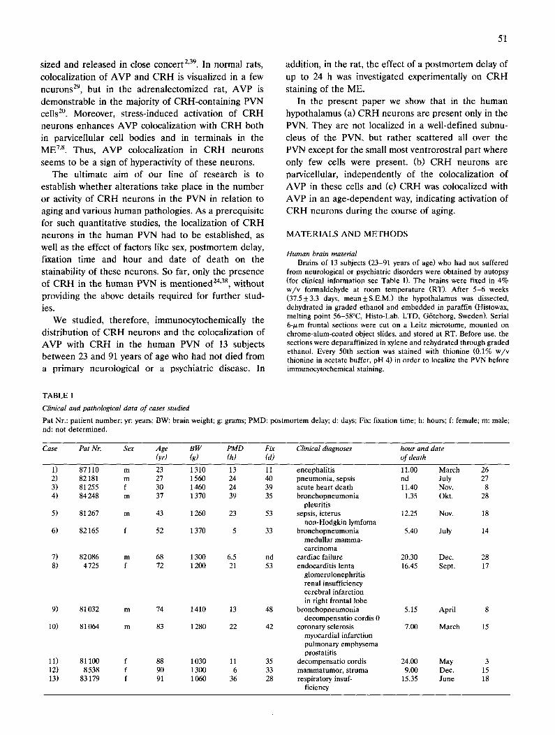
TABLE I
Clinical and pathological data of cases studied
Pat Nr.: patient number; yr: years; BW: brain weight; g: grams; PMD: postmortem delay; d: days; Fix: fixation time; h: hours; f: female; m: male; nd: not determined.
Case Pat Nr. Sex Age B W PMD Fix Clinical diagnoses hour and date (yr) (g) (h) (d) ofdeath
1) 87110 m 23 1310 13 11 2) 82181 m 27 1560 24 40 3) 81255 f 30 1460 24 39 4) 84248 m 37 1370 39 35
5) 81267 m 43 1260 23 53
6) 82165 f 52 1370 5 33
7) 82 086 m 68 1300 6.5 nd 8) 4 725 f 72 1200 21 53
9) 81032 m 74 1410 13 48
10) 81064 m 83 1280 22 42
11) 81 100 f 88 1030 11 35 12) 8 538 f 90 1300 6 33 13) 83179 f 91 1060 36 28
encephalitis 11.00 March 26 pneumonia, sepsis nd July 27 acute heart death 11.40 Nov. 8 bronchopneumonia 1.35 Okt. 28
pleuritis sepsis, icterus 12.25 Nov. 18
non-Hodgkin lymfoma bronchopneumonia 5.40 July 14
medullar mamma- carcinoma
cardiac failure 20.30 Dec. 28 endocarditis lenta 16.45 Sept. 17
glomerulonephritis renal insufficiency cerebral infarction in right frontal lobe
bronchopneumonia 5.15 April 8 decompensatio cordis 0
coronary sclerosis 7.00 March 15 myocardial infarction pulmonary emphysema prostatitis
decompensatio cordis 24.00 May 3 mammatumor, struma 9.00 Dec. 15 respiratory insuf- 15.35 June 18
ficiency

52
Localization of CRH in the human hypothalamus Immunocytochemical staining for CRH was done on a series of
sections taken at 600-/xm intervals throughout the region in which the PVN was present (as determined from the thionine-stained material). This part of the hypothalamus also contained parts of the supraoptic nucleus (SON) 17, suprachiasmatic nucleus 34 and sexually dimorphic nucleus 33.
The monoclonal rat anti-CRH 'PFU 83' (IgG2a subclass) 44 was used that was directed to the C-terminal part (amino acids 38-39) of ra t /human CRH. A radioimmunoassay (RIA) has proved it to be highly specific, with no significant binding of pituitary hormones and hypothalamic peptides other than CRH 44. lmmunocytochemical staining with PFU 83 in rat hypothalami showed staining patterns that were qualitatively similar to those obtained with a conventional CRH antiserum used earlier 7.
Using a modification of an immunocytochemical double staining 5, CRH was stained blue by alkaline phosphatase (AP). The sections were washed (i.e. 3× 10 min) in 0.05 M Tris containing 0.15 M NaC1 (TBS, pH 7.6). Incubations with PFU 83, diluted 1:100,000 in staining buffer (TBS with 0.5% v / v Triton X-100 and 0.25% w / v gelatin), were kept at RT for 30 min and subsequently at 4°C overnight in plastic boxes to prevent evaporation. The following day, the sections were washed and incubated for 30 min with an AP- labeled goat-anti-rat IgG (H + L) serum (KPL, Maryland) and diluted 50 times in staining buffer. Thereafter, the sections were washed and rinsed in 0.05 M Tris (pH 8.5). The AP activity was demonstrated by incubating exactly 10 min at RT with a filtered suspension of 2 mg Fast blue BB base (Sigma, St. Louis) in 10 ml 0.1 M Tris (pH 8.5) containing 2 mg naphtol-AS-AX phosphate (Sigma, St. Louis), 0.2 ml N,N-dimethylformamide and 3 mg levamisole (Sigma, St. Louis). Longer incubation did not result in further intensification of the blue cells but only in higher background staining. Finally the sections were washed and coverslipped with Kaisers Glyceringelatin (Merck).
The rostro-caudal distribution of CRtt cells in the PVN was determined on the right side of the brain (except in case Nr. 87110, where the tissue was not completely present on that side and the left side was used). The total PVN length was defined as the distance between the most rostral and most caudal thionine-stained section showing a cluster of at least 5 magnocellular neurons. The length of the PVN that contained CRH cells was defined as the distance between the most rostral and most caudal section which contained one or more CRH positive cells. This criterion of only one cell was used because of the low number of CRH neurons in some of the cases investigated. The length of the CRH-containing part of the PVN was expressed as a percentage of the total PVN length for each subject.
Colocalization of AVP and CRH Immunocytochemical double staining was performed on a second
series of sections, taken at 600-/zm intervals, in order to demonstrate colocalization of CRH and AVP. The staining was a modification of the method of Claassen et al. 5, resulting in blue CRH cells, red AVP cells, and purple CRH and AVP cells. In addition to the CRH staining described above, AVP was stained by a peroxidase-anti-per- oxidase (PAP). For immunostaining of AVP a rabbit antiserum 'Truus '42 was used. From this AVP antiserum oxytocin binding antibodies were removed by preadsorbing twice with oxytocin- glutaraldehyde-coupled sepharose beads (cf. ref. 25). For the first antibody incubation, Truus and PFU 83 were diluted in staining buffer, respectively, 1:300 and 1:100,000. The following day the sections were washed and incubated with goat anti-rabbit IgG serum, diluted 1:50 in staining buffer (1 h at RT). After washing, the sections were incubated with both an AP-labeled goat-anti-rat lgG ( H + L ) serum (K.P.L., Maryland, USA) and a rabbit PAP, diluted 1:50 and 1 : 1,000, respectively, in TBS. Thereafter, the sections were washed, and rinsed in 0.05 M Tris (pH 8.5). The AP activity was demonstrated as described above. The sections were subsequently washed and incubated (10 rain, RT) in filtered PAP-substrate pre- pared by dissolving 25 mg 3-amino-9-ethylcarbazole (Sigma, St. Louis) in 2.5 ml N,N-dimethylformamide, which was added to 50 ml 50 mM Na-acetate (pH 5.0) and activated by 20 /xl 30% H 2 0 2. Longer incubation did not result in more intensified cells. Only the back-
ground staining became higher. Finally, the sections were washed and coverslipped as described above.
Cellular profile area measurements In immunocytochemically double-stained sections of the PVN,
profile areas of AVP, CRH and both AVP and CRH containing cells (red, blue and purple, respectively) were measured by means of a Calcomp 2000 digitizer, using a Zeiss microscope with a PLAN 40 × objective and PLAN 12.5 × oculars. The profile area was measured only in neurons containing a cell nucleus.
Antibody specificity Immunocytochemical control stainings in the double staining were
performed with anti-CRH (PFU 83), preincubated with 10 -5 M CRH, and the AVP antibody preadsorbed twice with AVP. Pread- sorbtion was performed as the removal of cross-reacting antibodies (cf. ref. 25). Neither these controls, nor pre-immune serum or normal rat serum as first antibody revealed any staining (Fig. 3D).
A spot-blotting experiment, based upon the press-blotting proce- dure of Van der Sluis et al. 42, was performed in order to investigate possible cross-reactivities of the above antisera with the following peptides: synthetic rat CRH (Peninsula), three fragments of ra t /hu- man CRH consisting of aminoacids 1-20, 6-33, and 21-41 (kindly obtained from Dr. J. Rivier), AVP, oxytocin (both from Sigma, St. Louis), neurophysin-A (kindly obtained from Dr. B.T. Pickering, Bristol, UK), and a glycopeptide fragment (22-39) (kindly provided by Dr. J. van Nispen, Organon, Oss, The Netherlands). Spots con- taining 100 ng and a step-wise 1:3 dilution from 10 ng to 1.5 pg of the above mentioned peptides were applicated to a gelatin coated nitro- cellulose (NC) membrane in 1 ~1 spot buffer (i.e. 10% v /v dimethyl- formamide (BDH, Poole, UK) 1% v /v glycerol (BDH), 2.5% v / v Nonidet P-40 (Sigma, St. Louis), 2% w / v Servalyt 2-11 (Serva, Heidelberg, Germany) in aquadest), instead of the isoelectric fo- cussing gel as described in the original paper 42. Finally the peptides were fixed with 4% w / v formaldehyde to the gelatin coated NC membranes.
For the immunocytochemical staining of the NC membranes a PAP procedure 42 was used with either the purified AVP antibody (Truus, diluted 1:500), or the CRH antibody (PFU 83, diluted 1 : 105) or three rabbit antisera (diluted 1 : 1,000); anti-neurophysin-A (kindly obtained from Dr. A.G. Robinson, Pittsburgh, USA), anti-glyco- peptide 22-3930'43, and anti-oxytocin (O-1-V35). As secondary anti- bodies for the CRH antibody we used a biotinylated anti-rat anti- serum (Amersham, UK) and for the other primary antibodies a goat-anti-rabbit antiserum (Betsie), diluted 1:200 and 1:1,000, re- spectively, in staining buffer. For the peroxidase reaction an ABC- peroxidase kit (Vector Lab. Inc., Burlingame) or a PAP-labeled goat anti rabbit was used. Stainings for oxytocin, glycopeptide (22-39) and neurophysin-A with their homologous antisera were performed in order to control for the presence of these peptides on the NC membrane.
Quantification of the intmunocytochemically stained NC-mem- branes was performed according to Van der Sluis et al. 42. From each spot, the area and optical density were measured. For setting a 100% transmission, a gelatin-coated NC membrane without peptide spots was used. The corrected integrated optical density was obtained by multiplying area and optical density values and correcting for the blank value (spot buffer without peptide).
Effect of postmortem delay (PMD) on CRH staining in rats Nine male Wistar rats (strain: Cpb:WU, age: + 7 weeks, weight:
225-249 g, Harlan Cpb, Zeist, The Netherlands) were sacrificed at 10.00 a.m. by decapitation. The brains of three of these rats were immediately dissected and immersion-fixed in 4% w / v paraform- aldehyde (PMD 0 h). The brains of the other two groups of three rats were removed from the skull and fixed after storage of the intact skulls for 6 or 24 h at 4°C (PMD 6 and 24 h, respectively).
After 4 weeks of fixation at 4°C, all rat brains were embedded in paraffin, cut and prepared for immunocytochemistry in the same way as described for the human hypothalami. The PAP procedure de- scribed in the spot blot experiment was used for immunocytochem- istry.

Statistics Profile areas (Table IV) from CRH cells, AVP cells and cells
containing both CRH and AVP were analyzed by one way analysis of variance followed by a two-tailed t-test.
Analysis of semi-quantitative data In order to investigate whether differences in the number and
staining intensity of CRH-neurons in the PVN and in the staining intensity of the ME could be attributed to age, postmortem delay, fixation time or hour of death, Spearman correlation coefficients were determined after ranking the semi-quantitative data from Table II (_+ =1, + =2, + + =3, + + + =4), followed by a one-tailed significance test for correlation (Conover, 1980). The age-depend- ency of the colocalization of AVP with CRH was analysed by determining Spearman correlation coefficients of age vs. the ranked ratio of the number of purple cells with the number of purple and blue cells together, also followed by a one-tailed significance test for correlation (Conover, 1980).
Differences in the number and staining intensity of CRH neurons in the PVN and in the staining intensity of the ME between sexes, and differences in the ratio of purple/(purple + blue) cells between young subjects ( < 65 years of age) and old subjects (> 65 years of age), were analysed with the Mann-Whitney test followed by a two-tailed probability of Z corrected for ties. Correlation coefficients and significance values are only mentioned in the text if they reached significance.
For all statistical analyses SPSS-X (SPSS Inc, Chicago, USA) was used. Significance was defined at the 0.05 level.
Evaluation of the statistical procedures of the semi-quantitative data is presented in the discussion.
53
RESULTS
Localization of CRH in the human hypothalamus Immunocytochemical staining for CRH revealed im-
munoreactive neurons and nerve fibers in the PVN of all 13 subjects. In the cell bodies the reaction product was distributed over the entire cytoplasm (Fig. 1). No CRH cells were observed outside the PVN, i.e. neither in the suprachiasmatic nucleus (SCN), nor in the sexu- ally dimorphic nucleus (SDN), nor in the supraoptic nucleus (SON). In only one section a single CRH cell was seen in the SON (Nr. 4725) and one CRH cell was found lateral to the fornix (Nr. 81064).
The number of CRH neurons (a semi-quantitative estimate of the number of cells over the entire length of the PVN) appeared to vary greatly between the subjects (Table II). These differences have not yet been quantified. The CRH staining intensity of PVN cell bodies and ME nerve fibers showed also inter-individ- ual variation (Table II). Factors such as age, sex, fixa- tion time, PMD, hour of death and date of death
Fig. 1. Frontal paraffin sections (6 ~tm) through a human PVN stained (A) for thionine and (B) immunocytochemically for CRH. Note in (A) the clustering of the magnocellular cells forming the nucleus, and in (B) the cytoplasmatic staining of the parvicellular CRH neurons, the staining of
CRH fibers in this nucleus and the unstained magnocellular neuron (arrow). Bar represents 25/xm.

54
] ] I
PVN
I I
1 r a m
S PVN
11I
SON
PVN
SON
\
m
[] CRH
Pv.~) /
\
B.
C.
Relative PVN length ( CRH )
caudal !
Relative PVN length ( thionine ) rostral
PATIENT N R .
87110
82181
81255
84248
81267
82165
82086
4725
81032
81064
81100
8538
83179
-Y3 ..... I
J i I
12,'' J I

55
s e e m e d not to in f luence C R H sta in ing in tens i ty and
C R H cell number .
The rostro-caudal distribution o f CRH cells in the
P V N revea led tha t in only the small mos t ven t ro ros t r a l
pa r t of the P V N genera l ly no C R H cells were presen t .
In one case (Nr. 81100), a few C R H ceils were found in
the most ros t ra l par t . M o r e caudal ly , the first C R H
neurons a p p e a r (Fig. 2) in the cen t ra l pa r t of the P V N
(Fig. 2A). In the cen t ra l pa r t of the P V N the l a te ra l
b o u n d a r i e s for C R H neurons were m o r e or less ident i -
cal to those o b t a i n e d af te r th ion ine staining. The same
d i s t r ibu t ion was found in the cauda l pa r t of the PVN,
a l though in a lmos t ha l f o f the subjects the n u m b e r of
C R H cells was too low to c lear ly es tabl ish the b o u n d -
ar ies of the P V N a r e a tha t con t a ined C R H cells. T h e
m e d i a n length of the P V N pa r t tha t con t a ined C R H
cells was 67% of the to ta l P V N leng th (Tab le II). T h e
C R H cells were s p r e a d evenly over this pa r t of the
PVN.
TABLE II
CRH staining in human hypothalami
Cell number: + very low; + low; + + high; + + + very high Staining intensity: + faint; + + intense; + good; + + + very intense.
Case Total CRH- Ratio of CRH Intensity of PVN PVN CRH-PVN cell CRH fibers length length and total number cel ls median (izm) (l~m) PVN length in PVN in PVN eminence
1) 4800 1200 25% + + + + 2) 4800 3600 75% + + + + + + + + 3) 3 000 600 20% + + + + + 4) 4200 3600 75% + + + + + + + 5) 3600 2400 67% + + + + + 6) 4 200 1800 43% + + + + 7) 4200 1800 43% + + + + + 8) 5 400 3 600 67% + + + + + + + + + 9) 3600 2400 67% + + + + + + + +
10) 5 400 3 600 67% + _+ + + 11) 3000 3000 100% + + + + + + + + 12) 4200 3000 70% + + + + + + + + + 13) 4800 2400 50% + + + +
Colocalization o f A V P and CRH In the h u m a n PVN, an t i -AVP, a n t i - C R H and ant i -
C R H / A V P s ta in ing cells were v isua l ized as red, b lue
and purp le , respect ive ly (Fig. 3). The reac t ion p roduc t s
were d i s t r i bu t ed over the en t i re cytoplasm. T h e r ed
cells were c lear ly s ta ined, the major i ty of b lue cells
were fa int ly s t a ined and the p u r p l e cells were da rk ly
s t a ined (Fig. 3, Tab le III) , whe reas the b a c k g r o u n d
s ta ining was very low. Al so in this doub le s ta ining
pro tocol , the s ta in ing intensi ty of the C R H cells and
the n u m b e r of C R H ceils va r i ed grea t ly among differ-
en t subjects (Tab le III) . In the SON, only r ed s ta ined
( A V P ) cells were obse rved (Fig. 3)
Coloca l i za t ion of C R H and A V P in the P V N was
only found in subjects over abou t the age o f 40 years
(Table I I I and IV). The major i ty of C R H neurons were
s t a ined pu rp l e ( C R H plus A V P ) f rom the age of 52
years onwards , except the subjects Nr. 82086 and Nr.
83179 (68 and 91 years o f age, respect ively) , in which
the major i ty of C R H neurons were b lue (only CRH) .
These resul ts a re based upon two i n d e p e n d e n t bu t
ident ica l assessments by two r e sea rche r s (F .C.R. and
D.F.S. )
Cellular profile area measurements M e a s u r e m e n t s of the prof i le a reas f rom the t h ree
d i f fe ren t cell types r evea led tha t the m e a n prof i le a r ea
of the r ed (AVP) cells (327.0 /zm 2, _+ 16.3) was 2.2
t imes as large as the m e a n prof i le a r ea of the pu rp l e
( A V P plus C R H ) cells (148.5 /zm z, _+ 10.8) and 2.3
t imes as large as the m e a n prof i le a r ea of the b lue
( C R H ) cells (140.6 /xm 2, _+7.4). The r ed ( A V P ) cells
were s ignif icant ly l a rge r than the p u r p l e ( A V P and
C R H ) and b lue ( C R H ) cells ( P < 0.001).
Antibody specificity
Quantification o f the immunocytochemically stained NC-membranes showed b ind ing of the C R H an t ibody
' P F U 83' to in tac t C R H and f r agmen t 21-41 (Fig. 4).
P F U 83 did not b ind to spots con ta in ing the C R H
f ragmen t s 1 -20 or 6 -33 , AVP, neurophys in -A, gly-
c o p e p t i d e (22-39) or oxytocin. The A V P an t i s e rum
'T ruus ' , which had b e e n pur i f i ed to avoid cross- reac-
t ion with oxytocin, only r e a c t e d with A V P and not with
oxytocin, neu rophys in -A, g lycopep t ide (22-39) , C R H
or any of the C R H f ragmen t s (Fig. 4). The cont ro l
blots, which con t a ined neu rophys in -A , g lycopep t ide
r--
F ig . 2. A: topography of CRH cells in the human PVN from rostral (left) to caudal (right). F, fornix; OT, optic tract; III, third ventricle; PVN, paraventricular nucleus; CRH, the part of the PVN which contained CRH cells. B: length of the part of the PVN containing CRH cells expressed as percentage of the total length of the PVN. The total PVN length is represented by the upper horizontal line and put on 100% for each subject. The rest of the horizontal lines indicates the length (in %) and localization of the part of the PVN containing CRH neurons. C: projection of the CRH cells on the mediosagittal plane of the PVN in the hypothalamus. Scheme after Diericks and Vandesande 9. 1, PVN; la, part of PVN which contains CRH; 2, dorsolateral part of SON; 3, dorsomedial part of SON; 4, ventromedial part of SON; 5, SCN; 6, optic chiasm; 7, optic nerve; 8, origin of pituitary stalk; 9, mammillary body; 10, lamina terminalis; 11, anterior commissure; 12, axis of the fornix; 13, hypothalamic sulcus. Note
that CRH neurons are lacking in the small most ventrorostral PVN in all cases but one.

~q

57
TABLE IlI
lmmunocytochemical double staining for AVP and CRIt in the human PVN
CRH cells are blue, AVP cells red and CRH and AVP cells purple Cell profiles per section: - none; ± 1-5; + 6-9; + + 10-49; + + + 50-99; + 6-9; + + + + > 100. Staining intensity: - deficient; +__very faint; + faint; + + normal; + + + intense; + + + + very intense.
Case Majority Cell profiles Staining intensity of
of CRH blue cells purple cells red cells blue cells purple cells red cells cells
1) blue + + - + + + + _+ - + + 2) blue + + + - + + + + + - + 3) blue + + - + + + _+ - + 4) blue + + + + - + + + + + + - + + + 5) blue + + + + + + + + 6) purple + + + + + + + + + + + 7) blue + ± + -~ + + + + 8) purple + + + + + + + ± + + + + 9) purple + + + + + + + + + ± + + + + +
10) purple +_ + + + +_ + + 11) purple + + + + + + + + + + + + + + + + 12) purple + + + + + + + + + + + + + + 13) blue + + + + + + + +
( 2 2 - 3 9 ) o r oxy toc in , s h o w e d i m m u n o r e a c t i v i t y a f t e r
s t a i n i n g w i t h t h e i r h o m o l o g o u s a n t i s e r a (Fig. 4), p rov -
ing t h a t t h e p e p t i d e s w e r e i n d e e d b o u n d to t h e N C
m e m b r a n e .
E f f e c t o f p o s t m o r t e m de lay ( P M D ) o n C R H s ta in ing in
rats
In t h e ra t h y p o t h a l a m u s , n o C R H i m m u n o r e a c t i v i t y
was s e e n in t h e P V N , S O N or S C N a f t e r a P M D of 0, 6
o r 24 h. H o w e v e r , i n t e n s e C R H i m m u n o r e a c t i v i t y w a s
f o u n d in t h e e x t e r n a l z o n e o f t h e M E in all c a s e s (Fig .
5). T h e d i f f e r e n t P M D s d id n o t r e su l t in obv ious d i f f e r -
e n c e s in C R H s t a i n i n g i n t e n s i t i e s o f t h e M E ( T a b l e V).
DISCUSSION
d e c r e a s e d t r a n s p o r t r a t e o r a d i f f e r e n t p r o c e s s i n g o f
t h e p r e c u r s o r in h u m a n C R H n e u r o n s .
W e f o u n d C R H n e u r o n s in all 13 s u b j e c t s inves t i -
g a t e d , in sp i t e o f t h e fac t t h a t w e i n c u b a t e d m o u n t e d
s e c t i o n s f r o m p a r a f f i n - e m b e d d e d m a t e r i a l , w h i c h illus-
t r a t e s t h e sens i t iv i ty o f o u r i m m u n o c y t o c h e m i c a l p r o c e -
TABLE IV
Mean cellular profile areas of CRH cells, AI/P cells and AI/P and CRH cells in the human PI/N
area = mean cellular profile area (/xm2); S.E.M. = standard error of the mean; n = number of measured cells.
Case Blue cells Purple cells Red cells (only CRH) (CRH and A VP) (only A l/P)
area + sem n area +_ sem n area +_ sere n
In t h e h u m a n P V N , cel l b o d i e s c a n b e f o u n d t h a t 1) 161.34 12.81 18 - 2) 139.07 4.24 94 -
s t a in ve ry c l ea r ly fo r C R H 24'38 ( th i s ar t ic le) . In un - 3) 9 6 . 4 2 5.75 15 - -
t r e a t e d ra t s , t h e s e cel ls c a n n o t b e o b s e r v e d a n d 4) 137.10 4.39 119 - 5) 134.52 6.16 37 90.68 16.56 c o l c h i c i n e a d m i n i s t r a t i o n o r a d r e n a l e c t o m y a r e re- 6) 100.08 7.88 14 126.68 9.75
q u i r e d in o r d e r to v i sua l i ze C R H - p r o d u c i n g cel l b o d - 7) 178.08 9.34 21 120.00 2.34
ies 29. T h e l o n g P M D of t h e h u m a n s u b j e c t s as c o m - 8) 165.94 2.73 2 151.00 5.77 9) 128.37 9.68 20 155.85 4.93
p a r e d to r a t s is un l ike ly to b e r e s p o n s i b l e fo r t h e 10) 113.15 14.21 5 149.68 18.24
s p e c i e s d i f f e r e n c e s in s t a inab i l i t y o f cel l b o d i e s , s i nce 11) 172.07 10.96 16 169.16 4.76
no C R H s t a i n i n g w a s e v i d e n t in t h e P V N o f r a t s e v e n 12) 136.45 14.01 6 200.94 5.72 13) 164.58 13.91 11 172.63 8.69
at 24 h p o s t m o r t e m . It is n o t c l e a r at p r e s e n t w h e t h e r
th is s p e c i e s d i f f e r e n c e is d u e to i n c r e a s e d p r o d u c t i o n , mean 140.55 7.43 13 148.51 10.83
- 445.84 12.73 100 - 356.33 12.44 65 - 230.33 6.67 62 - 357.97 9.38 101 3 281.44 15.89 21
23 238.50 2.73 41 2 275.96 8.11 56
74 355.64 12.85 60 98 315.96 6.57 143 10 342.55 15.04 38
123 356.48 9.06 132 132 366.92 12.09 66
2 326.66 9.35 55
9 326.97 16.30 13
t--
Fig. 3. Immunocytochemical double staining on frontal paraffin sections (6 ~m) through the human hypothalamus. CRH cells stained blue, AVP cells red and CRH and AVP containing cells purple. A: section through the PVN of a young patient (Nr. 84248), not showing colocalization of AVP and CRH; B: section through the PVN of an old patient (Nr. 81032), showing red (*), blue (1") and purple cells ( • ) . (For profile areas of the different cell types, see Table IV). C: section through the SON of patient Nr. 81032, showing only red AVP cells. D: staining by AVP-preadsorbed anti-AVP and with 10 -5 M preincubated CRH anti-CRH of a section through the PVN of patient Nr. 81032, showing no
immunoreactive cells. Bar = 25/xm.

30
20
10
0 -2 , J , i i
0 0 0 1 0.01 0.1 1 10 100
A N T I B O D Y S P E C I F I C I T Y T E S T
o CRH intact & CRH 21-41 D CRH 1-21 ¢ CRH 6-33
• Vasopressin • Oxytocin • Neurophysin • Glycopeptide
Spot blot stained with anti-CRH ( PFU83 )
5 8
e: c
c
3
30
20
10
0 -2
Spot blot stained with purified anti-AVP ( Truus )
~ . i i i i )
0.001 0.01 0.1 1 10 100
30 Spot blots stained with the homologous
antisera of the applicate
2O
10
o " ~. ~ , - ' - - - - * - - - " _2 L. j i J i J
0 .001 0.01 0.1 1 10 100
A p p l i c a t e d a m o u n t o f p e p t i d e ( p m o l )
Fig. 4. Corrected integrated optical density (IODC) values of peptide spots blotted on gelatine coated nitrocellulose (NC). The IODC was corrected for (1) the optical density of gelatine coated NC without peptide ( = 100% transmission); (2) the optical density of the blank; and (3) the area of the spot. Different quantities (blank, 1.5 pg, 4.5 pg, 13 pg, 41 pg, 123 pg, 370 pg, 1.1 ng, 10 ng and 100 ng) of peptide were applicated to the NC in a volume of 1 p,l of spot buffer. The blank consists of spot buffer without peptide. The upper, middle and lower parts of the figure show the results from, respectively, the spot blot stained by the C R H antibody PFU 83, the spot blot stained by the AVP antibody Truus, which was purified for crossreactivity with oxytocin, and three different blots stained with their homologous
antibodies.
dure if we compare this to the literature. In mounted paraff in-embedded material, Ohtani et al. 23 could de- tect C R H immunoreactivity in only 5 out of the 23 cases. Therefore, they had to increase the sensitivity of the CRH staining in human material by using free- floating sections, which resulted in C R H positive cells and nerve fibers in all 7 cases examined 38.
In spite of the sensitivity of our method and the standardized way we performed it, considerable inter- individual differences in C R H staining intensity and cell number (based upon semi-quantitative observa- tions), were present in our material (Table II). These differences seem not to correlate ( P > 0.06) with fac- tors such as age, sex, PMD, fixation time and hour of death. Only the effect of PMD on CRH immunoreac- tivity in the hypothalamus has been investigated earlierl'38. The first group could not find with a RIA
an influence of PMD on C R H contents. However, they only investigated three subjects. The second group suggested the use of short PMDs after finding good CRH staining in tissue obtained within 3 h post- mortem. However, they have not investigated longer PMDs. Since our group showed that a longer PMD resulted in even bet ter staining of AVP positive cell bodies in the rat SCN 45, the need of short PMDs for
peptide immunocytochemistry in general can be ques- tioned. Indeed, our human hypothalamic C R H data (Table I) do not point to an effect of the PMD on CRH staining, neither in PVN cell bodies nor in ME fibers. Since it is difficult if not impossible to study one factor in isolation in human material, we also investi- gated the effect of PMD on C R H staining in the ME of the rat (Table V). This structure is of interest, since in the human material the intensity of the C R H staining of both, the ME and PVN cell bodies varied in a similar way (Table II). From the rat data it also seems that the C R H staining intensity is not affected by differences in postmortem delay.
Like postmortem delay, also different fixation times could theoretically lead to immunocytochemical stain- ing differences. In order to gain some information about the possible influence of these factors together with factors like age and sex on the immunocytochemi- cal results we obtained in this paper, we tried to analyse the data in a non-parametric way. However, the limited material does not allow very strong conclu- sions.
The same restriction holds for the possible influence of the time of death on the stainability of CRH-neu- rons, which is of interest since circadian changes of CRH are found in cerebrospinal fluid (e.g. Ref. 14). This restriction does not hold for the clear age depen- dent colocalization of AVP with CRH, because the

59
correlation of age vs. the ranked ratio's of purple cel ls /(purple + blue cells) is very high (rho = 0.8, P = 0.001), and the difference in ratio between young and old subjects is also very signifficant (P = 0.006).
In the SON of the human hypothalamus we did not
find CRH cells (see also refs. 24, 38), whereas in the
rat the presence of CRH cells in the SON has often been described = (for review see ref. 29). However, one
Fig. 5. lmmunocytochemical C R H staining of 6 /.tm paraffin sections through the median eminence of rats. The external zone of the median eminence is stained. A: no postmortem delay (PMD); B: PMD of 6 h; and C: PMD of 24 h. No difference in staining intensity was found.
Bar = 25/xm.

60
TABLE V
Effect of postmortem delay (PMD) on the intensity of CRH staining in the median eminence of 9 rats
+ good staining; + + intense staining; + + + very intense staining.
PMD 0 h PMD 6 h PMD 24 h
+ + + + + + + + + + + + ++ + + + + + + + +
patient with a high CRH cell number and very intense C R H staining in both ME and PVN showed one single CRH cell in the SON. This may indicate that in the
human SON CRH-producing cells occur and can be
made detectable following activation of the CRH sys- tem. The presence of radioimmunoassayable C R H in extracts of the human SON 26, favour this hypothesis. A
more likely explanation, however, is that the content of CRH fibers running along the SON to the neurohy- pophysis have been included by the dissection proce-
dure. The CRH neurons in the human PVN were all
parvicellular (Table III). They were dispersed all over the central and caudal part of the PVN, which is in contrast to the localization in a distinct subnucleus as in the rat 22'29'37. The species difference in the localiza-
tion of C R H neurons is in agreement with species differences found previously in the basic structure and topographical arrangement of AVP and oxytocin neu- rons in the PVN. In contrast to rats, in humans, cows, cats and guinea pigs no arrangement of the PVN into clear subnuclei was found 32. In this respect it seems
that the clear partition of the rat PVN into well de- fined subnuclei is the exception rather than the rule.
In the present study, the colocalization of AVP and CRH had been investigated for two reasons. Firstly, the distribution of the C R H neurons in the human PVN showed a large overlap with that of the AVP neurons, which could indicate that C R H and AVP are colocalized in a similar way as has been described for the rat (for review see ref. 29). Secondly, colocalization of AVP in C R H neurons in the rat was suggested to be a marker for the activity of C R H neurons 7's. In order
to investigate the colocalization of AVP and CRH, we used an immunocytochemical double staining proce- dure. This procedure has a major advantage to the alternative of staining adjacent paraffin sections with a single staining procedure because it is often difficult in paraff in-embedded tissue to recognize with certainty the same cell in the adjacent section because of the non-uniform way sections stretch. Immunocytochemi- cal double staining for AVP and C R H indeed showed
colocalization of C R H and AVP in the human PVN, but only in cases of over about 40 years of age (Table
III). This observation supports the view that the activ- ity of the CRH neurons increases with aging, which is in accordance with findings demonstrating hyperactiv-
ity of the human hypothalamo-pituitary-adrenal axis during aging 1°'13'47. Activation of AVP neurons in the
human PVN during aging has also been described (i) on the basis of changes in cell size 12 and nucleolus
size 19, but only in the age group of 80-100 years, and
(ii) on the basis of the number of cells expressing
AVP )~, which is higher in subjects older than 60 years of age. It might well be that in humans the parvicellu- lar CRH cells which start to synthesize AVP during the
course of aging contribute to a considerable degree to the increase found in AVP cell numbers during aging ~.
At the other hand, this possibility is not supported by the finding that the mean cellular profile area of AVP cells increases during aging ~2. Our data (Table IV) do not show a significantly larger mean cellular profile
area for AVP cells in the PVN of patients older than 80 years of age, but this group concerns only 4 patients.
The observation that C R H neurons are activated during the course of aging by coexpressing higher levels of AVP can not be confirmed by RIA because it is not possible to isolate PVN cells for assay without dissect- ing at the same time fibers containing either one or both of the peptides. Even if it were possible to isolate PVN cells, differences in AVP levels caused by the colocalization of AVP with CRH in the parvocellular neurons would be masked by the high AVP levels obtained from the majority of magnocellular AVP neu- rons present in the PVN. However, our observations can be confirmed by in situ hybridization.
Since in the human PVN, C R H neurons and neu- rons containing CRH and AVP were both found to be
parvicellular (Table IV), and because at a younger age only CRH neurons were present (Table III), one may wonder whether beyond the age of 43 years of age C R H neurons may start to synthesize AVP or alterna- tively parvicellular AVP neurons may begin to make CRH. The first possibility would fit with results ob- tained in the rat. In this species, two types of CRH neurons have been found after colchicine treatment and adrenalectomy, i.e. AVP deficient and AVP ex- pressing ones 48. The AVP-containing subset of CRH was selectively activated by stress, resulting in (i) deple- tion of neurosecretory vesicles from the axonal swellings in the external zone of the median eminence 4~, (ii) increased AVP stores and colocalization in CRH nerve terminals ~, and (iii) a five-fold elevation in number of the AVP containing C R H cell bodies 7. However, there is a difference with the data we obtained in the human

hypothalamus where no colocalization of CRH and AVP was found before activation of the CRH neurons before the age of 43 years. This species difference may be due to the colchicine treatment, resulting in a high AVP content in the CRH cell bodies, whereas in humans the AVP content of the CRH neurons before the age of 43 years might be too low for immunocyto- chemical detection. The second possibility can at pre-
sent also not be excluded. The colocalization of CRH and AVP did not result
from cross-reactivity of the antibodies, as was verified from the specificity tests for the antibodies. Since in the spot blot experiment the antigen is fixed in a similar way as in the tissue sections, it may give a good impression about the recognition of the antigen by the antibody in tissue sections during immunocytochem- istry. The observation that purple (CRH- and AVP- containing) cells were never present in double-stained sections of the human SON, whereas they were abun- dant in the PVN, also indicates that the anti-CRH antibody does not cross-react with AVP or parts of its precursor.
In the present study we make it clear that it will indeed be possible to study the CRH neurons in the PVN during disease states. However, one should con- sider that (1) the CRH cells are not localized in a distinct subnucleus and (2) the colocalization of CRH and AVP is an age-dependent phenomenon, which is possibly related to its activity status.
Acknowledgements. We are indebted to the Netherlands Brain Bank in the Netherlands Institute of Brain Research (coordinator Dr. R. Ravid) for providing us with brain material. We wish to thank Mr. B. Fisser for his technical assistance, Dr. C.W. Pool for his valuable advice on the antibody-specificity tests, Mr. H. Stoffels for preparing the illustrations, Mr. G. van der Meulen for making the photographs and Dr. M.A. Hofman for his statistical advice. We also appreciate the advice and help concerning the double staining provided by Dr. E. Claassen and Mrs. M. Fasbender (both of MBL, TNO, Rijswijk) and Mr. C. van der Loos (Pathological Anatomy, AMC, Amsterdam). We owe a lot to Dr. D.E. Oorscbot (University of Otago, New Zealand) and Mr. A. Janssen for improving the English of the manuscript. This investigation was supported by the network Neuro- sciences Amsterdam (project no. 89-23) and Mrs. E.J.M. Stevens. Part of this work had been presented at the Third IBRO World Congress of Neuroscience in Montreal ('91) with support of NWO (Grant SIR 15-412).
R E F E R E N C E S
1 Ackland, J.F., Ratter, S.J., Bourne, G.L. and Rees, L.H., Corti- cotrophin-releasing factor-like immunoreactivity and bioactivity of human fetal and adult hypothalami, J. Endocrinol., 108 (1986) 171-180.
2 Antoni, F.A., Hypothalamic control of adrenocorticotropin secre- tion: advances since the discovery of 41-residue corticotropin-re- leasing factor, Endocrinol. ReL'., 7 (1986) 351-378.
3 Bissette, G., Central nervous system CRF in stress: radioim- munoassay studies. In E.B. De Souza and C.B. Nemeroff (Eds.), Corticotropin-Releasing Factor." Basic and Clinical Studies of a Neuropeptide, CRC-Press Inc., Boca Raton, FL, 1990, pp. 21-28.
61
10
4 Brown, M.R., Fisher, L.A., Spiess, J., Rivier, C., Rivier, J. and Vale, W., Corticotropin-releasing factor: effects on the sympa- thetic nervous system and oxygen consumption, Life Sci., 30 (1982) 207-210.
5 Claassen, E., Adler, L.T. and Adler, F.J., Double immunocyto- chemical staining for the in situ study of allotype distribution during an anti-trinitrophenyl immune response in chimeric rab- bits, J. Histochem. Cytochem., 34 (1986) 989-994.
6 Conover, W.J., Practical Nonparametric Statistics, 2nd edn., Wi- ley, New York, 1980, pp. 252-256.
7 De Goeij, D.C.E., Jezova, D. and Tilders, F.J.H., Repeated stress enhances vasopressin synthesis in corticotropin releasing factor neurons in the paraventricular nucleus, Brain Res., 577 (1992) 165-168.
8 De Goeij, D.C.E., Kvetnansky, R., Whitnall, M.H., Jezova, D., Berkenbosch, F. and Tilders, F.J.H., Repeated stress induced activation of corticotropin-releasing factor (CRF) neurons en- hances vasopressin stores and colocalization with CRF in the median eminence of rats, Neuroendocrinology, 53 (1991) 150-159.
9 Diericks, K. and Vandesande, F., Immunocytochemical localiza- tion of the vasopressinergic and the oxytocinergic neurons in the human hypothalamus, Cell Tiss. Res, 184 (1977) 15-27. Dodt, C., Dinmann, J., Hruby, J., Sp~ith-Schwalbe, E., Born, J., Schiittler, R. and Fehm, L., Different regulation of adrenocorti- cotropin and cortisol secretion in young, mentally healthy elderly and patients with senile dementia of Alzheimer's type, J. Clin. Endocrinol. Metab., 72 (1991) 272-276.
11 Fisher, L.A., Rivier, J., Rivier, C., Spiess, J., Vale, W. and Brown, M.R., Corticotropin-releasing factor (CRF): central effects on mean arterial pressure and heart rate in rats, Endocrinology, 110 (1982) 2222-2224.
12 Fliers, E., Swaab, D.F., Pool, C.W. and Verwer, R.W.H., The vasopressin and oxytocin neurons in the human supraoptic and paraventricular nucleus; changes with aging and senile dementia, Brain Res., 342 (1985) 45-53.
13 Friedman, M., Green, M.F. and Sharland, D.E., Assessment of hypothalamic-pituitary-adrenal function in the geriatric group, J. Gerontol., 24 (1969) 292-297.
14 Garrick, N.A., Hill, J.L., Szele, F.G., Tomai, T.P., Gold, P.W. and Murphy, D.L., Corticotropin-releasing factor: a marked cir- cadian rhythm in primate cerebrospinal fluid peaks in the evening and is inversely related to the cortisol circadian rhythm, En- docrinology, 121 (1987) 1329-1334.
15 Geracioti Jr., T.D., Orth, D.N., Ekhator, N.N., Blumenkopf, B. and Loosen, P.T., Serial cerebrospinal fluid concentrations in healthy and depressed humans, 3. Clin. Endocrinol. MetaboL, 74 (1992) 1325-1330.
16 Gillies, G.E., Linton, L. and Lowry, P.J., Corticotropin releasing activity of the new CRF is potentiated several times by vaso- pressin, Nature, 299 (1982) 355-357.
17 Goudsmit, E., Hofman, M.A., Fliers, E. and Swaab, D.F., The supraoptic and paraventricular nuclei of the human hypothala- mus in relation to sex, age and Alzheimer's disease, NeurobioL Aging, 11 (1990)529-536.
18 Goudsmit, E., Neijmeijer-Leloux, A. and Swaab, D.F., The hu- man hypotbalamo-neurohypophyseal system in relation to devel- opment, aging and Alzheimer's disease, Prog. Brain Res., 93 (1992) 237-248.
19 Hoogendijk, J.E., Fliers, E., Swaab. D.F. and Verwer, R.W.H., Activation of vasopressin neurons in the human supraoptic and paraventricular nucleus in senescence and senile dementia, J. Neurol. Sci., 69 (1985) 291-299.
20 Kiss, J.Z., Mezey, E. and Skirboll, L., Corticotropin-releasing factor-immunoreactive neurons of the paraventricular nucleus become vasopressin reactive after adrenalectomy, Proc. Natl. Acad. Sci. USA, 81 (1984) 1854-1858.
21 Linton F.A., Tilders, F.J.H., Hodgkinson, S., Berkenbosch, F., Vermis, I. and Lowry, P.J., Stress-induced secretion of adreno- corticotropin in rats is inhibited by administration of antisera to ovine corticotropin-releasing factor and vasopressin. Endocrinol- ogy, 116 (1985) 966-970.
22 Merchenthaler, I., Vigh, S., Petrusz, P. and Schally, A.V., Im-

62
munocytochemical localization of corticotropin-releasing factor (CRF) in the rat brain, Am. J. Anat., 165 11982) 385-396.
23 Ohtani, H., Mouri, T., Sasaki, A. and Sasano, N., Immunoelec- tron microscopic study of corticotropin-releasing factor in the human hypothalamus and pituitary gland, Neuroendocrinology, 45 (1987) 104-108,
24 Pelletier, G., D~sy, L., C6t6, J. and Vaudry, H., Immunocyto- chemical localization of corticotropin-releasing factor-like im- munoreactivity in the human hypothalamus, Neurosci. Lett., 41 (1983) 259-263.
25 Pool, C.W., Madlener, S., Diegenbach, P.C., Sluiter, A.A. and Van der Sluis, P.J., Quantification of antiserum reactivity in immunocytochemistry, J. Histochem. Cytochem., 32 (1984) 921- 928.
26 Pralong, P.P., Linton, E.A., Favrod-Coune, C.A., Lowry, P.J., Muller, A.F. and Gaillard, R.C., Anatomical distribution of corti- cotropin-releasing factor and arginine vasopressin in the human hypothalamus; the effect of corticosteroids on their concentra- tions in human and rat hypothalami, J. Endocrinol., 2 (1990) 369-374.
27 Rivier, J., Spiess, J. and Vale, W., Characterization of rat hy- pothalamic corticotropin-releasing factor, Proc. Natl. Acad. Sci. USA, 80 (1983) 4851-4855.
28 Rivier, C. and Vale, W., Interaction of corticotropin-releasing factor and arginine-vasopressin on adrenocorticotropin secretion in vivo, Endocrinology, 113 (1983)939-942.
29 Sawchenko P.E. and Swanson, L.W., Organization of CRF im- munoreactive cells and fibers in the rat brain; immunohistochem- ical studies. In E.B. De Souza and C.B. Nemeroff (Eds.), Corti- cotropin-Releasing Factor." Basic and Clinical Studies of a Neu- ropeptide, CRC-Press, Inc., Boca Raton, FL, 1990, pp. 29-51.
30 Seger, M.A. and Burbach, J.P.H., The presence and in viw) biosynthesis of fragments of CPP (the C-terminal glycopeptide of the rat vasopressin precursor) in the hypothalamo-neurohypo- physeal system, Peptides, 8 (1987) 757-762.
31 Selye, H., A syndrome produced by diverse noxious agents, Nature, 138 (1987) 32.
32 Sofroniew, M.V., Vasopressin, oxytocin and their related neuro- physins. In A. Bj6rklund and T. H6kfelt (Eds.), Handbook of Chemical Neuroanatomy, I1ol. 4: GABA and Neuropeptides in the CNS, Part 1, Elsevier, Amsterdam, 1985, pp. 93-165.
33 Swaab, D.F. and Fliers, E., A sexually dimorphic nucleus in the human brain, Science, 228 (1985) 1112-1115.
34 Swaab, D.F., Fliers, E. and Partiman, T.S., The suprachiasmatic nucleus of the human brain in relation to sex, age and senile dementia, Brain Res., 342 11985) 37-44.
35 Swaab, D.F. and Pool, C.W., Specificity of oxytocin and vaso- pressin immunofluorescence, J. Endocrinol., 6 (1975) 263-272.
36 Swanson, L.W., Sawchenko, P.E., Rivier, J. and Vale, W.W., Organization of ovine corticotropin-releasing factor immunoreac- tive cells and fibers in the rat brain: an immunohistochemical study, Neuroendocrinology, 36 (1983) 165-186.
37 Swanson, L.W., Sawchenko, P.E. and Lind, R.W., Regulation of multiple peptides in CRF parvicellular neurosecretory neurons: implications for the stress response, Prog. Brain Res., 68 (1986) 169-190.
38 Takahashi, K., Mouri, T., Yamamoto, T., Itoi, K., Murakami, O., Yoshinaga, K. and Sasano, N., Corticotropin-releasing hormone in the human hypothalamus. Free-floating immunostaining method, Endocrinol. Jap., 36 (1989) 272-280.
39 Tilders F.J.H., De Goeij, D.C.E. and Berkenbosch, F., Turnover of corticotropin-releasing factor and vasopressin in the median eminence, and control of pituitary-adrenal activity. In F.C. Rose (Ed.), The Control of the Hypothalamo-Pituitary-Adrenocortical Axis, International University Press, Madison, 1989, pp. 7-24.
41) Vale, W.. Rivier, C., Brown, M.V., Spiess, J., Koob, G., Swanson, L., Bilezikjian, L., Bloom, F. and Rivier, J., Chemical and biologi- cal characterization of corticotropin-releasing factor, Recent Prog. Horm. Res., 39 (1983) 245-270.
41 Vale, W., Spiess, J., Rivier, C. and Rivier, J., Characterization of a 41-residue ovine hypothalamic peptide that stimulates secretion of corticotropin and/3-endorphin, Science, 213 (1981) 1394-1397.
42 Van der Sluis, P.J., Pool, C.W. and Sluiter, A.A., lmmunocyto- chemical detection of peptides and proteins on press-blots after direct tissue gel isoelectric focussing, Electrophoresis, 9 (1988) 654-661.
43 Van Leeuwen, F., Van der Beek, E., Seger, M., Burbach, P. and lvell, R., Age-related development of a heterozygous phenotype in solitary neurons of the homozygous Brattleboro rat, Proc. Natl. Acad. Sci. USA, 86 11989) 6417-6420.
44 Van Oers. J.W.A.M., Tilders, F.J.H. and Berkenbosch, F., Char- acterization and biological activity of a monoclonal antibody to ra t /human corticotropin-releasing factor, Endocrinology, 124 11989) 1239-1246.
45 Van Zwieten, E.J., Ravid, R., Van der Sluis, P.J., Sluiter, A.A., Pool, C.W., Smyth, D. and Swaab, D.F., Increased vasopressin immunoreactivity in the rat brain after a postmortem interval of 6 h, Brain Res., 550 (1991) 263-267.
46 Von Bardeleben, U. and Holsboer, F., Clinical studies with ovine and human CRF in psychiatric patients and normal controls. In E.B. De Souza and C.B. Nemeroff (Eds.), Corticotropin-Releasing Factor: Basic and Clinical Studies of a Neuropeptide, CRC-Press Inc., Boca Raton, FL, 1990, pp. 309-326.
47 Weiner, M.F., Davis, B.M., Mobs, R.C. and Davis, K.L., Influ- ence of age and relative weight on cortisol suppression in normal subjects, Am. J. Psychiatry, 144 (1987) 646-649.
48 Whitnall, M.H., Distributions of pro-vasopressin expression and pro-vasopressin deficient CRH neurons in the paraventricular hypothalamic nucleus of colchicine-treated normal and adrenal- ectomized rats, J. Comp. NeuroL, 275 (1988) 13-28.
49 Whitnall, M.H., Stress selectively activates the vasopressin-con- taining subset of corticotropin-releasing hormone neurons, Neu- roendocrinology, 50 11989) 702-707.