Relationship Between Vascular Endothelial Growth Factor and Nuclear Factor-κB in Renal Cell Tumors

NF-κB in the paraventricular nucleus modulatesneurotransmitters and contributes to sympathoexcitation inheart failure
Yu-Ming Kang1,a,*, Feng Gao4,a, Hui-Hua Li6,a, Jeffrey P Cardinale2, Carrie Elks2, Wei-JinZang3, Xiao-Jing Yu1, Yan-Yan Xu5, Jie Qi5, Qing Yang1, and Joseph Francis2,*
1Department of Physiology and Pathophysiology, Xi’an Jiaotong University CardiovascularResearch Center, Xi’an Jiaotong University School of Medicine, Xi’an, China2Comparative Biomedical Sciences, Louisiana State University, Baton Rouge, USA3Department of Pharmacology, Xi’an Jiaotong University School of Medicine, Xi’an, China4Department of Physiology, The Fourth Military Medical University, Xi’an, China5Department of Physiology, Shanxi Medical University, Taiyuan, China6Key Laboratory of Remodeling-related Cardiovascular Diseases, Department of Pathology,School of Basic Medical Sciences, Capital Medical University, Beijing, China
AbstractAims—Findings from our laboratory indicate that proinflammatory cytokines and theirtranscription factor, nuclear factor-kappaB (NF-κB), are increased in the hypothalamicparaventricular nucleus (PVN) and contribute towards the progression of heart failure. In thisstudy, we determined whether NF-κB activation within the PVN contributes tosympathoexcitation via interaction with neurotransmitters in the PVN during the pathogenesis ofheart failure.
Methods and results—Heart failure was induced in rats by left anterior descending coronaryartery ligation. Sham-operated control (SHAM) or heart failure rats were treated for 4 weeksthrough bilateral PVN infusion with SN50, SN50M or vehicle via osmotic minipump. Rats withheart failure treated with PVN vehicle or SN50M (inactive peptide for SN50) had increased levelsof glutamate, norepinephrine, tyrosine hydroxylase (TH), superoxide, gp91phox (a subunit ofNAD(P)H oxidase), phosphorylated IKKβ and NF-κB p65 activity, and lower levels of gamma-aminobutyric acid (GABA) and the 67-kDa isoform of glutamate decarboxylase (GAD67) in thePVN compared with those of SHAM rats. Plasma levels of cytokines, norepinephrine, epinephrineand angiotensin II, and renal sympathetic nerve activity (RSNA) were increased in heart failurerats. Bilateral PVN infusion of SN50 prevented, the decreases in PVN GABA and GAD67, andthe increases in RSNA and PVN glutamate, norepinephrine, TH, superoxide, gp91phox,phosphorylated IKKβ and NF-κB p65 activity observed in vehicle or SN50M treated heart failurerats. A same dose of SN50 given intraperitoneally did not affect neurotransmitters concentration inthe PVN and was similar to vehicle treated heart failure rats.
*Corresponding author: Yu-Ming Kang, M.D., Ph.D., Department of Physiology & Pathophysiology, Xi’an Jiaotong UniversitySchool of Medicine, Xi’an 710061, China, Phone: +86 2982657677, Fax: +86 2982657677, [email protected] or JosephFrancis, B.V.Sc., M.V.Sc., Ph.D., Comparative Biomedical Sciences, School of Veterinary Medicine, Louisiana State University,Baton Rouge, LA 70803, USA, Phone: +1 2255789752, Fax: +1 2255789895, [email protected] contributed equally to this study.Conflict of interest: none declared.
NIH Public AccessAuthor ManuscriptBasic Res Cardiol. Author manuscript; available in PMC 2012 November 1.
Published in final edited form as:Basic Res Cardiol. 2011 November ; 106(6): 1087–1097. doi:10.1007/s00395-011-0215-7.
NIH
-PA Author Manuscript
NIH
-PA Author Manuscript
NIH
-PA Author Manuscript

Conclusion—These findings suggest that NF-κB activation in the PVN modulatesneurotransmitters and contributes to sympathoexcitation in rats with ischemia-induced heartfailure.
KeywordsNF-κB; neurotransmitters; hypothalamic paraventricular nucleus; sympathetic nervous system;heart failure
1. IntroductionHeart failure (HF) is the end-stage manifestation of cardiac syndromes in which the heartfails to pump an adequate supply of blood to the body, often due to left ventriculardysfunction. Neurohumoral mechanisms play important roles in the pathophysiology of HFwith sympathoexcitation considered as the hallmark of HF. Plasma levels ofproinflammatory cytokines (PIC), such as tumor necrosis factor-alpha (TNF-α), interleukin(IL)-1beta (IL-1β) and IL-6, are also increased in HF patients, with their levels risingconcomitantly with cardiac dysfunction [37, 38] and severity of HF [13, 35, 40]. However,the functional role of PIC in HF remains poorly understood, especially withincardioregulatory regions of the brain. Under normal conditions, there is very low expressionof PIC in the brain, but during disease conditions such as HF, there is a marked up-regulation of cytokines in the brain [10].
A growing body of evidence suggests that immune-mediated mechanisms play importantroles in the pathogenesis of HF. In normal rats, an intracarotid injection of TNF-α elicits aprominent pressor response – characterized by activation of pre-sympathetic neurons in thehypothalamic paraventricular nucleus (PVN) and in the rostral ventrolateral medulla(RVLM) – with associated increases in arterial pressure, heart rate and renal sympatheticnerve activity (RSNA) [44]. In HF, TNF-α is increased in the heart, plasma andhypothalamus within minutes [11, 14], and the early appearance of TNF-α in the brain canbe largely prevented by interrupting the cardiac sympathetic afferent nerve signals [11, 14].Since PICs are too large to cross the blood-brain barrier, the origin of PIC in the centralnervous system (CNS) in HF remains a mystery. A recent study from our laboratorysuggests that TNF-α and IL-1β are increased in the hypothalamus, specifically the PVN, ofHF rats [13]. Furthermore, anti-cytokine therapy using etanercept, a synthetic TNF-αbinding agent, or pentoxifylline, a PIC production inhibitor [13, 15], decreased theexpression of PIC in the PVN and attenuated the neurohormonal excitation (NHE) observedin HF [17]. However, due to RENAISSANCE and RECOVER clinical trials, one mightargue against the cytokine hypothesis. However, the failure of these two clinical trials couldbe due to the peripheral targeting of only one cytokine. Hence, in this study, we aimed totarget central activation of nuclear factor-kappaB (NF-κB), considered to be the majortranscription factor regulating the inflammatory response by mediating the expression ofPIC, including TNF-α, IL-1β and IL-6, in various signal transduction pathways, in anattempt to limit the PIC cascade and sympathoexctiation seen in HF.
Increasing evidence demonstrates that NF-κB induced inflammation contributes to thepathophysiology of multiple disease states, including HF [41]. Functional NF-κB complexesare present in essentially all cell types in the CNS and activated NF-κB is the majorregulator facilitating the synthesis of several different injury-responsive cytokines inneurons [4]. Both in vivo and in vitro experiments have shown that PIC are effectiveactivators of NF-κB [16]. However, it is not known whether PIC induced within the brainupregulate NF-κB in the PVN, an important central integration site of sympathetic outflow
Kang et al. Page 2
Basic Res Cardiol. Author manuscript; available in PMC 2012 November 1.
NIH
-PA Author Manuscript
NIH
-PA Author Manuscript
NIH
-PA Author Manuscript

and cardiovascular regulation and contribute to NHE in HF. Based on current findings, NF-κB might be a potential target for the modulation of NHE in HF.
A previous study from our lab demonstrated that HF rats had increased neuronal excitationaccompanied by higher levels of glutamate and norepinephrine (NE) and lower levels ofgamma-aminobutyric acid (GABA) in the PVN when compared with SHAM rats [20, 21].In this study, we determined whether NF-κB activation in the PVN contributed tosympathoexcitation via interaction with neurotransmitters in the PVN during thepathogenesis of HF. The results from this study will lead to a better understanding of thedisease process and aid in designing new therapeutic strategies for the treatment of HF.
2. Materials and methods2.1 Animals
Adult male Sprague-Dawley rats (275–300g) were used for all experiments. Rats werehoused in temperature- (23±2°C) and light-controlled (12h light/dark cycle) animal quartersand were provided rat chow and tap water ad libitum. The Institutional Animal Care and UseCommittees of Xi’an Jiaotong University and Louisiana State University approved allprotocols. This investigation conforms to the “Guide for the Care and Use of LaboratoryAnimals” published by the US National Institutes of Health (NIH Publication No. 85-23,revised 1996).
2.2 General experimental protocolRats underwent implantation of bilateral PVN cannulae and were allowed a week forrecovery. Coronary artery ligation was then performed with the ischemic zone confirmedusing echocardiography. Subsequently, osmotic minipumps were implanted subcutaneouslyand connected to the cannulae for the continuous infusion of SN50 (2μg/h; a syntheticpeptide carrying the nuclear localization sequence of the NF-κB p50 subunit, whichcompetes for the cellular mechanisms mediating nuclear translocation and prevents NF-κBbinding to DNA without affecting the level of the inhibitory protein IκB; Enzo LifeSciences), SN50M (2μg/h; the inactive control peptide for the cell-permeable NF-κBinhibitory peptide SN50; Enzo Life Sciences), or vehicle directly into the PVN. At theconclusion of the study, conscious RSNA measurements were obtained. Another set of HFand SHAM rats were treated with intraperitoneally (IP) infusion of a similar dose of SN50(2μg/h) or SN50M (2μg/h), or vehicle over a 4-week treatment period.
2.3 Implantation of bilateral PVN cannulae for chronic infusion studiesThe method for PVN cannulation has been previously described [12]. Each rat wasanesthetized (ketamine+xylazine, ip), and the head placed into a stereotaxic apparatus. Askin incision was made, the skull opened and the dura carefully dissected parallel to thesinus vein. A stainless steel double cannula (Plastics One, Inc.) with a center-to-centerdistance of 0.5mm, was implanted into the PVN using an introducer, according tostereotaxic coordinates (1.8mm posterior to the bregma and 8.5mm ventral from the skullsurface) [36]. The cannula was fixed to the cranium using dental acrylic and two stainlesssteel screws. Animals received buprenorphine (0.01mg/kg, sc) immediately followingsurgery and 12h post-operation. The success rate of bilateral PVN cannulation is 65%. Atthe completion of the study, brains were sectioned to verify location of cannulae, and onlyanimals with verifiable bilateral PVN injection sites were used in the final analysis.
Kang et al. Page 3
Basic Res Cardiol. Author manuscript; available in PMC 2012 November 1.
NIH
-PA Author Manuscript
NIH
-PA Author Manuscript
NIH
-PA Author Manuscript

2.4 Coronary ligationRats underwent sterile surgery under anesthesia (ketamine+xylazine, ip) for induction of HFby ligation of the left anterior descending coronary artery, or the same surgery withoutvessel ligation (SHAM), as previously described [19, 21, 22, 24, 25].
2.5 Echocardiographic assessment of left ventricular functionEchocardiography was performed under ketamine sedation to assess left ventricular (LV)function as previously described [22, 24, 25]. From these measurements, LV ejectionfraction (LVEF) and LV end-diastolic volume (LVEDV) were reported.
2.6 Drug infusionWithin 24h of coronary ligation or sham operation, anesthetized (ketamine+xylazine, ip) ratsunderwent subcutaneous implantation of osmotic minipumps (Alzet, Model #1004).Minipumps were connected to the bilateral PVN cannulae for continuous infusion (0.11μl/h/side) of SN50, which inhibits nuclear translocation of NF-κB, at a total dose of 2μg/h;SN50M (2μg/h) or vehicle (0.11μl/h/side) over a 4-week period. Another set of HF andSHAM rats were treated with IP infusion of a similar dose of SN50 (2μg/h) or SN50M (2μg/h), or vehicle over a 4-week treatment period. The doses used in this study were determinedfrom preliminary studies based on a previous report from our laboratory [22].
2.7 Tissue microdissectionPalkovits’s microdissection procedure was used to isolate the PVN, as previously described[21, 33]
2.8 Measurement of PVN tissue levels of glutamate, GABA and NE, and of plasma NE andepinephrine
Tissue concentrations of glutamate and GABA were measured using HPLC withelectrochemical detection (ECD-300, Eicom Corporation, Japan) as previously described[21, 42]. Tissue NE concentration was measured using HPLC with electrochemical detection(HTEC-500, Eicom Corporation, Japan) as previously described [3, 21, 42]. Plasma NE andepinephrine (EPI) were measured using HPLC as previously described [18, 19].
2.9 Western blotMeasurement of PVN protein was performed as previously described [21, 31]. Briefly,protein extracted from the PVN was used for measurements of tyrosine hydroxylase (TH,Abcam) and the 67-kDa isoform of glutamate decarboxylase (GAD67, Abcam) expressionby western blot. Protein loading was controlled by probing all blots with β-actin antibody(Santa Cruz Biotechnology) and normalizing their protein intensities to that of β-actin. Thebands were analyzed using NIH Image J software.
2.10 Immunohistochemistry and ImmunofluorescenceCoronal sections from brains were obtained from the region approximately 1.80 mm fromthe bregma. Immunohistochemical labeling was performed in floating sections as previouslydescribed [20, 21] to identify Fra-like (Fra-LI, a marker of chronic neuronal activation;Santa Cruz Biotechnology) expression.
Immunofluorescence studies were performed as described previously [22, 23]. Superoxidegeneration was determined by fluorescent-labeled dihydroethidium (DHE; MolecularProbes) staining, as previously described [18]. The primary gp91phox antibodies were fromSanta Cruz Biotechnology, and the phosphorylated IKKβ (p-IKKβ) antibody was from Cell
Kang et al. Page 4
Basic Res Cardiol. Author manuscript; available in PMC 2012 November 1.
NIH
-PA Author Manuscript
NIH
-PA Author Manuscript
NIH
-PA Author Manuscript

Signaling Technology. DAPI for nuclear staining was from Molecular Probes. Positiveimmunofluorescent-staining cells were counted under confocal microscopy in 4 view fields(equal area) randomly selected from bilateral PVN transverse sections at about -1.80 mmfrom bregma. One sample consisted of the average of 4 view fields from a section.
2.11 Quantification of NF-κB p65 activity in the PVNThe NF-κB/p65 Active ELISA (Active Motif, USA) kit was used to measure the bindingactivity of free NF-κB p65 in nuclear extracts as described previously [1, 9]. The analysiswas done using a sandwich ELISA method according to the manufacturer’s instructions.
2.12 ELISA studiesPlasma and tissue cytokine levels were measured using ELISA (Biosource InternationalInc.) techniques, as previously described [21, 22, 25]. Plasma angiotenisn II (ANGII) wasmeasured using an EIA kit (Cayman Chemical Company) as previously described [22].
2.13 Electrophysiological recordings and anatomical measurementsArterial pressure (AP), heart rate (HR) and renal sympathetic nerve activity (RSNA) wererecorded. The general methods for recording and data analysis have been describedpreviously [21, 23]. Maximum RSNA was detected using an intravenous bolusadministration of sodium nitroprusside (SNP) [21, 34]. At the end of the experiment, thebackground noise, defined as the signal-recorded post-mortem, was subtracted from actualRSNA and subsequently expressed as percent of maximum (in response to SNP) [18, 19,32]. The left ventricular end-diastolic pressure (LVEDP), the right ventricle (RV)/bodyweight (BW) ratio and lung/BW ratio were measured as previously described [21, 22].
2.14 Statistical analysisAll data are expressed as mean ± SEM. Data were analyzed by two-way ANOVA. Multipletesting was corrected for by using Tukey’s test post hoc. Echocardiography data wereanalyzed with repeated measures ANOVA. A p-value of 0.05 was considered significant.
3. Results3.1 Echocardiography
At 24-hours post coronary artery ligation or sham operation, rats assigned to treatment withSN50, SN50M and vehicle were grouped based on echocardiographically-defined LVfunction (Table 1). The infarct sizes in this study ranged from 40%-50% of the LV. LVEFwas significantly reduced, and LVEDV and LVEDV/mass ratio were significantlyincreased, in rats with ischemic injury assigned to SN50, SN50M or vehicle treatments,when compared with sham-operated rats assigned to those same treatments. At 24-hours,however, there were no differences in LVEF, LVEDV, LVEDV/mass ratio or percentischemic zone (%IZ) among rats with ischemic injury assigned to SN50, SN50M or vehicletreatment. At 4 weeks, LVEDV and LVEDV/mass ratio were significantly higher than the24-hour baseline values in the SN50-, SN50M- and vehicle-treated HF rats, and LVEF wassignificantly lower in the SN50M- or vehicle-treated HF rats (Table 1). Moreover, LVEFwas higher in the HF rats that received SN50 when compared with the HF rats that receivedSN50M or vehicle. However, there were no significant differences in LVEDV, LVEDV/mass ratio or %IZ among the SN50-, SN50M- and vehicle-treated HF rats at 4 weeks.
3.2 PVN NeurotransmittersHF rats had elevated levels of NE and glutamate, and lower levels of GABA in the PVN.Four-week bilateral infusions of SN50 into the PVN prevented the decrease in PVN GABA
Kang et al. Page 5
Basic Res Cardiol. Author manuscript; available in PMC 2012 November 1.
NIH
-PA Author Manuscript
NIH
-PA Author Manuscript
NIH
-PA Author Manuscript

and the increases in PVN glutamate and NE in HF rats (Figure 1). However, IP treatmentwith the same dose of SN50 did not alter NE, glutamate, or GABA in the PVN of HF rats.
3.3 TH and GAD67 protein expression in the PVNWestern blot showed that HF rats had higher TH and lower GAD67 levels in the PVN whencompared with SHAM rats (Figure 2). Bilateral PVN infusion of SN50 for 4 weeksprevented the decrease in GAD67 and the increase in TH in the PVN of HF rats (Figure 2).
3.4 Fra-LI activity, an indicator of chronic neuronal activation, in the PVNCompared with SHAM rats, Fra-LI activity was higher in the PVN of HF rats. BilateralPVN infusions of SN50 prevented the increases in Fra-LI of HF rats (Figure 3).
3.5 NF-κB activation in the PVNHF rats showed increases in NF-κB p65 activity and p-IKKβ in the PVN. Four-weekbilateral infusion of SN50 into the PVN prevented the increase in PVN NF-κB p65 activityand p-IKKβ in HF rats (Figure 4).
3.6 Superoxide and NAD(P)H Oxidase in the PVNImmunofluorescence revealed that HF rats had more superoxide in the PVN, as determinedby fluorescent-labeled dihydroethidium (DHE) and the NAD(P)H oxidase subunit gp91phox,when compared with SHAM rats (Figure 5). Bilateral PVN infusion of SN50 for 4 weeksdecreased gp91phox and DHE in the PVN of HF rats (Figure 5).
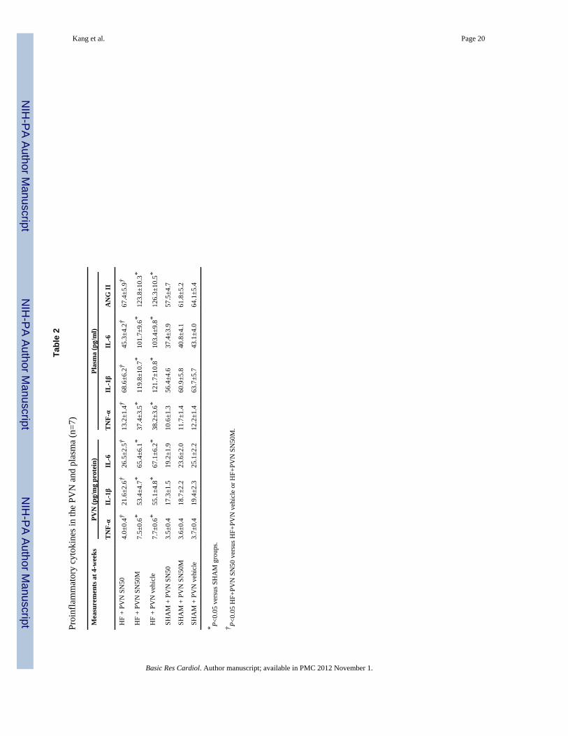
3.7 PVN levels of proinflammatory cytokinesPVN levels of TNF-α, IL-1β and IL-6 were higher in HF rats than in SHAM rats. PVNlevels of TNF-α, IL-1β and IL-6 were lower in HF rats that received bilateral PVN infusionof SN50 when compared to SN50M- or vehicle-treated HF rats (Table 2). Bilateral PVNinfusion of SN50 prevented the increases in PVN PIC seen in HF rats.
3.8 Plasma levels of proinflammatory cytokines, epinephrine and ANGIIHumoral indicators of HF paralleled the PVN findings. Plasma levels of epinephrine,ANGII, TNF-α, IL-1β and IL-6 were all higher in HF rats than in SHAM rats. Bilateral PVNinfusion of SN50 prevented the increases in plasma levels of epinephrine, ANGII, TNF-α,IL-1β and IL-6 in HF rats (Table 2 and Figure 7).
3.9 Renal sympathetic nerve activity (RSNA)At the conclusion of the study, conscious RSNA was measured 5h after rats recovered fromanesthesia, HF rats exhibited higher RSNA (% of max) when compared to SHAM rats;bilateral PVN infusion of SN50 inhibited RSNA in HF rats (Figure 6). Plasma NE, a markerof sympathetic activity, was also higher in HF rats than in SHAM rats. Bilateral PVNinfusion of SN50 prevented the increase in plasma NE of HF rats (Figure 7).
3.10 Functional/anatomical indicators of heart failureCompared with SHAM rats, HF rats had significantly higher LVEDP, RV/BW and lung/BWratio. SN50-treated HF rats had significantly lower LVEDP and lung/BW ratios thanSN50M- or vehicle-treated HF rats (Table 3).
Kang et al. Page 6
Basic Res Cardiol. Author manuscript; available in PMC 2012 November 1.
NIH
-PA Author Manuscript
NIH
-PA Author Manuscript
NIH
-PA Author Manuscript

4. DiscussionThe novel finding of the present study is that, brain cytokines induce an imbalance betweenexcitatory and inhibitory neurotransmitters in the PVN of HF rats, which contributes tosympathoexcitation. PVN treatment with the NF-κB inhibitor SN50 attenuated thisimbalance and decreased the exaggerated sympathetic activity in HF rats. A similar dose ofSN50 given peripherally did not restore the imbalance in the neurotransmitters in the PVNof HF animals, suggesting that NF-κB is activated within the PVN, and modulatesneurotransmitters thereby contributing to sympathoexcitation in rats with ischemia-inducedHF.
Recent evidence suggests that immune-mediated mechanisms play an important role in thepathogenesis of HF. Plasma PIC are increased in HF patients and their levels increase withthe severity of HF [2, 35]. Proinflammatory cytokines, including TNF-α, IL-1β and IL-6 [5,6, 29, 30], are released from the injured site into the circulation post-MI [27, 43]. Recentfindings show that cardiac spinal afferent nerves may serve as a potential source ofproduction of brain cytokines [14]. Regardless of the source, an overload of PIC in the braincan induce sympathoexcitation, impact cardiac function, and contribute towards thepathophysiology of cardiovascular diseases [24]. Although increased PIC were found in thebrains of HF rats and were considered as contributors to exaggerated sympathetic activity[11, 14], the mechanism by which PIC exert this effect is still unclear.
The hypothalamic PVN plays an important role in the regulation of sympathetic activation[26]. Neurotransmitters (both excitatory and inhibitory) have been demonstrated tocontribute to sympathetic activity, including NE, GABA and glutamate [39]. The PVNreceives dense catecholaminergic innervations from the caudal medulla, a region whichserves mainly as a relay site for sensory information. GABA is the principal inhibitoryneurotransmitter in the PVN with inputs originating mainly from local hypothalamicsources. These inputs are thought to impart limbic and cortical influences on PVNmechanisms. Furthermore, GABA has been shown to evoke a sympatho-inhibitory responsewithin the PVN. Microinjection of bicuculline, a GABAA receptor antagonist, into the PVNproduced an increase in renal sympathetic nerve activity, indicating that normally there is astrong tonic GABA-mediated inhibition of sympathetic neuronal firing [28]. Conversely,glutamate is the dominant excitatory neurotransmitter involved in neuroendocrineregulation, where injection of glutamate into the PVN causes increases in sympatheticactivity [28]. Likewise, the actions of NE mimic those of glutamate, primarily inciting anincrease in sympathetic outflow. A number of afferent mediators of PVN responses,including cytokines and neuropeptides, have been identified in HF. However, little attentionhas been directed towards identifying the neurotransmitter systems involved in sympatheticregulation in HF.
Increased PVN activation in HF results from the interplay of several neurotransmitters andneuromodulators in these sympathetic neurons. In addition, we also recently demonstratedthat PIC blockade within the CNS attenuated sympathetic activity in HF rats [8, 25].Presently, we found that RSNA is increased in rats with HF, along with increases in thePVN levels of the excitatory neurotransmitters glutamate and NE, as well as a decrease inthe levels of the inhibitory neurotransmitter GABA. The elevated basal level of NEconcentration in the PVN of HF rats may reflect a marked stimulation of ascendingnoradrenergic drive to the PVN, or alternatively, it may be a result of impaired NE re-uptake.
NF-κB is now considered a major transcription factor regulating the expression of PIC insignal transduction pathways. Functional NF-κB complexes are present in essentially all cell
Kang et al. Page 7
Basic Res Cardiol. Author manuscript; available in PMC 2012 November 1.
NIH
-PA Author Manuscript
NIH
-PA Author Manuscript
NIH
-PA Author Manuscript

types in the nervous system. In neurons, activated NF-κB is the major regulator facilitatingthe synthesis of several different injury-responsive cytokines including TNF-α and IL-6 [7].In this study, PVN treatment with SN50 (a selective inhibitor of NF-κB) reduced PICexpression and NF-κB p65 activity in the PVN of HF rats, indicating that NF-κB is involvedin regulating the production of PIC. The present study also demonstrates that PVN treatmentwith SN50 decreased plasma PIC (TNF-α, IL-1β, and IL-6), ANG II, and NE (a marker ofsympathetic activity) in HF. Since we did not see any effect after a similar dose of SN50given peripherally, we consider the reduction of plasma PIC and ANG II a direct result ofdecreases in both volume and pressure overload induced by sympathetic activity in thecardiovascular system; we further conclude that the reduction of plasma PIC is likely to bean indirect consequence of brain NF-κB inhibition. Thus, the present findings possiblyindicate that increased inflammatory molecule induced NF-κB activation within the PVNalters the delicate balance between excitatory and inhibitory neurotransmitters within thePVN, thereby contributing to the exaggerated sympathetic activity in HF rats.
In summary, the present study demonstrates that treatment of HF rats with PVN SN50attenuates the HF-induced increases in PIC, glutamate, and NE, and also attenuates the HF-induced decreases in GABA, in the PVN. Elevated excitatory neurotransmitters anddecreased inhibitory neurotransmitters in the PVN contribute to neurohumoral excitation inHF. SN50 could decrease the sympathoexcitation in HF by decreasing excitatoryneurotransmitters and increasing inhibitory neurotransmitters in the PVN.
AcknowledgmentsFunding Supported by National Natural Science Foundation of China (Nos. 81070199, 81025001), U.S. NationalInstitutes of Health (NIH) Grant RO1-HL-080544, and National Basic Research Program of China (Nos.2012CB517805, 2007CB512106).
References1. Agarwal D, Haque M, Sriramula S, Mariappan N, Pariaut R, Francis J. Role of proinflammatory
cytokines and redox homeostasis in exercise-induced delayed progression of hypertension inspontaneously hypertensive rats. Hypertension. 2009; 54:1393–1400.10.1161/HYPERTENSIONAHA.109.135459 [PubMed: 19841289]
2. Aker S, Belosjorow S, Konietzka I, Duschin A, Martin C, Heusch G, Schulz R. Serum but notmyocardial TNF-alpha concentration is increased in pacing-induced heart failure in rabbits. Am JPhysiol Regul Integr Comp Physiol. 2003; 285:463–469.10.1152/ajpregu.00153.2003
3. Barber M, Kasturi BS, Austin ME, Patel KP, MohanKumar SM, MohanKumar PS. Diabetes-induced neuroendocrine changes in rats: role of brain monoamines, insulin and leptin. Brain Res.2003; 964:128–135.10.1016/S0006-8993 (02)04091-X [PubMed: 12573521]
4. Bhakar AL, Tannis LL, Zeindler C, Russo MP, Jobin C, Park DS, MacPherson S, Barker PA.Constitutive nuclear factor-kappa B activity is required for central neuron survival. J Neurosci.2002; 22:8466–8475. doi:0270-6474/02/228466-10. [PubMed: 12351721]
5. Chappell D, Hofmann-Kiefer K, Jacob M, Rehm M, Briegel J, Welsch U, Conzen P, Becker BF.TNF-alpha induced shedding of the endothelial glycocalyx is prevented by hydrocortisone andantithrombin. Basic Res Cardiol. 2009; 104:79–89.10.1007/s00395-008-0749-5
6. Chorianopoulos E, Heger T, Lutz M, Frank D, Bea F, Katus HA, Frey N. FGF-inducible 14-kDaprotein (Fn14) is regulated via the RhoA/ROCK kinase pathway in cardiomyocytes and mediatesnuclear factor-kappaB activation by TWEAK. Basic Res Cardiol. 2010; 105:301–313.10.1007/s00395-009-0046-y [PubMed: 19629561]
7. Cowling RT, Gurantz D, Peng J, Dillmann WH, Greenberg BH. Transcription factor NF-kappa B isnecessary for up-regulation of type 1 angiotensin II receptor mRNA in rat cardiac fibroblasts treatedwith tumor necrosis factor-alpha or interleukin-1 beta. J Biol Chem. 2002; 277:5719–5724.10.1074/jbc.M107515200 [PubMed: 11600498]
Kang et al. Page 8
Basic Res Cardiol. Author manuscript; available in PMC 2012 November 1.
NIH
-PA Author Manuscript
NIH
-PA Author Manuscript
NIH
-PA Author Manuscript

8. Dunn AJ. Cytokine activation of the HPA axis. Ann N Y Acad Sci. 2000; 917:608–617.10.1111/j.1749-6632.2000.tb05426.x [PubMed: 11268389]
9. Elks CM, Mariappan N, Haque M, Guggilam A, Majid DS, Francis J. Chronic NF-κB blockadereduces cytosolic and mitochondrial oxidative stress and attenuates renal injury and hypertension inSHR. Am J Physiol Renal Physiol. 2009; 296:298–305.10.1152/ajprenal.90628.2008
10. Ferrari R. Tumor necrosis factor in CHF: a double facet cytokine. Cardiovasc Res. 1998; 37:554–559.10.1016/S0008-6363(97)00309-X [PubMed: 9659437]
11. Francis J, Chu Y, Johnson AK, Weiss RM, Felder RB. Acute myocardial infarction induceshypothalamic cytokine synthesis. Am J Physiol Heart Circ Physiol. 2004; 286:H2264–H2271.10.1152/ajpheart.01072.2003 [PubMed: 15148057]
12. Francis J, MohanKumar SM, MohanKumar PS. Correlations of norepinephrine release in theparaventricular nucleus with plasma corticosterone and leptin after systemic lipopolysaccharide:blockade by soluble IL-1 receptor. Brain Res. 2000; 867:180–187.10.1016/S0006-8993(00)02311-8 [PubMed: 10837812]
13. Francis J, Weiss RM, Johnson AK, Felder RB. Central mineralocorticoid receptor blockadedecreases plasma TNF-alpha after coronary artery ligation in rats. Am J Physiol Regul IntegrComp Physiol. 2003; 284:R328–R335.10.1152/ajpregu.00376.2002 [PubMed: 12529282]
14. Francis J, Zhang ZH, Weiss RM, Felder RB. Neural regulation of the proinflammatory cytokineresponse to acute myocardial infarction. Am J Physiol Heart Circ Physiol. 2004; 287:H791–R797.10.1152/ajpheart.00099.2004 [PubMed: 15277202]
15. Gao L, Wang W, Li YL, Schultz HD, Liu D, Cornish KG, Zucker IH. Sympathoexcitation bycentral ANG II: roles for AT1 receptor upregulation and NAD(P)H oxidase in RVLM. Am JPhysiol Heart Circ Physiol. 2005; 288:H2271–H2279.10.1152/ajpheart.00949.2004 [PubMed:15637113]
16. Gloire G, Legrand-Poels S, Piette J. NF-kappaB activation by reactive oxygen species: fifteenyears later. Biochem Pharmacol. 2006; 72:1493–1505.10.1016/j.bcp.2006.04.011 [PubMed:16723122]
17. Guggilam A, Cardinale JP, Mariappan N, Sriramula S, Haque M, Francis J. Central TNF inhibitionresults in attenuated neurohumoral excitation in heart failure: a role for superoxide and nitricoxide. Basic Res Cardiol. 2011; 106:273–286.10.1007/s00395-010-0146-8 [PubMed: 21246206]
18. Guggilam A, Haque M, Kerut EK, McIlwain E, Lucchesi P, Seghal I, Francis J. TNF-alphablockade decreases oxidative stress in the paraventricular nucleus and attenuatessympathoexcitation in heart failure rats. Am J Physiol Heart Circ Physiol. 2007; 293:H599–H609.10.1152/ajpheart.00286.2007 [PubMed: 17416605]
19. Guggilam A, Patel KP, Haque M, Ebenezer PJ, Kapusta DR, Francis J. Cytokine blockadeattenuates sympathoexcitation in heart failure: cross-talk between nNOS, AT-1R and cytokines inthe hypothalamic paraventricular nucleus. Eur J Heart Fai. 2008; 10:625–634.10.1016/j.ejheart.2008.05.004
20. Kang YM, Zhang AQ, Zhao XF, Cardinale J, Elks C, Cao XM, Zhang ZW, Francis J.Paraventricular nucleus corticotrophin releasing hormone contributes to sympathoexcitation viainteraction with neurotransmitters in heart failure. Basic Res Cardiol. 2011; 106(3):473–483.10.1007/s00395-011-0155-2 [PubMed: 21287352]
21. Kang YM, He RL, Yang LM, Qin DN, Guggilam A, Elks C, Yan N, Guo Z, Francis J. Braintumour necrosis factor-alpha modulates neurotransmitters in hypothalamic paraventricular nucleusin heart failure. Cardiovasc Res. 2009; 83:737–746.10.1093/cvr/cvp160 [PubMed: 19457890]
22. Kang YM, Ma Y, Elks C, Zheng JP, Yang ZM, Francis J. Cross-talk between cytokines and renin-angiotensin in hypothalamic paraventricular nucleus in heart failure: role of nuclear factor-kappaB.Cardiovasc Res. 2008; 79:671–678.10.1093/cvr/cvn119 [PubMed: 18469338]
23. Kang YM, Ma Y, Zheng JP, Elks C, Sriramula S, Yang ZM, Francis J. Brain nuclear factor-kappaB activation contributes to neurohumoral excitation in angiotensin II-induced hypertension.Cardiovasc Res. 2009; 82:503–512.10.1093/cvr/cvp073 [PubMed: 19246475]
24. Kang YM, Zhang ZH, Johnson RF, Yu Y, Beltz T, Johnson AK, Weiss RM, Felder RB. Noveleffect of mineralocorticoid receptor antagonism to reduce proinflammatory cytokines and
Kang et al. Page 9
Basic Res Cardiol. Author manuscript; available in PMC 2012 November 1.
NIH
-PA Author Manuscript
NIH
-PA Author Manuscript
NIH
-PA Author Manuscript

hypothalamic activation in rats with ischemia-induced heart failure. Circ Res. 2006; 99:758–766.10.1161/01.RES.0000244092.95152.86 [PubMed: 16960100]
25. Kang YM, Zhang ZH, Xue B, Weiss RM, Felder RB. Inhibition of brain proinflammatory cytokinesynthesis reduces hypothalamic excitation in rats with ischemia-induced heart failure. Am JPhysiol Heart Circ Physiol. 2008; 295:H227–H236.10.1152/ajpheart.01157.2007 [PubMed:18487441]
26. Kenney MJ, Weiss ML, Haywood JR. The paraventricular nucleus: an important component of thecentral neurocircuitry regulating sympathetic nerveoutflow. Acta Physiol Scand. 2003; 177(1):7–15.10.1046/j.1365-201X.2003.01042.x [PubMed: 12492774]
27. Kleinbongard P, Heusch G, Schulz R. TNFalpha in atherosclerosis, myocardial ischemia/reperfusion and heart failure. Pharmacol Ther. 2010; 127:295–314.10.1016/j.pharmthera.2010.05.002 [PubMed: 20621692]
28. Li YF, Jackson KL, Stern JE, Rabeler B, Patel KP. Interaction between glutamate and GABAsystems in the integration of sympathetic outflow by the paraventricular nucleus of thehypothalamus. Am J Physiol Heart Circ Physiol. 2006; 291:H2847–H2856.10.1152/ajpheart.00625.2005 [PubMed: 16877560]
29. Lacerda L, McCarthy J, Mungly SF, Lynn EG, Sack MN, Opie LH, Lecour S. TNFα protectscardiac mitochondria independently of its cell surface receptors. Basic Res Cardiol. 2010;105:751–762.10.1007/s00395-010-0113-4 [PubMed: 20680307]
30. Li S, Zhong S, Zeng K, Luo Y, Zhang F, Sun X, Chen L. Blockade of NF-kappaB by pyrrolidinedithiocarbamate attenuates myocardial inflammatory response and ventricular dysfunctionfollowing coronary microembolization induced by homologous microthrombi in rats. Basic ResCardiol. 2010; 105:139–150.10.1007/s00395-009-0067-6 [PubMed: 19823892]
31. Lupia E, Spatola T, Cuccurullo A, Bosco O, Mariano F, Pucci A, Ramella R, Alloatti G,Montrucchio G. Thrombopoietin modulates cardiac contractility in vitro and contributes tomyocardial depressing activity of septic shock serum. Basic Res Cardiol. 2010; 105:609–620.10.1007/s00395-010-0103-6 [PubMed: 20467749]
32. Liu JL, Irvine S, Reid IA, Patel KP, Zucker IH. Chronic exercise reduces sympathetic nerveactivity in rabbits with pacing-induced heart failure: A role for angiotensin II. Circulation. 2000;102:1854–1862.10.1161/01.CIR.102.15.1854 [PubMed: 11023943]
33. MohanKumar SM, MohanKumar PS, Quadri SK. Specificity of interleukin-1beta-induced changesin monoamine concentrations in hypothalamic nuclei: blockade by interleukin-1 receptorantagonist. Brain Res Bull. 1998; 47:29–34.10.1016/S0361-9230(98)00037-9 [PubMed: 9766386]
34. Nagura S, Sakagami T, Kakiichi A, Yoshimoto M, Miki K. Acute shifts in baroreflex control ofrenal sympathetic nerve activity induced by REM sleep and grooming in rats. J Physiol. 2004;558:975–983.10.1113/jphysiol.2004.064527 [PubMed: 15194739]
35. Nozaki N, Yamaguchi S, Shirakabe M, Nakamura H, Tomoike H. Soluble tumor necrosis factorreceptors are elevated in relation to severity of congestive heart failure. Jpn Circ J. 1997; 61:657–664.10.1253/jcj.61.657 [PubMed: 9276770]
36. Paxinos, G.; Watson, CR.; Emson, PC. The rat brain in stereotaxic coordinates. 2. San Diego, CA:Academic; 1987.
37. Schulz R, Heusch G. Tumor necrosis factor-alpha and its receptors 1 and 2: Yin and Yang inmyocardial infarction? Circulation. 2009; 119:1355–1357.10.1161/CIRCULATIONAHA.108.846105 [PubMed: 19255338]
38. Skyschally A, Gres P, Hoffmann S, Haude M, Erbel R, Schulz R, Heusch G. Bidirectional role oftumor necrosis factor-alpha in coronary microembolization: progressive contractile dysfunctionversus delayed protection against infarction. Circulation Research. 2007; 100:140–146.10.1161/01.RES.0000255031.15793.86 [PubMed: 17170366]
39. Swanson LW, Sawchenko PE. Paraventricular nucleus: a site for the integration of neuroendocrineand autonomic mechanisms. Neuroendocrinology. 1980; 31:410–417. [PubMed: 6109264]
40. Thielmann M, Dörge H, Martin C, Belosjorow S, Schwanke U, van De Sand A, Konietzka I,Büchert A, Krüger A, Schulz R, Heusch G. Myocardial dysfunction with coronarymicroembolization: signal transduction through a sequence of nitric oxide, tumor necrosis factor-
Kang et al. Page 10
Basic Res Cardiol. Author manuscript; available in PMC 2012 November 1.
NIH
-PA Author Manuscript
NIH
-PA Author Manuscript
NIH
-PA Author Manuscript

alpha, and sphingosine. Circulation Research. 2002; 90:807–813.10.1161/01.RES.0000014451.75415.36 [PubMed: 11964374]
41. Valen G, Yan ZQ, Hansson GK. Nuclear factor kappa-B and the heart. J Am Coll Cardiol. 2001;38:307–314.10.1016/s0735-1097(01)01377-8 [PubMed: 11499717]
42. Yang LM, Hu B, Xia YH, Zhang BL, Zhao H. Lateral habenula lesions improve the behavioralresponse in depressed rats via increasing the serotonin level in dorsal raphe nucleus. Behav BrainRes. 2008; 188:84–90.10.1016/j.bbr.2007.10.022 [PubMed: 18054396]
43. Zhang C, Wu J, Xu X, Potter BJ, Gao X. Direct relationship between levels of TNF-alphaexpression and endothelial dysfunction in reperfusion injury. Basic Res Cardiol. 2010; 105:453–464.10.1007/s00395-010-0083-6 [PubMed: 20091314]
44. Zhang ZH, Wei SG, Francis J, Felder RB. Cardiovascular and renal sympathetic activation byblood-borne TNF-alpha in rat: the role of central prostaglandins. Am J Physiol Regul Integr CompPhysiol. 2003; 284:R916–R927.10.1152/ajpregu.00406.2002 [PubMed: 12626358]
Kang et al. Page 11
Basic Res Cardiol. Author manuscript; available in PMC 2012 November 1.
NIH
-PA Author Manuscript
NIH
-PA Author Manuscript
NIH
-PA Author Manuscript

Figure 1.PVN levels of norepinephrine (NE), glutamate (GLU) and GABA in heart failure (HF) andsham operated (SHAM) rats treated for 4 weeks with SN50, SN50M or vehicle. HF rats hadhigher levels of NE (A) and glutamate (B), and lower levels of GABA (C) in the PVN. Four-week bilateral infusions of SN50 into the PVN prevented the decrease in PVN GABA andthe increases in PVN glutamate and NE in HF rats. PVN infusions of SN50M for 4 weeksdid not alter NE, glutamate, or GABA in the PVN of HF rats. IP treatment with the samedose of SN50 did not alter NE, glutamate, or GABA in the PVN of HF rats. *P<0.05 versusSHAM groups. †P<0.05 HF+PVN SN50 versus HF+PVN vehicle or HF+PVN SN50M.
Kang et al. Page 12
Basic Res Cardiol. Author manuscript; available in PMC 2012 November 1.
NIH
-PA Author Manuscript
NIH
-PA Author Manuscript
NIH
-PA Author Manuscript

Figure 2.Western blot of TH and GAD67 in the PVN showed that HF rats had higher levels of THand lower levels of GAD67 when compared with SHAM rats. Bilateral PVN infusions ofSN50 for 4 weeks decreased expression of TH, and increased GAD67 expression in thePVN of HF rats. *P<0.05 versus SHAM groups. †P<0.05 HF+PVN SN50 versus HF+PVNvehicle.
Kang et al. Page 13
Basic Res Cardiol. Author manuscript; available in PMC 2012 November 1.
NIH
-PA Author Manuscript
NIH
-PA Author Manuscript
NIH
-PA Author Manuscript

Figure 3.Fra-LI activity immunoreactivity in the PVN. HF rats had higher PVN levels of Fra-LI(black dots) immunoreactivity when compared with SHAM rats. Bilateral PVN infusions ofSN50 for 4 weeks decreased Fra-LI expression in the PVN of HF rats. PVN infusions ofSN50M for 4 weeks did not alter Fra-LI expression in the PVN of HF rats. *P<0.05 versusSHAM groups. †P<0.05 HF+PVN SN50 versus HF+PVN vehicle or HF+PVN SN50M.
Kang et al. Page 14
Basic Res Cardiol. Author manuscript; available in PMC 2012 November 1.
NIH
-PA Author Manuscript
NIH
-PA Author Manuscript
NIH
-PA Author Manuscript

Figure 4.NF-κB activation in the PVN. HF rats had higher level of NF-κB p65 activity and p-IKKβ inthe PVN. Four-week bilateral infusions of SN50 into the PVN prevented the increases inNF-κB p65 activity and p-IKKβ in the PVN of HF rats. (A) immunofluorescence for p-IKKβ (red) and neuronal nuclei (DAPI, blue) in the PVN. (B) group data showing thateffects of PVN infusions of SN50 on numbers of p-IKKβ positive neurons in the PVN of HFand SHAM rats. (C) group data showing that effects of PVN infusions of SN50 on NF-κBp65 activity in the PVN of HF and SHAM rats. PVN infusions of SN50M for 4 weeks didnot alter NF-κB p65 activity in the PVN of HF rats (C). *P<0.05 versus SHAM groups.†P<0.05 HF+PVN SN50 versus HF+PVN vehicle or HF+PVN SN50M.
Kang et al. Page 15
Basic Res Cardiol. Author manuscript; available in PMC 2012 November 1.
NIH
-PA Author Manuscript
NIH
-PA Author Manuscript
NIH
-PA Author Manuscript

Figure 5.Superoxide and NAD(P)H oxidase in the PVN. A: immunofluorescence for the NAD(P)Hoxidase subunit gp91phox and superoxide as determined by fluorescent-labeleddihydroethidium (DHE) in the PVN in different groups. B: comparison of gp91phox positiveneurons in the PVN in different groups. C: immunofluorescent intensity of DHE in the PVNof different groups of rats. *P<0.05 versus SHAM groups. †P<0.05 HF+PVN SN50 versusHF+PVN vehicle.
Kang et al. Page 16
Basic Res Cardiol. Author manuscript; available in PMC 2012 November 1.
NIH
-PA Author Manuscript
NIH
-PA Author Manuscript
NIH
-PA Author Manuscript

Figure 6.Renal sympathetic nerve activity (RSNA) was increased in HF rats compared with SHAMrats. Treatment with bilateral PVN infusions of SN50 for 4 weeks decreased RSNA of HFrats. PVN infusions of SN50M for 4 weeks did not alter RSNA of HF rats. *P<0.05 versusSHAM groups. †P<0.05 HF+PVN SN50 versus HF+PVN vehicle or HF+PVN SN50M.
Kang et al. Page 17
Basic Res Cardiol. Author manuscript; available in PMC 2012 November 1.
NIH
-PA Author Manuscript
NIH
-PA Author Manuscript
NIH
-PA Author Manuscript

Figure 7.Plasma NE and epinephrine (EPI) were higher in HF rats than in SHAM rats. Bilateral PVNinfusions of SN50 for 4 weeks prevented the increases in plasma NE and EPI of HF rats.PVN infusions of SN50M for 4 weeks did not alter plasma levels of NE and EPI in HF rats.*P<0.05 versus SHAM groups. †P<0.05 HF+PVN SN50 versus HF+PVN vehicle or HF+PVN SN50M.
Kang et al. Page 18
Basic Res Cardiol. Author manuscript; available in PMC 2012 November 1.
NIH
-PA Author Manuscript
NIH
-PA Author Manuscript
NIH
-PA Author Manuscript

NIH
-PA Author Manuscript
NIH
-PA Author Manuscript
NIH
-PA Author Manuscript
Kang et al. Page 19
Tabl
e 1
Echo
card
iogr
aphi
c m
easu
rem
ents
(n=1
4)
Mea
sure
men
ts at
24
Hou
rsH
F +
PVN
SN
50H
F +
PVN
SN
50M
HF
+ PV
N v
ehic
leSH
AM
+ P
VN
SN
50SH
AM
+ P
VN
SN
50M
SHA
M +
PV
N v
ehic
le
LVED
V (m
l)0.
74 ±
0.0
9*0.
73 ±
0.0
8*0.
70 ±
0.0
8*0.
34 ±
0.0
40.
36 ±
0.0
50.
37 ±
0.0
5
LVED
V/M
ass
1.07
± 0
.07*
1.08
± 0
.08*
1.10
± 0
.09*
0.55
± 0
.05
0.56
± 0
.05
0.57
± 0
.06
LVEF
0.36
± 0
.04*
0.37
± 0
.05*
0.39
± 0
.05*
0.83
± 0
.05
0.84
± 0
.06
0.85
± 0
.06
IZ (%
)49
±348
±347
±2--
----
----
----
----
Mea
sure
men
ts at
4 W
eeks
HF
+ PV
N S
N50
HF
+ PV
N S
N50
MH
F +
PVN
veh
icle
SHA
M +
PV
N S
N50
SHA
M +
PV
N S
N50
MSH
AM
+ P
VN
veh
icle
LVED
V (m
l)1.
34 ±
0.0
8*‡
1.42
± 0
.07*
‡1.
44 ±
0.0
7*‡
0.35
± 0
.04
0.37
± 0
.04
0.38
± 0
.04
LVED
V/M
ass
1.64
± 0
.11*
‡1.
75 ±
0.1
3*‡
1.78
± 0
.14*
‡0.
55 ±
0.0
50.
57 ±
0.0
50.
59 ±
0.0
5
LVEF
0.34
± 0
.03*
†0.
23 ±
0.0
3*‡
0.24
± 0
.03*
‡0.
85 ±
0.0
60.
83 ±
0.0
60.
82 ±
0.0
5
IZ (%
)47
± 3
50 ±
451
± 4
----
----
----
----
--
SHA
M: s
ham
-ope
rate
d co
ntro
l; H
F: h
eart
failu
re. L
VED
V: l
eft v
entri
cula
r end
-dia
stol
ic v
olum
e; L
VEF
: lef
t ven
tricu
lar e
ject
ion
frac
tion;
IZ%
: per
cent
isch
emic
zon
e. V
alue
s are
mea
n ±
SEM
.
* P<0.
05 v
ersu
s SH
AM
gro
ups.
† P<0.
05 H
F+PV
N S
N50
ver
sus H
F+PV
N v
ehic
le o
r HF+
PVN
SN
50M
.
‡ P<0.
05, 4
-wee
ks v
ersu
s 24-
hour
s val
ue.
Basic Res Cardiol. Author manuscript; available in PMC 2012 November 1.
NIH
-PA Author Manuscript
NIH
-PA Author Manuscript
NIH
-PA Author Manuscript
Kang et al. Page 20
Tabl
e 2
Proi
nfla
mm
ator
y cy
toki
nes i
n th
e PV
N a
nd p
lasm
a (n
=7)
Mea
sure
men
ts a
t 4-w
eeks
PVN
(pg/
mg
prot
ein)
Plas
ma
(pg/
ml)
TN
F-α
IL-1β
IL-6
TN
F-α
IL-1β
IL-6
AN
G II
HF
+ PV
N S
N50
4.0±
0.4†
21.6
±2.6
†26
.5±2
.5†
13.2
±1.4
†68
.6±6
.2†
45.3
±4.2
†67
.4±5
.9†
HF
+ PV
N S
N50
M7.
5±0.
6*53
.4±4
.7*
65.4
±6.1
*37
.4±3
.5*
119.
8±10
.7*
101.
7±9.
6*12
3.8±
10.3
*
HF
+ PV
N v
ehic
le7.
7±0.
6*55
.1±4
.8*
67.1
±6.2
*38
.2±3
.6*
121.
7±10
.8*
103.
4±9.
8*12
6.3±
10.5
*
SHA
M +
PV
N S
N50
3.5±
0.4
17.3
±1.5
19.2
±1.9
10.6
±1.3
56.4
±4.6
37.4
±3.9
57.5
±4.7
SHA
M +
PV
N S
N50
M3.
6±0.
418
.7±2
.223
.6±2
.011
.7±1
.460
.9±5
.840
.8±4
.161
.8±5
.2
SHA
M +
PV
N v
ehic
le3.
7±0.
419
.4±2
.325
.1±2
.212
.2±1
.463
.7±5
.743
.1±4
.064
.1±5
.4
* P<0.
05 v
ersu
s SH
AM
gro
ups.
† P<0.
05 H
F+PV
N S
N50
ver
sus H
F+PV
N v
ehic
le o
r HF+
PVN
SN
50M
.
Basic Res Cardiol. Author manuscript; available in PMC 2012 November 1.

NIH
-PA Author Manuscript
NIH
-PA Author Manuscript
NIH
-PA Author Manuscript
Kang et al. Page 21
Tabl
e 3
Hem
odyn
amic
and
ana
tom
ical
mea
sure
men
ts (n
=7)
Mea
sure
men
ts a
t 4-w
eeks
RV
/BW
(mg/
g)L
ung/
BW
(mg/
g)H
R (b
eats
/min
)M
AP
(mm
Hg)
PP (m
mH
g)L
VE
DP
(mm
Hg)
HF
+ PV
N S
N50
0.78
±0.0
8†5.
6±0.
4†33
7±12
98±7
31±4
17.7
2±1.
31†*
HF
+ PV
N S
N50
M1.
17±0
.10*
10.8
±0.6
*34
0±13
97±7
34±5
23.8
1±1.
80*
HF
+ PV
N v
ehic
le1.
20±0
.11*
11.2
±0.6
*34
2±13
95±6
33±5
23.7
4±1.
82*
SHA
M +
PV
N S
N50
0.61
±0.0
54.
7±0.
433
2±11
105±
835
±46.
14±1
.59
SHA
M +
PV
N S
N50
M0.
66±0
.05
5.0±
0.4
330±
1310
8±9
34±5
6.53
±1.4
2
SHA
M +
PV
N v
ehic
le0.
69±0
.06
5.3±
0.5
335±
1210
7±9
36±5
7.05
±1.5
6
SHA
M: s
ham
-ope
rate
d co
ntro
l; H
F: h
eart
failu
re; B
W: b
ody
wei
ght;
RV
: rig
ht v
entri
cula
r; H
R: h
eart
rate
; MA
P: m
ean
arte
rial p
ress
ure;
PP:
pul
se p
ress
ure;
LV
EDP:
left
vent
ricul
ar e
nd-d
iast
olic
pre
ssur
e.V
alue
s are
mea
n±SE
M.
* P<0.
05 v
ersu
s SH
AM
gro
ups.
† P<0.
05 H
F+PV
N S
N50
ver
sus H
F+PV
N v
ehic
le o
r HF+
PVN
SN
50M
.
Basic Res Cardiol. Author manuscript; available in PMC 2012 November 1.
Copyright © 2022 FDOKUMEN