IKKβ/NF-κB Activation Causes Severe Muscle Wasting in Mice
14
Cell, Vol. 119, 285–298, October 15, 2004, Copyright 2004 by Cell Press IKK/NF-B Activation Causes Severe Muscle Wasting in Mice improved by NF-B inhibition in MISR mice, consistent with a critical role for NF-B in the pathology of muscle wasting and establishing it as an important clinical Dongsheng Cai, 1,6 J. Daniel Frantz, 1,6,10 Nicholas E. Tawa, Jr., 2,7 Peter A. Melendez, 1,6,11 Byung-Chul Oh, 1,6 Hart G.W. Lidov, 3,8 Per-Olof Hasselgren, 2,7 Walter R. Frontera, 4,9 target for the treatment of muscle atrophy. Jongsoon Lee, 1,6 David J. Glass, 5 and Steven E. Shoelson 1,6, * Introduction 1 Research Division Joslin Diabetes Center Muscle wasting is a major feature of the cachexia asso- One Joslin Place ciated with diverse pathologies such as cancer, bacte- 2 Department of Surgery rial sepsis, AIDS, diabetes, and end-stage heart, kidney, Beth Israel Deaconess Medical Center and chronic obstructive pulmonary disease (Tisdale, 330 Brookline Avenue 1997; Argiles and Lopez-Soriano, 1999). During cachex- Boston, Massachusetts 02215 ia, muscle protein breakdown occurs well out of propor- 3 Division of Genetics tion to the catabolic effects of cachexia seen in other Children’s Hospital tissues, causing generalized weakness and debilitation, 300 Longwood Avenue which in respiratory muscles may be sufficiently severe Boston, Massachusetts 02115 to ultimately lead to asphyxia and death. Muscle wasting 4 Department of Physical Medicine seen in such catabolic states appears to be mediated and Rehabilitation predominantly by activation of ubiquitin- and protea- Spaulding Rehabilitation Hospital some-dependent pathways (Lecker et al., 1999), per- 125 Nashua Street haps through increases in the expression of relevant Boston, Massachusetts 02114 proteins, including ubiquitin E2-conjugating enzyme and 5 Muscle Program subunits of the proteasome. Genetic deletion of either Regeneron Pharmaceuticals of two recently discovered muscle-specific E3 ligases, 777 Old Saw Mill River Road atrogin-1/MAFbx or MuRF1 (muscle RING finger protein Tarrytown, New York 10591 1), protects against muscle atrophy following denerva- 6 Department of Medicine tion and disuse (Bodine et al., 2001; Glass, 2003). 7 Department of Surgery Several cytokines have been implicated in the patho- 8 Department of Pediatrics genesis of muscle wasting, most notably TNF-, a proin- 9 Department of Physical Medicine flammatory cytokine that was originally called “cachec- and Rehabilitation tin” (Tisdale, 1997; Argiles and Lopez-Soriano, 1999). Harvard Medical School Concentrations of TNF- are often elevated in the circu- Boston, Massachusetts 02115 lations of patients with sepsis or cancer, contributing to negative nitrogen balance—but, by itself, TNF- may be insufficient to cause muscle wasting (Moldawer et al., 1987; Fong et al., 1989; Mullen et al., 1990). IL-1, Summary IL-6, IFN-, TGF, LIF, and other related cytokines have been implicated as potential mediators of muscle wast- Muscle wasting accompanies aging and pathological ing or atrophy, suggesting that clinical syndromes might conditions ranging from cancer, cachexia, and diabe- involve synergistic effects of combinations of cytokines tes to denervation and immobilization. We show that (Argiles and Lopez-Soriano, 1999). activation of NF-B, through muscle-specific trans- Since NF-B in muscle is activated by disuse (Hunter genic expression of activated IB kinase (MIKK), et al., 2002) or sepsis (Penner et al., 2001), it might play causes profound muscle wasting that resembles clini- a role in the pathogenesis of these conditions, although cal cachexia. In contrast, no overt phenotype was seen alternative intracellular pathways including caspases upon muscle-specific inhibition of NF-B through ex- and JNK/AP-1 have also been shown to be activated in pression of IB superrepressor (MISR). Muscle loss muscle by cytokines (Coletti et al., 2002; Stewart et al., was due to accelerated protein breakdown through 2004). Consistent with a role for NF-B, in vitro blockade ubiquitin-dependent proteolysis. Expression of the E3 inhibits protein loss in C2C12 myotubes (Li and Reid, ligase MuRF1, a mediator of muscle atrophy, was in- 2000). Although detailed mechanisms of NF-B have creased in MIKK mice. Pharmacological or genetic been established in innate immunity, inflammation, and inhibition of the IKK/NF-B/MuRF1 pathway reversed apoptosis, its in vivo functions in muscle are unknown. muscle atrophy. Denervation- and tumor-induced mus- In cells of the immune and inflammatory systems, NF-B cle loss were substantially reduced and survival rates is a central integration site for proinflammatory signals and a master regulator of related target genes. Numer- *Correspondence: [email protected] ous inputs that activate NF-B in addition to TNF- and 10 Present address: Vertex Pharmaceuticals, 130 Waverly Street, IL-1 include bacterial cell wall and viral products, Cambridge, Massachusetts 02139. dsDNA, mitogens, and reactive oxygen species (ROS) 11 Present address: Hewlett Packard Caribe Ltd., P.O. Box 4048, Aguadilla, Puerto Rico 00603. (Baeuerle and Baltimore, 1996; Rothwarf and Karin,
-
Upload
hms-harvard -
Category
Documents
-
view
1 -
download
0
Transcript of IKKβ/NF-κB Activation Causes Severe Muscle Wasting in Mice

Cell, Vol. 119, 285–298, October 15, 2004, Copyright 2004 by Cell Press
IKK�/NF-�B Activation CausesSevere Muscle Wasting in Mice
improved by NF-�B inhibition in MISR mice, consistentwith a critical role for NF-�B in the pathology of musclewasting and establishing it as an important clinical
Dongsheng Cai,1,6 J. Daniel Frantz,1,6,10
Nicholas E. Tawa, Jr.,2,7 Peter A. Melendez,1,6,11
Byung-Chul Oh,1,6 Hart G.W. Lidov,3,8
Per-Olof Hasselgren,2,7 Walter R. Frontera,4,9 target for the treatment of muscle atrophy.Jongsoon Lee,1,6 David J. Glass,5
and Steven E. Shoelson1,6,* Introduction1Research DivisionJoslin Diabetes Center Muscle wasting is a major feature of the cachexia asso-One Joslin Place ciated with diverse pathologies such as cancer, bacte-2 Department of Surgery rial sepsis, AIDS, diabetes, and end-stage heart, kidney,Beth Israel Deaconess Medical Center and chronic obstructive pulmonary disease (Tisdale,330 Brookline Avenue 1997; Argiles and Lopez-Soriano, 1999). During cachex-Boston, Massachusetts 02215 ia, muscle protein breakdown occurs well out of propor-3 Division of Genetics tion to the catabolic effects of cachexia seen in otherChildren’s Hospital tissues, causing generalized weakness and debilitation,300 Longwood Avenue which in respiratory muscles may be sufficiently severeBoston, Massachusetts 02115 to ultimately lead to asphyxia and death. Muscle wasting4 Department of Physical Medicine seen in such catabolic states appears to be mediated
and Rehabilitation predominantly by activation of ubiquitin- and protea-Spaulding Rehabilitation Hospital some-dependent pathways (Lecker et al., 1999), per-125 Nashua Street haps through increases in the expression of relevantBoston, Massachusetts 02114 proteins, including ubiquitin E2-conjugating enzyme and5 Muscle Program subunits of the proteasome. Genetic deletion of eitherRegeneron Pharmaceuticals of two recently discovered muscle-specific E3 ligases,777 Old Saw Mill River Road atrogin-1/MAFbx or MuRF1 (muscle RING finger proteinTarrytown, New York 10591 1), protects against muscle atrophy following denerva-6 Department of Medicine tion and disuse (Bodine et al., 2001; Glass, 2003).7 Department of Surgery Several cytokines have been implicated in the patho-8 Department of Pediatrics genesis of muscle wasting, most notably TNF-�, a proin-9 Department of Physical Medicine flammatory cytokine that was originally called “cachec-
and Rehabilitation tin” (Tisdale, 1997; Argiles and Lopez-Soriano, 1999).Harvard Medical School Concentrations of TNF-� are often elevated in the circu-Boston, Massachusetts 02115 lations of patients with sepsis or cancer, contributing
to negative nitrogen balance—but, by itself, TNF-� maybe insufficient to cause muscle wasting (Moldawer etal., 1987; Fong et al., 1989; Mullen et al., 1990). IL-1�,SummaryIL-6, IFN-�, TGF�, LIF, and other related cytokines havebeen implicated as potential mediators of muscle wast-Muscle wasting accompanies aging and pathologicaling or atrophy, suggesting that clinical syndromes mightconditions ranging from cancer, cachexia, and diabe-involve synergistic effects of combinations of cytokinestes to denervation and immobilization. We show that(Argiles and Lopez-Soriano, 1999).activation of NF-�B, through muscle-specific trans-
Since NF-�B in muscle is activated by disuse (Huntergenic expression of activated I�B kinase � (MIKK),et al., 2002) or sepsis (Penner et al., 2001), it might playcauses profound muscle wasting that resembles clini-a role in the pathogenesis of these conditions, althoughcal cachexia. In contrast, no overt phenotype was seenalternative intracellular pathways including caspasesupon muscle-specific inhibition of NF-�B through ex-and JNK/AP-1 have also been shown to be activated inpression of I�B� superrepressor (MISR). Muscle lossmuscle by cytokines (Coletti et al., 2002; Stewart et al.,was due to accelerated protein breakdown through2004). Consistent with a role for NF-�B, in vitro blockadeubiquitin-dependent proteolysis. Expression of the E3inhibits protein loss in C2C12 myotubes (Li and Reid,ligase MuRF1, a mediator of muscle atrophy, was in-2000). Although detailed mechanisms of NF-�B havecreased in MIKK mice. Pharmacological or geneticbeen established in innate immunity, inflammation, andinhibition of the IKK�/NF-�B/MuRF1 pathway reversedapoptosis, its in vivo functions in muscle are unknown.muscle atrophy. Denervation- and tumor-induced mus-In cells of the immune and inflammatory systems, NF-�Bcle loss were substantially reduced and survival ratesis a central integration site for proinflammatory signalsand a master regulator of related target genes. Numer-
*Correspondence: [email protected] ous inputs that activate NF-�B in addition to TNF-� and10 Present address: Vertex Pharmaceuticals, 130 Waverly Street,IL-1� include bacterial cell wall and viral products,Cambridge, Massachusetts 02139.dsDNA, mitogens, and reactive oxygen species (ROS)11Present address: Hewlett Packard Caribe Ltd., P.O. Box 4048,
Aguadilla, Puerto Rico 00603. (Baeuerle and Baltimore, 1996; Rothwarf and Karin,
Cell286
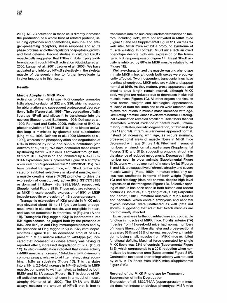
2000). NF-�B activation in these cells directly increases translocate into the nucleus; unrelated transcription fac-tors, including Oct1, were not activated in MIKK micethe production of a whole host of related proteins, in-
cluding cytokines and chemokines, immune- and anti- (Figure 1E and see Supplemental Figure S1C on the Cellweb site). MIKK mice exhibit a profound syndrome ofgen-presenting receptors, stress response and acute
phase proteins, and other regulators of apoptosis, growth, muscle wasting. In contrast, MISR mice lack an overtphenotype despite high-level expression of the trans-and host defense. Recent studies in cultured C2C12
muscle cells suggested that TNF-� inhibits myocyte dif- genic I�B� superrepressor (Figure 1F). Basal NF-�B ac-tivity is inhibited by 80% in MISR muscle relative to wtferentiation through NF-�B activation (Guttridge et al.,
2000; Langen et al., 2001; Ladner et al., 2003). We have (Figure 1E).We have characterized the muscle wasting phenotypeactivated and inhibited NF-�B selectively in the skeletal
muscle of transgenic mice to further investigate its in male MIKK mice, although both sexes were equiva-lently affected. Two independent transgenic lines havein vivo functions in this tissue.identical phenotypes. MIKK mice are viable and appearnormal at birth. As they mature, gross appearance andResultssnout-to-anus length remain normal, although MIKKbody weights are reduced due to decreases in skeletalMuscle Atrophy in MIKK Micemuscle mass (Figures 1G). All other organs and tissuesActivation of the I�B kinase (IKK) complex promoteshave normal weights and histological appearances.I�B� phosphorylation at S32 and S36, which is requiredMuscles of both the limbs and trunk were affected, andfor ubiqitination and subsequent proteasomal degrada-relative reductions in muscle mass increased with age.tion of I�B� (Yaron et al., 1998). The degradation of I�B�Circulating creatine kinase levels were normal. Histolog-liberates NF-�B and allows it to translocate into theical examination revealed smaller muscle fibers than wtnucleus (Baeuerle and Baltimore, 1996; Delhase et al.,littermates, without evidence of central nuclei, inflam-1999; Rothwarf and Karin, 2000). Activation of IKK� bymatory infiltrates, necrotic degeneration, or fibrosis (Fig-phosphorylation of S177 and S181 in its kinase activa-ures 1I and 1J). Intramuscular nerves appeared normal.tion loop is mimicked by glutamic acid substitutionsInstead of increasing with age, as occurs normally,(Ling et al., 1998; Delhase et al., 1999; Mercurio et al.,cross-sectional areas of muscle fibers in MIKK mice1999), whereas the phosphorylation and degradation ofdecreased with age (Figure 1H). Fiber and myonucleiI�B� is blocked by S32A and S36A substitutions (Vannumbers remained normal at earlier ages (SupplementalAntwerp et al., 1996). We have confirmed these resultsFigures S1D and S1E), suggesting ongoing atrophy inby showing that NF-�B in cells is activated through IKK�the absence of reduced myogenesis. Decreases in fiberSS177/181EE expression and inhibited by I�B� SS32/number seen in older animals (Supplemental Figure36AA expression (see Supplemental Figure S1A at http://S1D), along with replacement of muscle by fat (Figureswww.cell.com/cgi/content/full/119/2/285/DC1/). We1I and 1J), are suggestive of chronic atrophy and severehave created transgenic mice, with NF-�B either acti-muscle wasting (Mora, 1989). In mature mice, only so-vated or inhibited selectively in skeletal muscle, usingleus was unaffected in terms of both weight (Figurea muscle creatine kinase (MCK) promoter to drive the1G) and histology (data not shown), despite high-levelexpression of constitutively active IKK� SS177/181EEexpression of the transgene (Figure 1B). Selective spar-or dominant inhibitory I�B� SS32/36AA, respectivelying of soleus has been seen in both human and rodent(Supplemental Figure S1B). These mice are referred tocachexia (Tiao et al., 1997; Fang et al., 1998; Carpenteras MIKK (muscle-specific expression of IKK) or MISRand Karpati, 2001). Immature muscles in MIKK fetuses(muscle-specific expression of I�B superrepressor).and neonates, which contain embryonic and neonatalTransgenic expression of IKK� protein in MIKK micemyosin isoforms, were unaffected as well (data notwas elevated about 10- to 13-fold over basal endoge-shown), suggesting that adult fast twitch muscles arenous levels in skeletal muscle, was negligible in heart,predominantly affected.and was not detectable in other tissues (Figures 1A and
Ex vivo analyses further quantified size and contractile1B). Transgenic Flag-tagged IKK� is incorporated intofunction in muscles of MIKK mice. Tibialis anterior (TA)IKK signalosomes, as judged both by the presence ofmuscles from 12-week-old mice had normal numbersIKK� and IKK� in anti-Flag immunoprecipitates and byof muscle fibers, but fiber diameter and cross-sectionalthe presence of Flag-tagged IKK� in IKK� immunopre-area were 56% and 32% of normal, respectively. In addi-cipitates (Figure 1C). The decreased amount of I�B�tion to being small, muscles from MIKK mice exhibitedpresent in MIKK muscle relative to wild-type (wt) indi-functional deficits. Maximal force generated by singlecated that increased I�B kinase activity was having itsMIKK fibers was 23% of controls (Supplemental Figurereported effect, increased degradation of I�B� (FigureS1E), which corresponds to a 20% reduction when nor-1C). In vitro quantification indicated that kinase activitymalized by transverse area (Supplemental Figure S1F).in MIKK muscle is increased 10-fold in anti-IKK� immuneContraction (unloaded shortening) velocity was reducedcomplex assays, relative to wt littermates, using recom-by 21% in TA fibers from MIKK mice (Supplementalbinant I�B� as substrate (Figure 1D). This translatesFigure S1G).into a 15- � 2.5-fold increase in NF-�B activity in MIKK
muscle, compared to wt littermates, as judged by bothEMSA and ELISA assays (Figure 1E). This degree of NF- Reversal of the MIKK Phenotype by Transgenic
Suppression of I�B� Degradation�B activation matches that seen in a model of muscleatrophy (Hunter et al., 2002). The EMSA and ELISA Expression of I�B SS32/36AA (superrepressor) in mus-
cle does not induce an obvious phenotype; MISR miceassays measure the amount of NF-�B that is free to

NF-�B Activation Causes Muscle Wasting287
Figure 1. Profound Muscle Atrophy upon Muscle-Specific Activation of IKK�/NF-�B
Muscle lysates from MIKK mice and wt littermates were separated by SDS-PAGE, and proteins were analyzed by immunoblotting (IB) with(A) anti-Flag antibody that recognizes the transgene but not the endogenous protein and (B) anti-IKK� antibody that recognizes both proteins.(C) Proteins immunoprecipated (IP) from lysates of wt or MIKK muscle (n � 3) using anti-Flag or anti-IKK� antibodies were separated by SDS-PAGE and identified by immunoblotting with anti-Flag, anti-IKK�, anti-IKK�, or anti-IKK� antibodies. (Bottom panel) Tissue lysates wereseparated by SDS-PAGE, and I�B� was identified by immunoblotting with specific antibodies. (D) An immune complex assay used anti-IKK�
antibodies to immunoprecipitate IKK complexes from wt and MIKK muscle lysates. Anti-p38 antibody was used to immunoprecipitate p38kinase as control. [�-32P]ATP and either recombinant GST-I�B� or GST-ATF2, respectively, added as substrates, were separated by SDS-PAGE and detected by autoradiography. As loading controls, portions of the immunoprecipitates were separated by SDS-PAGE for Westernblotting of IKK� or p38. (E) NF-�B activity was measured in wt, MIKK, MISR, and MIKK x MISR gastrocnemius muscle using both EMSA-(upper) and ELISA-based DNA binding (lower) assays (n � 3). Oct1 activity was measured as control transcriptional factors for all samples.(F) Proteins immunoprecipated from lysates of wt or MISR muscle using anti-HA antibodies were analyzed by Western blotting using anti-HAand anti-I�B� antibodies. (G) Overall body and individual muscle and organ weights (black, white, or gray bars, respectively) of 12-week-oldMIKK mice are expressed as percentages of wt (n � 6). (H) Muscle fiber cross-sectional areas (CSA) were measured in transverse frozensections of EDL muscles at the indicated ages (wt, open; MIKK, black fill; n � 6–10). (I) Transverse sections taken at the midpoints of MIKKor wt legs and thighs were stained with hematoxylin and eosin (H&E) (12.5� magnification). (K) H&E-stained transverse sections of gastrocnemius(frozen) and quadriceps (fixed) muscles are from 20-week-old mice (200� magnification, scale bar � 35 �m). M, skeletal muscle; H, heart; L,liver; K, kidney; P, pancreas; S, soleus; E, extensor digitorum longus; Q, quadriceps; T, tibialis anterior; G, gastrocnemius; Pl, plantaris (*p
0.001; **p 108).

Cell288
Figure 2. Reversal of the MIKK Phenotype by Transgenic Expression of I�B� Superrepressor
MIKK and MISR mice were crossed to create compound transgenic MIKK x MISR mice. (A) Skin was stripped from the hind limbs of 20-week-old mice for gross comparisons of muscle mass. (B) Body weights of wt (pink), MISR (green), and MIKK x MISR (black) are superimposed,whereas body weights of MIKK mice (blue) are distinctly reduced (n � 6 in each group). (C) Cross-sectional areas for individual EDL musclefibers were measured in transverse frozen sections (n � 6 for each genotype; mean � SEM; *p 108 versus wt). (D) H&E-stained transversesections, taken at the midpoints of legs and thighs, were made from wt, MIKK, and MIKK x MISR mice.
are normal in terms of appearance, body weight (Figures specific expression of the I�B superrepressor in MIKKx MISR mice reduced NF-�B activity to levels seen in wt2A and 2B), individual muscle weight, and histology
(data not shown). MIKK and MISR mice were crossed animals (Figure 1E), despite continued high-level IKK�activity. Notably, the MIKK phenotype was completelyto determine whether the muscle loss phenotype was
dependent on NF-�B activation. Concomitant muscle- reversed in MIKK x MISR mice (Figure 2). Body weight

NF-�B Activation Causes Muscle Wasting289
(Figure 2B) and muscle mass were restored (Figures 2A effect favors degradation. In fact, the proteasome inhibi-tor MG-132 reduced protein breakdown in EDL musclesand 2D), and histology of MIKK x MISR muscle was
indistinguishable from wt controls (data not shown). of both MIKK and wt mice to similar levels (Figure 4A),suggesting that, in MIKK mice, increased protein degra-While fiber size in MIKK mice was reduced to �20% of
that in wt mice, this parameter was also restored to dation is largely mediated by ubiquitin- and proteasome-dependent pathways. Protein synthesis and breakdownnormal levels in MIKK x MISR (Figure 2C). These data
verify that the loss of muscle mass in MIKK mice is NF- rates were normal in soleus muscles of MIKK mice (datanot shown). Muscle wasting upon in vivo activation of�B dependent, as opposed to being mediated by IKK�
through an alternative pathway. IKK� and NF-�B thus appears to be mediated by ubiqui-tin-proteasome mechanisms. Our data is consistent withprevious suggestions that ubiquitin-dependent proteol-Pharmacological Reversal of the MIKK Phenotypeysis is involved in muscle atrophy (Jagoe and Goldberg,High-dose salicylates have been shown to inhibit IKK�2001; Glass, 2003; Li, 2003).and NF-�B (Kopp and Ghosh, 1994; Yin et al., 1998;
Yuan et al., 2001). We therefore asked whether salicylatetreatment had an effect in MIKK mice. Two distinct pro- NF-�B Activates Proteolysis but Not Cytokinetocols were followed, one to look at treatment and the Signaling in Muscleother to look at prevention of the muscle wasting pheno- To further explore potential mechanisms for muscletype. The treatment protocol was initiated after weaning wasting in MIKK mice, we asked whether cytokines,in 4-week-old affected mice. MIKK body weights in- such as those typically driven by NF-�B in other tissues,creased with salicylate treatment (Figure 3A); after 6 were being produced in MIKK muscle. Quantitative RT-months of therapy, body weight, muscle mass, and fiber PCR analyses revealed normal muscle mRNA levels forsize had approached normal levels (Figures 3B–3D). Sa- many of these cytokines, including TNF-�, IL-6, IL-1�,licylate therapy inhibited NF-�B activity in MIKK muscle IFN-�, IL-2, IL-8, IL-10, LIF, and CNTF and their recep-by 77% (Figure 3E). For the prevention protocol, the tors, including TNF-� receptors 1 and 2, IL-1� receptorssame regimen was used, except mice were treated dur- 1 and 2, IFN-� receptor, and IL-6 receptors � and �ing gestation and until weaning, at which time the pups (gp130) (Supplemental Table S2). Consistent with thewere treated with salicylate. Body weights and muscle mRNA data, circulating levels of TNF-� (wt 5.7 � 0.4mass were close to normal during the prevention proto- versus MIKK 5.9 � 0.5 pg/ml, n � 8, p � NS), IL-6 (belowcol (Figure 3F), suggesting that earlier intervention af- detection) and IL-1� (wt 3.7 � 0.4 versus MIKK 4.1 �fords an added benefit despite the fact that newborn 0.6 pg/ml, n � 8, p � NS) were normal in MIKK mice.muscle appears to be histologically normal. Much like These data suggest that, while NF-�B acts downstreamI�B� expression, high-dose salicylate rescued the MIKK of cachexia-inducing factors, in muscle, its activation isphenotype by inhibiting NF-�B signaling (Figure 3E). not sufficient to induce the transcription of cytokines
reportedly linked to muscle wasting. This is in contradis-tinction to NF-�B’s function in inflammation and innateCatabolism and Protein Turnover in MIKK Mice
Muscle wasting syndromes are catabolic and often ac- immunity, where many cytokines activate NF-�B andmany of the same cytokines and more are released incompanied by increases in resting energy expenditure
and thermogenesis (Argiles and Lopez-Soriano, 1999). a positive feedback response (Van Antwerp et al., 1996;Baeuerle and Baltimore, 1996; Delhase et al., 1999;MIKK mice and wt littermates were studied for 68 hr
in cages designed to measure a variety of metabolic Rothwarf and Karin, 2000).Gene microarray experiments were conducted to helpparameters. Food and water consumption and activity
levels were normal. However, O2 consumption and CO2 determine the genes perturbed by increased NF-�B ac-tivity in the muscles of MIKK mice, and the results drewand heat production were significantly elevated in MIKK
mice by 12%, 13%, and 17%, respectively (Supplemen- additional attention to elements of the proteasome deg-radation pathway. Subsequent RT-PCR experiments re-tal Figure S2).
Muscle wasting syndromes are often associated with vealed a 3.3-fold increase in mRNA levels for MuRF1(Supplemental Table S2), a muscle-specific E3 ligaseincreases in circulating and excreted protein breakdown
products (Jagoe and Goldberg, 2001). Concentrations (Bodine et al., 2001; Glass, 2003), and 2.4- to 2.8-foldincreases in mRNA for the C2 and C9 subunits of theof amino acids and their metabolites were elevated in
MIKK mouse urine by an average of 45% (Supplemental proteasome (Supplemental Table S2). mRNA levels forother associated genes, including ubiquitin, E2(14KD)Table S1). These findings suggest that the MIKK pheno-
type is associated with increased protein catabolism. and atrogin-1/MAFbx, were normal. Message levelswere normal as well for lysosomal and calcium-depen-A protein breakdown assay using EDL muscles tested
whether the catabolism was occurring in skeletal muscle dent proteases, including calpains 1 and 2 and cathep-sins B and D (Supplemental Table S1). In separate exper-and thus accounted for the apparent atrophy. Ex vivo
tyrosine release increased 1.9- to 2.4-fold in muscles iments, MuRF1 message (Figure 4C) and protein (Figure4D) levels were increased in MIKK mice and decreasedfrom 5- and 32-week- old MIKK mice (Figure 4A). A
complementary protein synthesis assay revealed 2.3- in MISR mice, compared to wt controls. Elevated MuRF1message and protein levels were concordantly reducedto 2.5-fold increases in MIKK muscle synthesis (Figure
4B). Although the fold increases in protein breakdown in doubly transgenic MIKK x MISR mice (Figures 4Cand 4D). To test whether MuRF1 is a NF-�B target, weand synthesis appeared to be balanced in these assays,
the markedly smaller size of MIKK muscle fibers demon- constructed a luciferase reporter assay using a 4.4 kBsegment of the MuRF1 promoter and transfected thisstrates that, at steady state in living animals, the net

Cell290
Figure 3. Reversal of the MIKK Phenotype using High-Dose Salicylate to Inhibit IKK�/NF-�B
Salicylate was dissolved in the drinking water (6 mg/ml) and incorporated in the chow (500 mg/kg). Treatment protocol, (A) Body and (B)muscle weights (gastrocnemius � TA � EDL � quadriceps � biceps), (C) cross-sectional areas of EDL muscles, and (D) H&E-stained transversesections of legs and thighs from 7-month-old mice (n � 6/group) treated with high-dose salicylate therapy that had been initiated at 4 weeksof age. (E) NF-�B activity was measured in gastrocnemius muscle lysates using EMSA and ELISA assays. Oct1 activity was measured ascontrol transcriptional factors for all samples. Wild-type and MIKK mice had been treated for 6 months with oral salicylate. (F) Preventionprotocol, weights of individual muscles from 6-month-old mice treated with high-dose salicylate initiated in utero (mothers and weanedoffspring received salicylate in the water and food; n � 6; mean � SEM). All panels, *p 0.05, **p 0.001, ***p 105.
into differentiated C2C12 myotubes. TNF-� or IL-1� 4E). These data fit recent observations showing thatMuRF1 mRNA is induced by IL-1�, sepsis, and reactivestimulation or coexpression of constitutively active IKK�
increased transcriptional activity by 4.6-, 2.3-, and 1.7- oxygen, all potential in vivo activators of NF-�B (Bodineet al., 2001; Wray et al., 2003; Li et al., 2003).fold, respectively (Figure 4E). Expression of the I�B�
superrepressor supported the suggestion that MuRF1 is To determine whether MuRF1 mediates NF-�B-drivenmuscle wasting, we crossed MIKK and MuRF1/an NF-�B target, as it reduced both basal and stimulated
expression under each of the tested conditions (Figure knockout mice (Bodine et al., 2001) to create MIKK x

NF-�B Activation Causes Muscle Wasting291
Figure 4. Ubiquitin-Dependent Proteolytic Pathway in MIKK Mice
Assays used isolated EDL muscles to simultaneously measure (A) protein breakdown and (B) protein synthesis (Tawa et al., 1997) (n � 6–7;*p 0.05; **p 0.01). The proteasome inhibitor MG-132 (10 �M) was added to the media in selected experiments. (C) Quantitative real-timeRT-PCR was used to quantify MuRF1 mRNA in TA muscles of wt, MIKK, MISR, and MIKK x MISR (n � 6–7/group). (D) Western blot analysesof (upper) MuRF1 protein in gastrocnemius and (lower) IKK�, I�B�, and MuRF1 in TA muscle (Tub � tubulin). (E) A reporter assay wasdeveloped using the promoter from MuRF1 linked to the luciferase gene. Differentiated C2C12 myotubes were cotransfected with the MuRF1reporter, control PRL-TK, and either pCMV-IKK� SS177/181EE (IKK� SE), pCMV-I�B� SS32/36AA (I�B� SR), or empty vector and stimulatedwith cytokines. (F) MIKK and MuRF1/ mice were crossed to obtain mice having the compound MIKK x MuRF1/ genotype. Body weightsof wt and MuRF1/ mice were indistinguishable, whereas weights of MIKK x MuRF1/ mice were intermediate between these and MIKKmice. (G) Cross-sectional areas of muscle fibers were measured in transverse sections of paraspinal and EDL muscles of wt, MIKK (M), andMIKK x MuRF1/ (M x M/) mice (n � 6/group). (H) Transverse sections show retroperitoneal muscles at the level of the pelvis. Black arrowspoint to the spine (midline) and pelvic bones (either side), and blue arrows point to sciatic nerves as landmarks (12.5� magnification). Allpanels, *p 0.05, **p 0.01, ***p 0.001.
MuRF1/ mice. Mice with the compound genotype had were 19% the size of wt fibers. Reductions in fiber sizein MIKK x MuRF1/ mice (55% of wt) were much lessintermediate body weights between wt and MIKK, dem-
onstrating an approximately 50% reversal of the muscle than in MIKK mice. Cross-sectional areas of EDL musclefibers in MIKK mice were equivalently reduced to �18%wasting phenotype by MuRF1 deletion (Figure 4F). Res-
cue was greatest for axial trunk muscles and less for the size of wt fibers. However, the rescue was of asmaller magnitude, as cross-sectional areas of MIKK xmuscles of the limbs (Figures 4G and 4H). Cross-sec-
tional areas of paraspinal muscle fibers in MIKK mice MuRF1/ EDL muscles were 32% of wt. The apparent

Cell292
differences in rescue efficiency between trunk and pe- in muscle mass of MISR mice (Figure 6C). This wasmanifested by a 33% reduction in cross-sectional fiberripheral muscles due to MuRF1 deletion may be a reflec-
tion of developmental as well as functional differences area in wt mice, compared to a 15% reduction in cross-sectional fiber area in littermate MISR mice (Figuresbetween these muscle groups. The incomplete rescue
seen in both axial and limb muscles demonstrates that 6D and 6F). Selective NF-�B blockade in muscle thusdecreases muscle wasting and prolongs survival in thisNF-�B activates other atrophic pathways in addition
to MuRF1. mouse model of cancer cachexia.
Relationships between Muscle NF-�BNF-�B Blockade Rescues Muscle Wastingand Insulin Resistancein Denervation and Cancer CachexiaWe have shown previously that activation of IKK� andSince NF-�B activity has been reported to increase inNF-�B promotes insulin resistance, whereas their inhibi-muscle in response to immobilization (Hunter et al.,tion improves insulin sensitivity (Yuan et al., 2001). We2002), we asked whether this was true as well in a re-therefore used MIKK and MISR mice to ask whetherlated, denervation model of muscle atrophy and whethermuscle IKK�/NF-�B modulates insulin sensitivity. MISRthe reverse, NF-�B inhibition, might decrease musclemice are normal in terms of insulin sensitivity, glucoseloss. NF-�B activity increased 9-fold in the hind limbtolerance, and lipid levels. Fasting blood glucose (102 �muscles of wt mice 14 days after severing their sciatic5.6 mg/dl) and insulin (0.77 � 0.12 ng/ml) levels in MISRnerves (Figure 5A). In contrast, NF-�B activity increasedmice are similar to wt littermates (97 � 7.3 mg/dl andonly 1.5-fold in the denervated muscle of MISR mice.0.81 � 0.19 ng/ml, respectively). Glucose concentra-Atrophy was readily apparent in denervated muscle,tions during glucose tolerance testing were similarlywith mass decreased by 51% relative to sham-operatedmatched between MISR mice and wt littermates (AUC:controls (Figure 5B). Fiber size (cross-sectional area)736 � 50 versus 787 � 94 mg/dl·h). Free fatty acid levelssimilarly decreased by 53% in denervated muscles (Fig-were equivalent, as well (wt, 1.64 � 0.21; MISR, 1.48 �ures 5C and 5D). Inhibition of NF-�B in MISR mice was0.33 mEq/l). Importantly, NF-�B inhibition in MISR miceaccompanied by a partial rescue of the denervation-afforded no protection against the development of obe-induced atrophy, including less loss of muscle masssity-induced insulin resistance. After 3 months of a high-(Figure 5B) and fiber size (Figures 5C and 5D) (38% p fat diet fasting blood glucose (120 � 17 versus 115 �0.05 and 28% p 0.01, respectively).14 mg/dl) and insulin (1.4 � 0.7 versus 1.2 � 0.8 ng/ml)Similar studies were conducted using a mouse modelconcentrations were similar in MISR mice versus wtfor cancer cachexia to determine the extent to whichcontrols, and glucose concentrations following a glu-NF-�B is involved in the pathogenesis of this disorder.cose challenge were similar in high-fat-fed MISR andLewis lung carcinoma (LLC) cells are often used to cre-wt mice (AUC: 972 � 130 versus 1019 � 105 mg/dl·h, re-ate a model for cancer cachexia-induced muscle wast-spectively).ing in mice. Subcutaneous injection of LLC cells leads
Activation of NF-�B in skeletal muscle does not causeto the growth of large tumors at the injection sites andMIKK mice to become insulin resistant. Fasting blooddiffuse tumors in the lungs. NF-�B activity, increasedglucose and insulin concentrations in 12- to 15-week-6-fold in the muscles of wt mice with tumors, was normalold MIKK mice (92 � 6.6 mg/dl and 0.37 � 0.07 ng/ml,in MISR mice with similar tumor burdens (Figure 6A).respectively) and wt littermate controls (95 � 3.8 mg/dlSince no tumor could be found in the muscle itself,and 0.34 � 0.05 ng/ml) were similar, as were glucoseincreased NF-�B activity was presumably due to the ef-levels during glucose tolerance testing (AUC: MIKK,fects of circulating “cachexia” factor(s). Body weights708 � 122 mg/dl·h; wt, 671 � 98 mg/dl·h). Ex vivo glu-typically begin to decrease within 1 week and continuecose uptake in isolated EDL muscles of MIKK and wtto decrease over the subsequent week after tumor cellmice was comparable (data not shown). In contrast,injection. Body weights of 8-month-old wt mice injectedserum-free fatty acid concentrations were elevated inwith LLC cells were decreased 6.6% on day 8, relativeMIKK mice (3.0 � 0.3 mEq/l) relative to wt littermatesto uninjected controls, and were decreased 13.2% by(1.8 � 0.2 mEq/l; p 0.01). The known associationday 12 (Figure 6B). In contrast, body weights of MISRbetween cachexia and elevations in circulating lipidslittermates decreased 1.6% on day 8 and 6.7% on daymay explain these findings (Rossi et al., 1995; Lopez-12, relative to uninjected MISR controls. The size andSoriano et al., 1996; Argiles and Lopez-Soriano, 1999).histology of the tumors at the injection sites (Figure 6F)We have concluded, since insulin sensitivity is not al-and tumor burden in the lungs (data not shown) weretered in MIKK or MISR mice, under both basal and pro-essentially identical in wt and MISR mice, suggesting thatvocative conditions, that IKK�, NF-�B, and MuRF1 me-the muscles of MISR mice were less responsive to thediate muscle wasting without being directly involved ineffects of the cachexia factor(s) in terms of both NF-�Bthe development of insulin resistance in this tissue.activation and muscle wasting. This was further suggested
using death as an endpoint, as all of the 2-month-old wtmice died within 28 days, whereas 40% of the MISR Discussionlittermates survived to 30 days (at which point they weresacrificed) (Figure 6E). This was presumably due to the NF-�B was activated or inhibited selectively in the skele-
tal muscle of transgenic mice through expression ofgreater degree of muscle wasting, particularly in therespiratory muscles, of wt versus MISR mice. At day 12, either constitutively active IKK� or a dominant inhibitory
form of I�B�. These mice are referred to as MIKK (mus-gastrocnemius and TA muscles of wt mice had de-creased in weight by 28%, compared to a 15% decrease cle-specific expression of IKK) or MISR (muscle-specific
NF-�B Activation Causes Muscle Wasting293
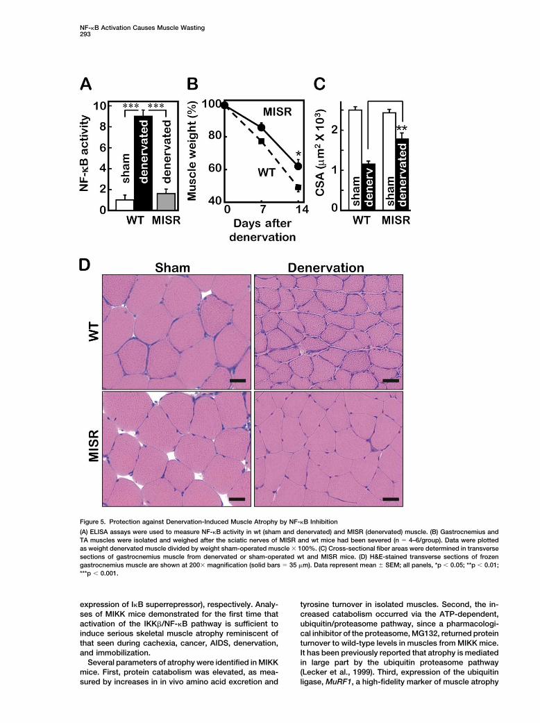
Figure 5. Protection against Denervation-Induced Muscle Atrophy by NF-�B Inhibition
(A) ELISA assays were used to measure NF-�B activity in wt (sham and denervated) and MISR (denervated) muscle. (B) Gastrocnemius andTA muscles were isolated and weighed after the sciatic nerves of MISR and wt mice had been severed (n � 4–6/group). Data were plottedas weight denervated muscle divided by weight sham-operated muscle � 100%. (C) Cross-sectional fiber areas were determined in transversesections of gastrocnemius muscle from denervated or sham-operated wt and MISR mice. (D) H&E-stained transverse sections of frozengastrocnemius muscle are shown at 200� magnification (solid bars � 35 �m). Data represent mean � SEM; all panels, *p 0.05; **p 0.01;***p 0.001.
expression of I�B superrepressor), respectively. Analy- tyrosine turnover in isolated muscles. Second, the in-creased catabolism occurred via the ATP-dependent,ses of MIKK mice demonstrated for the first time that
activation of the IKK�/NF-�B pathway is sufficient to ubiquitin/proteasome pathway, since a pharmacologi-cal inhibitor of the proteasome, MG132, returned proteininduce serious skeletal muscle atrophy reminiscent of
that seen during cachexia, cancer, AIDS, denervation, turnover to wild-type levels in muscles from MIKK mice.It has been previously reported that atrophy is mediatedand immobilization.
Several parameters of atrophy were identified in MIKK in large part by the ubiquitin proteasome pathway(Lecker et al., 1999). Third, expression of the ubiquitinmice. First, protein catabolism was elevated, as mea-
sured by increases in in vivo amino acid excretion and ligase, MuRF1, a high-fidelity marker of muscle atrophy

Cell294
Figure 6. NF-�B Inhibition Protects against Cancer-Induced Muscle Atrophy
Eight-month old mice were injected with LLC cells to induce tumor growth. (A) NF-�B activity was measured by ELISA assay in wt and MISRmuscle 12 days after subcutaneous injection of LLC cells. (B) Mice were weighed each day after injection of the LLC cells (n � 6); data arereported relative to uninjected controls. (C) Weights of gastrocnemius (Gas) and TA muscles and (D) cross-sectional fiber areas were measuredin transverse sections of gastrocnemius 12 days after injection of LLC cells. (E) Survival curves for 2-month-old wt and MISR mice afterinjection with identical numbers of LLC cells (n � 10–12; p value determined using a Wilcoxon test). (F) H&E-stained transverse sections ofgastrocnemius muscle are shown at 200� magnification (Solid bars � 35 �m). (Inserts) Tumors removed from the primary injection sites(diameter �1.5 cm). Data represent mean � SEM; all panels, *p 0.05; **p 0.01; ***p 0.001.

NF-�B Activation Causes Muscle Wasting295
(Bodine et al., 2001; Wray et al., 2003; Li et al., 2003),was shown to be greater in MIKK muscle relative tolevels seen in wild-type muscle. Fourth, and perhapsmost strikingly, muscle mass was significantly reducedin MIKK mice relative to wild-type controls, indicatingthat increased catabolism results in phenotypic atrophyand is not sufficiently compensated by an increase inprotein synthesis. The reduction in mass comes as aresult of reduced cross-sectional area of individual mus-cle fibers, as opposed to changes in fiber number. Thisis the situation seen in clinical settings of atrophy. Inthe quadriceps muscle of older MIKK animals, there issome replacement of muscle with adipose tissue, asoccurs in paraplegic patients after long-term denerva-tion-induced skeletal muscle atrophy (Mora, 1989).
When MIKK and MISR mice were crossed, the atrophicphenotype was reversed, thus demonstrating that the lossof muscle mass was not only induced by activation of NF-�B but could be blocked by genetic inactivation of NF-�B at the level of I�B�. NF-�B has previously been demon-strated to be activated upon treatment of C2C12 cells withthe cachectic factor TNF� (Guttridge et al., 2000; Li andReid, 2000; Langen et al., 2001; Ladner et al., 2003) andduring immobilization-induced decreases in muscle mass
Figure 7. Proposed Pathways that Integrate Muscle Hypertrophy(Hunter et al., 2002). Also, a previous in vitro study dem-and Atrophyonstrated that overexpression of I�B� could block lossRecent findings demonstrated the involvement of the Foxo familyof myosin in TNF�-treated C2C12 myotubes (Ladnerof transcription factors to be required for the activation of MuRF1et al., 2003). The current study is consistent with theand MAFbx/Atrogin-1 (Sandri et al., 2004; Stitt et al., 2004). By acti-previous data and provides the first in vivo evidence vating Akt, growth factors such as IGF-1, which induces hypertro-
that blockade of NF-�B can block atrophy. In addition phy, block the induction of MuRF1 and MAFbx/Atrogin-1 mRNA.to genetic complementation, a pharmacological inhibi- Activation of the IKK�/NF-�B axis, which is induced by cachexia,
is required to activate MuRF1 and is sufficient to induce muscletor of the NF-�B pathway, sodium salicylate, was suffi-atrophy (this work). The IKK�/NF-�B/MuRF1 pathway may be sup-cient to partially suppress the atrophic phenotype inpressed at multiple steps, including salicylate inhibition of IKK� andMIKK mice. High doses of sodium salicylate were pre-expression of the I�B� superrepressor to inhibit NF-�B, and byviously reported to block IKK� and NF-�B (Kopp and genetic ablation of MuRF1.
Ghosh, 1994; Yin et al., 1998; Yuan et al., 2001).To assess the physiologic role of the NF-�B pathway
in clinically relevant settings, MISR mice were subjected now be proposed (Figure 7) in which atrophic stimulito both denervation- and tumor-induced atrophy. In both such as denervation and tumor-induced factors activateconditions, blockade of the NF-�B pathway resulted in NF-�B, which in turn results in upregulation of MuRF1a significantly reduced loss of muscle mass as com- as well as other critical pathways which have yet topared to that observed in wild-type mice undergoing be elucidated.the same perturbations. These data suggest that the It is noteworthy that a second ubiquitin ligase, alterna-NF-�B pathway may be one of the long-sought, critical tively named MAFbx (Bodine et al., 2001) or atrogin-1triggers induced by atrophic stimuli, upstream of the (Gomes et al., 2001), was neither activated nor inhibitedproteolytic pathways that mediate catabolism. One indi- in the MIKK animals. MAFbx had been shown previouslycation that this may be the case is that activation of NF- to be activated in multiple models of atrophy and often�B in MIKK mice resulted in increased expression of the at levels greater than observed for MuRF1. The MIKKE3 ubiquitin ligase MuRF1. MuRF1 is upregulated in at animals are the first setting of atrophy to be reportedleast ten different settings of skeletal muscle atrophy where MAFbx is not upregulated. Thus, MAFbx upregu-(Bodine et al., 2001; Wray et al., 2003; Glass, 2003). lation is not required for NF-�B-induced muscle loss.Further, in MuRF1/ animals, the degree of atrophy is However, given the diverse settings that do result inreduced relative to wild-type controls (Bodine et al., MAFbx/atrogin upregulation, the implication is that a2001), reminiscent of the sort of reduction seen here in second, parallel pathway, distinct from NF-�B activa-MISR animals. When MIKK mice were crossed into a tion, is usually induced during atrophy. This idea is alsoMuRF1/ background, there was a significant reduction consistent with the MISR data, which shows significantin muscle loss, though less than that seen when MIKK but partial amelioration of denervation and tumor-inducedmice were crossed with MISR mice. Although MuRF1 is atrophy, indicating that the IKK�/NF-�B pathway is atherefore not the only critical mediator of muscle atrophy critical mediator of muscle loss in these clinical settingsto be activated by the NF-�B pathway, its ablation, even but not the only mediator.in the setting of continued activation of NF-�B, partially Many of what would typically be considered NF-�Bameliorates the atrophic phenotype. With this data, a target genes were not activated in muscles of MIKK
mice. Functions have been studied most extensively instepwise IKK�/NF-�B/MuRF1 signaling pathway can

Cell296
Histology and Blood/Urine Chemistryimmune and inflammatory cells and liver, where NF-�BCryostat and paraffin sections were stained with hematoxylin andorchestrates the synthesis of a host of mediators of celleosin using standard techniques. Cross-sectional areas were deter-survival and innate immune and inflammatory responses.mined for at least 50 fibers/animal, and six to ten animals were
Despite high-level activation of NF-�B in muscles of analyzed for each determination. NIH Image 1.6 software was usedMIKK mice, none of the typical cytokines, chemokines, for the analyses. Fiber numbers in EDL muscle were counted in theor associated receptors was upregulated as would be entire cross-section (n � 6 animals/group). Nuclei were counted in
similar sections of EDL muscle and normalized for fiber number (n �expected in these other cell types. While NF-�B may6 animals/group). Plasma creatine kinase activity was determinedbe activated in muscle by certain proinflammatory andusing the EC 2.7.3.2 UV test kit (Sigma). Circulating cytokines werecachexia-inducing cytokines, it must not drive their syn-measured using Luminex assays (Linco).
thesis as in other tissues. Therefore, NF-�B has dramati-cally different functions in distinct tissues. In muscle,
NF-�B Assaysthe catabolic actions of NF-�B provide a mechanism for Nuclear proteins were isolated from gastrocnemius muscle using athe sustained production of amino acids to be used as combination of mechanical and osmotic disruption as describedan energy source for other tissues. While this may be (Kumar and Boriek, 2003). Electrophoretic mobility shift assays
(EMSA, Promega) were conducted with 32P-labeled double-strandeda normal physiological response, for example, duringoligonucleotides having consensus recognition sequences for NF-starvation, NF-�B activation in muscle appears to have�B (5�-AGTTGAGGGGACTTTCCCAGG-3�) or Oct-1 (5�-TGTCGAATdeleterious and potentially disastrous consequencesGCAAATCACTAGAA-3�); unlabeled probe was added to negativeunder pathological conditions.controls. Protein-DNA complexes were separated using nondena-
It is of further interest to find signaling pathways that turing PAGE. Similar extracts were used for ELISA assays of NF-�Bcould dominantly suppress activation of NF-�B in skele- activity, using immobilized �B binding motifs (GGGGACTTTCCC) intal muscle, since activation of those pathways could a 96-well format (BD Biosciences) (Fiorucci et al., 2002). Bound
protein, recognized using anti-NF-�B p65 and IgG-HRP as primarypotentially help in suppressing atrophy. It was very re-and secondary antibodies, respectively, was quantified colorimetri-cently reported that activation of the Akt kinase, whichcally.was previously shown to cause skeletal muscle hyper-
trophy via upregulation of protein synthesis pathways,Kinase Assayscould also dominantly suppress upregulation of bothKinase activity was assessed in immune complexes precipitatedMuRF1 and MAFbx during atrophy (Sandri et al., 2004; from muscle lysates. For measurements of IKK activity, recombinant
Stitt et al., 2004). Akt has been shown to activate NF- GST-I�B� was used as a substrate for kinase activity precipitated�B in various cell types. However, data reported here with protein A Sepharose-immobilized anti-IKK� (Pharmingen). To
quantify p38 MAP kinase activity, GST-ATF2 (Santa Cruz) served asin conjunction with the Akt studies would seem to calla substrate for kinase activity associated with anti-p38 (Santa Cruz).into question whether Akt activates NF-�B in muscle,Proteins phosphorylated in the presence of [�-32P]ATP (20 �M) wereor at least suggests future studies focusing on an as-separated by SDS/PAGE, and incorporated radioactivity was quanti-sessment of the Akt/NF-�B pathway in skeletal muscle.fied using a phosphorimager (Storm, Molecular Dynamics).
The discovery that NF-�B activation is sufficient tocause skeletal muscle atrophy in vivo and that blockade
Ex Vivo Determinations of Protein Turnoverof the NF-�B pathway can ameliorate atrophy suggests and Fiber Size and Contractilitya new set of drug targets for clinical intervention during EDL or soleus muscles were secured to inert supports for the simul-cachexia, cancer, AIDS, and other settings of atrophy. taneous measurement of protein synthesis and degradation (Tawa
et al., 1997). For selected measurements, muscles were incubatedFor example, the inhibition of atrophy with high dosesin media supplemented with 0.5 mM cycloheximide, to suppressof sodium salicylate suggests that more specific inhibi-protein synthesis; or 10 �M MG-132, 25 �M E-64, or 10 mM methyl-tors of IKK� might be useful to block atrophy—sodiumamine, to suppress proteasomal, lysosomal, or Ca2�-dependentsalicylate itself has a range of other targets, and the proteolysis, respectively (Tawa et al., 1997). For assessments of
high doses of it required for IKK� inhibition are not well fiber size and contractile function, single, chemically skinned fiberstolerated due to side effects. MuRF1 has already been were isolated from TA and soleus muscles and analyzed at 15 C as
described (Larsson and Moss, 1993).suggested as a novel clinical target, and the data inthis study help to put the previous findings in a largersignaling context. Further study of the IKK�/NF-�B/ Cell Culture
C2C12 cells were cultured in DMEM supplemented with 5 mM glu-MuRF1 pathway will help to uncover other clinically use-cose, 20% fetal bovine serum (FBS), 2 mM glutamine, and penicillin/ful targets. This is critically important, because unfortu-streptomycin. Upon reaching confluence, cells were cultured innately, there are currently no drugs approved for theDMEM containing 25 mM glucose, 2% horse serum, 2 mM gluta-
treatment of skeletal muscle atrophy. mine, and penicillin/streptomycin. HEK 293 cells were cultured inDMEM containing 25 mM glucose, 10% FBS, 2 mM glutamine, and
Experimental Procedures penicillin/streptomycin. All cells were maintained at 37 C under7.5% CO2.Transgenic Mice
cDNAs encoding Flag-tagged full-length IKK� SS177/181EE (consti-MuRF1 and NF-�B Reporter Assaystutively active) or HA-tagged full-length I�B� SS32/36AA (superre-A 4.4 kB fragment of the mouse MuRF1 promoter was subclonedpressor) were subcloned into a truncated �-globin expression vectorinto the pGL3 firefly luciferase vector (Promega). C2C12 myotubes(Supplemental Figure S1A). A 3.3 kb fragment of DNA isolated fromwere cotransfected (Fugene, Roche) with pGL3-MuRF1, the controlthe mouse MCK promoter was subcloned upstream from exon 1 ofRellina luciferase vector pRL-TK, and matched amounts of pCMV-the �-globin cassette (Donoviel et al., 1996). Microinjected mouseempty, pCMV-IKK�SE, and/or pCMV-I�B�SR. HEK 293 cells wereoocytes were introduced into pseudopregnant females. Transgeniccotransfected with an NF-�B luciferase reporter provided by Dr.offspring were identified originally by Southern analyses, and subse-Gilmore (Sylla et al., 1998) and pCMV-IKK�SE and pCMV-I�B�SR.quent genotyping was by PCR. At least two independent lines wereC2C12 myotubes or HEK 293 cells were cultured overnight in DMEMobtained for both MIKK and MISR mice, and each had the same phe-
notype. lacking serum and treated for 90–120 min with 6 nM TNF-� (Calbio-

NF-�B Activation Causes Muscle Wasting297
chem) or IL-1� (R&D). Luciferase expression was quantified using Coletti, D., Yang, E., Marazzi, G., and Sassoon, D. (2002). TNF�
inhibits skeletal myogenesis through a PW1-dependent pathway bya Dual-Luciferase Reporter Assay (Promega).recruitment of caspase pathways. EMBO J. 21, 631–642.
Western Blotting Delhase, M., Hayakawa, M., Chen, Y., and Karin, M. (1999). PositiveAnimal tissues were homogenized and incubated for 60 min at 4 C in and negative regulation of I�B kinase activity through IKK� subunitlysis buffer, samples were separated by SDS/PAGE, and separated phosphorylation. Science 284, 309–313.proteins were transferred to nitrocellulose or PVDF membranes and
Donoviel, D.B., Shield, M.A., Buskin, J.N., Haugen, H.S., Clegg, C.H.,identified by immunoblotting. Primary antibodies included mono-
and Hauschka, S.D. (1996). Analysis of muscle creatine kinase geneclonal anti-Flag (Strategene), anti-HA (Cell Signaling), anti-myosin
regulatory elements in skeletal and cardiac muscles of transgenictype I and anti-myosin type II (Novocastra), anti-tubulin (Santa Cruz),
mice. Mol. Cell. Biol. 16, 1649–1658.anti-IKK� (PharMingen), rabbit anti-IKK�, anti-IKK�, anti-I�B�, (Santa
Fang, C.H., Li, B.G., Tiao, G., Wang, J.J., Fischer, J.E., and Hassel-Cruz), anti-�-actinin 3 (provided by Dr. Alan Beggs, Children’s Hospi-gren, P.O. (1998). The molecular regulation of protein breakdowntal), and anti-MuRF1 (Bodine et al., 2001). Secondary antibodiesfollowing burn injury is different in fast- and slow-twitch skeletalincluded GFP- and rodamine-conjugating anti-mouse and anti-rab-muscle. Int. J. Mol. Med. 1, 163–169.bit antibodies (Jackson ImmunoResearch) and HRP-conjugatedFiorucci, S., Antonelli, E., Distrutti, E., Del Soldato, P., Flower, R.J.,anti-rabbit and anti-mouse antibodies (Pierce).Clark, M.J., Morelli, A., Perretti, M., and Ignarro, L.J. (2002). NCX-1015, a nitric-oxide derivative of prednisolone, enhances regulatoryQuantitative Real-Time RT-PCRT cells in the lamina propria and protects against 2,4,6-trinitroben-Total RNA was extracted from mouse tissues or cultured cells usingzene sulfonic acid-induced colitis in mice. Proc. Natl. Acad. Sci.TRIzol (Invitrogen), and cDNA was synthesized using oligo (dT) prim-USA 99, 15770–15775.ers with the Advantage RT-for-PCR Kit (BD Biosciences). Primers
spanned intronic regions to generate 300–400 bp PCR products. Fong, Y., Moldawer, L.L., Marano, M., Wei, H., Barber, A., Manogue,PCR amplifications were quantified using the SYBRGreen PCR Mas- K., Tracey, K.J., Kuo, G., Fischman, D.A., and Cerami, A. (1989).ter Mix (Applied Biosystems). Results were normalized against tu- Cachectin/TNF or IL-1� induces cachexia with redistribution of bodybulin binding protein (TBP) and GAPDH gene expression. proteins. Am. J. Physiol. 256, R659–R665.
Glass, D.J. (2003). Signalling pathways that mediate skeletal muscleDenervation and Cancer Cachexia hypertrophy and atrophy. Nat. Cell Biol. 5, 87–90.Right sciatic nerves were severed in 16-week-old anaesthetized
Gomes, M.D., Lecker, S.H., Jagoe, R.T., Navon, A., and Goldberg,MISR mice and wt controls. Left sciatic nerves were exposed butA.L. (2001). Atrogin-1, a muscle-specific F-box protein highly ex-not severed to serve as sham-operated controls. Mice were sacri-pressed during muscle atrophy. Proc. Natl. Acad. Sci. USA 98,ficed after 7 or 14 days, and individual muscles were weighed. For14440–14445.the cancer cachexia model, Lewis lung carcinoma (LLC) cells (ATCC;Guttridge, D.C., Mayo, M.W., Madrid, L.V., Wang, C.Y., and Baldwin,5 � 106) were injected subcutaneously into the flanks of 8-month-A.S., Jr. (2000). NF-�B-induced loss of MyoD messenger RNA: possi-old C57Bl/6 mice. Mice were weighed daily and sacrificed 12 daysble role in muscle decay and cachexia. Science 289, 2363–2366.after injection. For survival studies, LLC cells were injected into the
flanks of 2-month-old C57Bl/6 mice. Hunter, R.B., Stevenson, E., Koncarevic, A., Mitchell-Felton, H., Es-sig, D.A., and Kandarian, S.C. (2002). Activation of an alternative
Statistical Analyses NF-�B pathway in skeletal muscle during disuse atrophy. FASEB J.Data are presented as mean � SEM. Statistical significance was 16, 529–538.determined using unpaired Student’s t tests. Jagoe, R.T., and Goldberg, A.L. (2001). What do we really know
about the ubiquitin-proteasome pathway in muscle atrophy? Curr.Acknowledgments Opin. Clin. Nutr. Metab. Care 4, 183–190.
Kopp, E., and Ghosh, S. (1994). Inhibition of NF-�B by sodium salicy-We dedicate this manuscript to the memory of our friend and col-late and aspirin. Science 265, 956–959.league, Valerie Fanikos. We wish to thank Dr. A. Beggs (Children’sKumar, A., and Boriek, A.M. (2003). Mechanical stress activates theHospital) and D. Guttridge (Ohio State University) for helpful discus-NF-�B pathway in skeletal muscle fibers: a possible role in Duchennesions and advice; and D. Sanoudou, M. Fareed, G. Krishnan, andmuscular dystrophy. FASEB J. 17, 386–396.members of the Shoelson Lab including M. Yuan, Y. Guo, E. Werner,
and L. Hansen for technical assistance. D.C. was supported by a Ladner, K.J., Caligiuri, M.A., and Guttridge, D.C. (2003). Tumor ne-Mentor-Based postdoctoral fellowship from the American Diabetes crosis factor-regulated biphasic activation of NF-�B is required forAssociation (S.E.S.). These studies were funded by NIH grants cytokine-induced loss of skeletal muscle gene products. J. Biol.DK45943 (S.E.S.), DK51729 (S.E.S.), and DK36836 (Joslin DERC); Chem. 278, 2294–2303.and the Helen and Morton Adler Chair (S.E.S.). Regeneron conducts Langen, R.C., Schols, A.M., Kelders, M.C., Wouters, E.F., and Jans-research and holds patents on intellectual property relating to the sen-Heininger, Y.M. (2001). Inflammatory cytokines inhibit myogenicmolecular mechanisms of muscle wasting. differentiation through activation of NF-�B. FASEB J. 15, 1169–1180.
Larsson, L., and Moss, R.L. (1993). Maximum velocity of shorteningReceived: April 5, 2004
in relation to myosin isoform composition in single fibres from humanRevised: August 31, 2004
skeletal muscles. J. Physiol. 472, 595–614.Accepted: September 2, 2004
Lecker, S.H., Solomon, V., Mitch, W.E., and Goldberg, A.L. (1999).Published: October 14, 2004Muscle protein breakdown and the critical role of the ubiquitin-proteasome pathway in normal and disease states. J. Nutr. 129,References227S–237S.
Li, Y.P. (2003). TNF-� is a mitogen in skeletal muscle. Am. J. Physiol.Argiles, J.M., and Lopez-Soriano, F.J. (1999). The role of cytokines285, C370–C376.in cancer cachexia. Med. Res. Rev. 19, 223–248.
Li, Y.P., and Reid, M.B. (2000). NF-�B mediates the protein lossBaeuerle, P.A., and Baltimore, D. (1996). NF-�B: ten years after. Cellinduced by TNF-� in differentiated skeletal muscle myotubes. Am.87, 13–20.J. Physiol. 279, R1165–R1170.Bodine, S.C., Latres, E., Baumhueter, S., Lai, V.K., Nunez, L., Clarke,Li, Y.P., Chen, Y., Li, A.S., and Reid, M.B. (2003). Hydrogen peroxideB.A., Poueymirou, W.T., Panaro, F.J., Na, E., Dharmarajan, K., et al.stimulates ubiquitin-conjugating activity and expression of genes(2001). Identification of ubiquitin ligases required for skeletal musclefor specific E2 and E3 proteins in skeletal muscle myotubes. Am.atrophy. Science 294, 1704–1708.J. Physiol. 285, C806–C812.Carpenter, S., and Karpati, G. (2001). Pathology of Skeletal Muscle
(Oxford: Oxford University Press). Ling, L., Cao, Z., and Goeddel, D.V. (1998). NF-�B-inducing kinase

Cell298
activates IKK-� by phosphorylation of Ser-176. Proc. Natl. Acad. Yuan, M., Konstantopoulos, N., Lee, J., Hansen, L., Li, Z.W., Karin,M., and Shoelson, S.E. (2001). Reversal of obesity- and diet-inducedSci. USA 95, 3792–3797.insulin resistance with salicylates or targeted disruption of Ikkb.Lopez-Soriano, J., Argiles, J.M., and Lopez-Soriano, F.J. (1996).Science 293, 1673–1677.Lipid metabolism in rats bearing the Yoshida AH-130 ascites hepa-
toma. Mol. Cell. Biochem. 165, 17–23.
Mercurio, F., Murray, B.W., Shevchenko, A., Bennett, B.L., Young,D.B., Li, J.W., Pascual, G., Motiwala, A., Zhu, H., Mann, M., andManning, A.M. (1999). I�B kinase (IKK)-associated protein 1, a com-mon component of the heterogeneous IKK complex. Mol. Cell. Biol.19, 1526–1538.
Moldawer, L.L., Svaninger, G., Gelin, J., and Lundholm, K.G. (1987).Interleukin 1 and tumor necrosis factor do not regulate protein bal-ance in skeletal muscle. Am. J. Physiol. 253, C766–C773.
Mora, M. (1989). Fibrous-adipose replacement in skeletal musclebiopsy. Eur. Heart J. Suppl. D 10, 103–104.
Mullen, B.J., Harris, R.B., Patton, J.S., and Martin, R.J. (1990). Re-combinant TNF-� chronically administered in rats: lack of cachecticeffect. Proc. Soc. Exp. Biol. Med. 193, 318–325.
Penner, C.G., Gang, G., Wray, C., Fischer, J.E., and Hasselgren, P.O.(2001). The transcription factors NF-�B and AP-1 are differentiallyregulated in skeletal muscle during sepsis. Biochem. Biophys. Res.Commun. 281, 1331–1336.
Rossi, F.F., Cangiano, C., Muscaritoli, M., Conversano, L., Torelli,G.F., and Cascino, A. (1995). Tumor-induced changes in host metab-olism: a possible marker of neoplastic disease. Nutrition 11,595–600.
Rothwarf, D.M., and Karin, M. (2000). The NF-�B activation pathway:a paradigm in information transfer from membrane to nucleus. Sci-ence’s STKE www.stke.org/cgi/content/full/OC_sigtrans;1999/5/re1.
Sandri, M., Sandri, C., Gilbert, A., Skurk, C., Calabria, E., Picard, A.,Walsh, K., Schiaffino, S., Lecker, S.H., and Goldberg, A.L. (2004).Foxo transcription factors induce the atrophy-related ubiquitin li-gase atrogin-1 and cause skeletal muscle atrophy. Cell 117,399–412.
Stewart, C.E., Newcomb, P.V., and Holly, J.M. (2004). Multifacetedroles of TNF-� in myoblast destruction: a multitude of signal trans-duction pathways. J. Cell. Physiol. 198, 237–247.
Stitt, T.N., Drujan, D., Clarke, B.A., Panaro, F., Timofeyva, Y., Kline,W.O., Gonzalez, M., Yancopoulos, G.D., and Glass, D.J. (2004). TheIGF-1/PI3K/Akt pathway prevents expression of muscle atrophy-induced ubiquitin ligases by inhibiting FOXO transcription factors.Mol. Cell 14, 395–403.
Sylla, B.S., Hung, S.C., Davidson, D.M., Hatzivassiliou, E., Malinin,N.L., Wallach, D., Gilmore, T.D., Kieff, E., and Mosialos, G. (1998).Epstein-Barr virus-transforming protein latent infection membraneprotein 1 activates transcription factor NF-kappaB through a path-way that includes the NF-�B-inducing kinase and the I�B kinasesIKK� and IKK�. Proc. Natl. Acad. Sci. USA 95, 10106–10111.
Tawa, N.E., Jr., Odessey, R., and Goldberg, A.L. (1997). Inhibitorsof the proteasome reduce the accelerated proteolysis in atrophyingrat skeletal muscles. J. Clin. Invest. 100, 197–203.
Tiao, G., Lieberman, M., Fischer, J.E., and Hasselgren, P.O. (1997).Intracellular regulation of protein degradation during sepsis is differ-ent in fast- and slow-twitch muscle. Am. J. Physiol. 272, R849–R856.
Tisdale, M.J. (1997). Biology of cachexia. J. Natl. Cancer Inst. 89,1763–1773.
Van Antwerp, D.J., Martin, S.J., Kafri, T., Green, D.R., and Verma,I.M. (1996). Suppression of TNF-�-induced apoptosis by NF-�B.Science 274, 787–789.
Wray, C.J., Mammen, J.M., Hershko, D.D., and Hasselgren, P.O.(2003). Sepsis upregulates the gene expression of multiple ubiquitinligases in skeletal muscle. Int. J. Biochem. Cell Biol. 35, 698–705.
Yaron, A., Hatzubai, A., Davis, M., Lavon, I., Amit, S., Manning, A.M.,Andersen, J.S., Mann, M., Mercurio, F., and Ben Neriah, Y. (1998).Identification of the receptor component of the I�B�-ubiquitin ligase.Nature 396, 590–594.
Yin, M.J., Yamamoto, Y., and Gaynor, R.B. (1998). The anti-inflamma-tory agents aspirin and salicylate inhibit the activity of I�B kinase-�.Nature 396, 77–80.