Inhibition of NF-κB in astrocytes is sufficient to delay ...
17
RESEARCH Open Access Inhibition of NF-κB in astrocytes is sufficient to delay neurodegeneration induced by proteotoxicity in neurons Y. X. Li 1 , O. C. M. Sibon 1 and P. F. Dijkers 1,2* Abstract Background: Most neurodegenerative diseases associated with protein aggregation are hallmarked by activation of astrocytes. However, how astrocytes are activated or which signaling pathways in astrocytes contribute to pathogenesis is not clear. One long-standing question is whether the responses in astrocytes are due to stress or damage in astrocytes themselves, or because of astrocytic responses to cellular stress or damage in neurons. Here, we examine responses in astrocytes induced by expression of disease-associated, aggregation-prone proteins in other cells. We also examine the consequences of these responses in astrocytes in a model for neurodegeneration. Methods: We first examined a role for intracellular astrocytic responses in a Drosophila model for Spinocerebellar ataxia type 3 (SCA3, also known as Machado–Joseph disease), a disease caused by expansion of the polyglutamine (polyQ) stretch in the ATXN3 gene. In this Drosophila SCA3 model, eye-specific expression of a biologically relevant portion of the ATXN3 gene, containing expanded polyQ repeats (SCA3 polyQ78 ) was expressed. In a candidate RNAi screen in the Drosophila SCA3 model, we analyzed whether downregulation of expression of specific genes in astrocytes affected degeneration induced by SCA3 polyQ78 expression in Drosophila eyes. We next examined the role of astrocytes in response to proteotoxic stress in neurons induced by SCA3 polyQ78 expression or amyloid beta peptides, associated with Alzheimer’s disease. Results: Eye-specific expression of SCA3 polyQ78 resulted in the presence of astrocytes in the eye, suggesting putative involvement of astrocytes in SCA3. In a candidate RNAi screen, we identified genes in astrocytes that can enhance or suppress SCA3 polyQ78 -induced eye degeneration. Relish, a conserved NF-κB transcription factor, was identified as an enhancer of degeneration. Activity of Relish was upregulated in our SCA3 model. Relish can exert its effect via Relish-specific AMPs, since downregulation of these AMPs attenuated degeneration. We next examined Relish signaling in astrocytes on neurodegeneration. Selective inhibition of Relish expression specifically in astrocytes extended lifespan of flies that expressed SCA3 polyQ78 exclusively in neurons. Inhibition of Relish signaling in astrocytes also extended lifespan in a Drosophila model for Alzheimer’s disease. (Continued on next page) * Correspondence: [email protected] 1 Department of Cell Biology, University of Groningen, Antonius Deusinglaan 1, 9713AV Groningen, The Netherlands 2 Hubrecht Institute for Developmental Biology and Stem Cell Research, Royal Netherlands Academy of Arts and Sciences (KNAW) and UMC Utrecht, Utrecht, The Netherlands © The Author(s). 2018 Open Access This article is distributed under the terms of the Creative Commons Attribution 4.0 International License (http://creativecommons.org/licenses/by/4.0/), which permits unrestricted use, distribution, and reproduction in any medium, provided you give appropriate credit to the original author(s) and the source, provide a link to the Creative Commons license, and indicate if changes were made. The Creative Commons Public Domain Dedication waiver (http://creativecommons.org/publicdomain/zero/1.0/) applies to the data made available in this article, unless otherwise stated. Li et al. Journal of Neuroinflammation (2018) 15:261 https://doi.org/10.1186/s12974-018-1278-2
-
Upload
khangminh22 -
Category
Documents
-
view
2 -
download
0
Transcript of Inhibition of NF-κB in astrocytes is sufficient to delay ...

RESEARCH Open Access
Inhibition of NF-κB in astrocytes issufficient to delay neurodegenerationinduced by proteotoxicity in neuronsY. X. Li1, O. C. M. Sibon1 and P. F. Dijkers1,2*
Abstract
Background: Most neurodegenerative diseases associated with protein aggregation are hallmarked by activation ofastrocytes. However, how astrocytes are activated or which signaling pathways in astrocytes contribute to pathogenesis isnot clear. One long-standing question is whether the responses in astrocytes are due to stress or damage in astrocytesthemselves, or because of astrocytic responses to cellular stress or damage in neurons. Here, we examine responses inastrocytes induced by expression of disease-associated, aggregation-prone proteins in other cells. We also examine theconsequences of these responses in astrocytes in a model for neurodegeneration.
Methods: We first examined a role for intracellular astrocytic responses in a Drosophila model for Spinocerebellar ataxiatype 3 (SCA3, also known as Machado–Joseph disease), a disease caused by expansion of the polyglutamine (polyQ)stretch in the ATXN3 gene. In this Drosophila SCA3 model, eye-specific expression of a biologically relevant portion of theATXN3 gene, containing expanded polyQ repeats (SCA3polyQ78) was expressed. In a candidate RNAi screen in theDrosophila SCA3 model, we analyzed whether downregulation of expression of specific genes in astrocytes affecteddegeneration induced by SCA3polyQ78 expression in Drosophila eyes. We next examined the role of astrocytes inresponse to proteotoxic stress in neurons induced by SCA3polyQ78 expression or amyloid beta peptides, associatedwith Alzheimer’s disease.
Results: Eye-specific expression of SCA3polyQ78 resulted in the presence of astrocytes in the eye, suggestingputative involvement of astrocytes in SCA3. In a candidate RNAi screen, we identified genes in astrocytes that canenhance or suppress SCA3polyQ78-induced eye degeneration. Relish, a conserved NF-κB transcription factor, wasidentified as an enhancer of degeneration. Activity of Relish was upregulated in our SCA3 model. Relish can exertits effect via Relish-specific AMPs, since downregulation of these AMPs attenuated degeneration. We next examinedRelish signaling in astrocytes on neurodegeneration. Selective inhibition of Relish expression specifically in astrocytesextended lifespan of flies that expressed SCA3polyQ78 exclusively in neurons. Inhibition of Relish signaling in astrocytesalso extended lifespan in a Drosophila model for Alzheimer’s disease.
(Continued on next page)
* Correspondence: [email protected] of Cell Biology, University of Groningen, Antonius Deusinglaan1, 9713AV Groningen, The Netherlands2Hubrecht Institute for Developmental Biology and Stem Cell Research, RoyalNetherlands Academy of Arts and Sciences (KNAW) and UMC Utrecht,Utrecht, The Netherlands
© The Author(s). 2018 Open Access This article is distributed under the terms of the Creative Commons Attribution 4.0International License (http://creativecommons.org/licenses/by/4.0/), which permits unrestricted use, distribution, andreproduction in any medium, provided you give appropriate credit to the original author(s) and the source, provide a link tothe Creative Commons license, and indicate if changes were made. The Creative Commons Public Domain Dedication waiver(http://creativecommons.org/publicdomain/zero/1.0/) applies to the data made available in this article, unless otherwise stated.
Li et al. Journal of Neuroinflammation (2018) 15:261 https://doi.org/10.1186/s12974-018-1278-2

(Continued from previous page)
Conclusions: Our data demonstrate that astrocytes respond to proteotoxic stress in neurons, and that these astrocyticresponses are important contributors to neurodegeneration. Furthermore, our data demonstrate that activation ofNF-κB transcription factor Relish in astrocytes, induced by proteotoxic stress in neurons, enhances neurodegeneration,and that specific Relish inhibition in astrocytes extends lifespan. Our data provide direct evidence for cell-non-autonomouscontributions of astrocytes to neurodegeneration, with possible implications for therapeutic interventions in multipleneurodegenerative diseases.
Keywords: Neurodegeneration, Astrocytes, NF-κB, Cell-non-autonomous, Drosophila, Polyglutamine diseases, Spinocerebellarataxia, Neuroimmunology
BackgroundA hallmark of many age-related neurodegenerative dis-eases is the presence of protein aggregates, which pre-cedes the onset of clinical symptoms. Genetic profilingof transcriptional changes in neurodegenerative dis-eases resulted in the identification of glial genes whosealtered expression could potentially contribute topathogenesis [1, 2]. Besides this, another characteristicin most, if not all neurodegenerative diseases associatedwith protein aggregation is the activation of astrocytes[3]. Recently, it was demonstrated that a subtype of as-trocytes is neurotoxic and is present in most neurode-generative diseases [4].The presence of aggregates in astrocytes can result in
their activation and subsequent signaling to neurons [5];reviewed in [6]. However, it is unclear whether signalsfrom aggregate-expressing neurons can modulate the ac-tivity of astrocytes. Furthermore, the putative conse-quences of signaling from astrocytes that are activatedby neuronal proteotoxic stress on the functioning ofneurons are not known.Astrocytes are involved in essential CNS functions, in-
cluding metabolic functions, regulating levels of neuro-transmitters, maintaining the blood-brain barrier, as wellas immune defense, thus contributing to neuronalhomeostasis [7]. Astrocytes are activated early in neuro-degenerative diseases at times that may precede theappearance of aggregates [6]. This activation only occursin specific areas of the brain, i.e., those that are primarilyaffected by the respective diseases, such as for examplethe striatum in Huntington’s disease (HD), the pons inSCA3, and the cortex in Alzheimer’s disease (AD) [8, 9].Alterations in function of astrocytes can contribute topathogenesis in a mouse model for HD, and thereforeastrocytes have been suggested as potential therapeutictargets [10]. In this model, the HD-associated,aggregation-prone protein was amongst others expressedin the brain, including neurons, microglia, and astro-cytes. Thus, in this model, cell-autonomous activation ofastrocytes (in response to proteotoxic stress) as well ascell-non-autonomous activation of astrocytes (in re-sponse to proteotoxic stress in neurons) could
contribute to pathogenesis. There is ample evidence ofcell-autonomous activation of astrocytes in the patho-genesis of neurodegenerative diseases (reviewed in [6,11]). However, how signaling from aggregate-expressingneurons can influence signaling in astrocytes and howresponses from astrocytes can modulate neurodegenera-tion is unknown.We investigated if and how astrocytes contribute to
neurodegenerative diseases in a cell-non-autonomousmanner, within the context of an intact animal. Examiningastrocytes in an in vivo model is key, given that theirmorphology and activity change when taken outside theirphysiological context (reviewed in [12]). For this, we con-ducted a dedicated RNAi screen to selectively knock downindividual genes in astrocytes in a Drosophila melanoga-ster model of the polyQ disease SCA3. In SCA3, theATXN3 gene contains an expanded CAG repeat (codingfor glutamine) that leads to the expression of a misfolded,aggregation-prone ATXN3 polyglutamine protein. Thesemisfolded polyQ-containing proteins accumulate intracel-lularly, resulting in neuronal damage and activation ofastrocytes [13]. In the brains of patients as well as of ani-mal models, proteolytic cleavage of a fragment of the genecontaining the polyQ-containing stretch can mediate thetoxicity of expanded polyQ [14–16]. We expressed thisbiologically relevant, truncated part of the ATXN3 genecontaining an expanded polyQ stretch (SCA3polyQ78)exclusively in Drosophila eyes and simultaneouslydownregulated expression (by RNAi) of candidate genesexclusively in astrocytes. This setup would provide insightinto the putative cell-non-autonomous contributions ofastrocytes to neurodegeneration induced by proteotoxicityin neurons. In a candidate RNAi screen, we identifiedgenes in astrocytes that cell-non-autonomously influenceddegeneration. This demonstrates the relevance of cell-non-autonomous signaling in astrocytes in neurodegener-ation. We further explored the contribution of NF-κBtranscription factor Relish, which was identified as anenhancer in our screen, and its activity was upregu-lated in our SCA3 model. We investigated the role ofRelish in astrocytes in SCA3, as well as in a modelfor Alzheimer’s disease.
Li et al. Journal of Neuroinflammation (2018) 15:261 Page 2 of 17
MethodsDrosophila strainsAll fly lines were maintained at 25 °C on standard fly foodunless indicated otherwise. The following stocks were ob-tained from the Bloomington Drosophila stock center(BDSC, Bloomington, Indiana, U.S.A.): UAS-MJD.tr-Q27(8149), UAS-MJD.tr-Q78 (8150), QUAS-mCD8-GFP(30003), UAS myr-RFP (7118), two tub-QS lines (52112and 30024), alrm-Gal4 (67031); GMR-Gal4 (1104); daugh-terless-Gal4 (8641); UAS-Relish RNAi #2 (33661); RelishE20 mutant (9457). The following stocks were fromVienna Drosophila Research Center (VDRC): UAS-RelishRNAi #1 (49414-GD), UAS-Attacin A RNAi (50320-GD),UAS-Cecropin A (42859-GD). The stocks used inthe screen are all from VDRC. UAS-GFP-Relish has beendescribed [17]. GMR-QF2 (BDSC #59283) nSyb-QF2(BDSC 51956), the CyO-tub-QS balancer as well as thepQUAST vector were a gift from C. Potter (Baltimore,MD, USA). UAS-necrotic-Abeta42 flies were a gift from D.Crowther (Cambridge University, Cambridge, UnitedKingdom) and have been described previously [18]. Allflies were backcrossed to w1118 flies. We generatedQUAST-SCA3polyQ27 and QUAST-SCA3polyQ78 transgenicflies by amplifying the corresponding cDNA from gDNAof stock #8149 (UAS-SCA3polyQ27) or #8150 (UAS-SCA3-polyQ78), using the following UAS-specific primers: 5′-ATAGGGAATTGGGAATTCGTT-3′ and 5′- CAATTATGTCACACCACAGAA-3′ and cloned into pUASTusing EcoRI and XbaI. We generated pQUAST-necroti-c-Aβ42 by amplifying from gDNA from UAS-necrotic-A-beta42 flies using 5′-cgaattcaacATGgcgagcaaagtctcgatc-3′and 5’-ctctagaTTACGCAATCACCACGCCGC-3′ andcloned into pQUAST using EcoRI and XbaI. Constructswere verified by sequencing. Transgenic fly lines weregenerated in the w1118 background, using BestGene(Chino Hills, CA, USA).
GeneticsTo independently and simultaneously manipulate geneexpression in either the eyes or neurons, or astrocytes,we used the QUAS-QF system to express constructs inneurons or in the eyes and UAS-Gal4 to manipulategene expression in astrocytes (using alrm-Gal4). Wesuppressed QF-dependent expression by expressing QSunder control of the tubulin promoter. A schematicdrawing of the genetic systems involved is shown inAdditional files 1a and 7b. To analyzeastrocyte-associated genes in SCA3polyQ78-induced eyedegeneration, we used the following fly line: GMR-QF2/(Y); QUAS-SCA3polyQ78:: alrm-Gal4/CyO-tub-QS. In thisline, expression of SCA3polyQ78 is suppressed by QS,which inhibits QF2. To analyze genes in astrocytes onthe degenerative SCA3 eye phenotype, we crossed thisline to different UAS constructs. To quantify eye
degeneration by analyzing levels of mCD8-GFP, we usedthe line GMR-QF2/(Y); QUAS-SCA3polyQ78/CyO-tub-QS:alrm-Gal4: QUAS-mCD8-GFP, and crossed them to UASlines or w1118 flies. The data depicted are all from femaleflies, although male flies yielded similar results. To analyzethe effect of SCA3polyQ78 in neurons and a possible effectof astrocytes on the SCA3polyQ78-induced pathogenesis,we used the following line: tub-QS/(y); QUAS-SCA3po-lyQ78:: alrm-Gal4; nSyb-QF2: tub-QS. We used two copiesof tub-QS, since one copy was not able to suppress the ex-pression of SCA3polyQ78. We supplemented adult flies withquinic acid to suppress QS and allow expression of SCA3-polyQ78 only in adulthood. Data of females are depicted, al-though data in males yielded similar results.
Analysis of eye degenerationFor each line we analyzed, we calculated the fraction ofthe eyes that showed a SCA3polyQ78-induced degenerativephenotype (necrotic spots) as shown previously [19] of atleast 40 flies. The data of eye degeneration that are shownare all of female flies, although male flies yielded similarresults. In all the figures, the percentage of eye degener-ation is calculated as the fraction of the eyes containingnecrotic spots. The results were average of at least threeindependent experiments −/+SEM. *p < 0.05; **p < 0.01.
Real-time quantification PCR (RT-qPCR)We collected 120 fly heads per point to isolate total RNAusing the RNeasy Mini Kit (Qiagen). To collect the heads,flies were frozen in liquid nitrogen and decapitated by vor-texing. Fly heads were collected using a mesh. M-MLV re-verse transcriptase (Invitrogen, 28025-013) was used totranscribe RNA into cDNA. Relative quantification of thegene expression level was determined in CFX ConnectTM(Bio-Rad) by using iQTM SYBR® Green Supermix(Bio-Rad Laboratories, Inc.). For all the samples, gene ex-pression levels were normalized to a housekeeping gene,ribosomal protein 49 (RP49). Analysis of knockdown ofgene expression was determined in adult flies.Primer pairs used for QPCR:
RP49: 5′-CCGCTTCAAGGGACAGTATC-3′/5′-GACAATCTCCTTGCGCTTCT-3′IM1: 5′-TGCCCAGTGCACTCAGTATC-3′/5′-GATCACATTTCCTGGATCGG-3′IM2: 5′-AAATACTGCAATGTGCACGG-3′/5′-ATGGTGCTTTGGATTTGAGG-3′AttA: 5′-ACAAGCATCCTAATCGTGGC-3′/5′-GGTCAGATCCAAACGAGCAT-3′AttC: 5′-CCAATGGCTTCAAGTTCGAT-3′/5′-AGGGTCCACTTGTCCACTTG-3′DptA: 5′-ACCGCAGTACCCACTCAATC-3′/5′-ACTTTCCAGCTCGGTTCTGA-3′
Li et al. Journal of Neuroinflammation (2018) 15:261 Page 3 of 17
CecA: 5′-GAACTTCTACAACATCTTCGT-3′/5′-TCCCAGTCCCTGGATTGT-3′Relish: 5′-AGCAGTGGCGCACTAAAGTT-3′/5′-GATGGCTGACCATTCGTTTT-3′
Results are average of at least three independent experi-ments −/+SEM. *p < 0.05; **p < 0.01; ***p < 0.001.
Western blottingFor examining HA-tagged SCA3polyQ27 or SCA3polyQ78
levels and aggregation and their effect on eye viability(using CD8-GFP), 2-day-old flies were used. At least 30fly heads per genotype were collected, lysed in Laemlibuffer by sonification, and boiled at 95 °C for 5 min.Samples were separated on 12.5% SDS-PAGE gels andtransferred to PVDF membranes (Millipore, Fisher Sci-entific). After blocking in 5% (w/v) non-fat dried milk inPBST for 1 h, the membrane was incubated with primaryantibody overnight at 4 °C. The following are antibodieswere used: rat anti-HA-Peroxidase (Roche DiagnosticsGmbH, Germany), rabbit anti-GFP (A11122; Invitrogen)mouse anti-RFP (6G6 ChromoTek) 6E10 Ab was pur-chased from Covenance and mouse anti-alpha tubulinfrom Sigma (T5138). After incubation with secondaryantibody, ECL (Amersham) signal was detected in aChemiDocTM Touch (Bio-Rad). The intensity of thebands was analyzed by using ImageJ (National Institutesof Health, USA). Quantification of western blots was ofat least three independent experiments unless indicatedotherwise, *p < 0.05; **p < 0.01.
LifespansTo determine the lifespan of flies, a maximum of 20 flieswas kept in a vial. At least 80 flies per condition wereanalyzed. Every 2 days flies were transferred into newvials containing fresh food. Data are representative offemale flies, although similar results were obtained withmales. The following fly lines were used to determinethe effect of genes in astrocytes on survival: (1) tub-QS/(y); QUAS-SCA3polyQ78:: alrm-Gal4/CyO; nSyb-QF2::tub-QS/TM6B (SCA3polyQ78 line), (2) tub-QS/(y);alrm-Gal4/CyO; nSyb-QF2: tub-QS/TM6B (control linefor SCA3polyQ78), (3) QUAS-necrotic-Abeta42:: alrm-Gal4/CyO-QS; nSyb-QF2 (Abeta42 line), and (4) alrm-Gal4/CyO-QS; nSyb-QF2 (control line for Abeta42). These lineswere used to test the effect of astrocyte-specific genes onflies that express SCA3polyQ78 in neurons in adulthood orAbeta42 peptides. Expression of SCA3polyQ78 was inducedin neurons of adult flies by inhibiting QS through supple-menting the food with quinic acid. Quinic acid was usedin a concentration of 250 mg/ml with fixed pH of7. 250 μl of quinic acid solution was put on top of the foodto cover the entire surface and used when the solutionwas completely absorbed into the food [20]. Lifespan
curves were analyzed in GraphPad Prism (GraphPad Soft-ware, San Diego, CA, USA) and statistical significance wasanalyzed by Log-rank (Mantel-Cox) test.
Climbing assayFor each cross, 5 vials of flies, containing a maximum of20 flies per vial, were analyzed. Here, we analyzed males,as the climbing ability in females varies more between theindividuals of a group. To determine the mobility of flies,we analyzed their climbing ability. For this, the vial was di-vided into four compartments, with the lowest compart-ment numbered with 1 (slow climbers) and the highestwith number 4 (fast climbers). Flies were tapped down tothe bottom of vials and were allowed 10 s to climb up,after which a picture was taken. The numbers of flies ineach compartment were counted for each time point. Toget better separation of the differences, flies in compart-ment 1 got a score of 1; flies in compartment 2 got a scoreof 3; flies in compartment 3 got a score of 5, and flies incompartment 4 got a score of 7. Subsequently, the averagescore of each time point was determined by dividing thetotal score by the total number of flies. The climbing testwas repeated three times for each group, and the averageof the three times was shown.
Drosophila brain and microscopyHeads of Drosophila females were collected in ice-cold PBS,fixed in 3.7% formaldehyde solution (0.1% triton x-100 inPBS) for 15 min, and washed five times in PBS. Next, Dros-ophila brains were dissected in PBS and stained with DAPIsolution (0.2 mg/ml, 2% BSA and 0.1% Triton x-100 inPBS) at room temperature. After washing with PBS forthree times, the brains were mounted in 80% glycerol onslides and imaged by a Leica SP8 confocal microscope.
ResultsExpression of a biologically relevant, truncated form ofthe human ATXN3 gene containing an expansion of theglutamine region (trSCA3 Q78, containing 78 glutamines,hereafter referred to as SCA3polyQ78) in Drosophila eyes re-sulted in progressive degeneration of photoreceptor cells[21], accompanied by eye depigmentation as well asdepigmentation accompanied by black necrotic spots(depigmented and necrotic, Fig. 1a; arrow indicates nec-rotic spots). No such effects were seen in control eyes oreyes expressing SCA3polyQ27, containing a non-pathogeniclength of glutamines (SCA3polyQ27; Fig. 1a) [21]. Quantifi-cation of the fraction of the eyes with a normal appear-ance, displaying depigmentation or necrotic spots isshown in Fig. 1b. Only expression of SCA3polyQ78 resultedin depigmentation or necrotic spots. We quantified theextent of degeneration by determining the fraction of theeyes containing necrotic spots. This quantification pro-vides a quick and reproducible means of analysis. To see
Li et al. Journal of Neuroinflammation (2018) 15:261 Page 4 of 17

25
130
100
5535
55
Mw (kD)
control SCA3polyQ78
depigmented necrotic
a
d
b
SCA3polyQ27control SCA3polyQ78
anti-GFP55
25
mCD8-GFP (eyes)+ RFP (astrocytes)
e
55anti-tubulin
anti-RFP
contro
l
SCA3polyQ
78
SCA3polyQ
27-
SCA3polyQ27
contro
l
SCA3polyQ
27
SCA3polyQ
780
50
100
normal eyedepigmentednecrotic
% o
f diff
eren
t eye
phe
noty
pes
contro
l
SCA3polyQ
27
SCA3polyQ
78
anti-HA
anti-tubulin
insoluble
soluble
c
RFP (astrocytes)
mCD8-GFP (eyes)
25
130
100
5535
Mw (kD)
anti-HA
0.0
0.5
1.0
1.5
2.0
RF
P/tu
buli n
rat
io
cont
rol
SCA3po
lyQ78
SCA3po
lyQ27
0.0
0.5
1.0
1.5
2.0
2.5
GF
P/t
ub
ul in
rat
io
cont
rol
SCA3po
lyQ78
SCA3po
lyQ27
Fig. 1 (See legend on next page.)
Li et al. Journal of Neuroinflammation (2018) 15:261 Page 5 of 17

whether SCA3polyQ78-induced degeneration was accompan-ied by decreased solubility of SCA3polyQ78, lysates of flyheads were analyzed. Expression of SCA3polyQ78 but notSCA3polyQ27 resulted in the presence of high molecularweight insoluble proteins (Fig. 1c), which have previ-ously been identified as aggregates using immunofluor-escence [22].We used another robust, sensitive way to quantify de-
generation, by coexpressing membrane-targeted mCD8-GFP [23] with SCA3polyQ27 or SCA3polyQ78 in the eyes.Expression of SCA3polyQ78 but not SCA3polyQ27 resulted ina decrease of mCD8-GFP levels, indicative of degeneration(Fig. 1d, top panel). Analysis of mCD8-GFP expressionlevels in SCA3polyQ78-expressing eyes in lysates of fly headsallows for quantification of degeneration (Fig. 1e). Boththe fraction of necrotic eyes and GFP levels were used asreadouts to determine the effect of downregulation of spe-cific genes in astrocytes on degeneration.To analyze a putative cell-non-autonomous role for as-
trocytes in SCA3polyQ78-induced eye degeneration, we usedtwo binary expression systems that act independently [20],UAS-Gal4 and QUAS-QF2 (Additional file 1a). Indeed,expression of fluorescent proteins in astrocytes (RFP) orthe eyes (GFP) shows specific non-overlapping expressionin the respective tissues (Additional file 1b). We analyzedwhether the location of astrocytes (expressing RFP) wasaltered upon expression of SCA3polyQ78 in the eyes. Weobserved the presence of astrocytes in SCA3polyQ78-ex-pressing eyes but not in control eyes or eyes expressingSCA3polyQ27 (Fig. 1d, bottom panel). The response inSCA3polyQ78-expressing flies is quite robust, as they all dis-played expression of RFP in the eyes (Additional file 2).The number of astrocytes in all samples in Fig. 1d is com-parable, as total RFP levels in fly heads were similar(Fig. 1e). Thus, the presence of astrocytes in SCA3poly-Q78-expressing eyes suggests a response in astrocytes,which warrants further investigation into a putativecell-non-autonomous role of astrocytes in degeneration.
Our Drosophila SCA3 model allows specific analysis ofcell-non-autonomous contributions of astrocytes to thedegenerative SCA3 eye phenotype. We selected genes(many of them evolutionarily conserved; Additional file 3)potentially involved in (1) communication between neu-rons and astrocytes, such as neuropeptides and theircognate receptors (2) immune signaling pathways, suchas nuclear factor kappa B (NF-κB) and (3) potential re-ceptors for cytokines, neuropeptides, aggregates, ordamage-associated molecular patterns. Some of thegenes that were selected have either been implicated incell-autonomous activation of astrocytes [6] or are up-regulated in the degenerative fly brain [24, 25]. However,the role of these genes in cell-non-autonomous activa-tion of astrocytes in response to proteotoxic stress inneurons and subsequent contribution to neurodegenera-tion still remained to be determined. In a candidateRNAi screen, RNAi constructs targeting a set of 156 se-lected genes were expressed exclusively in astrocytes inflies expressing SCA3polyQ78 exclusively in the eyes, andthe extent of eye degeneration was analyzed by deter-mining the fraction of necrotic spots. The screen re-vealed both suppressors (n = 26; RNAi against thesegenes enhanced degeneration) as well as enhancers (n = 20;RNAi against these genes reduces degeneration) of theSCA3 eye phenotype (Additional file 3). These resultsunderscore the relevance of astrocytes in the progression ofSCA3polyQ78-induced eye degeneration.One of the genes identified in the screen was NF-κB
transcription factor Relish (orthologous to mammalianNF-κB1). Downregulation of Relish expression by RNAi,but also downregulation of several other genes in theRelish pathway, suppressed the SCA3 phenotype. WhileNF-κB has been implicated in activation of astrocytes inneurodegeneration (reviewed in [6, 26]), acell-non-autonomous role of NF-κB activation in astro-cytes through signals from neurons and subsequent ef-fect on neurodegeneration remains to be determined. In
(See figure on previous page.)Fig. 1 Analysis and quantification of the effects induced by expression of SCA3polyQ78. a Representative pictures of a control eye, and eyesexpressing SCA3polyQ27 or SCA3polyQ78. Expression of SCA3polyQ78 but not SCA3polyQ27 in Drosophila eyes resulted in loss of pigment (“depigmented”),as well as the presence of necrotic patches (“necrotic”, indicated with arrow). b Quantification of the fraction of the eyes as shown in a that havea normal appearance, display a loss of pigment or contain necrotic patches; the fraction of SCA3polyQ78 eyes containing necrotic patches was usedas a quantitative measure for degeneration. Data are representative of at least three independent experiments. SEM = 0.863. c Western blot analysis offly head lysates expressing eye-specific, HA-tagged SCA3polyQ27, or SCA3polyQ78. A fraction of SCA3polyQ78 but not SCA3polyQ27 was SDS-insoluble.Tubulin was used as a control for equal loading. d Expression of mCD8-GFP in the eye to analyze SCA3polyQ78-induced eye degeneration and analysisof SCA3polyQ78 expression on the localization of astrocytes (expressing RFP). Top panel: GFP fluorescence of a representative control eye or eyescoexpressing SCA3polyQ27 or SCA3polyQ78. Bottom panel: the localization of myr-RFP-expressing (RFP) astrocytes was analyzed in control eyes or eyesexpressing SCA3polyQ27, or SCA3polyQ78. e Lysates of control flies or flies expressing mCD8-GFP in the eyes and myr-RFP in astrocytes together in theabsence or presence of SCA3polyQ27 or SCA3polyQ78 were analyzed for expression of HA-tagged SCA3, GFP, and RFP. Tubulin was used as an equalloading control. Genotypes in a–c: control, GMR-QF2/+. SCA3polyQ27, GMR-QF2/+; QUAS-SCA3polyQ27/+. SCA3polyQ78, GMR-QF2/+; QUAS-SCA3polyQ78/+.Genotypes in d, top panel: control, GMR-QF2/+; QUAS-mCD8-GFP/+. SCA3polyQ27, GMR-QF2/+; QUAS-SCA3polyQ27/+; QUAS-mCD8-GFP/+. SCA3polyQ78:GMR-QF2/+; QUAS-SCA3polyQ78/+; QUAS-mCD8-GFP/+. Bottom: control, GMR-QF2/+; alrm-Gal4::UAS-myr-RFP/+. SCA3polyQ27, GMR-QF2/+; alrm-Gal4::UAS-myr-RFP/QUAS-SCA3polyQ27. SCA3polyQ78: GMR-QF2/+; alrm-Gal4::UAS-myr-RFP/QUAS-SCA3polyQ78. Genotypes in e: as in d except that, where indicated,both QUAS-mCD8-GFP (eyes) or alrm-Gal4::UAS-myr-RFP (astrocytes) were expressed
Li et al. Journal of Neuroinflammation (2018) 15:261 Page 6 of 17

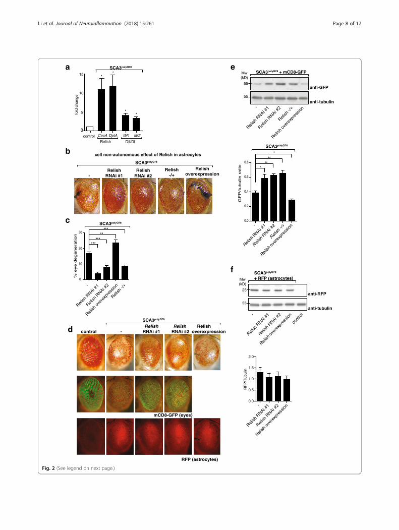
Drosophila, there are two independent NF-κB pathways,activating transcription factors Relish or Dif/Dorsal re-spectively. We examined expression of anti-microbial pep-tides (AMPs) in our SCA3 model, transcriptional NF-κBtargets which help fight infection [27]. In inflammatory re-sponses, these peptides are considered to aid in activatingand recruiting immune cells (reviewed in [28]). Whileboth NF-κB pathways are activated upon eye-specific ex-pression of SCA3polyQ78, activation of Relish was stronger:AMPs specific for Relish (CecA and DptA) were upregu-lated more than Dif/Dorsal-specific AMPs (IM1 and IM2;Fig. 2a and shown in [29]). Activation of Relish in SCA3po-lyQ78-expressing eyes occurred in astrocytes: GFP undercontrol of the promoter of a Relish-dependent gene wasexpressed predominantly in the astrocytes that werepresent in SCA3polyQ78-expressing eyes (Additional file 4a).In control eyes, no induction of GFP was seen.We further focused on the cell-non-autonomous role of
astrocyte-specific Relish signaling in SCA3. We first con-firmed the results of our screen by using two independentRelish RNAi constructs (efficacy of knockdown, seeAdditional file 4b) that both decreased SCA3polyQ78-in-duced degeneration (Fig. 2b). Inversely, the opposite effectwas seen upon overexpression of Relish. Modulation ofRelish expression in astrocytes did not affect morphologyof control eyes (Additional file 4c). In flies heterozygousfor Relish, degeneration was attenuated, similar toastrocyte-specific Relish RNAi. The effect of Relish in as-trocytes on eye degeneration was quantified (Fig. 2c) bydetermining the fraction of the eyes containing necroticspots. Importantly, the effects of Relish on degenerationare cell-non-autonomous, mediated via astrocytes: whencoexpressing SCA3polyQ78 and RNAi constructs targetingRelish in the eyes, eye degeneration was not attenuated(Additional file 4d).When membrane-targeted mCD8-GFP was used as a
readout for degeneration [23] in eyes expressing SCA3po-lyQ78, the protective or enhancing effects of theastrocyte-specific down- and upregulation of Relishlevels were confirmed (Fig. 2d for GFP images of differ-ent genotypes). To see whether modulating of Relish ex-pression in astrocytes affected the localization ofastrocytes, we coexpressed myr-RFP in astrocytes. Wedid not see an effect of modulation of Relish expressionon the localization of the RFP signal (Fig. 2d for RFP im-ages of different genotypes, Fig. 2f for western blot ana-lysis of lysates). These data suggest that Relish inastrocytes does not have an effect on the localization ofastrocytes in SCA3polyQ78-expressing eyes (additional im-ages are provided in Additional file 4e). We quantifiedthe effect of modulating Relish expression in astrocyteson mCD8-GFP levels in SCA3polyQ78-expressing eyes bywestern blot (Fig. 2e). GFP levels in flies heterozygousfor Relish were similar to those of Relish RNAi in
astrocytes, suggesting the importance of astrocyte-spe-cific Relish signaling in SCA3polyQ78-induced degener-ation. The effect of Relish on mCD8-GFP levels in theeye is specifically related to SCA3polyQ78-induced degen-eration, as in control flies (not expressing SCA3polyQ78)GFP levels were similar, irrespective of Relish expression(Additional file 4f). Together, these experimentsdemonstrate that in our SCA3 model, Relish is activated(and Dif/Dorsal to a lesser extent) and that Relish signalingin astrocytes enhances SCA3polyQ78-induced degeneration.Thus, eye-specific expression of SCA3polyQ78 promotedRelish activation in astrocytes, and this Relish activation en-hanced degeneration in a cell-non-autonomous manner.In the brain, Relish is activated in glia in aging flies as
well as in flies that are mutant for A-T mutated (ATM), akinase associated with ensuring genomic integrity andneurodegeneration [30, 31]. We analyzed contribution ofastrocytes to overall Relish signaling in the fly head in-duced by eye-specific expression of SCA3polyQ78. For this,we examined levels of Relish-dependent AMPs in flyheads expressing SCA3polyQ78 in the eyes together withastrocytes-specific targeting of Relish RNAi or Relish over-expression constructs, or in SCA3polyQ78-expressing fliesheterozygous for Relish. Expression of Relish RNAi con-structs in astrocytes in flies expressing eye-specific SCA3-polyQ78 resulted in decreased expression of Relish-specificAMPs (DptA and AttC) in the head to levels comparableto flies not expressing SCA3polyQ78 (Fig. 3a), whereas over-expression of Relish enhanced expression. SCA3 flies het-erozygous for Relish expressed levels of Relish targetgenes comparable to those expressing Relish RNAi con-structs in the astrocytes. Dif/Dorsal-specific target geneswere unaffected by \modulating Relish levels in astrocytes(Additional file 5). These data highlight the importance ofRelish signaling in astrocytes regarding Relish-dependentimmune signaling in the head.The presence of aggregated or misfolded proteins is
toxic to neurons [32]. Possibly, astrocytes could have aneffect on levels of protein aggregates. To see whetherRelish-specific signaling in astrocytes could have an ef-fect on solubility of SCA3polyQ78 in the eyes, we collectedheads of flies expressing eye-specific SCA3polyQ78 2 daysafter eclosion and analyzed the cell extract. ModulatingRelish expression in astrocytes or heterozygosity for Rel-ish did not affect total levels or solubility of SCA3polyQ78
(Fig. 3b, quantification of three independent experimentsin Fig. 3c). This suggests that the effect of Relish onSCA3polyQ78-induced degeneration is not the result of ei-ther a clearance of SCA3polyQ78 aggregates or of a shiftof the balance of soluble-insoluble SCA3polyQ78 towardsmore soluble SCA3polyQ78.To see whether Relish can modulate SCA3-induced eye
degeneration via Relish-dependent AMPs, we expressedRNAi constructs targeting two Relish-dependent AMPs
Li et al. Journal of Neuroinflammation (2018) 15:261 Page 7 of 17
55
55
a e
Relish
-/+
Relish
ove
rexp
ress
ion
Relish
RNAi #
1
Relish
RNAi #
2-
SCA3polyQ78
SCA3polyQ78 + mCD8-GFP
b
0.0
0.2
0.4
0.6
0.8
***
***
GF
P/tubulin
ratio
Relish
-/+
Relish
ove
rexp
ress
ion
Relish
RNAi #
1
Relish
RNAi #
2
SCA3polyQ78
-Relish
RNAi #1Relish
RNAi #2Relish
-/+Relish
overexpression
CecA
fold
cha
nge
15
10
5
0IM1
*
IM2
Mw (kD)
anti-GFP
0
10
20
30
Relish
-/+
Relish
ove
rexp
ress
ion
Relish
RNAi #
1
Relish
RNAi #
2
SCA3polyQ78
******
*****
% e
ye
de
ge
ne
ratio
n
cell non-autonomous effect of Relish in astrocytes
*
DptA
*
*
controlDif/DlRelish
SCA3polyQ78
-
anti-tubulin
-c
SCA3polyQ78
-Relish
RNAi #1Relish
RNAi #2control
RFP (astrocytes)
mCD8-GFP (eyes)
d
25
55
Relish
ove
rexp
ress
ion
Relish
RNAi #
1
Relish
RNAi #
2-
SCA3polyQ78 + RFP (astrocytes)Mw
(kD)
anti-RFP
anti-tubulin
cont
rol
f
f
0.0
0.5
1.0
1.5
2.0
RF
P/T
ubul
in
Relish
ove
rexp
ress
ion
Relish
RNAi #
1
Relish
RNAi #
2-
Relishoverexpression
Fig. 2 (See legend on next page.)
Li et al. Journal of Neuroinflammation (2018) 15:261 Page 8 of 17

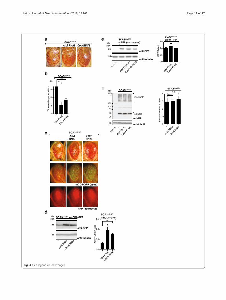
(AttA and CecA; efficacy of knockdown inAdditional file 6a). Astrocyte-specific downregulation ofthese AMPs attenuated the degenerative SCA3 eye pheno-type (Fig. 4a; quantification of degeneration in Fig. 4b),similar to decreasing Relish activity in astrocytes (Fig. 2a, b).No effect on eye morphology was seen when RNAi targetingAMPs was expressed in astrocytes (Additional file 6b).We next analyzed mCD8-GFP levels in the eyes as a
measure of degeneration in SCA3polyQ78-expressing eyesand examined the effect of astrocyte-specific knockdownof Relish-specific AMPs. Levels of mCD8-GFP were in-creased upon knockdown of AMPs, demonstrating at-tenuation of SCA3polyQ78-induced eye degeneration(Fig. 4c; analysis on western blot and quantification ofthree independent experiments in Fig. 4d). In control eyes,no effects of downregulation of Relish-specific AMPs ex-pression in astrocytes on mCD8-GFP levels in the eyeswere found (Additional file 6c). We also examinedwhether astrocyte-specific knockdown of Relish-specificAMPs could influence the presence of astrocytes in SCA3-polyQ78-expressing eyes, but did not see an effect (Fig. 4c),and the numbers of astrocytes were also not affected, asanalyzed by levels of myr-RFP in astrocytes (Fig. 4e).When we examined the effect of astrocyte-specific
downregulation of AMPs on SCA3polyQ78 levels or solubil-ity, we did not observe an effect (Fig. 4f; quantification ofthree independent experiments), similar to modulatingRelish levels in astrocytes. These data show that the effectof Relish on degeneration in our SCA3 model is, to a sig-nificant extent, mediated via the generation of AMPs, andthat the effect of AMPs does not occur via alterations inSCA3polyQ78 solubility.We next tested whether the observed effects of the
Relish pathway in astrocytes on (short term) degeneration
also translates into a (long term) survival benefit in fliesexpressing SCA3polyQ78 in neurons. Astrocyte-specificRelish inhibition may also extend lifespan in neurons ex-pressing SCA3polyQ78, given our observations of Relish inastrocytes on the SCA3 eye phenotype. We expressedSCA3polyQ78 pan-neuronally (not in astrocytes) and exam-ined effects of modulation of Relish expression specificallyin astrocytes (specificity of expression shown inAdditional file 7a) using the following two independentbinary expression systems (Additional file 7b): UAS-Gal4and QUAS-QF2. To avoid potential detrimental effects ofSCA3polyQ78 during development, we expressed SCA3po-lyQ78 in adult flies only, using the inducible “Q system,” inwhich the neuronally expressed transcription factor (QF2)that induces SCA3polyQ78 expression is coexpressed withQF-suppressor QS [20] (Additional file 7b). This way, ex-pression of SCA3polyQ78 is suppressed during development(Fig. 5a). Feeding quinic acid suppresses QS, thus alleviat-ing inhibition of QF2 resulting in the neuron-specific ex-pression of SCA3polyQ78, of which the extent ofaggregation increased over time (Fig. 5a). The lifespan offlies expressing SCA3polyQ78 in neurons was significantlyshortened (Fig. 5b). Quinic acid by itself did not affect life-span (Additional file 7c), excluding non-specific effects.Relish-dependent gene expression was increased in fliesthat expressed SCA3polyQ78 in neurons (Fig. 5c), similar toexpression of SCA3polyQ78 in the eyes (Fig. 2a).We next wished to examine whether modulating
Relish levels in astrocytes in adult flies could have aneffect on lifespan of flies that expressed SCA3polyQ78
pan-neuronally. For this, levels of Relish were modulatedin astrocytes of adult flies to exclude effects on develop-ment. Hereto, we used the transcription factor Gal4 todrive the expression of the Relish constructs. At a lower
(See figure on previous page.)Fig. 2 Effect of NF-κB transcription factor Relish in astrocytes on SCA3polyQ78-induced degeneration. a Quantification of Relish or Dif/Dorsalactivation in the head upon eye-specific expression of SCA3polyQ78. Heads of control flies or flies expressing SCA3polyQ78 in the eyes were analyzedfor expression of Relish target genes (CecA, DptA) or Dif/Dorsal target genes (IM-1, IM-2). b Effect of modulation of Relish expression in astrocyteson SCA3polyQ78-induced eye degeneration. Representative images of fly eyes expressing SCA3polyQ78 (−) or SCA3polyQ78 together with eitherastrocyte-specific expression of two independent RNAi constructs targeting Relish (Relish RNAi #1 and Relish RNAi #2), a Relish overexpressionconstruct (Relish overexpression) or flies heterozygous for Relish (Relish −/+). c Quantification of the fraction of degeneration of the eyes shown inb. In these and other eye quantification experiments, at least 80 eyes of females were counted per genotype. Data are representative of at leastthree independent experiments ± SEM. d The effect of modulation of Relish expression in astrocytes on GFP fluorescence in SCA3polyQ78-expressingeyes together with eye-specific mCD8-GFP and on the localization of astrocytes (expressing myr-RFP). Representative images of control eyes, eyesexpressing mCD8-GFP, and astrocyte-specific myr-RFP (RFP) or together with coexpression of SCA3polyQ78 in the absence or presence of RelishRNAi constructs (Relish RNAi #1 and Relish RNAi #2) or Relish overexpression in astrocytes. e Quantification of the effect of Relish signaling inastrocytes on SCA3polyQ78-induced degeneration by using eye-specific expression of mCD8-GFP. Levels of mCD8-GFP were determined in lysatesof fly heads of flies expressing SCA3polyQ78 together with mCD8-GFP in the eyes and the effect of astrocyte-specific expression of Relish RNAiconstructs (Relish RNAi #1 and Relish RNAi #2) in astrocytes, heterozygosity for Relish (Relish −/+), or overexpression of Relish in astrocytes wasanalyzed. Tubulin was used as a control for equal loading. Quantification of western blots is of at least three independent experiments. f Analysisof myr-RFP expression in astrocytes in lysates of control flies or flies expressing myr-RFP in astrocytes together with eye-specific SCA3polyQ78 in thepresence or absence of astrocyte-targeted Relish RNAi (Relish RNAi #1 or #2) or Relish overexpression. Quantification of western blots in twoindependent experiments. Genotypes: a control, GMR-QF2/+. SCA3polyQ78, GMR-QF2/+; QUAS- SCA3polyQ78/+. b, c -, GMR-QF2/+; QUAS-SCA3polyQ78:: alrm-Gal4/+. Relish RNAi #1, GMR-QF2/+; QUAS-SCA3polyQ78:: alrm-Gal4/UAS-Relish RNAi #1. Relish RNAi #2, GMR-QF2/+; QUAS-SCA3polyQ78:: alrm-Gal4/UAS-RelishRNAi #2. Relish overexpression, GMR-QF2/+; QUAS-SCA3polyQ78:: alrm-Gal4/UAS-GFP-Relish; Relish−/+, GMR-QF2/+; QUAS-SCA3polyQ78:: alrm-Gal4; RelishE20/+. d–f As in b, but with additional eye-specific expression of QUAS-CD8-GFP or astrocyte-specific expression of UAS-myr-RFP
Li et al. Journal of Neuroinflammation (2018) 15:261 Page 9 of 17
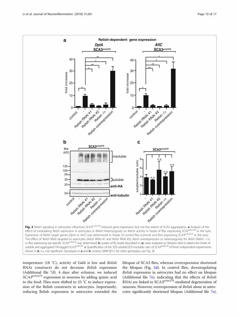
temperature (18 °C), activity of Gal4 is low and RelishRNAi constructs do not decrease Relish expression(Additional file 7d). 4 days after eclosion, we inducedSCA3polyQ78 expression in neurons by adding quinic acidto the food. Flies were shifted to 25 °C to induce expres-sion of the Relish constructs in astrocytes. Importantly,reducing Relish expression in astrocytes extended the
lifespan of SCA3 flies, whereas overexpression shortenedthe lifespan (Fig. 5d). In control flies, downregulatingRelish expression in astrocytes had no effect on lifespan(Additional file 7e), indicating that the effects of RelishRNAi are linked to SCA3polyQ78-mediated degeneration ofneurons. However, overexpression of Relish alone in astro-cytes significantly shortened lifespan (Additional file 7e),
25
aAttCDptA
0
10
20
30
40
cont
rol
Relish
RNAi #
1
Relish
RNAi #
2
Relish
-/+
Relish
ove
rexp
ress
ion
SCA3polyQ78 SCA3polyQ78
Relish-dependent gene expression
b
anti-HA
0
2
4
6
8
solu
ble
/inso
luble
ra
tio
Relish
RNAi #
1
Relish
RNAi #
2
Relish
-/+
Relish
ove
rexp
ress
ion
SCA3polyQ78 SCA3polyQ78
cont
rol
Relish
RNAi #
1
Relish
RNAi #
2
Relish
-/+
Relish
ove
rexp
ress
ion
130100
55
35
55
Mw (kD)
insoluble
soluble
-
fold
incre
ase
n.s.
n.s.
n.s.
n.s.
anti-tubulin -
-
c
***
***
*
0
fold
incre
ase
contr
ol
Relish
RNAi #
1
Relish
RNAi #
2
Relish
-/+
Relish
over
expr
essio
n
30
10
20
40
-
****
**
*
Fig. 3 Relish signaling in astrocytes influences SCA3polyQ78-induced gene expression but not the extent of SCA3 aggregation. a Analysis of theeffect of modulating Relish expression in astrocytes or Relish heterozygosity on Relish activity in heads of flies expressing SCA3polyQ78 in the eyes.Expression of Relish target genes (DptA or AttC) was determined in heads of control flies (control) and flies expressing SCA3polyQ78 in the eyes.The effect of Relish RNAi targeted to astrocytes (Relish RNAi #1 and Relish RNAi #2), Relish overexpression or heterozygosity for Relish (Relish −/+)in flies expressing eye-specific SCA3polyQ78 was determined. b Lysates of fly heads described in (a) were analyzed on Western blot to determine levels ofsoluble and aggregated HA-tagged SCA3polyQ78. c Quantification of the SDS-soluble/SDS-insoluble ratio of SCA3polyQ78 of three independent experimentsshown in b. n.s., not significant. Genotypes in a and b: control, GMR-QF/+; for other genotypes, see Fig. 2b
Li et al. Journal of Neuroinflammation (2018) 15:261 Page 10 of 17
a e
AttA R
NAi
CecA R
NAiAttA
RNAi
CecA R
NAi
d
GF
P/T
ub
ulin
ra
tio
1.5
1.0
0.5
0.0
AttA R
NAi
CecA R
NAi
55
55
anti-GFP
AttA R
NAi
CecA R
NAi
Mw(kD)
AttA RNAi CecA RNAi %
eye
de
ge
ne
ratio
n
Mw (kD)
AttA R
NAi
CecA R
NAi
solu
ble
/inso
lub
le r
atio
anti-HA
insoluble
soluble
cont
rol
25
130100
55
35
55
0
5
10
15
20
*
1
4
3
2
0
-
-
SCA3polyQ78
SCA3polyQ78
******
SCA3polyQ78 +mCD8-GFP
anti-tubulin-
-
- -
SCA3polyQ78 SCA3polyQ78
SCA3polyQ78
+mCD8-GFP
f
**
n.s.n.s.
anti-tubulin
b
SCA3polyQ78
-AttA RNAi
CecARNAi
c
RFP (astrocytes)
mCD8-GFP (eyes)
25
55
cont
rol
AttA R
NAi #1
CecA R
NAi #2-
SCA3polyQ78 + RFP (astrocytes)Mw
(kD)
anti-RFP
anti-tubulin0.0
0.5
1.0
1.5
RF
P/T
ubulin
AttA R
NAi
CecA R
NAi-
SCA3polyQ78
+myr-RFP
Fig. 4 (See legend on next page.)
Li et al. Journal of Neuroinflammation (2018) 15:261 Page 11 of 17

precluding analysis on Relish overexpression on SCA3po-lyQ78-mediated shortening of lifespan.In parallel, SCA3-related effects on motor function were
analyzed, measured as climbing ability. The SCA3poly-Q78-induced decrease in climbing ability was partiallyalleviated by knockdown of Relish in astrocytes(Additional file 7f). As was observed with modulating Rel-ish expression in astrocytes in SCA3polyQ78-expressingeyes, no effects of Relish in astrocytes on total brainSCA3polyQ78 aggregation load or levels could be detected(Additional file 7g).The effects of Relish modulation on SCA3-related de-
generation were not related to alterations of the levels ofSCA3polyQ78 aggregates (Fig. 3b and Additional file 7g).Therefore, we hypothesized that modulation of the Rel-ish pathway in astrocytes could be more generic andrelevant to a neurodegenerative disease associated withthe presence of extraneuronal aggregates, such as inAlzheimer’s disease. For this, we turned to an establishedDrosophila model of Alzheimer’s disease [18], in whichthe Amyloid beta (Abeta42; Aβ42) peptides are fused toa secretion signal peptide (of the necrotic gene), resultingin the secretion of Aβ42 peptides. When expressed inneurons (Additional file 7h), these Aβ42 peptides are se-creted and form extracellular aggregates in flies, reminis-cent of the plaques in human AD [33]. In this AD flymodel, flies displayed progressive neurodegeneration,with mobility and memory deficits, and reduced lifespan[18, 34]. In line with our hypothesis, the lifespan of fliesexpressing Aβ42 in neurons was shortened, but ex-tended when Relish expression was suppressed in astro-cytes, using two independent RNAi lines targeting Relish(Fig. 5e). This, together with the data on SCA3, demon-strates that modulating Relish signaling in astrocytes hasbeneficial effects in neurodegeneration, irrespective ofthe aggregation process that initiates it.Our data show that astrocytes can modulate the rate
of neurodegenerative disease progression in a cell-non-
autonomous manner. This is the first time to show thatneuron-specific expression of a pathological,degeneration-inducing protein results in activation ofastrocytes, which can subsequently influence symptomsof degeneration.
DiscussionAstrocytes become reactive during the course of a numberof age-related neurodegenerative diseases associated withaggregates [6], and several reports have suggested thatduring the course of disease astrocytes gain neurotoxicproperties ([4] reviewed in [6]). However, potential mecha-nisms for how astrocytes affect progression of disease haveyet been lacking, in part due to the fact that in most stud-ies disease-causing genes are expressed in both neuronaland non-neuronal cells in the brain. Expression ofaggregation-prone proteins in astrocytes can cell-non-au-tonomously influence neuronal viability [5], reviewed in[6]. However, putative cell-non-autonomous contributionsof astrocytes to neurodegeneration in response to neuronsexpressing aggregation-prone proteins are unclear. Here,we investigate how signaling in astrocytes can influenceneurons that express aggregation-prone, disease-causingproteins, thus investigating cell-non-autonomous contri-butions of astrocytes to neurodegeneration. We identifiedastrocytes in Drosophila eyes expressing SCA3polyQ78 butnot in control eyes or eyes expressing SCA3polyQ27
(Fig. 1d), suggesting a role for astrocytes. We further in-vestigated putative cell-non-autonomous contributions ofgenes in astrocytes in a candidate RNAi screen, where theeffect of RNAi-mediated downregulation of individualgenes in astrocytes on SCA3polyQ78-induced eye degener-ation was determined. This resulted in the identificationof astrocyte-specific enhancers and suppressors of theSCA3-related neurodegeneration (Additional file 3). Theseresults demonstrate, for the first time, the importance ofsignaling in astrocytes induced by specific expression ofaggregation-prone, neurodegeneration-associated proteins.
(See figure on previous page.)Fig. 4 Relish target genes influence SCA3polyQ78-induced degeneration but not SCA3polyQ78 aggregation or SCA3polyQ78-induced relocation ofastrocytes. a Representative images of flies expressing eye-specific SCA3polyQ78 compared to flies coexpressing RNAi constructs in astrocytestargeting Relish target genes AttA or CecA. b Quantification of the fraction of degeneration of the eyes shown in a. Data are representative of atleast three independent experiments. c Representative images of the effect of downregulation of Relish target genes AttA or CecA in astrocyteson SCA3-induced degeneration or the relocation of astrocytes upon expression of eye-specific SCA3polyQ78 by using eye-specific expression ofmCD8-GFP and astrocyte-targeted expression of myr-RFP (RFP). d Quantification of the effect of Relish target genes in astrocytes on SCA3-induceddegeneration by using membrane-targeted mCD8-GFP. Lysates of fly heads expressing mCD8-GFP and SCA3polyQ78 in the eyes were analyzed onwestern blot and compared to flies coexpressing RNAi constructs targeting AttA or CecA in astrocytes. Quantifications are of at least three independentexperiments. e No effect on Relish target genes on levels of astrocytes expressing myr-RFP. Lysates of fly heads of control flies or flies expressingmyr-RFP in astrocytes together with SCA3polyQ78 in the eyes in the presence or absence or AMP RNAi constructs (AttA RNAi or CecA RNAi) wereanalyzed on western blot for expression for myr-RFP and tubulin. Quantifications are of two independent experiments. f Analysis of the effect of Relishtarget genes in astrocytes on SDS-solubility of SCA3polyQ78 expressed in the eyes. Head lysates of flies expressing HA-tagged SCA3polyQ78 in the eyeswere compared to flies coexpressing RNAi constructs targeting AttA or CecA in astrocytes. Quantifications of the SDS-soluble/SDS-insoluble ratio are ofat least three independent experiments. Genotypes a -: GMR-QF2/+; QUAS-SCA3polyQ78:: alrm-Gal4/+. AttA RNAi, GMR-QF2/+; QUAS-SCA3polyQ78:: alrm-Gal4/UAS-AttA RNAi. CecA RNAi, GMR-QF2/+; QUAS-SCA3polyQ78:: alrm-Gal4/+; UAS-CecA RNAi/+. c, d Con: GMR-QF2/+, the rest of the genotypes as in abut with coexpression of QUAS-mCD8-GFP or UAS-myr-RFP. e, f Con: GMR-QF2/+; other samples as in a
Li et al. Journal of Neuroinflammation (2018) 15:261 Page 12 of 17

a
b
anti-tubulin
d
quinic acid+ + +- - ++ + +- +
adult flieslarvae
25
130100
55
35
55
Mw (kD)
+
surv
ial (
%)
0 20 40 600
20
40
60
80
100
AttC DptA
*
*
CecA
fold
incr
ease
0
5
10
15
age (days)1 10 20
anti-HA
age (days)
insoluble
soluble
age (days)
sur
vial
(%
)
neurons astrocytes
---
Aβ42Aβ42Aβ42
Relish RNAi#1Relish RNAi#2
****** ***
e
SCA3polyQ78
**
control
SCA3polyQ78 c
0 20 40 60 80 1000
20
40
60
80
100
0 20 40 600
20
40
60
80
100
day
neurons astrocytes
Relish RNAi#1SCA3polyQ78
Relish RNAi#2SCA3polyQ78
Relish overexpressionSCA3polyQ78
SCA3polyQ78 ---
******
******
sur
vial
(%
)
***SCA3polyQ78 control
Fig. 5 (See legend on next page.)
Li et al. Journal of Neuroinflammation (2018) 15:261 Page 13 of 17

Astrocytes in Drosophila share structural and functionalsimilarities with mammalian astrocytes: amongst others,they provide trophic support to neurons and are involvedin neurotransmitter recycling (reviewed in [35]). Therefore,genes that we identified in our screen may be relevant formammalian astrocytes as well.Conserved NF-κB transcription factor Relish was iden-
tified as an enhancer in our SCA3 model, and its activitywas upregulated in the head upon eye-specific SCA3po-lyQ78 expression. We therefore focused on Relish signal-ing for more in-depth analyses. Relish was specificallyactivated in astrocytes present in eyes expressing SCA3-polyQ78, but not in astrocytes elsewhere in the head(Additional file 4a). Downregulation of expression in as-trocytes of both Relish, but also Relish-dependent AMPsalleviated SCA3polyQ78-induced eye degeneration (Figs. 2and 4). In contrast, we did not see an effect of downreg-ulation of Relish expression on degeneration in eyescoexpressing SCA3polyQ78 and Relish RNAi constructs(Additional file 4d). Thus, eye-specific expression ofaggregation-prone SCA3polyQ78 results in cell-non-au-tonomous activation of NF-κB in astrocytes. We next in-vestigated possible cell-non-autonomous contributionsof astrocytes to neurodegeneration.Downregulation of Relish expression in astrocytes not
only delayed neurodegeneration (suggested by enhancedmobility, Additional file 7f) and extended lifespan of fliesexpressing SCA3polyQ78 in neurons (Fig. 5d), but also ex-tended lifespan in flies expressing neuronal Aβ42 peptides(Fig. 5e). We did not see an effect of downregulating Rel-ish expression in astrocytes on lifespan (Additional file 7e),underscoring the efficacy of using this pathway as a targetto inhibit the detrimental effects of SCA3polyQ78. However,overexpression of Relish in astrocytes was sufficient toshorten lifespan, demonstrating toxic effects of Relish.This is in agreement with earlier studies demonstratingthat expression of Relish or Relish-specific AMPs was
sufficient to induce neurodegeneration [30, 36]. Of par-ticular interest is that in aging flies Relish activity andlevels of AMPs were increased in both neurons and glia,and constitutes an important determinant of lifespan con-trol and the onset of neurodegeneration [31]. In additionto this, our data demonstrate the importance ofcell-non-autonomous Relish signaling in astrocytes inneurodegeneration associated with aggregates, which isgenerally aging-associated. The presence of aggregation-prone, neurodegeneration-associated proteins in neuronsresulted in Relish signaling in astrocytes, which subse-quently influenced neuronal functioning. Together, thesefindings suggest the importance of NF-κB activity in astro-cytes in age-associated neurodegenerative diseases.Our data thus provide direct evidence for earlier
suggestions that astrocytes are not only activated in re-sponse to neurodegeneration (as, e.g., demonstrated byelevation of GFAP, glial fibrillary acidic protein, a markerfor astrocyte activation) [5], but indeed can modulatedisease progression. The relevance of the cell-non-au-tonomous Relish activation in astrocytes is underscoredby the observation that downregulation of Relish in as-trocytes can influence disease progression. A toxic gainof function in astrocytes was also demonstrated in amice expressing human aggregation-prone TAR DNA-binding protein 43 (TDP-43) in neurons [37], resultingin alterations in protein expression in astrocytes.Depletion of an upregulated, astrocyte-specific factor re-duced neurotoxicity, underscoring the importance of as-trocytes to neurotoxicity. Our findings that genes in theNF-κB pathway are key mediators of such a response ex-tends the observation that astrocyte-specific, transientexpression of IKK (IκB kinase 2), resulted in neurode-generation in mice [38].While Lattke et al. showed sufficiency of astrocyte-spe-
cific NF-κB signaling in inducing neurodegeneration,our data now demonstrate that signals from neurons
(See figure on previous page.)Fig. 5 Analysis of SCA3polyQ78 expression in neurons and the effect of Relish in astrocytes on neuronal expression of SCA3polyQ78 or Abeta42. aInducible expression of SCA3polyQ78 in neurons can be suppressed during development. Control larvae or larvae expressing inducible SCA3polyQ78
were cultured on food with and without quinic acid. Flies expressing inducible SCA3polyQ78 were cultured on food with quinic acid for the timesindicated to induce SCA3polyQ78 expression. Levels and aggregation of HA-tagged SCA3polyQ78 were determined in lysates of larvae or fly heads onwestern blot. Tubulin was used to verify equal loading. b Effect of inducible SCA3polyQ78 on lifespan. Control flies or 4-day-old flies expressinginducible SCA3polyQ78 were cultured on food containing quinic acid and the lifespan was analyzed. c Expression of Relish target genes AttC, DptAand CecA was analyzed in heads of 15-day-old flies expressing SCA3polyQ78 in neurons. d Analysis of modulation of Relish expression specifically inastrocytes on the lifespan of flies expressing SCA3polyQ78 in neurons. Flies expressing SCA3polyQ78 in neurons induced as in b were compared toflies coexpressing astrocyte-targeted Relish RNAi (Relish RNAi #1 or #2) or overexpressing Relish in astrocytes. To induce changes in Relish expression only inadult flies, crosses were kept at 18 °C. 4-day-old progeny were shifted to 25 °C to allow expression of Relish constructs in astrocytes. e Downregulation ofRelish expression in astrocytes extends lifespan in an Alzheimer model. Lifespan of control flies and flies expressing Abeta42 in neurons were compared toflies expressing Abeta42 (Aβ42) in neurons and Relish RNAi constructs in astrocytes. Induction of Relish RNAi was done as in d. Genotypes: a -, tub-QS/+;alrm-Gal4/+; nSyb-QF2::tub-QS/+. SCA3polyQ78, tub-QS/+; alrm-Gal4::QUAS-SCA3polyQ78/+; nSyb-QF2::tub-QS/+. b, c Control, tub-QS/+; alrm-Gal4/+;nSyb-QF2::tub-QS/+. SCA3polyQ78, tub-QS/+; alrm-Gal4::QUAS-SCA3polyQ78/+; nSyb-QF2::tub-QS/+. d -, tub-QS/+; alrm-Gal4/+; nSyb-QF2::tub-QS/+.SCA3polyQ78, tub-QS/+; alrm-Gal4::QUAS-SCA3polyQ78/+; nSyb-QF::tub-QS/+; Relish RNAi #1, Relish RNAi #2, or Relish overexpression, as in SCA3polyQ78, butwith expression of UAS-Relish RNAi #1 or #2 or UAS-GFP-Relish in astrocytes. e -, alrm-Gal4/+; nSyb-QF2. Aβ42: alrm-Gal4::QUAS-Abeta42/+; nSyb-QF2/+.Relish RNAi (#1 or #2), alrm-Gal4:: QUAS-Abeta42/UAS-Relish RNAi #1 or #2); nSyb-QF2/+
Li et al. Journal of Neuroinflammation (2018) 15:261 Page 14 of 17
expressing aggregation-prone proteins suffice to inducecell-non-autonomous NF-κB activation in astrocytes.The importance of NF-κB activation in astrocytes wassuggested by date presented in Fig. 3a andAdditional file 5a. The presence of astrocytes inSCA3polyQ78-expressing eyes (Fig. 1d, Additional file 4)suggests recruitment of astrocytes, as shown for Alzhei-mer’s disease (reviewed in [39]). Downregulation of NF-κBin astrocytes did not appear to alter the presence ofastrocytes in SCA3polyQ78-expressing eyes (Fig. 2d,Additional file 4e), suggesting that additional signaling inastrocytes promotes the recruitment of astrocytes to de-generating cells. Together, these data demonstrate the ex-istence and importance of intercellular signaling betweenneurons and astrocytes as an important determinant forthe speed of neurodegeneration.The nature of the neuronal signals that trigger NF-κB
activation in astrocytes remains to be elucidated. Possibly,damage-associated molecular patterns (DAMPs), includ-ing SCA3polyQ78 aggregates, released from neurons can re-sult in activation of NF-κB in astrocytes. It is unlikely thatthe Relish canonical pathway, headed by PGRP-LC(reviewed in [27]), accounts for Relish activation, sincemodulation of PGRP-LC did not significantly affect theSCA3 eye phenotype (Additional file 1). Previous reportshave demonstrated Relish activation independent ofPGRP-LC, either by neuropeptides or nitric oxide [40, 41],which may account for Relish activation in our model.Whereas we show that astrocytes can directly modu-
late neuronal health, this appeared not to be associatedwith alteration in the burden of protein aggregates inthe brain. However, decreasing the levels of misfolded oraggregated proteins has been suggested as a therapeuticstrategy [42]. Furthermore, reduction of oxidativedamage can decrease degeneration [43]. Thus, a combin-ation of therapeutic strategies targeting aggregates,astrocyte-mediated neurotoxicity as well as oxidativedamage may have optimal effects in alleviatingaggregates-associated neurodegeneration.Targeting NF-κB signaling in astrocytes to alleviate
neurodegeneration may be more broadly applicable: thelifespan of flies expressing Aβ peptides, present in pa-tients with AD, was extended upon astrocyte-specific in-hibition of NF-κB (Fig. 5e). Thus, despite intracellularlocalization of aggregates in SCA3 and extracellularlocalization of Aβ peptides in AD, astrocyte-specificNF-κB inhibition extended lifespan in fly models forboth diseases. This suggests that targeting NF-κB in as-trocytes may be beneficial in other aggregates-associatedneurodegenerative diseases.
ConclusionIn conclusion, our data demonstrate that astrocytes re-spond to cellular stress or damage in neurons induced by
neuronal expression of disease-associated, aggregation-prone proteins. These astrocytic responses are importantmodulators of neurodegeneration. We demonstrate thatactivation of transcription factor NF-κB Relish occurs inastrocytes in response to proteotoxic stress in neurons,and that in this situation astrocyte-specific inhibition ofRelish enhances vitality and extends lifespan. While pro-teotoxic stress in astrocytes can contribute to neurodegen-eration, these data highlight the importance of astrocyticresponses to cellular stress or damage in neurons, andprovide putative therapeutic potential.
Additional files
Additional file 1: Genetic setup to analyze cell-non-autonomous orcell-autonomous roles of genes to SCA3. a To model SCA3 in Drosophilaeyes, we used the Q (QUAS-QF) system, in which a QUAS-SCA3polyQ78
(or QUAS-SCA3polyQ27) construct was induced by QF2 specifically expressedin the eyes, GMR-QF2. To downregulate gene expression in astrocytes,we used the UAS-Gal4 system. UAS-RNAi constructs were specificallyexpressed in astrocytes by using a Gal4 expressed in astrocytes, alrm-Gal4.Cell-autonomous roles of genes in SCA3 can be analyzed by coexpressingUAS-SCA3polyQ78 with UAS constructs in the eye, using eye-specific GMR-Gal4.b Independent expression of the QUAS-QF2 system (GFP in the eyes) andthe UAS-Gal4 system (RFP in astrocytes) in late pupa. Genotype, GMR-QF2/+;alrm-Gal4/UAS-myr-RFP; QUAS-mCD8-GFP/+. (PDF 186 kb)
Additional file 2: Analysis of localization of astrocytes induced by eye-specific expression of SCA3polyQ78. The eyes of control flies that expressmyr-RFP in astrocytes in the absence or presence of SCA3polyQ27 or SCA3-polyQ78 were analyzed for expression and localization of RFP. Genotypes:control, GMR-QF2/+; alrm-Gal4::UAS-myr-RFP/+. SCA3polyQ27, GMR-QF2/+;alrm-Gal4::UAS-myr-RFP/QUAS-SCA3polyQ27. SCA3polyQ78, GMR-QF2/+; alrm-Gal4::UAS-myr-RFP/QUAS-SCA3polyQ78. (PDF 4177 kb)
Additional file 3: RNAi screen to determine the contribution of genesin astrocytes to SCA3polyQ78-induced eye degeneration. Flies expressingSCA3polyQ78 in the eyes together with alrm-Gal4 were crossed to fly linescontaining UAS-RNAi constructs to specifically knock down genes inastrocytes. Progeny expressing SCA3polyQ78 in the eyes with individualgenes in astrocytes knocked down were analyzed for SCA3polyQ78-induced eye degeneration. The extent of degeneration was quantifiedand plotted in a table. The percentage of degeneration in SCA3polyQ78
eyes was set at 100% and the effect of RNAi-mediated gene knockdownin astrocytes was determined by the percentage of deviation from thecontrol. The suppressors of degeneration (in which RNAi-induced down-regulation of gene expression in astrocytes enhanced the SCA3polyQ78
phenotype, 60% or more deviation from the control) are shown inorange, the enhancers (40% or more deviation from the control) inpurple. Genotype of the crosses SCA3polyQ78, GMR-QF2/+; QUAS-SCA3polyQ78:: alrm-Gal4/+. RNAi: GMR-QF2/+; QUAS-SCA3polyQ78:: alrm-Gal4/+ together with UAS-RNAi. (PDF 149 kb)
Additional file 4: a Activation of Relish in SCA3polyQ78-expressing eyespredominantly occurred in astrocytes. (top) Representative picture of acontrol eye expressing GFP under the control of the Attacin promoter(att-GFP) and astrocytes expressing RFP or an eye coexpressing SCA3polyQ78.Pictures were taken from live eyes. (bottom) Representative picture of adissected eye expressing SCA3polyQ78, together with att-GFP andRFP-expressing astrocytes. Nuclei are stained with Hoechst (blue). Notethat in the bottom panel there is only an overlap between the RFP andGFP signal in the ommatidia. This may cause the imperfect overlapbetween the RFP and GFP signal in SCA3polyQ78-expressing flies in thetop panel: the RFP signal from the underlying astrocytes. b Efficacy of Relishknockdown. Flies expressing daughterless-Gal4 (da-Gal4, ubiquitouslyexpressed) were crossed to control flies (w1118) or fly lines containing RNAiconstructs targeting Relish (Relish RNAi #1 and Relish RNAi #2) andexpression of Relish in the progeny was determined. c Modulation of
Li et al. Journal of Neuroinflammation (2018) 15:261 Page 15 of 17
expression in astrocytes does not affect morphology of control eyes.Control flies or fly lines containing RNAi constructs targeting Relish (RelishRNAi #1 and Relish RNAi #2) or expressing Relish were crossed to alrm-Gal4flies, and the morphology of the eyes was determined. d No cell-autonomous effect of Relish on the degenerative SCA3polyQ78 eye pheno-type. Constructs targeting Relish were coexpressed in the eyes expressingSCA3polyQ78. e Expression of Relish RNAi in astrocytes does not influence therelocation of the astrocytes to the eye induced by eye-specific expression ofSCA3polyQ78. Images of multiple flies to show similar levels of RFP in theeyes between flies expressing eye-specific of SCA3polyQ78 and astrocyte-specific myr-RFP (RFP) in the absence or presence of Relish RNAi targeted toastrocytes. f Modulating Relish expression in astrocytes does not affect levelsof mCD8-GFP in the eyes. Lysates of fly heads expressing eye-specificmCD8-GFP were compared to fly heads expressing eye-specific mCD8-GFPtogether with astrocyte-specific knockdown or overexpression of Relishconstructs or flies heterozygous for Relish (Relish −/+). Analysis of themCD8-GFP/tubulin ratio of two independent experiments is shown below.Genotypes a control, GMR-QF2/+; alrm-Gal4::UAS-myr-RFP; att-GFP/+. SCA3po-lyQ78, GMR-QF2/+; alrm-Gal4::UAS-myr-RFP::QUAS-SCA3polyQ78; att-GFP/+. bControl, da-Gal4/+. Relish RNAi #1, UAS-Relish RNAi #1/+; da-Gal4/+. RelishRNAi #2, UAS-Relish RNAi #2/+; da-Gal4/+. c Control, alrm-Gal4/+. Relish RNAi#1, alrm-Gal4/UAS-Relish RNAi#1. Relish RNAi #2, alrm-Gal4/+; UAS-RelishRNAi#2/+. GFP-Relish: alrm-Gal4/UAS-GFP-Relish. d -, GMR-Gal4::UAS-SCA3po-lyQ78/+. Relish RNAi#1, GMR-Gal4:: UAS-SCA3polyQ78/UAS-Relish RNAi#1. RelishRNAi#2, GMR-Gal4::UAS-SCA3polyQ78/UAS-Relish RNAi #2. Relish overex-pression, GMR-Gal4::UAS-SCA3polyQ78/UAS-GFP-Relish. e As in Fig. 2d. f -,GMR-QF2/+; alrm-Gal4/+ QUAS-mCD8-GFP. Relish RNAi #1, GMR-QF2/+;alrm-Gal4/UAS-Relish RNAi #1; QUAS-mCD8-GFP/+. Relish RNAi #2, GMR-QF2/+; alrm-Gal4/UAS-Relish RNAi #2; QUAS-mCD8-GFP/+. Relish−/+,GMR-QF2/+; alrm-Gal4; Relish E20/QUAS-mCD8-GFP. Relish overexpression,GMR-QF2/+; QUAS- SCA3polyQ78:: alrm-Gal4/UAS-Relish QUAS-mCD8-GFP.(PDF 1700 kb)
Additional file 5: Effect of modulating Relish levels in astrocytes onDif/Dl-dependent gene expression in the head. Expression of Dif/Dl targetgenes (IM1 or IM2) was determined in the heads of control flies (control),flies expressing SCA3polyQ78 in the eyes (-), and the effect of Relish RNAitargeted to astrocytes (Relish RNAi #1 and Relish RNAi #2), Relishoverexpression, or SCA3polyQ78-expressing flies heterozygous for Relish(Relish −/+) on Dif/Dl target gene expression was determined bycomparing them to flies only expressing SCA3polyQ78. (PDF 125 kb)
Additional file 6: a Efficacy of knockdown of CeCA or AttA geneexpression. Flies expressing daughterless-Gal4 (da-Gal4) were crossed tocontrol flies (w1118) or fly lines containing RNAi constructs targeting AttAor CecA and expression of CeCA or AttA in the adult progeny wasdetermined. b Downregulating in astrocytes does not affect eyemorphology. Control flies or fly lines containing RNAi constructs targetingCeCA or AttA were crossed to alrm-Gal4 flies and the morphology of theeyes was determined. c No effect of modulating of Relish-dependent AMPexpression in astrocytes on mCD8-GFP levels in the eyes. Flies expressingeye-specific mCD8-GFP were compared to flies coexpressing RNAi constructstargeting AttA or CecA in astrocytes. Genotypes (a): control, da-Gal4/+. AttA,UAS− AttA/+ da-Gal4/+; CecA, UAS- CecA/+; da-Gal4/+. (b) Control, alrm-Gal4/+. AttA RNAi, alrm-Gal 4/UAS-AttA RNAi. CecA RNAi, alrm-Gal 4/+; UAS-CecARNAi/+. (c) As in (b), but with coexpression of QUAS-mCD8-GFP. (c) Control:GMR-QF2/+; alrm-Gal4/+; QUAS-mCD8GFP/+. AttA RNAi, GMR-QF2/+; alrm-Gal4/UAS-AttA RNA; QUAS-mCD8GFP/+. CecA RNAi, GMR-QF2/+; alrm-Gal4/+;UAS-CecA RNAi/QUAS-mCD8GFP. (PDF 289 kb)
Additional file 7: a The UAS-Gal4 and the QUAS-QF system functionindependently. QUAS-QF-driven GFP expression specifically in neurons(GFP); UAS-Gal4-driven expression of myr-RFP specifically in astrocytes(RFP). Nuclei are stained with Hoechst (blue). b Genetic setup to induciblyexpress SCA3polyQ78 in neurons and simultaneously modulate geneexpression in astrocytes. QUAS-QF was used to express SCA3polyQ78;UAS-Gal4 was used to modulate expression in astrocytes. Neuronallyexpressed QF2 (expressed under control of the pan-neuronal nSyb pro-moter) is suppressed by QS (expressed under control of the tubulin (tub)promoter). This suppression is alleviated by quinic acid, resulting inexpression of SCA3polyQ78. For details on the fly lines used, see experimentalprocedures. c Quinic acid does not affect lifespan. Control flies werecultured on standard fly food with or without quinic acid, and the fraction
of dead flies was determined over time. d Raising flies at 18 °C does not in-duce expression of Relish RNAi constructs. Progeny of flies ubiquitously ex-pressing RNAi constructs targeting Relish (Relish RNAi #1 and Relish RNAi #2)raised at 18 °C were analyzed for expression of Relish as in Additional file 4b.e Effect of modulating expression of Relish in astrocytes on lifespan. Thelifespan of control flies or flies expressing Relish RNAi or overexpressionconstructs specifically in astrocytes was analyzed. f Effect of modulatingRelish expression in astrocytes on impairment of mobility induced byneuronal SCA3
polyQ78expression. Control flies or flies expressing inducible
SCA3polyQ78
together with Relish RNAi or Relish overexpression constructstargeted to astrocytes were analyzed for mobility over time. Scoring wasdone as described in the experimental procedures. g Effect of modulatingRelish expression in astrocytes on SCA3
polyQ78levels or aggregation. Head
lysates of 15-day-old flies as in Fig. 5c were analyzed for expression ofHA-tagged SCA3
polyQ78on western blot. Tubulin was used as a control for
equal loading. h Expression levels of Abeta42. Two independent lines,QUAS-Abeta42 #5 and QUAS-Abeta42 #7 were analyzed for expression ofAbeta42 using 6E10 antibody. Tubulin was used as a control for equalloading. Genotypes: a alrm-Gal4::UAS-myr-RFP; nSyb-QF2/QUAS-mCD8-GFP. ctub-QS/+; alrm-Gal4/+; nSyb-QF2::tub-QS/+. d Genotypes as in Additional file4b. (e) -, tub-QS/+; alrm-Gal4/+; nSyb-QF2: tub-QS/+. Relish RNAi #1, RelishRNAi #2, or Relish overexpression, as in ‘-‘, but with UAS-Relish RNAi #1 or #2or UAS-GFP-Relish. f Genotypes as in Fig. 5d. g As in e. h Control, nSyb-QF2/+. Abeta42 #5 or #7, QUAS-Abeta42/+; nSyb-QF2/+. (PDF 609 kb)
AbbreviationsAD: Alzheimer’s disease; AMPs: Antimicrobial peptides; HD: Huntington’sdisease; NF-κB: Nuclear factor kappa B; PBS: Phosphate-buffered saline;SCA3: Spinocerebellar ataxia type 3; SDS-PAGE: Sodium dodecyl sulfatepolyacrylamide gel electrophoresis
AcknowledgementsWe thank H.H. Kampinga for critically reading the manuscript and usefulcomments, D. Crowther for providing Abeta flies, and C. Klämbt for providingthe alrm-Gal4 line.
FundingThis work was supported by a Rosalind Franklin Fellowship to P.F.D. and a“De Cock Stichting” grant to Y.X.L.
Availability of data and materialsAll data generated or analyzed during this study are included in this publishedarticle (and its supplementary information files).
Authors’ contributionsYXL and PFD designed the research plan, performed the work, and analyzedthe data. OCMS provided the feedback and editorial comments on the manuscript.All authors read and approved the final manuscript.
Ethics approvalAll applicable international, national, and/or institutional guidelines for thecare and use of animals were followed. This article does not contain anystudies with human participants performed by any of the authors.
Consent for publicationNot applicable.
Competing interestsThe authors declare that they have no competing interests.
Publisher’s NoteSpringer Nature remains neutral with regard to jurisdictional claims in publishedmaps and institutional affiliations.
Li et al. Journal of Neuroinflammation (2018) 15:261 Page 16 of 17

Received: 16 April 2018 Accepted: 12 August 2018
References1. Holtman IR, Raj DD, Miller JA, Schaafsma W, Yin Z, Brouwer N, Wes PD,
Möller T, Orre M, Kamphuis W, et al. Induction of a common microglia geneexpression signature by aging and neurodegenerative conditions: a co-expression meta-analysis; 2015. p. 1–18.
2. Wang M, Roussos P, McKenzie A, Zhou X, Kajiwara Y, Brennand KJ, De LucaGC, Crary JF, Casaccia P, Buxbaum JD, et al. Integrative network analysis ofnineteen brain regions identifies molecular signatures and networksunderlying selective regional vulnerability to Alzheimer’s disease. GenomeMed. 2016;8:104.
3. Heneka MT, Kummer MP, Latz E. Innate immune activation inneurodegenerative disease. Nat Rev Immunol. 2014;14:463–77.
4. Liddelow SA, Guttenplan KA, Clarke LE, Bennett FC, Bohlen CJ, Schirmer L,Bennett ML, Munch AE, Chung WS, Peterson TC, et al. Neurotoxic reactiveastrocytes are induced by activated microglia. Nature. 2017;541:481–7.
5. Faideau M, Kim J, Cormier K, Gilmore R, Welch M, Auregan G, Dufour N,Guillermier M, Brouillet E, Hantraye P, et al. In vivo expression ofpolyglutamine-expanded huntingtin by mouse striatal astrocytes impairsglutamate transport: a correlation with Huntington's disease subjects. HumMol Genet. 2010;19:3053–67.
6. Ben Haim L, Carrillo-de Sauvage M-A, Ceyzériat K, Escartin C. Elusive roles forreactive astrocytes in neurodegenerative diseases. Front Cell Neurosci. 2015;9:12957.
7. Oberheim NA, Goldman SA, Nedergaard M. Heterogeneity of astrocyticform and function. Methods Mol Biol. 2012;814:23–45.
8. Seidel K, Siswanto S, Fredrich M, Bouzrou M, Brunt ER, van Leeuwen FW,Kampinga HH, Korf HW, Rüb U, den Dunnen WFA. Polyglutamineaggregation in Huntington's disease and spinocerebellar ataxia type 3:similar mechanisms in aggregate formation. Neuropathol Appl Neurobiol.2015;42(42):153–66.
9. Yankner BA, Lu T, Loerch P. The aging brain. Ann Rev Pathol Mech Dis.2008;3:41–66.
10. Tong X, Ao Y, Faas GC, Nwaobi SE, Xu J, Haustein MD, Anderson MA, ModyI, Olsen ML, Sofroniew MV, Khakh BS. Astrocyte Kir4.1 ion channel deficitscontribute to neuronal dysfunction in Huntington’s disease model mice.Nat Neurosci. 2014;17:694–703.
11. Crotti A, Glass CK. The choreography of neuroinflammation in Huntington’sdisease. Trends Immunol. 2015;36:364–73.
12. Ransohoff RM. How neuroinflammation contributes to neurodegeneration.Science. 2016;353:777–83.
13. Evert BO, Vogt IR, Kindermann C, Ozimek L, de Vos RA, Brunt ER, Schmitt I,Klockgether T, Wüllner U. Inflammatory genes are upregulated in expandedataxin-3-expressing cell lines and spinocerebellar ataxia type 3 brains. JNeurosci Off J Soc Neurosci. 2001;21:5389–96.
14. Koch P, Breuer P, Peitz M, Jungverdorben J, Kesavan J, Poppe D, Doerr J,Ladewig J, Mertens J, Tüting T, et al. Excitation-induced ataxin-3aggregation in neurons from patients with Machado–Joseph disease.Nature. 2011;
15. Hübener J, Weber JJ, Richter C, Honold L, Weiss A, Murad F, Breuer P,Wüllner U, Bellstedt P, Paquet-Durand F, et al. Calpain-mediated ataxin-3cleavage in the molecular pathogenesis of spinocerebellar ataxia type 3(SCA3). Hum Mol Genet. 2013;22:508–18.
16. Goti D. A mutant Ataxin-3 putative-cleavage fragment in brains ofMachado-Joseph disease patients and transgenic mice is cytotoxic above acritical concentration. J Neurosci. 2004;24:10266–79.
17. Foley E, O'Farrell PH. Functional dissection of an innate immune responseby a genome-wide RNAi screen. PLoS Biol. 2004;2:E203.
18. Crowther DC, Page R, Chandraratna D, Lomas DA. A Drosophila model ofAlzheimer’s disease. Methods Enzymol. 2006;412:234–55.
19. Vos MJ, Zijlstra MP, Kanon B, van Waarde-Verhagen MA, Brunt ER,Oosterveld-Hut HM, Carra S, Sibon OC, Kampinga HH. HSPB7 is the mostpotent polyQ aggregation suppressor within the HSPB family of molecularchaperones. Hum Mol Genet. 2010;19:4677–93.
20. Potter CJ, Tasic B, Russler EV, Liang L, Luo L. The Q system: a repressiblebinary system for transgene expression, lineage tracing, and mosaic analysis.Cell. 2010;141:536–48.
21. Warrick JM, Paulson HL, Gray-Board GL, Bui QT, Fischbeck KH, Pittman RN,Bonini NM. Expanded polyglutamine protein forms nuclear inclusions andcauses neural degeneration in Drosophila. Cell. 1998;93:939–49.
22. Warrick JM, Morabito LM, Bilen J, Gordesky-Gold B, Faust LZ, Paulson HL,Bonini NM. Ataxin-3 suppresses polyglutamine neurodegeneration inDrosophila by a ubiquitin-associated mechanism. Mol Cell. 2005;18:37–48.
23. Burr AA, Tsou W-L, Ristic G, Todi SV. Using membrane-targeted greenfluorescent protein to monitor neurotoxic protein-dependent degenerationof Drosophila eyes. J Neurosci Res. 2014;92:1100–9.
24. Kadener S, Villella A, Kula E, Palm K, Pyza E, Botas J, Hall JC, Rosbash M.Neurotoxic protein expression reveals connections between the circadianclock and mating behavior in Drosophila. Proc Natl Acad Sci U S A. 2006;103:13537–42.
25. Favrin G, Bean DM, Bilsland E, Boyer H, Fischer BE, Russell S, Crowther DC,Baylis HA, Oliver SG, Giannakou ME. Identification of novel modifiers of Aβtoxicity by transcriptomic analysis in the fruitfly. Sci Rep. 2013;3:3512.
26. Liddelow SA, Barres BA. Reactive astrocytes: production, function, andtherapeutic potential. Immunity. 2017;46:957–67.
27. Lemaitre B, Hoffmann J. The host defense of Drosophila melanogaster.Annu Rev Immunol. 2007;25:697–743.
28. Al-Rayahi IA, Sanyi RH. The overlapping roles of antimicrobial peptides andcomplement in recruitment and activation of tumor-associatedinflammatory cells. Front Immunol. 2015;6:2.
29. Shieh SY, Bonini NM. Genes and pathways affected by CAG-repeat RNA-based toxicity in Drosophila. Hum Mol Genet. 2011;20:4810–21.
30. Petersen AJ, Rimkus SA, Wassarman DA. ATM kinase inhibition in glial cellsactivates the innate immune response and causes neurodegeneration inDrosophila. Proc Natl Acad Sci. 2012;109:E656–64.
31. Kounatidis I, Chtarbanova S, Cao Y, Hayne M, Jayanth D, Ganetzky B,Ligoxygakis P. NF-kappaB immunity in the brain determines fly lifespan inhealthy aging and age-related neurodegeneration. Cell Rep. 2017;19:836–48.
32. Ross CA, Poirier MA. Opinion: what is the role of protein aggregation inneurodegeneration? Nat Rev Mol Cell Biol. 2005;6:891–8.
33. De Strooper B, Karran E. The cellular phase of Alzheimer’s disease. Cell. 2016;164:603–15.
34. Iijima K, Liu H-P, Chiang A-S, Hearn SA, Konsolaki M, Zhong Y. Dissecting thepathological effects of human Aβ40 and Aβ42 in Drosophila: a potentialmodel for Alzheimer’s disease. Proc Natl Acad Sci U S A. 2004;101:6623–8.
35. Freeman MR, Rowitch DH. Evolving concepts of gliogenesis: a look wayback and ahead to the next 25 years. Neuron. 2013;80:613–23.
36. Cao Y, Chtarbanova S, Petersen AJ, Ganetzky B. Dnr1 mutations causeneurodegeneration in Drosophila by activating the innate immuneresponse in the brain. Proc Natl Acad Sci. 2013;110:E1752–60.
37. Bi F, Huang C, Tong J, Qiu G, Huang B, Wu Q, Li F, Xu Z, Bowser R, Xia XG,Zhou H. Reactive astrocytes secrete lcn2 to promote neuron death. ProcNatl Acad Sci U S A. 2013;110:4069–74.
38. Lattke M, Reichel SN, Magnutzki A, Abaei A, Rasche V, Walther P, Calado DP,Ferger B, Wirth T, Baumann B. Transient IKK2 activation in astrocytes initiatesselective non-cell-autonomous neurodegeneration. Mol Neurodegener.2017;12:16.
39. Glass CK, Saijo K, Winner B, Marchetto MC, Gage FH. Mechanisms underlyinginflammation in neurodegeneration. Cell. 2010;140:918–34.
40. An S, Dong S, Wang Q, Li S, Gilbert LI, Stanley D, Song Q. Insectneuropeptide bursicon homodimers induce innate immune and stressgenes during molting by activating the NF-κB transcription factor Relish.PLoS One. 2012;7:e34510.
41. Foley E, O'Farrell PH. Nitric oxide contributes to induction of innate immuneresponses to gram-negative bacteria in Drosophila. Genes Dev. 2003;17:115–25.
42. Stroo E, Koopman M, Nollen EAA, Mata-Cabana A. Cellular regulation ofamyloid formation in aging and disease. Front Neurosci. 2017;11:1119–7.
43. Snijder PM, Baratashvili M, Grzeschik NA, Leuvenink HG, Kuijpers L, HuitemaS, Schaap O, Giepmans BN, Kuipers J, Miljkovic JL, et al. Overexpression ofcystathionine gamma-lyase suppresses detrimental effects ofspinocerebellar ataxia type 3. Mol Med. 2015; https://doi.org/10.2119/molmed.2015.00221.
Li et al. Journal of Neuroinflammation (2018) 15:261 Page 17 of 17