Colocalization and Redistribution of Dishevelled and Actin during Wnt-induced Mesenchymal...
10
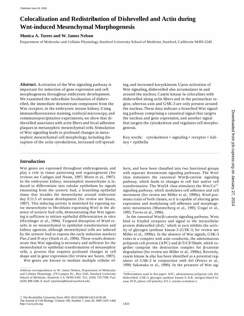
The Rockefeller University Press, 0021-9525/2000/06/1433/10 $5.00 The Journal of Cell Biology, Volume 149, Number 7, June 26, 2000 1433–1442 http://www.jcb.org 1433 Colocalization and Redistribution of Dishevelled and Actin during Wnt-induced Mesenchymal Morphogenesis Monica A. Torres and W. James Nelson Department of Molecular and Cellular Physiology, Stanford University School of Medicine, Stanford, California 94305-5345 Abstract. Activation of the Wnt signaling pathway is important for induction of gene expression and cell morphogenesis throughout embryonic development. We examined the subcellular localization of dishev- elled, the immediate downstream component from the Wnt receptor, in the embryonic mouse kidney. Using immunofluorescence staining, confocal microscopy, and coimmunoprecipitation experiments, we show that di- shevelled associates with actin fibers and focal adhesion plaques in metanephric mesenchymal cells. Stimulation of Wnt signaling leads to profound changes in meta- nephric mesenchymal cell morphology, including dis- ruption of the actin cytoskeleton, increased cell spread- ing, and increased karyokinesis. Upon activation of Wnt signaling, dishevelled also accumulates in and around the nucleus. Casein kinase Ie colocalizes with dishevelled along actin fibers and in the perinuclear re- gion, whereas axin and GSK-3 are only present around the nucleus. These data indicate a branched Wnt signal- ing pathway comprising a canonical signal that targets the nucleus and gene expression, and another signal that targets the cytoskeleton and regulates cell morpho- genesis. Key words: cytoskeleton • signaling • receptor • kid- ney • epithelia Introduction Wnt genes are expressed throughout embryogenesis, and play a role in tissue patterning and organogenesis (for reviews see Cadigan and Nusse, 1997; Moon et al., 1997). In the embryonic kidney, metanephric mesenchyme is in- duced to differentiate into tubular epithelium by signals emanating from the ureteric bud, a branching epithelial tissue that invades the mesenchyme around embryonic day E11.5 of mouse development (for review see Saxen, 1987). This inducing activity is mimicked by exposing na- ive mesenchyme to fibroblasts expressing Wnt1 in the ab- sence of ureteric bud cells, demonstrating that Wnt signal- ing is sufficient to initiate epithelial differentiation in vitro (Herzlinger et al., 1994). Targeted disruption of Wnt4 re- sults in no mesenchymal to epithelial transformation and kidney agenesis, although mesenchymal cells are induced by the ureteric bud to express the early induction markers Pax-2 and N-myc (Stark et al., 1994). These results demon- strate that Wnt signaling is necessary and sufficient for the mesenchymal to epithelial transformation of metanephric cells, a process that requires profound changes in cell shape and in gene expression (for review see Saxen, 1987). Wnt genes are known to mediate multiple cellular ef- fects, and have been classified into two functional groups with separate downstream signaling pathways. The Wnt1 class stimulates the canonical Wnt/b-catenin signaling pathway, which leads to changes in cell fate and/or cell transformation. The Wnt5A class stimulates the Wnt/Ca 21 signaling pathway, which modulates cell adhesion and cell movement (for review see Miller et al., 1999a). Wnt4 pos- sesses traits of both classes, as it is capable of altering gene expression and modulating cell adhesion and morphoge- netic movements (Munsterberg et al., 1995; Ungar et al., 1995; Torres et al., 1996). In the canonical Wnt/b-catenin signaling pathway, Wnts bind to frizzled receptors and signal to the intracellular protein dishevelled (dvl), 1 which in turn inhibits the activ- ity of glycogen synthase kinase-3 (GSK-3; for review see Miller et al., 1999a). In the absence of Wnt signals, GSK-3 exists in a complex with axin–conductin, the adenomatous polyposis coli protein (APC) and b-TrCP/Slimb, which to- gether comprise the destruction complex for b-catenin degradation (for review see Miller et al., 1999a). Recently, casein kinase Ie also has been identified as a potential reg- ulator of GSK-3 in conjunction with dvl (Peters et al., 1999; Sakanaka et al., 1999). In the presence of Wnt sig- Address correspondence to W. James Nelson, Department of Molecular and Cellular Physiology, 279 Campus Dr., Box 5345, Stanford University School of Medicine, Stanford, CA 94305-5345. Tel.: (650) 725-7596. Fax: (650) 498-5286. E-mail: [email protected] 1 Abbreviations used in this paper: APC, adenomatous polyposis coli; dvl, dishevelled; GSK-3, glycogen synthase kinase-3; ILK, integrin-linked ki- nase; PCP, planar cell polarity; ZO-1, zonula occludens-1. on January 27, 2014 jcb.rupress.org Downloaded from Published June 26, 2000
-
Upload
independent -
Category
Documents
-
view
0 -
download
0
Transcript of Colocalization and Redistribution of Dishevelled and Actin during Wnt-induced Mesenchymal...
The Rockefeller University Press, 0021-9525/2000/06/1433/10 $5.00The Journal of Cell Biology, Volume 149, Number 7, June 26, 2000 1433–1442http://www.jcb.org 1433
Colocalization and Redistribution of Dishevelled and Actin duringWnt-induced Mesenchymal Morphogenesis
Monica A. Torres and W. James Nelson
Department of Molecular and Cellular Physiology, Stanford University School of Medicine, Stanford, California 94305-5345
Abstract.
Activation of the Wnt signaling pathway is important for induction of gene expression and cell morphogenesis throughout embryonic development. We examined the subcellular localization of dishev-elled, the immediate downstream component from the Wnt receptor, in the embryonic mouse kidney. Using immunofluorescence staining, confocal microscopy, and coimmunoprecipitation experiments, we show that di-shevelled associates with actin fibers and focal adhesion plaques in metanephric mesenchymal cells. Stimulation of Wnt signaling leads to profound changes in meta-nephric mesenchymal cell morphology, including dis-ruption of the actin cytoskeleton, increased cell spread-
ing, and increased karyokinesis. Upon activation of Wnt signaling, dishevelled also accumulates in and
around the nucleus. Casein kinase I
e
colocalizes with dishevelled along actin fibers and in the perinuclear re-gion, whereas axin and GSK-3 are only present around the nucleus. These data indicate a branched Wnt signal-ing pathway comprising a canonical signal that targets the nucleus and gene expression, and another signal that targets the cytoskeleton and regulates cell morpho-genesis.
Key words: cytoskeleton • signaling • receptor • kid-ney • epithelia
Introduction
Wnt genes are expressed throughout embryogenesis, andplay a role in tissue patterning and organogenesis (forreviews see Cadigan and Nusse, 1997; Moon et al., 1997).In the embryonic kidney, metanephric mesenchyme is in-duced to differentiate into tubular epithelium by signalsemanating from the ureteric bud, a branching epithelialtissue that invades the mesenchyme around embryonicday E11.5 of mouse development (for review see Saxen,1987). This inducing activity is mimicked by exposing na-ive mesenchyme to fibroblasts expressing
Wnt1
in the ab-sence of ureteric bud cells, demonstrating that Wnt signal-ing is sufficient to initiate epithelial differentiation in vitro(Herzlinger et al., 1994). Targeted disruption of
Wnt4
re-sults in no mesenchymal to epithelial transformation andkidney agenesis, although mesenchymal cells are inducedby the ureteric bud to express the early induction markers
Pax-2
and
N-myc
(Stark et al., 1994). These results demon-strate that Wnt signaling is necessary and sufficient for themesenchymal to epithelial transformation of metanephriccells, a process that requires profound changes in cellshape and in gene expression (for review see Saxen, 1987).
Wnt
genes are known to mediate multiple cellular ef-
fects, and have been classified into two functional groupswith separate downstream signaling pathways. The
Wnt1
class stimulates the canonical Wnt/
b
-catenin signalingpathway, which leads to changes in cell fate and/or celltransformation. The
Wnt5A
class stimulates the Wnt/Ca
2
1
signaling pathway, which modulates cell adhesion and cellmovement (for review see Miller et al., 1999a).
Wnt4
pos-sesses traits of both classes, as it is capable of altering geneexpression and modulating cell adhesion and morphoge-netic movements (Munsterberg et al., 1995; Ungar et al.,1995; Torres et al., 1996).
In the canonical Wnt/
b
-catenin signaling pathway, Wntsbind to frizzled receptors and signal to the intracellularprotein dishevelled (dvl),
1
which in turn inhibits the activ-ity of glycogen synthase kinase-3 (GSK-3; for review seeMiller et al., 1999a). In the absence of Wnt signals, GSK-3exists in a complex with axin–conductin, the adenomatouspolyposis coli protein (APC) and
b
-TrCP/Slimb, which to-gether comprise the destruction complex for
b
-catenindegradation (for review see Miller et al., 1999a). Recently,casein kinase I
e
also has been identified as a potential reg-ulator of GSK-3 in conjunction with dvl (Peters et al.,1999; Sakanaka et al., 1999). In the presence of Wnt sig-
Address correspondence to W. James Nelson, Department of Molecularand Cellular Physiology, 279 Campus Dr., Box 5345, Stanford UniversitySchool of Medicine, Stanford, CA 94305-5345. Tel.: (650) 725-7596. Fax:(650) 498-5286. E-mail: [email protected]
1
Abbreviations used in this paper:
APC, adenomatous polyposis coli; dvl,dishevelled; GSK-3, glycogen synthase kinase-3; ILK, integrin-linked ki-nase; PCP, planar cell polarity; ZO-1, zonula occludens-1.
on January 27, 2014jcb.rupress.org
Dow
nloaded from
Published June 26, 2000
The Journal of Cell Biology, Volume 149, 2000 1434
nals, inhibition of GSK-3 results in the stabilization of
b
-catenin, which translocates into the nucleus and stimu-lates transcription by associating with LEF/TCF transcrip-tion factors (for reviews see Miller et al., 1999a; Barkeret al., 2000; Thorpe et al., 2000). The Wnt/Ca
2
1
signalingpathway also functions via a subclass of frizzled receptors,leading to release of intracellular calcium and activation ofprotein kinase C in a process that involves G protein acti-vation (for review see Miller et al., 1999a). The mecha-nisms by which Wnt pathways mediate cellular effectsother than changes in gene expression are very poorly un-derstood.
Although the Wnt/
b
-catenin pathway has been char-acterized extensively using genetic and biochemical ap-proaches in embryos and immortalized cell lines, it hasbeen very difficult to determine the subcellular localiza-tion of endogenous Wnt signaling proteins in these modelsystems. Endogenous
b
-catenin is present at adherensjunctions (for review see Aberle et al., 1996); in the nu-cleus (Yost et al., 1996), dvl is cytoplasmic (Fagotto et al.,1999) and may be associated with vesicle-like organelles(Miller et al., 1999b), and APC is present at the ends ofmicrotubules (Nathke et al., 1996; Mimori-Koyusue et al.,2000). The detailed subcellular localization of other en-dogenous Wnt signaling proteins is unknown.
We chose to study the subcellular distribution of endog-enous Wnt signaling proteins to gain insight into the possi-ble mechanisms by which Wnt signaling might target thecytoskeleton and, hence, changes in cell morphogenesis.We examined embryonic mouse kidney development be-cause Wnt signaling plays a role in the epithelial differenti-ation of metanephric mesenchymal cells in vivo (Stark etal., 1994) and because the epithelial phenotype of kidneytubule cells is very well understood. We focused on thesubcellular localization of dvl because it is the most proxi-mal intracellular branching site within the Wnt signalingpathway (Axelrod et al., 1998; Boutros et al., 1998; for re-view see Boutros and Mlodzik, 1999). Our results indicatea role for Wnt signaling and dvl in actin cytoskeleton orga-nization and cell morphogenesis.
Materials and Methods
Cell Culture
Kidneys from mouse CD1 E12 or E15 embryos were dissected and cul-tured for 3 d on collagen-coated coverslips. Embryonic E12 or E15 kid-neys were dissociated by incubating 10 kidneys/ml in HDF (147 mM NaCl,5.3 mM KCl, 5 mM dextrose, 4.2 mM NaHCO
3
, 0.5 mM EDTA) with0.0625% trypsin solution for 20 min at 37
8
C, followed by repeated pipet-ting to resuspend cells. This cell suspension was diluted 1:4 with serum-containing culture medium to inactivate the trypsin. An average of 50,000cells were plated on each collagen-coated coverslip in 1 ml total volume ofculture medium per well and grown for a total of 3 d. This procedure
yields a mix of
z
90% metanephric mesenchymal cells and 10% uretericbud cells, based on the expression of the ureteric bud and mesenchymalmarkers cytokeratin 8 (K8) and vimentin, respectively. Control and Wnt1expressing NIH 3T3 fibroblasts were provided by Dr. D. Herzlinger (Co-lumbia University, New York, NY), and cultured as described previously(Herzlinger et al., 1994). Where indicated, 60,000–100,000 of these NIH3T3 cells were added to each well of embryonic kidney cells plated 2 d ear-lier, and were cultured for an additional 24 h. In all cases, embryonic kid-neys and dissociated kidney cells were cultured in DME/Ham’s F-12 (1:1;GIBCO BRL) with 10% FCS and penicillin (0.5 U/ml), streptomycin (0.5mg/ml) and kanamycin (1 mg/ml).
Immunofluorescence Staining
Whole cultured embryonic kidneys and dissociated embryonic kidneycells were fixed as indicated in 100% methanol at
2
20
8
C for 10 min, in 2%paraformaldehyde in 75 mM lysine, 37.5 mM NaPO
4
and 0.1 M NaIO
4
atroom temperature for 10 min, or in 0.5% glutaraldehyde in 100 mM Pipes,pH 6.9, 4 mM MgCl
2
, 2 mM EGTA and 0.1% Triton X-100 (TX-100) atroom temperature for 10 min, followed by quenching in PBS (pH 8.0, 2.7mM KCl, 1.5 mM KH
2
PO
4
, 9.2 mM NaCl, 15.2 mM Na
2
HPO
4
), with 1 mg/ml NaBH
4
. After fixation, samples were washed in PBS, pH 7.5, and incu-bated overnight in blocking solution (PBS, pH 7.5 with 50 mM NH
4
Cl, 25mM poly-
L
-lysine, 25 mM glycine, 20% normal goat serum, 0.2% BSA) at4
8
C. When using mouse or rat mAbs, the blocking solution contained 18
m
g/ml unlabeled goat anti–mouse or anti–rat IgG (Jackson Immuno-Research Laboratories; Boehringer; Roche Molecular Biochemicals).Samples were incubated in blocking solution with the following primaryantibodies: rabbit polyclonal antiuvomorulin (provided by R. Kemler,Max-Planck Institut fur Immunbiologie, Freiburg, Germany; Vestweberand Kemler, 1984; Piepenhagen et al., 1995), rabbit polyclonal anti–dvl-1and -2, rabbit polyclonal antiaxin (all provided by K. Willert in the labora-tory of R. Nusse, Stanford, CA; Miller et al., 1999b, Willert et al., 1999),goat polyclonal anti–NH
2
terminus or COOH terminus dvl antibodies(Santa Cruz Biotechnology), mouse monoclonal antiactin (BoehringerMannheim; Roche Molecular Biochemicals), mouse monoclonal anti–E-cadherin, antipaxillin and anti–GSK-3 (all from Transduction Labora-tories). After washing the samples with PBS and 0.2% BSA, they were in-cubated in blocking solution with rhodamine- or FITC-conjugated goatanti–rabbit, goat anti–mouse or donkey anti–goat secondary antibodies, asindicated. The samples were washed in PBS, incubated in PBS with 10
m
g/ml Hoechst stain (Molecular Probes), washed in PBS, pH 7.5, andmounted in Vectashield (Vector Labs). Alternatively, nucleic acids werevisualized by incubating samples in 50
m
M SytoRed (Molecular Probes) in50 mM Tris-HCl, pH 7.5, washed in 50 mM Tris-HCl, pH 7.5, andmounted as above. All samples were analyzed using a confocal scanningmicroscope (Molecular Dynamics).
Protein Extraction, Immunoprecipitation, andWestern Blotting
Five E12, E15, or E16 embryonic mouse kidneys were homogenized onice in 300
m
l CSK buffer (10 mM Pipes, pH 6.8, 50 mM NaCl, 300 mM su-crose, 3 mM MgCl
2
, 0.5% TX-100, 1 mM pefablock, 10
m
g/ml DNase, 10
m
g/ml RNase) and centrifuged at 1,000
g
for 10 min at 4
8
C. The TX-100–soluble fraction was separated from the pellet, which was solubilized onice in 100
m
l Tris-SDS buffer (50 mM Tris-HCl, pH 7.5, 1% SDS, 2 mMEDTA, 1 mM pefablock, 10
m
g/ml DNase, 10
m
g/ml RNase). The SDSwas diluted to 0.1% by adding 900
m
l CSK buffer. Primary antibodies, asindicated, and protein A–Sepharose beads (Sigma Chemical Co.) were in-cubated with the TX-100 and SDS-soluble protein fractions ranging from2 h to overnight at 4
8
C, followed by washing three times in 1 ml CSKbuffer (or PBS, pH 7.5, in the case of actin coimmunoprecipitations). Im-munoprecipitated proteins were processed for SDS-PAGE (PAGE; 10%acrylamide gels) and transferred to 0.45-
m
m pore size nitrocellulose filtersfor Western blotting.
In all cases, antibodies used for Western blotting were the same asthose used in at least a subset of the immunofluorescence experiments.Dishevelled and axin were detected using rabbit polyclonal primary anti–dvl-1 and antiaxin antibodies provided by K. Willert and R. Nusse (Milleret al., 1999b; Willert et al., 1999). Actin was detected using mouse mono-clonal antiactin antibody (Boehringer; Roche Molecular Biochemicals).Paxillin and GSK-3 were detected using mouse monoclonal antipaxillinand mouse monoclonal anti–GSK-3 antibodies (Transduction Labora-tories). All primary antibodies were visualized using HRP-conjugatedanti-mouse or anti-rabbit secondary antibody (Amersham PharmaciaBiotech), except for GSK-3, which was detected using biotinylated anti-mouse antiserum, followed by HRP-conjugated streptavidin (Zymed) in-cubation. The HRP signal was visualized by ECL chemiluminescence re-agent (Amersham Pharmacia Biotech).
Scoring Wnt1 Effects on Embryonic Kidney Cells
In all cases, embryonic kidney cells were distinguished from NIH 3T3 cellsbecause of their large size, prominent stress fibers, and angular appear-ance. NIH 3T3 cells were easily identifiable by their small size and charac-teristic fibroblast morphology. The incidence of multinucleated cells, and
on January 27, 2014jcb.rupress.org
Dow
nloaded from
Published June 26, 2000
Torres and Nelson
Wnt-induced Morphogenesis
1435
increased cell spreading were scored by collecting a low magnification im-age from 10 random locations on a single coverslip per experiment. Thefrequency of morphological features listed above was determined at eachsite, and then an average and/or the total number of cells was calculatedfor each experiment. Multinucleated cells were identified by simulta-neously visualizing the actin cytoskeleton and Hoechst-stained nucleic ac-ids using a Zeiss Axioplan microscope. Nuclear dishevelled was visualizedusing a Molecular Dynamics confocal laser scanning microscope. SytoRednucleic acid stain (Molecular Probes Inc.) was used as above to detect nu-clei in intact cultured embryonic kidneys by confocal microscopy. Levelsof nuclear dishevelled were compared from cell to cell using pseudocolorimaging to quantitate differences in fluorescent signals. Disassembly ofthe actin cytoskeleton was scored by acquiring high magnification,pseudocolorized images from five random locations within a single cover-slip per experiment, and then determining the average number and aver-age length of actin stress fibers present. Cell spreading was quantitated bymeasuring the average area of cells in controls versus Wnt1 treatments.All measurements were carried out using NIH Image software (http://rsb.info.nih.gov/nih-image/index.html).
Results
Dishevelled Is Present in Multiple Subcellular Compartments in the Developing Kidney
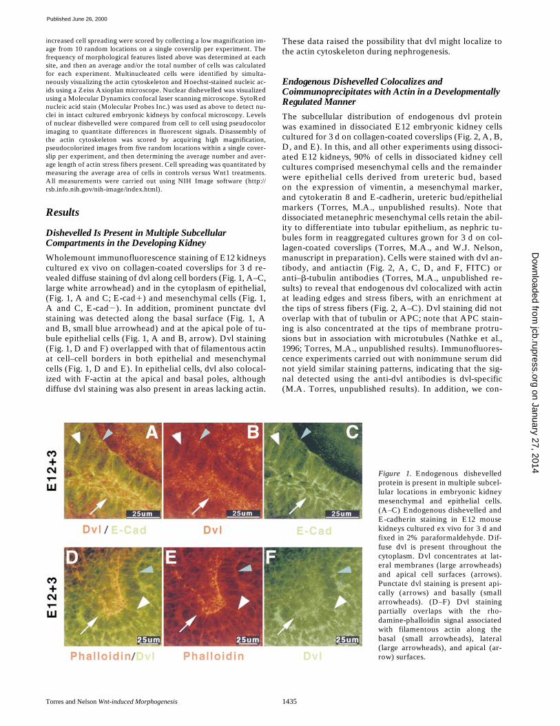
Wholemount immunofluorescence staining of E12 kidneyscultured ex vivo on collagen-coated coverslips for 3 d re-vealed diffuse staining of dvl along cell borders (Fig. 1, A–C,large white arrowhead) and in the cytoplasm of epithelial,(Fig. 1, A and C; E-cad
1
) and mesenchymal cells (Fig. 1,A and C, E-cad
2
). In addition, prominent punctate dvlstaining was detected along the basal surface (Fig. 1, Aand B, small blue arrowhead) and at the apical pole of tu-bule epithelial cells (Fig. 1, A and B, arrow). Dvl staining(Fig. 1, D and F) overlapped with that of filamentous actinat cell–cell borders in both epithelial and mesenchymalcells (Fig. 1, D and E). In epithelial cells, dvl also colocal-ized with F-actin at the apical and basal poles, althoughdiffuse dvl staining was also present in areas lacking actin.
These data raised the possibility that dvl might localize tothe actin cytoskeleton during nephrogenesis.
Endogenous Dishevelled Colocalizes and Coimmunoprecipitates with Actin in a Developmentally Regulated Manner
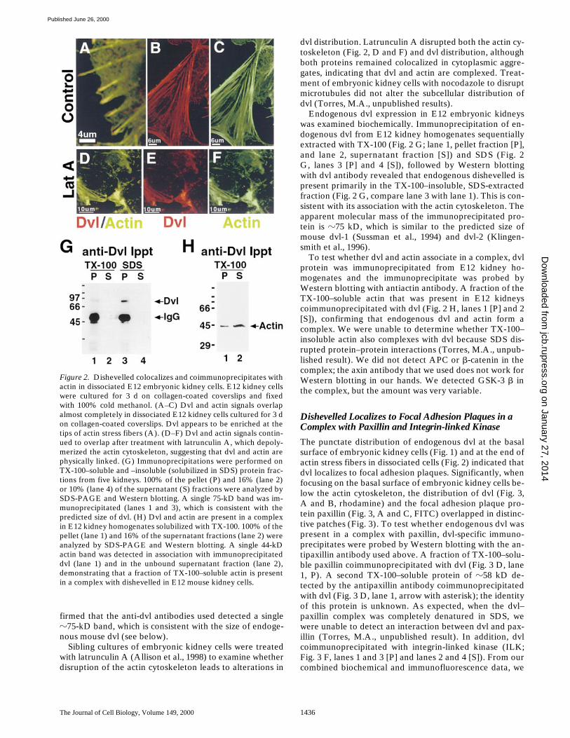
The subcellular distribution of endogenous dvl proteinwas examined in dissociated E12 embryonic kidney cellscultured for 3 d on collagen-coated coverslips (Fig. 2, A, B,D, and E). In this, and all other experiments using dissoci-ated E12 kidneys, 90% of cells in dissociated kidney cellcultures comprised mesenchymal cells and the remainderwere epithelial cells derived from ureteric bud, basedon the expression of vimentin, a mesenchymal marker,and cytokeratin 8 and E-cadherin, ureteric bud/epithelialmarkers (Torres, M.A., unpublished results). Note thatdissociated metanephric mesenchymal cells retain the abil-ity to differentiate into tubular epithelium, as nephric tu-bules form in reaggregated cultures grown for 3 d on col-lagen-coated coverslips (Torres, M.A., and W.J. Nelson,manuscript in preparation). Cells were stained with dvl an-tibody, and antiactin (Fig. 2, A, C, D, and F, FITC) oranti–
b
-tubulin antibodies (Torres, M.A., unpublished re-sults) to reveal that endogenous dvl colocalized with actinat leading edges and stress fibers, with an enrichment atthe tips of stress fibers (Fig. 2, A–C). Dvl staining did notoverlap with that of tubulin or APC; note that APC stain-ing is also concentrated at the tips of membrane protru-sions but in association with microtubules (Nathke et al.,1996; Torres, M.A., unpublished results). Immunofluores-cence experiments carried out with nonimmune serum didnot yield similar staining patterns, indicating that the sig-nal detected using the anti-dvl antibodies is dvl-specific(M.A. Torres, unpublished results). In addition, we con-
Figure 1. Endogenous dishevelledprotein is present in multiple subcel-lular locations in embryonic kidneymesenchymal and epithelial cells.(A–C) Endogenous dishevelled andE-cadherin staining in E12 mousekidneys cultured ex vivo for 3 d andfixed in 2% paraformaldehyde. Dif-fuse dvl is present throughout thecytoplasm. Dvl concentrates at lat-eral membranes (large arrowheads)and apical cell surfaces (arrows).Punctate dvl staining is present api-cally (arrows) and basally (smallarrowheads). (D–F) Dvl stainingpartially overlaps with the rho-damine-phalloidin signal associatedwith filamentous actin along thebasal (small arrowheads), lateral(large arrowheads), and apical (ar-row) surfaces.
on January 27, 2014jcb.rupress.org
Dow
nloaded from
Published June 26, 2000
The Journal of Cell Biology, Volume 149, 2000 1436
firmed that the anti-dvl antibodies used detected a single
z
75-kD band, which is consistent with the size of endoge-nous mouse dvl (see below).
Sibling cultures of embryonic kidney cells were treatedwith latrunculin A (Allison et al., 1998) to examine whetherdisruption of the actin cytoskeleton leads to alterations in
dvl distribution. Latrunculin A disrupted both the actin cy-toskeleton (Fig. 2, D and F) and dvl distribution, althoughboth proteins remained colocalized in cytoplasmic aggre-gates, indicating that dvl and actin are complexed. Treat-ment of embryonic kidney cells with nocodazole to disruptmicrotubules did not alter the subcellular distribution ofdvl (Torres, M.A., unpublished results).
Endogenous dvl expression in E12 embryonic kidneyswas examined biochemically. Immunoprecipitation of en-dogenous dvl from E12 kidney homogenates sequentiallyextracted with TX-100 (Fig. 2 G; lane 1, pellet fraction [P],and lane 2, supernatant fraction [S]) and SDS (Fig. 2G, lanes 3 [P] and 4 [S]), followed by Western blottingwith dvl antibody revealed that endogenous dishevelled ispresent primarily in the TX-100–insoluble, SDS-extractedfraction (Fig. 2 G, compare lane 3 with lane 1). This is con-sistent with its association with the actin cytoskeleton. Theapparent molecular mass of the immunoprecipitated pro-tein is
z
75 kD, which is similar to the predicted size ofmouse dvl-1 (Sussman et al., 1994) and dvl-2 (Klingen-smith et al., 1996).
To test whether dvl and actin associate in a complex, dvlprotein was immunoprecipitated from E12 kidney ho-mogenates and the immunoprecipitate was probed byWestern blotting with antiactin antibody. A fraction of theTX-100–soluble actin that was present in E12 kidneyscoimmunoprecipitated with dvl (Fig. 2 H, lanes 1 [P] and 2[S]), confirming that endogenous dvl and actin form acomplex. We were unable to determine whether TX-100–insoluble actin also complexes with dvl because SDS dis-rupted protein–protein interactions (Torres, M.A., unpub-lished result).
We did not detect APC or
b
-catenin in thecomplex; the axin antibody that we used does not work forWestern blotting in our hands. We detected GSK-3
b
inthe complex, but the amount was very variable.
Dishevelled Localizes to Focal Adhesion Plaques in a Complex with Paxillin and Integrin-linked Kinase
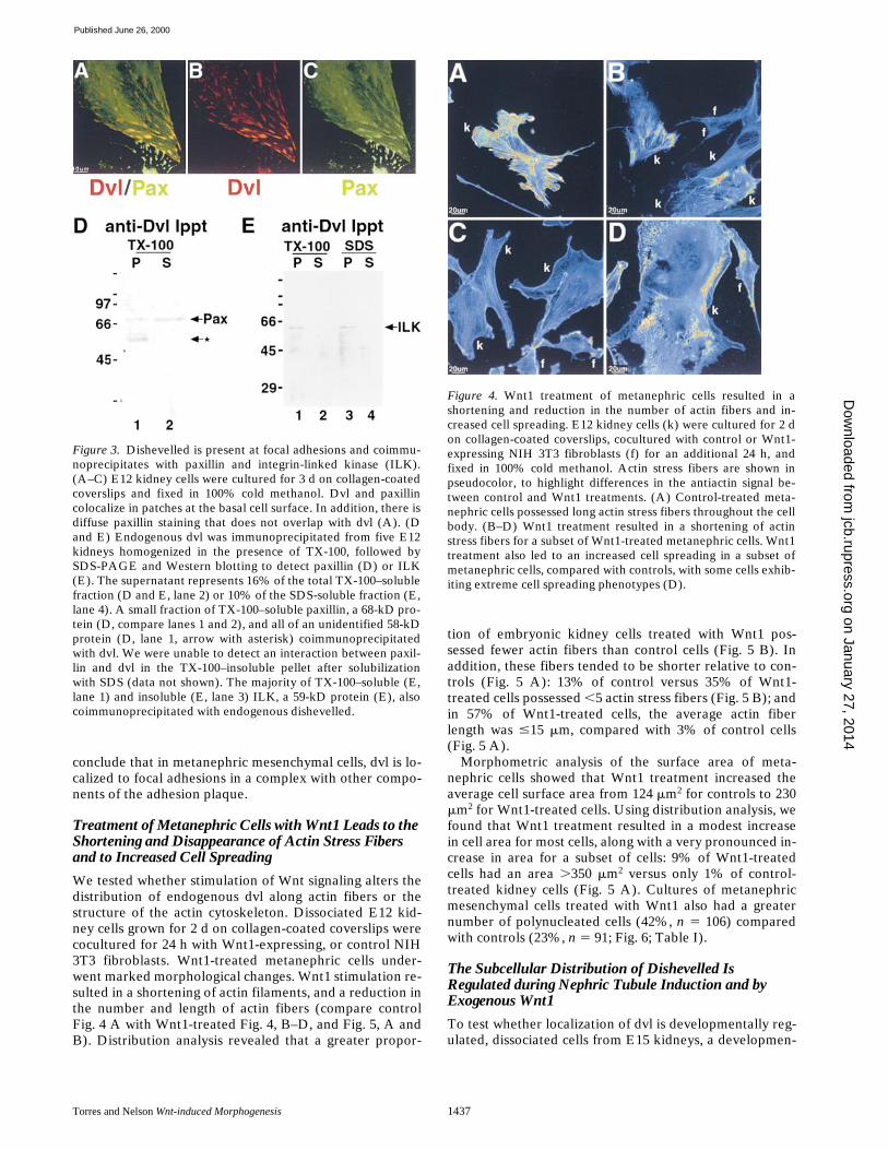
The punctate distribution of endogenous dvl at the basalsurface of embryonic kidney cells (Fig. 1) and at the end ofactin stress fibers in dissociated cells (Fig. 2) indicated thatdvl localizes to focal adhesion plaques. Significantly, whenfocusing on the basal surface of embryonic kidney cells be-low the actin cytoskeleton, the distribution of dvl (Fig. 3,A and B, rhodamine) and the focal adhesion plaque pro-tein paxillin (Fig. 3, A and C, FITC) overlapped in distinc-tive patches (Fig. 3). To test whether endogenous dvl waspresent in a complex with paxillin, dvl-specific immuno-precipitates were probed by Western blotting with the an-tipaxillin antibody used above. A fraction of TX-100–solu-ble paxillin coimmunoprecipitated with dvl (Fig. 3 D, lane1, P). A second TX-100–soluble protein of
z
58 kD de-tected by the antipaxillin antibody coimmunoprecipitatedwith dvl (Fig. 3 D, lane 1, arrow with asterisk); the identityof this protein is unknown. As expected, when the dvl–paxillin complex was completely denatured in SDS, wewere unable to detect an interaction between dvl and pax-illin (Torres, M.A., unpublished result). In addition, dvlcoimmunoprecipitated with integrin-linked kinase (ILK;Fig. 3 F, lanes 1 and 3 [P] and lanes 2 and 4 [S]). From ourcombined biochemical and immunofluorescence data, we
Figure 2. Dishevelled colocalizes and coimmunoprecipitates withactin in dissociated E12 embryonic kidney cells. E12 kidney cellswere cultured for 3 d on collagen-coated coverslips and fixedwith 100% cold methanol. (A–C) Dvl and actin signals overlapalmost completely in dissociated E12 kidney cells cultured for 3 don collagen-coated coverslips. Dvl appears to be enriched at thetips of actin stress fibers (A). (D–F) Dvl and actin signals contin-ued to overlap after treatment with latrunculin A, which depoly-merized the actin cytoskeleton, suggesting that dvl and actin arephysically linked. (G) Immunoprecipitations were performed onTX-100–soluble and –insoluble (solubilized in SDS) protein frac-tions from five kidneys. 100% of the pellet (P) and 16% (lane 2)or 10% (lane 4) of the supernatant (S) fractions were analyzed bySDS-PAGE and Western blotting. A single 75-kD band was im-munoprecipitated (lanes 1 and 3), which is consistent with thepredicted size of dvl. (H) Dvl and actin are present in a complexin E12 kidney homogenates solubilized with TX-100. 100% of thepellet (lane 1) and 16% of the supernatant fractions (lane 2) wereanalyzed by SDS-PAGE and Western blotting. A single 44-kDactin band was detected in association with immunoprecipitateddvl (lane 1) and in the unbound supernatant fraction (lane 2),demonstrating that a fraction of TX-100–soluble actin is presentin a complex with dishevelled in E12 mouse kidney cells.
on January 27, 2014jcb.rupress.org
Dow
nloaded from
Published June 26, 2000
Torres and Nelson
Wnt-induced Morphogenesis
1437
conclude that in metanephric mesenchymal cells, dvl is lo-calized to focal adhesions in a complex with other compo-nents of the adhesion plaque.
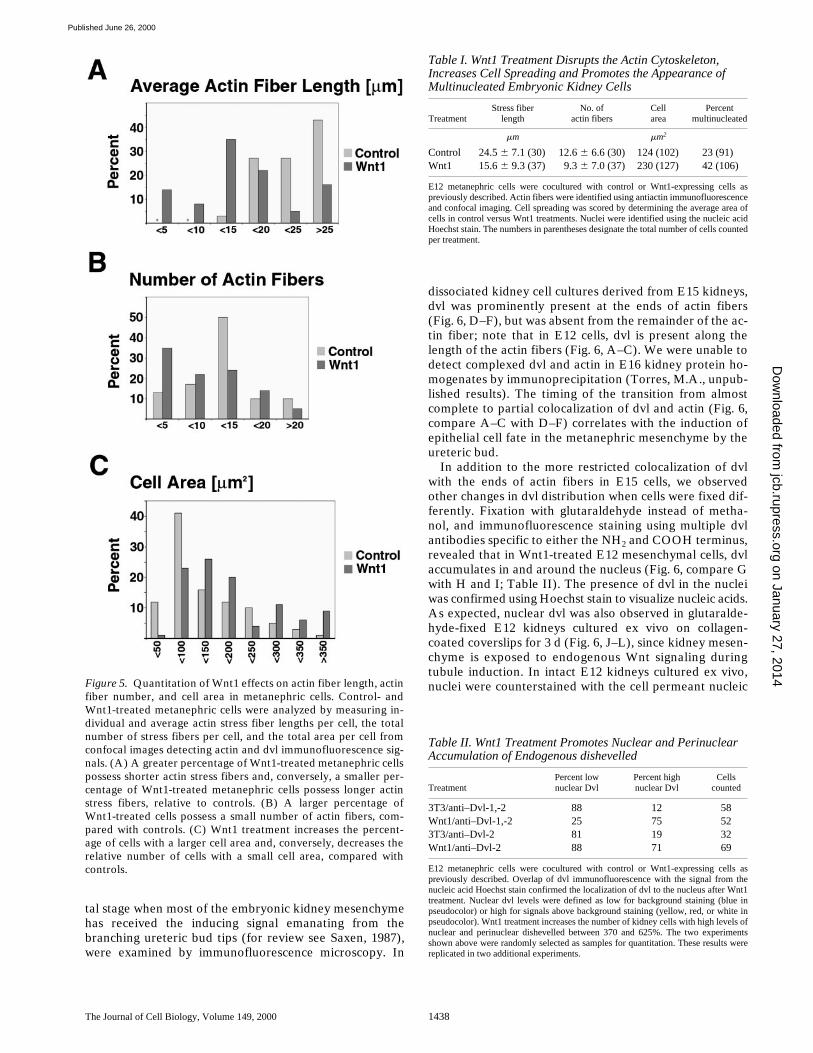
Treatment of Metanephric Cells with Wnt1 Leads to the Shortening and Disappearance of Actin Stress Fibers and to Increased Cell Spreading
We tested whether stimulation of Wnt signaling alters thedistribution of endogenous dvl along actin fibers or thestructure of the actin cytoskeleton. Dissociated E12 kid-ney cells grown for 2 d on collagen-coated coverslips werecocultured for 24 h with Wnt1-expressing, or control NIH3T3 fibroblasts. Wnt1-treated metanephric cells under-went marked morphological changes. Wnt1 stimulation re-sulted in a shortening of actin filaments, and a reduction inthe number and length of actin fibers (compare controlFig. 4 A with Wnt1-treated Fig. 4, B–D, and Fig. 5, A andB). Distribution analysis revealed that a greater propor-
tion of embryonic kidney cells treated with Wnt1 pos-sessed fewer actin fibers than control cells (Fig. 5 B). Inaddition, these fibers tended to be shorter relative to con-trols (Fig. 5 A): 13% of control versus 35% of Wnt1-treated cells possessed
,
5 actin stress fibers (Fig. 5 B); andin 57% of Wnt1-treated cells, the average actin fiberlength was
#
15
m
m, compared with 3% of control cells(Fig. 5 A).
Morphometric analysis of the surface area of meta-nephric cells showed that Wnt1 treatment increased theaverage cell surface area from 124
m
m
2
for controls to 230
m
m
2
for Wnt1-treated cells. Using distribution analysis, wefound that Wnt1 treatment resulted in a modest increasein cell area for most cells, along with a very pronounced in-crease in area for a subset of cells: 9% of Wnt1-treatedcells had an area
.
350
m
m
2
versus only 1% of control-treated kidney cells (Fig. 5 A). Cultures of metanephricmesenchymal cells treated with Wnt1 also had a greaternumber of polynucleated cells (42%,
n
5
106) comparedwith controls (23%,
n
5
91; Fig. 6; Table I).
The Subcellular Distribution of Dishevelled Is Regulated during Nephric Tubule Induction and byExogenous Wnt1
To test whether localization of dvl is developmentally reg-ulated, dissociated cells from E15 kidneys, a developmen-
Figure 3. Dishevelled is present at focal adhesions and coimmu-noprecipitates with paxillin and integrin-linked kinase (ILK).(A–C) E12 kidney cells were cultured for 3 d on collagen-coatedcoverslips and fixed in 100% cold methanol. Dvl and paxillincolocalize in patches at the basal cell surface. In addition, there isdiffuse paxillin staining that does not overlap with dvl (A). (Dand E) Endogenous dvl was immunoprecipitated from five E12kidneys homogenized in the presence of TX-100, followed bySDS-PAGE and Western blotting to detect paxillin (D) or ILK(E). The supernatant represents 16% of the total TX-100–solublefraction (D and E, lane 2) or 10% of the SDS-soluble fraction (E,lane 4). A small fraction of TX-100–soluble paxillin, a 68-kD pro-tein (D, compare lanes 1 and 2), and all of an unidentified 58-kDprotein (D, lane 1, arrow with asterisk) coimmunoprecipitatedwith dvl. We were unable to detect an interaction between paxil-lin and dvl in the TX-100–insoluble pellet after solubilizationwith SDS (data not shown). The majority of TX-100–soluble (E,lane 1) and insoluble (E, lane 3) ILK, a 59-kD protein (E), alsocoimmunoprecipitated with endogenous dishevelled.
Figure 4. Wnt1 treatment of metanephric cells resulted in ashortening and reduction in the number of actin fibers and in-creased cell spreading. E12 kidney cells (k) were cultured for 2 don collagen-coated coverslips, cocultured with control or Wnt1-expressing NIH 3T3 fibroblasts (f) for an additional 24 h, andfixed in 100% cold methanol. Actin stress fibers are shown inpseudocolor, to highlight differences in the antiactin signal be-tween control and Wnt1 treatments. (A) Control-treated meta-nephric cells possessed long actin stress fibers throughout the cellbody. (B–D) Wnt1 treatment resulted in a shortening of actinstress fibers for a subset of Wnt1-treated metanephric cells. Wnt1treatment also led to an increased cell spreading in a subset ofmetanephric cells, compared with controls, with some cells exhib-iting extreme cell spreading phenotypes (D).
on January 27, 2014jcb.rupress.org
Dow
nloaded from
Published June 26, 2000
The Journal of Cell Biology, Volume 149, 2000 1438
tal stage when most of the embryonic kidney mesenchymehas received the inducing signal emanating from thebranching ureteric bud tips (for review see Saxen, 1987),were examined by immunofluorescence microscopy. In
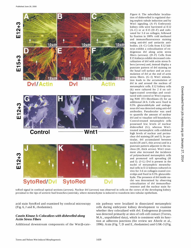
dissociated kidney cell cultures derived from E15 kidneys,dvl was prominently present at the ends of actin fibers(Fig. 6, D–F), but was absent from the remainder of the ac-tin fiber; note that in E12 cells, dvl is present along thelength of the actin fibers (Fig. 6, A–C). We were unable todetect complexed dvl and actin in E16 kidney protein ho-mogenates by immunoprecipitation (Torres, M.A., unpub-lished results). The timing of the transition from almostcomplete to partial colocalization of dvl and actin (Fig. 6,compare A–C with D–F) correlates with the induction ofepithelial cell fate in the metanephric mesenchyme by theureteric bud.
In addition to the more restricted colocalization of dvlwith the ends of actin fibers in E15 cells, we observedother changes in dvl distribution when cells were fixed dif-ferently. Fixation with glutaraldehyde instead of metha-nol, and immunofluorescence staining using multiple dvlantibodies specific to either the NH
2
and COOH terminus,revealed that in Wnt1-treated E12 mesenchymal cells, dvlaccumulates in and around the nucleus (Fig. 6, compare Gwith H and I; Table II). The presence of dvl in the nucleiwas confirmed using Hoechst stain to visualize nucleic acids.As expected, nuclear dvl was also observed in glutaralde-hyde-fixed E12 kidneys cultured ex vivo on collagen-coated coverslips for 3 d (Fig. 6, J–L), since kidney mesen-chyme is exposed to endogenous Wnt signaling duringtubule induction. In intact E12 kidneys cultured ex vivo,nuclei were counterstained with the cell permeant nucleicFigure 5. Quantitation of Wnt1 effects on actin fiber length, actin
fiber number, and cell area in metanephric cells. Control- andWnt1-treated metanephric cells were analyzed by measuring in-dividual and average actin stress fiber lengths per cell, the totalnumber of stress fibers per cell, and the total area per cell fromconfocal images detecting actin and dvl immunofluorescence sig-nals. (A) A greater percentage of Wnt1-treated metanephric cellspossess shorter actin stress fibers and, conversely, a smaller per-centage of Wnt1-treated metanephric cells possess longer actinstress fibers, relative to controls. (B) A larger percentage ofWnt1-treated cells possess a small number of actin fibers, com-pared with controls. (C) Wnt1 treatment increases the percent-age of cells with a larger cell area and, conversely, decreases therelative number of cells with a small cell area, compared withcontrols.
Table I. Wnt1 Treatment Disrupts the Actin Cytoskeleton, Increases Cell Spreading and Promotes the Appearance of Multinucleated Embryonic Kidney Cells
TreatmentStress fiber
lengthNo. of
actin fibersCell area
Percentmultinucleated
m
m
m
m
2
Control 24.5
6
7.1 (30) 12.6
6
6.6 (30) 124 (102) 23 (91)Wnt1 15.6
6
9.3 (37) 9.3
6
7.0 (37) 230 (127) 42 (106)
E12 metanephric cells were cocultured with control or Wnt1-expressing cells aspreviously described. Actin fibers were identified using antiactin immunofluorescenceand confocal imaging. Cell spreading was scored by determining the average area ofcells in control versus Wnt1 treatments. Nuclei were identified using the nucleic acidHoechst stain. The numbers in parentheses designate the total number of cells countedper treatment.
Table II. Wnt1 Treatment Promotes Nuclear and Perinuclear Accumulation of Endogenous dishevelled
TreatmentPercent lownuclear Dvl
Percent highnuclear Dvl
Cells counted
3T3/anti–Dvl-1,-2 88 12 58Wnt1/anti–Dvl-1,-2 25 75 523T3/anti–Dvl-2 81 19 32Wnt1/anti–Dvl-2 88 71 69
E12 metanephric cells were cocultured with control or Wnt1-expressing cells aspreviously described. Overlap of dvl immunofluorescence with the signal from thenucleic acid Hoechst stain confirmed the localization of dvl to the nucleus after Wnt1treatment. Nuclear dvl levels were defined as low for background staining (blue inpseudocolor) or high for signals above background staining (yellow, red, or white inpseudocolor). Wnt1 treatment increases the number of kidney cells with high levels ofnuclear and perinuclear dishevelled between 370 and 625%. The two experimentsshown above were randomly selected as samples for quantitation. These results werereplicated in two additional experiments.
on January 27, 2014jcb.rupress.org
Dow
nloaded from
Published June 26, 2000
Torres and Nelson
Wnt-induced Morphogenesis
1439
acid stain SytoRed and examined by confocal microscopy(Fig. 6, J and K, rhodamine).
Casein Kinase I
e
Colocalizes with dishevelled along Actin Stress Fibers
Additional downstream components of the Wnt/
b
-cate-
nin pathway were localized in dissociated metanephriccells during embryonic kidney development to examinewhether they colocalized with dvl. Endogenous
b
-cateninwas detected primarily at sites of cell–cell contact (Torres,M.A., unpublished data), which is consistent with its func-tion at adherens junctions (for review see Aberle et al.,1996). Axin (Fig. 7, D and E, rhodamine) and GSK-3 (Fig.
Figure 6. The subcellular localiza-tion of dishevelled is regulated dur-ing nephric tubule induction and byWnt1 signaling. (A–F) Embryonickidney cells were harvested at E12(A–C) or at E15 (D–F) and culti-vated for 3 d on collagen, followedby fixation in 100% cold methanoland immunofluorescence stainingusing anti-dvl and antiactin anti-bodies. (A–C) Cells from E12 kid-neys exhibit a colocalization of en-dogenous dvl along actin stressfibers (arrows). (D–F) Cells fromE15 kidneys exhibit decreased colo-calization of dvl with actin stress fi-bers (arrows) and, instead display apunctate pattern of dvl staining onthe basal cell surface and an accu-mulation of dvl at the end of actinstress fibers. (G–I) Wnt1 stimula-tion leads to the accumulation ofdvl in and around the nucleus ofmetanephric cells. E12 kidney cells(k) were cultured for 2 d on col-lagen-coated coverslips and cocul-tured with control or Wnt1-express-ing NIH 3T3 fibroblasts (f) for anadditional 24 h. Cells were fixed in0.5% glutaraldehyde and endoge-nous dvl was detected using anti-dvlantibodies. Pseudocolor was usedto quantify the amount of nucleardvl and to visualize cell boundaries.Control-treated metanephric cellspossessed low levels of nucleardishevelled (G), whereas Wnt1-treated metanephric cells exhibitedhigh levels of nuclear and perinu-clear dvl staining (H and I). In par-ticular, dvl accumulated betweennuclei (H and I, thin arrow) and in apunctate pattern adjacent to the nu-cleus (H, thick arrow). Wnt1 treat-ment also increased the incidenceof polynucleated metanephric cellsand promoted cell spreading (Hand I). (J–L) Dvl is present in thenuclei of metanephric mesenchy-mal cells in E12 kidneys cultured exvivo for 3 d on collagen-coated cov-erslips and fixed in 0.5% glutaralde-hyde. The presence of dvl inside nu-clei was determined by observingcolocalization of dvl immunofluo-rescence and the nuclear stain Sy-
toRed signal in confocal optical sections (arrows). Nuclear dvl (arrows) was observed in cells on the cortex of the developing kidneyproximal to the tips of ureteric bud branches (asterisk), where mesenchyme is induced to transform into tubular epithelium.
on January 27, 2014jcb.rupress.org
Dow
nloaded from
Published June 26, 2000
The Journal of Cell Biology, Volume 149, 2000 1440
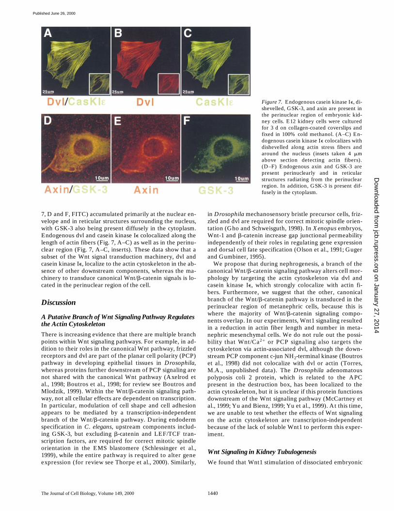
7, D and F, FITC) accumulated primarily at the nuclear en-velope and in reticular structures surrounding the nucleus,with GSK-3 also being present diffusely in the cytoplasm.Endogenous dvl and casein kinase I
e
colocalized along thelength of actin fibers (Fig. 7, A–C) as well as in the perinu-clear region (Fig. 7, A–C, inserts). These data show that asubset of the Wnt signal transduction machinery, dvl andcasein kinase I
e
, localize to the actin cytoskeleton in the ab-sence of other downstream components, whereas the ma-chinery to transduce canonical Wnt/
b
-catenin signals is lo-cated in the perinuclear region of the cell.
Discussion
A Putative Branch of Wnt Signaling Pathway Regulates the Actin Cytoskeleton
There is increasing evidence that there are multiple branchpoints within Wnt signaling pathways. For example, in ad-dition to their roles in the canonical Wnt pathway, frizzledreceptors and dvl are part of the planar cell polarity (PCP)pathway in developing epithelial tissues in
Drosophila
,whereas proteins further downstream of PCP signaling arenot shared with the canonical Wnt pathway (Axelrod etal., 1998; Boutros et al., 1998; for review see Boutros andMlodzik, 1999). Within the Wnt/
b
-catenin signaling path-way, not all cellular effects are dependent on transcription.In particular, modulation of cell shape and cell adhesionappears to be mediated by a transcription-independentbranch of the Wnt/
b
-catenin pathway. During endodermspecification in
C
.
elegans
, upstream components includ-ing GSK-3, but excluding
b
-catenin and LEF/TCF tran-scription factors, are required for correct mitotic spindleorientation in the EMS blastomere (Schlessinger et al.,1999), while the entire pathway is required to alter geneexpression (for review see Thorpe et al., 2000). Similarly,
in
Drosophila
mechanosensory bristle precursor cells, friz-zled and dvl are required for correct mitotic spindle orien-tation (Gho and Schweisguth, 1998). In
Xenopus
embryos,Wnt-1 and b-catenin increase gap junctional permeabilityindependently of their roles in regulating gene expressionand dorsal cell fate specification (Olson et al., 1991; Gugerand Gumbiner, 1995).
We propose that during nephrogenesis, a branch of thecanonical Wnt/b-catenin signaling pathway alters cell mor-phology by targeting the actin cytoskeleton via dvl andcasein kinase Ie, which strongly colocalize with actin fi-bers. Furthermore, we suggest that the other, canonicalbranch of the Wnt/b-catenin pathway is transduced in theperinuclear region of metanephric cells, because this iswhere the majority of Wnt/b-catenin signaling compo-nents overlap. In our experiments, Wnt1 signaling resultedin a reduction in actin fiber length and number in meta-nephric mesenchymal cells. We do not rule out the possi-bility that Wnt/Ca21 or PCP signaling also targets thecytoskeleton via actin-associated dvl, although the down-stream PCP component c-jun NH2-terminal kinase (Boutroset al., 1998) did not colocalize with dvl or actin (Torres,M.A., unpublished data). The Drosophila adenomatouspolyposis coli 2 protein, which is related to the APCpresent in the destruction box, has been localized to theactin cytoskeleton, but it is unclear if this protein functionsdownstream of the Wnt signaling pathway (McCartney etal., 1999; Yu and Bienz, 1999; Yu et al., 1999). At this time,we are unable to test whether the effects of Wnt signalingon the actin cytoskeleton are transcription-independentbecause of the lack of soluble Wnt1 to perform this exper-iment.
Wnt Signaling in Kidney Tubulogenesis
We found that Wnt1 stimulation of dissociated embryonic
Figure 7. Endogenous casein kinase Ie, di-shevelled, GSK-3, and axin are present inthe perinuclear region of embryonic kid-ney cells. E12 kidney cells were culturedfor 3 d on collagen-coated coverslips andfixed in 100% cold methanol. (A–C) En-dogenous casein kinase Ie colocalizes withdishevelled along actin stress fibers andaround the nucleus (insets taken 4 mmabove section detecting actin fibers).(D–F) Endogenous axin and GSK-3 arepresent perinuclearly and in reticularstructures radiating from the perinuclearregion. In addition, GSK-3 is present dif-fusely in the cytoplasm.
on January 27, 2014jcb.rupress.org
Dow
nloaded from
Published June 26, 2000
Torres and Nelson Wnt-induced Morphogenesis 1441
kidney cells results in the reorganization of the actin cy-toskeleton, increased cell spreading, and the appearanceof multiple nuclei within single metanephric cells (see be-low). These morphological changes are consistent with theprogram of epithelial differentiation observed in the em-bryonic kidney, which involves the formation of cell aggre-gates and cell proliferation (for review see Saxen, 1987).The ability of these dissociated mesenchymal cells to re-spond with the appropriate differentiation program is sup-ported by the fact that repelleted, dissociated mesenchy-mal cell cultures are capable of differentiating into tubularepithelium when cocultured with inducing ureteric budcells (Torres, M.A., and W.J. Nelson, manuscript in prepa-ration). It is possible that Wnt signaling mediates changesin cell shape during cell aggregation by modulating the ac-tin cytoskeleton in a dvl- and/or casein kinase Ie–depen-dent manner. However, additional experiments are re-quired to test whether dvl regulates the actin cytoskeletondirectly.
In addition, we propose that Wnt signaling plays a rolein increasing the apposition of aggregating cells by increas-ing cadherin-mediated cell adhesion (Bradley et al., 1993;Hinck et al., 1994), as well as by modulating integrin-medi-ated cell–substratum adhesion via dvl interacting with pax-illin and/or ILK at focal adhesions. We have found thatcommercially available, dvl-specific polyclonal antibodiesused to study the localization of dvl to the actin cytoskele-ton do not detect dvl at focal adhesions in parallel experi-ments (Torres, M.A., unpublished results). Therefore, weconclude that the polyclonal antibody we used detects aspecific epitope on dvl that is exposed at focal adhesions(Miller et al., 1999b; Willert et al., 1999). Presently, we areinvestigating the molecular nature of the association of dvlwith actin, paxillin, and ILK to understand the mechanismby which dvl might transduce Wnt signals to modulate theactin cytoskeleton and cell adhesion.
Coculture of metanephric mesenchymal cells with Wnt1-expressing fibroblasts resulted in an increased incidence ofmultinucleated metanephric cells. Previous studies havereported that activation of Wnt signaling at multiplepoints in the pathway can promote cell proliferation (forreview see Miller et al., 1999a). Therefore, we proposethat in our experiments, the presence of multiple nuclei re-flects a stimulation of cell division. The lack of cytokinesisthat presumably is responsible for the multinucleated cellsin cultured metanephric cells likely is due to the disruptionof the actin cytoskeleton, combined with the extensive cellspreading that occurs in our cell plating protocol and invitro culture conditions. In addition, the ability of dvl toform a complex with ILK, a putative oncogene (Novak etal., 1998; Wu et al., 1998), suggests that other signalingpathways might stimulate cell proliferation at least, inpart, by activating Wnt signaling via dvl, or by synergizingwith the Wnt signaling pathway.
Subcellular Localization of Wnt/b-Catenin Pathway Signaling Proteins
In metanephric mesenchyme, endogenous dvl, casein ki-nase Ie, GSK-3, and axin are all present in the perinuclearregion. Casein kinase Ie functions in conjunction with dvlto transduce canonical Wnt/b-catenin signals by inhibit-
ing GSK-3 activity (Peters et al., 1999; Sakanaka et al.,1999). GSK-3 and axin promote b-catenin degradationvia the proteosome pathway in the absence of Wnt sig-naling (for review see Miller et al., 1999a). The colocal-ization of proteins involved in Wnt-mediated b-cateninstabilization in close proximity to nuclear pores may besignificant, as b-catenin is able to traverse independentlyof the importin/karyopherin nuclear transport system(Fagotto et al., 1998). Interestingly, we find that perinu-clear dvl levels also increase with Wnt1 treatment, sug-gesting that dvl itself might be stabilized by the canonicalWnt/b-catenin signaling pathway in the perinuclear re-gion, followed by translocation of dvl into the nucleus, asobserved in our studies. Translocation of dvl into the nu-cleus raises the possibility that some transduction of Wntsignals upstream of b-catenin might occur within the nu-cleus itself. Further studies are required to determinewhether additional components of the Wnt signal trans-duction pathway are capable of accumulating in the nu-cleus as well.
Previously, ectopic green fluorescent protein–tagged dvlwas observed to translocate from vesicular structures tothe plasma membrane in response to planar cell polaritysignals, whereas Wnt/b-catenin signaling did not alter itssubcellular localization (Axelrod et al., 1998). Fagotta etal. (1999) also reported that over-expressed, ectopic HA-tagged dvl was present in cytoplasmic aggregates and atthe plasma membrane when myc-tagged axin was overex-pressed in the same cells. We found that accumulation ofendogenous dvl in the nucleus and perinuclear region afterWnt1 treatment was detectable only after fixing cells inglutaraldehyde. As previous studies used ectopically ex-pressed dvl-GFP and cells were fixed using different pro-tocols, we suggest that perinuclear dvl was not observed inthose cases because perinuclear dvl was not preserved, orbecause ectopic dvl does not accumulate in the same lo-cations as the endogenous protein. Nuclear localizationof endogenous cytoskeleton-associated proteins ZO-1(Gottardi et al., 1999), plakophilin-2a and -2b (Merthenset al., 1996), and -3 (Bonne et al., 1999), and profilin (May-boroda et al., 1997) also is dependent on extraction andfixation conditions. Some of the perinuclear dvl detectedin our experiments appeared to be associated with the ER(Torres, M.A., unpublished results), which is consistentwith localization of dvl to membrane-rich structures (forreview see Miller et al., 1999a,b).
In summary, we propose that during mouse embryonickidney development, Wnt signals mediate changes in cellshape during mesenchymal to epithelial transformation bytargeting the actin cytoskeleton via dvl and casein kinaseIe, but not the rest of the canonical Wnt/b-catenin signal-ing pathway. In addition, we hypothesize that Wnts medi-ate epithelial cell fate specification and cell proliferationby transducing signals in the perinuclear and nuclear re-gions of embryonic kidney cells. The novel component tothis second model is that dvl accumulates in and aroundthe nucleus, suggesting that even the upstream componentmost proximal to the frizzled receptor might transduce atleast part of the Wnt signal within the nucleus. Futurework is required to determine whether other Wnt signal-ing components are capable of entering the nucleus indi-vidually or as a complex, and what role perinuclear local-
on January 27, 2014jcb.rupress.org
Dow
nloaded from
Published June 26, 2000
The Journal of Cell Biology, Volume 149, 2000 1442
ization of Wnt signaling proteins plays in the transductionof the Wnt signal.
We thank members of the Nelson lab for helpful comments during thecourse of this work.
M.A. Torres was supported by a Minority Postdoctoral Fellowshipfrom the National Science Foundation, and this study was supported by aNational Institutes of Health grant to W.J. Nelson.
Submitted: 18 April 2000Revised: 16 May 2000Accepted: 16 May 2000
References
Aberle, H., H. Schwartz, and R. Kemler. 1996. Cadherin-catenin complex: pro-tein interactions and their implications for cadherin function. J. Cell. Bio-chem. 61:514–523.
Allison, D.W., V.I. Gelfand, I. Spector, and A.M. Craig. 1998. Role of actin inanchoring postsynaptic receptors in cultured hippocampal neurons: differen-tial attachment of NMDA versus AMPA receptors. J. Neurosci. 18:2423–2436.
Axelrod, J.D., J.R. Miller, J.M. Shulman, R.T. Moon, and N. Perrimon. 1998.Differential recruitment of dishevelled provides signaling specificity in theplanar cell polarity and wingless signaling pathways. Genes Dev. 12:2610–2622.
Barker, N., P.J. Morin, and H. Clevers. 2000. The yin-yang of TCF-b-cateninsignaling. Adv. Cancer Res. 77:1–24.
Bonne, S., J. Van Hengel, F. Nollet, P. Kools, and F. Van Roy. 1999. Plakophi-lin-3, a novel armadillo-like protein present in nuclei and desmosomes of ep-ithelial cells. J. Cell Sci. 112:2265–2276.
Boutros, M., and M. Mlodzik. 1999. Dishevelled: at the crossroads of divergentintracellular signaling pathways. Mech. Dev. 83:27–37.
Boutros, M., N. Paricio, D.I. Strutt, and M. Mlodzik. 1998. Dishevelled acti-vates JNK and discriminates between JNK pathways in planar polarity andwingless signaling. Cell. 94:109–118.
Bradley, R.S., P. Cowin, and A.M. Brown. 1993. Expression of Wnt-1 in PC12cells results in modulation of plakoglobin and E-cadherin and increased cel-lular adhesion. J. Cell Biol. 123:1857–1865.
Cadigan, K.M., and R. Nusse. 1997. Wnt signaling: a common theme in animaldevelopment. Genes Dev. 11:3286–3305.
Fagotto, F., U. Gluck, and B.M. Gumbiner. 1998. Nuclear localization signalindependent- and importin/karyopherin-independent nuclear import ofb-catenin. Curr. Biol. 8:181–190.
Fagotto, F., E.-H. Jho, L. Zeng, T. Kurth, T. Joos, C. Kaufman, and F. Constan-tini. 1999. Domains of axin involved in protein–protein interactions, Wntpathway inhibition, and intracellular localization. J. Cell Biol. 145:741–756.
Gho, M., and F. Schweisguth. 1998. Frizzled signaling controls orientation ofasymmetric sense organ precursor cell divisions in Drosophila. Nature. 393:178–181.
Gottardi, C.J., M. Arpin, A.S. Fanning, and D. Louvard. 1999. The junction-associated protein, zonula occludens-1, localizes to the nucleus before thematuration and during the remodeling of cell contacts. Proc. Natl. Acad. Sci.USA. 93:10779–10784.
Guger, K.A., and B.M. Gumbiner. 1995. Beta-catenin has Wnt-like activity andmimics the Nieuwkoop signaling center in Xenopus dorsal-ventral pattern-ing. Dev. Biol. 172:115–125.
Herzlinger, D., J. Qiao, D. Cohen, N. Ramakrishna, and A.M.C. Brown. 1994.Induction of kidney epithelial morphogenesis by cells expressing Wnt-1.Dev. Biol. 166:815–818.
Hinck, L., W.J. Nelson, and J. Papkoff. 1994. Wnt-1 modulates cell–cell adhe-sion in mammalian cells by stabilizing b-catenin binding to the cell adhesionprotein cadherin. J. Cell Biol. 124:729–741.
Klingensmith, J., Y. Yang, J.D. Axelrod, D.R. Beier, N. Perrimon, and D.J.Sussman. 1996. Conservation of dishevelled structure and function betweenflies and mice: isolation and characterization of Dvl-2. Mech. Dev. 58:15–26.
Lee, J.S., A. Ishimoto, and S. Yanagawa. 1999. Characterization of mouse di-shevelled (Dvl) proteins in the Wnt/Wingless signaling pathway. J. Biol.Chem. 274:21464–21470.
Mayboroda, O., K. Schluter, and B.M. Jockusch. 1997. Differential colocaliza-tion of profiliin with microfilaments in PtK2 cells. Cell. Motil. Cytoskel. 37:166–177.
McCartney, B., H.A. Dierick, C. Kirkpatrick, M.M. Moline, A. Baas, M. Peifer,and A. Bejsovec. 1999. Drosophila APC2 is a cytoskeletally associated pro-tein that regulates Wingless signaling in the embryonic epidermis. J. Cell
Biol. 146:1303–1318.Merthens, C., C. Kuhn, and W.W. Franke. 1996. Plakophilins 2a and 2b: consti-
tuitive proteins of dual location in the karyoplasm and the desmosomalplaque. J. Cell Biol. 135:1009–1025.
Miller, J.R., A.M. Hocking, J.D. Brown, and R.T. Moon. 1999a. Mechanismand function of signal transduction by the Wnt/b-catenin and Wnt/Ca21
pathways. Oncogene. 18:7860–7872.Miller, J.R., B.A. Rowning, C.A. Larabell, J.A. Yang-Snyder, R.L. Bates, and
R.T. Moon. 1999b. Establishment of the dorsal–ventral axis in Xenopus em-bryos coincides with the dorsal enrichment of dishevelled that is dependenton cortical rotation. J. Cell Biol. 146:427–437.
Mimori-Koyusue, Y., S. Nobuyuki, and S. Tsukita. 2000. Adenomatous polypo-sis coli (APC) protein moves along microtubules and concentrates at theirgrowing ends in epithelial cells. J. Cell Biol. 148:505–518.
Moon, R.T., J.D. Brown, and M.A. Torres. 1997. Wnts modulate cell fate andbehavior during vertebrate development. Trends Genet. 13:157–162.
Munsterberg, A.E., J. Kitajewski, D.A. Bumcrot, A.P. McMahon, and A.B.Lassar. 1995. Combinatorial signaling by Sonic hedgehog and Wnt familymembers induces myogenic bHLH gene expression in the somite. GenesDev. 9:2911–2922.
Nathke, I.S., C.L. Adams, P. Polakis, J.H. Sellin, and W.J. Nelson. 1996. The ad-enomatous polyposis coli tumor suppressor protein localizes to plasma mem-brane sites involved in active cell migration. J. Cell Biol. 134:165–179.
Novak, A., S.C. Hsu, C. Leung-Hagesteijn, G. Radeva, J. Papkoff, R. Monte-sano, C. Roskelley, R. Grosschedl, and S. Dedhar. 1998. Cell adhesion andthe integrin-linked kinase regulate the LEF-1 and beta-catenin signalingpathways. Proc. Natl. Acad. Sci. USA. 95:4374–4379.
Olson, D.J., J.L. Christian, and R.T. Moon. 1991. Effect of Wnt-1 and relatedproteins on gap junctional communication in Xenopus embryos. Science.252:1173–1176.
Peters, J.M., R.M. McKay, J.P. McKay, and J.M. Graff. 1999. Casein kinase Itransduces Wnt signals. Nature. 401:345–350.
Piepenhagen, P.A., L.L. Peters, S.E. Lux, and W.J. Nelson. 1995. Differentialexpression of Na1-K1-ATPase, ankyrin, fodrin, and E-cadherin along thekidney nephron. Am. J. Physiol. 269:C1417–C1432.
Sakanaka, C., P. Leong, L. Xu, S.D. Harrison, and L.T. Williams. 1999. Caseinkinase I epsilon in the Wnt pathway: regulation of beta-catenin function.Proc. Natl. Acad. Sci. USA. 96:12548–12552.
Saxen, L. 1987. Organogenesis of the kidney. In Dev. Cell Biol. Series. P.W.Barlow, P.B. Green, and C.C. Wylie, editors. Cambridge University Press.
Schlessinger, A., C.A. Shelton, J.N. Moloof, M. Meneghini, and B. Bowerman.1999. Wnt pathway components orient a mitotic spindle in the early C. ele-gans embryo without requiring gene transcription in the responding cell.Genes Dev. 13:2028–2038.
Stark, K., S. Vainio, G. Vassileva, and A.P. McMahon. 1994. Epithelial trans-formation of metanephric mesenchyme in the developing kidney is regulatedby Wnt-4. Nature. 372:679–683.
Sussman, D.J., J. Klingensmith, P. Salinas, P.S. Adams, R. Nusse, and N. Perri-mon. 1994. Isolation and characterization of a mouse homolog of the Dro-sophila segment polarity gene dishevelled. Dev. Biol. 166:73–86.
Thorpe, C.J., A. Schlesinger, and B. Bowerman. 2000. Wnt signalling in Cae-norhabditis elegans: regulating repressors and polarizing the cytoskeleton.Trends Cell Biol. 10:10–17.
Torres, M.A., J.A. Yang-Snyder, S.M. Purcell, A.A. DeMarais, L.L. McGrew,and R.T. Moon. 1996. Activities of the Wnt-1 class of secreted signaling fac-tors are antagonized by the Wnt-5A class and by a dominant negative cad-herin in early Xenopus development. J. Cell Biol. 133:1123–1137.
Ungar, A.R., G.M. Kelly, and R.T. Moon. 1995. Wnt4 affects morphogenesiswhen misexpressed in the zebrafish embryo. Mech. Dev. 52:153–164.
Vestweber, D., and R. Kemler. 1984. Rabbit antiserum against a purified sur-face glycoprotein decompacts mouse preimplantation embryos and reactswith specific adult tissues. Exp. Cell Res. 152:169–178.
Willert, K., S. Shibamoto, and R. Nusse. 1999. Wnt-induced dephosphorylationof axin releases b-catenin from the axin complex. Genes Dev. 13:1768–1773.
Wu, C., S.Y. Keightley, C. Leung-Hagesteijn, G. Radeva, M. Coppolino, S.Goicoechea, J.A. McDonald, and S. Dedhar. 1998. Integrin-linked proteinkinase regulates fibronectin matrix assembly, E-cadherin expression, and tu-morigenicity. J. Biol. Chem. 273:528–536.
Yost, C., M. Torres, J.M. Miller, E. Huang, D. Kimelman, and R.T. Moon. 1996.The axis-inducing activity, stability, and subcellular distribution of b-cateninis regulated in Xenopus embryos by glycogen synthase kinase-3. Genes Dev.10:1443–1454.
Yu, X., and M. Bienz. 1999. Ubiquitous expression of a Drosophila adenoma-tous polyposis coli homolog and its localization in cortical actin caps. Mech.Dev. 84:69–73.
Yu, X., L. Waltzer, and M. Bienz. 1999. APC homologue associated with adhe-sive zones of epithelial cells. Nat. Cell Biol. 1:144–151.
on January 27, 2014jcb.rupress.org
Dow
nloaded from
Published June 26, 2000