Immunodeficiency Virus Type 1 Infection Pathogenesis and ...
Published Ahead of Print 6 December 2006. 2007, 81(5):2497. DOI: 10.1128/JVI.01970-06. J. Virol.
KewalRamani and Li WuJian-Hua Wang, Alicia M. Janas, Wendy J. Olson, Vineet N. Immunodeficiency Virus Type 1DC-SIGN-Mediated Transmission of Human CD4 Coexpression Regulates
http://jvi.asm.org/content/81/5/2497Updated information and services can be found at:
These include:
REFERENCEShttp://jvi.asm.org/content/81/5/2497#ref-list-1at:
This article cites 64 articles, 37 of which can be accessed free
CONTENT ALERTS more»articles cite this article),
Receive: RSS Feeds, eTOCs, free email alerts (when new
http://journals.asm.org/site/misc/reprints.xhtmlInformation about commercial reprint orders: http://journals.asm.org/site/subscriptions/To subscribe to to another ASM Journal go to:
on Decem
ber 2, 2013 by guesthttp://jvi.asm
.org/D
ownloaded from
on D
ecember 2, 2013 by guest
http://jvi.asm.org/
Dow
nloaded from
JOURNAL OF VIROLOGY, Mar. 2007, p. 2497–2507 Vol. 81, No. 50022-538X/07/$08.00�0 doi:10.1128/JVI.01970-06Copyright © 2007, American Society for Microbiology. All Rights Reserved.
CD4 Coexpression Regulates DC-SIGN-Mediated Transmission ofHuman Immunodeficiency Virus Type 1�
Jian-Hua Wang,1 Alicia M. Janas,1 Wendy J. Olson,1 Vineet N. KewalRamani,2 and Li Wu1*Department of Microbiology and Molecular Genetics, Medical College of Wisconsin, Milwaukee, Wisconsin 53226,1 and
HIV Drug Resistance Program, National Cancer Institute, Frederick, Maryland 217022
Received 11 September 2006/Accepted 20 November 2006
Dendritic cells (DCs) potently stimulate the cell-cell transmission of human immunodeficiency virus type 1(HIV-1). However, the mechanisms that underlie DC transmission of HIV-1 to CD4� T cells are not fullyunderstood. DC-SIGN, a C-type lectin, efficiently promotes HIV-1 trans infection. DC-SIGN is expressed inmonocyte-derived DCs (MDDCs), macrophage subsets, activated B lymphocytes, and various mucosal tissues.MDDC-mediated HIV-1 transmission to CD4� T cells involves DC-SIGN-dependent and -independent mech-anisms. DC-SIGN transmission of HIV-1 depends on the donor cell type. HIV-1 Nef can upregulate DC-SIGNexpression and promote DC-T-cell clustering and HIV-1 spread. Nef also downregulates CD4 expression;however, the effect of the CD4 downmodulation on DC-mediated HIV-1 transmission has not been examined.Here, we report that CD4 expression levels correlate with inefficient HIV-1 transmission by monocytic cellsexpressing DC-SIGN. Expression of CD4 on Raji B cells strongly impaired DC-SIGN-mediated HIV-1 trans-mission to T cells. By contrast, enhanced HIV-1 transmission was observed when CD4 molecules on MDDCsand DC-SIGN-CD4-expressing cell lines were blocked with specific antibodies. Coexpression of CD4 andDC-SIGN in Raji cells promoted the internalization and intracellular retention of HIV-1. Interestingly,internalized HIV-1 particles were sorted and confined to late endosomal compartments that were positive forCD63 and CD81. Furthermore, in HIV-1-infected MDDCs, significant downregulation of CD4 by Nef expres-sion correlated with enhanced viral transmission. These results suggest that CD4, which is present at variouslevels in DC-SIGN-positive primary cells, is a key regulator of HIV-1 transmission.
Understanding human immunodeficiency virus (HIV)-hostcell interactions and defining the mechanisms of cell-mediatedvirus transmission are essential for developing effective strat-egies to combat HIV-1 infection (60). Dendritic cells (DCs)perform a pivotal role in the induction and regulation of adap-tive immune responses (3). DCs are proposed to be among thefirst cells that encounter HIV-1 at the mucosa and play animportant and multifaceted role in HIV-1 infection (7, 39, 60).Coculture of HIV-1-pulsed DCs with CD4� T cells dramati-cally enhances the infection of the T cells (7, 39, 40). DC-captured HIV-1 is directed to synaptic junctions or infectioussynapses that form between DCs and CD4� T cells, whichfacilitate HIV-1 trans infection (32). However, the mechanismsunderlying DC-enhanced HIV-1 trans infection are not fullyunderstood.
A C-type lectin, DC-specific intercellular adhesion molecule3 (ICAM-3)-grabbing nonintegrin (DC-SIGN, also known asCD209), functions as an attachment factor of HIV-1 and fa-cilitates DC-mediated viral transmission (18, 19). DC-SIGN-expressing DCs from human rectal mucosa efficiently bind andtransfer HIV-1 to CD4� T cells (23). A recent study indicatedthat DC-SIGN induced on activated primary B-lymphocytespotentiates HIV-1 transmission to CD4� T cells (41). More-over, the suppression of DC-SIGN expression can impair theformation of the infectious synapse between DCs and T cells,
which inhibits the transmission of X4 HIV-1 to T cells (1, 2).Those studies implicate DC-SIGN in the pathogenesis of HIV.
DC-SIGN-independent mechanisms are also involved inDC-mediated HIV-1 trans infection of CD4� T cells (4, 21, 22,53, 59, 63). The mechanisms or alternative molecule(s) thataccounts for the DC-SIGN-independent HIV-1 transmissionby DCs has not currently been elucidated. Despite the detec-tion of DC-SIGN in monocyte-derived DCs (MDDCs), mac-rophage subpopulations, activated B cells, and other humantissues (13, 19, 21, 23, 24, 31, 41, 49, 50), major DC subsets invivo, including myeloid DCs, plasmacytoid DCs, and Langer-hans cells, do not express DC-SIGN (54, 55), suggesting thatthese cells utilize DC-SIGN-independent mechanisms of HIV-1transmission. Nonetheless, DC-SIGN-dependent and -indepen-dent transmission seems to rely on the access of pathways thatdirect virus synaptic junctions between cells, suggesting that com-mon underlying mechanisms may be utilized.
Interestingly, Nef can upregulate cell surface expression ofDC-SIGN and significantly increase the clustering of HIV-1-infected MDDCs with T lymphocytes (51). The Nef protein ofHIV-1 and simian immunodeficiency virus (SIV) is requiredfor efficient viral replication and AIDS pathogenicity in HIV-1-infected humans or SIV-infected macaques (10, 11, 26, 27).The mechanisms by which the Nef protein acts as a pathogenicfactor in vivo are not fully understood, although a recent find-ing suggests that the inability of lentivirus Nef to suppressCD4� T-cell activation correlates with viral pathogenesis (45).It has been reported that HIV-1 Nef expression is required forefficient viral replication in cocultures of MDDCs and T cells(37). Notably, the Nef protein downregulates the cell surfaceexpression of the HIV-1 receptors CD4 and CCR5, which
* Corresponding author. Mailing address: Department of Microbi-ology and Molecular Genetics, Medical College of Wisconsin, 8701Watertown Plank Road, BSB 203, Milwaukee, WI 53226. Phone: (414)456-4075. Fax: (414) 456-6535. E-mail: [email protected].
� Published ahead of print on 6 December 2006.
2497
on Decem
ber 2, 2013 by guesthttp://jvi.asm
.org/D
ownloaded from
protects the infected cells from superinfection (34, 38). Nef-mediated downregulation of major histocompatibility complexclass I facilitates the immune evasion of HIV-1-infected cellsfrom recognition by cytotoxic T lymphocytes (8, 46). HIV-1and SIV Nef proteins can downmodulate CD28 and disruptT-cell activation (52). However, it is unknown whether thedownregulation of any of these molecules affects DC-mediatedHIV-1 transmission to target cells, particularly the downmodu-lation of CD4, the primary HIV-1 receptor.
Here, we report that the cis expression of CD4 stronglyimpairs DC-SIGN-mediated HIV-1 transmission to T cells andalters intracellular viral trafficking. Rescue of HIV-1 transmis-sion to T cells was observed when CD4 molecules on donor cellsurfaces were blocked with specific antibodies known to inter-rupt the binding of the HIV-1 envelope protein (Env). Inter-estingly, in HIV-1-infected MDDCs, significant downregula-tion of CD4 by Nef expression correlates with enhanced viraltransmission. Our results suggest that CD4 levels may modu-late DC-mediated HIV-1 transmission, which provides a newinsight into HIV-1 pathogenesis.
MATERIALS AND METHODS
Plasmids. The following plasmids have been previously described: the HIV-1proviral vector pNL-Luc-E�R� (HIV-Luc), which contains a firefly luciferasereporter gene (9), HIV-1 Env expression plasmids pJRFL and pHXB2, DC-SIGN expression vector pMX-DC-SIGN (59, 63), and the vesicular stomatitisvirus G protein-expressing construct pL-VSV-G (57). The human CD4-express-ing construct pCEN-CD4 with a neomycin-resistant gene was obtained from DanLittman (New York University Medical Center). Wild-type (WT) HIV-1 proviralvectors pNLAD8 (R5 tropic) and pNL4-3 (X4 tropic) (14) were kind gifts fromEric Freed (National Cancer Institute, Frederick, MD). HIV-1 nef-inactivatedproviral vector pNLAD8�nef (37) was a kind gift from Olivier Schwartz (PasteurInstitute, France). pNL4-3�nef was generated by inserting a frameshift mutationat the unique XhoI site of the viral genome as described previously (37). Nefexpression by HIV-1 proviral vectors was confirmed by Western blotting.
Primary cells. Human peripheral blood mononuclear cells were separatedfrom buffy coat units of healthy donors (provided by the Blood Center ofWisconsin, Milwaukee, WI) using Histopaque (Sigma-Aldrich) gradient centrif-ugation. CD14� monocytes and CD4� T cells were isolated separately fromperipheral blood mononuclear cells using anti-human CD14- and anti-humanCD4-coated magnetic particles according to the manufacturer’s instructions (BDBiosciences). Immature MDDCs were generated from purified monocytestreated with 50 ng/ml of granulocyte-macrophage colony-stimulating factor andinterleukin 4 (IL-4) (R&D Systems) for 5 days as previously described (59, 63).CD4� T cells were activated using phytohemagglutinin (Sigma-Aldrich) as de-scribed previously (57) and cultured in RPMI 1640 medium with 10% fetalbovine serum (FBS) (Atlanta Biologicals) in the presence of recombinant IL-2(NIH AIDS Research and Reference Reagent Program).
Cell lines. The following cell lines have been previously described (59, 63): thehuman B-cell line Raji, monocytic cell line THP-1ATCC, and their derived stableDC-SIGN-expressing cell lines, human embryonic kidney cell line HEK293T,human T-cell line Hut/CCR5, and HIV-1 indicator cell line GHOST/X4/R5. Thehuman monocytic cell line U937 (ATCC CRL-1593.2) was obtained from theAmerican Type Culture Collection (ATCC). The stable DC-SIGN-expressingU937 cell line was generated by transduction of the parental cells with thepMX-DC-SIGN retroviral vector, which was then followed by fluorescence-activated cell sorting to obtain high levels of DC-SIGN expression as describedpreviously (63). U937/DC-SIGN cells were cultured in RPMI 1640 medium with10% FBS. The stable CD4-expressing cell lines Raji/CD4 and Raji/DC-SIGN/CD4 were generated separately by electroporation of Raji cells and Raji/DC-SIGN cells with pCEN-CD4, followed by neomycin selection and fluorescence-activated cell sorting as described previously (62). CD4-expressing cells werecultured in RPMI 1640 medium (Invitrogen) with 10% FBS in the presence ofneomycin (0.5 mg/ml).
Flow cytometry and antibodies. To assess expression levels of cell surface CD4,DC-SIGN, CCR5, or CXCR4, various cells (1 � 105 cells) were stained withspecific monoclonal antibodies (mAbs) or isotype-matched immunoglobulin G(IgG) controls as previously described (63). Phycoerythrin (PE)-conjugated DC-
SIGN mAb (clone 120507) was purchased from R&D Systems. Purified CXCR4mAb (clone 44717; R&D Systems) was obtained through the NIH AIDS Re-search and Reference Reagent Program. PE- or fluorescein isothiocyanate-conjugated mouse anti-human CD4 (clone S3.5), PE- or fluorescein isothiocya-nate-conjugated goat anti-mouse IgG, and purified normal human and mouseIgG were purchased from Caltag Laboratories. PE-conjugated CCR5 mAb(clone 2D7/CCR5) and mouse IgG isotype control mAbs were purchased fromthe BD Biosciences. Stained cells were analyzed using a FACSCalibur flowcytometer (Becton Dickinson).
HIV stocks. Single-cycle, infectious HIV-1 stocks were generated by calciumphosphate cotransfections of HEK293T cells with pNL-Luc-E�R� and expres-sion plasmids for Env of HIV-1JRFL or HIV-1HXB2 as described previously (63).The infectivities of the virus stocks were evaluated by limiting dilution onGHOST/X4/R5 cells as described previously (63). Aldrithiol-2 (AT-2)-inacti-vated HIV-1 Bal/Supt1-CCR5 cl30 (16, 42) was a kind gift from Jeffery Lifson(AIDS Vaccine Program, SAIC, Frederick, MD).
HIV-1 binding and internalization assay. Raji cells and their derivatives (3 �105 cells) were incubated separately with AT-2-inactivated HIV-1 (20 ng ofp24-equivalent viruses) for 2 h at 37°C or 4°C. Cells were then washed thoroughlyand lysed with 1% Triton X-100 buffer for Gag p24 quantification using anenzyme-linked immunosorbent assay (anti-p24-coated plates were purchasedfrom the AIDS Vaccine Program, SAIC, Frederick, MD) as previously described(62). To test the protease sensitivity of cell-associated HIV-1, virus-pulsed cellswere treated with 0.25% trypsin (Invitrogen) at room temperature for 4 min, andcells were subsequently neutralized with culture medium containing 10% FBSand washed prior to cell lysis.
HIV-1 transmission and infection assays. HIV-1 transmission and direct in-fection assays using luciferase viruses were performed as described previously(63). For the antibody-blocking experiments, DCs or cell lines were preincubatedwith 10 �g/ml of either anti-CD4 (clone M-T441; Ancell Corporation), anti-human CD4 complex mAb B4 (NIH AIDS Research and Reference ReagentProgram), or anti-DC-SIGN (clone 120526; R&D Systems) at room temperaturefor 30 min prior to HIV-1 incubation. Cell lysates were obtained 2 days afterinfection and analyzed for luciferase activity with a commercially available kit(Promega).
For DC infection and transmission assays using replication-competent HIV-1,immature MDDCs (3 � 105 cells) were incubated separately with WT and �NefR5 or X4 HIV-1 (50 ng p24 equivalent) for 16 h; cells were then washedthoroughly and cultured for the indicated times. Cell-free supernatants from theHIV-1-infected DCs were harvested for Gag p24 quantification at 3, 5, and 7 dayspostinfection (dpi). In parallel, the infected DCs (1.5 � 105 cells) were washedintensively and then cocultured with Hut/CCR5 cells (1.5 � 105 cells) for anadditional 3 days. Cell-free supernatants were harvested for p24 quantificationusing an enzyme-linked immunosorbent assay. As a control, Hut/CCR5 cells(1 � 105 cells) were pulsed with the replication-competent viruses (5 ng p24) for2 h, washed thoroughly, and cultured for 3 days before harvesting supernatantsfor p24 quantification.
Electron microscopy. Raji/DC-SIGN and Raji/DC-SIGN/CD4 cells were in-cubated separately with AT-2-inactivated HIV-1 (2 �g of p24-equivalent virusesper 106 cells) at 37°C for 2 h, cells were washed and immediately fixed with 2%glutaraldehyde (Sigma-Aldrich) in phosphate-buffered saline (PBS) for 30 minon ice, and the samples were then processed for conventional transmissionelectron microscopy as previously described (12). To observe the intracellularviral retention, AT-2-inactivated HIV-1-pulsed cells were cultured for an addi-tional 24 h prior to the fixation and sample preparation. Thin sections wereexamined with a Hitachi H-600 transmission electron microscope operating at75 kV.
Confocal microscopy. Raji/DC-SIGN cells or Raji/DC-SIGN/CD4 cells (6 �105 cells) were incubated with AT-2-inactivated HIV-1 (40 ng of p24-equivalentviruses) at 37°C for 2 h and washed twice with 1 ml PBS containing 2% FBS. Thefollowing immunostaining was performed at room temperature unless specified.Washed cells were attached to poly-L-lysine-coated microscope slides (Poly-sciences) at 37°C for 20 min and fixed with 4% paraformaldehyde (Sigma-Aldrich) in PBS for 20 min, and cells were permeabilized with 0.1% Triton X-100in PBS for 2 min and then incubated with 0.1 M glycine and PBS containing 3%bovine serum albumin (Sigma-Aldrich) for 20 min. Cells were first stained withmouse anti-human CD63 (a kind gift from Amy Hudson, Medical College ofWisconsin) or anti-CD81 (2 �g/ml) (clone JS-81; BD-Pharmingen) for 60 minand washed three times with PBS containing 3% bovine serum albumin. Cellswere then stained with Alexa Fluor 568-labeled goat anti-mouse IgG (2 �g/ml;Molecular Probes) for 60 min and washed as described above. Cells were stainedsubsequently with Alexa Fluor 488-labeled mAb against HIV-1 p24 (2 �g/ml)(clone 24-2; NIH AIDS Research and Reference Reagent Program) for 60 min.
2498 WANG ET AL. J. VIROL.
on Decem
ber 2, 2013 by guesthttp://jvi.asm
.org/D
ownloaded from

The p24 mAbs were fluorescently labeled with the Zenon One Alexa Fluor 488labeling kit according to the manufacturer’s protocol (Molecular Probes). Fol-lowing immunostaining, cells were fixed, washed, and mounted with Fluoro-mount G (Electron Microscopy Sciences); cells were then examined using a laserscanning confocal microscope (Leica TCS SP2).
Statistical analyses. Statistical analyses were performed using Wilcoxon’spaired t test or Mann-Whitney’s unpaired t test with Prism software.
RESULTS
CD4 expression levels correlate with monocytic cell restric-tion of DC-SIGN-mediated HIV-1 transmission. Previous stud-ies reported that DC-SIGN-mediated HIV-1 transmission iscell type dependent (53, 61, 62). The cell type restriction of theDC-SIGN function in HIV-1 transmission provides a valuablemeans to explore the mechanisms of DC-mediated HIV-1transmission and cellular features that regulate the viral trans-fer. To better understand the mechanism of cell type restric-tion of DC-SIGN-mediated HIV-1 transmission, a screen forcell types that are permissive or restrictive for DC-SIGN-me-diated HIV-1 transmission was performed with more than 20different human cell lines and MDDCs (61, 62; L. Wu et al.,
unpublished data). The screen revealed that cell surface CD4expression levels correlate with the restriction phenotype ofmonocytic cell lines (Fig. 1). After pulsing with single-cycleluciferase HIV-1, Raji/DC-SIGN cells and immature MDDCsefficiently transferred HIV-1 to cocultured CD4� CCR5� hu-man T cells (Hut/CCR5) (Fig. 1A). By contrast, the monocyticcell lines THP-1 and U937 did not support HIV-1 trans infec-tion despite high levels of exogenous DC-SIGN expression(Fig. 1A). These two monocytic cell lines that expressed highlevels of endogenous CD4 were restrictive for HIV-1 transinfection, whereas permissive Raji/DC-SIGN cells were nega-tive for CD4 as measured by flow cytometry (Fig. 1B). Com-pared with Raji/DC-SIGN cells, immature MDDCs that ex-press moderate levels of CD4, but similar levels of DC-SIGN,supported less efficient HIV-1 transmission (Fig. 1). BecauseHIV-1 can also interact with CD4, we hypothesized that CD4expression might regulate DC-SIGN-mediated HIV-1 trans in-fection.
CD4 expression impairs DC-SIGN-mediated HIV transmis-sion. To test whether CD4 expression impairs DC-SIGN-me-
FIG. 1. CD4 expression levels correlate with monocytic cell restriction of DC-SIGN-mediated HIV-1 transmission. (A) Monocytic cell linesrestrict DC-SIGN-mediated HIV-1 transmission. The HIV-1 transmission assay was performed as described previously (59). Immature MDDCsand DC-SIGN-expressing donor cell lines were pulsed separately with single-cycle luciferase HIV-1 (R5 EnvJRFL) and cocultured with Hut/CCR5target cells. Donor cells alone were used as controls. The data show the means � standard deviations (SD) of triplicate wells of cell samples. Onerepresentative experiment out of four is shown. cps, counts per second. (B) Surface expression levels of DC-SIGN and CD4 of immature MDDCsand DC-SIGN-expressing cell lines. (Top) Staining of DC-SIGN with mAbs against DC-SIGN (solid peaks) or isotypic controls (open peaks).(Bottom) Staining of CD4 with mAbs against CD4 (gray peaks, arrowheads) or isotypic controls (black peaks). Antibody staining (FL2-H) isdepicted by the histogram plots along the x axis.
VOL. 81, 2007 CD4 IMPAIRS DC-SIGN-MEDIATED HIV-1 TRANSFER 2499
on Decem
ber 2, 2013 by guesthttp://jvi.asm
.org/D
ownloaded from

diated HIV-1 transmission, CD4-negative Raji cells and Raji/DC-SIGN cells were used to generate Raji/CD4 and Raji/DC-SIGN/CD4 stable cell lines, respectively. Comparable levels ofcell surface expression of DC-SIGN and CD4 were detected inthe appropriate cells by flow cytometry (Fig. 2A). These Raji-derived cells were then used as donor cells and examined forR5 HIV-1 transmission to Hut/CCR5 cells or activated humanprimary CD4� T cells. Similar to the negative control Rajicells, Raji/CD4 cells did not support HIV-1 transmission (Fig.2B), whereas Raji/DC-SIGN cells efficiently transferred single-cycle R5 HIV-1 to various target T cells (Fig. 2B). Hut/CCR5target cells were more susceptible to viral infection than pri-mary T-cell targets, which is most likely due to higher consti-tutive expression of viral receptors by the stable cell line (Wuet al., unpublished). Thus, the expression of CD4 in Raji/DC-SIGN cells dramatically impairs their capacity to transfer R5HIV-1 to cocultured T cells.
Coexpression of CD4 and DC-SIGN in Raji cells enhancesX4 HIV-1 susceptibility. It has been shown that cis expression
of DC-SIGN allows for a more efficient entry of HIV-1through viral receptors (29). To determine whether the coex-pression of CD4 and DC-SIGN on Raji cells targets HIV-1 toproductive cis infection, these cells were challenged with sin-gle-cycle HIV-1. Parental Raji cells and CD4- and DC-SIGN-expressing derivatives were separately pulsed with R5 or X4HIV-1 and cultured 3 days before detection of viral infection.Hut/CCR5 cells were used as positive controls for R5 and X4HIV-1 infection. Cell surface expression of CCR5 and CXCR4on these cells was examined by immunostaining and flow cy-tometry. Similar levels of CXCR4 expression were observed on
FIG. 2. CD4 expression impairs DC-SIGN-mediated HIV-1 trans-mission. (A) Cell surface expression of DC-SIGN and CD4 of Raji-derived cells. Cells were double stained with DC-SIGN and CD4 mAbsand then analyzed by flow cytometry. Antibody staining (FL1-H andFL2-H) is depicted by the dot plots along the x and y axes. (B) Coex-pression of CD4 and DC-SIGN impairs HIV-1 transmission. TheHIV-1 transmission assay was performed using single-cycle luciferaseHIV-1 (R5 EnvJRFL) as previously described (59). The Hut/CCR5T-cell line or CD4� primary T cells were used as target cells. The datashow the means � SD of triplicate wells of infected cells. One repre-sentative experiment out of four is shown. cps, counts per second. FIG. 3. Coexpression of CD4 and DC-SIGN promotes X4 HIV-1
infection. (A) Cell surface expression of CCR5 and CXCR4. Cells weredouble stained with mAbs against CCR5 and CXCR4 and then analyzedby flow cytometry. Antibody staining (FL1-H and FL2-H) is depicted bythe dot plots along the x and y axes. (B) HIV-1 direct infection. R5- andX4-tropic single-cycle luciferase (Luc) HIV-1 was used separately to in-fect Raji cells and derivatives. Cells were washed after viral exposure andcultured for 3 days before the detection. Hut/CCR5 cells were used aspositive controls. The data show the means � SD of duplicate wells ofinfected cells. One representative experiment out of three is shown. cps,counts per second.
2500 WANG ET AL. J. VIROL.
on Decem
ber 2, 2013 by guesthttp://jvi.asm
.org/D
ownloaded from
Raji cells and Raji derivatives. CCR5 was negative on theseB-cell lines. High levels of CCR5 and CXCR4 expression weredetected on Hut/CCR5 cells (Fig. 3A). As expected, CD4-negative Raji cells and Raji/DC-SIGN cells were not permis-sive to HIV-1 infection, and none of the Raji cell lines weresusceptible to R5 HIV-1 due to the absence of the coreceptorCCR5 (Fig. 3A and B). By contrast, ectopic CD4 expressionand endogenous CXCR4 expression rendered Raji cells highlysusceptible to X4 HIV-1 infection. Compared with Raji/CD4cells, the coexpression of CD4 and DC-SIGN enhanced X4HIV-1 infection about 10-fold (P � 0.001) (Fig. 3B).
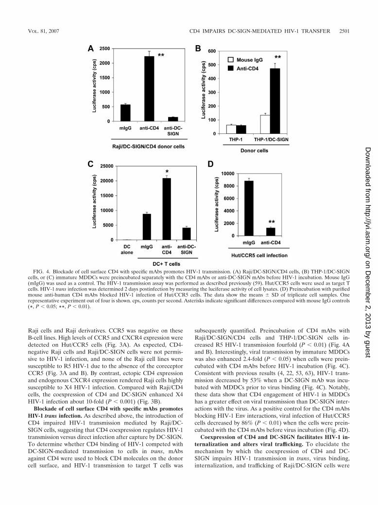
Blockade of cell surface CD4 with specific mAbs promotesHIV-1 trans infection. As described above, the introduction ofCD4 impaired HIV-1 transmission mediated by Raji/DC-SIGN cells, suggesting that CD4 coexpression regulates HIV-1transmission versus direct infection after capture by DC-SIGN.To determine whether CD4 binding of HIV-1 competed withDC-SIGN-mediated transmission to cells in trans, mAbsagainst CD4 were used to block CD4 molecules on the donorcell surface, and HIV-1 transmission to target T cells was
subsequently quantified. Preincubation of CD4 mAbs withRaji/DC-SIGN/CD4 cells and THP-1/DC-SIGN cells in-creased R5 HIV-1 transmission fourfold (P � 0.01) (Fig. 4Aand B). Interestingly, viral transmission by immature MDDCswas also enhanced 2.4-fold (P � 0.05) when cells were prein-cubated with CD4 mAbs before HIV-1 incubation (Fig. 4C).Consistent with previous results (4, 22, 53, 63), HIV-1 trans-mission decreased by 53% when a DC-SIGN mAb was incu-bated with MDDCs prior to virus binding (Fig. 4C). Notably,these data show that CD4 engagement of HIV-1 in MDDCshas a greater effect on viral transmission than DC-SIGN inter-actions with the virus. As a positive control for the CD4 mAbsblocking HIV-1 Env interactions, viral infection of Hut/CCR5cells decreased by 86% (P � 0.01) when the cells were prein-cubated with the CD4 mAbs before virus incubation (Fig. 4D).
Coexpression of CD4 and DC-SIGN facilitates HIV-1 in-ternalization and alters viral trafficking. To elucidate themechanism by which the coexpression of CD4 and DC-SIGN impairs HIV-1 transmission in trans, virus binding,internalization, and trafficking of Raji/DC-SIGN cells were
FIG. 4. Blockade of cell surface CD4 with specific mAbs promotes HIV-1 transmission. (A) Raji/DC-SIGN/CD4 cells, (B) THP-1/DC-SIGNcells, or (C) immature MDDCs were preincubated separately with the CD4 mAbs or anti-DC-SIGN mAbs before HIV-1 incubation. Mouse IgG(mIgG) was used as a control. The HIV-1 transmission assay was performed as described previously (59). Hut/CCR5 cells were used as target Tcells. HIV-1 trans infection was determined 2 days postinfection by measuring the luciferase activity of cell lysates. (D) Preincubation with purifiedmouse anti-human CD4 mAbs blocked HIV-1 infection of Hut/CCR5 cells. The data show the means � SD of triplicate cell samples. Onerepresentative experiment out of four is shown. cps, counts per second. Asterisks indicate significant differences compared with mouse IgG controls(*, P � 0.05; **, P � 0.01).
VOL. 81, 2007 CD4 IMPAIRS DC-SIGN-MEDIATED HIV-1 TRANSFER 2501
on Decem
ber 2, 2013 by guesthttp://jvi.asm
.org/D
ownloaded from

compared with those of Raji/DC-SIGN/CD4 cells. To exam-ine the cell-virus interactions in the absence of potentiallyconfounding effects of productive viral infection, we used AT-2-inactivated HIV-1 in these experiments. Previous studiesshowed that AT-2-inactivated HIV-1 is conformationally au-thentic and interacts with MDDCs similarly to infectious viruses(16, 56).
To measure virus binding and internalization, Raji/DC-SIGN cells and Raji/DC-SIGN/CD4 cells were pulsed sepa-rately with AT-2-inactivated R5 HIV-1 at 4°C or 37°C for 2 h,and after rigorous washing, cells were lysed, and HIV-1 Gagp24 was quantified. To test the proteolysis sensitivity of cell-associated HIV-1, cells were treated with trypsin after virusincubation. At 4°C, HIV-1 binding to Raji/DC-SIGN/CD4 cellswas nearly twofold higher than that to Raji/DC-SIGN cells(Fig. 5A). Trypsin treatment greatly reduced cell-associatedHIV-1 to background levels of Raji cell controls (Fig. 5A anddata not shown), indicating that virus remained on the cellsurface upon binding at 4°C. By contrast, HIV-1 internaliza-tion by Raji/DC-SIGN/CD4 cells at 37°C was fivefold greater(P � 0.01) than that by Raji/DC-SIGN cells. At 37°C, 80% ofRaji/DC-SIGN/CD4 cell-associated HIV-1 was resistant to
proteolysis, whereas the Raji/DC-SIGN cell-associated HIV-1was largely sensitive to trypsin (Fig. 5A).
Next, HIV-1 trafficking in these cells was visualized usingelectron and confocal microscopy. After a 2-h HIV-1 exposureto cells at 37°C, only cell surface-bound HIV-1 particles werefound on Raji/DC-SIGN cells, in apparent association withclathrin-coated pits (Fig. 5B, left); however, in addition to thecell surface-bound HIV-1 particles (Fig. 5B, upper right), virusparticles were observed within intracellular compartments ofRaji/DC-SIGN/CD4 cells, which were close to cell membranes(Fig. 5B, lower right). Immunostaining and confocal micros-copy indicated that HIV-1 internalized within Raji/DC-SIGN/CD4 cells colocalized with the late endosomal markers CD63and CD81 (Fig. 5C and D), whereas HIV-1 bound to Raji/DC-SIGN cell surfaces did not colocalize with these markers. Inaddition, compared with Raji/DC-SIGN cells, the late en-dosomal compartments were concentrated and polarizedwithin Raji/DC-SIGN/CD4 cells after exposure to HIV-1(Fig. 5C and D).
Collectively, these data indicate that the coexpression ofCD4 and DC-SIGN in Raji cells significantly enhances HIV-1internalization within late endosomal compartments, suggest-
FIG. 5. Coexpression of DC-SIGN and CD4 facilitates HIV-1 internalization and alters viral trafficking. (A) Coexpression of DC-SIGN andCD4 enhances HIV-1 binding and internalization. Raji/DC-SIGN cells or Raji/DC-SIGN/CD4 cells were separately incubated with AT-2-inactivated R5 HIV-1 for 2 h. Cells were washed and treated with trypsin or medium and lysed for HIV-1 p24 quantification. The data representthe means � SD of triplicate cell samples. One representative experiment out of 10 is shown. *, significant differences compared withRaji/DC-SIGN cells (P � 0.01). (B) Raji/DC-SIGN/CD4 cells internalize HIV-1 in intracellular vesicles. Cells were washed after exposure toAT-2-inactivated HIV-1 and were prepared immediately for electron microscopy. Scale bars, 100 nm. (C and D) HIV-1 colocalized with the lateendosomal markers CD63 (C) and CD81 (D) in Raji/DC-SIGN/CD4 cells. Cells were pulsed with AT-2-inactivated HIV-1, coimmunostained, andanalyzed by confocal microscopy. Green, HIV-1 p24; red, CD63 or CD81. Magnification, �60.
2502 WANG ET AL. J. VIROL.
on Decem
ber 2, 2013 by guesthttp://jvi.asm
.org/D
ownloaded from

ing that the altered viral trafficking contributes to the CD4inhibition of DC-SIGN-mediated HIV-1 trans infection.
HIV-1 retention in Raji cells coexpressing CD4 and DC-SIGN. To examine the stability of HIV-1 internalized in Raji/DC-SIGN/CD4 cells, cells were cultured for an additional 24 hafter 2 h of exposure of AT-2-inactivated HIV-1 and thenanalyzed by electron microscopy. No viral particles were ob-served in mock control cells without incubation of AT-2-inac-tivated HIV-1 (data not shown). Only cell surface-associatedvirus particles were observed from Raji/DC-SIGN cells (Fig.6A). By contrast, in addition to abundant HIV-1 particlesbound on the cell surfaces of Raji/DC-SIGN/CD4 cells, intactvirus particles were still confined to intracellular compartmentsin these cells after a 24-h culture (Fig. 6B and C). A highmagnification of the boxed area in Fig. 6B indicated that intactHIV-1 particles were trapped in the endosomal compartments(Fig. 6C). Thus, HIV-1 internalized by Raji/DC-SIGN/CD4cells can be retained in the cells for at least 24 h.
Downregulation of CD4 by Nef expression in DCs correlateswith enhanced viral transmission. We next wished to examinethe effect of CD4 levels on DC-mediated HIV-1 transmission.In addition to DC-mediated HIV-1 trans infection, long-termviral transfer from DCs to T cells depends on HIV-1 infectionof DCs (6, 30, 35, 56). MDDCs express both CD4 and DC-SIGN and are susceptible to HIV-1 infection, albeit at reducedreplication levels (35). HIV-1 Nef downregulates cell surfaceexpression of CD4 (38) but upregulates DC-SIGN expression(51). We thus hypothesized that HIV-1 Nef expression uponviral infection may facilitate DC-mediated HIV-1 transmission
through the modulation of CD4 and/or DC-SIGN expression.To test this model, WT and nef-inactivated (�nef) HIV-1 in-fection upon viral transmission by MDDCs was examined.
First, levels of viral replication of R5 (NLAD8) or X4(NL4-3) WT and �nef HIV-1 in Hut/CCR5 target cells werecompared by using equivalent inoculums. By quantification ofGag p24 in cell-free supernatants, comparable levels of virusreplication were detected between WT and �nef HIV-1-in-fected Hut/CCR5 cells at 3 dpi (Fig. 7A). HIV-1 production byinfected immature MDDCs was then measured. WT and �nefHIV-1-infected MDDCs showed similar virus replication ki-netics (Fig. 7B). In MDDCs, viral replication of X4 HIV-1 wassignificantly lower than that of R5 viruses (Fig. 7B), which isconsistent with previous studies showing that susceptibility ofDCs to R5 HIV is generally higher than that to X4 HIV due tothe higher expression of CCR5 or the lack of CXCR4 expres-sion on DC surfaces (20, 25, 35, 36, 48, 64).
At 3, 5, and 7 dpi, DCs were washed thoroughly and thencocultured with Hut/CCR5 cells for an additional 3 days toquantify viral transmission. Cell-free supernatants of the co-cultures were then measured for p24 production. Interestingly,compared with �nef HIV-1-infected MDDCs, Nef-expressing,WT NLAD8 HIV-1 infection significantly enhanced DC-me-diated viral transmission twofold, sixfold (P � 0.01), and four-fold (P � 0.01) at 3, 5, and 7 days after DC infection, respec-tively. As expected, X4-tropic NL4-3 replicated more weakly inthe cocultures using preinfected MDDCs, but the presence ofNef increased virus production twofold and threefold (P �0.05) at 5 and 7 days after DC infection, respectively (Fig. 7C).
FIG. 6. Raji/DC-SIGN/CD4 cells retain intracellular HIV-1 parti-cles for at least 24 h. Cells were incubated with AT-2-inactivated R5HIV-1 at 37°C for 2 h, washed, and cultured for an additional 24 h.Cells were then analyzed by electron microscopy. (A) Raji/DC-SIGNcells. Arrows indicated cell surface-associated HIV-1 particles.(B) Raji/DC-SIGN/CD4 cells. (C) Higher-magnification image of theboxed area from panel B. Arrows indicate the intracellular compart-ments that trapped intact HIV-1 particles. Scale bars, 100 nm.
VOL. 81, 2007 CD4 IMPAIRS DC-SIGN-MEDIATED HIV-1 TRANSFER 2503
on Decem
ber 2, 2013 by guesthttp://jvi.asm
.org/D
ownloaded from
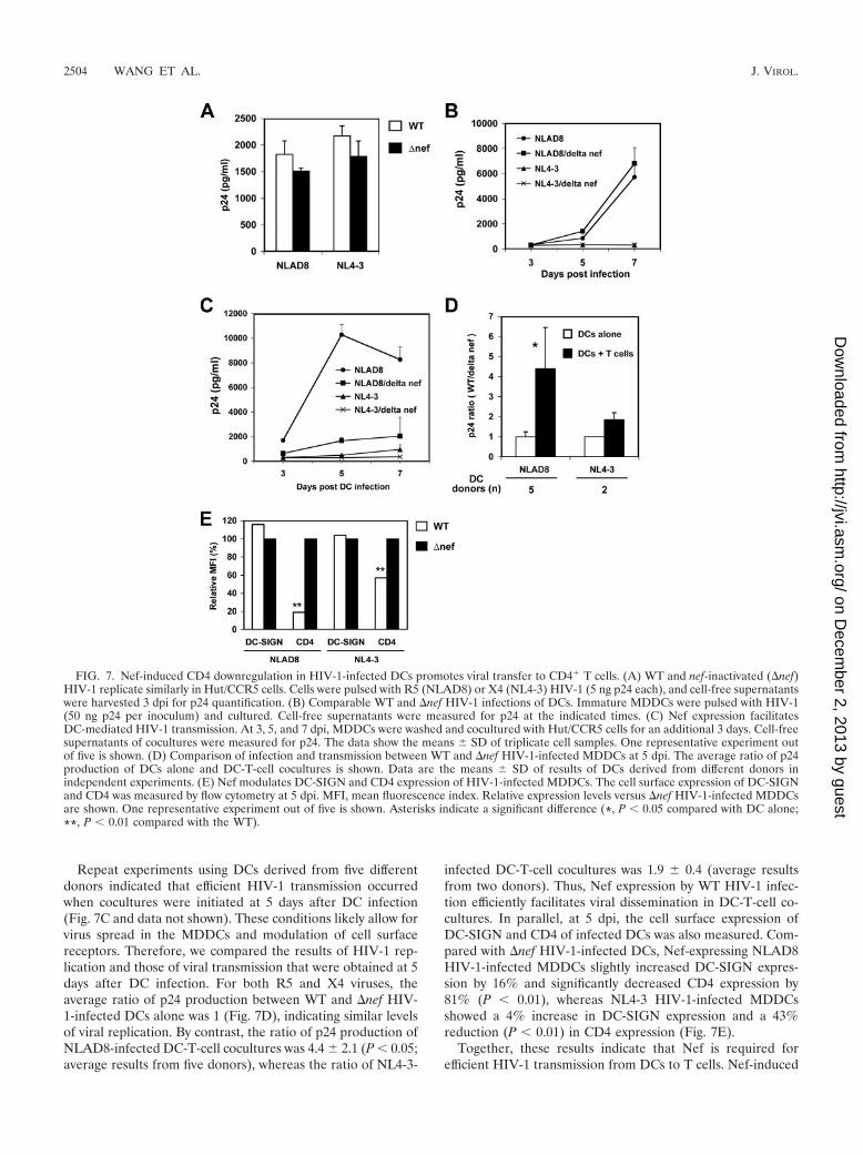
Repeat experiments using DCs derived from five differentdonors indicated that efficient HIV-1 transmission occurredwhen cocultures were initiated at 5 days after DC infection(Fig. 7C and data not shown). These conditions likely allow forvirus spread in the MDDCs and modulation of cell surfacereceptors. Therefore, we compared the results of HIV-1 rep-lication and those of viral transmission that were obtained at 5days after DC infection. For both R5 and X4 viruses, theaverage ratio of p24 production between WT and �nef HIV-1-infected DCs alone was 1 (Fig. 7D), indicating similar levelsof viral replication. By contrast, the ratio of p24 production ofNLAD8-infected DC-T-cell cocultures was 4.4 � 2.1 (P � 0.05;average results from five donors), whereas the ratio of NL4-3-
infected DC-T-cell cocultures was 1.9 � 0.4 (average resultsfrom two donors). Thus, Nef expression by WT HIV-1 infec-tion efficiently facilitates viral dissemination in DC-T-cell co-cultures. In parallel, at 5 dpi, the cell surface expression ofDC-SIGN and CD4 of infected DCs was also measured. Com-pared with �nef HIV-1-infected DCs, Nef-expressing NLAD8HIV-1-infected MDDCs slightly increased DC-SIGN expres-sion by 16% and significantly decreased CD4 expression by81% (P � 0.01), whereas NL4-3 HIV-1-infected MDDCsshowed a 4% increase in DC-SIGN expression and a 43%reduction (P � 0.01) in CD4 expression (Fig. 7E).
Together, these results indicate that Nef is required forefficient HIV-1 transmission from DCs to T cells. Nef-induced
FIG. 7. Nef-induced CD4 downregulation in HIV-1-infected DCs promotes viral transfer to CD4� T cells. (A) WT and nef-inactivated (�nef)HIV-1 replicate similarly in Hut/CCR5 cells. Cells were pulsed with R5 (NLAD8) or X4 (NL4-3) HIV-1 (5 ng p24 each), and cell-free supernatantswere harvested 3 dpi for p24 quantification. (B) Comparable WT and �nef HIV-1 infections of DCs. Immature MDDCs were pulsed with HIV-1(50 ng p24 per inoculum) and cultured. Cell-free supernatants were measured for p24 at the indicated times. (C) Nef expression facilitatesDC-mediated HIV-1 transmission. At 3, 5, and 7 dpi, MDDCs were washed and cocultured with Hut/CCR5 cells for an additional 3 days. Cell-freesupernatants of cocultures were measured for p24. The data show the means � SD of triplicate cell samples. One representative experiment outof five is shown. (D) Comparison of infection and transmission between WT and �nef HIV-1-infected MDDCs at 5 dpi. The average ratio of p24production of DCs alone and DC-T-cell cocultures is shown. Data are the means � SD of results of DCs derived from different donors inindependent experiments. (E) Nef modulates DC-SIGN and CD4 expression of HIV-1-infected MDDCs. The cell surface expression of DC-SIGNand CD4 was measured by flow cytometry at 5 dpi. MFI, mean fluorescence index. Relative expression levels versus �nef HIV-1-infected MDDCsare shown. One representative experiment out of five is shown. Asterisks indicate a significant difference (*, P � 0.05 compared with DC alone;**, P � 0.01 compared with the WT).
2504 WANG ET AL. J. VIROL.
on Decem
ber 2, 2013 by guesthttp://jvi.asm
.org/D
ownloaded from
CD4 downregulation in HIV-1-infected MDDCs correlateswith enhanced viral transmission.
DISCUSSION
The cell type dependency of DC-SIGN-mediated HIV-1transmission provides a useful model system to investigatethe mechanisms underlying DC-mediated viral transfer (61,62). We previously discovered that monocytic THP-1 cellsdo not support DC-SIGN-mediated HIV-1 transmission, al-though it was widely assumed in the literature that such cellsrecapitulated DC transmission of HIV-1 (61). Here, weidentify an underlying mechanism for the monocytic cellrestriction of DC-SIGN-mediated HIV-1 transmission andpresent a model by which HIV-1 might exploit primary cellswith similar characteristics to potentiate viral spread toCD4� T cells.
In this report, we show that CD4 expression levels regulateDC-SIGN and MDDC transmission of HIV-1. Monocytic celltype restriction of DC-SIGN-mediated HIV-1 transmissioncorrelates with CD4 expression, and this impairment can beblocked with CD4 antibodies that prevent interactions withHIV-1 Env. This restriction can be re-created by the expres-sion of CD4 in Raji/DC-SIGN cells, which are otherwise per-missive to DC-SIGN-mediated HIV-1 transmission. In thepresence of an appropriate HIV-1 coreceptor, cells engineeredto coexpress CD4 and DC-SIGN are preferentially infected. Bycontrast, CD4/DC-SIGN double-positive cells lacking a viralcoreceptor efficiently internalize and trap HIV-1. Using pri-mary cells expressing CD4 and DC-SIGN, we observed thatHIV-1 infection changes the transmission properties of thesecells. Specifically, infection by Nef-positive HIV-1 efficientlypromotes DC-mediated HIV-1 transmission, which correlateswith CD4 downregulation in these cells. Collectively, thesedata suggest that modulations of CD4 levels may play an im-portant regulatory role in DC-mediated HIV-1 transmission.
DC-mediated HIV-1 transmission to CD4� T cells involvesat least two pathways (60), namely, HIV-1 trans infection with-out viral infection in DCs, and long-term viral transfer de-pended on the HIV-1 replication in infected DCs (6, 30, 35,56). It has been reported that certain DC subsets and macro-phages in vivo express DC-SIGN, CD4, and HIV-1 coreceptors(24, 29, 50), and both cell types were proposed to facilitatemucosal transmission of HIV-1 (47). Thus, it is conceivablethat these types of cells are more susceptible to HIV-1 infec-tion due to the coexpression of CD4, DC-SIGN, and viralcoreceptors, which may augment viral dissemination by DCsand macrophages using the cis infection pathway. Upon HIV-1infection, the Nef protein may convert these cells into morepotent HIV-1 transmitters through a significant downregula-tion of CD4. CD4 and DC-SIGN molecules have dileucine-based internalization motifs in the cytoplasmic domains (5, 19),and the dileucine motifs of CD4 and DC-SIGN are critical forendocytosis function and regulation by Nef (51). AlthoughMDDCs have provided a convenient tool to study DC-HIVinteractions, further examinations of bona fide DC subsets exvivo for viral infection and transmission would be beneficial forunderstanding the contribution of DCs to AIDS pathogenesis.
Internalization of HIV-1 and distinct viral trafficking havebeen suggested to be important for DC-mediated HIV-1 trans
infection (28, 53); however, it is unclear how internalized viri-ons remain infectious, recycle back to the cell surface, and aretransferred to the T cells. By contrast, our viral binding andelectron microscopy results suggest that HIV-1 internalizationin Raji/DC-SIGN cells is not required for efficient HIV-1transmission. Consistent with our observations, Burleigh andcolleagues recently reported that DC-SIGN-mediated HIV-1internalization is dispensable for both trans infection of T cellsand the retention of viral infectivity (6). We found that thecoexpression of CD4 and DC-SIGN in Raji cells significantlypromoted viral internalization and altered viral trafficking tolate endosomal compartments (also known as multivesicularbodies [MVBs]). Although endocytosis-mediated HIV-1 entrycan lead to productive viral infection (15, 44), only X4 HIV-1,but not R5 HIV-1, replicated in CD4-expressing Raji cells.These results suggest that the fusion-mediated entry of X4HIV-1 leads to productive infection in these cells; however,endocytosed R5 HIV-1 is trapped or inactivated in acidifiedendosomes and is eventually degraded in lysosomes. Indeed,compared with Raji/DC-SIGN cells, we have observed signif-icant colocalization of the HIV-1 p24 protein with lysosome-associated membrane protein 1 in Raji/DC-SIGN/CD4 cells(data not shown), suggesting enhanced HIV-1 degradationwithin lysosomes in CD4-DC-SIGN-expressing cells.
Interestingly, it was shown that HIV-1 captured by immatureMDDCs is also internalized to MVBs, and infectious HIV-1 isconstitutively released into the extracellular milieu in associa-tion with the exosomes to initiate viral transfer to T cells (58).Similarly, HIV-1 particles internalized by mature MDDCs arealso sequestered into MVBs (17). In contrast to the Raji/DC-SIGN/CD4 cells, it remains unclear how internalized HIV-1circumvents the intracellular degradation machinery in DCsand recycles back to the DC surface to be transferred to the Tcells. Further investigations of HIV-1 trafficking in DCs incomparison to Raji/DC-SIGN/CD4 cells may help us to under-stand this unique feature of DC-mediated viral transmission.
The blocking of exposed CD4 on DCs with specific mAbspromoted HIV-1 trans infection, most likely by impairingvirus internalization and intracellular degradation. Similar re-sults were confirmed using DCs generated from different do-nors and using different CD4 mAbs in independent experi-ments (data not shown). However, the enhancement of HIV-1transmission by CD4 mAb blocking appears to be limited, with2.4-fold and 4-fold increases in MDDCs and various cell lines,respectively. It is conceivable that other regions of CD4 out-side the gp120 binding domain contribute to the inhibition ofDC-SIGN-mediated HIV-1 transmission. In addition, duringthe 2-day coculture with T-cell targets, the newly synthesizedCD4 proteins on the donor cell surfaces diminished the block-ade effect of CD4 mAb. In DCs, CD4 mAb alone may notcompletely block HIV-1 internalization due to the coexpres-sion of DC-SIGN or other C-type lectins. These results alsosuggest that CD4 expression is not the sole cellular determi-nant that controls the efficiency of DC-SIGN-mediated HIV-1trans infection. For example, we previously reported that CD4-negative human erythroleukemic K562 cells do not efficientlytransmit HIV-1 despite high levels of exogenous DC-SIGNexpression (62). Presumably, another unidentified cellularfactor(s) can also influence DC-SIGN-mediated HIV-1
VOL. 81, 2007 CD4 IMPAIRS DC-SIGN-MEDIATED HIV-1 TRANSFER 2505
on Decem
ber 2, 2013 by guesthttp://jvi.asm
.org/D
ownloaded from

transmission, given its cell type dependence in CD4-nega-tive cells (61, 62).
Nef-expressing HIV-1-infected DCs promoted viral trans-mission to cocultured T cells. Indeed, Nef modulation of DC-SIGN and CD4 expression was observed despite the limitedlevels of DC-SIGN upregulation. These results provide anadditional context to consider previous observations of Neffacilitating DC-mediated HIV-1 spread to T cells (37). It hasbeen reported that HIV-1 transmission efficiency can be en-hanced by the maturation of DCs (32, 43). To examine thepossibility that Nef-promoted HIV-1 transmission results fromNef-induced DC maturation, maturation markers (such asHLA-DR, CD83, and CD86) of WT and �nef HIV-1-infectedDCs were compared, but no Nef-induced upregulation of thematuration markers was observed (unpublished data). It hasalso been reported that Nef-expressing immature MDDCsstimulate T-cell activation but without upregulating DC mat-uration markers (33). We confined our analyses to a trans-formed CD4� T cell that is constitutively activated. With rest-ing, primary CD4� T cells, it is likely that Nef may alsofacilitate DC-mediated HIV-1 transmission by promoting DC-T-cell interactions or by enhancing T-cell clustering (51).
In summary, we find that CD4 coexpression with DC-SIGNenhances HIV-1 internalization and retention but strongly im-pairs HIV-1 transmission to T cells. The blocking of CD4 onthe DC surfaces promotes HIV-1 trans infection. Significantly,Nef facilitates DC-mediated HIV-1 transmission, which corre-lates with Nef-induced downregulation of CD4. These dataprovide a novel insight into cellular characteristics that influ-ence DC-mediated HIV-1 dissemination and highlight Nef’srole as a multifunctional pathogenic factor capable of regulat-ing these processes.
ACKNOWLEDGMENTS
We thank Mark McNally for critical reading of the manuscript. Wethank Eric Freed, Amy Hudson, Jeffery Lifson, and Olivier Schwartzfor the kind gift of reagents. The following reagents were obtainedthrough the AIDS Research and Reference Reagent Program, Divi-sion of AIDS, NIAID, NIH: recombinant IL-2 from Maurice Gately,Hoffmann-La Roche Inc.; CXCR4 mAb (44717.111); CD4 complexmAb B4 from United Biomedical, Inc.; and p24 mAb from MichaelMalim.
This work was supported by a grant (R01-AI068493) to L.W. fromthe National Institutes of Health (NIH). V.N.K. is supported by intra-mural research funds from the National Cancer Institute, NIH.
A.M.J. and W.J.O. contributed equally to this study.
REFERENCES
1. Arrighi, J. F., M. Pion, E. Garcia, J. M. Escola, Y. van Kooyk, T. B.Geijtenbeek, and V. Piguet. 2004. DC-SIGN-mediated infectious synapseformation enhances X4 HIV-1 transmission from dendritic cells to T cells. J.Exp. Med. 200:1279–1288.
2. Arrighi, J. F., M. Pion, M. Wiznerowicz, T. B. Geijtenbeek, E. Garcia, S.Abraham, F. Leuba, V. Dutoit, O. Ducrey-Rundquist, Y. van Kooyk, D.Trono, and V. Piguet. 2004. Lentivirus-mediated RNA interference of DC-SIGN expression inhibits human immunodeficiency virus transmission fromdendritic cells to T cells. J. Virol. 78:10848–10855.
3. Banchereau, J., and R. M. Steinman. 1998. Dendritic cells and the control ofimmunity. Nature 392:245–252.
4. Baribaud, F., S. Pohlmann, G. Leslie, F. Mortari, and R. W. Doms. 2002.Quantitative expression and virus transmission analysis of DC-SIGN onmonocyte-derived dendritic cells. J. Virol. 76:9135–9142.
5. Bentham, M., S. Mazaleyrat, and M. Harris. 2003. The di-leucine motif inthe cytoplasmic tail of CD4 is not required for binding to human immuno-deficiency virus type 1 Nef, but is critical for CD4 down-modulation. J. Gen.Virol. 84:2705–2713.
6. Burleigh, L., P.-Y. Lozach, C. Schiffer, I. Staropoli, V. Pezo, F. Porrot, B.
Canque, J.-L. Virelizier, F. Arenzana-Seisdedos, and A. Amara. 2006. Infec-tion of dendritic cells (DCs), not DC-SIGN-mediated internalization ofhuman immunodeficiency virus, is required for long-term transfer of virus toT cells. J. Virol. 80:2949–2957.
7. Cameron, P. U., P. S. Freudenthal, J. M. Barker, S. Gezelter, K. Inaba, andR. M. Steinman. 1992. Dendritic cells exposed to human immunodeficiencyvirus type-1 transmit a vigorous cytopathic infection to CD4� T cells. Sci-ence 257:383–387.
8. Collins, K. L., B. K. Chen, S. A. Kalams, B. D. Walker, and D. Baltimore.1998. HIV-1 Nef protein protects infected primary cells against killing bycytotoxic T lymphocytes. Nature 391:397–401.
9. Connor, R. I., B. K. Chen, S. Choe, and N. R. Landau. 1995. Vpr is requiredfor efficient replication of human immunodeficiency virus type-1 in mono-nuclear phagocytes. Virology 206:935–944.
10. Daniel, M. D., F. Kirchhoff, S. C. Czajak, P. K. Sehgal, and R. C. Desrosiers.1992. Protective effects of a live attenuated SIV vaccine with a deletion in thenef gene. Science 258:1938–1941.
11. Deacon, N. J., A. Tsykin, A. Solomon, K. Smith, M. Ludford-Menting, D. J.Hooker, D. A. McPhee, A. L. Greenway, A. Ellett, C. Chatfield, V. A. Lawson,S. Crowe, A. Maerz, S. Sonza, J. Learmont, J. S. Sullivan, A. Cunningham,D. Dwyer, D. Dowton, and J. Mills. 1995. Genomic structure of an attenuatedquasi species of HIV-1 from a blood transfusion donor and recipients.Science 270:988–991.
12. DeMasi, J., and P. Traktman. 2000. Clustered charge-to-alanine mutagen-esis of the vaccinia virus H5 gene: isolation of a dominant, temperature-sensitive mutant with a profound defect in morphogenesis. J. Virol. 74:2393–2405.
13. Engering, A., S. J. van Vliet, K. Hebeda, D. G. Jackson, R. Prevo, S. K. Singh,T. B. Geijtenbeek, H. van Krieken, and Y. van Kooyk. 2004. Dynamic pop-ulations of dendritic cell-specific ICAM-3 grabbing nonintegrin-positive im-mature dendritic cells and liver/lymph node-specific ICAM-3 grabbing non-integrin-positive endothelial cells in the outer zones of the paracortex ofhuman lymph nodes. Am. J. Pathol. 164:1587–1595.
14. Englund, G., T. S. Theodore, E. O. Freed, A. Engelman, and M. A. Martin.1995. Integration is required for productive infection of monocyte-derivedmacrophages by human immunodeficiency virus type 1. J. Virol. 69:3216–3219.
15. Fackler, O. T., and B. M. Peterlin. 2000. Endocytic entry of HIV-1. Curr.Biol. 10:1005–1008.
16. Frank, I., M. Piatak, Jr., H. Stoessel, N. Romani, D. Bonnyay, J. D. Lifson,and M. Pope. 2002. Infectious and whole inactivated simian immunodefi-ciency viruses interact similarly with primate dendritic cells (DCs): differen-tial intracellular fate of virions in mature and immature DCs. J. Virol.76:2936–2951.
17. Garcia, E., M. Pion, A. Pelchen-Matthews, L. Collinson, J. F. Arrighi, G.Blot, F. Leuba, J. M. Escola, N. Demaurex, M. Marsh, and V. Piguet. 2005.HIV-1 trafficking to the dendritic cell-T-cell infectious synapse uses a path-way of tetraspanin sorting to the immunological synapse. Traffic 6:488–501.
18. Geijtenbeek, T. B., D. S. Kwon, R. Torensma, S. J. van Vliet, G. C. vanDuijnhoven, J. Middel, I. L. Cornelissen, H. S. Nottet, V. N. KewalRamani,D. R. Littman, C. G. Figdor, and Y. van Kooyk. 2000. DC-SIGN, a dendriticcell-specific HIV-1-binding protein that enhances trans-infection of T cells.Cell 100:587–597.
19. Geijtenbeek, T. B., R. Torensma, S. J. van Vliet, G. C. van Duijnhoven, G. J.Adema, Y. van Kooyk, and C. G. Figdor. 2000. Identification of DC-SIGN, anovel dendritic cell-specific ICAM-3 receptor that supports primary immuneresponses. Cell 100:575–585.
20. Granelli-Piperno, A., E. Delgado, V. Finkel, W. Paxton, and R. M. Steinman.1998. Immature dendritic cells selectively replicate macrophagetropic (M-tropic) human immunodeficiency virus type 1, while mature cells efficientlytransmit both M- and T-tropic virus to T cells. J. Virol. 72:2733–2737.
21. Granelli-Piperno, A., A. Pritsker, M. Pack, I. Shimeliovich, J. F. Arrighi,C. G. Park, C. Trumpfheller, V. Piguet, T. M. Moran, and R. M. Steinman.2005. Dendritic cell-specific intercellular adhesion molecule 3-grabbing non-integrin/CD209 is abundant on macrophages in the normal human lymphnode and is not required for dendritic cell stimulation of the mixed leukocytereaction. J. Immunol. 175:4265–4273.
22. Gummuluru, S., M. Rogel, L. Stamatatos, and M. Emerman. 2003. Bindingof human immunodeficiency virus type 1 to immature dendritic cells canoccur independently of DC-SIGN and mannose binding C-type lectin recep-tors via a cholesterol-dependent pathway. J. Virol. 77:12865–12874.
23. Gurney, K. B., J. Elliott, H. Nassanian, C. Song, E. Soilleux, I. McGowan,P. A. Anton, and B. Lee. 2005. Binding and transfer of human immunode-ficiency virus by DC-SIGN� cells in human rectal mucosa. J. Virol. 79:5762–5773.
24. Jameson, B., F. Baribaud, S. Pohlmann, D. Ghavimi, F. Mortari, R. W.Doms, and A. Iwasaki. 2002. Expression of DC-SIGN by dendritic cells ofintestinal and genital mucosae in humans and rhesus macaques. J. Virol.76:1866–1875.
25. Kawamura, T., F. O. Gulden, M. Sugaya, D. T. McNamara, D. L. Borris,M. M. Lederman, J. M. Orenstein, P. A. Zimmerman, and A. Blauvelt. 2003.R5 HIV productively infects Langerhans cells, and infection levels are reg-
2506 WANG ET AL. J. VIROL.
on Decem
ber 2, 2013 by guesthttp://jvi.asm
.org/D
ownloaded from
ulated by compound CCR5 polymorphisms. Proc. Natl. Acad. Sci. USA100:8401–8406.
26. Kestler, H. W., III, D. J. Ringler, K. Mori, D. L. Panicali, P. K. Sehgal, M. D.Daniel, and R. C. Desrosiers. 1991. Importance of the nef gene for mainte-nance of high virus loads and for development of AIDS. Cell 65:651–662.
27. Kirchhoff, F., T. C. Greenough, D. B. Brettler, J. L. Sullivan, and R. C.Desrosiers. 1995. Brief report: absence of intact nef sequences in a long-termsurvivor with nonprogressive HIV-1 infection. N. Engl. J. Med. 332:228–232.
28. Kwon, D. S., G. Gregorio, N. Bitton, W. A. Hendrickson, and D. R. Littman.2002. DC-SIGN-mediated internalization of HIV is required for trans-en-hancement of T cell infection. Immunity 16:135–144.
29. Lee, B., G. Leslie, E. Soilleux, U. O’Doherty, S. Baik, E. Levroney, K.Flummerfelt, W. Swiggard, N. Coleman, M. Malim, and R. W. Doms. 2001.cis expression of DC-SIGN allows for more efficient entry of human andsimian immunodeficiency viruses via CD4 and a coreceptor. J. Virol. 75:12028–12038.
30. Lore, K., A. Smed-Sorensen, J. Vasudevan, J. R. Mascola, and R. A. Koup.2005. Myeloid and plasmacytoid dendritic cells transfer HIV-1 preferentiallyto antigen-specific CD4� T cells. J. Exp. Med. 201:2023–2033.
31. Lore, K., A. Sonnerborg, C. Brostrom, L. E. Goh, L. Perrin, H. McDade,H. J. Stellbrink, B. Gazzard, R. Weber, L. A. Napolitano, Y. van Kooyk, andJ. Andersson. 2002. Accumulation of DC-SIGN�CD40� dendritic cells withreduced CD80 and CD86 expression in lymphoid tissue during acute HIV-1infection. AIDS 16:683–692.
32. McDonald, D., L. Wu, S. M. Bohks, V. N. KewalRamani, D. Unutmaz, andT. J. Hope. 2003. Recruitment of HIV and its receptors to dendritic cell-Tcell junctions. Science 300:1295–1297.
33. Messmer, D., J. M. Jacque, C. Santisteban, C. Bristow, S. Y. Han, L. Vil-lamide-Herrera, E. Mehlhop, P. A. Marx, R. M. Steinman, A. Gettie, and M.Pope. 2002. Endogenously expressed nef uncouples cytokine and chemokineproduction from membrane phenotypic maturation in dendritic cells. J. Im-munol. 169:4172–4182.
34. Michel, N., I. Allespach, S. Venzke, O. T. Fackler, and O. T. Keppler. 2005.The Nef protein of human immunodeficiency virus establishes superinfectionimmunity by a dual strategy to downregulate cell-surface CCR5 and CD4.Curr. Biol. 15:714–723.
35. Nobile, C., C. Petit, A. Moris, K. Skrabal, J. P. Abastado, F. Mammano, andO. Schwartz. 2005. Covert human immunodeficiency virus replication indendritic cells and in DC-SIGN-expressing cells promotes long-term trans-mission to lymphocytes. J. Virol. 79:5386–5399.
36. Patterson, S., A. Rae, N. Hockey, J. Gilmour, and F. Gotch. 2001. Plasma-cytoid dendritic cells are highly susceptible to human immunodeficiencyvirus type 1 infection and release infectious virus. J. Virol. 75:6710–6713.
37. Petit, C., F. Buseyne, C. Boccaccio, J. P. Abastado, J. M. Heard, and O.Schwartz. 2001. Nef is required for efficient HIV-1 replication in coculturesof dendritic cells and lymphocytes. Virology 286:225–236.
38. Piguet, V., O. Schwartz, S. Le Gall, and D. Trono. 1999. The downregulationof CD4 and MHC-I by primate lentiviruses: a paradigm for the modulationof cell surface receptors. Immunol. Rev. 168:51–63.
39. Pope, M., M. G. Betjes, N. Romani, H. Hirmand, P. U. Cameron, L. Hoffman,S. Gezelter, G. Schuler, and R. M. Steinman. 1994. Conjugates of dendriticcells and memory T lymphocytes from skin facilitate productive infectionwith HIV-1. Cell 78:389–398.
40. Pope, M., S. Gezelter, N. Gallo, L. Hoffman, and R. M. Steinman. 1995. Lowlevels of HIV-1 infection in cutaneous dendritic cells promote extensive viralreplication upon binding to memory CD4� T cells. J. Exp. Med. 182:2045–2056.
41. Rappocciolo, G., F. J. Jenkins, H. R. Hensler, P. Piazza, M. Jais, L.Borowski, S. C. Watkins, and C. R. Rinaldo, Jr. 2006. DC-SIGN is a receptorfor human herpesvirus 8 on dendritic dells and macrophages. J. Immunol.176:1741–1749.
42. Rossio, J. L., M. T. Esser, K. Suryanarayana, D. K. Schneider, J. W. Bess,Jr., G. M. Vasquez, T. A. Wiltrout, E. Chertova, M. K. Grimes, Q. Sattentau,L. O. Arthur, L. E. Henderson, and J. D. Lifson. 1998. Inactivation of humanimmunodeficiency virus type 1 infectivity with preservation of conforma-tional and functional integrity of virion surface proteins. J. Virol. 72:7992–8001.
43. Sanders, R. W., E. C. de Jong, C. E. Baldwin, J. H. Schuitemaker, M. L.Kapsenberg, and B. Berkhout. 2002. Differential transmission of humanimmunodeficiency virus type 1 by distinct subsets of effector dendritic cells.J. Virol. 76:7812–7821.
44. Schaeffer, E., V. B. Soros, and W. C. Greene. 2004. Compensatory linkbetween fusion and endocytosis of human immunodeficiency virus type 1 inhuman CD4 T lymphocytes. J. Virol. 78:1375–1383.
45. Schindler, M., J. Munch, O. Kutsch, H. Li, M. L. Santiago, F. Bibollet-Ruche, M. C. Muller-Trutwin, F. J. Novembre, M. Peeters, V. Courgnaud, E.Bailes, P. Roques, D. L. Sodora, G. Silvestri, P. M. Sharp, B. H. Hahn, andF. Kirchhoff. 2006. Nef-mediated suppression of T cell activation was lost ina lentiviral lineage that gave rise to HIV-1. Cell 125:1055–1067.
46. Schwartz, O., V. Marechal, S. Le Gall, F. Lemonnier, and J. M. Heard. 1996.Endocytosis of major histocompatibility complex class I molecules is inducedby the HIV-1 Nef protein. Nat. Med. 2:338–342.
47. Shattock, R. J., and J. P. Moore. 2003. Inhibiting sexual transmission ofHIV-1 infection. Nat. Rev. Microbiol. 1:25–34.
48. Smed-Sorensen, A., K. Lore, J. Vasudevan, M. K. Louder, J. Andersson, J. R.Mascola, A. L. Spetz, and R. A. Koup. 2005. Differential susceptibility tohuman immunodeficiency virus type 1 infection of myeloid and plasmacytoiddendritic cells. J. Virol. 79:8861–8869.
49. Soilleux, E. J., L. S. Morris, B. Lee, S. Pohlmann, J. Trowsdale, R. W. Doms,and N. Coleman. 2001. Placental expression of DC-SIGN may mediateintrauterine vertical transmission of HIV. J. Pathol. 195:586–592.
50. Soilleux, E. J., L. S. Morris, G. Leslie, J. Chehimi, Q. Luo, E. Levroney, J.Trowsdale, L. J. Montaner, R. W. Doms, D. Weissman, N. Coleman, and B.Lee. 2002. Constitutive and induced expression of DC-SIGN on dendritic celland macrophage subpopulations in situ and in vitro. J. Leukoc. Biol. 71:445–457.
51. Sol-Foulon, N., A. Moris, C. Nobile, C. Boccaccio, A. Engering, J. P. Abastado,J. M. Heard, Y. van Kooyk, and O. Schwartz. 2002. HIV-1 Nef-inducedupregulation of DC-SIGN in dendritic cells promotes lymphocyte clusteringand viral spread. Immunity 16:145–155.
52. Swigut, T., N. Shohdy, and J. Skowronski. 2001. Mechanism for down-regulation of CD28 by Nef. EMBO J. 20:1593–1604.
53. Trumpfheller, C., C. G. Park, J. Finke, R. M. Steinman, and A. Granelli-Piperno. 2003. Cell type-dependent retention and transmission of HIV-1 byDC-SIGN. Int. Immunol. 15:289–298.
54. Turville, S. G., J. Arthos, K. Mac Donald, G. Lynch, H. Naif, G. Clark, D.Hart, and A. L. Cunningham. 2001. HIV gp120 receptors on human den-dritic cells. Blood 98:2482–2488.
55. Turville, S. G., P. U. Cameron, A. Handley, G. Lin, S. Pohlmann, R. W.Doms, and A. L. Cunningham. 2002. Diversity of receptors binding HIV ondendritic cell subsets. Nat. Immunol. 3:975–983.
56. Turville, S. G., J. J. Santos, I. Frank, P. U. Cameron, J. Wilkinson, M.Miranda-Saksena, J. Dable, H. Stossel, N. Romani, M. Piatak, Jr., J. D.Lifson, M. Pope, and A. L. Cunningham. 2004. Immunodeficiency virusuptake, turnover, and 2-phase transfer in human dendritic cells. Blood 103:2170–2179.
57. Unutmaz, D., V. N. KewalRamani, S. Marmon, and D. R. Littman. 1999.Cytokine signals are sufficient for HIV-1 infection of resting human T lym-phocytes. J. Exp. Med. 189:1735–1746.
58. Wiley, R. D., and S. Gummuluru. 2006. Immature dendritic cell-derivedexosomes can mediate HIV-1 trans infection. Proc. Natl. Acad. Sci. USA103:738–743.
59. Wu, L., A. A. Bashirova, T. D. Martin, L. Villamide, E. Mehlhop, A. O.Chertov, D. Unutmaz, M. Pope, M. Carrington, and V. N. KewalRamani.2002. Rhesus macaque dendritic cells efficiently transmit primate lentivirusesindependently of DC-SIGN. Proc. Natl. Acad. Sci. USA 99:1568–1573.
60. Wu, L., and V. N. KewalRamani. 2006. Dendritic-cell interactions with HIV:infection and viral dissemination. Nat. Rev. Immunol. 6:859–868.
61. Wu, L., T. D. Martin, M. Carrington, and V. N. KewalRamani. 2004. Raji Bcells, misidentified as THP-1 cells, stimulate DC-SIGN-mediated HIV trans-mission. Virology 318:17–23.
62. Wu, L., T. D. Martin, Y. C. Han, S. K. Breun, and V. N. KewalRamani. 2004.Trans-dominant cellular inhibition of DC-SIGN-mediated HIV-1 transmis-sion. Retrovirology 1:14.
63. Wu, L., T. D. Martin, R. Vazeux, D. Unutmaz, and V. N. KewalRamani. 2002.Functional evaluation of DC-SIGN monoclonal antibodies reveals DC-SIGN interactions with ICAM-3 do not promote human immunodeficiencyvirus type 1 transmission. J. Virol. 76:5905–5914.
64. Zaitseva, M., A. Blauvelt, S. Lee, C. K. Lapham, V. Klaus-Kovtun, H.Mostowski, J. Manischewitz, and H. Golding. 1997. Expression and functionof CCR5 and CXCR4 on human Langerhans cells and macrophages: impli-cations for HIV primary infection. Nat. Med. 3:1369–1375.
VOL. 81, 2007 CD4 IMPAIRS DC-SIGN-MEDIATED HIV-1 TRANSFER 2507
on Decem
ber 2, 2013 by guesthttp://jvi.asm
.org/D
ownloaded from
Copyright © 2022 FDOKUMEN