Human Immunodeficiency Virus-Associated Vasculopathy in Transgenic Mice
6
JOURNAL OF VIROLOGY, 0022-538X/97/$04.0010 June 1997, p. 4809–4814 Vol. 71, No. 6 Copyright © 1997, American Society for Microbiology Human Immunodeficiency Virus-Associated Vasculopathy in Transgenic Mice BRAD T. TINKLE, 1 LIEN NGO, 1 PAUL A. LUCIW, 3 TOM MACIAG, 2 AND GILBERT JAY 1 * Departments of Virology 1 and Molecular Biology, 2 Jerome H. Holland Laboratory, Rockville, Maryland 20855, and Department of Medical Pathology, University of California, Davis, California 95616 3 Received 15 October 1996/Accepted 13 March 1997 There is substantial clinical evidence for the development of vascular disorders in human immunodeficiency virus (HIV)-infected individuals, particularly in the form of vasculitis. Transgenic mice carrying a replication- defective HIV-1 provirus with selective deletion of the gag, pol, and env genes developed extensive vasculopathy. Restricted expression of HIV nonstructural genes in smooth muscle cells was accompanied by the migration and proliferation of these cells in blood vessels of all sizes and at different body sites. The frequent infiltration observed in the hypertrophic vessel walls occurred predominantly in the adventitia and was composed of primarily T cells and occasionally plasma cells. The intimal thickening generated significant luminal narrow- ing in some vessels, and the restricted blood flow led to ischemia in the affected tissues. Interestingly, the endothelium did not appear to support HIV gene expression or be involved in the pathological process. This transgenic model provides an opportunity to dissect the mechanism underlying HIV-associated vasculopathy. Human immunodeficiency virus type 1 (HIV-1) is capable of infecting multiple cell types either through the cell surface CD4 receptor or through various non-CD4 mechanisms (10, 14, 23, 36). The pathological consequences may depend on the cell type(s) involved. For example, the immune dysfunction observed among HIV-1-infected individuals is a consequence of infection of one or more types of leukocytes (11) whereas infection of renal epithelial cells may result in HIV-associated nephropathy (2). Several vascular disorders have been documented as a con- sequence of HIV-1 infection. These include systemic vasculitis (4, 6, 15, 16, 20, 21, 24, 34), formation of “cotton-wool spots” in the eye due to vasculitis-induced ischemic injury (18, 26), and thrombotic thrombocytopenic purpura (7, 8, 19). The vas- culitis affects small and medium-sized vessels and is apparently more common among pediatric AIDS patients (6, 20, 21). It is characterized by intimal and medial thickening, resulting from hyperplasia of smooth muscle cells (SMCs). The vascular and perivascular infiltrates are composed of mainly T lymphocytes and mononuclear cells and a few plasma cells. A similar finding has also been made with simian immunodeficiency virus infec- tion of rhesus monkeys, in which approximately 20% of the animals develop arteriopathy (5). HIV-1-infected patients typically express autoantibodies and circulating immune complexes (32), and the vasculitis observed among infected patients has been attributed to immune com- plex formation or deposition in a few of the reported cases (15, 18). However, some of the systemic vascular lesions involving SMC proliferation observed associated with HIV-1 infection do not appear to involve an inflammatory response (20, 21). In addition, HIV-1 antigens can be found in vascular and perivas- cular cells but not in the infiltrate, which is composed primarily of CD8 1 T cells (4, 15). Therefore, it remains unclear whether inflammatory cells are necessary to induce the SMC migration and proliferation or are a result of vascular injury. We have generated transgenic mice carrying a defective HIV-1 provirus (strain SF2) with specific deletion of the gag, pol, and env genes (33) (Fig. 1). Mice from two independent transgenic lines were analyzed for histopathological evidence of vascular lesions. A survey of blood vessels detected in par- affin-embedded sections of the major tissues from each mouse has revealed characteristic vascular changes in over 60% of transgenic animals (11 of 18 mice between 9 and 20 months of age from the low-expressing N4 line and 6 of 10 mice between 27 and 46 days of age from the high-expressing Q5 line), but in none of their nontransgenic littermates. These lesions were noted in vessels of different sizes and were present in various organs, including the brain, heart, kidney, pancreas, mesentery, uterus, and spleen. Figure 2a shows a renal artery from a healthy, nontransgenic control animal. This vessel exhibits a single layer of luminal vascular endothelium, two to three layers of SMCs in the media, and an outer collagen-enriched adventitia. In contrast, the renal artery of a transgenic mouse shows extensive cellular infiltrates within the vessel wall (Fig. 2b). This artery exhibits an irregular pattern of SMCs that are hyperplastic and that have rounded vesicular nuclei. These lesions are observed at multiple sites within the same animal. The vascular infiltrates in the transgenic mice are localized primarily to the adventitia but can be seen infrequently transcending the medial and in- timal layers (Fig. 2c). This infiltrate is composed primarily of lymphoid cells, with scattered plasma cells and sparse mono- cytes but no granulocytes. The predominant T-cell infiltration is confirmed by the use of an antibody directed against CD3 (data not shown). However, some vessels demonstrate no im- * Corresponding author. FIG. 1. Schematic of the replication-defective HIV provirus used for pro- ducing transgenic mice. The pHIV-del construct contains two deletions, located between nucleotides 311 to 4454 and 5601 to 7930. This modified provirus is shown colinear with the 59 and 39 LTRs, as well as the open reading frames. 4809 on February 8, 2015 by guest http://jvi.asm.org/ Downloaded from
-
Upload
independent -
Category
Documents
-
view
0 -
download
0
Transcript of Human Immunodeficiency Virus-Associated Vasculopathy in Transgenic Mice
JOURNAL OF VIROLOGY,0022-538X/97/$04.0010
June 1997, p. 4809–4814 Vol. 71, No. 6
Copyright © 1997, American Society for Microbiology
Human Immunodeficiency Virus-Associated Vasculopathyin Transgenic Mice
BRAD T. TINKLE,1 LIEN NGO,1 PAUL A. LUCIW,3 TOM MACIAG,2 AND GILBERT JAY1*
Departments of Virology1 and Molecular Biology,2 Jerome H. Holland Laboratory, Rockville, Maryland 20855,and Department of Medical Pathology, University of California, Davis, California 956163
Received 15 October 1996/Accepted 13 March 1997
There is substantial clinical evidence for the development of vascular disorders in human immunodeficiencyvirus (HIV)-infected individuals, particularly in the form of vasculitis. Transgenic mice carrying a replication-defective HIV-1 provirus with selective deletion of the gag, pol, and env genes developed extensive vasculopathy.Restricted expression of HIV nonstructural genes in smooth muscle cells was accompanied by the migrationand proliferation of these cells in blood vessels of all sizes and at different body sites. The frequent infiltrationobserved in the hypertrophic vessel walls occurred predominantly in the adventitia and was composed ofprimarily T cells and occasionally plasma cells. The intimal thickening generated significant luminal narrow-ing in some vessels, and the restricted blood flow led to ischemia in the affected tissues. Interestingly, theendothelium did not appear to support HIV gene expression or be involved in the pathological process. Thistransgenic model provides an opportunity to dissect the mechanism underlying HIV-associated vasculopathy.
Human immunodeficiency virus type 1 (HIV-1) is capable ofinfecting multiple cell types either through the cell surfaceCD4 receptor or through various non-CD4 mechanisms (10,14, 23, 36). The pathological consequences may depend on thecell type(s) involved. For example, the immune dysfunctionobserved among HIV-1-infected individuals is a consequenceof infection of one or more types of leukocytes (11) whereasinfection of renal epithelial cells may result in HIV-associatednephropathy (2).
Several vascular disorders have been documented as a con-sequence of HIV-1 infection. These include systemic vasculitis(4, 6, 15, 16, 20, 21, 24, 34), formation of “cotton-wool spots”in the eye due to vasculitis-induced ischemic injury (18, 26),and thrombotic thrombocytopenic purpura (7, 8, 19). The vas-culitis affects small and medium-sized vessels and is apparentlymore common among pediatric AIDS patients (6, 20, 21). It ischaracterized by intimal and medial thickening, resulting fromhyperplasia of smooth muscle cells (SMCs). The vascular andperivascular infiltrates are composed of mainly T lymphocytesand mononuclear cells and a few plasma cells. A similar findinghas also been made with simian immunodeficiency virus infec-tion of rhesus monkeys, in which approximately 20% of theanimals develop arteriopathy (5).
HIV-1-infected patients typically express autoantibodies andcirculating immune complexes (32), and the vasculitis observedamong infected patients has been attributed to immune com-plex formation or deposition in a few of the reported cases (15,18). However, some of the systemic vascular lesions involvingSMC proliferation observed associated with HIV-1 infectiondo not appear to involve an inflammatory response (20, 21). Inaddition, HIV-1 antigens can be found in vascular and perivas-cular cells but not in the infiltrate, which is composed primarilyof CD81 T cells (4, 15). Therefore, it remains unclear whetherinflammatory cells are necessary to induce the SMC migrationand proliferation or are a result of vascular injury.
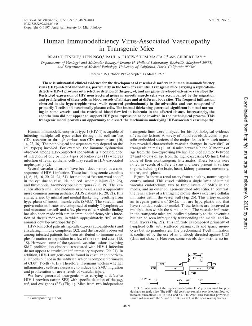
We have generated transgenic mice carrying a defectiveHIV-1 provirus (strain SF2) with specific deletion of the gag,pol, and env genes (33) (Fig. 1). Mice from two independent
transgenic lines were analyzed for histopathological evidenceof vascular lesions. A survey of blood vessels detected in par-affin-embedded sections of the major tissues from each mousehas revealed characteristic vascular changes in over 60% oftransgenic animals (11 of 18 mice between 9 and 20 months ofage from the low-expressing N4 line and 6 of 10 mice between27 and 46 days of age from the high-expressing Q5 line), but innone of their nontransgenic littermates. These lesions werenoted in vessels of different sizes and were present in variousorgans, including the brain, heart, kidney, pancreas, mesentery,uterus, and spleen.
Figure 2a shows a renal artery from a healthy, nontransgeniccontrol animal. This vessel exhibits a single layer of luminalvascular endothelium, two to three layers of SMCs in themedia, and an outer collagen-enriched adventitia. In contrast,the renal artery of a transgenic mouse shows extensive cellularinfiltrates within the vessel wall (Fig. 2b). This artery exhibitsan irregular pattern of SMCs that are hyperplastic and thathave rounded vesicular nuclei. These lesions are observed atmultiple sites within the same animal. The vascular infiltratesin the transgenic mice are localized primarily to the adventitiabut can be seen infrequently transcending the medial and in-timal layers (Fig. 2c). This infiltrate is composed primarily oflymphoid cells, with scattered plasma cells and sparse mono-cytes but no granulocytes. The predominant T-cell infiltrationis confirmed by the use of an antibody directed against CD3(data not shown). However, some vessels demonstrate no im-
* Corresponding author.
FIG. 1. Schematic of the replication-defective HIV provirus used for pro-ducing transgenic mice. The pHIV-del construct contains two deletions, locatedbetween nucleotides 311 to 4454 and 5601 to 7930. This modified provirus isshown colinear with the 59 and 39 LTRs, as well as the open reading frames.
4809
on February 8, 2015 by guest
http://jvi.asm.org/
Dow
nloaded from

4810 NOTES J. VIROL.
on February 8, 2015 by guest
http://jvi.asm.org/
Dow
nloaded from

mune cell infiltration. Figure 2d depicts a pancreatic artery thatis occluded primarily because of SMC proliferation. The ap-parent lack of fibrosis suggests that the hypertrophic growth isnot the result of an inflammatory response but may actuallyprecede it.
Both the internal and external elastic lamina (IEL and EEL,respectively [stained black]) which separate the three well-defined layers of the artery wall may be revealed by Verhoeff’siron hematoxylin-van Geissan staining of an uninvolved vesselfrom a nontransgenic mouse (Fig. 2e). The intima is composedof a single layer of endothelial cells and is delimited on itsouter aspect by the IEL. The media consists of SMCs (stainedbrown) arranged in multiple lamellae and bounded on theluminal side by the IEL and on the abluminal side by the EEL.The adventitia, the outermost layer of the artery and delimited
on the luminal aspect by the EEL, is made up of collagenbundles and elastic fibers (stained red). Immunostaining of aserial section with an antibody directed against SMC a-actin(13) shows the activity predominantly in the medial layer (Fig.2f). A similar analysis of an involved vessel from a transgenicmouse reveals hypercellularity of the intimal layer on the lu-minal side of the IEL and hypocellularity in the medial layer(Fig. 2g). The hypercellular intima contains numerous a-actin-positive SMCs (Fig. 2h). This likely represents migration ofSMCs from the media through the IEL and subsequent pro-liferation within the intima. The hypocellular area juxtaposedto the IEL is fibrotic because of collagen deposition (stainedred). The EEL is somewhat fragmented and segmentally thick-ened. In other vessels, the fragmentation of the elastic laminais overwhelming. The cellular infiltrate is localized distally to
FIG. 3. Splenic infarcts in HIV-1 transgenic mice. (a) Low-magnification view of the spleen from a nontransgenic control mouse (hematoxylin and eosin; 310). (b)Low-magnification view of the spleen from a transgenic mouse with splenic infarct (hematoxylin and eosin; 310). (c) High-magnification view of a splenic white pulpwith a central arteriole from a control mouse (hematoxylin and eosin; 3100). (d) High-magnification view of the white pulp area with an enlarged central arteriole froma transgenic mouse (hematoxylin and eosin, 3100).
FIG. 2. Vascular lesions in HIV-1 transgenic mice. (a) Uninvolved renal artery from a nontransgenic mouse (hematoxylin and eosin; 360). (b) Renal artery from atransgenic mouse showing extensive infiltration within the outer adventitia and disorganized SMCs (hematoxylin and eosin; 360). (c) Mesenteric artery from the sametransgenic mouse as in panel b showing extensive infiltration, disorganized SMCs with rounded vesicular nuclei, and an area of fibrosis (arrow) (hematoxylin and eosin; 360).(d) Pancreatic artery from a different transgenic mouse that is occluded by SMC proliferation and exhibiting minimal immune cell infiltrates (hematoxylin and eosin; 360).(e) Vessel from a nontransgenic control with intact IEL and EEL (Verhoeff’s iron hematoxylin-van Geissan; 3120). (f) Serial section of vessel in panel e immunostainedwith an antibody against SMC a-actin (Mayer’s hematoxylin counterstain; 3120). (g) Intimal thickening in a vessel from a different transgenic mouse (Verhoeff’s ironhematoxylin-van Geissan; 3120). (h) Serial section of vessel in panel g immunostained with an antibody against SMC a-actin (Mayer’s hematoxylin counterstain; 3120).
VOL. 71, 1997 NOTES 4811
on February 8, 2015 by guest
http://jvi.asm.org/
Dow
nloaded from
the EEL in the collagen-enriched adventitia. It is interesting tonote that the endothelium remains intact and apparently un-involved (Fig. 2h).
Such vascular changes are detected in a majority of thetransgenic mice and may have significant functional conse-quences. Prolonged proliferation of SMCs may cause signifi-cant luminal narrowing, and restricted blood flow may resultin ischemia. Indeed, some of the transgenic mice exhibitedsplenic infarcts, while other demonstrated evidence of renal
thrombosis. For example, while the spleen of a nontransgenicmouse is composed of well-defined areas of white pulp (Fig.3a), the spleen of a transgenic littermate exhibited extensivecell death in the same areas as a result of a splenic infarct (Fig.3b). Unlike the central arteriole in the white pulp of the un-affected spleen (Fig. 3c), that in the transgenic mouse is en-larged and is surrounded by abundant cellular debris (Fig. 3d).Seemingly, the arteriopathy observed in the transgenic mouseresembles many of the clinical features seen among HIV-in-
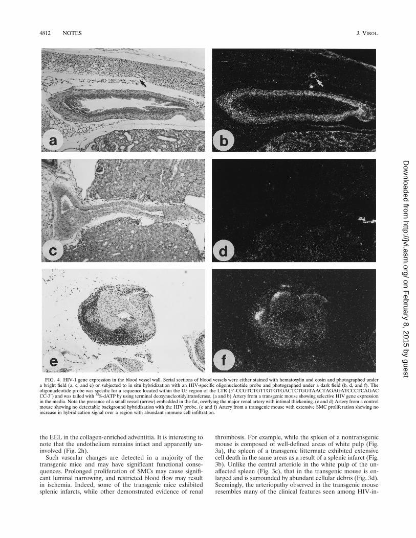
FIG. 4. HIV-1 gene expression in the blood vessel wall. Serial sections of blood vessels were either stained with hematoxylin and eosin and photographed undera bright field (a, c, and e) or subjected to in situ hybridization with an HIV-specific oligonucleotide probe and photographed under a dark field (b, d, and f). Theoligonucleotide probe was specific for a sequence located within the U5 region of the LTR (59-CCGTCTGTTGTGTGACTCTGGTAACTAGAGATCCCTCAGACCC-39) and was tailed with 35S-dATP by using terminal deoxynucleotidyltransferase. (a and b) Artery from a transgenic mouse showing selective HIV gene expressionin the media. Note the presence of a small vessel (arrow) embedded in the fat, overlying the major renal artery with intimal thickening. (c and d) Artery from a controlmouse showing no detectable background hybridization with the HIV probe. (e and f) Artery from a transgenic mouse with extensive SMC proliferation showing noincrease in hybridization signal over a region with abundant immune cell infiltration.
4812 NOTES J. VIROL.
on February 8, 2015 by guest
http://jvi.asm.org/
Dow
nloaded from

fected patients (4, 6, 15, 16, 20, 21, 24, 34) and may be acomplicating factor and even lethal (24).
The cell type responsible for initiating this series of changesin the vascular wall may be identified by an analysis of the siteof HIV gene expression. In situ hybridization with an HIVprobe demonstrates the accumulation of viral transcripts in themedia of a vessel from a transgenic mouse (Fig. 4a and b), butnot in a similar region from a control mouse (Fig. 4c and d). Inthe transgenic vessel wall, where there is no detectable immuneinfiltration but clear evidence of a neointima (Fig. 4a), hybrid-ization is observed homogeneously throughout the mediallayer and, to a lesser extent, the neointima (Fig. 4b). Therestricted expression of HIV genes in SMCs correlates wellwith the proliferation of these cells in vivo. Analysis of a hy-pertrophic vessel with asymmetric immune cell infiltration(Fig. 4e) reveals viral transcripts in the resident SMCs but notin the infiltrating lymphocytes (Fig. 4f). This may suggest thatinfiltration of T cells is not likely to be the initiating event inthis pathological change.
The analysis of two independent transgenic mouse lines,each carrying and expressing the HIV regulatory and accessorygenes under the control of the HIV long terminal repeat(LTR), has revealed histopathological evidence of vasculopa-thy which is highly reminiscent of that detected in HIV-in-fected individuals (4, 6, 15, 16, 20, 21, 34). Our findings suggestthat SMC proliferation is central to the observed changes inthe blood vessel wall and is likely a consequence of HIV geneexpression in these cells. The intimal and medial thickeningcan progress to significant luminal narrowing and tissue necro-sis resulting from ischemia. It is likely that one or more of theHIV regulatory proteins is responsible for inducing the loss ofgrowth arrest in the SMCs. Further studies to define the HIVgene(s) responsible for inducing the pathological changes willfacilitate our understanding of the underlying biochemicalmechanism. Implicit in our findings is the suggestion that theSMC is a direct target for HIV infection. Interestingly, HIVhas been shown to infect a variety of cell types either throughthe CD4 receptor or by various non-CD4 mechanisms (10, 14,23, 36). It will be important to determine whether HIV geneexpression can be detected in the involved vessel walls in HIV-infected patients and if SMCs in culture can be infected byHIV.
The immune cell infiltration, characterized predominantlyby T cells with occasional inclusion of B cells, may be second-ary to the SMC hypertrophy. This suggestion is supported byour observation that SMC expansion was not always accompa-nied by immune cell infiltration and that HIV gene expressioncould not be detected in the infiltrating cells. The lack ofgranulocytes and macrophages in the infiltrate and the local-ization of infiltrating cells to the adventitia rather than thesubendothelium most likely exclude immune complex deposi-tion as a causative factor in this type of systemic vasculitis.However, these findings are not inconsistent with autoimmunedisease in which T-cell infiltrates predominate.
This transgenic model may prove useful not only in the studyof HIV-associated vasculopathy but also in our understandingof atherosclerosis. It has been hypothesized that lesions ofatherosclerosis develop as a protective, inflammatory-fibropro-liferative response to injury of the arterial wall (30). It has beenspeculated that insult to the endothelium results in an inflam-matory response which involves the adherence of peripheralblood monocytes and T lymphocytes to the endothelium andinvasion of the arterial wall by these cells. At these sites,activation of monocytes to macrophages results in the releaseof growth factors which can induce SMC migration and pro-liferation within the intima of the injured artery.
The SMC proliferation we observed differs significantly fromthe response-to-injury hypothesis in several ways. First, weobserved no evidence for dysfunctional changes in the endo-thelium. The single layer of endothelial cells invariably re-mained intact and morphologically normal at the variousstages of the disease process. Second, we detected no evidencefor monocyte-derived macrophages. The infiltrate is composedof predominately T lymphocytes and some B cells and has notbeen detected in the subendothelium. Based upon these dif-ferences, the SMC proliferation in our transgenic mice is likelydue to the activity of growth-inducing signals or changes indig-enous to the SMCs which support HIV gene expression. Sig-naling from the endothelium or the macrophages does notappear to be part of the process.
While platelet-derived growth factor (9, 29, 31) (PDGF) andfibroblast growth factor (1, 3, 22, 28) (FGF) gene families arewell recognized as key regulatory effectors of endothelial celland SMC cell proliferation in vitro, their contribution to thehypertrophic events in this transgenic model is not known.Although PDGF is recognized as a mediator of SMC migrationin vivo (12, 17), somatic gene transfer experiments have sug-gested that it is limited in its ability to promote vascular SMChypertrophy in vivo (27). In contrast, the prominent angiogenicand SMC hypertrophic events mediated by a secretory form ofFGF-1 following somatic gene transfer in vivo (25) suggest thatmembers of the FGF gene family may be functional in thepromotion of vascular SMC hypertrophy in vivo. Since thevascular SMC is potentially a rich source of FGF-1 (35, 37), itis possible that the function of the replication-defective HIV-1provirus may be to influence the ability of FGF-1 to perform asan SMC mitogen.
We thank Lisa Ruiz for help in the preparation of the manuscript.This work was supported by U.S. Public Health Service grant
CA53633 to G.J.
REFERENCES1. Baird, A., and P. Bohlen. 1990. Fibroblast growth factors, p. 369–418. In
M. B. Sporn and A. B. Roberts (ed.), Handbook of experimental pharma-cology: peptide growth factors and their receptors I. Springer-Verlag, Berlin,Germany.
2. Bourgoignie, J. J., C. Oritz-Butcher, D. F. Green, and V. Pardo. 1994. Renaldisease and AIDS, p. 597–608. In S. Broder, T. C. Merigan, Jr., and D.Bolognesi (ed.), Textbook of AIDS medicine. Williams & Wilkins, Philadel-phia, Pa.
3. Burgess, W. H., and T. Maciag. 1989. The heparin-binding (fibroblast)growth factor family of proteins. Annu. Rev. Biochem. 58:575–606.
4. Calabrese, L. H., M. Estes, B. Yen-Lieberman, M. R. Proffitt, R. Tubbs, A. J.Fishleder, and K. H. Levin. 1989. Systemic vasculitis in association withhuman immunodeficiency virus infection. Arthritis Rheum. 32:569–576.
5. Chalifoux, L. V., M. A. Simon, D. R. Pauley, J. J. MacKey, M. S. Wyand, andD. J. Ringler. 1992. Arteriopathy in macaques infected with simian immu-nodeficiency virus. Lab. Invest. 67:338–349.
6. Cho, E.-S., and L. R. Sharer. 1990. Central nervous system in HIV infection,p. 43–63. In V. V. Joshi (ed.), Pathology of AIDS and other manifestationsof HIV infection. Igaku-Shoin, New York, N.Y.
7. Chu, Q. D., L. J. Medeiros, A. E. Fisher, R. F. Chaquette, and J. P. Crowley.1995. Thrombotic thrombocytopenic purpura and HIV infection. South.Med. J. 88:82–88.
8. del Arco, A., M. A. Martinez, J. M. Pena, C. Gamallo, J. J. Gonzalez, F. J.Barbado, and J. J. Vazquez. 1993. Thrombotic thrombocytopenic purpuraassociated with human immunodeficiency virus infection: demonstration ofp24 antigen in endothelial cells. Clin. Infect. Dis. 17:360–363.
9. Deuel, T. F. 1987. Polypeptide growth factors: roles in normal and abnormalcell growth. Annu. Rev. Cell. Biol. 3:443–492.
10. Fauci, A. S. 1993. Multifactorial nature of human immunodeficiency virusdisease: implications for therapy. Science 262:1011–1018.
11. Feinberg, M. B., and W. C. Greene. 1992. Molecular insights into humanimmunodeficiency virus type 1 pathogenesis. Curr. Opin. Immunol. 4:466–474.
12. Ferns, G. A. A., E. W. Raines, K. H. Sprugel, A. S. Motani, M. A. Reidy, andR. Ross. 1991. Inhibition of neointimal smooth muscle accumulation afterangioplasty by an antibody to PDGF. Science 253:1129–1132.
13. Gabbiani, G., O. Kocher, W. S. Bloom, J. Vandekerckhove, and K. Weber.
VOL. 71, 1997 NOTES 4813
on February 8, 2015 by guest
http://jvi.asm.org/
Dow
nloaded from

1984. Actin expression in smooth muscle cells of rat aortic intimal thicken-ing, human atheromatous plaque, and cultured rat aortic media. J. Clin.Invest. 73:148–152.
14. Gallo, R. C., and L. Montagnier. 1988. AIDS in 1988. Sci. Am. 259:40–48.15. Gherardi, R., L. Belec, C. Mhiri, F. Gray, M.-C. Lescs, A. Sobel, L. Guillevin,
and J. Wechsler. 1993. The spectrum of vasculitis in human immunodefi-ciency virus-infected patients. Arthritis. Rheum. 36:1164–1174.
16. Gherardi, R., C. Mhiri, M. Baudrimont, E. Roullet, J.-P. Berry, and J.Poirier. 1991. Iron pigment deposits, small vessel vasculitis, and erythroph-agocytosis in the muscle of human immunodeficiency virus-infected patients.Hum. Pathol. 22:1187–1194.
17. Heldin, C.-H., and B. Westermark. 1990. Platelet-derived growth factor:mechanism of action and possible in vivo function. Cell Regul. 1:555–566.
18. Holland, G. N. 1994. Ocular sequelae, p. 473–487. In S. Broder, T. C.Merigan, Jr., and D. Bolognesi (ed.), Textbook of AIDS medicine. Williams& Wilkins, Philadelphia, Pa.
19. Jorda, M., M. M. Rodriguez, and R. A. Reik. 1994. Thrombocytic thrombo-cytopenic purpura as the cause of death in an HIV-positive child. Pediatr.Pathol. 14:919–925.
20. Joshi, V. V., B. Pawel, E. Conner, L. Sharer, J. M. Oleske, S. Morrison, andJ. Marin-Garcia. 1987. Arteriopathy in children with acquired immune de-ficiency syndrome. Pediatr. Pathol. 7:261–275.
21. Kabus, D., and M. A. Greco. 1991. Arteriopathy in children with AIDS:microscopic changes in the vasa vasorum with gross irregularities of theaortic limb. Pediatr. Pathol. 11:793–795.
22. Klagsbrun, M., and E. R. Edelman. 1989. Biological and biochemical prop-erties of fibroblast growth factors. Implications for the pathogenesis of ath-erosclerosis. Arteriosclerosis 9:269–278.
23. Levy, J. A. 1993. Pathogenesis of human immunodeficiency virus infection.Microbiol. Rev. 57:183–289.
24. Mezzaroma, I., C. Carini, A. Cirelli, and F. Aiuti. 1987. HIV infection,vasculitis and immune complexes. AIDS 1:131–132.
25. Nabel, E. G., Z.-Y. Yang, G. Plautz, R. Forough, X. Zhan, C. C. Haudens-child, T. Maciag, and G. J. Nabel. 1993. Recombinant fibroblast growthfactor-1 promotes intimal hyperplasia and angiogenesis in arteries in vivo.Nature 362:844–846.
26. Pepose, J. S., G. N. Holland, M. S. Nestor, A. J. Cochran, and R. Y. Foos.
1985. Acquired immune deficiency syndrome: pathogenic mechanisms ofocular disease. Ophthalmology 92:472–484.
27. Pompili, V. J., D. Gordon, H. San, Z. Yang, D. W. M. Muller, G. J. Nabel,and E. G. Nabel. 1995. Expression and function of a recombinant PDGF Bgene in porcine arteries. Arterioscler. Thromb. Vasc. Biol. 15:2254–2264.
28. Presta, M., D. Moscatelli, J. Joseph-Silverstein, and D. B. Rifkin. 1986.Purification from a human hepatoma cell line of a basic fibroblast growthfactor-like molecule that stimulates capillary endothelial cell plasminogenactivator production, DNA synthesis, and migration. Mol. Cell. Biol. 6:4060–4066.
29. Raines, E. W., D. F. Bowen-Pope, and R. Ross. 1990. Platelet-derived growthfactor, p. 173–262. In M. B. Sporn and A. B. Roberts (ed.), Handbook ofexperimental pharmacology: peptide growth factors and their receptors I.Springer-Verlag, Berlin, Germany.
30. Ross, R. 1993. Atherosclerosis: a defense mechanism gone awry. Am. J.Pathol. 143:987–1002.
31. Ross, R., E. W. Raines, and D. F. Bowen-Pope. 1986. The biology of platelet-derived growth factor. Cell 46:155–169.
32. Stroncek, D. F., W. E. Kline, M. E. Clay, L. B. Plachta, R. B. Stricker, H.Hollander, J. S. Greenspan, and K. M. Skubitz. 1992. Antibodies to granu-locytes in patients infected with human immunodeficiency virus. J. Lab. Clin.Med. 119:724–731.
33. Tinkle, B. T., H. Ueda, L. Ngo, P. A. Luciw, K. Shaw, C. A. Rosen, and G. Jay.Transgenic dissection of HIV genes involved in lymphoid depletion. J. Clin.Invest., in press.
34. Valeriano-Marcet, J., L. Ravichandran, and L. D. Kerr. 1990. HIV associ-ated systemic necrotizing vasculitis. J. Rheumatol. 17:1091–1093.
35. Weich, H. A., N. Iberg, M. Klagsbrun, and J. Folkman. 1990. Expression ofacidic and basic fibroblast growth factors in human and bovine vascularsmooth muscle cells. Growth Factors 2:313–320.
36. Weiss, R. A. 1994. The virus and its target cells, p. 15–20. In S. Broder, T. C.Merigan, Jr., and D. Bolognesi (ed.), Textbook of AIDS medicine. Williams& Wilkins, Philadelphia, Pa.
37. Winkles, J. A., and C. G. Gay. 1991. Serum, phorbol ester, and polypeptidemitogens increase class 1 and 2 heparin-binding (acidic and basic fibroblast)growth factor gene expression in human vascular smooth muscle cells. CellGrowth Differ. 2:531–540.
4814 NOTES J. VIROL.
on February 8, 2015 by guest
http://jvi.asm.org/
Dow
nloaded from