Therapeutic antagonists of microRNAs deplete leukemia-initiating cell activity
Upload
independentCategory
view
3download
0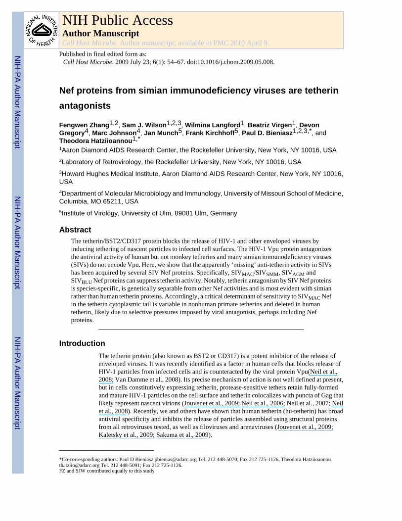
Nef proteins from simian immunodeficiency viruses are tetherinantagonists
Fengwen Zhang1,2, Sam J. Wilson1,2,3, Wilmina Langford1, Beatriz Virgen1, DevonGregory4, Marc Johnson4, Jan Munch5, Frank Kirchhoff5, Paul D. Bieniasz1,2,3,*, andTheodora Hatziioannou1,*1Aaron Diamond AIDS Research Center, the Rockefeller University, New York, NY 10016, USA2Laboratory of Retrovirology, the Rockefeller University, New York, NY 10016, USA3Howard Hughes Medical Institute, Aaron Diamond AIDS Research Center, New York, NY 10016,USA4Department of Molecular Microbiology and Immunology, University of Missouri School of Medicine,Columbia, MO 65211, USA5Institute of Virology, University of Ulm, 89081 Ulm, Germany
AbstractThe tetherin/BST2/CD317 protein blocks the release of HIV-1 and other enveloped viruses byinducing tethering of nascent particles to infected cell surfaces. The HIV-1 Vpu protein antagonizesthe antiviral activity of human but not monkey tetherins and many simian immunodeficiency viruses(SIVs) do not encode Vpu. Here, we show that the apparently ‘missing’ anti-tetherin activity in SIVshas been acquired by several SIV Nef proteins. Specifically, SIVMAC/SIVSMM, SIVAGM andSIVBLU Nef proteins can suppress tetherin activity. Notably, tetherin antagonism by SIV Nef proteinsis species-specific, is genetically separable from other Nef activities and is most evident with simianrather than human tetherin proteins. Accordingly, a critical determinant of sensitivity to SIVMAC Nefin the tetherin cytoplasmic tail is variable in nonhuman primate tetherins and deleted in humantetherin, likely due to selective pressures imposed by viral antagonists, perhaps including Nefproteins.
IntroductionThe tetherin protein (also known as BST2 or CD317) is a potent inhibitor of the release ofenveloped viruses. It was recently identified as a factor in human cells that blocks release ofHIV-1 particles from infected cells and is counteracted by the viral protein Vpu(Neil et al.,2008; Van Damme et al., 2008). Its precise mechanism of action is not well defined at present,but in cells constitutively expressing tetherin, protease-sensitive tethers retain fully-formedand mature HIV-1 particles on the cell surface and tetherin colocalizes with puncta of Gag thatlikely represent nascent virions (Jouvenet et al., 2009; Neil et al., 2006; Neil et al., 2007; Neilet al., 2008). Recently, we and others have shown that human tetherin (hu-tetherin) has broadantiviral specificity and inhibits the release of particles assembled using structural proteinsfrom all retroviruses tested, as well as filoviruses and arenaviruses (Jouvenet et al., 2009;Kaletsky et al., 2009; Sakuma et al., 2009).
*Co-corresponding authors: Paul D Bieniasz [email protected] Tel. 212 448-5070; Fax 212 725-1126, Theodora [email protected] Tel. 212 448-5091; Fax 212 725-1126.FZ and SJW contributed equally to this study
NIH Public AccessAuthor ManuscriptCell Host Microbe. Author manuscript; available in PMC 2010 April 9.
Published in final edited form as:Cell Host Microbe. 2009 July 23; 6(1): 54–67. doi:10.1016/j.chom.2009.05.008.
NIH
-PA Author Manuscript
NIH
-PA Author Manuscript
NIH
-PA Author Manuscript

The mechanism by which HIV-1 Vpu antagonizes hu-tetherin is not fully understood, butoverexpressed HIV-1 Vpu reduces the overall levels of tetherin in cells and inhibits itsappearance at the cell surface(Bartee et al., 2006; Van Damme et al., 2008). Furthermore,HIV-1 Vpu and hu-tetherin co-localize, and Vpu prevents the co-localization of hu-tetherinwith nascent HIV-1 particles(Jouvenet et al., 2009; Neil et al., 2008). However, while tetherinproteins from non-hominid primates are potent inhibitors of HIV-1 particle release, they cannotbe counteracted by HIV-1 Vpu(McNatt et al., 2009). Portions of primate tetherin genes,including sequences encoding the transmembrane domain that governs sensitivity toantagonism by Vpu, are unusually divergent, and exhibit clear evidence of positive selection(McNatt et al., 2009). Thus, HIV-1 has apparently acquired a biological activity (i.e. Vpu), thathas specifically evolved to antagonize the tetherin variant expressed in its host species.
Although hu-tetherin inhibits the release of particles assembled using a diverse array ofretroviral structural proteins, only a subset of the primate lentiviruses encode Vpu. Thus, itseemed reasonable to suppose that SIVs have evolved alternative mechanisms to evade tetherinin their natural hosts. Indeed, earlier work indicated that the HIV-2 envelope protein couldenhance particle release from cells that were subsequently shown to express hu-tetherin(Abadaet al., 2005; Bour et al., 1996; Bour and Strebel, 1996; Varthakavi et al., 2003). Given thisprecedent, it was quite plausible that the envelope proteins of SIVs might have a similarfunction. Consistent with this idea, the Ebola virus envelope protein has recently been reportedto be a tetherin antagonist(Kaletsky et al., 2009).
However, as reported herein, we found that the envelope protein of SIVMAC, a macaquelentivirus that is closely related to HIV-2, did not antagonize macaque tetherin proteins. Rather,Nef proteins from SIVMAC and several other SIVs antagonize primate tetherins. Notably,tetherin antagonism by SIV Nef proteins was species-specific, and each SIV Nef was poorlyactive against human tetherin. Furthermore, the cytoplasmic tail of tetherin, which, like thetransmembrane domain, has been evolving under positive selection in primates(McNatt et al.,2009), contains a discrete motif that is deleted in humans and variable in other primates andgoverns sensitivity to antagonism by SIVMAC Nef. Thus, several primate lentiviruses that lackVpu have acquired the ability to antagonize tetherin using their Nef proteins.
ResultsInhibition of SIVMAC particle release by tetherin proteins
Hu-tetherin can inhibit the release of particles assembled using the structural proteins (Gagand/or GagPol) of a wide variety of retroviruses(Jouvenet et al., 2009), raising the question ofhow retroviruses that lack a Vpu gene are efficiently released from infected cells that mightordinarily express tetherin. Among the retroviruses previously tested for sensitivity to hu-tetherin were the primate lentiviruses, SIVMAC and SIVAGMSab, neither of which encode aVpu protein(Jouvenet et al., 2009). However, it has previously been shown that at least somestrains of HIV-2, a virus that shares a recent common ancestor with SIVMAC, encode anenvelope protein that has Vpu-like activity (Abada et al., 2005; Bour et al., 1996; Bour andStrebel, 1996; Varthakavi et al., 2003). Therefore, we tested whether the release of particlesgenerated by a full-length SIVMAC239 proviral construct could be inhibited by tetherin proteinsfound in macaque species, specifically two variants from rhesus macaques (rh-tetherin-1 and-2) and one from pig-tailed macaques (pgt-tetherin). Strikingly, each of the macaque tetherinproteins was a rather poor inhibitor of SIVMAC239 particle release (Figure 1A). This was thecase whether or not the SIVMAC239 proviral DNA construct encoded a functional Env protein.Thus, these results suggested that SIVMAC239 might encode a tetherin antagonist other thanthe Env protein.
Zhang et al. Page 2
Cell Host Microbe. Author manuscript; available in PMC 2010 April 9.
NIH
-PA Author Manuscript
NIH
-PA Author Manuscript
NIH
-PA Author Manuscript

Next we co-expressed an SIVMAC239 provirus with varying amounts of human and macaquetetherin proteins. As previously shown for SIVMAC239 particles assembled using only the Gagand Pol proteins, the release of authentic SIVMAC239 virions appeared highly sensitive to hu-tetherin (Figure 1B, D). In contrast, tetherin proteins from macaques appeared to besignificantly less effective inhibitors of SIV MAC239 virion release (Figure 1C, D, Figure S1).
These findings suggested the possibility that another SIVMAC protein might be a macaque-tetherin-specific antagonist. The ability of Nef to regulate the levels of cell surface proteinsprompted us to examine whether it might fulfill this role. Strikingly, the release of virionsgenerated by a proviral plasmid lacking Nef (SIVMAC239(delNef)) appeared more sensitive toinhibition by macaque tetherins than SIVMAC239(WT) (Figure 1C,D Figure S1). In contrast,hu-tetherin inhibited SIVMAC239(WT) release almost as well as it inhibited SIVMAC(delNef)release (Figure 1B, D). Thus, Nef deletion specifically sensitized SIVMAC239 to inhibition bymacaque tetherins.
Scanning electron microscopic analysis of cells cotransfected with plasmids expressingSIVMAC239 Gag and either rh-tetherin-1 or hu-tetherin revealed that either tetherin induced theaccumulation of SIV MAC239 VLPs on the surface of cells (Figure 1E). Moreover, we havepreviously reported that HIV-1 Gag-GFP VLPs tethered to the plasma membrane by hu-tetherin can be internalized resulting in the accumulation of intracellular HIV-1 Gag-GFP(Neil et al., 2006; Neil et al., 2008) and similar findings were obtained with SIVMAC239 Gag-GFP and rh-tetherin-1 (Figure 1F), Specifically, in 293T cells stably expressing rh-tetherin-1,fluorescence microscopic analysis indicated that nearly half the cells transiently expressingSIVMAC239 Gag-GFP contained prominent intracellular accumulations of Gag-GFP (Figure1F,G). Crucially, these accumulations decreased in frequency when increasing amounts of anSIVMAC239 Nef expression plasmid were cotransfected, or when cells that did not expresstetherin were used(Figure 1F,G). Thus, macaque and human tetherin proteins block particlerelease in the same manner, and SIVMAC239 Nef antagonizes the tethering activity of macaquetetherin.
Nef proteins from divergent primate lentiviruses antagonize tetherin in a species-specificmanner
Vpu is present in only a minority of primate lentiviruses, while Nef is present in all primatelentiviruses isolated thus far. Therefore, to test whether Nef proteins from lentiviruses otherthan SIVMAC might also be capable of counteracting tetherin, several of them were expressedin place of HIV-1 Nef in the context of a replication competent HIV-1 proviral construct. TheHIV-1 Vpu protein was inactivated in this construct, to test the effects of Nef on particle releasein the absence of confounding factors. Proviral plasmids bearing the various Nef proteins wereco-transfected with increasing amounts of plasmids expressing hu-, rh-1-, pgt-, and Africangreen monkey (agm-) tetherin proteins. Western blot analyses indicated proviruses expressingthe various Nef proteins generated HIV-1 virions with similar efficiency in the absence oftetherins (Figure 2A–H).
The generation of extracellular virions by cells transfected with the parental proviral plasmid(which lacked both Nef and Vpu) was strongly inhibited by each of the four tetherins (Figure2A, Figure S2A), in the absence of effects on cell associated Gag expression. A matched HIV-1provirus, which lacked Nef but retained Vpu, was largely resistant to hu-tetherin. However,Vpu did not antagonize agm-tetherin, rh-tetherin or pgt-tetherin (McNatt et al., 2009), (Figure2B, Figure S2B). A construct that lacked Vpu but retained HIV-1 Nef behaved essentially thesame as the construct that lacked both Vpu and Nef (Figure 2C, Figure S2C). Thus, HIV-1Vpu specifically antagonized hu-tetherin but not monkey tetherins, while HIV-1 Nef did notantagonize any of the tetherin proteins.
Zhang et al. Page 3
Cell Host Microbe. Author manuscript; available in PMC 2010 April 9.
NIH
-PA Author Manuscript
NIH
-PA Author Manuscript
NIH
-PA Author Manuscript

Notably, replacement of HIV-1 Nef with SIVMAC Nef resulted in substantial resistance to bothpgt- and rh-tetherin (Figure 2D, Figure S2D). Importantly, this effect was quite specific to themacaque tetherins; and SIVMAC Nef only marginally enhanced particle release when inhibitionwas imposed by hu-tetherin or agm-tetherin. Conversely, SIVAGMSab Nef conferred a greaterdegree of resistance to agm-tetherin than did SIVMAC Nef (Figure 2E, Figure S2E), andSIVAGMSab Nef was also active against the macaque tetherins. However, SIVAGMSab Nefwas similar to SIVMAC Nef in that it was a poor antagonist of hu-tetherin (Figure 2E, FigureS2E). Although this sample size is small, we note that in the two cases examined where theparental viruses lack a vpu gene (SIVMAC and SIVAGMSab), the Nef proteins were able toantagonize the antiviral activity of tetherins found in the corresponding simian host species.
We also tested Nef proteins from three other SIVs, with the caveat that the SIV Nef proteinswere not derived from the same host species as the tetherins. Nonetheless, an HIV-1 proviralconstruct expressing the Nef protein from SIVBLU was largely resistant to pgt-tetherin and rh-tetherin (Figure 2F, Figure S2F). Conversely, Nef proteins from SIVRCM and SIVGSN appearedlargely unable to antagonize the tetherin proteins (Figure 2G, H Figure S2G, H). However, inaddition to the fact that Nef and tetherin proteins from non-orthologous species were tested,SIVGSN naturally encodes a Vpu protein(Courgnaud et al., 2002), which potentially wouldobviate the requirement for this activity in Nef. An additional caveat with the aforementionedexperiment is that the SIV Nef proteins were expressed in the Nef position of an HIV-1 provirus,and may be present at higher or lower levels than in their parental proviruses. Additionally,antibodies to the SIVRCM and SIVGSN proteins were not available, therefore we were unableto demonstrate that these Nef proteins were expressed, although we have previously shownthat infection by nearly identical proviruses caused efficient CD4 downregulation (Schindleret al., 2006). Nonetheless, three of the five SIV Nef proteins tested in these assays exhibitedapparent tetherin antagonizing activity. The sensitivity or otherwise of a given tetherin proteinto antagonism by Nef did not correlate with its expression level (Figure S3), which was assessedunder identical conditions to those used for the assays in Figure 2 and Figure S2.
To exclude the possibility the aforementioned findings were the result of some unanticipatedconsequence of inserting foreign nef genes into the HIV-1 genome in cis, we determinedwhether Nef proteins, expressed in trans, would relieve tetherin imposed blocks. A Vpu andNef-defective HIV-1 proviral plasmid was cotransfected with a fixed amount of each tetherinprotein, and varying amounts of plasmids expressing the Nef proteins that exhibited apparenttetherin antagonizing activity (Figure 2, Figure S2). We also included HIV-1 Nef as a control,and an additional SIVAGM Nef protein, from SIVAGMTan. As expected, western blot andinfectivity assays revealed that particle release was strongly inhibited by each of the tetherinproteins, and co-expression of HIV-1 Nef in trans had little, if any, ability to relieve thesetetherin-imposed blocks (Figure 3A, Figure S4A). Conversely, SIVMAC Nef relieved theinhibition of particle release imposed by macaque tetherins but its effect on particle release inthe presence of hu-tetherin or agm-tetherin was marginal (Figure 3B, Figure S4B). BothSIVAGMSab and SIVAGMTan Nef restored particle release from cells expressing rh-, pgt- oragm-tetherin but were less active in relieving the block imposed by hu-tetherin (Figure 3C,D,Figure S4C,D). Notably these two SIVAGM derived proteins were the only Nef proteins thateffectively relieved inhibition by agm-tetherin. Finally, SIVBLU Nef efficiently antagonizedthe macaque tetherin proteins, but it also had some ability to enhance particle release in thepresence of hu- and agm-tetherin (Figure 3E Figure S4E). However, effects on hu-tetherin andagm tetherin were clearly reduced in magnitude as compared to effects on macaque tetherins.Overall, the effects of the various Nef proteins, expressed in trans, on HIV-1 particle releasecorroborated those obtained when they were expressed in cis. However, we were able to detectadditional, albeit slight, anti-tetherin effects when Nef was expressed in trans (for example,SIVBLU Nef slightly antagonized hu-tetherin (Figure 3E)). We suspect that the smalldifferences in results obtained using the two assays reflect differences in the level of Nef
Zhang et al. Page 4
Cell Host Microbe. Author manuscript; available in PMC 2010 April 9.
NIH
-PA Author Manuscript
NIH
-PA Author Manuscript
NIH
-PA Author Manuscript

expression, which is likely higher when Nef is expressed in trans. Notably, a modest degreeof variation in the levels of expression of the various tetherin proteins (Figure S5), assessedunder identical conditions to those used in the assays in Figure 3 and Figure S4, did not correlatewith the ability of a given tetherin protein to be antagonized by Nef. Moreover, the Nef proteinsthat did not antagonize tetherins (HIV-1 Nef) or Nef proteins that did antagonize monkeytetherins (SIVMAC239 Nef or SIVAGMTan Nef) did not appear to inhibit expression of any ofthe tetherin proteins (Figure S5).
The effects of Nef proteins on particle yield measured using infectious virion yield assayscorrelated well with those assessed using western blotting (Figure S4). However, in additionto their tetherin antagonizing effects, each Nef protein tested enhanced the intrinsicinfectiousness of HIV-1 particles in the absence of tetherins. Nonetheless, the tetherin-independent effect on virion infectiousness was quite similar, and modest, irrespective of whichNef protein was expressed (Figure S4). Together, however, the compound effect of Nef proteinson particle release and infectiousness, resulted in quite large effects on overall infectious virionyield, approaching two orders of magnitude in some cases.
SIV Nef proteins can enhance virion release from simian cellsIf endogenously expressed monkey tetherins inhibit virion release and are antagonized by SIVNef proteins then Nef should enhance particle release from simian cells, particularly when theyare treated with IFNα, a known enhancer of tetherin expression. We first determined the effectof Nef on SIVMAC239 particle release from rhesus macaque 221 T-cells in single cyclereplication assays, following infection with VSV-G-pseudotyped SIVMAC239(WT) orSIVMAC239(del Nef). Notably, 221 expressed tetherin in the absence of IFNα, and the levelsof tetherin mRNA were modestly increased by IFNα treatement (Figure S6A). Western blottinganalysis indicated that equivalently infected cells release fewer SIVMAC239(del Nef) virionsthan SIVMAC239(WT) virions, even though Gag was equally expressed (Figure 4A, FigureS6B). This effect was exacerbated when the infected 221 cells were treated with IFN-α.Notably, the discrepancy in particle release between SIVMAC239(WT) and SIVMAC239(del Nef)was not observed when human 293T cells were used in an otherwise identical single cyclereplication assay (Figure 4A, Figure S6C). Thus, stimulation of particle release by Nef wasclearly host cell-type dependent. Notably, transmission electron microscopic analysis of 221cells revealed very obvious accumulation of virions associated with the plasma membrane ofcells when they were infected with SIVMAC239 (delNef) but not when they were infected withSIVMAC239(WT) (Figure 4B). This phenotype was observed in otherwise untreated 221 cellsbut was especially striking when the infected 221 cells were treated with IFNα. Furthermore,these virions often appeared to be tethered together (Figure 4B, (iv) inset), Thus, the inefficientrelease of Nef-defective SIVMAC239 particles was accompanied by accumulation of virions atthe plasma membrane, recapitulating previous observations of Vpu-defective HIV-1 virionsin hu-tetherin expressing human cells.
We also examined whether SIVAGMSab Nef could enhance particle release from African greenmonkey COS-7 cells. In this case, VSV-G pseudotyped HIV-1 constructs encoding either noNef, HIV-1 Nef or SIVAGMSab Nef were used to infect COS-7 cells. Subsequent westernblotting analysis indicated that SIVAGMSab Nef significantly increased the yield of HIV-1particles from HIV-1 infected COS-7 cells, as compared to HIV-1 Nef or no Nef (Figure 4C,Figure S6D) Again this effect was present in the absence of IFNα treatment, suggesting thatCOS-7 cells express some tetherin, but was exacerbated by IFN-α. Overall, both SIVMAC239and SIVAGMSab Nef proteins promoted particle release and alleviated the accumulation oftethered virus particles at the plasma membrane, in cells derived from relevant host-species.
Zhang et al. Page 5
Cell Host Microbe. Author manuscript; available in PMC 2010 April 9.
NIH
-PA Author Manuscript
NIH
-PA Author Manuscript
NIH
-PA Author Manuscript

A critical determinant of sensitivity to antagonism by SIVMAC Nef is variable in monkeytetherins and deleted in human tetherin
The amino-terminus of tetherin is the only portion of the protein that is ordinarily exposed tothe cell cytoplasm and was, therefore, the most likely determinant of sensitivity to antagonismby SIV Nef proteins. We constructed two plasmids expressing chimeric tetherins, in which theN-termini of rh-tetherin and hu-tetherin (residues 1–21 and 1–26, respectively) werereciprocally exchanged, generating huCT-rh-tetherin and rhCT-hu-tetherin respectively(Figure 5A). Each of these chimeric tetherin proteins inhibited the release of HIV-1 particlesin the absence of Vpu or Nef (Figure 5B). Notably, hu-tetherin and the huCT-rh-tetherinchimera also inhibited the release of HIV-1 particles generated by proviral plasmids thatencoded SIVMAC239 Nef in cis, assessed using both western blot or infectious virion assays(Figure 5B, C). Conversely, the rhCT-hu-tetherin protein, like rh-tetherin did not efficientlyinhibit the generation of HIV-1 particles generated in the presence of SIVMAC239 Nef. Similarexperiments showed that the release of Nef-defective SIVMAC239 virions was inhibited by eachof the intact and chimeric tetherins. Conversely, the intact, Nef-expressing SIVMAC239construct was sensitive to inhibition by hu-tetherin and huCT-rh-tetherin, but was resistant tothe rhCT-hu-tetherin and rh-tetherin proteins (Figure 5D,E). Thus, determinants in thecytoplasmic tails of rh- and hu-tetherins govern their sensitivity and resistance, respectively,to antagonism by SIVMAC Nef.
The cytoplasmic tails of rh-tetherins differ from hu-tetherin in three short clusters of amino-acids, including a five-residue sequence that is deleted specifically in the human lineage.Therefore, we constructed three plasmids expressing hu-tetherin in which each variant aminoacid cluster was substituted for its equivalent in rh-tetherin-1; (hu(PIL), hu (KM) and hu(GDWIK), Figure 6A). Each of these tetherin proteins was expressed and inhibited the releaseof SIVMAC239(del Nef) virions with approximately equal efficiency, as assessed by westernblotting assays (Figure 6B, D). Moreover, hu(PIL) and hu(KM) tetherin proteins efficientlyinhibited the release of SIVMAC239(WT) virions and were, thus, apparently insensitive toantagonism by SIVMAC239 Nef (Figure 6C, D). Conversely, restoration of the ‘missing’ 5amino acid GDIWK motif in hu-tetherin conferred sensitivity to antagonism by SIVMAC239Nef. Indeed, hu(GDIWK) was as sensitive to antagonism by SIVMAC239 Nef as was rhCT-huprotein that encoded the complete rh-tetherin-1 cytoplasmic tail (Figure 6C, D).
The cytoplasmic tail of tetherin has been evolving under positive selection, and the 5-residuemotif that is deleted in humans contains codons that exhibit a high probability of beingpositively selected in other primate species(McNatt et al., 2009). Notably, rh-tetherin-1 andrh-tetherin-2 differ at one amino acid within this motif, encoding GDIWK and DDIWKrespectively (Figure S7A). Moreover, agm-tetherin, which is insensitive to antagonism bySIVMAC239 Nef (Figure 2, Figure 3) contains a single substitution relative to rh-tetherin-2tetherin at this motif and encodes DDICK (Figure S7A). To further determine the role of thisvariable motif in determining susceptibility to antagonism by SIVMAC239 Nef, we generatedplasmids expressing rh-tetherin lacking this motif (rh(ΔGDWIK)), as well as single amino acidsubstitution mutants (W17C) of rh-tetherin-1 and rh-tetherin-2 (rh-1(GDICK) and rh-2(DDICK)). Notably, either the 5 amino acid deletion or the single amino-acid substitution, inthe context of either the rh-tetherin-1 or rh-tetherin-2, conferred resistance to antagonism bySIVMAC239 Nef (Figure S7B, C). Thus, naturally occurring deletions and substitutions withinthe same small motif, found in humans and African green monkeys, can confer resistance toantagonism by Nef proteins found in an SIV from another species.
Tetherin antagonism is genetically separable from other SIVMAC239 Nef activitiesWe next determined whether mutations that specifically abolish known biological activities ofSIVMAC239 Nef affected its ability to antagonize tetherin. As is the case with most other
Zhang et al. Page 6
Cell Host Microbe. Author manuscript; available in PMC 2010 April 9.
NIH
-PA Author Manuscript
NIH
-PA Author Manuscript
NIH
-PA Author Manuscript

biological activities of Nef proteins, mutation of the N-terminal myristate acceptor (G2A)abolished the ability of SIVMAC239Nef to antagonize rh-tetherin (Figure 7A, B). Conversely,mutation of a single residue (Y223F) that abolishes the ability of SIVMAC239 Nef todownregulate MHC-I but leaves other activities of SIVMAC239 Nef, such as CD4downregulation, intact (Schindler et al., 2004) had little effect on tetherin antagonizing activity.However, a series of three mutants that share the D204R substitution and have lost CD4 andCD28 downregulation activity, but maintain MHC-I downregulation activity(Schindler et al.,2004), were not effective rh-tetherin-1 antagonists (Figure 7A, B). Notably, none of thesemutations had any effect on Nef expression levels (Schindler et al., 2004). Moreover, ananalysis of 5 naturally occurring Nef proteins derived from the same lentivirus lineage asSIVMAC239 revealed clear differences in tetherin antagonizing activity (Figure 7C,D).Specifically, two SIVSMM derived Nef proteins were efficient rh-tetherin-1 antagonists, while2 of 3 HIV-2 Nef proteins were devoid of tetherin antagonizing activity and a third exhibitedweak rh-tetherin-1 antagonizing activity. Because these Nef proteins all share the ability todownregulate CD4, MHC-I and CD3(Munch et al., 2005), these data show that tetherinantagonism is genetically separable from several known biological activities of Nef, but mayshare some mechanistic properties with CD4 downregulation. Additionally, these data hint thatnatural variation in the ability of closely related Nef proteins to act as tetherin antagonists maybe influenced by the host species in which the corresponding virus replicates.
DiscussionHere we report that Nef proteins can enhance virion particle release via antagonism of tetherinproteins. Notably, this activity was not present in HIV-1 Nef, but 5 out of 7 SIV Nef proteinstested, from 5 different SIV lineages, exhibited tetherin-antagonizing activity. Importantly, thisNef function exhibited striking species specificity; none of the SIV Nef proteins were highlyactive against hu-tetherin, but in both cases where tetherins and Nef proteins were derived fromthe same host species (macaque and agm) efficient antagonism was observed.
Previously, we showed that the HIV-1 Vpu protein also antagonizes tetherins in a species-specific manner. Determinants of sensitivity to Vpu reside in the hu-tetherin transmembranedomain, which exhibits a strong signature of positive or diversifying selection, suggesting thepossibility that Vpu, or a similar viral antagonist of tetherin, has imposed selective pressure onthis portion of the tetherin gene. Notably, we also observed signatures of positive selection insequences encoding the tetherin N-terminal cytoplasmic tail(McNatt et al., 2009). Obviously,given the finding that the cytoplasmic tail of tetherin can determine sensitivity or resistance toantagonism by SIVMAC Nef, then Nef proteins are reasonable candidates for antagonists thatimpose diversifying selection pressure on this tetherin domain.
The mechanism by which SIV Nef proteins antagonize tetherins remains to be determined.Preliminary experiments did not reveal a clear effect of SIVMAC or SIVBLU Nef proteins onrh-tetherin cell surface expression (data not shown). Nonetheless, HIV-1 and SIV Nef proteinsdownregulate several other cell surface proteins, most notably CD4 and MHC-I (Collins et al.,1998; Garcia and Miller, 1991; Schindler et al., 2006; Schwartz et al., 1996; Stumptner-Cuvelette et al., 2001), by recruiting these receptors to the endocytic machinery or byredirecting them to lysosomes (reviewed in (Kirchhoff et al., 2008; Roeth and Collins,2006)). The HIV-1 Vpu protein excludes tetherin from sites of particle assembly at the plasmamembrane and inhibits cell surface expression of tetherin(Jouvenet et al., 2009; Van Dammeet al., 2008). However, further work will be required to determine precisely how antagonismof tetherin function by Nef and Vpu proteins is achieved.
The ability to antagonize tetherin has apparently arisen at least twice, perhaps three times duringthe evolution of primate lentiviruses and, strikingly, the acquisition of this function has
Zhang et al. Page 7
Cell Host Microbe. Author manuscript; available in PMC 2010 April 9.
NIH
-PA Author Manuscript
NIH
-PA Author Manuscript
NIH
-PA Author Manuscript

occurred in completely different ways, employing Nef, Vpu and perhaps Env proteins. Ofinterest is the fact that both Nef and Vpu proteins, to varying degrees, remove or antagonizemolecules (CD4 and tetherin) that are both capable of inhibiting the release of infectious viralparticles from the cell surface(Lama et al., 1999; Ross et al., 1999). Moreover, both appear tobe multifunctional proteins whose activities partially coincide. Overall these findingsunderscore the functional plasticity of primate lentiviral genomes in general, and vpu and nefgenes in particular.
While the ability of Vpu and Nef proteins to antagonize tetherin is clearly demonstrable, itremains unproven that these activities are critical in vivo. SIVMAC239 chimeras that encodeHIV-1 Nef, and therefore lack a predicted tetherin antagonist, are less pathogenic than intactSIVMAC239, but are capable of causing simian AIDS in some animals(Alexander et al., 1999;Kirchhoff et al., 1999). Thus, tetherin antagonism might not be essential for pathogenesis.However, given the plasticity of Nef function it will be interesting to determine whether HIV-1Nef can acquire tetherin antagonizing activity during SIVMAC239/SHIV replication in vivo.Additionally, HIV-1 Vpu, despite lacking the ability to antagonize macaque tetherins, enhancesreplication in the context of SHIV infection of pig-tailed macaques(Stephens et al., 2002).Thus, other functions of Vpu and Nef, may be critical in vivo. Nonetheless, the independentacquisition of tetherin antagonizing activity that is specific to the host species of a given HIV/SIV by Nef and Vpu strongly suggests that these activities confer a selective advantage invivo.
Finally, we note that three of the five known ‘non-essential’ accessory genes in primatelentiviruses, namely vif, vpu and nef, are now demonstrated to encode antagonists of innate orintrinsic immunity. Overall, these findings emphasize the extent to which intrinsic and innatehost defenses are likely to have imposed evolutionary pressure on primate lentiviruses,influenced species tropism and provided the impetus for the acquisition of new genes andbiological activities to enable immunodeficiency virus replication in an intrinsically hostileenvironment.
Experimental proceduresPlasmid construction
Plasmids expressing the various HA-epitope tagged tetherin proteins were constructed aspreviously described (Jouvenet et al., 2009; McNatt et al., 2009) using the cDNA sequencesand oligonucleotides listed in Supplementary table 1. Two variants of rhesus macaque tetherinwere used (rh-1 and rh-2) that differ at a single amino acid position in the cytoplasmic tail. Nofunctional difference between the two variants was detected, and the variants were usedinterchangeably. Mutant human and rhesus tetherins were generated by PCR using long oligosdirected to the 5’ end of the coding sequence (Supplementary table 1). To generate theSIVMAC proviral clones used in this study, the full-length sequence of SIVMAC239 wasassembled from plasmids p239SpSp5’ and p239SpE3’ (Kestler et al., 1990)(NIH AIDSReagent program) into a low copy number (pXF3) backbone. For the SIVMAC(delNef)derivative of this clone, overlapping primers and PCR were used to introduce premature stopcodon at codons 58 and 59 of Nef, immediately 3’ to the Env stop codon, using the primerslisted in Supplementary table 2. HIV-1-based proviral plasmids expressing Nef proteins fromvarious lentiviruses have been previously described(Munch et al., 2007; Schindler et al.,2006). To generate vpu-defective derivatives thereof, restriction fragments encompassing thenef region were cloned into pBRHIV-1NL4-3Δvpu containing a deletion of nucleotides 3 to120 of the vpu coding frame. For the experiments in which Nef proteins were expressed intrans, Nef coding sequences from HIV-1, SIVMAC239, SIVBLU, SIVAGMSab and SIVAGMTanwere amplified by PCR using primers introducing EcoRI and SalI sites at the 5’ end and 3’end of the coding sequence respectively (Supplementary table 2). The PCR products were
Zhang et al. Page 8
Cell Host Microbe. Author manuscript; available in PMC 2010 April 9.
NIH
-PA Author Manuscript
NIH
-PA Author Manuscript
NIH
-PA Author Manuscript

inserted into the pIRES2-GFP expression vector (Clontech). SIVMAC239 Nef was also insertedinto pCR3.1. pCG-IRESGFP vectors expressing the SIVMAC239 mutant, HIV-2 and SIVSMMNef proteins used in Figure 7 have been described previously(Schindler et al., 2004).
Cell lines and transfection293T and TZMbl cells (which express CD4 and CCR5 and contain a lacZ reporter gene underthe control of an HIV-1 LTR) were maintained under standard conditions. All transfectionexperiments were performed in 293T cells, which do not constitutively express tetherin, thatwere seeded in 24-well plates at a concentration of 2.5×10⁵ cells/well and transfected thefollowing day using polyethylenimine (PolySciences). A 293T-;derived cell line stablyexpressing rh-tetherin was derived by inserting the rh-tetherin-1 cDNA into a retroviral vector(LHCX) which was then used to transduce 293T cells, which were then selected in hygromycin.
To measure tetherin inhibition of SIVMAC239 particle release, cells were transfected with 400ngof an SIVMAC239 proviral plasmid or the SIVMAC (delNef) derivative, along with increasingamounts (3.12ng, 6.25ng, 12.5ng, 25ng, or 50ng) of each tetherin-HA expression plasmid. Insome experiments (Figure 5), the amount of SIVMAC proviral plasmid DNA was increased to500ng and the amounts of tetherin plasmid were increased (to 0ng, 12.5ng, 25ng, 50ng, 100ng,200ng/well). For assays using the HIV-1 proviral plasmids encoding various SIV Nef proteins,cells were transfected with 200ng of each proviral plasmid and varying amounts (0ng, 3.12ng,6.25ng, or 12.5ng) of each tetherin-HA plasmid. Alternatively, 500ng of HIV-1 proviralplasmid and 0ng, 50ng, 100ng, and 200ng of tetherin expression plasmid were used.
For assays in which Nef proteins were expressed in trans, cells were transfected with 400ngof the HIV-1 proviral plasmid that lacked both Nef and Vpu, along with 25ng of each tetherin-HA expression plasmid and varying amounts (0ng, 25ng, 50ng or 100ng) of each HIV-1 orSIV Nef expression plasmid. In all transfection experiments, the total amount of DNA washeld constant within the experiment by supplementing the transfection with empty expressionvector.
To generate the VSV-G pseudotyped viruses in Figure 4, 10µg of proviral construct and 2µgof VSV-G expression plasmid were used to transfect a 10cm dish of 293T cells. Viral stockswere titered on TZMbl indicator cells.
Virus release assays and infectivity assaysAt 48 hrs post transfection, virion containing culture supernatants were harvested, clarified bylow speed centrifugation and filtered (0.2 µm). Infectious virus release was determined byinoculating TZMbl indicator cells, plated the previous day in 96 well plates at 8×10³ cells/well,with 50µl of serially diluted supernatants. At 48 hrs after infection, β-galactosidase activitywas determined using GalactoStar reagent (Perkin Elmer). The remainder of the virioncontaining supernatant (750µl) was layered onto 400µl of 20% sucrose in PBS and centrifugedat 20,000 g for 2 hours at 4°C. Virion pellets, and corresponding virion producing cells weredissolved in SDS PAGE loading buffer. Virion and cell lysates were separated on 4–12%acrylamide gels, blotted onto nitrocellulose membranes and probed with anti-HIV-1-p24CA(183-H12-5C, which also recognizes SIVMAC239 p27CA) or anti-HA (Covance) monoclonalantibodies. Subsequently, blots were probed with anti-mouse HRP-conjugated goat secondaryantibodies (Jackson) and proteins revealed using chemiluminescence detection reagents(Pierce). Alternatively, for quantitative western blotting, blots were simultaneously orindividually probed with anti-CA and rabbit anti-HA antibodies, followed by goat anti-rabbitand anti-mouse antibodies conjugated to IRDye680 and IRDye800CW, respectively.Fluorescent signals were detected and quantitated using a LICOR Odyssey scanner.
Zhang et al. Page 9
Cell Host Microbe. Author manuscript; available in PMC 2010 April 9.
NIH
-PA Author Manuscript
NIH
-PA Author Manuscript
NIH
-PA Author Manuscript

MicroscopyCells were seeded on 3.5-cm, glass-bottomed dishes coated with poly-L-lysine (Mattek) Thefollowing day, they were transfected with 1.6µg of a mixture of plasmids expressing untaggedSIVMAC239 Gag and SIVMAC239 Gag-GFP, along with 1.6 µg of empty pCR3.1 or pCR3.1/SIVMAC239 Nef, using Lipofectamine 2000. Cells were fixed 24 h later and observed bydeconvolution microscopy using an Olympus IX70-based Deltavision microscopy suite(Tokyo, Japan) as described previously(Neil et al., 2006). For transmission electronmicroscopy studies, 221 cells were fixed 48 hrs post-infection, using 4% paraformaldehyde,for 10 min. Cells were then pelleted by centrifugation and processed for electron microscopyas described previously(Neil et al., 2006). Alternatively, HT1080 cells were cotransfected with200ng each of a plasmid expressing a codon optimized SIV Gag-IRES GFP cassette and eitherrh-tetherin-1 or hu-tetherin expression plasmids inspected by fluorescent and scanning electronmicroscopy using a Hitachi S4700 field emission SEM (University of Missouri ElectronMicroscopy Core Facility) as described previously(Zhadina et al., 2007).
Supplementary MaterialRefer to Web version on PubMed Central for supplementary material.
AcknowledgmentsWe thank members of the Bieniasz Lab and Stuart Neil for helpful discussions. This work was supported by grantsfrom the NIH, R01AI078788 (to TH), R01AI050111 (to PDB), and R01AI067057 (to FK), the DeutscheForschungsgemeinschaft and the Howard Hughes Medical Institute.
ReferencesAbada P, Noble B, Cannon PM. Functional domains within the human immunodeficiency virus type 2
envelope protein required to enhance virus production. J Virol 2005;79:3627–3638. [PubMed:15731257]
Alexander L, Du Z, Howe AY, Czajak S, Desrosiers RC. Induction of AIDS in rhesus monkeys by arecombinant simian immunodeficiency virus expressing nef of human immunodeficiency virus type1. J Virol 1999;73:5814–5825. [PubMed: 10364333]
Bartee E, McCormack A, Fruh K. Quantitative membrane proteomics reveals new cellular targets of viralimmune modulators. PLoS Pathog 2006;2:e107. [PubMed: 17238276]
Bour S, Schubert U, Peden K, Strebel K. The envelope glycoprotein of human immunodeficiency virustype 2 enhances viral particle release: a Vpu-like factor? J Virol 1996;70:820–829. [PubMed: 8551620]
Bour S, Strebel K. The human immunodeficiency virus (HIV) type 2 envelope protein is a functionalcomplement to HIV type 1 Vpu that enhances particle release of heterologous retroviruses. J Virol1996;70:8285–8300. [PubMed: 8970948]
Collins KL, Chen BK, Kalams SA, Walker BD, Baltimore D. HIV-1 Nef protein protects infected primarycells against killing by cytotoxic T lymphocytes. Nature 1998;391:397–401. [PubMed: 9450757]
Courgnaud V, Salemi M, Pourrut X, Mpoudi-Ngole E, Abela B, Auzel P, Bibollet-Ruche F, Hahn B,Vandamme AM, Delaporte E, et al. Characterization of a novel simian immunodeficiency virus witha vpu gene from greater spot-nosed monkeys (Cercopithecus nictitans) provides new insights intosimian/human immunodeficiency virus phylogeny. J Virol 2002;76:8298–8309. [PubMed: 12134035]
Garcia JV, Miller AD. Serine phosphorylation-independent downregulation of cell-surface CD4 by nef.Nature 1991;350:508–511. [PubMed: 2014052]
Jouvenet N, Neil SJ, Zhadina M, Zang T, Kratovac Z, Lee Y, McNatt M, Hatziioannou T, Bieniasz PD.Broad-spectrum inhibition of retroviral and filoviral particle release by tetherin. J Virol 2009;83:1837–1844. [PubMed: 19036818]
Kaletsky RL, Francica JR, Agrawal-Gamse C, Bates P. Tetherin-mediated restriction of filovirus buddingis antagonized by the Ebola glycoprotein. Proc Natl Acad Sci U S A 2009;106:2886–2891. [PubMed:19179289]
Zhang et al. Page 10
Cell Host Microbe. Author manuscript; available in PMC 2010 April 9.
NIH
-PA Author Manuscript
NIH
-PA Author Manuscript
NIH
-PA Author Manuscript

Kestler H, Kodama T, Ringler D, Marthas M, Pedersen N, Lackner A, Regier D, Sehgal P, Daniel M,King N, et al. Induction of AIDS in rhesus monkeys by molecularly cloned simian immunodeficiencyvirus. Science 1990;248:1109–1112. [PubMed: 2160735]
Kirchhoff F, Munch J, Carl S, Stolte N, Matz-Rensing K, Fuchs D, Haaft PT, Heeney JL, Swigut T,Skowronski J, et al. The human immunodeficiency virus type 1 nef gene can to a large extent replacesimian immunodeficiency virus nef in vivo. J Virol 1999;73:8371–8383. [PubMed: 10482588]
Kirchhoff F, Schindler M, Specht A, Arhel N, Munch J. Role of Nef in primate lentiviralimmunopathogenesis. Cell Mol Life Sci 2008;65:2621–2636. [PubMed: 18438604]
Lama J, Mangasarian A, Trono D. Cell-surface expression of CD4 reduces HIV-1 infectivity by blockingEnv incorporation in a Nef- and Vpu-inhibitable manner. Curr Biol 1999;9:622–631. [PubMed:10375528]
McNatt MW, Zang T, Hatziioannou T, Bartlett M, Fofana IB, Johnson WE, Neil SJ, Bieniasz PD. Species-specific activity of HIV-1 Vpu and positive selection of tetherin transmembrane domain variants.PLoS Pathog 2009;5:e1000300. [PubMed: 19214216]
Munch J, Rajan D, Schindler M, Specht A, Rucker E, Novembre FJ, Nerrienet E, Muller-Trutwin MC,Peeters M, Hahn BH, et al. Nef-mediated enhancement of virion infectivity and stimulation of viralreplication are fundamental properties of primate lentiviruses. J Virol 2007;81:13852–13864.[PubMed: 17928336]
Munch J, Schindler M, Wildum S, Rucker E, Bailer N, Knoop V, Novembre FJ, Kirchhoff F. Primarysooty mangabey simian immunodeficiency virus and human immunodeficiency virus type 2 nefalleles modulate cell surface expression of various human receptors and enhance viral infectivity andreplication. J Virol 2005;79:10547–10560. [PubMed: 16051847]
Neil SJ, Eastman SW, Jouvenet N, Bieniasz PD. HIV-1 Vpu promotes release and prevents endocytosisof nascent retrovirus particles from the plasma membrane. PLoS Pathog 2006;2:e39. [PubMed:16699598]
Neil SJ, Sandrine V, Sundquist WI, Bieniasz PD. An Interferon-alpha-Induced Tethering MechanismInhibits HIV-1 and Ebola Virus Particle Release but Is Counteracted by the HIV-1 Vpu Protein. CellHost and Microbe 2007;2:193–203. [PubMed: 18005734]
Neil SJ, Zang T, Bieniasz PD. Tetherin inhibits retrovirus release and is antagonized by HIV-1 Vpu.Nature 2008;451:425–430. [PubMed: 18200009]
Roeth JF, Collins KL. Human immunodeficiency virus type 1 Nef: adapting to intracellular traffickingpathways. Microbiol Mol Biol Rev 2006;70:548–563. [PubMed: 16760313]
Ross TM, Oran AE, Cullen BR. Inhibition of HIV-1 progeny virion release by cell-surface CD4 is relievedby expression of the viral Nef protein. Curr Biol 1999;9:613–621. [PubMed: 10375525]
Sakuma T, Noda T, Urata S, Kawaoka Y, Yasuda J. Inhibition of Lassa and Marburg virus productionby tetherin. J Virol 2009;83:2382–2385. [PubMed: 19091864]
Schindler M, Munch J, Brenner M, Stahl-Hennig C, Skowronski J, Kirchhoff F. Comprehensive analysisof nef functions selected in simian immunodeficiency virus-infected macaques. J Virol2004;78:10588–10597. [PubMed: 15367626]
Schindler M, Munch J, Kutsch O, Li H, Santiago ML, Bibollet-Ruche F, Muller-Trutwin MC, NovembreFJ, Peeters M, Courgnaud V, et al. Nef-mediated suppression of T cell activation was lost in alentiviral lineage that gave rise to HIV-1. Cell 2006;125:1055–1067. [PubMed: 16777597]
Schwartz O, Marechal V, Le Gall S, Lemonnier F, Heard JM. Endocytosis of major histocompatibilitycomplex class I molecules is induced by the HIV-1 Nef protein. Nat Med 1996;2:338–342. [PubMed:8612235]
Stephens EB, McCormick C, Pacyniak E, Griffin D, Pinson DM, Sun F, Nothnick W, Wong SW,Gunderson R, Berman NE, et al. Deletion of the vpu sequences prior to the env in a simian-humanimmunodeficiency virus results in enhanced Env precursor synthesis but is less pathogenic for pig-tailed macaques. Virology 2002;293:252–261. [PubMed: 11886245]
Stumptner-Cuvelette P, Morchoisne S, Dugast M, Le Gall S, Raposo G, Schwartz O, Benaroch P. HIV-1Nef impairs MHC class II antigen presentation and surface expression. Proc Natl Acad Sci U S A2001;98:12144–12149. [PubMed: 11593029]
Zhang et al. Page 11
Cell Host Microbe. Author manuscript; available in PMC 2010 April 9.
NIH
-PA Author Manuscript
NIH
-PA Author Manuscript
NIH
-PA Author Manuscript

Van Damme N, Goff D, Katsura C, Jorgenson RL, Mitchell R, Johnson MC, Stephens EB, Guatelli J.The interferon-induced protein BST-2 restricts HIV-1 release and is downregulated from the cellsurface by the viral Vpu protein. Cell Host Microbe 2008;3:245–252. [PubMed: 18342597]
Varthakavi V, Smith RM, Bour SP, Strebel K, Spearman P. Viral protein U counteracts a human hostcell restriction that inhibits HIV-1 particle production. Proc Natl Acad Sci U S A 2003;100:15154–15159. [PubMed: 14657387]
Zhadina M, McClure MO, Johnson MC, Bieniasz PD. Ubiquitin-dependent virus particle budding withoutviral protein ubiquitination. Proc Natl Acad Sci U S A 2007;104:20031–20036. [PubMed: 18056634]
Zhang et al. Page 12
Cell Host Microbe. Author manuscript; available in PMC 2010 April 9.
NIH
-PA Author Manuscript
NIH
-PA Author Manuscript
NIH
-PA Author Manuscript

Figure 1. Effects of Env and Nef proteins on SIVMAC239 particle release(A) Western blot analysis of wildtype (WT) and envelope deleted (del Env) SIVMAC239 particlerelease in the presence of tetherin-HA proteins from rhesus (rh) and pig-tailed macaques (pgt).Cells were transfected with 400ng of proviral plasmid and 40ng of each tetherin expressionplasmid. Cell and virion lysates were probed with an anti-capsid monoclonal and/or rabbit anti-HA antibody, and signals detected using fluorescent secondary antibodies. The numbers beloweach lane indicate quantitation (LICOR) of the p27CA present in the virion pellet.(B,C) Western blot analysis of wildtype (WT) and Nef-deleted (del Nef) SIVMAC239 particlerelease in the presence of the hu-tetherin (B) or rh-tetherin-1 (C) proteins. Cells weretransfected with a fixed amount (400ng) of proviral plasmid and varying amounts (50, 25, 12.5,
Zhang et al. Page 13
Cell Host Microbe. Author manuscript; available in PMC 2010 April 9.
NIH
-PA Author Manuscript
NIH
-PA Author Manuscript
NIH
-PA Author Manuscript

6.25, 3.12 or 0ng) of each tetherin expression plasmid. Cell and virions lysates were probedwith an anti-capsid and anti-HA epitope monoclonal antibodies, and signals detected usingchemiluminescence reagents. The results shown are representative of at least three separateexperiments.(D) Quantitation of virion release inhibition by tetherin. P27CA in virion samples from panel(B) and (C) was measured using quantitative fluorescence-based western blotting (LICOR).(E) Representative scanning EM images of cells transfected with pSIVGag-IRESGFP andeither no tetherin, rh-tetherin-1 or hu-tetherin. Cells were selected for imaging based on similarlevels of GFP fluorescence. Scale bar indicates 2µm.(F) Representative fluorescence micrographs of unmanipulated or rh-tetherin-1 expressing293T cells transfected with plasmids expressing SIVMAC239 Gag-GFP fusion protein, in thepresence or absence of SIVMAC239 Nef.(G) Quantitation of the proportion of cells displaying both plasma membrane and internal (PM+Int) or plasma membrane only (PM) intense accumulations of SIVMAC239 Gag-GFP, underthe same experimental conditions as in (F) (see (F) for examples). Cells were transfected with0µg (−), 0.8µg(+) or 1.6µg(++) of pCR3.1/Nef. Values represent the mean ± SD of triplicatedeterminations and are representative of two independent experiments. At least 100 individualcells were evaluated under each condition.
Zhang et al. Page 14
Cell Host Microbe. Author manuscript; available in PMC 2010 April 9.
NIH
-PA Author Manuscript
NIH
-PA Author Manuscript
NIH
-PA Author Manuscript

Figure 2. Effects of Nef substitution on HIV-1 particle release in the presence of human and monkeytetherin proteins(A–H) Western blot analysis of HIV-1 particle release. A fixed amount (200ng) of each proviralplasmid was co-transfected with empty vector (none) or with increasing amounts (3.12ng,6.25ng, or 12.5ng, left to right) of African green monkey (agm), human (hu), pig-tailedmacaque (pgt), or rhesus macaque (rh-1) tetherin-HA expression plasmids. Cell and virionlysates were probed with an anti-capsid monoclonal antibody. Proviral plasmids carryingneither Nef nor Vpu (A) or HIV-1 Vpu (B) were used as controls. Thereafter, proviral plasmidslacking Vpu, but encoding HIV-1 Nef (C), SIVMAC Nef (D), SIVAGMSab Nef (E), SIVBLUNef (F), SIVRCM Nef (G), or SIVGSN Nef (H) were used. Samples derived from each viral
Zhang et al. Page 15
Cell Host Microbe. Author manuscript; available in PMC 2010 April 9.
NIH
-PA Author Manuscript
NIH
-PA Author Manuscript
NIH
-PA Author Manuscript

construct were run on a single gel, vertical lines are for visual guidance only. Left panels arefilm images following detection using chemiluminescence reagents, right panels show theresults of p24CA quantitation in virion samples measured using quantitative fluorescence-based western blotting (LICOR). Results are from a single experiment and are representativeof at least three separate experiments.
Zhang et al. Page 16
Cell Host Microbe. Author manuscript; available in PMC 2010 April 9.
NIH
-PA Author Manuscript
NIH
-PA Author Manuscript
NIH
-PA Author Manuscript

Figure 3. SIV Nef proteins expressed in trans can relieve inhibition of HIV-1 particle release bytetherins(A–E) Western blot analysis of HIV-1 particle release. A fixed amount (200ng) of each proviralplasmid was co-transfected with a fixed amount (12.5ng) of rhesus macaque (rh-1), pig-tailedmacaque (pgt), human (hu) or African green monkey (agm) tetherin-HA expression plasmids,along with varying amounts (0ng, 25ng, 50ng, 100ng) of HIV-1 Nef (A), SIVMAC Nef (B),SIVAGMSab Nef (C), SIVAGMTan Nef (D), or SIVBLU Nef (E) expression plasmids. Cell andvirion lysates were probed with an anti-capsid monoclonal antibody. Samples derived usingeach Nef protein were run on a single gel, vertical lines are for visual guidance only. Left panelsare film images following detection using chemiluminescence reagents, right panels show the
Zhang et al. Page 17
Cell Host Microbe. Author manuscript; available in PMC 2010 April 9.
NIH
-PA Author Manuscript
NIH
-PA Author Manuscript
NIH
-PA Author Manuscript

results of p24CA quantitation in virion samples measured using quantitative fluorescence-based western blotting (LICOR). Results are from a single experiment and are representativeof at least three separate experiments.
Zhang et al. Page 18
Cell Host Microbe. Author manuscript; available in PMC 2010 April 9.
NIH
-PA Author Manuscript
NIH
-PA Author Manuscript
NIH
-PA Author Manuscript

Figure 4. SIV Nef proteins can enhance virion release and relieve virion tethering in simian cells(A) Rhesus macaque 221 cells and human 293T cells were infected with VSV-G pseudotypedSIVMAC239(WT) or SIVMAC239(del Nef), as indicated, at an MOI of 0.2 for 24h, then washedand incubated with 0, 10, 100 or 1000U/ml of IFNα for 24h. Cell and virion lysates were probedwith anti-CA antibodies and signals revealed using fluorescence detection (LICOR, see FigureS6A, B for quantitation).(B) Rhesus macaque 221 cells were infected with VSV-G pseudotyped SIVMAC239(WT) (iand iii) or SIVMAC239(del Nef) (ii and iv), as indicated, at an MOI of 2 for 24h, then washedand incubated in the absence (i and ii) or presence (iii and iv) of 1000U/ml of IFNα for 24h.Cells were then fixed and examined using transmission electron microscopy. Scale bar =500nm. (iv) inset, shows an expanded view of virions apparently tethered to each other.(C) African green monkey COS-7 cells were infected with VSV-G pseudotyped HIV-1encoding no Nef, HIV-1 Nef or SIVAGMSab Nef, as indicated, at an MOI of 1 for 24h, thenwashed and incubated with 0, 10, 100 or 1000U/ml of IFNα for 24h. Cell and virion lysateswere probed with anti-CA antibodies and signals revealed using fluorescence detection(LICOR, see Figure S6C for quantitation of virion release).Results are representative of at least three independent experiments.
Zhang et al. Page 19
Cell Host Microbe. Author manuscript; available in PMC 2010 April 9.
NIH
-PA Author Manuscript
NIH
-PA Author Manuscript
NIH
-PA Author Manuscript

Figure 5. The cytoplasmic tail of tetherin governs sensitivity to antagonism by SIVMAC Nef(A) Schematic representation of the intact and chimeric tetherin proteins generated by exchangeof sequences encoding the hu-tetherin and rh-tetherin cytoplasmic domains, generating huCT-rh-tetherin and rhCT-hu tetherin.(B) A fixed amount of (500ng) an HIV-1 proviral plasmid lacking both Vpu and Nef (upperpanels) or lacking Vpu and encoding SIVMAC Nef (lower panels) was transfected either alone(− lanes) or along with 50ng of each intact or chimeric tetherin-HA expression plasmid depictedin (A) (+ lanes). The panels show western blots of cell and virion lysates probed with anti-capsid or anti-HA monoclonal antibodies, and detection with chemiluminescence based
Zhang et al. Page 20
Cell Host Microbe. Author manuscript; available in PMC 2010 April 9.
NIH
-PA Author Manuscript
NIH
-PA Author Manuscript
NIH
-PA Author Manuscript

reagents. Numbers below each lane represent measurements of virion associated p24 measuredusing quantitative fluorescent western blotting (LICOR).(C) An HIV-1 proviral plasmid carrying SIVMAC Nef (500ng) was cotransfected withincreasing amounts (0ng, 50ng, 100ng, 200ng) of each tetherin expression plasmid depictedin (A). Infectious virion yield was measured as in Figure 3. Results are the mean± SD of tripicatedeterminations from a single experiment and are representative of three independentexperiments(D) Western blot analysis of SIVMAC239(WT) and SIVMAC239 (delNef) particle release in thepresence of the intact and chimeric tetherin-HA proteins. Cells were transfected with a fixedamount (500ng) of each SIVMAC239 proviral plasmid and varying amounts (0, 50ng, 100ng)of each tetherin expression plasmid. The panels show western blots of cell and virion lysatesprobed with anti-capsid or anti-HA monoclonal antibodies, and detection withchemiluminescence based reagents. Numbers below each lane represent measurements ofvirion associated p27 measured using quantitative fluorescent western blotting (LICOR).(E) An SIVMAC239(WT) proviral plasmid was cotransfected with increasing amounts (0ng,13ng, 25ng, 50ng, 100ng, 200ng) of each intact or chimeric tetherin expression plasmiddepicted in (A). Infectious virion yield in the culture supernatant was measured as in Figure 3.Results are the mean± SD of tripicate determinations from a single experiment and arerepresentative of three independent experiments
Zhang et al. Page 21
Cell Host Microbe. Author manuscript; available in PMC 2010 April 9.
NIH
-PA Author Manuscript
NIH
-PA Author Manuscript
NIH
-PA Author Manuscript

Figure 6. Insertion of a ‘missing’ five amino acid sequence in hu-tetherin confers sensitivity to Nefantagonism(A) Alignment of the cytoplasmic tails of hu-tetherin and rh-tetherin-1 N-terminal cytoplasmictails with differences in rh-tetherin-1 highlighted. Also shown are the three mutant hu-tetherinconstructs that were used.(B) Western blot analysis of the expression of the mutant hu-tetherin proteins and inhibitionof SIVMAC239(del Nef) particle release. Cells were transfected with a fixed amount (500ng)of proviral plasmid and 0, 12.5ng, 25ng, 50ng, 100ng or 200ng of each tetherin expressionplasmid. Blots of cell lysates were simultaneously probed with rabbit anti-HA and (upperpanels) and anti-CA monoclonal antibodies (middle panels). Lysates of pelleted virions were
Zhang et al. Page 22
Cell Host Microbe. Author manuscript; available in PMC 2010 April 9.
NIH
-PA Author Manuscript
NIH
-PA Author Manuscript
NIH
-PA Author Manuscript

probed with anti-CA antibodies only. Signals were detected using fluorescent detectionreagents (LICOR).(C) Same as (B), except that SIVMAC239(WT) was used in place of SIVMAC239(del Nef).(D) Quantitative analysis of virion associated p27CA, determined using the western blotsshown in (B) for SIVMAC239(del Nef) and (C) for SIVMAC239(WT).Results are representative of at least three independent experiments.
Zhang et al. Page 23
Cell Host Microbe. Author manuscript; available in PMC 2010 April 9.
NIH
-PA Author Manuscript
NIH
-PA Author Manuscript
NIH
-PA Author Manuscript
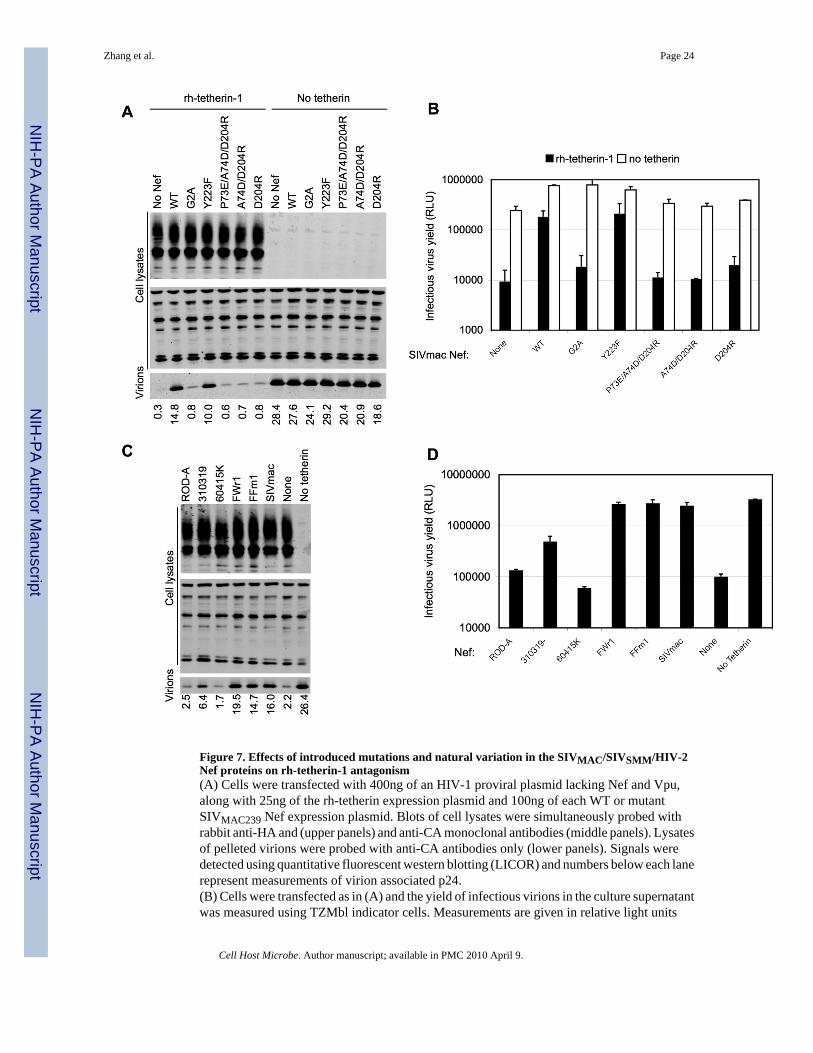
Figure 7. Effects of introduced mutations and natural variation in the SIVMAC/SIVSMM/HIV-2Nef proteins on rh-tetherin-1 antagonism(A) Cells were transfected with 400ng of an HIV-1 proviral plasmid lacking Nef and Vpu,along with 25ng of the rh-tetherin expression plasmid and 100ng of each WT or mutantSIVMAC239 Nef expression plasmid. Blots of cell lysates were simultaneously probed withrabbit anti-HA and (upper panels) and anti-CA monoclonal antibodies (middle panels). Lysatesof pelleted virions were probed with anti-CA antibodies only (lower panels). Signals weredetected using quantitative fluorescent western blotting (LICOR) and numbers below each lanerepresent measurements of virion associated p24.(B) Cells were transfected as in (A) and the yield of infectious virions in the culture supernatantwas measured using TZMbl indicator cells. Measurements are given in relative light units
Zhang et al. Page 24
Cell Host Microbe. Author manuscript; available in PMC 2010 April 9.
NIH
-PA Author Manuscript
NIH
-PA Author Manuscript
NIH
-PA Author Manuscript

(RLU) following detection of β-galactosidase expression using a chemiluminescent assay.Results are the mean± SD of tripicate determinations from a single experiment and arerepresentative of two independent experiments.(C) Cells were transfected with 400ng of an HIV-1 proviral plasmid lacking Nef and Vpu,along with 25ng of the rh-tetherin expression plasmid and 50ng of each HIV-2, SIVSMM orSIVMAC239 Nef expression plasmid. Blots were probed and analysed as in (A) and numbersbelow each lane represent measurements of virion associated p24.(D) Cells were transfected as in (C) and the yield of infectious virions in the culture supernatantwas measured as in (B). Results are the mean± SD of tripicate determinations from a singleexperiment and are representative of two independent experiments.
Zhang et al. Page 25
Cell Host Microbe. Author manuscript; available in PMC 2010 April 9.
NIH
-PA Author Manuscript
NIH
-PA Author Manuscript
NIH
-PA Author Manuscript
Copyright © 2022 FDOKUMEN