Cutaneous Manifestations in Children with Diabetes Mellitus ...
Upload
independentCategory
view
2download
0
PEDIATRICSREVIEW ARTICLE
published: 25 July 2014doi: 10.3389/fped.2014.00077
Pulmonary manifestations of primary immunodeficiencydisorders in childrenMilos Jesenak 1*, Peter Banovcin1, Barbora Jesenakova1 and Eva Babusikova2*1 Center for Diagnosis and Treatment of Primary Immunodeficiencies, Department of Pediatrics, Jessenius Faculty of Medicine, Comenius University in Bratislava,
Martin, Slovakia2 Department of Medical Biochemistry, Jessenius Faculty of Medicine, Comenius University in Bratislava, Martin, Slovakia
Edited by:Luis Garcia-Marcos, University ofMurcia, Spain
Reviewed by:Michael B. Anthracopoulos, UniversityHospital of Patras, GreeceKelvin D. MacDonald, Oregon Health& Science University, USA
*Correspondence:Milos Jesenak and Eva Babusikova,Center for Diagnosis and Treatment ofPrimary Immunodeficiencies,Departments of Pediatrics andMedical Biochemistry, JesseniusFaculty of Medicine, ComeniusUniversity in Bratislava, Kollarova 2,Martin 036 59, Slovakiae-mail: [email protected];[email protected]
Primary immunodeficiencies (PIDs) are inherited disorders in which one or several com-ponents of immune system are decreased, missing, or of non-appropriate function.Thesediseases affect the development, function, or morphology of the immune system. Thegroup of PID comprises more than 200 different disorders and syndromes and the numberof newly recognized and revealed deficiencies is still increasing. Their clinical presentationand complications depend on the type of defects and there is a great variability in the rela-tionship between genotypes and phenotypes. A variation of clinical presentation acrossvarious age categories is also presented and children could widely differ from adult patientswith PID. Respiratory symptoms and complications present a significant cause of morbid-ity and also mortality among patients suffering from different forms of PIDs and they areobserved both in children and adults. They can affect primarily either upper airways (e.g.,sinusitis and otitis media) or lower respiratory tract [e.g., pneumonia, bronchitis, bronchiec-tasis, and interstitial lung diseases (ILDs)]. The complications from lower respiratory tractare usually considered to be more important and also more specific for PIDs and they deter-minate patients’ prognosis. The spectrum of the causal pathogens usually demonstratestypical pattern characteristic for each PID category. The respiratory signs of PIDs can bedivided into infectious (upper and lower respiratory tract infections and complications) andnon-infectious (ILDs, bronchial abnormalities – especially bronchiectasis, malignancies, andbenign lymphoproliferation). Early diagnosis and appropriate therapy can prevent or at leastslow down the development and course of respiratory complications of PIDs.
Keywords: infectious complications, inheritance, immune system dysregulation, interstitial lung diseases, respira-tory tract, non-infectious complications, primary immunodeficiencies
INTRODUCTIONScience of primary immunodeficiencies (PIDs) represents a fas-cinating and rapidly developing part of modern medicine andclinical immunology. PIDs are inherited disorders of immune sys-tem in which one or several immune components are decreased,missing, or of non-appropriate function. These diseases affect thedevelopment, function, or morphology of the immune system (1).Since the first official scientific publication of the PID case in 1952(2), more than 200 other diseases were described and character-ized. The heterogeneity of the PIDs, the variability of their clinicalmanifestations, inconsistence in the relationship between geno-type and phenotype and involvement of whatever organ or tissuesupports the interdisciplinary character of these diseases, whichrequires multidisciplinary approach in their management. Hand-in-hand, continual education of health professionals and laics aswell is urgently needed. There are on-going efforts of the edu-cation of laic and medical community, which is aimed on theincrease of public awareness of the PID. The early diagnosis isusually associated with better prognosis and sooner appropriatetherapeutic strategies. From the symptomatic diagnosis that wasbased on the detection of various immune system defects after the
onset of clinical symptoms and complications, we shifted towardcomplicated algorithms consisting from different immunologicaltests accompanied by molecular-genetic analysis in the selectedcases (Figure 1). The biggest challenge of current immunologyis to establish the diagnosis of PID even before the onset of theclinical symptoms, just to start the right therapy as soon as pos-sible and to prevent the possible complications and consequencesof non-treated or non-appropriately treated disease. Several coun-tries have recently introduced into the praxis the pilot programs ofneonatal screening for selected immunodeficiencies [especially forsevere combined immunodeficiency (SCID) diseases, but also forataxia telangiectasia and some severe humoral immunodeficien-cies]. On the other hand, for the most severe diseases, the prenataldiagnosis in cases of positive family history for particular PID isfully recommended (3).
The group of PIDs is getting more and more heterogeneousand the number of newly recognized and revealed deficiencies isincreasing year by year. The majority of PIDs is monogenic, but insome cases, the polygenic basic is suspected. There are still manyPIDs with unknown or not-fully described and characterizedgenetic background [e.g., common variable immunodeficiency
www.frontiersin.org July 2014 | Volume 2 | Article 77 | 1

Jesenak et al. Pulmonary manifestations of primary immunodeficiencies
(CVID) or selective deficiency of IgA]. PIDs are usually rare dis-eases with overall prevalence of 1:10,000 live births. However, someof the PIDs can be found quite frequently (e.g., selective deficiencyof IgA, deficiencies of IgG subclasses, deficiency of mannose-binding lectin, etc.) (5). The most frequent immunodeficienciesyield usually mild course. Some of the frequent immunodefi-ciencies can be even asymptomatic during the lifetime and theirclinical significant of questionable and discussed. In case of thecombination of different “mild” defects in immunity, the clinicalmanifestation becomes more possible and evident.
The classification of PIDs underwent a long way and was elab-orated regularly. The understanding of the particular diseasesallowed the creation of the new categories and groups of relateddiseases of immune system. According to the current knowl-edge, the immunodeficiencies are classified into the eight major
FIGURE 1 | General clinical course of primary immunodeficiencies anddifferent approaches in establishment of the diagnosis [adapted andmodified from Ref. (4)].
categories based on the primarily involved immune componentand associated symptoms and signs (6):
1. predominantly humoral (antibody) deficiencies,2. combined T-cell and B-cell immunodeficiencies,3. other well defined immunodeficiency syndromes,4. congenital defects of number and/or function of phagocytes,5. complement deficiencies,6. defects of immune dysregulation,7. autoinflammatory disorders,8. defects in innate immunity.
Among all the immunodeficiencies, antibody deficiencies arethe most frequent and comprise approximately 70–75% of allPIDs. These patients are typically characterized by different respi-ratory symptoms and complications due to the inherited immunedefect. In children, respiratory symptoms are typical initial presen-tation of various PIDs. However, also the other groups and classesof PIDs can be associated with significant respiratory morbidityand manifestations (Table 1). Through two simple widely avail-able test – serum immunoglobulin concentration (IgG, IgA, IgM,and ±IgE) and differential leukocyte cell count – the majority ofthe PID can be detected and revealed. Therefore, these two simplextests can be in general recommended as screening tools for PIDsin primary care.
RESPIRATORY MANIFESTATIONS OF PRIMARYIMMUNODEFICIENCIESRespiratory symptoms and complications present a significantcause of morbidity and also mortality among patients sufferingfrom different forms of PIDs (Table 2). Computed tomography(CT) in combination with other imaging techniques, clinical tests,and laboratory examinations play an important role in detecting,characterizing, and quantifying the extent and kind of lung dam-age (9). Another important role of the imaging techniques lies also
Table 1 | Respiratory presentations and complications of primary immunodeficiencies [adapted according to Bierry et al. (7) andTouw et al. (8)].
Non-infectious complications Infectious
complications
Chronic lung
disease
Chronic
inflammatory
diseases
Benign
lymphoproliferative
disease
Malign
neoplasma
RESPIRATORY COMPLICATIONS OF PRIMARY IMMUNODEFICIENCIES
Bronchial abnormalities
(bronchiectasis, bronchial wall
thickening, atelectasis, mucus plugs,
emphysema, bullae, pneumatocoele)
Otitis Fibrosis Granulomas Parenchymal
lymphoid hyperplasia
Solid organ
tumors
(leiomyoma,
adenocarcinoma)
Lung parenchyma abnormalities
(nodules, cavity)
Rhino/sinusitis Pulmonary
hypertension
Interstitial lung
disease
Reactive follicular
hyperplasia
Lymphomas
Ventilation abnormalities (obstructive,
restrictive, combined)
Bronchitis Cor pulmonale Mediastinal
lymphadenopathy
Thymic tumors
Laryngeal angioedema Pneumonia Respiratory
failure
Lung metastasis
Empyema Allergies
Lung abscess
Frontiers in Pediatrics | Pediatric Pulmonology July 2014 | Volume 2 | Article 77 | 2
Jesenak et al. Pulmonary manifestations of primary immunodeficiencies
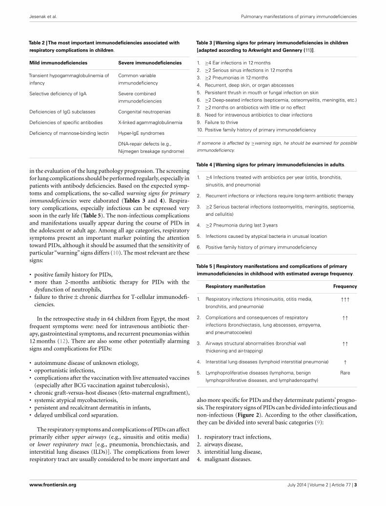
Table 2 |The most important immunodeficiencies associated with
respiratory complications in children.
Mild immunodeficiencies Severe immunodeficiencies
Transient hypogammaglobulinemia of
infancy
Common variable
immunodeficiency
Selective deficiency of IgA Severe combined
immunodeficiencies
Deficiencies of IgG subclasses Congenital neutropenias
Deficiencies of specific antibodies X-linked agammaglobulinemia
Deficiency of mannose-binding lectin Hyper-IgE syndromes
DNA-repair defects (e.g.,
Nijmegen breakage syndrome)
in the evaluation of the lung pathology progression. The screeningfor lung complications should be performed regularly, especially inpatients with antibody deficiencies. Based on the expected symp-toms and complications, the so-called warning signs for primaryimmunodeficiencies were elaborated (Tables 3 and 4). Respira-tory complications, especially infectious can be expressed verysoon in the early life (Table 5). The non-infectious complicationsand manifestations usually appear during the course of PIDs inthe adolescent or adult age. Among all age categories, respiratorysymptoms present an important marker pointing the attentiontoward PIDs, although it should be assumed that the sensitivity ofparticular “warning” signs differs (10). The most relevant are thesesigns:
• positive family history for PIDs,• more than 2-months antibiotic therapy for PIDs with the
dysfunction of neutrophils,• failure to thrive± chronic diarrhea for T-cellular immunodefi-
ciencies.
In the retrospective study in 64 children from Egypt, the mostfrequent symptoms were: need for intravenous antibiotic ther-apy, gastrointestinal symptoms, and recurrent pneumonias within12 months (12). There are also some other potentially alarmingsigns and complications for PIDs:
• autoimmune disease of unknown etiology,• opportunistic infections,• complications after the vaccination with live attenuated vaccines
(especially after BCG vaccination against tuberculosis),• chronic graft-versus-host diseases (feto-maternal engraftment),• systemic atypical mycobacteriosis,• persistent and recalcitrant dermatitis in infants,• delayed umbilical cord separation.
The respiratory symptoms and complications of PIDs can affectprimarily either upper airways (e.g., sinusitis and otitis media)or lower respiratory tract [e.g., pneumonia, bronchiectasis, andinterstitial lung diseases (ILDs)]. The complications from lowerrespiratory tract are usually considered to be more important and
Table 3 | Warning signs for primary immunodeficiencies in children
[adapted according to Arkwright and Gennery (11)].
1. ≥4 Ear infections in 12 months
2. ≥2 Serious sinus infections in 12 months
3. ≥2 Pneumonias in 12 months
4. Recurrent, deep skin, or organ abscesses
5. Persistent thrush in mouth or fungal infection on skin
6. ≥2 Deep-seated infections (septicemia, osteomyelitis, meningitis, etc.)
7. ≥2 months on antibiotics with little or no effect
8. Need for intravenous antibiotics to clear infections
9. Failure to thrive
10. Positive family history of primary immunodeficiency
If someone is affected by ≥warning sign, he should be examined for possible
immunodeficiency.
Table 4 | Warning signs for primary immunodeficiencies in adults.
1. ≥4 Infections treated with antibiotics per year (otitis, bronchitis,
sinusitis, and pneumonia)
2. Recurrent infections or infections require long-term antibiotic therapy
3. ≥2 Serious bacterial infections (osteomyelitis, meningitis, septicemia,
and cellulitis)
4. ≥2 Pneumonia during last 3 years
5. Infections caused by atypical bacteria in unusual location
6. Positive family history of primary immunodeficiency
Table 5 | Respiratory manifestations and complications of primary
immunodeficiencies in childhood with estimated average frequency.
Respiratory manifestation Frequency
1. Respiratory infections (rhinosinusitis, otitis media,
bronchitis, and pneumonia)
↑↑↑
2. Complications and consequences of respiratory
infections (bronchiectasis, lung abscesses, empyema,
and pneumatocoeles)
↑↑
3. Airways structural abnormalities (bronchial wall
thickening and air-trapping)
↑↑
4. Interstitial lung diseases (lymphoid interstitial pneumonia) ↑
5. Lymphoproliferative diseases (lymphoma, benign
lymphoproliferative diseases, and lymphadenopathy)
Rare
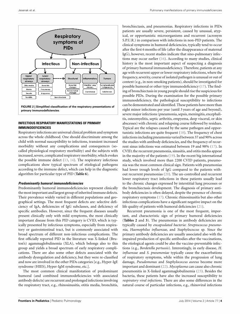
also more specific for PIDs and they determinate patients’ progno-sis. The respiratory signs of PIDs can be divided into infectious andnon-infectious (Figure 2). According to the other classification,they can be divided into several basic categories (9):
1. respiratory tract infections,2. airways disease,3. interstitial lung disease,4. malignant diseases.
www.frontiersin.org July 2014 | Volume 2 | Article 77 | 3
Jesenak et al. Pulmonary manifestations of primary immunodeficiencies
FIGURE 2 | Simplified classification of the respiratory presentations ofprimary immunodeficiencies.
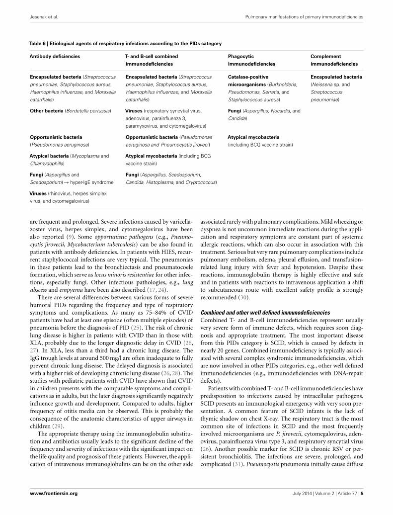
INFECTIOUS RESPIRATORY MANIFESTATIONS OF PRIMARYIMMUNODEFICIENCIESRespiratory infections are universal clinical problem and symptomacross the whole childhood. One should discriminate among thechild with normal susceptibility to infections, transient increasedmorbidity without any complications and consequences (so-called physiological respiratory morbidity) and the subjects withincreased, severe, complicated respiratory morbidity, which evokesthe possible immune defect (13, 14). The respiratory infectiouscomplications show typical spectrum of etiological pathogensaccording to the immune defect, which can help in the diagnosticalgorithm for particular type of PID (Table 6).
Predominantly humoral immunodeficienciesPredominantly humoral immunodeficiencies represent clinicallythe most important and largest group of inherited immune defects.Their prevalence widely varied in different populations and geo-graphical settings. The most frequent defects are: selective defi-ciency of IgA, deficiencies of IgG subclasses, and deficiency ofspecific antibodies. However, since the frequent diseases usuallypresent clinically only with mild symptoms, the most clinicallyimportant disease from this PID category is CVID, which is typ-ically presented by infectious symptoms, especially from respira-tory or gastrointestinal tract, but is commonly associated withbroad spectrum of different non-infectious complications. Thefirst officially reported PID in the literature was X-linked (Bru-ton’s) agammaglobulinemia (XLA), which belongs also to thisgroup and yields a broad spectrum of early respiratory compli-cations. There are also some other defects associated with theantibody dysregulation and deficiency, but they were re-classifiedand now are involved in the other PIDs categories [e.g., Hyper-IgEsyndrome (HIES), Hyper-IgM syndrome, etc.].
The most common clinical manifestation of predominanthumoral (and combined immunodeficiencies with associatedantibody defects) are recurrent and prolonged infections involvingthe respiratory tract, e.g., rhinosinusitis, otitis media, bronchitis,
bronchiectasis, and pneumonias. Respiratory infections in PIDspatients are usually severe, persistent, caused by unusual, atyp-ical, or opportunistic microorganisms and recurrent (acronymS.P.U.R.) in comparison with infections in non-PID patients. Theclinical symptoms in humoral deficiencies, typically tend to occurafter the first 6 months of life (after the disappearance of maternalIgG), however, recent studies indicate that sino-pulmonary infec-tions may occur earlier (16). According to many studies, clinicalhistory is the most important aspect of suspecting a diagnosisof primary humoral immunodeficiency. Therefore, patients at anyage with recurrent upper or lower respiratory infections, where thefrequency, severity, course of isolated pathogen is unusual or out ofcontext (e.g., in non-smoking patients), should be investigated forpossible humoral or other type immunodeficiency (17). The find-ing of bronchiectasis in young people should rise the suspicious forpossible PIDs. During the examination for the possible primaryimmunodeficiency, the pathological susceptibility to infectionscan be demonstrated and identified. These patients have more thaneight minor infections per year (until 3 years of age and beyond),severe major infections (pneumonia, sepsis, meningitis, encephali-tis, osteomyelitis, septic arthritis, empyema, deep visceral, or skinabscesses) with chronic and relapsing course followed by residues.Typical are the relapses caused by the same pathogen and oppor-tunistic infections are quite frequent (18). The frequency of chestinfections including pneumonia varied between 37 and 90% acrossthe studies with antibody deficiencies, and the frequency of recur-rent sinus infections was estimated between 19 and 98% (17). InCVID, the recurrent pneumonia, sinusitis, and otitis media appearin the majority of the patients (19). In the recent big internationalstudy, which involved more than 2200 CVID patients, pneumo-nia was the most common clinical sign. Patients with pneumoniashad lower trough levels of IgG compared to the patients with-out recurrent pneumonias (20). The un-controlled and recurrentlower respiratory tract infections in these patients usually leadto the chronic changes expressed by interstitial lung processes orthe bronchiectasis development. The diagnosis of primary anti-body deficiencies is often delayed, despite the presence of chronicrespiratory symptoms (17). Chronic rhinosinusitis but also otherinfectious complications have a significant negative impact on thelife quality of patients with humoral deficiencies (21).
Recurrent pneumonia is one of the most frequent, impor-tant, and characteristic sign of primary humoral deficiencies(Tables 2 and 3). The pneumonias in antibody deficiencies aretypically caused by encapsulated bacteria: Streptococcus pneumo-nia, Haemophilus influenzae, and Staphylococcus sp. Since theprimary antibody deficiencies are usually associated also with theimpaired production of specific antibodies after the vaccinations,the etiological agents could be also the vaccine-preventable infec-tions (e.g., Bordetella pertussis). Interestingly, in early disease, H.influenzae and S. pneumoniae typically cause the exacerbationsof respiratory symptoms, while within the progression of lungdamage, Pseudomonas and Staphylococcus aureus become moreimportant and dominant (22). Mycoplasma can cause also chronicpneumonitis in X-linked agammaglobulinemia (23). Besides thebacteria, these patients have also the increased susceptibility torespiratory viral infections. There are also some differences in thenatural course of particular infections, e.g., rhinoviral infections
Frontiers in Pediatrics | Pediatric Pulmonology July 2014 | Volume 2 | Article 77 | 4
Jesenak et al. Pulmonary manifestations of primary immunodeficiencies
Table 6 | Etiological agents of respiratory infections according to the PIDs category.
Antibody deficiencies T- and B-cell combined
immunodeficiencies
Phagocytic
immunodeficiencies
Complement
immunodeficiencies
Encapsulated bacteria (Streptococcus
pneumoniae, Staphylococcus aureus,
Haemophilus influenzae, and Moraxella
catarrhalis)
Encapsulated bacteria (Streptococcus
pneumoniae, Staphylococcus aureus,
Haemophilus influenzae, and Moraxella
catarrhalis)
Catalase-positive
microorganisms (Burkholderia,
Pseudomonas, Serratia, and
Staphylococcus aureus)
Encapsulated bacteria
(Neisseria sp. and
Streptococcus
pneumoniae)
Other bacteria (Bordetella pertussis) Viruses (respiratory syncytial virus,
adenovirus, parainfluenza 3,
paramyxovirus, and cytomegalovirus)
Fungi (Aspergillus, Nocardia, and
Candida)
Opportunistic bacteria
(Pseudomonas aeruginosa)
Opportunistic bacteria (Pseudomonas
aeruginosa and Pneumocystis jiroveci )
Atypical mycobacteria
(including BCG vaccine strain)
Atypical bacteria (Mycoplasma and
Chlamydophilla)
Atypical mycobacteria (including BCG
vaccine strain)
Fungi (Aspergillus and
Scedosporium)→hyper-IgE syndrome
Fungi (Aspergillus, Scedosporium,
Candida, Histoplasma, and Cryptococcus)
Viruses (rhinovirus, herpes simplex
virus, and cytomegalovirus)
are frequent and prolonged. Severe infections caused by varicella-zoster virus, herpes simplex, and cytomegalovirus have beenalso reported (9). Some opportunistic pathogens (e.g., Pneumo-cystis jirovecii, Mycobacterium tuberculosis) can be also found inpatients with antibody deficiencies. In patients with HIES, recur-rent staphylococcal infections are very typical. The pneumoniasin these patients lead to the bronchiectasis and pneumatocoeleformation, which serve as locus minoris resistentiae for other infec-tions, especially fungi. Other infectious pathologies, e.g., lungabscess and empyema have been also described (17, 24).
There are several differences between various forms of severehumoral PIDs regarding the frequency and type of respiratorysymptoms and complications. As many as 75–84% of CVIDpatients have had at least one episode (often multiple episodes) ofpneumonia before the diagnosis of PID (25). The risk of chroniclung disease is higher in patients with CVID than in those withXLA, probably due to the longer diagnostic delay in CVID (26,27). In XLA, less than a third had a chronic lung disease. TheIgG trough levels at around 500 mg/l are often inadequate to fullyprevent chronic lung disease. The delayed diagnosis is associatedwith a higher risk of developing chronic lung disease (26, 28). Thestudies with pediatric patients with CVID have shown that CVIDin children presents with the comparable symptoms and compli-cations as in adults, but the later diagnosis significantly negativelyinfluence growth and development. Compared to adults, higherfrequency of otitis media can be observed. This is probably theconsequence of the anatomic characteristics of upper airways inchildren (29).
The appropriate therapy using the immunoglobulin substitu-tion and antibiotics usually leads to the significant decline of thefrequency and severity of infections with the significant impact onthe life quality and prognosis of these patients. However, the appli-cation of intravenous immunoglobulins can be on the other side
associated rarely with pulmonary complications. Mild wheezing ordyspnea is not uncommon immediate reactions during the appli-cation and respiratory symptoms are constant part of systemicallergic reactions, which can also occur in association with thistreatment. Serious but very rare pulmonary complications includepulmonary embolism, edema, pleural effusion, and transfusion-related lung injury with fever and hypotension. Despite thesereactions, immunoglobulin therapy is highly effective and safeand in patients with reactions to intravenous application a shiftto subcutaneous route with excellent safety profile is stronglyrecommended (30).
Combined and other well defined immunodeficienciesCombined T- and B-cell immunodeficiencies represent usuallyvery severe form of immune defects, which requires soon diag-nosis and appropriate treatment. The most important diseasefrom this PIDs category is SCID, which is caused by defects innearly 20 genes. Combined immunodeficiency is typically associ-ated with several complex syndromic immunodeficiencies, whichare now involved in other PIDs categories, e.g., other well definedimmunodeficiencies (e.g., immunodeficiencies with DNA-repairdefects).
Patients with combined T- and B-cell immunodeficiencies havepredisposition to infections caused by intracellular pathogens.SCID presents an immunological emergency with very soon pre-sentation. A common feature of SCID infants is the lack ofthymic shadow on chest X-ray. The respiratory tract is the mostcommon site of infections in SCID and the most frequentlyinvolved microorganisms are P. jirovecii, cytomegalovirus, aden-ovirus, parainfluenza virus type 3, and respiratory syncytial virus(26). Another possible marker for SCID is chronic RSV or per-sistent bronchiolitis. The infections are severe, prolonged, andcomplicated (31). Pneumocystis pneumonia initially cause diffuse
www.frontiersin.org July 2014 | Volume 2 | Article 77 | 5
Jesenak et al. Pulmonary manifestations of primary immunodeficiencies
interstitial infiltrates, which progress to alveolar infiltrates, whichcan be focal and asymmetric (26). Although pneumonia causedby P. jirovecii is a common presentation feature of SCID, it israrely recognized. The diagnosis should be estimated throughbronchoalveolar lavage or lung biopsy. The diagnosis is suspecteddue to infiltrates on chest X-ray and presence of respiratory dis-tress, especially in combination with other symptoms of PID suchas diarrhea, failure to thrive, or thrush. Pneumocystis infectionalone in child with SCID does not lessen the chances of success-ful hematopoietic stem cell transplantation (HSCT), which is theunique causal therapeutic option for these patients (32). Respi-ratory viruses, particularly paramyxoviruses and adenoviruses arecommon significant pathogens in these patients with significantworsening effect of BMT outcome. Aggressive treatment (virosta-tics, immunoglobulins – intravenous, subcutaneous, or nebulized,corticosteroids) may reduce viral replication, lung damage andmay improve respiratory functions and outcome. No treatmentcan probably results in viral clearance without successful T-cellengraftment (33).
Hyper-IgE syndrome (Job’s syndrome) is a complex combinedimmunodeficiency. Till today, at least three types of HIES werediscriminated (autosomal dominant form – caused by mutationin signal transducer and activator of transcription, STAT3; andtwo autosomal recessive forms – caused by mutation in genefor tyrosine kinase 2, TYK2 or gene for dedicator of cytokine-sis, DOCK8). In general, in HIES the lung symptoms are verycommon and early presentation of the disease. At the beginning,the recurrent sino-pulmonary infections are caused predomi-nantly by S. aureus, and less frequently with S. pneumonia andH. influenzae. Recurrent pneumonias are typical clinical featurefor all the three types of HIES. The healing after infections isusually aberrant and the result is the formation of bronchiectasisand pneumatocoeles, which are considered to be the pathogenicmarker for autosomal dominant form of HIES (STAT3 mutation)whereas they were not reported in DOCK8 or TYK2 deficiencies.The pneumatocoeles can be occupied by Aspergillus or Scedospo-rium and are difficult to manage and treat. Pneumatocoeles areunusual in children infections and their appearance should alertto an unusual diagnosis are the very least (34, 35). Ones theparenchymal lung damage is present the spectrum of pulmonarypathogens shifts toward the spectrum seen in cystic fibrosis –Pseudomonas aeruginosa and non-tuberculous mycobacteria. Thelung complication in HIES may be due to the impaired Th17cell differentiation (36). Prior to pyogenic pneumonias, also HIEScan be clinically presented by P. jirovecii pneumonia, however, ingeneral this complication is rare. P. jirovecii can cause pneumo-nia in patients with HIES both with and without chronic lungdiseases (37).
Patients with Hyper-IgM syndromes could have distinct clinicalinfectious complications based on the type of genetic backgroundand exact type of the syndrome. While autosomal form of HIMSpresent as typical humoral PID, the X-linked form shows thespectrum of pathogens similar to the patients with combinedimmunodeficiencies. The underlying infectious cause of the pneu-monias includes encapsulated bacteria, CMV, histoplasmosis, andP. jirovecii. Fungal pneumonias caused by Candida, Cryptococcus,and Histoplasma can be also found (38).
Pneumonia and chronic lung disease can be observed alsoin the patients with DNA-repair deficiencies (ataxia telangiecta-sia, Bloom syndrome, and Nijmegen breakage syndrome). Thepatients with ataxia telangiectasia are susceptible to recurrent viraland bacterial infections, but have also the increased risk of differentmalignancies, especially lymphoreticular. The increased sensitivityto ionizing radiation should be taken into account when indicatingimaging in patients with DNA-repair defects (31).
Phagocytic immunodeficienciesPhagocytic diseases are caused either by the decrease numberand/or dysfunction of phagocytes. This group of PIDs is quiterare, however, in case of organ or deep skin abscesses, chronicdermatitis, persistent fungal infections, or delayed umbilical cordseparation, one should think about them. The most importantdisease from this PIDs category is chronic granulomatous dis-eases (CGD), which is caused by the inability of the phagocytesto produce reactive oxygen species for intracellular killing ofingested microorganism. Another important group of phagocyticimmunodeficiencies is presented by different form of congen-ital neutropenia (e.g., severe congenital neutropenia and cyclicneutropenia).
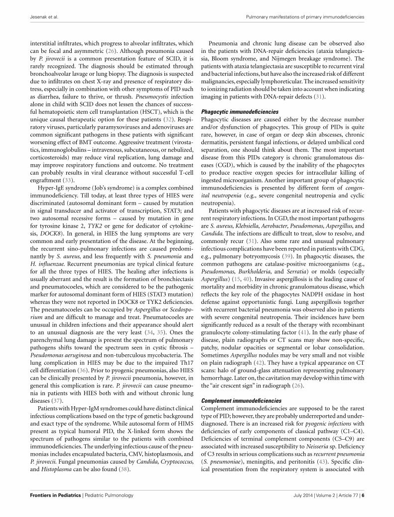
Patients with phagocytic diseases are at increased risk of recur-rent respiratory infections. In CGD, the most important pathogensare S. aureus, Klebsiella, Aerobacter, Pseudomonas, Aspergillus, andCandida. The infections are difficult to treat, slow to resolve, andcommonly recur (31). Also some rare and unusual pulmonaryinfectious complications have been reported in patients with CDG,e.g., pulmonary botryomycosis (39). In phagocytic diseases, thecommon pathogens are catalase-positive microorganisms (e.g.,Pseudomonas, Burkholderia, and Serratia) or molds (especiallyAspergillus) (15, 40). Invasive aspergillosis is the leading cause ofmortality and morbidity in chronic granulomatous disease, whichreflects the key role of the phagocytes NADPH oxidase in hostdefense against opportunistic fungi. Lung aspergillosis togetherwith recurrent bacterial pneumonia was observed also in patientswith severe congenital neutropenia. Their incidences have beensignificantly reduced as a result of the therapy with recombinantgranulocyte colony-stimulating factor (41). In the early phase ofdisease, plain radiographs or CT scans may show non-specific,patchy, nodular opacities or segmental or lobar consolidation.Sometimes Aspergillus nodules may be very small and not visibleon plain radiograph (42). They have a typical appearance on CTscans: halo of ground-glass attenuation representing pulmonaryhemorrhage. Later on, the cavitation may develop within time withthe “air crescent sign” in radiograph (26).
Complement immunodeficienciesComplement immunodeficiencies are supposed to be the raresttype of PID; however, they are probably underreported and under-diagnosed. There is an increased risk for pyogenic infections withdeficiencies of early components of classical pathway (C1–C4).Deficiencies of terminal complement components (C5–C9) areassociated with increased susceptibility to Neisseria sp. Deficiencyof C3 results in serious complications such as recurrent pneumonia(S. pneumoniae), meningitis, and peritonitis (43). Specific clin-ical presentation from the respiratory system is associated with
Frontiers in Pediatrics | Pediatric Pulmonology July 2014 | Volume 2 | Article 77 | 6
Jesenak et al. Pulmonary manifestations of primary immunodeficiencies
hereditary angioedema, which is clinically manifested by recur-rent angioedemas in different part of the body. Potentially life-threatening are the angioedemas located to larynx (15). The age ofonset, frequency of attacks, and the factors triggering upper airwayswelling are variable among different patients. To avoid a fatal out-come of laryngeal swelling, the therapy should be administered assoon as possible. The new available drugs for the treatment of acuteattacks significantly improved the prognosis of these patients (44).
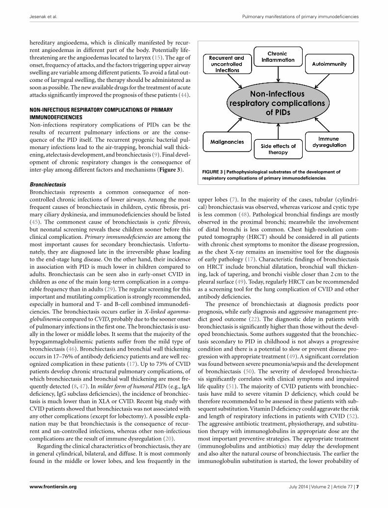
NON-INFECTIOUS RESPIRATORY COMPLICATIONS OF PRIMARYIMMUNODEFICIENCIESNon-infections respiratory complications of PIDs can be theresults of recurrent pulmonary infections or are the conse-quence of the PID itself. The recurrent pyogenic bacterial pul-monary infections lead to the air-trapping, bronchial wall thick-ening, atelectasis development, and bronchiectasis (9). Final devel-opment of chronic respiratory changes is the consequence ofinter-play among different factors and mechanisms (Figure 3).
BronchiectasisBronchiectasis represents a common consequence of non-controlled chronic infections of lower airways. Among the mostfrequent causes of bronchiectasis in children, cystic fibrosis, pri-mary ciliary dyskinesia, and immunodeficiencies should be listed(45). The commonest cause of bronchiectasis is cystic fibrosis,but neonatal screening reveals these children sooner before thisclinical complication. Primary immunodeficiencies are among themost important causes for secondary bronchiectasis. Unfortu-nately, they are diagnosed late in the irreversible phase leadingto the end-stage lung disease. On the other hand, their incidencein association with PID is much lower in children compared toadults. Bronchiectasis can be seen also in early-onset CVID inchildren as one of the main long-term complication in a compa-rable frequency than in adults (29). The regular screening for thisimportant and mutilating complication is strongly recommended,especially in humoral and T- and B-cell combined immunodefi-ciencies. The bronchiectasis occurs earlier in X-linked agamma-globulinemia compared to CVID, probably due to the sooner onsetof pulmonary infections in the first one. The bronchiectasis is usu-ally in the lower or middle lobes. It seems that the majority of thehypogammaglobulinemic patients suffer from the mild type ofbronchiectasis (46). Bronchiectasis and bronchial wall thickeningoccurs in 17–76% of antibody deficiency patients and are well rec-ognized complication in these patients (17). Up to 73% of CVIDpatients develop chronic structural pulmonary complications, ofwhich bronchiectasis and bronchial wall thickening are most fre-quently detected (8, 47). In milder form of humoral PIDs (e.g., IgAdeficiency, IgG subclass deficiencies), the incidence of bronchiec-tasis is much lower than in XLA or CVID. Recent big study withCVID patients showed that bronchiectasis was not associated withany other complications (except for lobectomy). A possible expla-nation may be that bronchiectasis is the consequence of recur-rent and un-controlled infections, whereas other non-infectiouscomplications are the result of immune dysregulation (20).
Regarding the clinical characteristics of bronchiectasis, they arein general cylindrical, bilateral, and diffuse. It is most commonlyfound in the middle or lower lobes, and less frequently in the
FIGURE 3 | Pathophysiological substrates of the development ofrespiratory complications of primary immunodeficiencies.
upper lobes (7). In the majority of the cases, tubular (cylindri-cal) bronchiectasis was observed, whereas varicose and cystic typeis less common (48). Pathological bronchial findings are mostlyobserved in the proximal bronchi; meanwhile the involvementof distal bronchi is less common. Chest high-resolution com-puted tomography (HRCT) should be considered in all patientswith chronic chest symptoms to monitor the disease progression,as the chest X-ray remains an insensitive tool for the diagnosisof early pathology (17). Characteristic findings of bronchiectasison HRCT include bronchial dilatation, bronchial wall thicken-ing, lack of tapering, and bronchi visible closer than 2 cm to thepleural surface (49). Today, regularly HRCT can be recommendedas a screening tool for the lung complication of CVID and otherantibody deficiencies.
The presence of bronchiectasis at diagnosis predicts poorprognosis, while early diagnosis and aggressive management pre-dict good outcome (22). The diagnostic delay in patients withbronchiectasis is significantly higher than those without the devel-oped bronchiectasis. Some authors suggested that the bronchiec-tasis secondary to PID in childhood is not always a progressivecondition and there is a potential to slow or prevent disease pro-gression with appropriate treatment (49). A significant correlationwas found between severe pneumonia/sepsis and the developmentof bronchiectasis (50). The severity of developed bronchiecta-sis significantly correlates with clinical symptoms and impairedlife quality (51). The majority of CVID patients with bronchiec-tasis have mild to severe vitamin D deficiency, which could betherefore recommended to be assessed in these patients with sub-sequent substitution.Vitamin D deficiency could aggravate the riskand length of respiratory infections in patients with CVID (52).The aggressive antibiotic treatment, physiotherapy, and substitu-tion therapy with immunoglobulins in appropriate dose are themost important preventive strategies. The appropriate treatment(immunoglobulins and antibiotics) may delay the developmentand also alter the natural course of bronchiectasis. The earlier theimmunoglobulin substitution is started, the lower probability of
www.frontiersin.org July 2014 | Volume 2 | Article 77 | 7
Jesenak et al. Pulmonary manifestations of primary immunodeficiencies
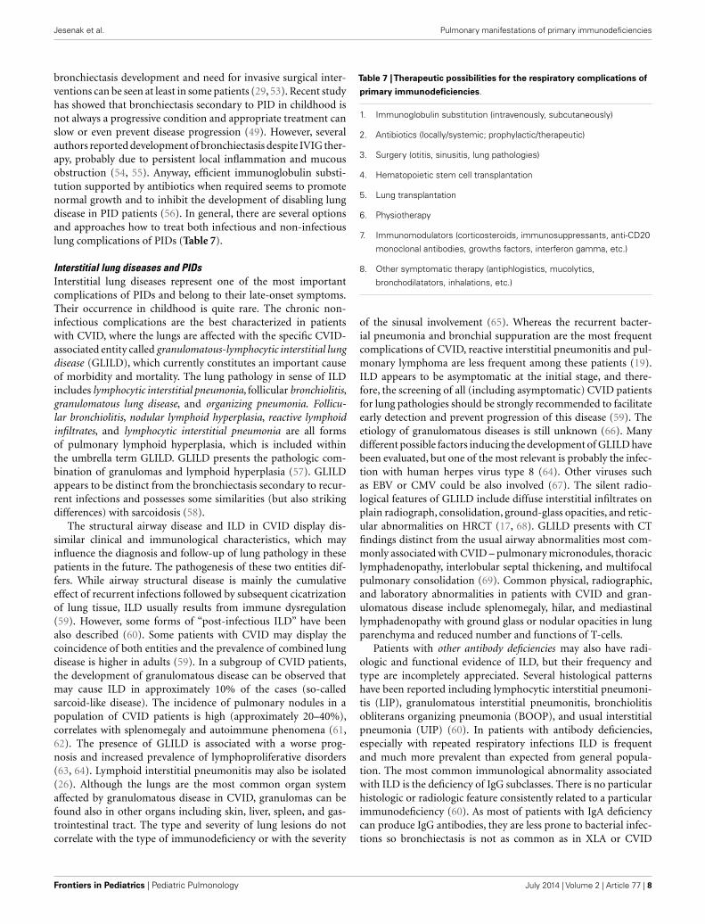
bronchiectasis development and need for invasive surgical inter-ventions can be seen at least in some patients (29, 53). Recent studyhas showed that bronchiectasis secondary to PID in childhood isnot always a progressive condition and appropriate treatment canslow or even prevent disease progression (49). However, severalauthors reported development of bronchiectasis despite IVIG ther-apy, probably due to persistent local inflammation and mucousobstruction (54, 55). Anyway, efficient immunoglobulin substi-tution supported by antibiotics when required seems to promotenormal growth and to inhibit the development of disabling lungdisease in PID patients (56). In general, there are several optionsand approaches how to treat both infectious and non-infectiouslung complications of PIDs (Table 7).
Interstitial lung diseases and PIDsInterstitial lung diseases represent one of the most importantcomplications of PIDs and belong to their late-onset symptoms.Their occurrence in childhood is quite rare. The chronic non-infectious complications are the best characterized in patientswith CVID, where the lungs are affected with the specific CVID-associated entity called granulomatous-lymphocytic interstitial lungdisease (GLILD), which currently constitutes an important causeof morbidity and mortality. The lung pathology in sense of ILDincludes lymphocytic interstitial pneumonia, follicular bronchiolitis,granulomatous lung disease, and organizing pneumonia. Follicu-lar bronchiolitis, nodular lymphoid hyperplasia, reactive lymphoidinfiltrates, and lymphocytic interstitial pneumonia are all formsof pulmonary lymphoid hyperplasia, which is included withinthe umbrella term GLILD. GLILD presents the pathologic com-bination of granulomas and lymphoid hyperplasia (57). GLILDappears to be distinct from the bronchiectasis secondary to recur-rent infections and possesses some similarities (but also strikingdifferences) with sarcoidosis (58).
The structural airway disease and ILD in CVID display dis-similar clinical and immunological characteristics, which mayinfluence the diagnosis and follow-up of lung pathology in thesepatients in the future. The pathogenesis of these two entities dif-fers. While airway structural disease is mainly the cumulativeeffect of recurrent infections followed by subsequent cicatrizationof lung tissue, ILD usually results from immune dysregulation(59). However, some forms of “post-infectious ILD” have beenalso described (60). Some patients with CVID may display thecoincidence of both entities and the prevalence of combined lungdisease is higher in adults (59). In a subgroup of CVID patients,the development of granulomatous disease can be observed thatmay cause ILD in approximately 10% of the cases (so-calledsarcoid-like disease). The incidence of pulmonary nodules in apopulation of CVID patients is high (approximately 20–40%),correlates with splenomegaly and autoimmune phenomena (61,62). The presence of GLILD is associated with a worse prog-nosis and increased prevalence of lymphoproliferative disorders(63, 64). Lymphoid interstitial pneumonitis may also be isolated(26). Although the lungs are the most common organ systemaffected by granulomatous disease in CVID, granulomas can befound also in other organs including skin, liver, spleen, and gas-trointestinal tract. The type and severity of lung lesions do notcorrelate with the type of immunodeficiency or with the severity
Table 7 |Therapeutic possibilities for the respiratory complications of
primary immunodeficiencies.
1. Immunoglobulin substitution (intravenously, subcutaneously)
2. Antibiotics (locally/systemic; prophylactic/therapeutic)
3. Surgery (otitis, sinusitis, lung pathologies)
4. Hematopoietic stem cell transplantation
5. Lung transplantation
6. Physiotherapy
7. Immunomodulators (corticosteroids, immunosuppressants, anti-CD20
monoclonal antibodies, growths factors, interferon gamma, etc.)
8. Other symptomatic therapy (antiphlogistics, mucolytics,
bronchodilatators, inhalations, etc.)
of the sinusal involvement (65). Whereas the recurrent bacter-ial pneumonia and bronchial suppuration are the most frequentcomplications of CVID, reactive interstitial pneumonitis and pul-monary lymphoma are less frequent among these patients (19).ILD appears to be asymptomatic at the initial stage, and there-fore, the screening of all (including asymptomatic) CVID patientsfor lung pathologies should be strongly recommended to facilitateearly detection and prevent progression of this disease (59). Theetiology of granulomatous diseases is still unknown (66). Manydifferent possible factors inducing the development of GLILD havebeen evaluated, but one of the most relevant is probably the infec-tion with human herpes virus type 8 (64). Other viruses suchas EBV or CMV could be also involved (67). The silent radio-logical features of GLILD include diffuse interstitial infiltrates onplain radiograph, consolidation, ground-glass opacities, and retic-ular abnormalities on HRCT (17, 68). GLILD presents with CTfindings distinct from the usual airway abnormalities most com-monly associated with CVID – pulmonary micronodules, thoraciclymphadenopathy, interlobular septal thickening, and multifocalpulmonary consolidation (69). Common physical, radiographic,and laboratory abnormalities in patients with CVID and gran-ulomatous disease include splenomegaly, hilar, and mediastinallymphadenopathy with ground glass or nodular opacities in lungparenchyma and reduced number and functions of T-cells.
Patients with other antibody deficiencies may also have radi-ologic and functional evidence of ILD, but their frequency andtype are incompletely appreciated. Several histological patternshave been reported including lymphocytic interstitial pneumoni-tis (LIP), granulomatous interstitial pneumonitis, bronchiolitisobliterans organizing pneumonia (BOOP), and usual interstitialpneumonia (UIP) (60). In patients with antibody deficiencies,especially with repeated respiratory infections ILD is frequentand much more prevalent than expected from general popula-tion. The most common immunological abnormality associatedwith ILD is the deficiency of IgG subclasses. There is no particularhistologic or radiologic feature consistently related to a particularimmunodeficiency (60). As most of patients with IgA deficiencycan produce IgG antibodies, they are less prone to bacterial infec-tions so bronchiectasis is not as common as in XLA or CVID
Frontiers in Pediatrics | Pediatric Pulmonology July 2014 | Volume 2 | Article 77 | 8
Jesenak et al. Pulmonary manifestations of primary immunodeficiencies
(43). Anyway, there are several reports of chronic lung disease alsoin selective IgA deficiency, especially when associated with IgG2
deficiency (70).Pneumatocoeles are the typical clinical radiological finding in
the patients with HIES. They are typical warning characteristicsign for autosomal dominant HIES and they can present the locusfor other possible infections, e.g., fungal.
Tumors of respiratory tract in primary immunodeficienciesMalignancy is after infections the second leading cause of death inPIDs. The majority of the tumors is associated with the Epstein–Barr virus (30–60% of cases). In general, the risk of develop-ing malignancies varies from 1 to 25%, but the children withCVID and Wiskott–Aldrich syndrome are at greater risk (31).We can discriminate between benign lymphoproliferative diseases(parenchymal lymphoid hyperplasia, reactive follicular hyperpla-sia) and malignancies. The enlargement of mediastinal lymph nodecan occasionally lead to superior vena caval syndrome and CVIDwas recommended to be included in differential diagnosis of ILDsand hilar lymphadenopathy (71).
Regarding the solid lung tumors, there are only few case reportsin the literature (leiomyoma, pulmonary adenocarcinoma) andthis respiratory complication is considered to be in general rarein PID patients (72, 73). Pulmonary comprise due to metas-tasis (origin from, e.g., gastric carcinoma) is more often seenthan primary pulmonary malignancy (7). Thymoma may occurin combined immunodeficiency and this rare entity is calledGood syndrome. Good syndrome occurs in 1–6% of patientswith primary humoral immunodeficiencies (74). However, pul-monary lymphoma seems to be a serious complication associatedwith CVID or other PIDs (19, 75). Patients with CVID frequentlydevelop lymphoproliferative disease and the risk for malignantlymphoma is increased by more than 300-fold. Non-Hodgkin’slymphoma (especially high-grade B-cell lymphoma) is more com-mon than Hodgkin’s lymphoma. Approximately 8% patients withCVID develop non-Hodgkin’s lymphoma and in general, <1%of patients with humoral PID develop Hodgkin’s lymphoma(76). There is also a possibility of benign lymphoproliferativedisease in PID, e.g., parenchymal lymphoid hyperplasia of thelungs (77). Some benign conditions (e.g., lymphocytic interstitialpneumonia) can transform into malignant lymphoma (78).
Other respiratory complications of PIDsIn patients with humoral immunodeficiencies, the respiratoryallergic or allergy-like symptoms such as dyspnea, rhinitis, orasthma can be frequently seen (29). Many patients despite theappropriate treatment present with chronic productive cough,which is usually the hallmark of chronic sinusitis or bronchitis(79). Despite some similarities in the clinical picture of primaryciliary dyskinesia and CVID, these two entities require differenttherapeutic approach. There is a case report in the literature withboth diseases in one patient. Therefore, in a patient with alreadyestablished diagnosis of chronic lung disease with the deteriora-tion of clinical course, the search for secondary diagnosis shouldbe recommended (80, 81).
Several cases of primary pulmonary hypertension were alsodescribed in association with primary immunodeficiency (82).
Primary pulmonary dysgenesis or even agenesis was discussedto be considered as a part of the spectrum anomalies associatedwith velocardiofacial syndrome (83).
Recently, a new immunodeficiency syndrome called pulmonaryalveolar proteinosis has been described. It is characterized by accu-mulation of pulmonary surfactant, respiratory insufficiency andincreased infections due to associated immunodeficiency. Thissyndrome belongs to the category of phagocytic diseases andis a result of the disturbed surfactant catabolism in alveolarmacrophages. It can be either acquired (due to autoantibod-ies against GM-CSF) or congenital caused by CSF2RA muta-tions. CSF2RA encodes the GM-CSF receptor α protein. Thesepatients have increased susceptibility to opportunistic microbialpathogens and increased mortality from un-controlled infec-tions (84). Another new syndrome in two 46XX sisters with fatallung fibrosis, profound combined immunodeficiency and gonadaldysgenesis was also recently described. Comparative genomehybridization and analysis of genes known to be associated withsevere immune defects in infancy or gonadal dysgenesis showedno abnormality (85).
The delay in the diagnosis of PID results in the poorer prognosisand considerable morbidity, particularly recurrent pneumonias,resulting in structural lung damage such as bronchiectasis, pul-monary hypertension, and ultimately cor pulmonale (17). Theend-stage lung disease with the development of cor pulmonaleand respiratory insufficiency has been documented as the mostcommon cause of morbidity in primary humoral immunodefi-ciencies (25). However, non-infectious lung diseases may occurdespite optimal immunoglobulin therapy (28). Therefore, patientsdeveloping chronic respiratory symptoms should be managedin a multidisciplinary team, including chest physician, as pro-gression of lung disease may occur despite apparently optimalimmunoglobulin therapy (17).
DIAGNOSIS OF RESPIRATORY COMPLICATIONS OF PIDsDue to the chronic involvement of the respiratory tract (bacterialpneumonia, abscesses, fungal lung disease, and interstitial pneu-monia) in PIDs, the changes of lung functions could be logicallyexpected. Although different patterns are typically associated withdistinct type of immune defects, there is a substantial overlapin imaging findings (26). Screening examinations, such as lungfunction testing (LFT) and HRCT of the chest, should be used toevaluate pulmonary status in PID patients (86).
Lung function testing in PIDsSeveral studies evaluated the changes in pulmonary functions.Normal lung functions can be seen in 45–74% patients. In general,the ventilatory disturbances evaluated by lung function tests canbe observed in the majority of the patients with CVID or XLA(87). There are some inter-disease differences, e.g., patients withCVID have more commonly abnormal lung tests compared to thepatients with X-linked agammaglobulinemia (88). An obstruc-tive pattern is reported most frequently, although restrictive pul-monary disease is well recognized (17). Small airways involvementleads to ventilation abnormalities and chronic obstructive disease,which are usually irreversible (7). A reduced rate of carbon monox-ide uptake and restrictive ventilatory pattern is typically found in
www.frontiersin.org July 2014 | Volume 2 | Article 77 | 9

Jesenak et al. Pulmonary manifestations of primary immunodeficiencies
CVID patients with ILD (63). A significant correlation betweenthe HRCT score and the predicted values in pulmonary func-tion test has been confirmed in several studies (46), while theothers did not find such associations (49, 87). There is a nega-tive correlation between a number of episodes of pneumonia andlung function tests. Recent study has shown that the longitudinaldecline of lung functions can be observed in patients with PID.Chronic persistent cough can be used as a marker of the reductionof lung functions (87). One study reported an improvement oflung function tests despite bronchiectasis progression on HRCTin patients after the initiation of immunoglobulin treatment andauthors concluded that LFT may serve as a toll for evaluation of theclinical response to immunoglobulin therapy (89). The changes inlung function could be used as an indicator of immunoglobulintreatment efficacy. Based on the significant association betweenthe dose of IVIG and spirometric indices, the use of higher IVIGdoses as a protection of lung function decline was recommended(90). Lung functional tests can be used to monitor patients at amore regular basis, although featured by decreased sensitivity forcomplications (8).
Imaging techniques in PIDsThere is an essential role of different imaging techniques, especiallyCT in the identification and monitoring of pulmonary changesand complications in CVID or some other specific immunod-eficiencies. One of the most important diagnostic tools for thedetection, quantifying, and characterizing the extent and kind oflung damage is CT. HRCT is the reference standard for the detec-tion of bronchiectasis. The early sign of the possible developmentof bronchiectasis is the bronchial wall thickening. HRCT appearsto be more sensitive for the detection of pneumopathies (withspecial emphasis on bronchial changes and interstitial lesions) inPID patients than chest X-ray (55, 91). Both HRCT and helicalCT proved to be useful tools for monitoring of lung changes asso-ciated with PID, especially in symptomatic patients with negativeradiographic findings (92).
The patients with severe PID should be regularly examinedfor the possible respiratory symptoms and complications. In gen-eral, LFT can be recommended every 6–12 months with repeatedchest X-ray. CT examination should be repeated within 5–10 yearsafter initial presentation of PID unless there is a specific indica-tion or changes in clinical status. If significant abnormalities arefound,a repeated CT should be performed every 3–5 years depend-ing on the type of abnormality or clinical stability. If patientsare prescribed potentially toxic therapies, e.g., high-dose steroidsfor GLILD, CT examination should be performed approximately6 months after the initiation of therapy. Subsequent CT examina-tions depend on clinical, X-ray, and lung function test changes,but in general should be repeated annually for 1–2 years followedby a decreased frequency is stability is achieved (9, 79).
A special approach should be given to the patients withincreased radiation sensitivity (DNA-repair defects, also the part ofCVID patients). In these cases, an alternative radiation-free tech-nique alternative to CT scan or chest X-ray could be MRI evaluationof the pulmonary changes and alterations. In recent study, CT andMRI findings were comparable for moderate to severe degrees ofbronchial and parenchymal alterations, but weaker concordance
between CT and MRI scan was found for lower scores of bronchialabnormalities. Therefore, it should be admitted that CT allowsbetter identification of peripheral airways changes. However, fur-ther studies are needed to image quality and involvement of thismethod into the diagnostic algorithm in PID (93). Actually, low-dose HRCT is still considered to be the standard and the mostsensitive method for identification of structural abnormalities andpulmonary complications in CVID at the time of diagnosis and atregular time-points during follow-up, however, with the properfollow-up interval yet to be determined. Annual testing (bothspirometry and transfer factor) is useful in the assessment of PIDpatients, and should not be confined to those with radiologicalevidence of lung disease (27, 94).
In a study with 58 pediatric patients with PID or cancer (12with PID) suffering from different respiratory symptoms, thebronchoscopy and bronchoalveolar lavage was shown to be clin-ically useful and established an overall diagnostic rate 94% ofpatients. Infection rate was 74.2% and infectious agent was iso-lated in 53% of the cases. In PID patients, different agents wereisolated (Pseudomonas, Enterobacter, E. coli, Acinetobacter, Proteus,and Aspergillus). P. jirovecii was identified only in patients with-out prophylactic therapy with trimethoprim–sulfamethoxazole.Authors stated that these two methods should be considered as aninitial diagnostic tool in pediatric PID patients (95).
CONCLUSIONRespiratory system is the most common site of the clinical man-ifestations of different PID both in adults and in children. Infec-tious and non-infectious respiratory complications determinatethe patients’ prognosis. To reduce the morbidity associated withPID the greater awareness of respiratory complications for suchpatients should be raised. Regular examinations by the appropri-ate tests should reveal the respiratory pathologies in early stagesand should be used also for the monitoring of already existingabnormalities. In general, due to the raising awareness of PIDs theprognosis of these patients gradually improves.
ACKNOWLEDGMENTSThe work was supported by the projects: Measurement of kinet-ics of cilia in respiratory tract (ITMS 262202200019) and VEGA1/0252/14.
REFERENCES1. Madkaikar M, Mishra A, Ghosh K. Diagnostic approach to primary immunod-
eficiency disorders. Indian Pediatr (2013) 50:579–86. doi:10.1007/s13312-013-0171-4
2. Bruton OC. Agammaglobulinemia. Pediatrics (1952) 9:722–8.3. Puck JM. The case for newborn screening for severe combined immun-
odeficiency and related disorders. Ann N Y Acad Sci (2011) 1246:108–17.doi:10.1111/j.1749-6632.2011.06346.x
4. Lindegren ML, Kobrynski L, Rasmussen SA, Moore CA, Grosse SD, Vander-ford ML, et al. Applying public health strategies to primary immunodeficiencydisorders. MMWR Recomm Rep (2004) 53(RR01):1–28.
5. Stiehm RE. The four most common immunodeficiencies. Adv Exp Med Biol(2007) 601:15–26. doi:10.1007/978-0-387-72005-0_2
6. Al-Herz W, Bousfiha A, Casanova JL, Chatila T, Conley ME, Cunningham-Rundles C, et al. Primary immunodeficiency diseases: an update on the classifi-cation from the international union of immunological societies expert commit-tee for primary immunodeficiency. Front Immunol (2014) 5:162. doi:10.3389/fimmu.2014.00162
Frontiers in Pediatrics | Pediatric Pulmonology July 2014 | Volume 2 | Article 77 | 10
Jesenak et al. Pulmonary manifestations of primary immunodeficiencies
7. Bierry G, Boileau J, Barnig C, Gasser B, Korganow AS, Buy X, et al. Thoracis man-ifestations of primary humoral immunodeficiency: a comprehensive review.Radiographics (2009) 29:1909–20. doi:10.1148/rg.297095717
8. Touw CM, van de Ven AA, de Jong PA, Terheggen-Lagro S, Beek E, Sanders EA,et al. Detection of pulmonary complications in common variable immunode-ficiency. Pediatr Allergy Immunol (2010) 21:793–805. doi:10.1111/j.1399-3038.2009.00963.x
9. Hampson FA, Chandra A, Screaton NJ, Condliffe A, Kumararatne DS, ExleyAR, et al. Respiratory disease in common variable immunodeficiency andother primary immunodeficiency disorders. Clin Radiol (2012) 67:587–95.doi:10.1016/j.crad.2011.10.028
10. O’Sullivan MD, Cant AJ. The 10 warning signs: a time for a change? Curr OpinAllergy Clin Immunol (2012) 12:588–94. doi:10.1097/ACI.0b013e3283591534
11. Arkwright PD, Gennery AR. Ten warning signs of primary immunodeficiency: anew paradigm is needed for the 21st century. Ann N Y Acad Sci (2011) 1238:7–14.doi:10.1111/j.1749-6632.2011.06206.x
12. Reda SM, Afifi HM, Amine MM. Primary immunodeficiency diseases inEgyptian children: a single-center study. J Clin Immunol (2009) 29:343–51.doi:10.1007/s10875-008-9260-x
13. Jesenak M, Ciljakova M, Rennerova Z, Babusikova E, Banovcin P. Recurrent res-piratory infections in children – definition, diagnostic approach, treatment andprevention. In: Martin-Loeches I, editor. Bronchitis. Rijeka: InTech (2011). p.119–48.
14. Jesenak M, Majtan J, Rennerova Z, Kyselovic J, Banovcin P, Hrubisko M.Immunomodulatory effect of pleuran (β-glucan from Pleurotus ostreatus) inchildren with recurrent respiratory tract infections. Int Immunopharmacol(2013) 15:395–9. doi:10.1016/j.intimp.2012.11.020
15. Jesenak M, Banovcin P, Freiberger T. Hereditary angioedema in paediatric andadolescent age – in centre experience. Lek Obz (2013) 62:245–9.
16. Winkelstein JA, Marino MC, Lederman HM, Jones SM, Sullivan K, BurksAW, et al. X-linked agammaglobulinemia: report on a United States registryof 201 patients. Medicine (2006) 85:193–202. doi:10.1097/01.md.0000229482.27398.ad
17. Wood P, Stanworth S, Burton J, Jones A, Peckham DG, Green T, et al. Recog-nition, clinical diagnosis and management of patients with primary anti-body deficiencies: a systemic review. Clin Exp Immunol (2007) 149:410–23.doi:10.1111/j.1365-2249.2007.03432.x
18. Wahn V. When is susceptibility to infections abnormal? Pediatr Allergy Immunol(2011) 22:650–1. doi:10.1111/j.1399-3038.2011.01217.x
19. Cadranel J, Bouvry D, Wislez M. Manifestations respiratoires au cours dudeficit immunitaire commun variable de l’adulte. Rev Mal Respir (2003) 20:126–33.
20. Gathmann B, Mahlaoui N, Gerard L, Oksenhendler E, Warnatz K, Schulze I,et al. Clinical picture and treatment of 2212 patients with common variableimmunodeficiency. J Allergy Clin Immunol (2014) 134:116–26. doi:10.1016/j.jaci.2013.12.1077
21. Mendieta-Flores E, Del-Rivero-Hernandez LG, Zavala-Perez M, Segura-MendezNH. Rhinosinusitis and its impact on the quality of life in patients with commonvariable immunodeficiency. Rev Alerg Mex (2012) 59:60–4.
22. Tarzi MD, Grigoriadou S, Carr SB, Kuitert LM, Longhurst HJ. Clinical immunol-ogy review series: an approach to the management of pulmonary disease in pri-mary antibody deficiency. Clin Exp Immunol (2008) 155:147–55. doi:10.1111/j.1365-2249.2008.03851.x
23. Furr PM, Taylor-Robinson D, Webster AD. Mycoplasmas and ureaplasmas inpatients with hypogammaglobulinaemia and their role in arthritis: micro-biological observations over twenty years. Ann Rheum Dis (1994) 53:183–7.doi:10.1136/ard.53.3.183
24. Havlicekova Z, Jesenak M, Freiberger T, Banovcin P. Biomed. Pap. Med. Fac. Univ.Palacky Olomouc Czech Repub (2013). doi:10.5507/bp.2013.011
25. Cunningham-Rundles C, Bodian C. Common variable immunodeficiency: clin-ical and immunological features of 248 patients. Clin Immunol (1999) 92:34–48.doi:10.1006/clim.1999.4725
26. Notarangelo LD, Plebani A, Mazzolari E, Soresina A, Bondioni MP. Geneticcauses of bronchiectasis: primary immune deficiencies and the lung. Respiration(2007) 74:264–75. doi:10.1159/000101784
27. Aghamohammadi A, Allahverdi A, Abolhassani H, Moazzami K, Alizadeh H,Gharagozlou M, et al. Comparison of pulmonary diseases in common variable
immunodeficiency and X-linked agammaglobulinaemia. Respirology (2010)15:289–95. doi:10.1111/j.1440-1843.2009.01679.x
28. Quartier P, Debre M, De Blic J, De Sauverzac R, Sayegh N, Jabado N, et al. Earlyand prolonged intravenous immunoglobulin replacement therapy in childhoodagammaglobulinemia: a retrospective survey of 31 patients. J Pediatr (1999)134:589–96. doi:10.1016/S0022-3476(99)70246-5
29. Urschel S, Kayikci L, Wintergerst U, Notheis G, Jansson A, Belohradsky BH.Common variable immunodeficiency in children: delayed diagnosis despite typ-ical clinical presentation. J Pediatr (2009) 154:888–94. doi:10.1016/j.jpeds.2008.12.020
30. Stiehm RE. Adverse effects of human immunoglobulin therapy. Transfus MedRev (2013) 27:171–8. doi:10.1016/j.tmrv.2013.05.004
31. Jeanes AC, Owens CM. Chest imaging in the immunocompromised child. Pae-diatr Respir Rev (2002) 3:59–69. doi:10.1053/prrv.2002.0186
32. Berrington JE, Flood TJ, Abinun M, Galloway A, Cant AJ. Unsuspected Pneumo-cystis carinii pneumonia at presentation of severe primary immunodeficiency.Arch Dis Child (2000) 82:144–7. doi:10.1136/adc.82.2.144
33. Crooks BN, Taylor CE, Turner AJ, Osman HK, Abinun M, Flood TJ, et al. Res-piratory viral infections in primary immune deficiencies: significance and rele-vance to clinical outcome in a single BMT unit. Bone Marrow Transplant (2000)26:1097–102. doi:10.1038/sj.bmt.1702656
34. Cant A, Battersby A. When to think of immunodeficiency? In: Curtis N, edi-tor. Hot Topics in Infection and Immunity in Children. New York, NY: SpringerScience+Business Media (2013). p. 167–77.
35. Almyroudis NG, Holland SM, Segal BH. Invasive aspergillosis in primaryimmunodeficiencies. Med Mycol (2005) 43(Suppl 1):S247–59. doi:10.1080/13693780400025203
36. Sowerwine KJ, Holland SM, Freeman AF. Hyper-IgE syndrome update. Ann NY Acad Sci (2012) 1250:25–32. doi:10.1111/j.1749-6632.2011.06387.x
37. Freeman AF, Davis J, Anderson VJ, Barson W, Darnell DN, Puck JM, et al. Pneu-mocystis jiroveci infection in patients with hyper-immunoglobulin E syndrome.Pediatrics (2006) 118:e1271–5. doi:10.1542/peds.2006-0311
38. Dosanjh A. Chronic pediatric pulmonary disease and primary humoral anti-body based immune disease. Respir Med (2011) 105:511–4. doi:10.1016/j.rmed.2010.11.013
39. Paz HL, Little BJ, Ball WC, Winkelstein JA. Primary pulmonary botryomycosis –a manifestation of chronic granulomatous disease. Chest (1992) 101:1160–2.doi:10.1378/chest.101.4.1160
40. Jesenak M, Havlicekova Z, Banovcin P, Stasia MJ. Chronic granulomatous dis-ease caused by a novel mutation in a 2-month-old boy with multifocal splenicabscesses. J Investig Allergol Clin Immunol (2013) 23:137–8.
41. Dale DC, Cottle TE, Fier CJ, Bolyard AA, Bonilla MA, Boxer LA, et al.Severe chronic neutropenia: treatment and follow-up of patients in the severechronic neutropenia international registry. Am J Hematol (2003) 72:82–93.doi:10.1002/ajh.10255
42. Copley SJ. Application of computed tomography in childhood respiratory infec-tions. Br Med Bull (2002) 61:263–79. doi:10.1093/bmb/61.1.263
43. Buckley RH. Pulmonary complications of primary immunodeficiencies.Paediatr Respir Rev (2004) 5(Suppl A):225–33. doi:10.1016/S1526-0542(04)90043-7
44. Papadopoulou-Alataki E. Upper airway considerations in hereditaryangioedema. Curr Opin Allergy Clin Immunol (2010) 10:20–5. doi:10.1097/ACI.0b013e328334f629
45. Bouyahia O, Essadem L, Matoussi N, Gharsallah L, Fitouri Z, Mrad Mazigh S,et al. Etiology and outcome of bronchiectasis in children: a study of 41 patients.Tunis Med (2008) 86:996–9.
46. Gharagozlou M, Ebrahimi FA, Farhoudi A, Aghamohammadi A, Bemanian MH,Chavoshzadeh Z, et al. Pulmonary complications in primary hypogammaglob-ulinemia: a survey by high resolution CT scan. Monaldi Arch Chest Dis (2006)65:69–74.
47. Thickett MK, Kumararatne DS, Banerjee AK, Dudley R, Stableforth DE.Common variable immune deficiency: respiratory manifestations, pulmonaryfunction and high-resolution CT scan findings. Q J Med (2002) 95:655–62.doi:10.1093/qjmed/95.10.655
48. Obregon RG, Lynch DA, Kaske T, Newell JD, Kirkpatrick CH. Radiologic find-ings of adult primary immunodeficiency disorders. Contribution of CT. Chest(1994) 106:490–5. doi:10.1378/chest.106.2.490
www.frontiersin.org July 2014 | Volume 2 | Article 77 | 11

Jesenak et al. Pulmonary manifestations of primary immunodeficiencies
49. Haidopoulou K, Calder A, Jones A, Jaffe A, Sonnappa S. Bronchiectasis secondaryto primary immunodeficiency in children: longitudinal changes in structure andfunction. Pediatr Pulmonol (2009) 44:669–75. doi:10.1002/ppul.21036
50. Chapel H, Lucas M, Lee M, Bjorkander J, Webster D, Grimbacher B,et al. Common variable immunodeficiency disorders: division into distinctclinical phenotypes. Blood (2008) 112:277–86. doi:10.1182/blood-2007-11-124545
51. Galindo-Pacheco LV, Amaya-Mejia AS, O’Farrill-Romanillos PM, Del Rivero-Hernandez LG, Segura-Mendez NH. Quality of life in adults with variablecommon immunodeficiency and bronchiectasis. Rev Alerg Mex (2013) 60:123–8.
52. Amaya-Mejia AS, O’Farrill-Romanillos PM, Galindo-Pacheco LV,Vargas-OrtegaG, Mendoza-Zubieta V, Del Rivero-Hernandez LG, et al. Vitamin D deficiency inpatients with common variable immunodeficiency, with autoimmune diseasesand bronchiectasis. Rev Alerg Mex (2013) 60:110–6.
53. Cantani A, Ferrara M. The lung and primary immunodeficiency. AllergolImmunopathol (Madr) (1988) 16:429–37.
54. Quinti I, Soresina A, Spadaro G, Martino S, Donnano S, Agostani C, et al. Long-term follow-up and outcome of a large cohort of patient with common variableimmunodeficiency. J Clin Immunol (2007) 27:308–16. doi:10.1007/s10875-007-9075-1
55. Mogica Martinez MD, Garcia Lara S, Silva Very R, Montano Velazquez B, CruzMerida A, Santibanez Bustamante J, et al. Neumopathies in patients with pri-mary immunodeficiencies in treatment with intravenous gammaglobulin. RevAlerg Mex (2007) 54:14–9.
56. Bjorkander J, Bake B, Hanson LA. Primary hypogammaglobulinaemia: impairedlung function and body growth with delayed diagnosis and inadequate treat-ment. Eur J Respir Dis (1984) 65:529–36.
57. Maglione PJ, Ko HM, Beasley MB, Strauchen JA, Cunningham-Rundles C.Tertiary lymphoid neogenesis is a component of pulmonary hyperplasia inpatients with common variable immunodeficiency. J Allergy Clin Immunol(2014) 133:535–42. doi:10.1016/j.jaci.2013.08.022
58. Bianchi MP, Letovanec I, Spertini F, Nicod LP, Lazor R. Granulomatous lympho-cytic interstitial lung disease in common variable immunodeficiency. Rev MedSuisse (2013) 9:2175–80.
59. van de Ven AA, de Jong PA, Hoytema van Konijnenburg DP, Kessels OA, BoesM, Sanders EA, et al. Airway and interstitial lung disease are distinct entitiesin paediatric common variable immunodeficiency. Clin Exp Immunol (2011)165:235–42. doi:10.1111/j.1365-2249.2011.04425.x
60. Popa V, Colby TV, Reich SB. Pulmonary interstitial disease in Ig deficiency. Chest(2002) 122:1594–603. doi:10.1378/chest.122.5.1594
61. Bondioni MP, Soresina A, Lougaris V, Gatta D, Plebani A, Maroldi R. Commonvariable immunodeficiency: computed tomography evaluation of bronchopul-monary changes including nodular lesions in 40 patients. Correlation withclinical and immunological data. J Comput Assist Tomogr (2010) 34:395–401.doi:10.1097/RCT.0b013e3181cad9da
62. Park JE, Beal I, Dilworth JP, Tormey V, Haddock J. The HRCT appearances ofgranulomatous pulmonary disease in common variable immune deficiency. EurJ Radiol (2005) 54:359–64. doi:10.1016/j.ejrad.2004.09.005
63. Bates CA, Ellison MC, Lynch DA, Cool CD, Brown KK, Routes JM.Granulomatous-lymphocytic lung disease shortens survival in common vari-able immunodeficiency. J Allergy Clin Immunol (2004) 114:415–21. doi:10.1016/j.jaci.2004.05.057
64. Wheat WH, Cool CD, Morimoto Y, Rai PR, Kirkpatrick CH, Lindenbaum BA,et al. Possible role of human herpesvirus 8 in the lymphoproliferative disor-ders in common variable immunodeficiency. J Exp Med (2005) 202:479–84.doi:10.1084/jem.20050381
65. Bondioni MP, Duse M, Plebani A, Soresina A, Notarangelo LD, Berlucchi M,et al. Pulmonary and sinusal changes in 45 patients with primary immunod-eficiencies: computed tomography evaluation. J Comput Assist Tomogr (2007)31:620–8. doi:10.1097/RCT.0b013e31802e3c11
66. Morimoto Y, Routes JM. Granulomatous disease in common variable immun-odeficiency. Curr Allergy Asthma Rep (2005) 5:370–5. doi:10.1007/s11882-005-0008-x
67. Stewart JP, Egan JJ, Ross AJ, Kelly BG, Lok SS, Hasleton PS, et al. The detec-tion of Epstein-Barr virus DNA in lung tissue from patients with idio-pathic pulmonary fibrosis. Am J Respir Crit Care Med (1999) 159:1336–41.doi:10.1164/ajrccm.159.4.9807077
68. Park JH, Levinson AI. Granulomatous-lymphocytic interstitial lung disease(GLILD) in common variable immunodeficiency (CVID). Clin Immunol (2010)134:97–103. doi:10.1016/j.clim.2009.10.002
69. Torigian DA, LaRosa DF, Levinson AI, Litzky LA, Miller WT Jr. Granulomatous-lymphocytic interstitial lung disease associated with common variable immun-odeficiency: CT findings. J Thorac Imaging (2008) 23:162–9. doi:10.1097/RTI.0b013e318166d32f
70. Bjorkander J, Bake B, Oxelius VA, Hanson LA. Impaired lung function in patientswith IgA deficiency and low levels of IgG2 or IgG3. N Engl J Med (1985)313:720–4. doi:10.1056/NEJM198509193131203
71. Sacco O, Fragonese B, Picco P, Faraci M, Facchetti P, Pistoia V, et al. Commonvariable immunodeficiency presenting in a girl as lung infiltrates and mediasti-nal adenopathies leading to severe “superior vena caval” syndrome. Eur Respir J(1996) 9:1958–61. doi:10.1183/09031936.96.09091958
72. User IR, Dogru D, Talim B, Orhan D, Karnak I. Endobronchial, pulmonary andliver leiomyomata in a child with primary immune deficiency. Eur J Pediatr Surg(2010) 20:423–5. doi:10.1055/s-0030-1254144
73. Oztop I, Demirkan B, Tarhan O, Kayahan H, Yilmaz U, Kargi A, et al. The devel-opment of pulmonary adenocarcinoma in a patient with Job’s syndrome, a rareimmunodeficiency condition. Tumori (2004) 90:132–5.
74. Agarwal S, Cunningham-Rundles C. Thymoma and immunodeficiency (Goodsyndrome): a report of 2 unusual cases and review of the literature.Ann Allergy Asthma Immunol (2007) 98:185–90. doi:10.1016/S1081-1206(10)60695-3
75. Bal A, Gupta A, Sodhi KS, Das A, Singh S. Multifocal extranodal non-Hodgkinlymphoma involving both the lungs and brain in a child with primary immun-odeficiency. J Pediatr Hematol Oncol (2008) 30(2008):317–9. doi:10.1097/MPH.0b013e318161aa3b
76. Cunningham-Rundles C, Lieberman P, Hellman G, Chaganti RS. Non-Hodgkinlymphoma in common variable immunodeficiency. Am J Hematol (1991)37:69–74. doi:10.1002/ajh.2830370202
77. Kajiwara S, Sakai S, Soeda H, Takahashi N, Okafuji T, Yoshimitsu K, et al. Mul-tifocal nodular lymphoid hyperplasia of the lung. J Thorac Imaging (2005)20:239–41. doi:10.1097/01.rti.0000158404.40711.26
78. Fishback N, Koss M. Update on lymphoid interstitial pneumonitis. Curr OpinPulm Med (1996) 2:429–33. doi:10.1097/00063198-199609000-00014
79. Rusconi F, Panisi C, Dellepiane RM, Cardinale F, Chini L, Martire B, et al.Pulmonary and sinus diseases in primary humoral immunodeficiencies withchronic productive cough. Arch Dis Child (2003) 88:1101–5. doi:10.1136/adc.88.12.1101
80. Skorpinski EW, Kung SJ, Yousef E, McGeady SJ. Diagnosis of common vari-able immunodeficiency in a patients with primary ciliary dyskinesia. Pediatrics(2007) 119:e1203–5. doi:10.1542/peds.2006-2396
81. Modrzewska K, Wiatr E, Langfort R, Oniszh K, Roszkowski-Sliz K. Commonvariable immunodeficiency in a patient with suspected sarcoidosis. PneumonolAlergol Pol (2009) 77:91–6.
82. Bolkova NV. Primary immunodeficiency state in a child with the pulmonaryhypertension syndrome. Arkh Patol (1985) 47:63–6.
83. Cunningham ML, Perry RJ, Eby PR, Gibson RL, Opheim KE, Manning SC. Pri-mary pulmonary dysgenesis in velocardiofacial syndrome: a second patient. AmJ Med Genet A (2003) 121A:177–9. doi:10.1002/ajmg.a.20142
84. Trapnell BC, Carey BC, Uchida K, Suzuki T. Pulmonary alveolar proteinosis, aprimary immunodeficiency of impaired GM-CSF stimulation of macrophages.Curr Opin Immunol (2009) 21:514–21. doi:10.1016/j.coi.2009.09.004
85. Somech R, Somers GR, Chitayat D, Grunebaum E, Atkinson A, Kolomietz E,et al. Fatal lung fibrosis associated with immunodeficiency and gonadal dysge-nesis in 46XX sisters – a new syndrome. Am J Med Genet A (2008) 146A:8–14.doi:10.1002/ajmg.a.32014
86. Busse PJ, Farzan S, Cunningham-Rundles C. Pulmonary complications of com-mon variable immunodeficiency. Ann Allergy Asthma Immunol (2007) 98:1–8.doi:10.1016/S1081-1206(10)60853-8
87. Costa-Carvalho BR, Wandalsen GF, Pulici G, Aranda CS, Sole D. Pulmonarycomplications in patients with antibody deficiency. Allergol Immunopathol(Madr) (2011) 39:128–32. doi:10.1016/j.aller.2010.12.003
88. Sweinberg SK, Wodell RA, Grodofsky MP, Greene JM, Conley ME.Retrospective analysis of the incidence of pulmonary disease in hypogam-maglobulinemia. J Allergy Clin Immunol (1991) 88:96–104. doi:10.1016/0091-6749(91)90306-9
Frontiers in Pediatrics | Pediatric Pulmonology July 2014 | Volume 2 | Article 77 | 12
Jesenak et al. Pulmonary manifestations of primary immunodeficiencies
89. de Gracia J, Vendrell M, Alvarez A, Pallisa E, Rodrigo MJ, de la Rosa D, et al.Immunoglobulin therapy to control lung damage in patients with common vari-able immunodeficiency. Int Immunopharmacol (2004) 4:745–53. doi:10.1016/j.intimp.2004.02.011
90. Chen Y, Stirling RG, Paul E, Hore-Lacy F, Thompson BR, Douglass JA.Longitudinal decline in lung function in patients with primary immunoglobulindeficiencies. J Allergy Clin Immunol (2011) 127:1414–7. doi:10.1016/j.jaci.2011.03.041
91. Feydy A, Sibilia J, De Kerviler E, Zagdanski AM, Chevret S, Fermand JP, et al.Chest high resolution CT in adults with primary humoral immunodeficiency.Br J Radiol (1996) 69:1108–16. doi:10.1259/0007-1285-69-828-1108
92. Gattoni F, Tagliaferri B, Boioli F, Mazzoleni C, Uslenghi CM. Comput-erized tomography of the lungs in patients with congenital immunodefi-ciency. Comparison with clinicoradiologic assessment. Radiol Med (1999) 98:26–35.
93. Serra G, Milito C, Mitrevski M, Granata G, Martini H, Pesce AM, et al.Lung MRI as a possible alternative to CT scan for patients with primaryimmune deficiencies and increased radiosensitivity. Chest (2011) 140:1581–9.doi:10.1378/chest.10-3147
94. Rich AL, Le Jeune IR, McDermott L, Kinnear WJ. Serial lung function tests inprimary immune deficiency. Clin Exp Immunol (2007) 151:110–3. doi:10.1111/j.1365-2249.2007.03550.x
95. Efrati O, Gonik U, Bielorai B, Modan-Moses D, Neumann Y, Szeinberg A, et al.Fiberoptic bronchoscopy and bronchoalveolar lavage for the evaluation of pul-monary disease in children with primary immunodeficiency and cancer. PediatrBlood Cancer (2007) 48:324–9. doi:10.1002/pbc.20784
Conflict of Interest Statement: The authors declare that the research was conductedin the absence of any commercial or financial relationships that could be construedas a potential conflict of interest.
Received: 24 February 2014; accepted: 07 July 2014; published online: 25 July 2014.Citation: Jesenak M, Banovcin P, Jesenakova B and Babusikova E (2014) Pulmonarymanifestations of primary immunodeficiency disorders in children. Front. Pediatr. 2:77.doi: 10.3389/fped.2014.00077This article was submitted to Pediatric Pulmonology, a section of the journal Frontiersin Pediatrics.Copyright © 2014 Jesenak, Banovcin, Jesenakova and Babusikova. This is an open-access article distributed under the terms of the Creative Commons Attribution License(CC BY). The use, distribution or reproduction in other forums is permitted, providedthe original author(s) or licensor are credited and that the original publication in thisjournal is cited, in accordance with accepted academic practice. No use, distribution orreproduction is permitted which does not comply with these terms.
www.frontiersin.org July 2014 | Volume 2 | Article 77 | 13
Copyright © 2022 FDOKUMEN