Clinical and laboratory aspects of common variable immunodeficiency
20
Anais da Academia Brasileira de Ciências (2004) 76(4): 707-726 (Annals of the Brazilian Academy of Sciences) ISSN 0001-3765 www.scielo.br/aabc Clinical and laboratory aspects of common variable immunodeficiency CRISTINA M. KOKRON 1,2 , PAOLO R. ERRANTE 3 , MYRTHES T. BARROS 1,2 , GISELE V. BARACHO 3 , MARISTELA M. CAMARGO 3 , JORGE KALIL 1,2,4 and LUIZ V. RIZZO 3 1 Serviço de Imunologia, Av. Dr. Enéas de Carvalho Aguiar, 155, 8 ◦ andar, bloco 3, 05403-010 São Paulo, SP, Brasil 2 Institute for Investigation in Immunology 3 Departamento de Imunologia, ICB-USP,Av. Prof. Lineu Prestes, 1730, 05508-900 São Paulo, SP, Brasil 4 Fundação Zerbini, Av. Brigadeiro Faria Lima, 1884, 2 ◦ andar, Edifício Cal Center, Jardim Paulistano, 01451-000 São Paulo, SP, Brasil Manuscript received on February 27, 2004; accepted for publication on March 1, 2004; contributed by Jorge Kalil* ABSTRACT Common variable immunodeficiency (CVID) is an immunological disorder characterized by defective an- tibody production, recurrent infections, most notably of the respiratory tract, autoimmune phenomena and cancer. Some CVID patients may also present disturbances of the cellular immune response such as a decrease in the number and proportion of different lymphocyte populations, diminished lymphoproliferative response to mitogens and antigens, altered production of cytokines, and deficient expression of cell-surface molecules. Most Brazilian CVID patients included in this study show a decrease in T and B lymphocyte counts in the peripheral blood. Furthermore, their lymphocytes are more susceptible to apoptosis following activation than normal individuals, and they have a decrease in the expression of activation molecules like CD25, CD69, CD40L and CD70. Moreover, they show a decreased synthesis of IL-4 and IL-5 in comparison with normal individuals. The increase in susceptibility to apoptosis following activation, may also be responsible for the decrease in the expression of activation molecules and CD40L, decrease in Th2 cytokines synthesis, and in the number of T and B circulating cells. In this study we discuss some of these immunological disturbances correlating them to the patients’ clinical features and comparing our patients’ findings to the literature. Key words: immunodeficiency, humans, antibody, cytokines, clinical features. INTRODUCTION CommonVariable Immunodeficiency (CVID) is one of the most frequent primary immunodeficiencies diagnosed in humans, its incidence will vary from 1 to 10,000 to 1 to 2,000,000 people depending on the study (Tiller and Liddle 2000, Cunningham- Rundles 2001, Kainulainen et al. 2001, Sneller 2001). Interestingly, the disease is very rare among *Member Academia Brasileira de Ciências Correspondence to: Luiz Vicente Rizzo E-mail: [email protected] Asians; the incidence in Japan is estimated at 1 in every 2 million inhabitants. The first case of CVID was described in 1953 by Janeway and colleagues (Janeway et al. 1953). They described a 39-year- old female with bronchiectasis, recurrent pulmonary infections, sinusitis, otitis and one episode of menin- gitis due to Haemophilus influenzae. Although an electrophoresis of serum proteins was not performed hypogammaglobulinemia was the presumed diag- nosis due to the lack of isohemagglutinins in the serum and the remission of the recurrent infection An Acad Bras Cienc (2004) 76 (4)
Transcript of Clinical and laboratory aspects of common variable immunodeficiency
Anais da Academia Brasileira de Ciências (2004) 76(4): 707-726(Annals of the Brazilian Academy of Sciences)ISSN 0001-3765www.scielo.br/aabc
Clinical and laboratory aspects of common variable immunodeficiency
CRISTINA M. KOKRON1,2, PAOLO R. ERRANTE3, MYRTHES T. BARROS1,2,GISELE V. BARACHO3, MARISTELA M. CAMARGO3, JORGE KALIL1,2,4
and LUIZ V. RIZZO3
1Serviço de Imunologia, Av. Dr. Enéas de Carvalho Aguiar, 155, 8◦ andar, bloco 3, 05403-010 São Paulo, SP, Brasil2Institute for Investigation in Immunology
3Departamento de Imunologia, ICB-USP, Av. Prof. Lineu Prestes, 1730, 05508-900 São Paulo, SP, Brasil4Fundação Zerbini, Av. Brigadeiro Faria Lima, 1884, 2◦ andar,
Edifício Cal Center, Jardim Paulistano, 01451-000 São Paulo, SP, Brasil
Manuscript received on February 27, 2004; accepted for publication on March 1, 2004;
contributed by Jorge Kalil*
ABSTRACT
Common variable immunodeficiency (CVID) is an immunological disorder characterized by defective an-
tibody production, recurrent infections, most notably of the respiratory tract, autoimmune phenomena and
cancer. Some CVID patients may also present disturbances of the cellular immune response such as a decrease
in the number and proportion of different lymphocyte populations, diminished lymphoproliferative response
to mitogens and antigens, altered production of cytokines, and deficient expression of cell-surface molecules.
Most Brazilian CVID patients included in this study show a decrease in T and B lymphocyte counts in the
peripheral blood. Furthermore, their lymphocytes are more susceptible to apoptosis following activation than
normal individuals, and they have a decrease in the expression of activation molecules like CD25, CD69,
CD40L and CD70. Moreover, they show a decreased synthesis of IL-4 and IL-5 in comparison with normal
individuals. The increase in susceptibility to apoptosis following activation, may also be responsible for the
decrease in the expression of activation molecules and CD40L, decrease in Th2 cytokines synthesis, and in
the number of T and B circulating cells. In this study we discuss some of these immunological disturbances
correlating them to the patients’ clinical features and comparing our patients’ findings to the literature.
Key words: immunodeficiency, humans, antibody, cytokines, clinical features.
INTRODUCTION
CommonVariable Immunodeficiency (CVID) is one
of the most frequent primary immunodeficiencies
diagnosed in humans, its incidence will vary from
1 to 10,000 to 1 to 2,000,000 people depending
on the study (Tiller and Liddle 2000, Cunningham-
Rundles 2001, Kainulainen et al. 2001, Sneller
2001). Interestingly, the disease is very rare among
*Member Academia Brasileira de CiênciasCorrespondence to: Luiz Vicente RizzoE-mail: [email protected]
Asians; the incidence in Japan is estimated at 1 in
every 2 million inhabitants. The first case of CVID
was described in 1953 by Janeway and colleagues
(Janeway et al. 1953). They described a 39-year-
old female with bronchiectasis, recurrent pulmonary
infections, sinusitis, otitis and one episode of menin-
gitis due to Haemophilus influenzae. Although an
electrophoresis of serum proteins was not performed
hypogammaglobulinemia was the presumed diag-
nosis due to the lack of isohemagglutinins in the
serum and the remission of the recurrent infection
An Acad Bras Cienc (2004) 76 (4)

708 CRISTINA M. KOKRON ET AL.
after the institution of gammaglobulin replacement.
Other cases were described soon after, in 1955 Rose-
can and colleagues reported two male patients
with hypogammaglobulinemia and splenomegaly;
and Wall and Sasla described two adult patients with
hypogammaglobulinemia and recurrent bacterial in-
fections (Wall and Sasla 1955). In 1961, Wollheim
described several women with recurrent infections
associated with hypogammaglobulinemia in two re-
gions in Sweden, at the time he suggested a hered-
itary nature to the disease (Wollheim 1961). In
1967, Kirkpatrick and Schimke described patients
with ‘‘familiar’’ hypogammaglobulinemia and very
low levels of IgM (Kirkpatrick and Schimke 1967).
In 1968 Kamin and colleagues observed that T cells
from some patients with ‘‘familiar’’ hypogamma-
globulinemia proliferated poorly when stimulated
in vitro with phytohemagglutinin (PHA), suggest-
ing for the first time that besides the low levels of
antibodies other immunological disorder could be
associated with the disease, and even be responsi-
ble for the defective humoral response (Kamin et
al. 1968). In 1971, Cooper and collaborators de-
scribed patients with hypogammaglobulinemia and
normal levels of B lymphocytes carrying surface
immunoglobulins in the peripheral blood (Cooper
et al. 1971). Furthermore, these individuals dis-
played normal germinal center formation after anti-
genic stimulation. The authors suggested then that
the disease could be caused by a defect in the ter-
minal differentiation of B lymphocytes into plasma
cells. They were also the first to suggest that the
variable nature of the patients described in the lit-
erature was an integral part of the characteristics of
the disease thus implying the designation of vari-
able hypogammaglobulinemia. The distinction of
first using the name ‘‘Common variable Immunod-
eficiency’’ in a widely published journal, however,
belongs to Douglas and Geha, independently (Dou-
glas et al. 1974, Geha et al. 1974).
Today, it is known that the disease may be
caused by multiple gene defects, many of which
have been described and will be discussed bellow.
The clinical spectrum of CVID is broad, and it may
present at any age, from childhood through late adult
life. Most of the patients have a history of recurrent
respiratory tract infections, but it may include au-
toimmune phenomena, bowel infections and granu-
lomatous disease. CVID patients also have an in-
creased incidence of malignancies. Clinical and
laboratory criteria have been established for the di-
agnosis of CVID by the Pan-American Group on
Immunodeficiencies (PAGID), the European Soci-
ety for Immunodeficiencies (ESID) and the Clinical
Immunology Committee of the International Union
of Immmunological Societies (IUIS). These criteria
may be found on each society’s home page, http://
www.clinimmsoc.org/pagid/, http://www.esid.org/,
http://www.iuisonline.org/, respectively. It is im-
portant to note that the diagnosis of CVID,
specially during the first years of life can only be
made after other immunodeficiencies such as hyper-
IgM syndrome (Kaneko et al. 2000, D’Addio et al.
2001, Durandy 2001), X-linked agammaglobuline-
mia (Van der Hilst et al. 2002) and X-linked lym-
phoproliferative syndrome (Aghamohammadi et
al. 2003, Elenitoba-Johnson and Jaffe 1997, Fontan
Casariego 2001, Morra et al. 2001, Soresina et al.
2002) have been excluded. In adult patients, it is
important to rule out lymphomas and Good’s syn-
drome, most notably when hypogammaglobuline-
mia is diagnosed in females above the age of 40
(Gompels et al. 2003, Belmonte et al. 1989, Akai
et al. 1996). In our service, patients have been
diagnosed with CVID when they present clinically
relevant immunodeficiency signs and symptoms and
serum IgG and IgA levels two standard deviations
bellow the average for their age, if serum levels were
normal before the age of two. Additional criteria
include low or absent isohemagglutinins, poor anti-
body response to vaccines and when other causes for
hypogammaglobulinemia were ruled out. Patients
with a reduction in IgG levels (two standard devia-
tions bellow de average for the age group) but not in
IgA levels that fulfill the other conditions are con-
sidered with CVID whenever they present clinical
An Acad Bras Cienc (2004) 76 (4)
CELLULAR IMMUNITY CHANGES IN CVID 709
symptoms and signs compatible with this immun-
odeficiency (see above).
CVID patients generally have a normal num-
ber of T and B lymphocytes in the peripheral blood;
these are more often than not equipped with their
surface antigen receptors and are capable of recog-
nizing antigen in a first encounter. However, B cells
from these patients become incapable of differenti-
ating in plasma cells in vivo (Cunningham-Rundles
and Bodian 1999, Stevens et al. 1980), resulting
in hypogammaglobulinemia. This defect on B cell
maturation results in susceptibility to recurrent in-
fections of the respiratory tract mostly by Haemo-
philus influenza or Streptococcus pneumoniae, oti-
tis, sinusitis and intestinal infections by Giardia lam-
blia, Campylobacter jejuni and or Candida albi-
cans. The frequent pulmonary infections may lead
to bronchiectasis and chronic obstructive pulmonary
disease (Kainulainen et al. 2001, Sneller 2001,
Cunningham-Rundles and Bodian 1999,Abonia and
Castells 2002, Hausser et al. 1883, Martinez Garcia
et al. 2001).
Diseases with similar characteristics have also
been shown to appear spontaneously in animals such
as the horse, the llama and even dogs (Flaminio et
al. 2002, Davis et al. 2000, Lobetti 2000, Day et al.
1997). Hypogammaglobulinemia with decreased B
cell memory, hallmarks of CVID, was induced in
transgenic mice through the inactivation of a specific
gene, the X-Box protein 1, or XBP-1 (Reimold et
al. 2001). Although the animal models, both spon-
taneous and induced, are important and their study
may yield significant information regarding human
disease, due to the variable nature of this immun-
odeficiency and the multiple genes that may cause,
studies in humans are pivotal to the understanding of
this complex disease, to a better definition of patient
subsets and consequent better management of these
individuals.
All data shown here were obtained from pro-
tocols approved by the Internal Review Board at
the Biomedical Sciences Institute and ‘‘Hospital das
Clínicas’’, University of São Paulo.
B LYMPHOCYTES IN CVID
Patients with CVID may have normal or decreased
levels of B cells in the periphery (Caballero et al.
1984, Farrant 1991, Farrant et al. 1989, 1985, 1994).
In some of the patients with normal levels of B
cells these will have a mature phenotype bearing
surface immunoglobulins, others will only possess
cells with an immature phenotype characterized by
an enlarged volume, restricted usage of VH families
and decreased mutating capacity at the hypervari-
able regions of the immunoglobulin genes (Braun et
al. 1992a, b, 1991).
Schwartz and colleagues identified a group of
patients with a distinctive defect on the transduc-
tion signals necessary for differentiation into plasma
cells. Using Sendai virus loaded on vesicles contain-
ing plasma membrane (PM) from normal B cells,
they observed a group of patients with a deficit in
phosphorilation of certain tyrosine kinases in which
the transplant of PM reconstituted the phosphorila-
tion capacity. Nevertheless, in an other group this
procedure was inefficient, suggesting a primordial
defect (Schwartz et al. 1999), which we have been
investigating and seems traced to a chaperone in-
volved in immunoglobulin RNA processing (Bara-
cho GV et al., manuscript in preparation).
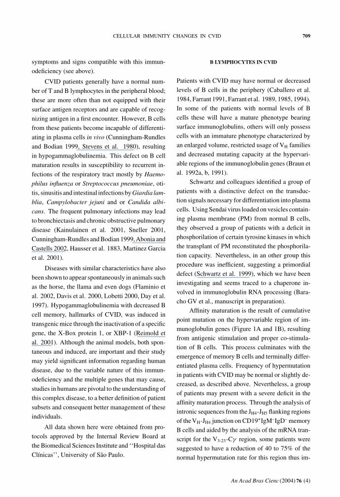
Affinity maturation is the result of cumulative
point mutation on the hypervariable region of im-
munoglobulin genes (Figure 1A and 1B), resulting
from antigenic stimulation and proper co-stimula-
tion of B cells. This process culminates with the
emergence of memory B cells and terminally differ-
entiated plasma cells. Frequency of hypermutation
in patients with CVID may be normal or slightly de-
creased, as described above. Nevertheless, a group
of patients may present with a severe deficit in the
affinity maturation process. Through the analysis of
intronic sequences from the JH4-JH5 flanking regions
of the VH-JH4 junction on CD19+IgM–IgD– memory
B cells and aided by the analysis of the mRNA tran-
script for the V3-23-Cγ region, some patients were
suggested to have a reduction of 40 to 75% of the
normal hypermutation rate for this region thus im-
An Acad Bras Cienc (2004) 76 (4)

710 CRISTINA M. KOKRON ET AL.
Figure 1A: B Figure 1A: B cell development and cell development and aa putative step involved putative step involved CVIDCVID developmentdevelopment
Adapted from Ballow, J Allergy Clin Immunol 2002
CVIDIgAD
Fig. 1A – Steps involved in the development of B cells and their maturation. The differentiation into antibody forming cells may
be compromised in CVID patients. The figure shows the immunoglobulin gene rearrangements involved in the process and the steps
necessary for antibody production and secretion, highlighting the changes that are antigen-independent and consequently autonomous
from T cell interaction, and those that are T cell dependent and may be affected in CVID patients.
pairing B cell differentiation, antibody synthesis and
memory development. The functional analysis of
T cells from these patients confirmed that the defect
was restricted to B lymphocytes (Levy et al. 1998),
which was confirmed independently (Bonhomme et
al. 2000).
The putative defect on B cell activation was
studied by Nonoyma and colleagues in 22 patients
with CVID by in vitro stimulation of patients’ B cells
in the presence of combinations of anti-CD40, IL-
4, IL-10 and in the presence or absence of CDw32L
cells. Four groups of patients were identified regard-
ing B cell response to these multiple stimuli; a group
incapable of producing IgM, IgA or IgG, a group that
synthesized IgM an IgG but not IgA, a third group
that produced IgM and low levels of IgA and IgG,
and a forth group that produced minute amounts of
IgM. The subsequent analysis of IgG subclasses in
the patients that synthesized this antibody isotype
revealed that IgG4 was most affected followed by
IgG2, whereas the synthesis of IgG3 and IgG1 was
normal. Similarly, in the patients capable of produc-
ing IgA, the secretion of IgA2 was more affected that
that of IgA1. These finding suggest an hierarchy in
the synthesis of isotypes and subclasses that seem to
correspond to the location of the heavy chain gene in
the human genome on chromosome 14 (Nonoyama
et al. 1993). The same phenomena was confirmed
by other studies but using bacterial polysaccharides
and cytokines as the stimulants for antibody produc-
An Acad Bras Cienc (2004) 76 (4)
CELLULAR IMMUNITY CHANGES IN CVID 711
Genes involved in B cell differentiation into plasma Genes involved in B cell differentiation into plasma cellscells
PrePre BB CellCell
PrePre TT cellcell
ILIL--7R7R--
PU.1PU.1highhigh
PU.1PU.1lowlow
MyeloidMyeloid progenitorprogenitor
ILIL--7R7R--
ILIL--7R+7R+
Notch 1Notch 1
E2AE2A
EBFEBF
PaxPax 55
PrePre NKNK cellcell
Id2Id2 switching switching
Bcl6Bcl6 BlimpBlimp--11
PaxPax 55
XBPXBP--11ATF6ATF6
IRE1IRE1
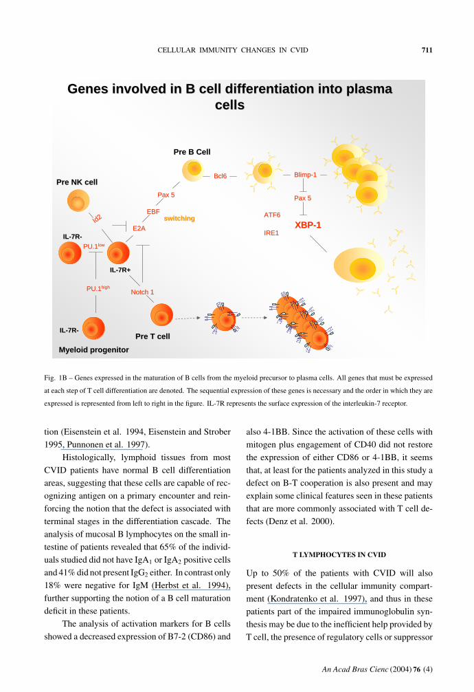
Fig. 1B – Genes expressed in the maturation of B cells from the myeloid precursor to plasma cells. All genes that must be expressed
at each step of T cell differentiation are denoted. The sequential expression of these genes is necessary and the order in which they are
expressed is represented from left to right in the figure. IL-7R represents the surface expression of the interleukin-7 receptor.
tion (Eisenstein et al. 1994, Eisenstein and Strober
1995, Punnonen et al. 1997).
Histologically, lymphoid tissues from most
CVID patients have normal B cell differentiation
areas, suggesting that these cells are capable of rec-
ognizing antigen on a primary encounter and rein-
forcing the notion that the defect is associated with
terminal stages in the differentiation cascade. The
analysis of mucosal B lymphocytes on the small in-
testine of patients revealed that 65% of the individ-
uals studied did not have IgA1 or IgA2 positive cells
and 41% did not present IgG2 either. In contrast only
18% were negative for IgM (Herbst et al. 1994),
further supporting the notion of a B cell maturation
deficit in these patients.
The analysis of activation markers for B cells
showed a decreased expression of B7-2 (CD86) and
also 4-1BB. Since the activation of these cells with
mitogen plus engagement of CD40 did not restore
the expression of either CD86 or 4-1BB, it seems
that, at least for the patients analyzed in this study a
defect on B-T cooperation is also present and may
explain some clinical features seen in these patients
that are more commonly associated with T cell de-
fects (Denz et al. 2000).
T LYMPHOCYTES IN CVID
Up to 50% of the patients with CVID will also
present defects in the cellular immunity compart-
ment (Kondratenko et al. 1997), and thus in these
patients part of the impaired immunoglobulin syn-
thesis may be due to the inefficient help provided by
T cell, the presence of regulatory cells or suppressor
An Acad Bras Cienc (2004) 76 (4)
712 CRISTINA M. KOKRON ET AL.
cells (Braun et al. 1991), the latter was associated
in the past with the inversion in the CD4:CD8 ratio
observed in the peripheral blood of some patients
(see bellow). Kamin and collaborators were the first
to identify a deficit in the proliferative response of
T lymphocytes to PHA in patients with ‘‘familiar
hypogammaglobulinemia’’, which was later named
CVID (Kamin et al. 1968). Therefore, many authors
have suggested that this disease is a combined im-
munodeficiency (Sneller 2001, Duarte et al. 1990).
Among the patients that we follow 56% of them
(40/71) present a decrease in the CD4/CD8 ratio
(< 1.0), due to increased CD8+ lymphocytes, di-
minished CD4+ lymphocytes or both. Furthermore,
from 34 patients who were submitted to delayed type
hypersensitivity skin test, 21 were non-responsive.
There are many reports, either as series of pa-
tients or as isolated case reports that support the
notion that some patients with CVID have a cel-
lular defect associated with the hypogammaglob-
ulinemia. These studies, describe infections that
are commonly associated with T cell deficits and
vary from severe infection with viruses such as the
cytomegalovirus (CMV), or herpes zoster (CMV)
(Kainulainen et al. 1999, Linde et al 1988, Mul-
lighan et al. 1996, Tarr et al. 2001, Witte et al.
2000), and also include infection with intracellu-
lar parasites like the Toxoplasma gondii or Strongy-
loides stercorallis (Duarte et al. 1990, Naspitz et al.
1987, Mushiake et al. 1993, Bonilla et al. 1997).
The TCR/CD3 complex is fundamental for the
antigen recognition and activation of T cell. It is as-
sociated with two families of tyrosine kinases Src
and Syk. Src family kinases will phosphorylate
the Immune Receptor Tyrosine Associated Motif
(ITAM), for instance on the cytoplasmatic domain
of the CD3/ξ chain. Following this phosphoryla-
tion, the enzyme ZAP-70 is recruited and further
phosphorylate the ξ and ε CD3 chains. Although the
analysis of the nucleotide sequence of the ITAMs on
the CD3/ξ or on the ZAP-70 enzyme did not show
any differences when compared to normal individu-
als, a defect in the interaction of ZAP-70 with p23ξ
leading to defective activation of T cells through the
TRC was described (Boncristiano et al. 2000). In
contrast, Majolini and colleagues were unable to re-
produce these results, although, they recognized a
defect in the phosphorylation cascade that follows
antigen recognition by the TCR in CVID patients
(Majolini et al. 1997).
Farrington and colleagues reported a decreased
expression of CD40L mRNA on activated T cells
from patients as compared to controls, although the
study did not specify the clinical status of the pa-
tients which could potentially interfere with the ex-
pression of this molecule, the authors proposed that
this marker should be used to classify the patients
based on the presence or absence of a T lymphocyte
defect (Farrington et al. 1994).
Cytokines play an essential role on antibody
synthesis and the regulation of the immune response
in general. Reports on the importance of cytokines
for the development of CVID have been presented
briefly above and will be discussed in depth here.
North and collaborators and Cambronero and
collaborators observed that IL-12 synthesis in vitro
by LPS-stimulated monocytes from the peripheral
blood was increased in CVID patients. In turn,
this could be responsible for the increase in the fre-
quency of IFN-γ producing T cells, as defined by
intracellular staining for this cytokine. The authors
suggest that this changes would represent a shift to
a polarized Th1 response in these patients which
would account for the decreased antibody produc-
tion and some of the granulomatous features of the
disease in some individuals affected (North et al.
1996, Cambronero et al. 2000). Although inter-
esting as a concept and even useful to classify yet
another subtype of CVID patients, it is important
to note that some other studies have failed to show
the same increase in IFN-γ producing T cells, some
have reported normal levels of this cytokines, while
others have reported diminished levels (Matricardi
et al. 1984, Paganelli et al. 1988, Pastorelli et al.
1989a).
IL-10 is another cytokine whose importance
in CVID has been underlined. Lymphocytes from
CVID patients have been reported to secrete
An Acad Bras Cienc (2004) 76 (4)
CELLULAR IMMUNITY CHANGES IN CVID 713
less IL-10 when activated than normal lymphocytes
(Aukrust et al. 2000, Holm et al. 2003). In con-
trast, monocyte-derived IL-10 has been implicated
in the diminished production of IL-2 by some pa-
tients and as a contributor to the proliferative deficit
observed in a subset of individuals with CVID (Zhou
et al. 1998). Interestingly, the effect of IL-10 on IL-
2 synthesis by T lymphocytes from CVID patients
and its effect on antibody synthesis in vitro was only
observed in the presence of IL-4 and anti-CD40 anti-
body but not in the absence of IL-4 (Punnonen et al.
1997). It is noteworthy that defects on IL-2 produc-
tion have also been described independently from
the interaction with other cytokines (Fischer et al.
1993, Nonoyama et al. 1994).
Changes in the number and function of CD8+
T lymphocytes have been described in CVID pa-
tients for a long time. Enhanced numbers of these
cells, followed or not by a decrease on CD4+ T cells,
leading to an inversion of the CD4+/CD8+ are char-
acteristic of a subset of CVID patients with severe
compromise of the cellular immunity (Jaffe et al.
1993a, b). Changes in the expression of CD40L and
other activation molecules expressed in the surface
of T cells have also been reported, in association to
changes in cytokine synthesis (North et al. 1998),
as described above, or independently (Errante PR et
al. manuscript in preparation).
The antigen-specific T cell response has also
been reported as deficient in some patients with
CVID. The generation of memory cells to several
antigens seems compromised, for example, memory
cells for purified protein derivative (PPD), tetanus
toxoid (TT), keyhole limpet hemocyanin (KLH) and
a peptide derive from the env gene on HIV have
all being reported as lacking in exposed individuals
with CVID (Kondratenko et al. 1997, Funauchi et
al. 1995, Stagg et al. 1993, 1994). In addition,
mRNA for IL-12 (Farrington et al. 1994), IL-2, IL-
4 (Pastorelli et al. 1989a, b, 1990), IFN-γ (Paganelli
et al. 1988), IL-5 and IFN-γ (Sneller and Strober
1990a, b) has also been reported as decreased in
response to specific antigenic stimulation in PBMCs
from CVID patients.
CLINICAL CHARACTERISTICS OF THEPATIENTS WITH CVID
Immunodeficiency in CVID patients is often un-
apparent during the first years of life. Although
patients less than three years old have been diag-
nosed correctly with CVID, diagnosis during early
childhood is not the norm and the disease is most
commonly diagnosed in teenagers (Cunningham-
Rundles and Bodian 1999, Abonia and Castells
2002, Piqueras et al. 2003), with the peak in diag-
nosis occurring between the ages of 13 and 30 years
old (Tiller and Liddle 2000, Cunningham-Rundles
and Bodian 1999, Aghamohammadi et al. 2002,
Pfluger et al. 2000, Strober and Chua 2000). In-
terestingly, immunodeficiency symptoms may not
be manifested until much later, and many cases of
diagnosis after 65 years of age are described, in-
cluding a case with the primary diagnosis made in a
88-year-old patient (Hawkins et al. 2003). Because
most physicians are unaware of this condition it is
often misdiagnosed, frequently confused withAIDS
(Kaczmarski et al. 1994), severe asthma or autoim-
mune diseases (Fontan Casariego 2001, Rusconi et
al. 2003, Kart et al. 2003, Heeney et al. 2003, De
Vera and Yu 2003, Byrne et al. 2003).
CVID has no gender preference, although
women tend to manifest the disease later in life, after
the diagnosis the survival rate is similar for both
sexes (Cunningham-Rundles 2001, Cunningham-
Rundles and Bodian 1999, Martinez Garcia et al.
2001, Pfluger et al. 2000, Strober and Chua 2000,
Gottesman et al. 1999, Manson et al. 2000).
Between 1980 and 2003, our group followed at
the Division of Clinical Immunology and Allergy of
University of São Paulo Medical School, a reference
service for adult patients, 71 patients with CVID.
Patients followed during this period had a similar
distribution of gender (38 males and 33 females), in
agreement with the medical literature cited above.
The follow-up period for our patients varied from
2 months to 23 years (mean number of years of
follow-up = 4.5). The age range of patients currently
under our care is between 15 and 78 years. Most
An Acad Bras Cienc (2004) 76 (4)
714 CRISTINA M. KOKRON ET AL.
0
20
40
60
80
100
Recurrent infections Autoimmunity Allergy Malignancies
Perc
enta
ge o
f pat
ients
affe
cted
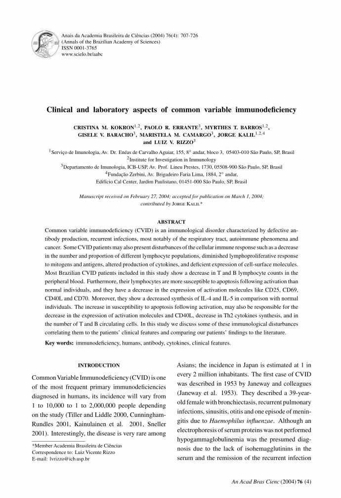
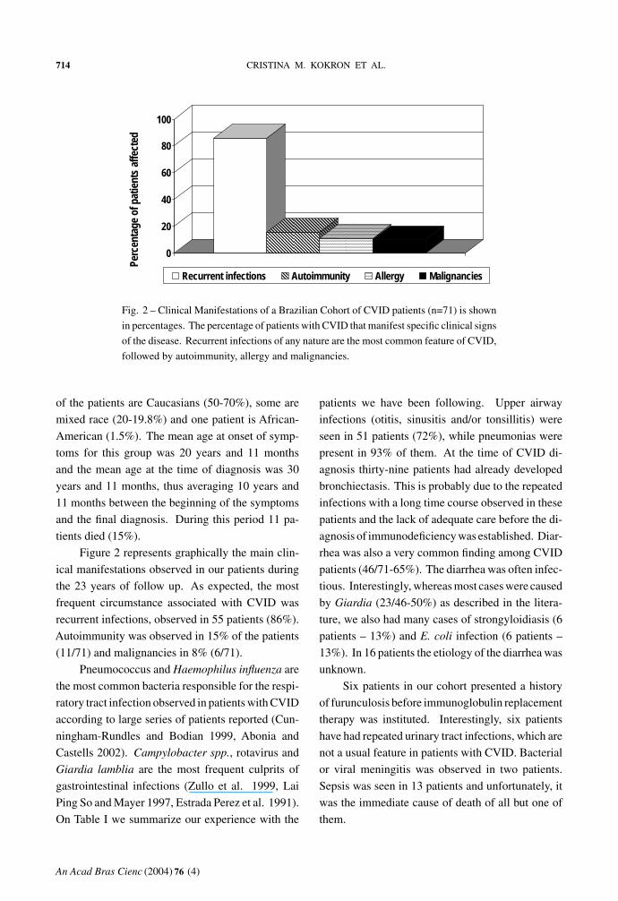
Fig. 2 – Clinical Manifestations of a Brazilian Cohort of CVID patients (n=71) is shown
in percentages. The percentage of patients with CVID that manifest specific clinical signs
of the disease. Recurrent infections of any nature are the most common feature of CVID,
followed by autoimmunity, allergy and malignancies.
of the patients are Caucasians (50-70%), some are
mixed race (20-19.8%) and one patient is African-
American (1.5%). The mean age at onset of symp-
toms for this group was 20 years and 11 months
and the mean age at the time of diagnosis was 30
years and 11 months, thus averaging 10 years and
11 months between the beginning of the symptoms
and the final diagnosis. During this period 11 pa-
tients died (15%).
Figure 2 represents graphically the main clin-
ical manifestations observed in our patients during
the 23 years of follow up. As expected, the most
frequent circumstance associated with CVID was
recurrent infections, observed in 55 patients (86%).
Autoimmunity was observed in 15% of the patients
(11/71) and malignancies in 8% (6/71).
Pneumococcus and Haemophilus influenza are
the most common bacteria responsible for the respi-
ratory tract infection observed in patients with CVID
according to large series of patients reported (Cun-
ningham-Rundles and Bodian 1999, Abonia and
Castells 2002). Campylobacter spp., rotavirus and
Giardia lamblia are the most frequent culprits of
gastrointestinal infections (Zullo et al. 1999, Lai
Ping So and Mayer 1997, Estrada Perez et al. 1991).
On Table I we summarize our experience with the
patients we have been following. Upper airway
infections (otitis, sinusitis and/or tonsillitis) were
seen in 51 patients (72%), while pneumonias were
present in 93% of them. At the time of CVID di-
agnosis thirty-nine patients had already developed
bronchiectasis. This is probably due to the repeated
infections with a long time course observed in these
patients and the lack of adequate care before the di-
agnosis of immunodeficiency was established. Diar-
rhea was also a very common finding among CVID
patients (46/71-65%). The diarrhea was often infec-
tious. Interestingly, whereas most cases were caused
by Giardia (23/46-50%) as described in the litera-
ture, we also had many cases of strongyloidiasis (6
patients – 13%) and E. coli infection (6 patients –
13%). In 16 patients the etiology of the diarrhea was
unknown.
Six patients in our cohort presented a history
of furunculosis before immunoglobulin replacement
therapy was instituted. Interestingly, six patients
have had repeated urinary tract infections, which are
not a usual feature in patients with CVID. Bacterial
or viral meningitis was observed in two patients.
Sepsis was seen in 13 patients and unfortunately, it
was the immediate cause of death of all but one of
them.
An Acad Bras Cienc (2004) 76 (4)

CELLULAR IMMUNITY CHANGES IN CVID 715
TABLE I
Most common infectious diseases in a cohort of Brazilian patients with CVID.
Number of patients %
Recurrent tonsillitis, sinusitis, otitis 51 72
Pneumonia 66 93
Infectious diarrhea
Giardiasis 23 32
Strongyloidiasis 6 8
E. coli 6 8
Non-identified agent 16 22
Bacterial skin infections 6 8
Urinary infections 10 14
Meningitis 3 4
Sepsis 13 18
Arthritis 4 6
Opportunistic infections
Candidiasis 4 6
Herpes I/II 2 3
Herpes Zoster 4 6
HPV 1 1.5
Toxoplasmosis
Ocular 1 1.5
Neurotoxoplasmosis 1 1.5
CMV - 1 1.5
Cryptococcosis 1 1.5
Tuberculosis 5 7
Other
Schistosomiasis 1 1.5
Paracoccidioidomycosis 1 1.5
Opportunistic infections like herpes zoster,
genital HPV, tuberculosis, candidiasis, toxoplasmo-
sis, cryptococcosis and CMV were observed in 13
patients – 18% – (in some patients the episodes were
repetitive and there were more than one type of op-
portunistic infection). Five patients reported tuber-
culosis (7%) prior to the diagnosis of CVID, but dis-
ease was only confirmed in one of the patients and
it is possible that severe pneumonia was confused
for tuberculosis because of the difficulties in treat-
ing an immunodeficient patient with conventional
antibiotic therapy. Fungal infections by Cryptococ-
cus neoformans and protozoan infections by Pneu-
mocystis carinni, Toxoplasma gondii e Cryptospori-
dium spp (Tzipori 1988, Esolen et al. 1992, Macura
et al. 2003), have been occasionally described in
these patients. Viral infections are also common and
Herpes, Epstein-Barr and CMV are among the most
frequently reported (Cunningham-Rundles and Bo-
dian 1999, Webster 2001). Another virus reported
as chronically affecting CVID patients is the par-
vovirus B19, leading to aplastic anemia (Chuhjo et
An Acad Bras Cienc (2004) 76 (4)
716 CRISTINA M. KOKRON ET AL.
al. 1999).
Approximately a third of the patients with
CVID will develop some autoimmune phenomena,
including hemolytic anemia, purpura, autoimmune
hepatitis, thyroiditis, diabetes, dermatomiositis and
Graves disease, among others (Heeney et al. 2003,
Andon Saavedra et al. 1996, Cunningham-Rundles
2002, Lee et al. 1993). In our series of patients
autoimmune disease was observed in 11 individuals
(15,5%). Contrary to what is described in the liter-
ature, most of them males (n=8). Furthermore, the
autoimmune disorder that is described most often
among the CVID patients is Idiopathic Thrombocy-
topenic Purpura, which was not observed among our
patients.
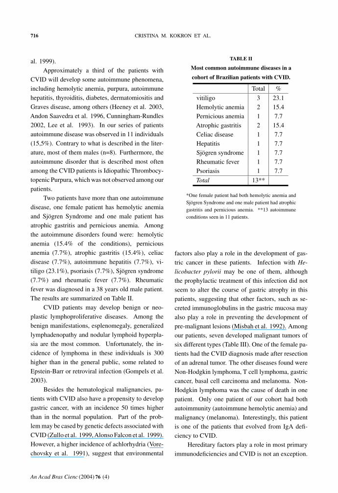
Two patients have more than one autoimmune
disease, one female patient has hemolytic anemia
and Sjögren Syndrome and one male patient has
atrophic gastritis and pernicious anemia. Among
the autoimmune disorders found were: hemolytic
anemia (15.4% of the conditions), pernicious
anemia (7.7%), atrophic gastritis (15.4%), celiac
disease (7.7%), autoimmune hepatitis (7.7%), vi-
tiligo (23.1%), psoriasis (7.7%), Sjögren syndrome
(7.7%) and rheumatic fever (7.7%). Rheumatic
fever was diagnosed in a 38 years old male patient.
The results are summarized on Table II.
CVID patients may develop benign or neo-
plastic lymphoproliferative diseases. Among the
benign manifestations, esplenomegaly, generalized
lymphadenopathy and nodular lymphoid hyperpla-
sia are the most common. Unfortunately, the in-
cidence of lymphoma in these individuals is 300
higher than in the general public, some related to
Epstein-Barr or retroviral infection (Gompels et al.
2003).
Besides the hematological malignancies, pa-
tients with CVID also have a propensity to develop
gastric cancer, with an incidence 50 times higher
than in the normal population. Part of the prob-
lem may be cased by genetic defects associated with
CVID (Zullo et al. 1999,Alonso Falcon et al. 1999).
However, a higher incidence of achlorhydria (Vore-
chovsky et al. 1991), suggest that environmental
TABLE II
Most common autoimmune diseases in a
cohort of Brazilian patients with CVID.
Total %
vitiligo 3 23.1
Hemolytic anemia 2 15.4
Pernicious anemia 1 7.7
Atrophic gastritis 2 15.4
Celiac disease 1 7.7
Hepatitis 1 7.7
Sjögren syndrome 1 7.7
Rheumatic fever 1 7.7
Psoriasis 1 7.7
Total 13**
*One female patient had both hemolytic anemia andSjögren Syndrome and one male patient had atrophicgastritis and pernicious anemia. **13 autoimmuneconditions seen in 11 patients.
factors also play a role in the development of gas-
tric cancer in these patients. Infection with He-
licobacter pylorii may be one of them, although
the prophylactic treatment of this infection did not
seem to alter the course of gastric atrophy in this
patients, suggesting that other factors, such as se-
creted immunoglobulins in the gastric mucosa may
also play a role in preventing the development of
pre-malignant lesions (Misbah et al. 1992). Among
our patients, seven developed malignant tumors of
six different types (Table III). One of the female pa-
tients had the CVID diagnosis made after resection
of an adrenal tumor. The other diseases found were
Non-Hodgkin lymphoma, T cell lymphoma, gastric
cancer, basal cell carcinoma and melanoma. Non-
Hodgkin lymphoma was the cause of death in one
patient. Only one patient of our cohort had both
autoimmunity (autoimmune hemolytic anemia) and
malignancy (melanoma). Interestingly, this patient
is one of the patients that evolved from IgA defi-
ciency to CVID.
Hereditary factors play a role in most primary
immunodeficiencies and CVID is not an exception.
An Acad Bras Cienc (2004) 76 (4)

CELLULAR IMMUNITY CHANGES IN CVID 717
TABLE III
Neoplasias developed in a cohort of Brazilian
patients with CVID.
Total (n=71) %
Non-Hodgkin lymphoma 1 1.5
T cell lymphoma 2 3
Gastric adenocarcinoma 1 1.5
Basal cell carcinoma 1 1.5
Melanoma 1 1.5
Adrenal tumor 1 1.5
Total 7 (10%)
However, because of the polymorphic nature of this
disease it has been hard to assign a specific pattern
of transmission of disease, although the association
with IgA deficiency has been better characterized
in this aspect. Nevertheless, there are many re-
ports of individuals who develop CVID and no spe-
cific hereditary association is found (Webster 2001).
Specific genetic defects will be discussed bellow. In
the cohort we are currently following, immunode-
ficiency diseases have been diagnosed in the fam-
ilies of nine patients. Six relatives with IgA defi-
ciency were found, low IgG levels (2 persons) and
CVID (5 persons). One of the female patients had a
daughter with IgA deficiency, a son with transitory
hypogammaglobulinemia and a cousin with CVID.
Another female patient had a son with low IgG lev-
els and a niece with IgA deficiency. A third female
patient, who has Sjögren syndrome beside CVID,
has two children (a son and a daughter) with IgA
deficiency and SLE. These patients’ families also
reported cases of autoimmunity and cancer. Au-
toimmunity was seen in the families of 7 patients
(inflammatory bowel disease; thyroiditis; diabetes;
SLE and scleroderma).
The median follow-up time for these patients
was 4.5 years (range 1 month – 28.3 years, some
of them were being treated elsewhere before). We
observed 11 deaths (15.5%), four females and seven
males. This difference was not statistically signif-
icant. The main death causes were: sepsis, in 7
patients with bronchiectasis, one patient with lym-
phoma, one patient with malabsorption syndrome
and one patient with neurocryptococcosis; and prob-
able P. carinii infection in one patient with acute
respiratory insufficiency.
Treatment of CVID
The basis for the treatment of patients with CVID is
the timely replacement of serum gammaglobulins.
Initially, replacement was performed by the subcu-
taneous or intramuscular injection of pooled gam-
maglobulin from blood donors. For the past twenty
years, the availability of gammaglobulins for intra-
venous injection (IVIG) has greatly improved the
outcome of these patients. Ordinarily, IVIG is used
once or twice a month with an attack dose of IVIG at
600 mg/kg of body weight and follow up injections
aiming at keeping the blood levels of immunoglob-
ulins above 400 mg/dL. Although other immune
deficits associated with CVID, e.g. immunoglob-
ulins are absent in blood fluids like the saliva and
gastric juice, the increase in serum antibody has
proven effective in preventing pulmonary infections,
sepsis and meningitis (Cunningham-Rundles 2001,
Cunningham-Rundles and Bodian 1999). It is note-
worthy that the administration of large doses of im-
munoglobulins intravenously has very well reported
immunomodulatory effects that are frequently used
in the treatment of autoimmune diseases. These im-
munomodulatory properties may contribute to the
overall effect IVIG has on CVID patients. It was
shown that it may cause an increase in the expression
of IL-2 by CD4+ and more importantly CD4+28–. It
can also increase the expression of TNF-α in these
same cells (Sewell et al. 1999, 1998).
Adjuvant therapy has been tried in CVID in an
attempt to improve the results obtained with IVIG,
especially in view of the poor effect IVIG has on pre-
venting cancer in these patients. Recombinant IL-2,
pure or polymerized with poly-ethylene-glycol, is
the most commonly use adjuvant therapy (Cunnin-
gham-Rundles et al. 1995, 2001, Rump et al. 1997).
Although the results on preventing malignancies are
unclear, the addition of IL-2 decreases the incidence
An Acad Bras Cienc (2004) 76 (4)
718 CRISTINA M. KOKRON ET AL.
of severe disease and it increases the overall level
of serum IgG, which may be due to direct stimu-
lation of the patient’s own B cells to secrete anti-
bodies. Less conventional therapies such as cimeti-
dine, which was thought to enhance IgA secretion,
bacterial products and vitamins have been tried spo-
radically (Litzman et al. 1996a, Segal et al. 1989,
Saxon et al. 1993, Della Bella et al. 1997).
In our group, patients diagnosed before 1994
(10-15.6%) were treated with plasma or intramus-
cular (IM) immunoglobulin. Following the avail-
ability of IVIG at the Hospital das Clínicas da Fac-
uldade de Medicina da Universidade de São Paulo,
starting in 1995 all patients have received IVIG as
described above. Three patients from the cohort we
describe here died before IVIG treatment was es-
tablished. Two patients were diagnosed elsewhere
and received IM immunoglobulin irregularly up to
the time (1.5 and 10 y) they were referred to our
service.
It is clear that the establishment of immunolog-
ical criteria that allow the early detection of CVID
and consequently the implementation of therapy will
contribute to prolonging the life of patients with this
disease and enhance their quality of life. Large se-
ries of studies as described above and our own expe-
rience with the 71 cases described here show that the
patients with earlier diagnosis and treatment survive
longer and with less sequellae than those who are di-
agnosed later, especially if a diagnosis is made after
pulmonary complications have been established.
GENETIC DEFFECTS ASSOCIATED WITH CVIDAND THE RELATIONSHIP WITH TOTAL IgA
DEFICIENCY (IgAD)
The characterization of specific genetic defects lead-
ing to failures in the immune response that charac-
terize CVID is pivotal for the understanding of the
different subsets of patients with this disease and to
comprehend the differential evolution of each pa-
tients. Furthermore, as these defects are identified
and correlated with specific symptoms one can better
understand the effector and regulatory mechanism of
the human immune system.
The inducible co-stimulatory protein (ICOS) is
a member of the CD28/CTLA-4 family and is ex-
pressed specifically by T lymphocytes, interacting
with its ligand B7H on the surface of B cells and
macrophages. This interaction will increase the ex-
pression of CD40L and the proliferation of T lym-
phocytes. ICOS also possess antiapoptotic prop-
erties and induces the secretion of many cytokines
that act on B cells (Grimbacher et al. 2003a, Sporici
and Perrin 2001). Recently, Grimbacher and col-
leagues have described an autossomic recessive dis-
order in 4 patients with CVID out of 32 studied.
These patients came from two families with a pos-
sible common ancestor (Grimbacher et al. 2003b).
The patients had a homozygous partial deletion of
the ICOS gene. Laboratory analysis of these indi-
viduals revealed, besides hypogammaglobulinemia,
B cell lymphopenia, normal T cell response to PHA,
PWM, mAb-anti-CD3 and TT with proliferation and
synthesis of TNF-α, IFN-γ , IL-2, IL-4, IL-10 and
IL-13. Interestingly, B cells from these individuals
stimulated in the presence of T CD4+, IL-2 and SAC
were capable of secreting all isotypes of antibodies
in vitro. It is noteworthy that the IgG subclasses
were not studied. We were unable to detect this or
any mutation in the ICOS gene in any of the 38 pa-
tients studied, out of the 56 we currently follow.
A few patients with IgAD may develop CVID
over time. Furthermore, the frequency of relatives
from CVID patients with low levels of IgA is much
higher than what expected in the general popula-
tion (Carvalho Neves Forte et al. 2000, Cham et al.
2002). This has led to the hypothesis that IgAD and
some cases of CVID represent two different poles
of a spectrum of antibody deficiency (Ashman et al.
1992, Litzman et al. 1996b). Although the precise
molecular defect that causes IgAD is still unrecog-
nized, in many patients with CVID or IgAD associ-
ation with specific regions inside the MHC loci have
been described.
An association with the MHC III region on
chromosome 6 has been described by many authors.
A high frequency of null alleles for the C4a com-
ponent of the Complement systems was observed in
An Acad Bras Cienc (2004) 76 (4)
CELLULAR IMMUNITY CHANGES IN CVID 719
patients with IgAD. In a study including CVID and
IgAD patients it was observed that 12 out of 19 pa-
tients with CVID (63%, p < 0, 01) and nine out of
sixteen patients with IgAD (56%, p>0, 01) showed
rare alleles for C2 and or a deletion of the genes
that encodes 21-OHase A. In another study, nine out
of eleven patients with a deletion of C4a also car-
ried HLA-A1, -B8, -C4AQ0, -C4B1, -BFS e -DR3
haplotypes, all previously associated with IgAD and
or CVID (Schaffer et al. 1989, Schroeder et al.
1998, Schroeder 2000). The putative gene for IgAD
was then named IGAD1. A genome-wide genetic
linkage analysis to study IGAD1 in a large num-
ber of IgAD/CVID patients implicated the HLA-
DQ/DR as a major susceptibility locus (Kralovicova
et al. 2003).
Other studies have suggested that certain genes
in the MHC III region are on linkage disequilibrium
with alleles from the MHC I and or II (De La Con-
cha et al. 1999), registering an association with the
HLA-DR3, -B8, -A1 haplotypes. In these regions
the susceptibility types for CVID and IgA would
be related to the HLA-D821/D823 e HLA-B8 hap-
lotypes, localized at the G1/AIF1 and HLA-B re-
gions, respectively (Schroeder et al. 1998, Schroeder
2000). Another studied suggested that the associa-
tion with CVID and IgAD was in the loss of spe-
cific genes on the region 6p21.3 (Vorechovsky et al.
2000, 2001).
The findings described above suggest that a
common defect for all patients with CVID is un-
likely, nevertheless groups of patients with similar
defects can be found and thus they must be typed
and studied accordingly.
CONCLUDING REMARKS
CVID is a complex disease very likely caused by
multiple different gene defects resulting in similar
but not identical clinical signs. Besides the devas-
tating effect that it has on its carriers and the conse-
quent medical quagmire it represents, CVID offers
us the opportunity to understand the functioning of
the immune system and thus grant us with unique
tools to learn how to modulate it, not only to im-
prove the quality of life of patients with CVID but
also patients with other immune-mediated diseases.
CVID is the most frequent primary immunod-
eficiency followed by clinical immunologists and
it should be recognized by the physicians as soon
as possible in order to avoid the complications de-
termined by the delay in the diagnosis and conse-
quently a better life quality.
ACKNOWLEDGMENTS
This work was supported by grants from Fundação
de Amparo a Pesquisa do Estado de São Paulo
(FAPESP), Conselho Nacional de Desenvolvimento
Científico e Tecnológico (CNPq), Ministério da
Ciência e Tecnologia (MCT) through the Institu-
tos do Milênio Program. GVB is a recipient of a
PhD scholarship from FAPESP. LVR is a recipient
of a personal grant for scientific achievement from
CNPq.
The authors thank the patients with CVID and
their families for their trust and cooperation. They
also thank the staff of the Hospital das Clínicas da
Universidade de São Paulo for the excellent care
they provide to the patients.
RESUMO
A imunodeficiência comum variável (CVID) é uma doen-
ça caracterizada por hipogamaglobulinemia, infecções
recorrentes, especialmente das vias aéreas, enfermidades
auto-imunes e neoplasias. Alguns pacientes com CVID
possuem diversos distúrbios do sistema imune como al-
terações no número e proporção de diferentes populações
leucocitárias; resposta proliferativa linfocitária diminuída
para antígenos e mitógenos; produção alterada de citoci-
nas e alteração na expressão de moléculas de superfí-
cie. Neste trabalho, são discutidas várias destas altera-
ções imunológicas procurando correlacioná-las aos acha-
dos clínicos dos pacientes e incorporar aos dados da litera-
tura os nossos próprios achados.
Palavras-chave: imunodeficiência, humanos, anticor-
pos, citocinas, dados clínicos.
An Acad Bras Cienc (2004) 76 (4)
720 CRISTINA M. KOKRON ET AL.
REFERENCES
Abonia JP and Castells MC. 2002. Common variable
immunodeficiency. Allergy Asthma Proc 23: 53–57.
Aghamohammadi A, Moein M, Farhoudi A, Pour-
pak Z, Rezaei N, Abolmaali K, Movahedi M,
Gharagozlou M, Ghazi BM, Mahmoudi M,
Mansouri D, Arshi S, Trash NJ, Akbari H, Sher-
kat R, Hosayni RF, Hashemzadeh A, Moham-
madzadeh I, Amin R, Kashef S, Alborzi A,
Karimi A and Khazaei H. 2002. Primary immun-
odeficiency in Iran: first report of the National Reg-
istry of PID in Children and Adults. J Clin Immunol
22: 375–380.
Aghamohammadi A, Kanegane H, Moein M,
Farhoudi A, Pourpak Z, Movahedi M, Gharago-
zlou M, Zargar AA and Miyawaki T. 2003. Iden-
tification of an SH2D1A mutation in a hypogamma-
globulinemic male patient with a diagnosis of com-
mon variable immunodeficiency. Int J Hematol 78:
45–47.
Akai M, Ishizaki T, Sasaki F, Ameshima S, Shige-
mori K, Higashi T and Nakai T. 1996. Immunod-
eficiency with thymoma (Good’s syndrome) similar
to sino-bronchial syndrome. Nihon Kyobu Shikkan
Gakkai Zasshi 34: 829–832.
Alonso Falcon F, Codoceo Alquinta R, Polanco
Allue I, Aguado Gil A and Fontancasariego
G. 1999. Study of gastrointestinal polypeptides con-
trolling gastric acid secretion in patients with primary
antibody deficiency. Rev Esp Enferm Dig 91: 54–60.
Andon Saavedra C, Deben Ariznavarreta G,
Batlle Fonrodona J, Ramirez Cereceda C and
Saavedra A. 1996. Autoimmune hemolytic ane-
mia in common variable immunodeficiency. Sangre
(Barc) 41: 404–405.
Ashman RF, Schaffer FM, Kemp JD, Yokoyama WM,
Zhu ZB, Cooper MD and Volanakis JE. 1992. Ge-
netic and immunologic analysis of a family contain-
ing five patients with common-variable immune de-
ficiency or selective IgA deficiency. J Clin Immunol
12: 406–414.
Aukrust P, Muller F, Ueland T, Svardal AM, Berge
RK and Froland SS. 2000. Decreased vitamin A
levels in common variable immunodeficiency: vi-
tamin A supplementation in vivo enhances immuno-
globulin production and downregulates inflammatory
responses. Eur J Clin Invest 30: 252–259.
Belmonte E, Yebra M, Marazuela M and Merino J.
1989. Common variable immunodeficiency. Report
of a case simulating lymphoma. Med Clin (Barc) 92:
597.
Boncristiano M, Majolini MB, D’Elios MM, Pacini
S, Valensin S, Ulivieri C, Amedei A, Falini B,
Del Prete G, Telford JL and Baldari CT. 2000.
Defective recruitment and activation of ZAP-70 in
common variable immunodeficiency patients with T
cell defects. Eur J Immunol 30: 2632–2638.
Bonhomme D, Hammarstrom L, Webster D, Chapel
H, Hermine O, Le Deist F, Lepage E, Romeo PH
and Levy Y. 2000. Impaired antibody affinity matu-
ration process characterizes a subset of patients with
common variable immunodeficiency. J Immunol
165: 4725–4730.
Bonilla HF, Chenoweth CE, Tully JG, Blythe LK,
Robertson JA, Ognenovski VM and Kauffman
CA. 1997. Mycoplasma felis septic arthritis in a pa-
tient with hypogammaglobulinemia. Clin Infect Dis
24: 222–225.
Braun J, Galbraith L, Valles-Ayoub Y and Saxon
A. 1991. Human immunodeficiency resulting from
a maturational arrest of germinal center B cells. Im-
munol Lett 27: 205–208.
Braun J, Berberian L, King L, Sanz I and Govan
HL 3rd. 1992a. Restricted use of fetal VH3 im-
munoglobulin genes by unselected B cells in the
adult. Predominance of 56p1-like VH genes in com-
mon variable immunodeficiency. J Clin Invest 89:
1395–1402.
Braun J, Saxon A, Wall R and Morrison SL. 1992b.
The second century of the antibody. Molecular per-
spectives in regulation, pathophysiology, and thera-
peutic applications. West J Med 157: 158–168.
Byrne MF, Royston D and Patchett SE. 2003. Asso-
ciation of common variable immunodeficiency with
atypical collagenous colitis. Eur J Gastroenterol
Hepatol 15: 1051–1053.
Caballero FM, Brown WR, Kohler PF and Hay-
ward AR. 1984. B cell numbers and responses in pa-
tients with common variable immunodeficiency and
nodular lymphoid hyperplasia of the bowel. J Clin
Lab Immunol 13: 59–63.
Cambronero R, Sewell WA, North ME, Webster
AD and Farrant J. 2000. Up-regulation of IL-12
An Acad Bras Cienc (2004) 76 (4)
CELLULAR IMMUNITY CHANGES IN CVID 721
in monocytes: a fundamental defect in common vari-
able immunodeficiency. J Immunol 164: 488–494.
Carvalho Neves Forte W, Ferreira de Carvalho
Junior F, Damaceno N, Vidal Perez F, Gonzales
Lopes C and Mastroti RA. 2000. Evolution of IgA
deficiency to IgG subclass deficiency and common
variable immunodeficiency. Allergol Immunopathol
(Madr) 28: 18–20.
Cham B, Bonilla MA and Winkelstein J. 2002. Neu-
tropenia associated with primary immunodeficiency
syndromes. Semin Hematol 39: 107–112.
Chuhjo T, Nakao S and Matsuda T. 1999. Success-
ful treatment of persistent erythroid aplasia caused
by parvovirus B19 infection in a patient with com-
mon variable immunodeficiency with low-dose im-
munoglobulin. Am J Hematol 60: 222–224.
Cooper MD, Lawton AR and Bockman DE. 1971.
Agammaglobulinaemia with B lymphocytes. Spe-
cific defect of plasma-cell differentiation. Lancet 2:
791–794.
Cunningham-Rundles C. 2001. Common variable
immunodeficiency. Curr Allergy Asthma Rep 1:
421–429.
Cunningham-Rundles C. 2002. Hematologic compli-
cations of primary immune deficiencies. Blood Rev
16: 61–64.
Cunningham-Rundles C and Bodian C. 1999. Com-
mon variable immunodeficiency: clinical and im-
munological features of 248 patients. Clin Immunol
92: 34–48.
Cunningham-Rundles C, Kazbay K, Zhou Z and
Mayer L. 1995. Immunologic effects of low-dose
polyethylene glycol-conjugated recombinant human
interleukin-2 in common variable immunodeficiency.
J Interferon Cytokine Res 15: 269–276.
Cunningham-Rundles C, Bodian C, Ochs HD, Mar-
tin S, Reiter-Wong M and Zhuo Z. 2001. Long-
term low-dose IL-2 enhances immune function in
common variable immunodeficiency. Clin Immunol
100: 181–190.
D’Addio F, Giunta R, Scarfiglieri D, De Fanis U,
Dalla Mora L, Pezone L, Bresciano E, Man-
cino D and Lucivero G. 2001. Late onset immun-
odeficiency with hypo-IgG and hyper-IgM, T CD4+
lymphocytopenia and vitiligo. Recenti Prog Med 92:
392–394.
Davis WC, Heirman LR, Hamilton MJ, Parish SM,
Barrington GM, Loftis A and Rogers M. 2000.
Flow cytometric analysis of an immunodeficiency
disorder affecting juvenile llamas. Vet Immunol Im-
munopathol 74: 103–120.
Day MJ, Power C, Oleshko J and Rose M. 1997.
Low serum immunoglobulin concentrations in re-
lated Weimaraner dogs. J Small Anim Pract
38: 311–315.
De La Concha EG, Fernandez-Arquero M, Mar-
tinez A, Vidal F, Vigil P, Conejero L, Garcia-
Rodriguez MC and Fontan G. 1999. HLA class II
homozygosity confers susceptibility to common vari-
able immunodeficiency (CVID). Clin Exp Immunol
116: 516–520.
De Vera M and Yu BH. 2003. Recurrent staphylococ-
cal infections and chronic dermatitis in a 45-year-old
man. Ann Allergy Asthma Immunol 91: 244–250.
Della Bella S, Vanoli M, Bazzi S and Scorza R.
1997. Successful treatment of common variable im-
munodeficiency and related disorders with cimeti-
dine and zinc sulfate. Int J Clin Lab Res 27: 79–80.
Denz A, Eibel H, Illges H, Kienzle G, Schlesier
M and Peter HH. 2000. Impaired up-regulation
of CD86 in B cells of ‘‘type A’’ common variable
immunodeficiency patients. Eur J Immunol 30:
1069–1077.
Douglas SD, Kamin RM and Fudenberg HH. 1974.
Letter: Lymphocytes in common variable (adult ‘‘ac-
quired’’) hypogammaglobulinaemia. Lancet 2: 955.
Duarte AJ, Vasconcelos DM, Sato MN, Sales JM,
Yamaguchi NH, Brigido LF, Ko-Huey J, Yama-
shiro-Kanashiro EH, Kaneno R, Tanji MM et
al. 1990. Common variable immunodeficiency (hy-
pogammaglobulinemia of late onset or acquired
hypogammaglobulinemia): initial follow-up of 11
cases. Rev Hosp Clin Fac Med São Paulo 45:
95–104.
Durandy A. 2001. Terminal defects of B lymphocyte
differentiation. Curr Opin Allergy Clin Immunol 1:
519–524.
Eisenstein EM and Strober W. 1995. Evidence for a
generalized signaling abnormality in B cells from pa-
tients with common variable immunodeficiency. Adv
Exp Med Biol 371B: 699–704.
An Acad Bras Cienc (2004) 76 (4)

722 CRISTINA M. KOKRON ET AL.
Eisenstein EM, Chua K and Strober W. 1994. B cell
differentiation defects in common variable immun-
odeficiency are ameliorated after stimulation with
anti-CD40 antibody and IL-10. J Immunol 152:
5957–5968.
Elenitoba-Johnson KS and Jaffe ES. 1997. Lympho-
proliferative disorders associated with congenital im-
munodeficiencies. Semin Diagn Pathol 14: 35–47.
Esolen LM, Fasano MB, Flynn J, Burton A and Led-
erman HM. 1992. Pneumocystis carinii osteomyeli-
tis in a patient with common variable immunodefi-
ciency. N Engl J Med 326: 999–1001.
Estrada Perez V, Perez De La Serna J, Garcia Pare-
des J, Cortes Leon M, Gutierrez Marcos FM
and Estrada Saiz RV. 1991. Digestive manifes-
tations of common variable immunodeficiency. Rev
Clin Esp 188: 142–146.
Farrant J. 1991. T and B cell defects in common vari-
able immunodeficiency. Immunol Invest 20: 143–
150.
Farrant J, Bryant AE, Lever AM, Edwards AJ,
Knight SC and Webster AD. 1985. Defective low-
density cells of dendritic morphology from the blood
of patients with common variable hypogammaglob-
ulinaemia: low immunoglobulin production on stim-
ulation of normal B cells. Clin Exp Immunol 61:
189–194.
Farrant J, Bryant A, Almandoz F, Spickett G,
Evans SW and Webster AD. 1989. B cell function
in acquired ‘‘common-variable’’ hypogammaglob-
ulinemia: proliferative responses to lymphokines.
Clin Immunol Immunopathol 51: 196–204.
Farrant J, Spickett G, Matamoros N, Copas D, Her-
nandez M, North M, Chapel H and Webster
AD. 1994. Study of B and T cell phenotypes in
blood from patients with common variable immun-
odeficiency (CVID). Immunodeficiency 5: 159–169.
Farrington M, Grosmaire LS, Nonoyama S, Fischer
SH, Hollenbaugh D, Ledbetter JA, Noelle RJ,
Aruffo A and Ochs HD. 1994. CD40 ligand ex-
pression is defective in a subset of patients with com-
mon variable immunodeficiency. Proc Natl Acad Sci
USA 91: 1099–1103.
Fischer MB, Hauber I, Vogel E, Wolf HM, Man-
nhalter JW and Eibl MM. 1993. Defective inter-
leukin-2 and interferon-gamma gene expression in
response to antigen in a subgroup of patients with
common variable immunodeficiency. J Allergy Clin
Immunol 92: 340–352.
Flaminio MJ, Lacombe V, Kohn CW and Antczak
DF. 2002. Common variable immunodeficiency in a
horse. J Am Vet Med Assoc 221: 1296–1302, 1267.
Fontan Casariego G. 2001. Primary immunodeficien-
cies. Clinical features and variant forms. Allergol
Immunopathol (Madr) 29: 101–107.
Funauchi M, Farrant J, Moreno C and Webster
AD. 1995. Defects in antigen-driven lymphocyte
responses in common variable immunodeficiency
(CVID) are due to a reduction in the number of anti-
gen-specific CD4+ T cells. Clin Exp Immunol 101:
82–88.
Geha RS, Schneeberger E, Merler E and Rosen
FS. 1974. Heterogeneity of ‘‘acquired’’ or common
variable agammaglobulinemia. N Engl J Med 291:
1–6.
Gompels MM, Hodges E, Lock RJ, Angus B, White H,
Larkin A, Chapel HM, Spickett GP, Misbah SA
and Smith JL. 2003. Lymphoproliferative disease
in antibody deficiency: a multi-centre study. Clin
Exp Immunol 134: 314–320.
Gottesman SR, Haas D, Ladanyi M and Amorosi EL.
1999. Peripheral T cell lymphoma in a patient with
common variable immunodeficiency disease: case
report and literature review. Leuk Lymphoma 32:
589–595.
Grimbacher B, Warnatz K and Peter HH. 2003a.
The immunological synapse for B-cell memory: the
role of the ICOS and its ligand for the longevity of
humoral immunity. Curr Opin Allergy Clin Immunol
3: 409–419.
Grimbacher B, Hutloff A, Schlesier M, Glocker
E, Warnatz K, Drager R, Eibel H, Fischer B,
Schaffer AA, Mages HW, Kroczek RA and Pe-
ter HH. 2003b. Homozygous loss of ICOS is asso-
ciated with adult-onset common variable immunod-
eficiency. Nat Immunol 4: 261–268.
Hausser C, Virelizier JL, Buriot D and Griscelli C.
1983. Common variable hypogammaglobulinemia
in children. Clinical and immunologic observations
in 30 patients. Am J Dis Child 137: 833–837.
Hawkins CA, Blasioli J, McCarthy CS, Yip D, Hur-
witz MD, Jain S and Cook MC. 2003. Recent onset
granulomatous common variable immunodeficiency
in an 88-year-old woman. Pathology 35: 81–83.
An Acad Bras Cienc (2004) 76 (4)
CELLULAR IMMUNITY CHANGES IN CVID 723
Heeney MM, Zimmerman SA and Ware RE. 2003.
Childhood autoimmune cytopenia secondary to un-
suspected common variable immunodeficiency. J Pe-
diatr 143: 662–665.
Herbst EW, Armbruster M, Rump JA, Buscher HP
and Peter HH. 1994. Intestinal B cell defects in
common variable immunodeficiency. Clin Exp Im-
munol 95: 215–221.
Holm AM, Aukrust P, Aandahl EM, Muller F,
Tasken K and Froland SS. 2003. Impaired se-
cretion of IL-10 by T cells from patients with com-
mon variable immunodeficiency-involvement of pro-
tein kinase A type I. J Immunol 170: 5772–5777.
Jaffe JS, Eisenstein E, Sneller MC and Strober W.
1993a. T-cell abnormalities in common variable im-
munodeficiency. Pediatr Res 33: S24-27; discussion
S27–28.
Jaffe JS, Strober W and Sneller MC. 1993b. Func-
tional abnormalities of CD8+ T cells define a unique
subset of patients with common variable immunode-
ficiency. Blood 82: 192–201.
Janeway CA, Apt L and Gitlin D. 1953. Agamma-
globulinemia. Trans Assoc Am Phys 66: 200–204.
Kaczmarski RS, Webster AD, Moxham J, Davison F,
Sutherland S and Mufti GJ. 1994. CD4+ lym-
phocytopenia due to common variable immunodefi-
ciency mimicking AIDS. J Clin Pathol 47: 364–366.
Kainulainen L, Nikoskelainen J, Vuorinen T, Tevo-
la K, Liippo K and Ruuskanen O. 1999. Viruses
and bacteria in bronchial samples from patients with
primary hypogammaglobulinemia. Am J Respir Crit
Care Med 159: 1199–1204.
Kainulainen L, Nikoskelainen J and Ruuskanen
O. 2001. Diagnostic findings in 95 Finnish patients
with common variable immunodeficiency. J Clin Im-
munol 21: 145–149.
Kamin RM, Fudenberg HH and Douglas SD. 1968.
A genetic defect in ‘‘acquired’’ agamaglobulinemia.
Proc Nat Acad Sci USA 60: 881–885.
Kaneko H, Fukao T, Inoue R, Kasahara K, Matsui
E and Kondo N. 2000. Long-term study of a female
hyper-IgM immunodeficiency. Exp Clin Immuno-
genet 17: 173–178.
Kart L, Tor M, Altin R, Tekin IO, Sayarluoglu H,
Ustundag Y and Gun BD. 2003. Common variable
immunodeficiency in an adult with recurrent pneu-
monia. Monaldi Arch Chest Dis 59: 84–87.
Kirkpatrick CH and Schimke RN. 1967. Paternal im-
munoglobulin abnormalities in congenital hypogam-
maglobulinemia. J Am Med Assoc 200: 105–110.
Kondratenko I, Amlot PL, Webster AD and Far-
rant J. 1997. Lack of specific antibody response in
common variable immunodeficiency (CVID) asso-
ciated with failure in production of antigen-specific
memory T cells. MRC Immunodeficiency Group.
Clin Exp Immunol 108: 9–13.
Kralovicova J, Hammarstrom L, Plebani A, Webster
AD and Vorechovsky I. 2003. Fine-scale mapping
at IGAD1 and genome-wide genetic linkage analysis
implicate HLA-DQ/DR as a major susceptibility lo-
cus in selective IgA deficiency and common variable
immunodeficiency. J Immunol 170: 2765–2775.
Lai Ping So A and Mayer L. 1997. Gastrointestinal
manifestations of primary immunodeficiency disor-
ders. Semin Gastrointest Dis 8: 22–32.
Lee AH, Levinson AI and Schumacher Jr HR.
1993. Hypogammaglobulinemia and rheumatic dis-
ease. Semin Arthritis Rheum 22: 252–264.
Levy Y, Gupta N, Le Deist F, Garcia C, Fischer
A, Weill JC and Reynaud CA. 1998. Defect in
IgV gene somatic hypermutation in common vari-
able immuno-deficiency syndrome. Proc Natl Acad
Sci USA 95: 13135–13140.
Linde A, Hammarstrom L and Smith CI. 1988. IgG
subclass distribution of antiviral antibodies in com-
mon variable immunodeficiency: effect of substitu-
tion therapy. Clin Immunol Immunopathol 49: 341–
348.
Litzman J, Lokaj J and Gerylovova A. 1996a. Orally
administered bacterial lysate Broncho-Vaxom for
the treatment of common variable immunodeficiency.
Allerg Immunol (Paris) 28: 81–85.
Litzman J, Burianova M, Thon V and Lokaj J. 1996b.
Progression of selective IgA deficiency to common
variable immunodeficiency in a 16 year old boy. Al-
lergol Immunopathol (Madr) 24: 174–176.
Lobetti R. 2000. Common variable immunodeficiency
in miniature dachshunds affected with Pneumonocys-
tis carinii pneumonia. J Vet Diagn Invest 12: 39–45.
Macura AB, Macura-Biegun A and Pawlik B. 2003.
Susceptibility to fungal infections of nails in patients
with primary antibody deficiency. Comp Immunol
Microbiol Infect Dis 26: 223–232.
An Acad Bras Cienc (2004) 76 (4)
724 CRISTINA M. KOKRON ET AL.
Majolini MB, D’Elios MM, Boncristiano M, Galie-
ni P, Del Prete G, Telford JL and Baldari
CT. 1997. Uncoupling of T-cell antigen receptor
and downstream protein tyrosine kinases in common
variable immunodeficiency. Clin Immunol Immuno-
pathol 84: 98–102.
Manson DE, Sikka S, Reid B and Roifman C.
2000. Primary immunodeficiencies: a pictorial im-
munology primer for radiologists. Pediatr Radiol 30:
501–510.
Martinez Garcia MA, De Rojas MD, Nauffal
Manzur MD, Munoz Pamplona MP, Compte
Torrero L, Macian V and Perpina Tordera M.
2001. Respiratory disorders in common variable im-
munodeficiency. Respir Med 95: 191–195.
Matricardi PM, Capobianchi MR, Paganelli R, Fac-
chini J, Sirianni MC, Seminara R, Dianzani F
and Aiuti F. 1984. Interferon production in primary
immunodeficiencies. J Clin Immunol 4: 388–394.
Misbah SA, Chapel HM, Johnston BJ, Ali MH,
Reed PI and O’Sullivan D. 1992. Attempt to
reverse atrophic gastritis associated with common
variable immunodeficiency. J Clin Gastroenterol 15:
354–355.
Morra M, Silander O, Calpe S, Choi M, Oettgen H,
Myers L, Etzioni A, Buckley R and Terhorst C.
2001. Alterations of the X-linked lymphoprolifera-
tive disease gene SH2D1A in common variable im-
munodeficiency syndrome. Blood 98: 1321–1325.
Mullighan CG, Read SJ, Bird AG, Kurtz JB, Chapel
HM and Welsh KI. 1996. Human cytomegalovirus
infection is not increased in common variable im-
munodeficiency. J Clin Immunol 16: 272–277.
Mushiake K, Motoyoshi F, Kondo N, Shimizu H and
Orii T. 1993. Long-term follow up of patients with
common variable immunodeficiency treated with in-
travenous immunoglobulin: reevaluation of intra-
venous immunoglobulin replacement therapy. IVIG
therapy in CVID. Biotherapy 7: 101–107.
Naspitz CK, Sole D and Leser PG. 1987. Immuno-
modulator therapy with cimetidine in patients with
common variable immunodeficiency. Rev Paul Med
105: 47–50.
Nonoyama S, Farrington M, Ishida H, Howard M
and Ochs HD. 1993. Activated B cells from patients
with common variable immunodeficiency proliferate
and synthesize immunoglobulin. J Clin Invest 92:
1282–1287.
Nonoyama S, Farrington ML and Ochs HD. 1994.
Effect of IL-2 on immunoglobulin production by anti-
CD40-activated human B cells: synergistic effect
with IL-10 and antagonistic effect with IL-4. Clin
Immunol Immunopathol 72: 373–379.
North ME, Ivory K, Funauchi M, Webster AD, Lane
AC and Farrant J. 1996. Intracellular cytokine
production by human CD4+ and CD8+ T cells from
normal and immunodeficient donors using directly
conjugated anti-cytokine antibodies and three-colour
flow cytometry. Clin Exp Immunol 105: 517–522.
North ME, Webster AD and Farrant J. 1998. Pri-
mary defect in CD8+ lymphocytes in the antibody
deficiency disease (common variable immunodefi-
ciency): abnormalities in intracellular production of
interferon-gamma (IFN-gamma) in CD28+ (‘cyto-
toxic’) and CD28- (‘suppressor’) CD8+ subsets. Clin
Exp Immunol 111: 70–75.
Paganelli R, Capobianchi MR, Ensoli B, D’Offizi
GP, Facchini J, Dianzani F and Aiuti F. 1988.
Evidence that defective gamma interferon production
in patients with primary immunodeficiencies is due
to intrinsic incompetence of lymphocytes. Clin Exp
Immunol 72: 124–129.
Pastorelli G, Roncarolo MG, Touraine JL, Per-
onne G, Tovo PA and De Vries JE. 1989a. Pe-
ripheral blood lymphocytes of patients with common
variable immunodeficiency (CVI) produce reduced
levels of interleukin-4, interleukin-2 and interferon-
gamma, but proliferate normally upon activation by
mitogens. Clin Exp Immunol 78: 334–340.
Pastorelli G, Roncarolo MG, Touraine JL, Rous-
set F, Pene J and De Vries JE. 1989b. Interleukin-
4 suppresses immunoglobulin production by periph-
eral blood lymphocytes of patients with common
variable immunodeficiency (CVI) induced by su-
pernatants of T cell clones. Clin Exp Immunol 78:
341–347.
Pastorelli G, Roncarolo MG, Peronne C, Tovo PA
and DE Vries JE. 1990. The capacity of interleukin-
4 to induce in vitro IgE synthesis by B cells of patients
with common variable immunodeficiency. Clin Exp
Immunol 82: 120–127.
Pfluger H, Helbling A, Mordasini C and Pichler
WJ. 2000. CVID (common variable immunodefi-
An Acad Bras Cienc (2004) 76 (4)
CELLULAR IMMUNITY CHANGES IN CVID 725
ciency): heterogeneous clinical manifestation of the
commonest symptomatic primary immunodeficiency
disease. Schweiz MedWochenschr 130: 1590–1599.
Piqueras B, Lavenu-Bombled C, Galicier L, Berge-
ron-Van Der Cruyssen F, Mouthon L, Chevret
S, Debre P, Schmitt C and Oksenhendler E.
2003. Common variable immunodeficiency patient
classification based on impaired B cell memory dif-
ferentiation correlates with clinical aspects. J Clin
Immunol 23: 385–400.
Punnonen J, Kainulainen L, Ruuskanen O, Nikos-
kelainen J and Arvilommi H. 1997. IL-4 syner-
gizes with IL-10 and anti-CD40 MoAbs to induce B-
cell differentiation in patients with common variable
immunodeficiency. Scand J Immunol 45: 203–212.
Reimold AM, Iwakoshi NN, Manis J, Vallabhajo-
syula P, Szomolanyi-Tsuda E, Gravallese EM,
Friend D, Grusby MJ, Alt F and Glimcher LH.
2001. Plasma cell differentiation requires the tran-
scription factor XBP-1. Nature 412: 300–307.
Rump JA, Jahreis A, Schlesier M, Stecher S and Pe-
ter HH. 1997. A double-blind, placebo-controlled,
crossover therapy study with natural human IL-2
(nhuIL-2) in combination with regular intravenous
gammaglobulin (IVIG) infusions in 10 patients with
common variable immunodeficiency (CVID). Clin
Exp Immunol 110: 167–173.
Rusconi F, Panisi C, Dellepiane RM, Cardinale F,
Chini L, Martire B, Bonelli N, Felisati G and
Pietrogrande MC. 2003. Pulmonary and sinus dis-
eases in primary humoral immunodeficiencies with
chronic productive cough. Arch Dis Child 88:
1101–1105.
Saxon A, Keld B, Braun J, Dotson A and Sidell N.
1993. Long-term administration of 13-cis retinoic
acid in common variable immunodeficiency: circu-
lating interleukin-6 levels, B-cell surface molecule
display, and in vitro and in vivo B-cell antibody pro-
duction. Immunology 80: 477–487.
Schaffer FM, Palermos J, Zhu ZB, Barger BO,
Cooper MD and Volanakis JE. 1989. Individuals
with IgA deficiency and common variable immunod-
eficiency share polymorphisms of major histocom-
patibility complex class III genes. Proc Natl Acad
Sci USA 86: 8015–8019.
Schroeder Jr HW. 2000. Genetics of IgA deficiency
and common variable immunodeficiency. Clin Rev
Allergy Immunol 19: 127–140.
Schroeder Jr HW, Zhu ZB, March RE, Campbell RD,
Berney SM, Nedospasov SA, Turetskaya RL,
Atkinson TP, Go RC, Cooper MD and Volanakis
JE. 1998. Susceptibility locus for IgA deficiency
and common variable immunodeficiency in the HLA-
DR3, -B8, -A1 haplotypes. Mol Med 4: 72–86.
Schwartz R, Porat YB, Handzel Z, Sthoeger Z,
Garty BZ, Confino-Cohen R, Levy J and Zan-
Bar I. 1999. Identification of a subset of common
variable immunodeficiency patients with impaired B-
cell protein tyrosine phosphorylation. Clin Diagn
Lab Immunol 6: 856–860.
Segal R, Dayan M, Epstein N, Zecler E, Peller S,
Michalevitch R, Brautbar C and Moses E. 1989.
Common variable immunodeficiency: a family study
and therapeutic trial with cimetidine. J Allergy Clin
Immunol 84: 753–761.
Sewell WA, Marshall SE, Mullighan CG and Bird
AG. 1998. A hypogammaglobulinaemic man with
respiratory failure. Postgrad Med J 74: 503–504.
Sewell WA, North ME, Cambronero R, Webster
AD and Farrant J. 1999. In vivo modulation of
cytokine synthesis by intravenous immunoglobulin.
Clin Exp Immunol 116: 509–515.
Sneller MC. 2001. Common variable immunodefi-
ciency. Am J Med Sci 321: 42–48.
Sneller MC and Strober W. 1990a. Abnormalities in
the expression of T cell activation genes in patients
with common variable immunodeficiency. Trans As-
soc Am Physicians 103: 163–173.
Sneller MC and Strober W. 1990b. Abnormalities
of lymphokine gene expression in patients with com-
mon variable immunodeficiency. J Immunol 144:
3762–3769.
Soresina A, Lougaris V, Giliani S, Cardinale F,
Armenio L, Cattalini M, Notarangelo LD and
Plebani A. 2002. Mutations of the X-linked lym-
phoproliferative disease gene SH2D1A mimicking
common variable immunodeficiency. Eur J Pediatr
161: 656–659.
Sporici RA and Perrin PJ. 2001. Costimulation of
memory T-cells by ICOS: a potential therapeutic tar-
get for autoimunity? Clin Immunol 100: 263–269.
An Acad Bras Cienc (2004) 76 (4)
726 CRISTINA M. KOKRON ET AL.
Stagg A, Knight SC, Webster AD and Farrant J.
1993. Failure of primary and secondary T cell re-
sponses in common variable immunodeficiency. Im-
munodeficiency 4: 13–14.
Stagg AJ, Funauchi M, Knight SC, Webster AD and
Farrant J. 1994. Failure in antigen responses by T
cells from patients with common variable immunod-
eficiency (CVID). Clin Exp Immunol 96: 48-53.
Stevens RH, Tamaroff M and Saxon A. 1980. In-
ability of patients with common variable hypogam-
maglobulinemia to generate lymphjoblastoid B cells
following booster immunization. Clin Immunol Im-
munopathol 16: 336–343.
Strober W and Chua K. 2000. Common variable
immunodeficiency. Clin Rev Allergy Immunol 19:
157–181.
Tarr PE, Sneller MC, Mechanic LJ, Economides A,
Eger CM, Strober W, Cunningham-Rundles C
and Lucey DR. 2001. Infections in patients with
immunodeficiency with thymoma (Good syndrome).
Report of 5 cases and review of the literature. Medi-
cine (Baltimore) 80: 123–133.
Tiller JR TL and Liddle KJ. 2000. Immunodeficiency
problems in children. JSC Med Assoc 96: 225–228.
Tzipori S. 1988. Cryptosporidiosis in perspective. Adv
Parasitol 27: 63–129.
Van Der Hilst JC, Smits BW and Van Der Meer
JW. 2002. Hypogammaglobulinaemia: cumulative
experience in 49 patients in a tertiary care institution.
Neth J Med 60: 140–147.
Vorechovsky I, Litzman J, Lokaj J and Sobotkova R.
1991. Family studies in common variable immunod-
eficiency. J Hyg Epidemiol Microbiol Immunol 35:
17–26.
Vorechovsky I, Cullen M, Carrington M, Ham-
marstrom L and Webster AD. 2000. Fine mapping
of IGAD1 in IgA deficiency and common variable
immunodeficiency: identification and characteriza-
tion of haplotypes shared by affected members of 101
multiple-case families. J Immunol 164: 4408-4416.
Vorechovsky I, Webster AD and Hammarstrom L.
2001. Mapping genes underlying complex disorders:
progress on IgA deficiency and common variable im-
munodeficiency. Adv Exp Med Biol 495: 183–190.
Wall RL and Sasla WS. 1955. Adult agamaglobuline-
mia. Arch Intern Med 95: 33–36.
Webster ADB. 2001. Common variable immunode-
ficiency. Humoral Immunodeficiencies. Immunol
Allergy Clin North America 21: 1–22.
Witte T, Werwitzke S and Schmidt RE. 2000.
CMV complications in common variable immunod-
eficiency. Immunobiology 202: 194–198.
Wollheim FA. 1961. Inherited ‘‘acquired’’ hypogam-
maglobulinaemia. Lancet 1: 316–317.
Zhou Z, Huang R, Danon M, Mayer L and Cunnin-
gham-Rundles C. 1998. IL-10 production in com-
mon variable immunodeficiency. Clin Immunol Im-
munopathol 86: 298-304.
Zullo A, Romiti A, Rinaldi V, Vecchione A, Tomao
S, Aiuti F, Frati L and Luzi G. 1999. Gastric
pathology in patients with common variable immun-
odeficiency. Gut 45: 77–81.
An Acad Bras Cienc (2004) 76 (4)