Secreted aspartic proteases of Candida albicans activate the NLRP3 inflammasome
Upload
independentCategory
view
0download
0
Epithelial Cells Activate Plasmacytoid Dendritic CellsImproving Their Anti-HIV ActivityChristian Rodriguez Rodrigues1, Mercedes Cabrini1, Federico Remes Lenicov1, Juan Sabatte1, Ana
Ceballos1, Carolina Jancic2, Silvina Raiden2, Matıas Ostrowski1, Claudia Silberstein3, Jorge Geffner1,2*
1 Centro Nacional de Referencia para el SIDA, Facultad de Medicina, Universidad de Buenos Aires, Argentina, 2 IIHEMA, Academia Nacional de Medicina, Buenos Aires,
Argentina, 3 Laboratorio de Fisiopatogenia, Departamento de Fisiologıa, Facultad de Medicina, Universidad de Buenos Aires, Buenos Aires, Argentina
Abstract
Plasmacytoid dendritic cells (pDCs) play a major role in anti-viral immunity by virtue of their ability to produce high amountsof type I interferons (IFNs) and a variety of inflammatory cytokines and chemokines in response to viral infections. Sincerecent studies have established that pDCs accumulate at the site of virus entry in the mucosa, here we analyzed whetherepithelial cells were able to modulate the function of pDCs. We found that the epithelial cell lines HT-29 and Caco-2, as wellas a primary culture of human renal tubular epithelial cells (HRTEC), induced the phenotypic maturation of pDCs stimulatingthe production of inflammatory cytokines. By contrast, epithelial cells did not induce any change in the phenotype ofconventional or myeloid DCs (cDCs) while significantly stimulated the production of the anti-inflammatory cytokine IL-10.Activation of pDCs by epithelial cells was prevented by Bafilomycin A1, an inhibitor of endosomal acidification as well as bythe addition of RNase to the culture medium, suggesting the participation of endosomal TLRs. Interestingly, the cross-talkbetween both cell populations was shown to be associated to an increased expression of TLR7 and TLR9 by pDCs and theproduction of LL37 by epithelial cells, an antimicrobial peptide able to bind and transport extracellular nucleic acids into theendosomal compartments. Interestingly, epithelium-activated pDCs impaired the establishment of a productive HIVinfection in two susceptible target cells through the stimulation of the production of type I IFNs, highlighting the anti-viralefficiency of this novel activation pathway.
Citation: Rodriguez Rodrigues C, Cabrini M, Remes Lenicov F, Sabatte J, Ceballos A, et al. (2011) Epithelial Cells Activate Plasmacytoid Dendritic Cells ImprovingTheir Anti-HIV Activity. PLoS ONE 6(12): e28709. doi:10.1371/journal.pone.0028709
Editor: Mauricio Rojas, University of Pittsburgh, United States of America
Received May 18, 2011; Accepted November 14, 2011; Published December 7, 2011
Copyright: � 2011 Rodriguez Rodrigues et al. This is an open-access article distributed under the terms of the Creative Commons Attribution License, whichpermits unrestricted use, distribution, and reproduction in any medium, provided the original author and source are credited.
Funding: This study was supported by grants from the Consejo Nacional de Investigaciones Cientıficas y Tecnicas, the Buenos Aires University School ofMedicine, and the Agencia Nacional de Promocion Cientıfica y Tecnologica (Argentina). The funders had no role in study design, data collection and analysis,decision to publish, or preparation of the manuscript.
Competing Interests: The authors have declared that no competing interests exist.
* E-mail: [email protected]
Introduction
Plasmacytoid dendritic cells (pDCs) play a critical role in anti-
viral immunity. These cells develop fully in the bone marrow and
are released into the blood stream comprising about 0.2% to 0.5%
of peripheral blood mononuclear cells [1–3]. Recognition of viral
nucleic acids by TLR7 and TLR9 triggers the activation of pDCs.
This results in an increased expression of costimulatory and MHC
class I and class II molecules, the production of inflammatory
cytokines and specially the production of large amounts of type I
IFNs, almost 100 to 1000-fold higher than the production
mediated by other cell types [4,5]. Not only viral nucleic acids
but also host DNA appears to be able to activate pDCs. Studies
performed in LES and psoriasis models suggest that recognition of
self DNA by TLR9 triggers a sustained production of type I IFNs
which promotes T cell-mediated autoimmunity favoring disease
progression [4,6].
Under steady-state conditions pDCs migrate from the periph-
eral blood to the T-cell rich areas of lymph nodes, mucosal-
associated lymphoid tissues and spleen [7,8]. Human blood pDCs
express L-selectin and PSGL1, the counter-ligand of P- and E-
selectins. They drive the emigration of pDCs from the blood into
lymph nodes across high endothelial venules [7,8]. pDCs are
usually difficult to detect in peripheral tissues such as skin and
mucosa. However, high numbers of pDCs have been found in
injured tissues of autoimmune patients with lupus erythematosus
(LES), psoriasis, Sjogren’s syndrome, and multiple sclerosis [4,9].
Moreover, during the course of viral infections large numbers of
pDCs are recruited to inflamed mucosa providing innate immune
protection against mucosal viral infection in situ [3,4,9–12]. These
observations suggest that under different pathologic conditions
pDCs are recruited to the mucosa in the proximity of epithelial
cells that line mucosal surfaces.
The infiltration of pDCs into infected or inflamed tissues
appears to involve the participation of a number of chemokine
receptors such as CCR1, CCR2, CCR5, CXCR3 and CXCR4
[7,8]. pDCs also express CCR9, the receptor for the chemokine
CCL25, which drives the homing of pDCs to the small intestine
[7,8]. Not only chemokines, but also compounds released or
produced in the context of tissue damage, such as adenosine, the
heme-binding protein fragment peptide F2L, and C5a appear to
participate in the recruitment of pDCs to inflamed tissues by
interacting with the specific receptors A1, the formyl peptide
receptor known as FPRL2, and the C5a receptor, respectively
[13–15]. Finally, pDCs express ChemR23, a G-protein-coupled
receptor, which drives the migration of pDCs in response to
chemerin, a chemoattractant released by inflamed tissues and
tumors [16].
PLoS ONE | www.plosone.org 1 December 2011 | Volume 6 | Issue 12 | e28709

Most viral infections are transmitted through mucosal epithe-
lium, which provides the first line of defense against invading
pathogens. The fact that pDCs accumulate at site of virus entry in
the mucosa open the question whether epithelial cells were able to
modulate the function of pDCs. A large number of studies have
analyzed the ability of epithelium to modulate the function profile
of conventional or myeloid dendritic cells (cDCs). By contrast, to
our knowledge, no previous studies have analyzed the influence of
epithelium on the function of pDCs. In this study we show that
epithelial cells induce the activation of pDC. Epithelial cells
efficiently stimulated the phenotypic maturation of pDCs, the
production of inflammatory cytokines and improved the anti-HIV
activity of pDCs. Our results support a new mechanism through
which epithelial cells might contribute to host protection against
virus infection.
Results
Epithelial cells induce the phenotypic maturation and theproduction of inflammatory cytokines by pDCs
pDCs were purified from the blood of healthy adult human
volunteers. PBMCs were isolated using Ficoll-Hypaque density
centrifugation, and pDCs were positively selected using BDCA-4
magnetic beads. The purity of pDCs was higher than 93% (range
93–98%) and their expression of CD123 and HLA-DR is shown in
Figure 1A. In a first set of experiments we analyzed whether two
epithelial cell lines, HT-29 and Caco-2 were able to up-regulate
the expression of HLA-DR, CD86, CD83, and CD40 in pDCs.
Cell lines were grown to confluence in 96 well, flat bottom plates,
and 16105 pDCs were added to each well in a final volume of
0.2 ml. Control pDCs were cultured alone. After 12 h of culture,
pDCs were harvested from the culture and their phenotype was
analyzed by flow cytometry. Figure 1B shows that both, HT-29
and Caco-2 cell lines effectively induced the up-regulation of
HLA-DR, CD86, CD83 and CD40 in pDCs while the expression
of CCR7 remained unchanged (not shown). By contrast, no
stimulatory effect was observed when pDCs were cultured with
non-epithelial cells such as the T cell line MT-2, the osteosarcoma
cell line GHOST, or human fibroblasts (data not shown).
Further studies were done to analyze the degree of activation of
the epithelial cell lines used in our experiments. It was performed
by studying the production of the chemokines MCP-1 and IL-8.
Previous studies have shown that both chemokines are produced at
very low levels in resting Caco-2 cells and their production is
strongly stimulated upon cellular activation [17,18]. Figure 1Cshows that Caco-2 cells cultured alone produce very low amounts
of IL-8 and MCP-1 suggesting that they are in a resting state. As
expected, treatment with TNF-a resulted in a marked stimulation
in the production of both chemokines. Interestingly, the culture of
Caco-2 cells with pDCs also resulted in a marked stimulation of
the production of MCP-1 and IL-8 (Figure 1C), suggesting that
the culture of pDCs with epithelial cells results, not only in the
phenotypic maturation of pDCs, but also in the activation of
epithelial cells.
To analyze whether activation of pDCs by epithelial cells
required the physical interaction between both cell populations, a
new set of experiments was performed using 24-transwell
chambers with a polycarbonate filter (0.2 mm pore size). HT-29
cells were grown to confluence on the filter, and pDCs were
cultured alone in the lower chamber or together with epithelial
cells in the upper chamber. Cells were cultured for 12 h at 37uCand the expression of HLA-DR, CD83, CD86 and class I HLA
molecules was then analyzed by flow cytometry. Figure 2 shows
that pDCs cultured together with epithelial cells in the upper
chamber up-regulated the expression of all the markers analyzed.
By contrast, no changes in the phenotype of pDCs were observed
for those cells incubated alone in the lower chamber. This suggests
that epithelial cells activate pDCs in a cell contact-dependent
manner. Consistent with this notion, we observed that pDCs did
not increase the expression of HLA-DR, CD83, CD86 and class I
HLA molecules when cultured for 12 h with supernatants
collected from confluent Caco-2 or HT-29 cells (not shown).
We next analyzed whether epithelial cells were also able to
stimulate the production of inflammatory cytokines by pDCs. To
this aim, epithelial cells and pDCs were cultured together for 12 h
and the levels of TNF-a, IL-1b, and IL-6 were measured in cell
supernatants by ELISA. Figures 3 A–C show that very low or
undetectable levels of these cytokines were detected when
epithelial cells or pDCs were cultured alone. By contrast, high
levels of TNF-a, IL-1b, and IL-6 were observed when epithelial
cells and pDCs were cultured together. Supernatants collected
from Caco-2 or HT-29 cells grown to confluence failed to induce
any production of cytokines by pDCs suggesting that epithelial
cells stimulate the production of inflammatory cytokines by pDCs
in a cell contact-dependent manner. The kinetic of IL-1bproduction is shown in Figure 3D. High levels of IL-1bproduction were observed as early as 6 h after the addition of
pDCs to confluent epithelial cells. As expected, by analyzing the
presence of intracellular TNF-a and IL-1b by flow cytometry we
found that pDCs (Figure 3E), but not epithelial cells (data not
shown), were the source of these cytokines.
We then asked whether epithelial cells were also able to activate
conventional or myeloid DCs (cDCs). These cells were obtained
from human monocytes cultured for 5 days with GM-CSF plus IL-
4. As described for pDCs, cDCs (16105 cells, purity .85%) were
cultured for 12 h with confluent monolayers of HT-29 cells in 96
well, flat bottom plates, in a final volume of 0.2 ml. Then, cDCs
were harvested and their phenotype was analyzed by flow
cytometry. As a positive control for the induction of phenotypic
maturation of cDCs we used LPS-treated cDCs. Contrasting with
the results observed for pDCs, HT-29 cells did not induce any
change in the phenotype of cDCs (Figures 4A and B). Moreover,
as shown in Figure 4C, HT-29 and Caco-2 cells were unable to
stimulate the production of the inflammatory cytokines TNF-aand IL-12p70 while they significantly stimulated the production of
IL-10. We conclude that pDCs, but not cDCs, are activated in a
pro-inflammatory profile by epithelial cells.
Primary human renal tubular epithelial cells (HRTEC)induce the phenotypic maturation and the production ofinflammatory cytokines by pDCs
Our previous results were obtained using epithelial cell lines. In
order to establish whether primary cultures of epithelial cells were
also able to activate pDCs, we performed a new set of experiments
using primary human renal proximal tubular cells, obtained as
described under Materials and Methods. Cells were grown to
confluence in 96 well, flat bottom plates. pDCs were added
(16105/well) to confluent epithelial cells and after 12 h of culture
the phenotype of pDCs was analyzed by flow cytometry.
Figure 5A shows that HRTEC increase the expression of HLA-
DR, CD83, and CD80 by pDCs in a similar fashion than the
epithelial cell lines HT-29 and Caco-2. Moreover, HRTEC
markedly increased the production of TNF-a, IL-6, and IL-1b by
pDCs (Figure 5B). Interestingly, and in contrast with the
observations made with the epithelial cell lines HT-29 and
Caco-2 we found that supernatants from HRTEC cultured alone
significantly induced the up-regulation of HLA-DR expression and
the stimulation of IL-6 production by pDCs, suggesting that
Epithelium and Plasmacytoid Dendritic Cells
PLoS ONE | www.plosone.org 2 December 2011 | Volume 6 | Issue 12 | e28709

Epithelium and Plasmacytoid Dendritic Cells
PLoS ONE | www.plosone.org 3 December 2011 | Volume 6 | Issue 12 | e28709

primary epithelial cells might activate pDCs, at least in part,
through cell contact-independent mechanisms.
Analysis of the mechanisms through which epithelialcells induce the activation of pDCs
Plasmacytoid DCs might induce deleterious effects on epithelial
cells leading to the expression or release of damage associated
molecular patters (DAMPs) or self-nucleic acid which in turn
induce the activation of pDCs. In a first set of experiments we
analyzed whether pDCs might affect epithelial cell integrity. The
cell line HT-29 was incubated for 18 h with or without pDCs and
the viability of epithelial cells was then analyzed by flow cytometry
using Annexin V and propidium iodide. Figure 6A shows that
pDCs did not induce deleterious effects on epithelial cell integrity.
Positive control represents epithelial cells cultured for 18 h in
protein-free medium (cell death .90%). Further studies were then
performed by analyzing the transepithelial electrical resistance
(TEER) of HRTEC cultured with or without pDCs. HRTEC
were grown to confluence on a polycarbonate filter (0.2 mm pore
size) in the upper chamber of a 24-transwell plate. Then, epithelial
cells were incubated for 18 h together with pDCs (26105) in the
upper chamber or without pDCs. At different time points TEER
was measured, as described under Materials and Methods. As
shown in Figure 6B the culture of HRTEC with pDCs did not
result in any change in the transepithelial resistance of HRTEC.
We conclude that pDCs do not affect epithelial cell integrity.
The main pathway leading to the activation of pDCs is
mediated by the recognition of RNA and DNA by TLR7 and
TLR9, respectively [1,5]. Not only microbial nucleic acids, but
also self-nucleic acids are able to activate endosomal TLRs [4,6].
Even though pDCs did not induce deleterious effects on epithelial
cells, we speculated that the cross-talk between epithelial cells and
pDCs could sensitize pDCs to the activation by self-nucleic acids,
which might gain access to the extracellular space through two
major mechanisms; the spontaneous death of a reduced fraction of
epithelial cells or the active release by epithelial cells of small
membrane vesicles containing nucleic acids (exosomes) [19,20].
To analyze the participation of endosomal TLRs in the activation
of pDCs, and considering that the effective recognition of nucleic
acids by endosomal TLRs requires the maturation and acidifica-
tion of the endosomes [21], we carried out a new set of
experiments using Bafilomycin A1, an antagonist of the vacuolar
Figure 2. Epithelial cells induce the phenotypic maturation of pDCs in a cell contact-dependent manner. Experiments were performedusing 24-transwell chambers with a polycarbonate filter (0.2 mm pore size). HT-29 cells were grown to confluence on the filter. pDCs (36105) werecultured alone in the lower chamber or in contact with the monolayer of epithelial cells, in the upper chamber. Control cells were cultured in theupper chamber without epithelial cells. After 12 h of culture, pDCs were harvested and the expression of HLA-DR, CD83, CD86, and MHC class I wasanalyzed in the gate of CD123+ cells by flow cytometry. The relative mean fluorescence intensity (MFI) of isotype controls were in all cases lower than5 (not shown). The MFI for control cells (for all the markers analyzed) was assigned to the value of 100, and the MFI for pDCs cultured alone in thelower chamber (X) or those cultured in contact with the monolayer of epithelial cells in the upper chamber (Y) was calculated using the equation: X orY6100/MFI of control pDCs. Histograms show a representative experiment (n = 4–8). Graph bars show the MFI of HLA-DR, CD83, CD86, and MHC classI in the gate of CD123+ cells. Results are the mean 6 SEM of 6–7 experiments. (* p,0.05 vs control).doi:10.1371/journal.pone.0028709.g002
Figure 1. Epithelial cells induce the phenotypic maturation of pDCs. The epithelial cell lines HT-29 and Caco-2 were grown to confluence in96 well flat bottom plates. pDCs (16105/200 ml) were cultured alone (controls) or with epithelial cells for 12 h. Then, pDCs were harvested and theirphenotype was analyzed by flow cytometry. (A) Dot-plots illustrating the purity of pDCs and the expression of CD123 and HLA-DR. (B) The expressionof HLA-DR, CD86, CD83, and CD40 in the gate of CD123+ cells is shown for pDCs cultured alone (open histograms) or in the presence of epithelialcells (grey-filled histograms). Black-filled histograms represent isotype controls (they were similar for pDCs cultured alone or in the presence ofepithelial cells). A representative experiment (n = 5–7) is shown. Graph bars show the relative mean fluorescence intensity (MFI) of HLA-DR, CD86,CD83, and CD40 in the gate of CD123+ cells, for pDCs cultured alone or in the presence of epithelial cells. The MFI of pDCs cultured alone is assignedthe value of 100. Results are the mean 6 SEM of 5–7 experiments. (* p,0.05 vs pDCs cultured alone). (C) Caco-2 cells were grown to confluence in 96well flat bottom plates. Cells were then cultured for 12 h in the absence or presence of TNF-a (50 ng/ml) or pDCs (16105/200 ml) and the productionof the chemokines MCP-1 and IL-8 was assessed by ELISA. Results are the mean 6 SEM of four experiments performed in duplicate. (*p,0.05 vscontrols).doi:10.1371/journal.pone.0028709.g001
Epithelium and Plasmacytoid Dendritic Cells
PLoS ONE | www.plosone.org 4 December 2011 | Volume 6 | Issue 12 | e28709

type proton ATPase responsible for endosomal acidification [22].
Figure 7A shows that Bafilomycin A1 markedly prevented the up-
regulation of CD83 in pDCs as well as the production of IL-6, IL-
1b, and TNF-a stimulated by Caco-2 cells. As expected,
Bafilomicyn A1 almost completely abrogated the activation of
pDCs by CpG-containing oligonucleotides (Figure 7B) without
affecting the activation of pDCs induced by CD40L-expressing
fibroblasts (Figure 7C). Together, these results suggest that
endosomal TLRs are involved in the activation of pDCs induced
by epithelial cells.
Interestingly, when exploring the expression of TLR7 and
TLR9 in pDCs we found that the expression of both receptors was
up-regulated as a consequence of the incubation of pDCs with
epithelial cells (Figure 7C). This suggests that epithelial cells could
sensitize pDCs to endosomal TLR ligands. Moreover, supporting
that self-nucleic acids are involved in the activation of pDCs by
epithelial cells, we observed that depletion of extracellular RNA
from the cultures by RNase A prevented the up-regulation of
CD83 expression in pDCs induced by epithelial cells. By contrast,
no inhibitory effect was observed using DNase I (Figure 7D).
These results suggest that self-RNA, but not self-DNA, is involved
in the activation of pDCs. Usually, self-nucleic acids are rapidly
degraded in the extracellular space and fail to access to endosomal
TLRs [23]. This explains why the culture with necrotic cells does
not lead to the activation of pDCs [24–26]. In line with this, we
did not observe any activation of pDCs when they were cultured
with necrotic (freeze-thawed) Caco-2 cells (data not shown).
Recently, Ganguly and colleagues [27] showed that the antimi-
crobial peptide LL37 binds self-RNA and promotes RNA
transport into the endosomal compartments of pDCs enabling
extracellular RNA to activate TLR7. Since activated epithelial
cells are one of the major sources of LL37 [28], we evaluated
whether incubation with pDCs resulted in an increased expression
of LL37 by Caco-2 cells. Results in Figure 7E show that pDCs
enhance the expression of LL37 in Caco-2 cells. Overall, these
results suggest that the cross-talk between pDCs and epithelial cells
leads to the activation of both cell populations enhancing the
ability of pDCs to sense extracellular self-RNA.
Epithelial cells improve the anti-HIV activity mediated bypDCs
Previous studies have shown that pDCs prevent the infection of
target cells by HIV in vitro [29,30]. To analyze whether epithelial
cells were able to improve the anti-HIV activity mediated by
pDCs, we performed a new set of experiments using GHOST cells
expressing CD4, the coreceptor CXCR4 and a Tat-dependent
green fluorescent protein (GFP) reporter cassette. GHOST cells
were cultured with HIV-1IIIB (X4 tropic) in the absence or
presence of pDCs previously incubated, or not, with confluent
epithelial cells. Infection was detected by flow cytometric
Figure 3. Epithelial cells stimulate the production of inflammatory cytokines by pDCs. (A–C) The epithelial cell lines HT-29 and Caco-2were grown to confluence in 96 well flat bottom plates. pDCs (16105/200 ml) were cultured alone (controls), with HT-29 or Caco-2 cells, or withsupernatants (SN) collected from confluent HT-29 or Caco-2 cells cultured for 12 h. Then, the production of TNF-a, IL-1b, and IL-6 was evaluated in cellsupernatants by ELISA. Results are the mean 6 SEM of eight experiments performed in duplicate. (*p,0.05 vs controls). The levels of TNF-a and IL-1bproduce by HT-29 and Caco-2 cells cultured alone were undetectable (below the detection limit of the ELISA assays). (D) Kinetic of the production ofIL-1b by pDCs cultured with the epithelial cell line HT-29. A representative experiment (n = 2) is shown. (E) Caco-2 cells were grown to confluence in96 well flat bottom plates. pDCs (16105/200 ml) were cultured alone or with Caco-2 cells for 12 h. In the assays directed to evaluate the production ofTNF-a, Brefeldin A (10 mg/ml) was added during the last 6 h of culture. Then, the production of TNF-a and IL-1b was evaluated by intracellularstaining and flow cytometry. A representative experiment (n = 3) is shown.doi:10.1371/journal.pone.0028709.g003
Epithelium and Plasmacytoid Dendritic Cells
PLoS ONE | www.plosone.org 5 December 2011 | Volume 6 | Issue 12 | e28709

quantitation of GFP-expressing cells, after 48 h of culture.
Figure 8A shows that pDCs prevented the infection of GHOST
cells and that this anti-HIV activity was further improved using
pDCs preincubated with epithelial cells during 12 h. No anti-HIV
activity was observed using supernatants collected from pDCs or
HT-29 cells cultured alone. By contrast, supernatants collected
Figure 4. Epithelial cells induce neither the phenotypic maturation nor the stimulation of the production of inflammatory cytokinesby cDCs. Conventional DCs were obtained from human monocytes (.85% purity) cultured for 5 days with GM-CSF plus IL-4. The epithelial cell linesHT-29 and Caco-2 were grown to confluence in 96 well, flat bottom plates. DCs (16105/200 ml) were cultured alone for 12 h, in the absence orpresence of 100 ng/ml of LPS or in the presence of confluent monolayers of HT-29 or Caco-2 cells, in 96 well flat bottom plates. (A) Dot-plotsillustrating the purity of cDCs and the expression of CD1a and HLA-DR. (B) The expression of CD83, MHC class I, and CD86 was analyzed by flowcytometry in the gate of CD1a+ cells and a representative experiment (n = 5–7) is shown. Graph bars show the MFI of CD83, MHC class I, and CD86.The MFI of cDCs cultured alone is assigned the value of 100. Results are the mean 6 SEM of 7–10 experiments. (* p,0.05 vs cDCs). (C) The productionof TNF-a, IL-12p70, and IL-10 was evaluated in cell supernatants by ELISA. Results are the mean 6 SEM of 7–10 experiments performed in duplicate.(* p,0.05 vs controls).doi:10.1371/journal.pone.0028709.g004
Epithelium and Plasmacytoid Dendritic Cells
PLoS ONE | www.plosone.org 6 December 2011 | Volume 6 | Issue 12 | e28709

from pDCs cultured for 12 h with monolayers of HT-29 cells
induced a strong anti-HIV activity. This supports that factors
released by HT-29-treated pDCs are responsible for the anti-HIV
activity. Similar results were observed when GHOST cells
expressing CD4, the coreceptor CCR5 and a Tat-dependent
green fluorescent protein (GFP) reporter cassette were infected
with HIV-1 BaL (R5 tropic). Figure 8B shows that pDCs
prevented GHOST cell infection by HIV-1 BaL, and that this
anti-HIV activity was further improved using pDCs preincubated
with epithelial cells. Since type I IFNs represent the most
important anti-viral factors produced by pDCs we also analyzed
whether they account for the anti-viral effect mediated by pDCs.
Figure 8B shows that the addition of a blocking antibody directed
to the receptor for type I IFNs almost completely prevented the
anti-viral effect mediated by pDCs. Consistent with these results,
Figure 8C shows that the epithelial cell lines HT-29 and Caco-2,
but not the osteosarcoma cell line GHOST, markedly stimulated
the production of IFN-a by pDCs. We finally analyzed the anti-
HIV activity of pDCs using the T-cell line MT-2 as target cell.
Infection was evaluated by measuring the levels of the HIV-1
antigen p24 in cell supernatants by ELISA. Consistent with the
results observed using GHOST cells, we found that preincubation
with HT-29 cells resulted in the stimulation of the anti-HIV
activity mediated by pDCs (Figure 8D).
Discussion
In the present study we show for the first time that epithelial
cells activate pDCs. Activation of pDCs results in an increased
expression of molecules responsible for antigen presentation, the
Figure 5. Primary human renal tubular epithelial cells (HRTEC) induce the activation of pDCs. Primary human renal proximal tubular cells,obtained as described under Materials and Methods, were grown to confluence in 96 well, flat bottom plates. pDCs (16105/well) were cultured for12 h alone (controls), with confluent HRTEC, or with supernatants collected from confluent HRTEC incubated alone for 12 h. Then, the phenotype ofpDCs in the gate of CD123+ cells was analyzed by flow (A) Histograms of representative experiments (n = 6) are shown. The MFI of isotype controlswere in all cases lower than 5 (not shown). Graph bars show the relative mean fluorescence intensity (MFI) of HLA-DR, CD83, and CD80 for pDCscultured alone or in the presence of epithelial cells or epithelial cell supernatants. The MFI of pDCs cultured alone is assigned the value of 100. Resultsare the mean 6 SEM of 7–8 experiments performed in duplicate. (* p,0.05 vs pDCs). (B) The production of TNF-a, IL-6, and IL-1b were assessed in cellsupernatants by ELISA. *p,0.05 vs pDCs.doi:10.1371/journal.pone.0028709.g005
Epithelium and Plasmacytoid Dendritic Cells
PLoS ONE | www.plosone.org 7 December 2011 | Volume 6 | Issue 12 | e28709

production of inflammatory cytokines, and an improved ability of
pDCs to prevent the infection of target cells by HIV through a
pathway dependent on the production of type I IFNs. Previous
reports from the Rescigno group [31–34] showed that through the
release of soluble factors such as TGF-b, retinoic acid, and thymic
stromal lymphopoietin (TSLP), epithelial cells efficiently stimulate
the differentiation of cDCs into a tolerogenic profile able to drive the
development of CD4+CD25+Foxp3+ regulatory T cells. Consistent
with these observations we found that epithelial cells stimulated the
production of IL-10 without inducing the phenotypic maturation or
the production of inflammatory cytokines by cDCs. Together, these
observations support the notion that epithelial cells exert opposite
effects on pDCs and cDCs.
The main pathway leading to the activation of pDCs is
mediated by the recognition of single-stranded RNA and double-
stranded DNA by TLR7 and TLR9, respectively. This leads to the
recruitment of MyD88 and to the assembly of a multiprotein signal
transduction complex responsible for the activation of IRF7, the
master transcription factor involved in the induction of type I IFNs
that is expressed at high constitutive levels in pDCs but not other
PBMC populations [4,5]. It should be noted that, contrasting with
cDCs which can be fully activated by a broad range of stimuli
including a variety of pathogen-associated molecular patterns
(PAMPs), danger-associated molecular patterns (DAMPs), cyto-
kines and chemokines [35–37], only few stimuli besides microbial
nucleic acids appear to be able to activate pDCs. Among them,
CD40L and endothelial microparticles resulting from vascular
endothelium injury. It has been shown that upon activation
by CD40L, pDCs up-regulate antigen presenting, adhesion and
costimulatory molecules increasing the production of type I IFNs
and their ability to activate naive T cells [1,38,39]. Contrasting
results, on the other hand, have been published regarding the
ability of CD40L-stimulated pDCs to drive TCD4+ response into
a Th1 vs Th2 polarization profile [1,38,39]. A different pattern of
activation is induced by endothelial microparticles which stimu-
lated the up-regulation of costimulatory molecules, the production
of the inflammatory cytokines IL-6 and IL-8 but were completely
unable to stimulate the production of type I IFNs [40].
The ability to activate pDCs appears to be selectively expressed
by epithelial cells. In fact, no activation of pDCs was observed
when they were cultured with non-epithelial cells such as the T cell
line MT-2, the osteosarcoma cell line GHOST, or human
fibroblasts (Rodriguez Rodrigues C, unpublished results). While
the early events involved in the cross-talk between pDCs and
epithelial cells remain to be defined, our results suggest that
incubation with epithelial cells sensitize pDCs to effectively
recognize self-nucleic acids released in the extracellular space. In
fact, we found that the activation of pDCs was markedly inhibited
by the addition of RNase as well as by the inhibitor of endosomal
maturation Bafilomycin A1, suggesting that the recognition of self-
RNA by endosomal TLRs is involved. This recognition might be
facilitated by changes in both cell populations; an enhanced
expression of endosomal TLRs by pDCs, and the increased
production of the antimicrobial peptide LL37 by epithelial cells.
In line with these results, and consistent with previous reports
[24–26], we observed that the exposure of pDCs to necrotic
epithelial cells did not induce any activation of pDCs, suggesting
that an active cross-talk between both cell populations is required.
Figure 6. Plasmacytoid DCs do not affect the integrity of epithelial cells. (A) The epithelial cell line HT-29 was grown to confluence in 96well flat bottom plates. Then, they were cultured in the absence or presence of pDCs (16105/200 ml) for 18 h, and the viability of epithelial cells wasanalyzed by flow cytometry using Annexin V and propidium iodide. Representative dot-plots are shown. Positive control represents epithelial cellsculture for 18 hs in culture medium without fetal calf serum (protein-free medium). Graph bars show the percentages of epithelial cell viability forepithelial cells cultured alone or in the presence of pDCs. Results are the mean 6 SEM of 4 experiments performed in duplicate. (B) Primary humanrenal tubular epithelial cells (HRTEC) were grown to confluence on a polycarbonate filter (0.2 mm pore size) in the upper chamber of a 24-transwellplate. Then, epithelial cells were incubated for 18 h together with pDCs (26105) in the upper chamber or in the absence of pDCs. At different timepoints the transepithelial electrical resistance (TEER) was measured, as described under Materials and Methods. A representative experiment (n = 3) isshown.doi:10.1371/journal.pone.0028709.g006
Epithelium and Plasmacytoid Dendritic Cells
PLoS ONE | www.plosone.org 8 December 2011 | Volume 6 | Issue 12 | e28709

Figure 7. Analysis of the mechanisms through which epithelial cells induce the activation of pDCs. (A–C) pDCs (16105/200 ml) were pre-incubated or not with Bafilomycin A1 (50 ng/ml) for 30 min. Then, they were cultured for 12 h at 37uC alone or in the presence of confluent Caco-2cells (A), the CpG-containing oliguncleotide 2006 (2.5 mg/ml) (B), confluent control fibroblasts or confluent CD40L-expressing fibroblasts (C). Then,the expression of CD83 and the production of the cytokines IL-6, IL-1b and TNF-a was evaluated by flow cytometry and ELISA, respectively.Representative histograms (n = 3–4) are shown. Graph bars show a representative experiment made by duplicate (D) pDCs (16105/200 ml) werecultured alone or in presence of confluent Caco-2 cells for 12 h. Then, the expression of TLR7 and TLR9 was evaluated by intracellular staining andflow cytometry. A representative experiment (n = 3) is shown. (E) Caco-2 were grown to confluence in 96 well flat bottom plates. pDCs (16105/200 ml)were cultured alone (controls) or with Caco-2 cells for 12 h, in the absence or presence of DNase I (15 mg/ml) or RNase A (10 mg/ml). Then, pDCs wereharvested and the expression of CD83 was analyzed by flow cytometry in the gate of CD123-positive cells. Dot-plots from a representativeexperiment (n = 4) are shown. (F) Caco-2 cells were grown to confluence in 96 well flat bottom plates. They were cultured for 12 h with or withoutpDCs (16105/200 ml). Then, the expression of LL37 by Caco-2 cells was analyzed by intracellular staining and flow cytometry, in the gate of CD123-negative cells. A representative experiment (n = 4) is shown.doi:10.1371/journal.pone.0028709.g007
Epithelium and Plasmacytoid Dendritic Cells
PLoS ONE | www.plosone.org 9 December 2011 | Volume 6 | Issue 12 | e28709
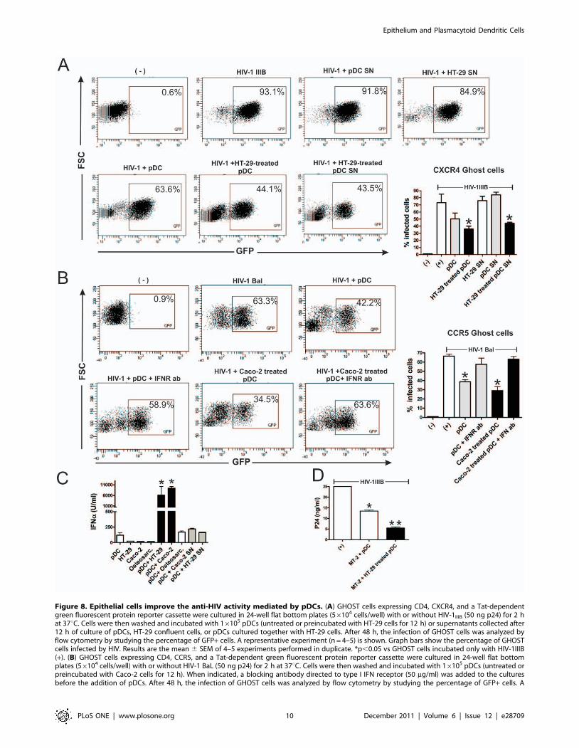
Figure 8. Epithelial cells improve the anti-HIV activity mediated by pDCs. (A) GHOST cells expressing CD4, CXCR4, and a Tat-dependentgreen fluorescent protein reporter cassette were cultured in 24-well flat bottom plates (56104 cells/well) with or without HIV-1IIIB (50 ng p24) for 2 hat 37uC. Cells were then washed and incubated with 16105 pDCs (untreated or preincubated with HT-29 cells for 12 h) or supernatants collected after12 h of culture of pDCs, HT-29 confluent cells, or pDCs cultured together with HT-29 cells. After 48 h, the infection of GHOST cells was analyzed byflow cytometry by studying the percentage of GFP+ cells. A representative experiment (n = 4–5) is shown. Graph bars show the percentage of GHOSTcells infected by HIV. Results are the mean 6 SEM of 4–5 experiments performed in duplicate. *p,0.05 vs GHOST cells incubated only with HIV-1IIIB(+). (B) GHOST cells expressing CD4, CCR5, and a Tat-dependent green fluorescent protein reporter cassette were cultured in 24-well flat bottomplates (56104 cells/well) with or without HIV-1 BaL (50 ng p24) for 2 h at 37uC. Cells were then washed and incubated with 16105 pDCs (untreated orpreincubated with Caco-2 cells for 12 h). When indicated, a blocking antibody directed to type I IFN receptor (50 mg/ml) was added to the culturesbefore the addition of pDCs. After 48 h, the infection of GHOST cells was analyzed by flow cytometry by studying the percentage of GFP+ cells. A
Epithelium and Plasmacytoid Dendritic Cells
PLoS ONE | www.plosone.org 10 December 2011 | Volume 6 | Issue 12 | e28709

Our observation indicating that epithelial cell lines activate
pDCs in a contact-dependent mode could be explained consid-
ering that RNA is rapidly degraded in the extracellular space [23].
Moreover, the cationic nature of the antimicrobial peptide LL37
[27] might lead to its rapid association to cell membranes. How to
explain the ability of primary epithelial cells cultures to activate
pDCs, at least in part, through cell contact-independent
mechanisms? Primary epithelial cells would display a higher
ability than epithelial cell lines to release self-RNA, to produce the
antimicrobial peptide LL37, and/or to stimulate the expression of
endosomal TLRs by pDCs. Further experiments are needed to test
these hypotheses.
Plasmacytoid DCs play a critical role in the immune response to
different virus including HIV-1. Groot and colleagues [30] have
shown that cDCs and pDCs have opposing roles on HIV-1
infection of T cells. The authors reported that cDCs enhance
HIV-1 infection through the capture of the virus and subsequent
transmission to T cells, and also that differently maturated cDCs
have different HIV-1 transmission efficiencies. By contrast, pDCs
inhibit HIV-1 replication in T cells through a mechanism
dependent on the production of IFN-a and undefined small
anti-viral molecules. Our results are in line with those of Groot et
al. [30] and support that pDCs effectively prevent HIV-1 infection
of target cells. However, the authors observed that unstimulated
pDCs or pDCs activated by different maturation stimuli such as
the TLR synthetic agonist R-848, poly (I:C) or fixed Staphylococus
aureus Cowan strain I bacteria (SAC) show a similar ability to
prevent the infection of T cells by HIV [30]. By contrast, our
results show that activation of pDCs by epithelial cells results in an
enhanced ability to inhibit HIV replication through a type I IFN-
dependent pathway. Consistent with these results, Meyers and
colleagues [29] have previously shown that activation by CpG
ODN markedly increase the anti-HIV activity mediated by pDCs.
In conclusion, we have shown that epithelial cells are able to
activate pDCs. Because pDCs are usually difficult to detect in the
normal mucosa [7,8], they would rarely interact with the
epithelium under steady-state conditions. However, during the
course of viral infections, autoimmune and allergic diseases, pDCs
are recruited to the mucosa [13,15,41–43] thus favoring their
interaction with epithelial cells lining the gastrointestinal, genito-
urinary, and respiratory tracts. This might enable the cross-talk
between both cell populations leading to the activation of pDCs.
This novel pathway of pDC activation may contribute not only to
anti-viral immune response but also to the development of tissue
injury in allergic and autoimmune processes.
Materials and Methods
ReagentsLipopolysaccharide from Escherichia coli, trypsin, collagenase
type I, dimethyl sulfoxide, endothelial cell growth factor, and
recombinant human granulocyte-macrophage colony-stimulating
factor (GM-CSF) were from Sigma-Aldrich (St. Louis, MO).
Bafilomycin A1 (Streptomyces griseus) was from Merk (Darmstadt,
Germany), RNase A (bovine pancreas) was from Fermentas
(Buenos Aires, Argentine), DNase I (bovine pancreas) was from
Invitrogen (Carlsbad, CA). Recombinant human interleukin-4 (IL-4)
was from Preprotech (Rocky Hill, NJ) or R&D Systems (Minneap-
olis, MN). Ficoll-Hypaque and Percoll were from Amersham
Pharmacia Biotech (Piscataway, NJ). The sequence of the phospho-
diester CpG-containing oligonucleotide used was: 2006, TCGTCG-
TTTTGTCGTTTTGTCGTT (Sigma Genosys, St. Louis, MO).
Isolation of pDCs and preparation of conventionaldendritic cells (cDCs)
Peripheral blood mononuclear cells (PBMCs) were isolated from
healthy volunteers by standard density gradient centrifugation on
Ficoll-Hypaque. pDCs were isolated using immunomagnetic cell
sorting (BDCA-4 cell isolation kit, Miltenyi Biotec; Germany)
according to the manufacturer’s instructions. The purity of pDCs
was checked by FACS using MAb directed to CD123 and HLA-
DR and was found to be .93%. Monocytes were purified by
centrifugation on a discontinuous Percoll gradient with modifica-
tions of a previously described method [44]. Briefly, PBMCs were
suspended in Ca2+/Mg2+-free Tyrode solution supplemented with
0.2% EDTA and incubated during 30 min at 37uC. During this
incubation, the osmolarity of the medium was gradually increased
from 290 to 360 osmol/liter by the addition of 9% NaCl. Three
different Percoll fractions were layered in polypropylene tubes:
50% at the bottom, followed by 46 and 40%. PBMCs (56106/ml)
were layered at the top, and they were centrifuged at 4006 g for
20 min at 4uC. Monocytes were recovered at the 50/46%
interface. The purity was checked by FACS using an anti-CD14
MAb and was found to be .85%. To obtain cDCs, monocytes
were cultured in RPMI 1640 medium (Life Technologies, Grand
Island, NY) supplemented with 10% heat-inactivated fetal calf
serum, 50 U of penicillin/ml, 50 mg of streptomycin/ml (Life
Technologies) (complete culture medium) at 106 cells/ml with
10 ng/ml of IL-4 and 10 ng/ml of GM-CSF, as described by
Sallusto and Lanzavecchia [45]. On day 5, the cells were analyzed
by FACS.
Cell lines and virusHuman intestinal HT-29 and Caco-2 cell lines (generous gift of
Dr Martın Rumbo, National University of La Plata, Argentina)
were grown at 37uC in Dulbecco’s modified Eagle’s medium
(Cellgro-Mediatech, VA) containing 10% heat-inactivated fetal
calf serum (Gibco; Invitrogen), penicillin/streptomycin (100 U/ml),
2 mM L-glutamin, 1 mM pyruvate, and 0.1 mM nonessential
amino acids (all from Life Technologies, Grand Island, NY) at
pH 7.3. Epithelial cells were removed from the wells using a
solution of 0.25% Trypsin-EDTA 1 mM prepared in Hank’s
balanced salt solution without Ca2+ and Mg2+. The human
osteosarcoma cell line GHOST, expressing CD4, the HIV
coreceptors CXCR4 or CCR5, and a Tat-dependent green
fluorescent protein reporter cassette [46], and the T cell line MT-
2 were obtained through the AIDS Research and Reference
representative experiment (n = 4) is shown. Graph bars show the percentage of GHOST cells infected by HIV-1. Results are the mean 6 SEM of 4experiments performed in duplicate. (* p,0.05 vs GHOST cells incubated only with HIV-1 BaL). (C) The epithelial cell lines HT-29 and Caco-2, and theosteosarcoma line GHOST (osteosarc) were grown to confluence in 96 well flat bottom plates. pDCs (16105/200 ml) were cultured for 12 h alone(controls), with HT-29, Caco-2 cells, GHOST cells or with supernatants collected from HT-29 or Caco-2 grown to confluence and cultured for 12 h.Then, the production of IFN-a was evaluated in cell supernatants by ELISA. Results are the mean 6 SEM of five experiments performed in duplicate. (*p,0.05 vs controls). (D) The T cell line MT-2 (16105 cells) was cultured with HIV-1IIIB (50 ng p24) for 2 h at 37uC. Cells were then washed andincubated with 16105 pDCs, previously incubated for 12 h with or without confluent HT-29 cells. Infection of MT-2 cells was determined bymeasuring p24 antigen levels in cell supernatants 48 h post-infection. Results are the mean 6 SEM of four experiments performed in duplicate. (*p,0.05 vs MT-2+HIV-1 and ** p,0.05 vs MT-2+HIV-1+pDCs).doi:10.1371/journal.pone.0028709.g008
Epithelium and Plasmacytoid Dendritic Cells
PLoS ONE | www.plosone.org 11 December 2011 | Volume 6 | Issue 12 | e28709

Reagent Program, Division of AIDS, National Institute of Allergy
and Infectious Disease, National Institutes of Health. GHOST cells
were grown in DMEM medium (Cellgro-Mediatech) supplemented
with 10% heat-inactivated fetal calf serum, L-glutamine (2 mM),
penicillin/streptomycin (100 U/ml), G418 (300 mg/ml), hygromy-
cin (100 mg/ml) and puromycin (1 mg/ml) (all from Life Technol-
ogies) in 24-well flat bottom plates (56104 cells/well). Cells were
detached using 0.25% trypsin. Murine fibroblasts stably transfected
with a human CD40L cDNA and their control counterpart were
provided by Dr. Claire Hivroz from Institut Curie, Paris. The
CXCR4-using HIV-1 IIIB and the CCR5-using HIV-1 BaL were
from the AIDS Research and Reference Reagent Program. The
HIV-1 IIIB isolate was obtained from H9HTLV-IIIB supernatants
while HIV-1 BaL was grown in human monocyte-derived
macrophages. The viruses were concentrated by ultracentrifugation
at 28,000 rpm for 90 min at 4uC (L2-65B ultracentrifuge; Beckman
Coulter), and the virus pellet was suspended in RPMI 1640
medium. p24 antigen levels were determined by ELISA (Vironos-
tika, Biomerieux, Argentina), and virus input into assays was a
function of p24 antigen concentration. The virus stocks as well as all
the cell cultures were free of Mycoplasma as measured by the
Mycoplasma PCR detection kit (VenorHGeM), Cambridge, UK).
Isolation and culture of human renal tubular epithelialcells
Human renal tubular epithelial cells (HRTEC) were isolated
from kidneys removed from different adult patients undergoing
nephrectomies for renal cell carcinoma from the ‘‘Unidad de
Urologıa, Hospital Nacional Profesor A. Posadas, Buenos Aires,
Argentina’’. The removal of the portion of the renal tissue for
research purposes was approved by the ethics committee of the
University of Buenos Aires. The cortex was dissected from the
renal medulla and the primary culture of the HRTEC was
performed as previously described [47]. Briefly, the cortical
fragments were cultured for 1 h at 37uC in a buffer containing
0.1% collagenase type I. Cells were washed and suspended in
RPMI 1640 medium supplemented with 5% heat-inactivated fetal
calf serum, 2 mM L-glutamine, and 100 U/ml penicillin/
streptomycin. Cells were grown at 37uC to confluence. The cell
isolates were trypsinized, concentrated in heat-inactivated fetal calf
serum containing 5% dimethyl sulfoxide and stored in liquid
nitrogen for subsequent use. Cells were then cultured in flasks or in
96-well, flat bottom plates in complete culture medium supple-
mented with 1% endothelial cell growth factor and were used
between three and five passages. By light microscopy, more than
95% of the cells had similar morphologies. These cells were
confirmed as epithelial cells by positive staining for cytokeratins
(Sigma-Aldrich). The presence of fibroblasts was ruled out by the
lack of reactivity with an antibody directed to the human fibroblast
common antigen (Dako, Denmark). Furthermore, the cells were
also negative for staining with an antibody directed to the
endothelial cell antigen PECAM (CD31) (Dako).
Culture of pDCs with epithelial cellsEpithelial cells were grown to confluence in 96 well, flat bottom
plates. When the cultures reached 90–100% confluence, 16105
pDCs were added to each well, and cells were cultured in 0.2 ml of
RPMI 1640 culture medium supplemented with 10% heat-
inactivated fetal calf serum, 50 U of penicillin/ml, 50 mg of
streptomycin/ml. Plasmacytoid dendritic cells cultured without
epithelial cells and epithelial cells cultured alone were used as
controls. After 12 h of culture, the levels of the cytokines TNF-a,
IL-1b, IL-6, IFN-a, MCP-1 and IL-8 in cell supernatants were
analyzed by ELISA (R&D Systems or Amersham Biosciences)
following the manufacturer’s recommendations, and the pheno-
type of pDCs was analyzed by flow cytometry. Experiments
carried out with cDCs were done in a similar way. In some
experiments pDCs were cultured with supernatants harvested
from confluent epithelial cell cultured during 12 h at 37uC. When
indicated coculture of pDCs and epithelial cells were performed in
24-transwell chambers with a polycarbonate filter (0.2 mm pore
size). In these experiments epithelial cells were grown to
confluence on the filter, and pDCs (36105) were added in the
upper chamber, enabling the contact between pDCs and epithelial
cells, or in the lower chamber. After 12 h of culture, pDCs were
harvested and their phenotype was analyzed by flow cytometry.
Flow cytometryFluorescein isothiocyanate (FITC), phycoerythrin (PE), or
allophycocyanin (APC) conjugated MAbs directed to CD1a,
CD14, CD80, CD86, CD40, HLA-DR, CD83, CD123, TNF-a,
IL-1b (BD Pharmingen, San Diego, CA), LL37 (Santa Cruz
Biotechnology, Santa Cruz, CA), and TLR-9 (eBioscience, San
Diego, CA). Rabbit polyclonal anti-TLR7 was from IMGENEX
(San Diego, CA). In all cases, isotype-matched control antibodies
were used, and a gate (R1) was defined in the analysis to exclude
all nonviable cells and debris, based on size and propidium iodine
staining. Analysis was performed by using a FACS flow cytometer
and CellQuest software (BD Biosciences, San Jose, CA). The
results are expressed as the mean fluorescence intensity or as the
percentage of positive cells.
Quantitation of cellular apoptosis by annexin-V bindingand flow cytometry
It was performed using an apoptosis detection kit (Immunotech,
Marseille, France). In brief, cells were labelled with annexin-V-
FITC for 20 min at 4uC and with propidium iodide immediately
before evaluation of fluorescence by flow cytometry.
Transepithelial electrical resistanceTo estimate the integrity of the HRTC monolayer, transepi-
thelial electrical resistance (TEER) was measured as described
[47,48] using a Millicell-ERS electric resistance system (Millipore,
Bedford, MA, USA). Briefly, HRTC were grown to confluence on
a polycarbonate filter (0.2 mm pore size) in the upper chamber of a
24-transwell plate. Then, epithelial cells were incubated for 18 h
together with pDCs (26105) in the upper chamber or in the
absence of pDCs. TEER was measured at 0, 3 and 18 h after the
addition of pDCs. Data were corrected for the resistance of the
empty filter.
HIV-1 infection assaysGHOST cells expressing CD4, CXCR4 and a Tat-dependent
green fluorescent protein (GFP) reporter cassette were infected by
HIV-1 IIIB (X4 tropic) while GHOST cells expressing CD4 and
CCR5 were infected with HIV-1 BaL (R5 tropic). In all cases, cells
were cultured with HIV-1 (50 ng p24) for 2 h at 37uC. Cells were
then washed and pDCs (16105) (untreated or preincubated with
HT-29 cells for 12 h) were added to GHOST cells. Infection of
GHOST cells was analyzed by flow cytometry at 48 h post-
infection by studying the percentage of GFP+ cells, as previously
described [49]. To neutralize the activity of type I IFNs, a specific
blocking antibody directed to the common receptor IFN-R was
used (clone MMHAR-2, PBL interferon Source, NJ, USA).
Infection of MT-2 cells was performed using HIV-1 IIIB and it
was evaluated by measuring p24 antigen levels in cell supernatants
at 48 h post-infection by ELISA.
Epithelium and Plasmacytoid Dendritic Cells
PLoS ONE | www.plosone.org 12 December 2011 | Volume 6 | Issue 12 | e28709

Statistical analysisAll statistical comparisons were performed by using one-way
analysis of variance with Dunnett post-test. P values,0.05 were
considered statistically significant.
Author Contributions
Conceived and designed the experiments: CRR JG. Performed the
experiments: CRR MC FRL JS AC CJ SR MO CS. Analyzed the data:
CRR JG. Wrote the paper: CRR JG.
References
1. Asselin-Paturel C, Trinchieri G (2005) Production of type I interferons:plasmacytoid dendritic cells and beyond. J Exp Med 202: 461–465.
2. Liu YJ (2005) IPC: professional type 1 interferon-producing cells and
plasmacytoid dendritic cell precursors. Annu Rev Immunol 23: 275–306.3. Zhang Z, Wang FS (2005) Plasmacytoid dendritic cells act as the most
competent cell type in linking antiviral innate and adaptive immune responses.Cell Mol Immunol 2: 411–417.
4. Swiecki M, Colonna M. Unraveling the functions of plasmacytoid dendritic
cells during viral infections, autoimmunity, and tolerance. Immunol Rev 234:142–162.
5. Gotoh K, Tanaka Y, Nishikimi A, Nakamura R, Yamada H, et al. Selectivecontrol of type I IFN induction by the Rac activator DOCK2 during TLR-
mediated plasmacytoid dendritic cell activation. J Exp Med 207: 721–730.6. Charles J, Chaperot L, Salameire D, Di Domizio J, Aspord C, et al.
Plasmacytoid dendritic cells and dermatological disorders: focus on their role
in autoimmunity and cancer. Eur J Dermatol 20: 16–23.7. Sozzani S, Vermi W, Prete AD, Facchetti F. Trafficking properties of plas-
macytoid dendritic cells in health and disease. Trends Immunol.8. Colonna M, Trinchieri G, Liu YJ (2004) Plasmacytoid dendritic cells in
immunity. Nat Immunol 5: 1219–1226.
9. Gilliet M, Cao W, Liu YJ (2008) Plasmacytoid dendritic cells: sensing nucleicacids in viral infection and autoimmune diseases. Nat Rev Immunol 8: 594–606.
10. Wang H, Peters N, Schwarze J (2006) Plasmacytoid dendritic cells limit viralreplication, pulmonary inflammation, and airway hyperresponsiveness in
respiratory syncytial virus infection. J Immunol 177: 6263–6270.
11. Lund JM, Linehan MM, Iijima N, Iwasaki A (2006) Cutting Edge: Plasmacytoiddendritic cells provide innate immune protection against mucosal viral infection
in situ. J Immunol 177: 7510–7514.12. Hartmann E, Graefe H, Hopert A, Pries R, Rothenfusser S, et al. (2006) Analysis
of plasmacytoid and myeloid dendritic cells in nasal epithelium. Clin VaccineImmunol 13: 1278–1286.
13. Schnurr M, Toy T, Shin A, Hartmann G, Rothenfusser S, et al. (2004) Role of
adenosine receptors in regulating chemotaxis and cytokine production ofplasmacytoid dendritic cells. Blood 103: 1391–1397.
14. Devosse T, Guillabert A, D’Haene N, Berton A, De Nadai P, et al. (2009)Formyl peptide receptor-like 2 is expressed and functional in plasmacytoid
dendritic cells, tissue-specific macrophage subpopulations, and eosinophils.
J Immunol 182: 4974–4984.15. Gutzmer R, Kother B, Zwirner J, Dijkstra D, Purwar R, et al. (2006) Human
plasmacytoid dendritic cells express receptors for anaphylatoxins C3a and C5aand are chemoattracted to C3a and C5a. J Invest Dermatol 126: 2422–2429.
16. Vermi W, Riboldi E, Wittamer V, Gentili F, Luini W, et al. (2005) Role ofChemR23 in directing the migration of myeloid and plasmacytoid dendritic cells
to lymphoid organs and inflamed skin. J Exp Med 201: 509–515.
17. Sonnier DI, Bailey SR, Schuster RM, Lentsch AB, Pritts TA. TNF-alpha inducesvectorial secretion of IL-8 in Caco-2 cells. J Gastrointest Surg 14: 1592–1599.
18. Kim JM, Lee JY, Yoon YM, Oh YK, Youn J, et al. (2006) NF-kappa Bactivation pathway is essential for the chemokine expression in intestinal
epithelial cells stimulated with Clostridium difficile toxin A. Scand J Immunol
63: 453–460.19. Lasser C, Alikhani VS, Ekstrom K, Eldh M, Paredes PT, et al. (2011) Human
saliva, plasma and breast milk exosomes contain RNA: uptake by macrophages.J Transl Med 9: 9.
20. Kesimer M, Scull M, Brighton B, DeMaria G, Burns K, et al. (2009)Characterization of exosome-like vesicles released from human tracheobronchial
ciliated epithelium: a possible role in innate defense. FASEB J 23: 1858–1868.
21. Barton GM, Kagan JC (2009) A cell biological view of Toll-like receptorfunction: regulation through compartmentalization. Nat Rev Immunol 9:
535–542.22. Russo C, Cornella-Taracido I, Galli-Stampino L, Jain R, Harrington E, et al.
(2011) Small molecule Toll-like receptor 7 agonists localize to the MHC class II
loading compartment of human plasmacytoid dendritic cells. Blood 117:5683–5691.
23. Barton GM, Kagan JC, Medzhitov R (2006) Intracellular localization of Toll-like receptor 9 prevents recognition of self DNA but facilitates access to viral
DNA. Nat Immunol 7: 49–56.
24. Lovgren T, Eloranta ML, Bave U, Alm GV, Ronnblom L (2004) Induction ofinterferon-alpha production in plasmacytoid dendritic cells by immune
complexes containing nucleic acid released by necrotic or late apoptotic cellsand lupus IgG. Arthritis Rheum 50: 1861–1872.
25. Bave U, Alm GV, Ronnblom L (2000) The combination of apoptotic U937 cellsand lupus IgG is a potent IFN-alpha inducer. J Immunol 165: 3519–3526.
26. Barrat FJ, Meeker T, Gregorio J, Chan JH, Uematsu S, et al. (2005) Nucleicacids of mammalian origin can act as endogenous ligands for Toll-like receptors
and may promote systemic lupus erythematosus. J Exp Med 202: 1131–1139.
27. Ganguly D, Chamilos G, Lande R, Gregorio J, Meller S, et al. (2009) Self-RNA-antimicrobial peptide complexes activate human dendritic cells through TLR7
and TLR8. J Exp Med 206: 1983–1994.
28. Chromek M, Slamova Z, Bergman P, Kovacs L, Podracka L, et al. (2006) The
antimicrobial peptide cathelicidin protects the urinary tract against invasive
bacterial infection. Nat Med 12: 636–641.
29. Meyers JH, Justement JS, Hallahan CW, Blair ET, Sun YA, et al. (2007) Impact
of HIV on cell survival and antiviral activity of plasmacytoid dendritic cells.PLoS One 2: e458.
30. Groot F, van Capel TM, Kapsenberg ML, Berkhout B, de Jong EC (2006)Opposing roles of blood myeloid and plasmacytoid dendritic cells in HIV-1
infection of T cells: transmission facilitation versus replication inhibition. Blood
108: 1957–1964.
31. Iliev ID, Spadoni I, Mileti E, Matteoli G, Sonzogni A, et al. (2009) Human
intestinal epithelial cells promote the differentiation of tolerogenic dendritic cells.Gut 58: 1481–1489.
32. Rescigno M, Lopatin U, Chieppa M (2008) Interactions among dendritic cells,macrophages, and epithelial cells in the gut: implications for immune tolerance.
Curr Opin Immunol 20: 669–675.
33. Iliev ID, Matteoli G, Rescigno M (2007) The yin and yang of intestinal epithelialcells in controlling dendritic cell function. J Exp Med 204: 2253–2257.
34. Rimoldi M, Chieppa M, Salucci V, Avogadri F, Sonzogni A, et al. (2005)Intestinal immune homeostasis is regulated by the crosstalk between epithelial
cells and dendritic cells. Nat Immunol 6: 507–514.
35. Guermonprez P, Valladeau J, Zitvogel L, Thery C, Amigorena S (2002) Antigen
presentation and T cell stimulation by dendritic cells. Annu Rev Immunol 20:
621–667.
36. Reis e Sousa C (2006) Dendritic cells in a mature age. Nat Rev Immunol 6:
476–483.
37. Sabatte J, Maggini J, Nahmod K, Amaral MM, Martinez D, et al. (2007)
Interplay of pathogens, cytokines and other stress signals in the regulation of
dendritic cell function. Cytokine Growth Factor Rev 18: 5–17.
38. Cella M, Facchetti F, Lanzavecchia A, Colonna M (2000) Plasmacytoid
dendritic cells activated by influenza virus and CD40L drive a potent TH1polarization. Nat Immunol 1: 305–310.
39. Liu YJ, Blom B (2000) Introduction: TH2-inducing DC2 for immunotherapy.Blood 95: 2482–2483.
40. Angelot F, Seilles E, Biichle S, Berda Y, Gaugler B, et al. (2009) Endothelial cell-
derived microparticles induce plasmacytoid dendritic cell maturation: potentialimplications in inflammatory diseases. Haematologica 94: 1502–1512.
41. Li Q, Estes JD, Schlievert PM, Duan L, Brosnahan AJ, et al. (2009) Glycerolmonolaurate prevents mucosal SIV transmission. Nature 458: 1034–1038.
42. Skrzeczynska-Moncznik J, Stefanska A, Zabel BA, Kapinska-Mrowiecka M,Butcher EC, et al. (2009) Chemerin and the recruitment of NK cells to diseased
skin. Acta Biochim Pol 56: 355–360.
43. Penna G, Sozzani S, Adorini L (2001) Cutting edge: selective usage ofchemokine receptors by plasmacytoid dendritic cells. J Immunol 167:
1862–1866.
44. Chuluyan HE, Issekutz AC (1993) VLA-4 integrin can mediate CD11/CD18-
independent transendothelial migration of human monocytes. J Clin Invest 92:
2768–2777.
45. Sallusto F, Lanzavecchia A (1994) Efficient presentation of soluble antigen by
cultured human dendritic cells is maintained by granulocyte/macrophagecolony-stimulating factor plus interleukin 4 and downregulated by tumor
necrosis factor alpha. J Exp Med 179: 1109–1118.
46. Vodros D, Tscherning-Casper C, Navea L, Schols D, De Clercq E, et al. (2001)
Quantitative evaluation of HIV-1 coreceptor use in the GHOST3 cell assay.
Virology 291: 1–11.
47. Silberstein C, Pistone Creydt V, Gerhardt E, Nunez P, Ibarra C (2008)
Inhibition of water absorption in human proximal tubular epithelial cells inresponse to Shiga toxin-2. Pediatr Nephrol 23: 1981–1990.
48. Schmitz H, Rokos K, Florian P, Gitter AH, Fromm M, et al. (2002)Supernatants of HIV-infected immune cells affect the barrier function of human
HT-29/B6 intestinal epithelial cells. Aids 16: 983–991.
49. Sabatte J, Ceballos A, Raiden S, Vermeulen M, Nahmod K, et al. (2007)Human seminal plasma abrogates the capture and transmission of human
immunodeficiency virus type 1 to CD4+ T cells mediated by DC-SIGN. J Virol81: 13723–13734.
Epithelium and Plasmacytoid Dendritic Cells
PLoS ONE | www.plosone.org 13 December 2011 | Volume 6 | Issue 12 | e28709
Copyright © 2022 FDOKUMEN