Bardess-Bytes-50-Qlik-Tips-to-Accelerate-Your-Development ...
Upload
independentCategory
view
3download
0
G
T
1
Sf
1
2
PC
3
4
a5b6
7
a8
9
A10
R11
R12
A13
A
14
K15
N16
T17
S18
D19
T20
P21
122
23
a24
d25
u26
n27
h28
o29
a30
31
b32
t33
m34
s35
Q1
0d
ARTICLE IN PRESS
TED
PR
OO
F
Model
OX 50398 1–7
Toxicology xxx (2009) xxx–xxx
Contents lists available at ScienceDirect
Toxicology
journa l homepage: www.e lsev ier .com/ locate / tox ico l
ingle-walled carbon nanotubes activate platelets and accelerate thrombusormation in the microcirculation
eter Bihari a,∗, Martin Holzera, Marc Praetnera, Janos Fentb, Max Lerchenbergera,hristoph A. Reichela, Markus Rehberga, Susan Lakatosb, Fritz Krombacha
Walter Brendel Centre of Experimental Medicine, Ludwig-Maximilians-Universität München, Marchioninistrasse 27, D-81377 Munich, GermanyMilitary Health Center, Hungarian Defence Forces, Budapest, Hungary
r t i c l e i n f o
rticle history:eceived 8 May 2009eceived in revised form 3 August 2009ccepted 12 August 2009vailable online xxx
eywords:anoparticleshrombosisingle-walled carbon nanotubesiesel exhaust particlesitanium dioxideolystyrene beads
a b s t r a c t
Objectives: Although ambient nanoparticles have been shown to exert prothrombotic effects, manufac-tured nanoparticles are in this aspect less well investigated. Thus, the aim of this study was to characterizethe effects of diesel, titanium dioxide rutile, and single-walled carbon nanotube nanoparticles on (i)platelet activation in vitro and (ii) on macro- and microcirculatory thrombus formation in vivo.Methods: Platelet P-selectin expression was measured by flow cytometry after incubation of whole bloodwith diesel (0.1 mg/mL), titanium dioxide (0.1 mg/mL) or single-walled nanotubes (0.001–0.1 mg/mL).Platelet–granulocyte complexes were analyzed in whole blood and platelet aggregometry was performedwith platelet-rich plasma. Upon systemic administration of nanoparticles (1 mg/kg) to anesthetized mice,ferric chloride-induced thrombus formation was measured in small mesenteric arteries using in vivomicroscopy. In separate experiments, diesel (1 mg/kg), titanium dioxide (1 mg/kg), or single-walled nan-otubes (0.01–1 mg/kg) were injected into anesthetized mice and light/dye-induced thrombus formationwas investigated in the cremasteric microcirculation.
EC
Results: Diesel and titanium dioxide nanoparticles did not activate platelets or exert prothrombotic effects.In contrast, single-walled nanotubes significantly increased platelet P-selectin expression, the numberof platelet–granulocyte complexes, and platelet aggregability in vitro, and reduced the occlusion time inmesenteric arteries as well as in cremasteric arterioles.Conclusion: Our study shows that single-walled carbon nanotubes, but not diesel or titanium dioxidenanoparticles, induce platelet activation in vitro and exert prothrombotic effects in the microcirculation
36
37
38
39
40
41
42
43
44
45
46
NC
OR
Rin vivo.
. Introduction
Nanoparticles are defined as matter at dimensions betweenpproximately 1 and 100 nm. In the last decades, there has been aramatic increase in research, technology, and production of man-factured nanoparticles. As the production of nanoparticles and theumber of new applications including medical use rapidly grow, itas become important to determine the hazards of nanoparticlesn human health (Borm et al., 2006; Maynard et al., 2006; Nel etl., 2006).
Epidemiological studies have demonstrated an association
Please cite this article in press as: Bihari, P., et al., Single-walled carbon namicrocirculation. Toxicology (2009), doi:10.1016/j.tox.2009.08.011
Uetween exposure to ambient nanoparticles, i.e. the ultrafine frac-ion of particulate matter in the air, and increased cardiovascular
orbidity and mortality (Brook, 2008; Mills et al., 2009). There areeveral mechanisms discussed to play a role in the effects of ambi-
∗ Corresponding author. Tel.: +49 89218076538.E-mail address: [email protected] (P. Bihari).
47
48
49
300-483X/$ – see front matter © 2009 Published by Elsevier Ireland Ltd.oi:10.1016/j.tox.2009.08.011
© 2009 Published by Elsevier Ireland Ltd.
ent nanoparticles on the cardiovascular system. Indirect effects aremediated by inflammatory mediators, produced during pulmonaryinflammation that is induced by particles deposited in the lung.These mediators are proposed to change the autonomic controlof the heart, alter the activity of the coagulatory system, or acti-vate endothelial cells (Delfino et al., 2005; Mills et al., 2007). Anemerging idea is that nano-sized particles, in addition to induc-ing inflammation in the lung, are also able to translocate into thecirculation (Kreyling et al., 2002; Nemmar et al., 2002a, 2005),reach distal organs, and directly influence the thrombotic process(Nemmar et al., 2004a).
In spite of the increasing epidemiological and experimen-tal data about prothrombotic effects of ambient nanoparticlesand the fact that manufactured nanoparticles are produced in
notubes activate platelets and accelerate thrombus formation in the
high amounts, the effects of manufactured nanoparticles on the 50
thrombotic process are less investigated. It has been shown 51
that amine-modified polystyrene nanoparticles augmented and 52
carboxyl-modified polystyrene nanoparticles inhibited thrombus 53
formation in femoral veins (Nemmar et al., 2002b). Carbon nan- 54

IN
T
G
T
2 ology
o55
a56
a57
e58
e59
e60
261
c62
63
s64
a65
I66
t67
i68
m69
t70
e71
o72
h73
w74
a75
t76
t77
p78
279
280
81
c82
b83
E84
d85
H86
(87
w88
r89
p90
U91
292
93
094
F95
(96
s97
c98
o99
d100
w101
h102
a103
0104
N105
o106
m107
p108
s109
2110
111
t112
B113
0114
2115
116
p117
c118
c119
t120
121
122
123
124
125
126
127
128
129
130
131
132
133
134
135
136
137
138
139
140
141
142
143
144
145
146
147
148
149
150
151
152
153
154
155
156
157
158
159
160
161
162
163
164
165
166
167
168
169
170
171
172
173
174
175
176
177
178
179
180
181
182
183
ARTICLE
UN
CO
RR
EC
Model
OX 50398 1–7
P. Bihari et al. / Toxic
tubes were found to cause platelet agglomeration in vitro and tougment ferric chloride-induced thrombosis in vivo (Radomski etl., 2005). TiO2 nanorods increased ADP-induced platelet agglom-ration in vitro and decreased the platelet count in vivo (Nemmart al., 2008). Intravenous administration of quantum dots (Geyst al., 2008) or mesoporous silicate nanoparticles (Hudson et al.,008) was shown to induce vascular thrombosis in the pulmonaryirculation.
While most of the studies on nanoparticle-induced thrombo-is investigated large vessels, only few recent publications havenalyzed the effects of nanoparticles in the microcirculation.nhaled carbon black nanoparticles enhanced platelet accumula-ion on the endothelium of postsinusoidal venules and sinusoidsn the hepatic microcirculation (Khandoga et al., 2004) and amine-
odified polystyrene nanoparticles had a prothrombotic effect inhe microcirculation (Silva et al., 2005). However, effects of dieselxhaust, titanium dioxide rutile, and single-walled carbon nan-tube nanoparticles on thrombus formation in the microcirculationave not yet been investigated. Therefore, the objective of our studyas (i) to characterize the effects of these nanoparticles on platelet
ctivation in vitro, (ii) to asses their impact on thrombus forma-ion in small arteries and in the microcirculation in vivo, and (iii)o compare these effects with those induced by surface-modifiedolystyrene beads as benchmark particles.
. Materials and methods
.1. Materials
Titanium(IV) oxide nanopowder 99.5% rutile ∼10 nm × 40 nm (TiO2), 10×oncentrated PBS, fluorescein isothiocyanate dextran 150 kD (FITC), epinephrine-itartrate, adenosine 5′-diphosphate (ADP), and lipopolysaccharide fromscherichia Coli O111:B4 (LPS) were purchased from Sigma–Aldrich, Schnell-orf, Germany. Human serum albumin 50 g/L was from Baxter Deutschland Gmbh,eidelberg, Germany. Amine- and carboxyl-modified polystyrene nanoparticles
60 nm) were purchased from Bangs Laboratories, Fishers, USA. S-purified single-all nanotubes (SWNT), outer diameter: <2 nm, length: 1–5 �m were from SES
esearch, Houston, USA. SRM 2975 diesel exhaust particulate matter (DEP) wasurchased from the National Institute of Standards and Technology, Gaithersburg,SA.
.2. Preparation of nanoparticles
DEP, TiO2, or SWNT stock solutions were prepared at a concentration of.23 mg/ml in distilled water using sonication with 4.2 × 10⁵ kJ/m3 specific energy.or in vitro experiments with human blood, 30 �l of 50 mg/ml human serum albuminend concentration 1.5 mg/ml) and for in vivo mouse experiments 30 �l of mouseerum was given to 870 �l of dispersion before the addition of 100 �l of a 10× con-entrated PBS solution. To prepare dispersions at lower concentrations, dilutionsf the stock solution were used. The end concentrations of nanoparticles in theispersion were 0.002, 0.02 and 0.2 mg/ml. The vehicle was prepared in the sameay, but instead of a nanoparticle dispersion 870 �l of distilled water was added touman serum albumin or mouse serum and to concentrated PBS. Dispersions withmine- and carboxyl-modified polystyrene nanoparticles were prepared in PBS at.2 mg/ml without sonication or addition of human serum albumin or mouse serum.anoparticles tend to form coarse agglomerates in physiological solutions. The sizef the polystyrene nanoparticles in the dispersion with physiological solution waseasured to be about 60 nm. The size of other nanoparticles in dispersion with
hysiological solution was less than 290 nm after addition of serum as dispersiontabiliser (Bihari et al., 2008a).
.3. Endotoxin measurement
Nanoparticle dispersions prepared by the above method were tested withhe LAL (Limulus amoebocyte lysate) kinetic chromogenic assay (Lonza, Verviers,elgium). The endotoxin content in DEP, TiO2 and SWNT dispersions was below.5 EU/ml.
.4. Measurement of platelet P-selectin and platelet–granulocyte complexes
Please cite this article in press as: Bihari, P., et al., Single-walled carbon namicrocirculation. Toxicology (2009), doi:10.1016/j.tox.2009.08.011
The effect of nanoparticles on platelet activation and on the formation oflatelet–granulocyte complexes in human whole blood was determined by flowytometry (FACScan, Becton Dickinson, USA). Human blood was collected from theubital vein of healthy volunteers into citrate anticoagulant containing Vacuetteest tubes (Greiner, Austria, Kremsmünster). All samples were obtained with the
PRESS
ED P
RO
OF
xxx (2009) xxx–xxx
approval of the local Ethical Committee after the donor had given informed con-sent. 100 �l of citrated blood was incubated with 100 �l of vehicle or 100 �l ofnanoparticle dispersion for 10 min at room temperature. Dispersions for carboxyl-,amine-modified polystyrene bead, DEP and TiO2 nanoparticles had a concentrationof 0.2 mg/ml and SWNT dispersions had concentrations of 0.002, 0.02 or 0.2 mg/ml.After incubation, samples were diluted 5-fold with 0.35% bovine serum albumin con-taining Tyrodes’ buffer: BSA-Tyr (10 mM Hepes, 137 mM NaCl, 2.8 mM KCl, 1 mMMgCl2, 12 mM NaHCO3, 0.4 mM Na2HPO4, 5.5 mM glucose, pH 7.4). As a positivecontrol, samples were incubated with 1 �M ADP (final concentration) for 10 min.Samples were stained according to the manufacturer’s instructions. To check non-specific binding of the antibodies, appropriate isotype control antibodies were used.After staining, samples were diluted 50-fold with BSA-Tyr buffer (500-fold final dilu-tion of blood). To minimize the spontaneous activation of platelets, no washing stepswere used.
To measure P-selectin expression on platelets, whole blood was incubated withPE-labeled anti-CD62P antibodies (Dako, Glostrup, Denmark) and FITC-labeled anti-CD41 antibodies (Immunotech, Marseilles, France). The CD41 platelet marker wasused as a trigger signal for data collection, the platelets were gated on the FS-SSdot plot and the mean CD62P fluorescence intensity (MFI) was measured for CD41positive events.
To measure platelet–granulocyte complexes, FITC-labeled anti-CD41and PC5-labeled anti-CD15 (Immunotech, France, Marseilles) were used.Platelet–granulocyte complexes were detected as double-positive events in thegranulocyte gate. Here, the CD15-PC5 granulocyte marker was used as a triggersignal. Since a 500-fold dilution of blood was used in these measurements, coinci-dence of platelets and granulocytes did not result in false double positivity (Bihariet al., 2008b; Fent et al., 2008). The amount of platelet–granulocyte complexes wasdetermined as percentage of CD41 positivity in the CD15-positive gate.
2.5. Aggregometry
The effect of nanoparticles on in vitro aggregation of human platelets wasdetected with a Chronolog Whole Blood Lumi-Aggregometer type 560 C (Chrono-Log, Havertown, USA) by measuring the optical density in plastic cuvettes at 37 ◦Cwith continuous stirring (1000 rpm). The reaction mixture contained 400 �l ofplatelet-rich plasma (PRP) and 100 �l of 0.5 mg/ml nanoparticles. Platelet-pureplasma (PPP) containing the same amount of nanoparticles served as reference. Afterrunning samples for at least 10 min, 1 �M of ADP was added to check the aggrega-tion ability of platelets. Data were collected both with a two-channel recorder anda computer. Aggregation in the presence of nanoparticles is given as a percentageof that induced by ADP.
2.6. Animals
C57BL/6NCrl mice were purchased from Charles River (Sulzfeld, Germany).All experiments were performed with male mice with a body weight of 15–22 g(18.6 ± 1.6 g) for mesenteric thrombosis and 20–27 g (24.0 ± 1.5 g) for cremastericthrombosis experiments. Animals were raised in a specific pathogen-free environ-ment and later housed under conventional conditions with free access to food andwater. All experiments were performed according to the German legislation for theprotection of animals.
2.7. Preparation of mouse serum
C57BL/6NCrl male mice (Charles River, Sulzfeld, Germany) were anesthetizedwith isoflurane-N2O (FiO2 0.35, 0.015 L/L isoflurane; Forene; Abbott GmbH, Wies-baden, Germany). Blood was taken by heart puncture and allowed to clot. The bloodwas centrifuged with 1400 × g for 20 min and the supernatant was taken. Serumsamples were pooled and aliquots were stored at −20 ◦C until use.
2.8. Ferric chloride-induced thrombosis in small mesenteric arteries
2.8.1. Surgical procedureMice were anesthetized using a ketamine/xylazine mixture (100 mg/kg
ketamine and 10 mg/kg xylazine) administered by intraperitoneal injection. Thecarotid artery was cannulated in a retrograde manner for the administration of FITC-dextran, nanoparticles, or vehicle. For intravital microscopy, the mesentery wasexteriorized gently through a midline abdominal incision. At the end of the experi-ment, blood was collected from the heart for measurement of blood cell counts witha Coulter Ac T 8 haematology analyser (Beckman Coulter, Fullerton, USA).
2.8.2. Intravital microscopy
notubes activate platelets and accelerate thrombus formation in the
The set-up for intravital microscopy was centred around an Zeiss Axiotech 184
upright microscope, equipped for fluorescence epi-illumination microscopy. Light 185
from a 100-W HBO source was directed onto the specimen via a FITC filter cube (Ex 186
450-490, FT 510, LP 520). Microscopic images were obtained with Epiplan lenses 187
(10×/NA 0.2) and recorded with a black and white CCD video camera (FK 6990A-IQ, 188
Pieper, Schwerte, Germany) and a digital video recorder (Sony DSR-45P DVCAM). 189

IN PRESS
TD
PR
OO
F
G
T
ology xxx (2009) xxx–xxx 3
2190
191
m192
c193
n194
b195
i196
d197
(198
a199
s200
m201
a202
s203
a204
t205
2206
2207
208
s209
p210
M211
a212
a213
a214
v215
a216
t217
d218
o219
A220
o221
2222
223
u224
W225
o226
G227
fi228
M229
l230
3231
i232
2233
234
a235
d236
2237
c238
T239
S240
b241
c242
b243
T244
i245
t246
T247
e248
w249
t250
f251
b252
t253
v254
2255
4256
2257
258
W259
(260
H261
p262
Fig. 1. (A) Representative flow cytometry histograms of platelet P-selectin mea-surements after 10 min incubation of human whole blood either with PBS (dashedline), or with 1 �M ADP (solid line) or with 100 �g/mL SWNT (filled area). (B)Platelet P-selectin expression. Human whole blood was incubated with PBS (n = 44),ADP (n = 44) or nanoparticles for 10 min. The concentrations of amine- (n = 23),carboxyl-modified polystyrene (n = 18), DEP (n = 16), or TiO2 (n = 15) nanoparticles
263
264
265
266
267
268
269
270
271
272
273
274
275
276
277
278
279
280
281
282
283
ARTICLE
UN
CO
RR
EC
Model
OX 50398 1–7
P. Bihari et al. / Toxic
.8.3. Experimental protocolThe experiments were performed as described earlier (Andre et al., 2003) with
inor modifications. After surgical preparation, DEP, TiO2, and SWNT nanoparti-les at a concentration of 1 mg/kg (in a volume of 5 �L/g body weight), polystyreneanoparticles at a concentration of 0.5 mg/kg body weight (in a volume of 2.5 �L/gody weight), and vehicle or physiological saline were injected through the catheter
nto the mice 10 min prior to induction of thrombosis in order to yield a uniformistribution of the particles in the microcirculation. One small mesenteric arterydiameters 140–230 �m) was recorded per animal. Vessel wall injury was gener-ted at 10 min after nanoparticle application by placing a 1 mm × 2 mm filter paperaturated with a 5% FeCl3 solution over the artery for 7 min. Occlusion time waseasured between induction of vessel wall injury and complete occlusion of the
rtery. In vessels without complete occlusion until the end of the recording, occlu-ion time was considered 40 min. As a positive control, mice were given epinephrinet a blood concentration of 12.5 �M intra-arterially (blood volume was estimatedo be 2 mL) immediately prior to induction of thrombosis.
.9. Light/dye induced thrombosis in the cremasteric microcirculation
.9.1. Surgical procedureLight/dye induced thrombosis in the cremaster microcirculation was done in a
eparate set of experiments. The surgical preparation of the cremaster muscle waserformed as originally described by Baez with minor modifications (Baez, 1973).ice were anesthetized using a ketamine/xylazine mixture (100 mg/kg ketamine
nd 10 mg/kg xylazine) administered by intraperitoneal injection. The left femoralrtery was cannulated in a retrograde manner for the administration of FITC-dextrannd nanoparticles or vehicle. The right cremaster muscle was exposed through aentral incision of the scrotum. The muscle was opened ventrally in a relativelyvascular zone, using careful electrocautery to stop any bleeding, and spread overhe pedestal of a custom-made microscopic stage. Epididymis and testicle wereetached from the cremaster muscle and placed into the abdominal cavity. Through-ut the surgical procedure, the muscle was superfused with warm Ringer solution.t the end of each experiment, blood was collected from the heart for measurementf blood cell counts.
.9.2. Intravital microscopyThe set-up for intravital microscopy was centred around an Olympus BX 50
pright microscope (Olympus Microscopy, Hamburg, Germany). Light from a 75-xenon source was narrowed to a near monochromatic beam of a wavelength
f 488 nm by a galvanometric scanner (Polychrome II, TILL Photonics, Gräfelfing,ermany) and directed onto the specimen via a fluorescein isothiocyanate (FITC)lter cube equipped with dichroic and emission filters (DCLP 500, LP515, Olympusicroscopy). Microscopic images were obtained with Olympus water immersion
enses [20×/NA 0.5 and 60×/NA 0.9], recorded with a CCD camera (IMAGO S/N82KLO345; TILL Photonics GmbH, Gräfelfing, Germany) and subjected to digital
mage analysis (TILLvisION 4.0; TILL Photonics GmbH, Gräfelfing, Germany).
.9.3. Experimental protocolLight/dye-induced thrombosis was performed as described earlier (Rumbaut et
l., 2006) with slight modifications. The light intensity was measured by a photo-iode at 488 nm at the exit of the light source daily and maintained to be between.65 and 2.75 mA. After surgical preparation, polystyrene nanoparticles at a con-entration of 0.5 mg/kg body weight (in a volume of 2.5 �L/g body weight), DEP andiO2 at a concentration of 1 mg/kg body weight (in a volume of 5 �L/g body weight),WNT at a concentration of 0.01, 0.1 and 1 mg/kg body weight (in a volume of 5 �L/gody weight), and vehicle or physiological saline (control) were injected through theatheter. The nanoparticles were administered 10 min prior to induction of throm-osis in order to yield a uniform distribution of the particles in the microcirculation.hereafter, 4 ml/kg body weight FITC-dextran (2.5%) was given. To verify comparablentravascular FITC concentrations among experimental groups, digital images wereaken from one venule and the mean grey fluorescence intensity was measured.en minutes after the application of nanoparticles, photoactivation was induced byxposing a vessel segment of 300 �m length to continuous epi-illumination with aavelength of 488 nm. An Olympus water immersion lens [60×/NA 0.9] was used
o focus the light onto the cremaster and to obtain fluorescent images. Thrombusormation was quantified in one arteriole (25–35 �m) and one venule (30–50 �m)y analyzing the time when platelets became adherent to the vessel wall (onset) andhe time required for complete occlusion of the vessel (cessation). Occlusion time inessels without complete occlusion until the end of the recording was considered0 min for venules and 40 min for arterioles. As a positive control, mice were givenmg/kg body weight LPS intraperitoneally 4 h prior to induction of thrombosis.
.10. Statistical analysis
Please cite this article in press as: Bihari, P., et al., Single-walled carbon namicrocirculation. Toxicology (2009), doi:10.1016/j.tox.2009.08.011
Data analysis was performed with a statistical software package (SigmaStat forindows, Jandel Scientific, Erkrath, Germany). ANOVA followed by Dunnett test
comparison vs. control) for in vivo data and repeated measures ANOVA followed byolm-Sidak test for human in vitro data was used for the estimation of stochasticrobability. For analyzes of the aggregometry data one-sample t-test was used. p
Ewere 100 �g/ml each. SWNT concentrations were 1 �g/ml (n = 11), 10 �g/mL (n = 14)and 100 �g/mL (n = 25). ADP was added at a final concentration of 1 �M. Mean CD62Pfluorescence intensities (MFI) of CD41-positive events were analyzed (*p < 0.05 vs.control).
values <0.05 were considered statistically significant. Data are given as mean valuesand standard error.
3. Results
3.1. Platelet P-selectin expression and platelet–granulocytecomplexes
Human platelet P-selectin expression was measured to analyzedirect effects of nanoparticles on platelet activation. Carboxyl-modified polystyrene, DEP, and TiO2 nanoparticles did not changeCD62P (P-selectin) expression on platelets as compared to con-trols. In contrast, addition of ADP, amine-modified polystyrene, orSWNT nanoparticles to human whole blood increased P-selectinexpression on platelets (Fig. 1).
As an additional marker of platelet activation, the forma-tion of platelet–granulocyte complexes was analyzed. Similar tothe results from the P-selectin expression measurements, ADP,amine-modified polystyrene, and SWNT nanoparticles significantlyincreased the percentage of CD41 positivity in the granulocyte gateas compared to control experiments. In contrast, carboxyl-modifiedpolystyrene, DEP, and TiO2 nanoparticles had no effect on the num-ber of platelet–granulocyte complexes (Fig. 2).
3.2. Platelet aggregometry
notubes activate platelets and accelerate thrombus formation in the
To further characterize platelet activation induced by amine- 284
modified polystyrene and SWNT nanoparticles, the effect of these 285
nanoparticles on human platelet aggregation was analyzed. Addi- 286
tion of SWNT nanoparticles to PRP reduced the optical density as 287

ARTICLE IN PRESS
TR
OO
F
G Model
TOX 50398 1–7
4 P. Bihari et al. / Toxicology xxx (2009) xxx–xxx
Fig. 2. (A) Representative flow cytometry histograms of platelet–granulocytecomplexes measured in human whole blood after incubation with PBS (dashedline), 1 �M ADP (solid line) or 100 �g/mL SWNT (filled area) for 10 min. (B)Platelet–granulocyte complexes. Human whole blood was incubated with PBS(n = 16), ADP (n = 16), or nanoparticles for 10 min. The concentration of amine- (n = 9),cnc
c288
d289
b290
t291
s292
w293
3294
295
c296
l297
h298
w299
3300
301
i302
i303
a304
p305
m306
o307
308
b309
s310
v
Fig. 3. Platelet aggregation. Optical density changes of PRP vs. PPP were measuredafter addition of amine-modified polystyrene (n = 18) or SWNT (n = 13) nanoparticles
311
312
313
314
315
316
317
318
319
320
321
those in control experiments (Fig. 7). There were also no significant 322
differences in the onset time of thrombus formation in arterioles. 323
While cessation times were not changed after application of DEP 324
or TiO2, SWNT injection dose-dependently decreased the cessation 325
UN
CO
RR
EC
arboxyl- (n = 9) modified polystyrene, DEP (n = 9), TiO2 (n = 9) or SWNT (n = 10)anoparticles was 100 �g/ml. ADP was given at a final concentration of 1 �M. Per-ent of CD41-positive granulocytes is shown (*p < 0.05 vs. control).
ompared to PPP incubated with SWNT. Changes in the opticalensity after addition of amine-modified polystyrene beads wereelow the threshold of background noise (Fig. 3). It is noteworthyhat SWNT did not induce the usual initial increase in optical den-ity due to platelet shape change and the optical density changeas not as rapid as by ADP induced platelet aggregation (Fig. 4).
.3. Mouse blood counts
There were no differences in platelet, leukocyte and erythrocyteounts, haematocrit, haemoglobin concentration, mean corpuscu-ar volume, mean corpuscular haemoglobin, and mean corpuscularaemoglobin concentration of erythrocytes among groups treatedith vehicle or nanoparticles (data not shown).
.4. Mesenteric thrombosis
Effects of nanoparticles on thrombus formation were analyzedn small mesenteric arteries. As a positive control, systemic admin-stration of epinephrine significantly decreased the occlusion times compared to injection of physiological saline. Amine-modifiedolystyrene nanoparticles significantly diminished and carboxyl-
Please cite this article in press as: Bihari, P., et al., Single-walled carbon namicrocirculation. Toxicology (2009), doi:10.1016/j.tox.2009.08.011
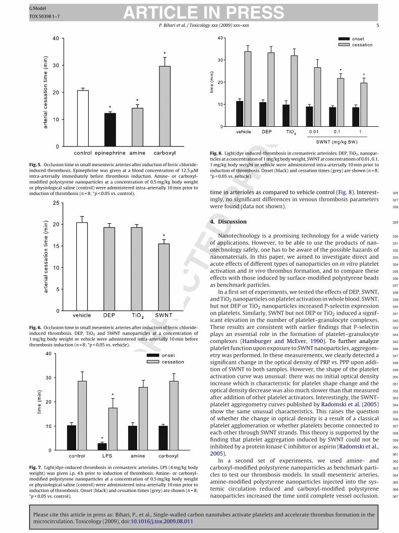
odified polystyrene nanoparticles significantly increased thecclusion time as compared to controls (Fig. 5).
There were no significant differences in arterial occlusion timeetween DEP-, TiO2- or vehicle-treated animals. In contrast, occlu-ion time was significantly decreased in SWNT- as compared toehicle-treated animals (Fig. 6).
ED P
to both solutions. The concentration of nanoparticles was 100 �g/ml. After runningsamples for at least 10 min, ADP was added at a final concentration of 1 �M to PRP.Aggregation in the presence of nanoparticles is given as percentage of that inducedby ADP (*p < 0.05 vs. zero).
3.5. Cremasteric thrombosis
Microvascular thrombotic effects of nanoparticles were ana-lyzed in the cremasteric microcirculation. To assure comparableintravascular FITC dextran concentrations in all experiments, fluo-rescence intensity was measured in one venule in each experiment.There were no significant differences in the fluorescent intensityamong experimental groups (data not shown). As a positive con-trol, LPS significantly decreased the onset time and the cessationtime in arterioles. In contrast to our observation in small mesen-teric arteries, onset and cessation times in mice treated with amine-or carboxyl-modified polystyrene nanoparticles did not differ from
notubes activate platelets and accelerate thrombus formation in the
Fig. 4. Platelet aggregation curves from three representative experiments. Opti-cal density changes of PRP vs. PPP were measured after addition of PBS (1),amine-modified polystyrene (2), or SWNT (3) nanoparticles to both solutions. Theconcentration of the nanoparticles was 100 �g/ml. After running samples for at least10 min, ADP was added at a final concentration of 1 �M to PRP.
Please cite this article in press as: Bihari, P., et al., Single-walled carbon namicrocirculation. Toxicology (2009), doi:10.1016/j.tox.2009.08.011
ARTICLE IN PRESS
UN
CO
RR
ECT
OF
G Model
TOX 50398 1–7
P. Bihari et al. / Toxicology xxx (2009) xxx–xxx 5
Fig. 5. Occlusion time in small mesenteric arteries after induction of ferric chloride-induced thrombosis. Epinephrine was given at a blood concentration of 12.5 �Mintra-arterially immediately before thrombosis induction. Amine- or carboxyl-modified polystyrene nanoparticles at a concentration of 0.5 mg/kg body weightor physiological saline (control) were administered intra-arterially 10 min prior toinduction of thrombosis (n = 8; *p < 0.05 vs. control).
Fig. 6. Occlusion time in small mesenteric arteries after induction of ferric chloride-induced thrombosis. DEP, TiO2 and SWNT nanoparticles at a concentration of1 mg/kg body weight or vehicle were administered intra-arterially 10 min beforethrombosis induction (n = 8; *p < 0.05 vs. vehicle).
Fig. 7. Light/dye-induced thrombosis in cremasteric arterioles. LPS (4 mg/kg bodyweight) was given i.p. 4 h prior to induction of thrombosis. Amine- or carboxyl-modified polystyrene nanoparticles at a concentration of 0.5 mg/kg body weightor physiological saline (control) were administered intra-arterially 10 min prior toinduction of thrombosis. Onset (black) and cessation times (grey) are shown (n = 8;*p < 0.05 vs. control).
Fig. 8. Light/dye induced-thrombosis in cremasteric arterioles. DEP, TiO2, nanopar-
326
327
328
329
330
331
332
333
334
335
336
337
338
339
340
341
342
343
344
345
346
347
348
349
350
351
352
353
354
355
356
357
358
359
360
361
362
ED P
ROticles at a concentration of 1 mg/kg body weight, SWNT at concentrations of 0.01, 0.1,
1 mg/kg body weight or vehicle were administered intra-arterially 10 min prior toinduction of thrombosis. Onset (black) and cessation times (grey) are shown (n = 8;*p < 0.05 vs. vehicle).
time in arterioles as compared to vehicle control (Fig. 8). Interest-ingly, no significant differences in venous thrombosis parameterswere found (data not shown).
4. Discussion
Nanotechnology is a promising technology for a wide varietyof applications. However, to be able to use the products of nan-otechnology safely, one has to be aware of the possible hazards ofnanomaterials. In this paper, we aimed to investigate direct andacute effects of different types of nanoparticles on in vitro plateletactivation and in vivo thrombus formation, and to compare theseeffects with those induced by surface-modified polystyrene beadsas benchmark particles.
In a first set of experiments, we tested the effects of DEP, SWNT,and TiO2 nanoparticles on platelet activation in whole blood. SWNT,but not DEP or TiO2 nanoparticles increased P-selectin expressionon platelets. Similarly, SWNT but not DEP or TiO2 induced a signif-icant elevation in the number of platelet–granulocyte complexes.These results are consistent with earlier findings that P-selectinplays an essential role in the formation of platelet–granulocytecomplexes (Hamburger and McEver, 1990). To further analyzeplatelet function upon exposure to SWNT nanoparticles, aggregom-etry was performed. In these measurements, we clearly detected asignificant change in the optical density of PRP vs. PPP upon addi-tion of SWNT to both samples. However, the shape of the plateletactivation curve was unusual: there was no initial optical densityincrease which is characteristic for platelet shape change and theoptical density decrease was also much slower than that measuredafter addition of other platelet activators. Interestingly, the SWNT-platelet aggregometry curves published by Radomski et al. (2005)show the same unusual characteristics. This raises the questionof whether the change in optical density is a result of a classicalplatelet agglomeration or whether platelets become connected toeach other through SWNT strands. This theory is supported by thefinding that platelet aggregation induced by SWNT could not beinhibited by a protein kinase C inhibitor or aspirin (Radomski et al.,2005).
In a second set of experiments, we used amine- and
notubes activate platelets and accelerate thrombus formation in the
carboxyl-modified polystyrene nanoparticles as benchmark parti- 363
cles to test our thrombosis models. In small mesenteric arteries, 364
amine-modified polystyrene nanoparticles injected into the sys- 365
temic circulation reduced and carboxyl-modified polystyrene 366
nanoparticles increased the time until complete vessel occlusion. 367

IN
T
G
T
6 ology
F368
p369
a370
b371
o372
a373
i374
s375
j376
m377
f378
t379
e380
s381
w382
l383
t384
a385
a386
l387
388
T389
t390
d391
s392
c393
fi394
v395
t396
e397
c398
p399
o400
c401
p402
2403
404
T405
(406
c407
e408
d409
o410
f411
w412
r413
t414
i415
t416
p417
m418
b419
S420
a421
a422
(423
424
t425
c426
m427
r428
b429
430
e431
i432
i433
434
435
436
437
438
439
440
441
442
443
444
445
446
447
448
449
450
451
452
453
454
455
456
457
458
459
460
461
462
463
464
465
466
467
468
469
470
471
472
473
474
475
476
477
478
479
480
481
482
483
484
485
486
487
488
489
490
491
492
493
494
495
496
497
498
ARTICLE
UN
CO
RR
EC
Model
OX 50398 1–7
P. Bihari et al. / Toxic
urthermore, amine-modified polystyrene nanoparticles activatedlatelets as measured by increased expression of platelet P-selectinnd elevated numbers of platelet–granulocyte complexes in wholelood. These results are in agreement with previously publishedbservations (Nemmar et al., 2002b). Interestingly, incubation ofmine-modified polystyrene nanoparticles with platelets did notnduce platelet aggregation in our experiments. These observationsuggest that positively charged polystyrene nanoparticles provokeust modest platelet activation. Interestingly, amine- or carboxyl-
odified polystyrene nanoparticles had no effect on thrombusormation in cremasteric microvessels. A possible explanation forhe inconsistent results in the two thrombosis models might bexplained by methodical differences in the induction of thrombo-is. In the cremaster model, light/dye-induced thrombosis was usedhich, in contrast to ferric chloride application, activates endothe-
ial cells without endothelial denudation. Thus, we suppose thathe antithrombotic and prothrombotic effects of carboxyl- andmine-modified polystyrene beads, respectively, are sufficient toffect endothelial damage-dependent (mesenterium) but endothe-ial damage-independent (cremaster) thrombus formation.
In a final set of experiments, prothrombotic effects of DEP,iO2, and SWNT nanoparticles were investigated in small mesen-eric arteries and in the microcirculation in vivo. Surprisingly, weid not detect platelet activation or augmented thrombosis inmall mesenteric arteries or in the microcirculation upon appli-ation of DEP nanoparticles. These data are in contrast to previousndings demonstrating accelerated thrombus formation in largeessels after instillation (Nemmar et al., 2003a,b, 2004b) or sys-emic administration (Nemmar et al., 2007) of DEPs. A possiblexplanation for this inconsistency might be that DEP nanoparti-les are not able to directly and acutely modulate the thromboticrocess. Also TiO2 nanoparticles had no effect on platelet activationr thrombus formation in small mesenteric arteries or in the micro-irculation. Interestingly, TiO2 nanorods increased ADP-inducedlatelet agglomeration in vitro and decreased the platelet count4 h after instillation (Nemmar et al., 2008).
In small mesenteric arteries, SWNT decreased thrombosis time.hese results are in line with the findings by Radomski et al.2005) who found similar effects of SWNT on thrombosis in thearotid artery of rats. Interestingly, SWNT had a more pronouncedffect in the microcirculation. The cessation time decreased dose-ependently upon application of SWNT. Even low concentrationsf SWNT such as 0.01 mg/kg body weight augmented thrombusormation. Although the cessation times were decreased, thereas only a slight non-significant decline in the onset times. These
esults indicate that platelet–platelet binding or stabilisation of thehrombus has changed rather than the initial platelet–endothelialnteraction. Thus, our observations indicate that SWNT exertheir prothrombotic effects in the microcirculation by activatinglatelets rather than endothelial cells. In addition, SWNT aug-ent platelet aggregation either by activation of platelets or
y mimicking molecular bridges between platelets. Furthermore,WNT-activated platelets have been shown to release gelatinases,nd the changing balance between proaggregatory MMP-2 andntiaggregatory MMP-9 may further augment thrombus formationRadomski et al., 2005).
The crucial difference between DEP or TiO2 and SWNT nanopar-icles regarding prothrombotic effects might be specific surfaceharacteristics and aspect ratio. The surface characteristics of SWNTight be responsible for platelet activation and the high aspect
atio of SWNT may ensure platelet aggregation by forming bridges
Please cite this article in press as: Bihari, P., et al., Single-walled carbon namicrocirculation. Toxicology (2009), doi:10.1016/j.tox.2009.08.011
etween platelets and thus augmenting thrombus formation.The most interesting observation of this study is that SWNT
xert prothrombotic effects not only in small mesenteric arter-es but also in the microcirculation. This finding is of particularmportance since the microcirculation plays an essential role in
PRESS
ED P
RO
OF
xxx (2009) xxx–xxx
various organ functions. Thrombotic events in the microcirculationreduce tissue perfusion and might result in functional disturbances.Moreover, SWNT are also suggested for drug-delivery applications.For effective drug-delivery, however, drug-containing SWNT haveto reach the target cells through the circulation. In this context,microthrombotic effects of SWNT are relevant because trapping ofSWNT by microthrombi may potentially reduce the availability ofdrugs and, therefore, result in therapy failure.
In conclusion, our data demonstrate that DEP and TiO2 nanopar-ticles injected into healthy mice have no effect on platelet activationor thrombus formation, while SWNT induce activation and aggre-gation of platelets, and exert prothrombotic effects in small arteriesas well as in the microcirculation.
Conflict of interest statement
The authors declare that there are no conflicts of interest.
Acknowledgements
This study was supported by European Commission grantNMPT-CT-2006-032777 (NANOSH). The views and opinionsexpressed in this publication do not necessarily reflect those of theEuropean Commission.
References
Andre, P., Delaney, S.M., LaRocca, T., Vincent, D., DeGuzman, F., Jurek, M., Koller, B.,Phillips, D.R., Conley, P.B., 2003. P2Y12 regulates platelet adhesion/activation,thrombus growth, and thrombus stability in injured arteries. J. Clin. Invest. 112,398–406.
Baez, S., 1973. An open cremaster muscle preparation for the study of blood vesselsby in vivo microscopy. Microvasc. Res. 5, 384–394.
Bihari, P., Vippola, M., Schultes, S., Praetner, M., Khandoga, A.G., Reichel, C.A., Coester,C., Tuomi, T., Rehberg, M., Krombach, F., 2008a. Optimized dispersion of nanopar-ticles for biological in vitro and in vivo studies. Part Fibre Toxicol. 5, 14.
Bihari, P., Fent, J., Hamar, J., Furesz, J., Lakatos, S., 2008b. An easy-to-use prac-tical method to measure coincidence in the flow cytometer—the case ofplatelet–granulocyte complex determination. J. Biochem. Biophys. Methods 70,1080–1085.
Borm, P.J., Robbins, D., Haubold, S., Kuhlbusch, T., Fissan, H., Donaldson, K., Schins,R., Stone, V., Kreyling, W., Lademann, J., Krutmann, J., Warheit, D., Oberdorster,E., 2006. The potential risks of nanomaterials: a review carried out for ECETOC.Part Fibre Toxicol. 3, 11.
Brook, R.D., 2008. Cardiovascular effects of air pollution. Clin. Sci. (Lond.) 115,175–187.
Delfino, R.J., Sioutas, C., Malik, S., 2005. Potential role of ultrafine particles in associa-tions between airborne particle mass and cardiovascular health. Environ. HealthPerspect. 113, 934–946.
Fent, J., Bihari, P., Furesz, J., Hamar, J., Lakatos, S., 2008. Impact of coincidence ongranulocyte–platelet complex determination by flow cytometry is evaluated bya novel computer simulation model of coincidence. J. Biochem. Biophys. Meth-ods 70, 1086–1090.
Geys, J., Nemmar, A., Verbeken, E., Smolders, E., Ratoi, M., Hoylaerts, M.F., Nemery,B., Hoet, P.H., 2008. Acute toxicity and prothrombotic effects of quantum dots:impact of surface charge. Environ. Health Perspect. 116, 1607–1613.
Hamburger, S.A., McEver, R.P., 1990. GMP-140 mediates adhesion of stimulatedplatelets to neutrophils. Blood 75, 550–554.
Hudson, S.P., Padera, R.F., Langer, R., Kohane, D.S., 2008. The biocompatibility ofmesoporous silicates. Biomaterials 29, 4045–4055.
Khandoga, A., Stampfl, A., Takenaka, S., Schulz, H., Radykewicz, R., Kreyling, W.,Krombach, F., 2004. Ultrafine particles exert prothrombotic but not inflamma-tory effects on the hepatic microcirculation in healthy mice in vivo. Circulation109, 1320–1325.
Kreyling, W.G., Semmler, M., Erbe, F., Mayer, P., Takenaka, S., Schulz, H., Oberdorster,G., Ziesenis, A., 2002. Translocation of ultrafine insoluble iridium particles fromlung epithelium to extrapulmonary organs is size dependent but very low. J.Toxicol. Environ. Health A 65, 1513–1530.
Maynard, A.D., Aitken, R.J., Butz, T., Colvin, V., Donaldson, K., Oberdorster, G., Philbert,M.A., Ryan, J., Seaton, A., Stone, V., Tinkle, S.S., Tran, L., Walker, N.J., Warheit, D.B.,2006. Safe handling of nanotechnology. Nature 444, 267–269.
notubes activate platelets and accelerate thrombus formation in the
Mills, N.L., Tornqvist, H., Robinson, S.D., Gonzalez, M.C., Soderberg, S., Sandstrom, T., 499
Blomberg, A., Newby, D.E., Donaldson, K., 2007. Air pollution and atherothrom- 500
bosis. Inhal. Toxicol. 19 (Suppl. 1), 81–89. 501
Mills, N.L., Donaldson, K., Hadoke, P.W., Boon, N.A., MacNee, W., Cassee, F.R., Sand- 502
strom, T., Blomberg, A., Newby, D.E., 2009. Adverse cardiovascular effects of air 503
pollution. Nat. Clin. Pract. Cardiovasc. Med. 6, 36–44. 504

ING
T
ology
N505
506
N507
508
509
N510
511
512
N513
514
515
N516
517
518
N519
520
521
N522
523
524
525
526
527
528
529
530
531
532
533
534
535
536
J.F., Burns, A.R., 2006. Endotoxin enhances microvascular thrombosis in mouse 537
ARTICLEModel
OX 50398 1–7
P. Bihari et al. / Toxic
el, A., Xia, T., Madler, L., Li, N., 2006. Toxic potential of materials at the nanolevel.Science 311, 622–627.
emmar, A., Hoet, P.H., Vanquickenborne, B., Dinsdale, D., Thomeer, M., Hoylaerts,M.F., Vanbilloen, H., Mortelmans, L., Nemery, B., 2002a. Passage of inhaled par-ticles into the blood circulation in humans. Circulation 105, 411–414.
emmar, A., Hoylaerts, M.F., Hoet, P.H., Dinsdale, D., Smith, T., Xu, H., Vermylen, J.,Nemery, B., 2002b. Ultrafine particles affect experimental thrombosis in an invivo hamster model. Am. J. Respir. Crit. Care Med. 166, 998–1004.
emmar, A., Hoet, P.H., Dinsdale, D., Vermylen, J., Hoylaerts, M.F., Nemery, B., 2003a.Diesel exhaust particles in lung acutely enhance experimental peripheral throm-bosis. Circulation 107, 1202–1208.
emmar, A., Nemery, B., Hoet, P.H., Vermylen, J., Hoylaerts, M.F., 2003b. Pulmonaryinflammation and thrombogenicity caused by diesel particles in hamsters: roleof histamine. Am. J. Respir. Crit. Care Med. 168, 1366–1372.
Please cite this article in press as: Bihari, P., et al., Single-walled carbon namicrocirculation. Toxicology (2009), doi:10.1016/j.tox.2009.08.011
UN
CO
RR
ECT
emmar, A., Hoylaerts, M.F., Hoet, P.H., Nemery, B., 2004a. Possible mechanismsof the cardiovascular effects of inhaled particles: systemic translocation andprothrombotic effects. Toxicol. Lett. 149, 243–253.
emmar, A., Hoet, P.H., Vermylen, J., Nemery, B., Hoylaerts, M.F., 2004b. Pharmaco-logical stabilization of mast cells abrogates late thrombotic events induced bydiesel exhaust particles in hamsters. Circulation 110, 1670–1677.
PRESSxxx (2009) xxx–xxx 7
Nemmar, A., Hamoir, J., Nemery, B., Gustin, P., 2005. Evaluation of particle transloca-tion across the alveolo-capillary barrier in isolated perfused rabbit lung model.Toxicology 208, 105–113.
Nemmar, A., Al-Maskari, S., Ali, B.H., Al-Amri, I.S., 2007. Cardiovascular and lunginflammatory effects induced by systemically administered diesel exhaust par-ticles in rats. Am. J. Physiol. Lung Cell Mol. Physiol. 292, L664–670.
Nemmar, A., Melghit, K., Ali, B.H., 2008. The acute proinflammatory and prothrom-botic effects of pulmonary exposure to rutile TiO2 nanorods in rats. Exp. Biol.Med. (Maywood) 233, 610–619.
Radomski, A., Jurasz, P., Alonso-Escolano, D., Drews, M., Morandi, M., Malinski, T.,Radomski, M.W., 2005. Nanoparticle-induced platelet aggregation and vascularthrombosis. Br. J. Pharmacol. 146, 882–893.
Rumbaut, R.E., Bellera, R.V., Randhawa, J.K., Shrimpton, C.N., Dasgupta, S.K., Dong,
notubes activate platelets and accelerate thrombus formation in the
ED P
RO
OFcremaster venules via a TLR4-dependent, neutrophil-independent mechanism. 538
Am. J. Physiol. Heart Circ. Physiol. 290, H1671–H1679. 539
Silva, V.M., Corson, N., Elder, A., Oberdorster, G., 2005. The rat ear vein model for 540
investigating in vivo thrombogenicity of ultrafine particles (UFP). Toxicol. Sci. 541
85, 983–989. 542
Copyright © 2022 FDOKUMEN