Ionising radiation immediately impairs synaptic plasticity-associated cytoskeletal signalling...
10
Ionising Radiation Immediately Impairs Synaptic Plasticity-Associated Cytoskeletal Signalling Pathways in HT22 Cells and in Mouse Brain: An In Vitro/In Vivo Comparison Study Stefan J. Kempf 1 , Sonja Buratovic 2 , Christine von Toerne 3 , Simone Moertl 1 , Bo Stenerlo ¨w 4 , Stefanie M. Hauck 3 , Michael J. Atkinson 1,5 , Per Eriksson 2 , Soile Tapio 1 * 1 Institute of Radiation Biology, Helmholtz Zentrum Mu ¨ nchen, German Research Center for Environmental Health GmbH, Neuherberg, Germany, 2 Department of Environmental Toxicology, Uppsala University, Uppsala, Sweden, 3 Research Unit Protein Science, Helmholtz Zentrum Mu ¨ nchen, German Research Center for Environmental Health GmbH, Neuherberg, Germany, 4 Division of Biomedical Radiation Sciences, Rudbeck Laboratory, Uppsala University, Uppsala, Sweden, 5 Chair of Radiation Biology, Technical University Munich, Munich, Germany Abstract Patients suffering from brain malignancies are treated with high-dose ionising radiation. However, this may lead to severe learning and memory impairment. Preventive treatments to minimise these side effects have not been possible due to the lack of knowledge of the involved signalling pathways and molecular targets. Mouse hippocampal neuronal HT22 cells were irradiated with acute gamma doses of 0.5 Gy, 1.0 Gy and 4.0 Gy. Changes in the cellular proteome were investigated by isotope-coded protein label technology and tandem mass spectrometry after 4 and 24 hours. To compare the findings with the in vivo response, male NMRI mice were irradiated on postnatal day 10 with a gamma dose of 1.0 Gy, followed by evaluation of the cellular proteome of hippocampus and cortex 24 hours post-irradiation. Analysis of the in vitro proteome showed that signalling pathways related to synaptic actin-remodelling were significantly affected at 1.0 Gy and 4.0 Gy but not at 0.5 Gy after 4 and 24 hours. We observed radiation-induced reduction of the miR-132 and Rac1 levels; miR-132 is known to regulate Rac1 activity by blocking the GTPase-activating protein p250GAP. In the irradiated hippocampus and cortex we observed alterations in the signalling pathways similar to those in vitro. The decreased expression of miR-132 and Rac1 was associated with an increase in hippocampal cofilin and phospho-cofilin. The Rac1-Cofilin pathway is involved in the modulation of synaptic actin filament formation that is necessary for correct spine and synapse morphology to enable processes of learning and memory. We suggest that acute radiation exposure leads to rapid dendritic spine and synapse morphology alterations via aberrant cytoskeletal signalling and processing and that this is associated with the immediate neurocognitive side effects observed in patients treated with ionising radiation. Citation: Kempf SJ, Buratovic S, von Toerne C, Moertl S, Stenerlo ¨ w B, et al. (2014) Ionising Radiation Immediately Impairs Synaptic Plasticity-Associated Cytoskeletal Signalling Pathways in HT22 Cells and in Mouse Brain: An In Vitro/In Vivo Comparison Study. PLoS ONE 9(10): e110464. doi:10.1371/journal.pone. 0110464 Editor: Xiangming Zha, University of South Alabama, United States of America Received July 17, 2014; Accepted September 8, 2014; Published October 20, 2014 Copyright: ß 2014 Kempf et al. This is an open-access article distributed under the terms of the Creative Commons Attribution License, which permits unrestricted use, distribution, and reproduction in any medium, provided the original author and source are credited. Data Availability: The authors confirm that all data underlying the findings are fully available without restriction. The raw-files of the obtained MS-MS spectra can be found under http://storedb.org/project_details.php?projectid = 38 with the ProjectID 38. Funding: The research was supported by a grant from the European Community’s Seventh Framework Programme (EURATOM) contract no 295552 (CEREBRAD - Cognitive and Cerebrovascular Effects Induced by Low Dose Ionizing Radiation). The funder had no role in study design, data collection, analysis and interpretation, decision to publish, or preparation of the manuscript. Competing Interests: The authors have declared that no competing interests exist. * Email: [email protected] Introduction Ionising radiation is frequently used during treatment of central nervous system (CNS) malignancies. Normally, the patient is exposed to a total radiation dose of 20–50 Gy that is given in fractions of 2–4 Gy to reduce the side-effects. Still, immediate detrimental decline in cognition and visual memory are widely observed [1,2]. Epidemiological studies indicate that even moderate radiation doses may lead to acute and permanent deficits in learning and memory [3–5], in particular if the exposure occurred during childhood [6,7]. Approximately 200,000 children worldwide were treated with X-rays for ringworm of the scalp (Tinea capitis), with head doses ranging from 0.7 to 1.7 Gy [8,9]: Long-term side-effects on cognition were evaluated 10 to 29 years later, showing that psychiatric disorders were more often diagnosed in exposed children than in not exposed ones [10]. A follow-up study with 11,000 irradiated Israeli Tinea capitis children showed similar long-term effects after radiation exposure including lower exam- ination scores, intelligence quotients, and a small increase in the frequency of mental retardation [4]. The cognitive damage in people exposed early in life may be a consequence of the immature state of the brain when ionising radiation was applied. On its way to adolescence, the brain undergoes various remodelling processes on molecular and structural levels called the brain growth spurt [11]. It includes PLOS ONE | www.plosone.org 1 October 2014 | Volume 9 | Issue 10 | e110464
-
Upload
helmholtz-muenchen -
Category
Documents
-
view
0 -
download
0
Transcript of Ionising radiation immediately impairs synaptic plasticity-associated cytoskeletal signalling...
Ionising Radiation Immediately Impairs SynapticPlasticity-Associated Cytoskeletal Signalling Pathways inHT22 Cells and in Mouse Brain: An In Vitro/In VivoComparison StudyStefan J. Kempf1, Sonja Buratovic2, Christine von Toerne3, Simone Moertl1, Bo Stenerlow4,
Stefanie M. Hauck3, Michael J. Atkinson1,5, Per Eriksson2, Soile Tapio1*
1 Institute of Radiation Biology, Helmholtz Zentrum Munchen, German Research Center for Environmental Health GmbH, Neuherberg, Germany, 2 Department of
Environmental Toxicology, Uppsala University, Uppsala, Sweden, 3 Research Unit Protein Science, Helmholtz Zentrum Munchen, German Research Center for
Environmental Health GmbH, Neuherberg, Germany, 4 Division of Biomedical Radiation Sciences, Rudbeck Laboratory, Uppsala University, Uppsala, Sweden, 5 Chair of
Radiation Biology, Technical University Munich, Munich, Germany
Abstract
Patients suffering from brain malignancies are treated with high-dose ionising radiation. However, this may lead to severelearning and memory impairment. Preventive treatments to minimise these side effects have not been possible due to thelack of knowledge of the involved signalling pathways and molecular targets. Mouse hippocampal neuronal HT22 cells wereirradiated with acute gamma doses of 0.5 Gy, 1.0 Gy and 4.0 Gy. Changes in the cellular proteome were investigated byisotope-coded protein label technology and tandem mass spectrometry after 4 and 24 hours. To compare the findings withthe in vivo response, male NMRI mice were irradiated on postnatal day 10 with a gamma dose of 1.0 Gy, followed byevaluation of the cellular proteome of hippocampus and cortex 24 hours post-irradiation. Analysis of the in vitro proteomeshowed that signalling pathways related to synaptic actin-remodelling were significantly affected at 1.0 Gy and 4.0 Gy butnot at 0.5 Gy after 4 and 24 hours. We observed radiation-induced reduction of the miR-132 and Rac1 levels; miR-132 isknown to regulate Rac1 activity by blocking the GTPase-activating protein p250GAP. In the irradiated hippocampus andcortex we observed alterations in the signalling pathways similar to those in vitro. The decreased expression of miR-132 andRac1 was associated with an increase in hippocampal cofilin and phospho-cofilin. The Rac1-Cofilin pathway is involved inthe modulation of synaptic actin filament formation that is necessary for correct spine and synapse morphology to enableprocesses of learning and memory. We suggest that acute radiation exposure leads to rapid dendritic spine and synapsemorphology alterations via aberrant cytoskeletal signalling and processing and that this is associated with the immediateneurocognitive side effects observed in patients treated with ionising radiation.
Citation: Kempf SJ, Buratovic S, von Toerne C, Moertl S, Stenerlow B, et al. (2014) Ionising Radiation Immediately Impairs Synaptic Plasticity-AssociatedCytoskeletal Signalling Pathways in HT22 Cells and in Mouse Brain: An In Vitro/In Vivo Comparison Study. PLoS ONE 9(10): e110464. doi:10.1371/journal.pone.0110464
Editor: Xiangming Zha, University of South Alabama, United States of America
Received July 17, 2014; Accepted September 8, 2014; Published October 20, 2014
Copyright: � 2014 Kempf et al. This is an open-access article distributed under the terms of the Creative Commons Attribution License, which permitsunrestricted use, distribution, and reproduction in any medium, provided the original author and source are credited.
Data Availability: The authors confirm that all data underlying the findings are fully available without restriction. The raw-files of the obtained MS-MS spectracan be found under http://storedb.org/project_details.php?projectid = 38 with the ProjectID 38.
Funding: The research was supported by a grant from the European Community’s Seventh Framework Programme (EURATOM) contract no 295552 (CEREBRAD -Cognitive and Cerebrovascular Effects Induced by Low Dose Ionizing Radiation). The funder had no role in study design, data collection, analysis andinterpretation, decision to publish, or preparation of the manuscript.
Competing Interests: The authors have declared that no competing interests exist.
* Email: [email protected]
Introduction
Ionising radiation is frequently used during treatment of central
nervous system (CNS) malignancies. Normally, the patient is
exposed to a total radiation dose of 20–50 Gy that is given in
fractions of 2–4 Gy to reduce the side-effects. Still, immediate
detrimental decline in cognition and visual memory are widely
observed [1,2]. Epidemiological studies indicate that even
moderate radiation doses may lead to acute and permanent
deficits in learning and memory [3–5], in particular if the exposure
occurred during childhood [6,7].
Approximately 200,000 children worldwide were treated with
X-rays for ringworm of the scalp (Tinea capitis), with head doses
ranging from 0.7 to 1.7 Gy [8,9]: Long-term side-effects on
cognition were evaluated 10 to 29 years later, showing that
psychiatric disorders were more often diagnosed in exposed
children than in not exposed ones [10]. A follow-up study with
11,000 irradiated Israeli Tinea capitis children showed similar
long-term effects after radiation exposure including lower exam-
ination scores, intelligence quotients, and a small increase in the
frequency of mental retardation [4].
The cognitive damage in people exposed early in life may be a
consequence of the immature state of the brain when ionising
radiation was applied. On its way to adolescence, the brain
undergoes various remodelling processes on molecular and
structural levels called the brain growth spurt [11]. It includes
PLOS ONE | www.plosone.org 1 October 2014 | Volume 9 | Issue 10 | e110464

fundamental neuronal architecture changes such as growth of
axons and dendrites to enable the formation and deletion of
synaptic contacts [12]. The brain is especially susceptible to
damage if exposed to ionising radiation during this developmental
period. It has been shown that toxic agents given to mice within
the susceptibility window around postnatal day ten lead to
disruption of adult brain function [13,14]. Further, a synergistic
effect between toxicants and ionising radiation given on postnatal
day ten has been shown [15]. Interestingly, the brain growth spurt
is species-dependent as in human beings it lasts until the age of
three to four years whereas in rodents it corresponds to the second
and fourth postnatal weeks [16].
Especially the hippocampus is a highly radiation-sensitive brain
region involved in learning and memory consolidation. Irradiation
may lead to changes in the neurogenic niche of the dentate gyrus
of the hippocampus by depleting neural stem and progenitor cells
[17–20]. Nevertheless, the low frequency of life-long newly
generated neurons in this region may suggest that other brain
regions and biological targets may also be of importance in the
manifestation of long-lasting cognitive defects after radiation
treatment. It has been shown recently that the mature neuronal
networks of the hippocampus are highly-radiation sensitive [21].
Thus, ionising radiation may have adverse effects on the effective
neurotransmission by altering the synaptic plasticity of the brain.
Synaptic plasticity is a dynamic process involving rapid cytoskel-
etal organisation on the dendrite and spine morphology to
modulate signal transmission. Defects in synaptic plasticity and
dendrite or spine morphology have been observed in cognitive
diseases such as Alzheimer’s [22], Rett syndrome [23] and Down’s
syndrome [24], emphasising not only the role of the hippocampus
but also that of the cortex in this process.
The aim of this study was (i) to determine the role of synaptic
plasticity-associated cytoskeletal signalling pathways in the acute
radiation response in vitro and in vivo and (ii) to compare these
alterations. We show here that dendritic spine morphology-
associated proteins and signalling pathways such as the Rac1-
Cofilin pathway were rapidly altered after in vitro exposure to a
dose of 1.0 Gy in primary immortalised neurons of the mouse
hippocampal cells (HT22). Similar alterations were confirmed in
the hippocampus and cortex of NMRI mice irradiated on
postnatal day ten that represents a developmental stage within
the brain growth spurt in mice.
Materials and Methods
Ethics statement, irradiation of animals and tissuecollection
Experiments were carried out in accordance with the European
Communities Council Directive of 24 November 1986 (86/609/
EEC), after approval from the local ethical committees (Uppsala
University and the Agricultural Research Council) and by the
Swedish Committee for Ethical Experiments on Laboratory
Animals. All animal experiments were performed under trained
personnel, and all efforts were made to minimise animal suffering.
Male NMRI mice were total body irradiated on postnatal day
10 (PND 10) with a single exposure to gamma irradiation (137Cs,
0.20 Gy/min) at doses of 0 (sham-irradiated control) and 1.0 Gy
(Rudbeck Laboratory, Uppsala University). Dose verification was
done with an ionisation chamber (Markus chamber type 23343,
PTW-Freiburg) and was homogeneous within 63% over the
10 cm dish area where mice were positioned during irradiation
procedure. Neonates from each litter were irradiated together.
Animals were sacrificed via cervical dislocation. Brains were
excised and transferred to ice-cold PBS, rinsed carefully, and
dissected under stereomicroscopic inspection under cold condi-
tions. Hippocampi and cortices without meninges from each
hemisphere were separately sampled, gently rinsed in ice-cold PBS
and snap-frozen in liquid nitrogen. Samples were stored at 280uCuntil isolation of protein and RNA.
Irradiation and harvesting of cellsHT22 cells (immortalised primary neurons from the mouse
hippocampus) were kindly provided from J. Lewerenz (Depart-
ment of Neurology, University Hospital Hamburg-Eppendorf,
Hamburg, Germany) [25]. The cells were grown in high glucose
DMEM media (PAA Laboratories, E15-840) supplemented with
10% foetal bovine serum (PAA Laboratories, A15-101) without
antibiotics in T75 tissue flask at 37uC with 5% C02 in air. They
were irradiated in the exponential growth phase with doses of
0 Gy (sham), 0.5 Gy, 1.0 Gy or 4.0 Gy of c-rays (137Cs, 0.48 Gy/
min) (HWM-D 2000, Waelischmiller Engineering, Germany). For
each dose group and time point, four independent flasks were
seeded and irradiated. At four and 24 hours post-irradiation cells
were rinsed with ice-cold PBS and enzymatically detached with
accutase (Invitrogen). After blocking of the accutase reaction with
media containing 10% foetal bovine serum and splitting of cell
volume in two equal parts for total RNA and protein isolation of
each tissue flask, the cells were centrifuged and washed once with
ice-cold PBS. This centrifugation and washing step was repeated,
followed by cell pelleting via centrifugation. Pelleted cells were
frozen at 280uC until total protein and RNA content were
isolated.
Isolation of total protein and RNAa) Isolation of total protein. HT22 cell pellets or individual
frozen hippocampi and cortices were homogenised with 6 M
guanidine hydrochloride (SERVA Electrophoresis GmbH, Ger-
many) on ice using a manual plastic mortar. Homogenates were
briefly vortexed, sonicated, and cleared by centrifugation
(20,0006g, 1 hour, 4uC). The supernatants were collected and
stored at 220uC before further use. Total protein content was
determined using Bradford assay (Thermo Fisher) following the
manufacturer’s instructions.
b) Total RNA isolation. Total RNA from HT22 cell pellets
or individual frozen hippocampi and cortices was isolated and
purified by mirVanaTM Isolation Kit (Ambion) according to the
manufacturer’s instructions. Total RNA was eluted with nuclease-
free water. The optical density (OD) ratio of 260/280 was
measured using a Nanodrop spectrophotometer (PeqLab Biotech-
nology; Germany); it ranged between 1.9 and 2.1. Eluates were
stored at 220uC until further analysis.
Mass spectrometry-based proteome analysisa) Isotope coded protein label (ICPL) analysis of proteins,
1D PAGE separation and in-gel digest. In total, four
individual replicates of HT22 cells were used for proteomic
analysis at each radiation dose and time point. Total protein
lysates were labelled with ICPL reagents (SERVA Electrophoresis
GmbH, Germany) according to the manufacturer’s instructions.
Briefly, individual protein lysates (20 mg in 20 ml of 6 M guanidine
hydrochloride from each biological sample) were reduced,
alkylated and labelled with the respective ICPL-reagent as follows:
control with ICPL-0, 0.5 Gy sample with ICPL-4, 1.0 Gy sample
with ICPL-6 and 4.0 Gy sample with ICPL-10. All labelled
samples representing each radiation dose at one time point (4 and
24 hours) were combined and overnight precipitated with 80%
acetone at 220uC to purify the labelled protein content.
Ionising Radiation Affects Synaptic Plasticity in Brain
PLOS ONE | www.plosone.org 2 October 2014 | Volume 9 | Issue 10 | e110464

Biological replicates from the in vivo mouse study included
animals from at least three different litters. Four biological
replicates from hippocampus and five from cortex were used for
both control and irradiated groups. The samples were labelled
with ICPL reagents as follows: control with ICPL-0 and 1.0 Gy
sample with ICPL-6. These labelled samples were further treated
as described for the HT22 cells.
Protein precipitates were separated by 12% SDS-polyacryl-
amide gel electrophoresis followed by Coomassie Blue staining.
Gel lanes were cut into at least four equal slices, destained, and
trypsinised overnight as described recently [26]. Peptides were
extracted and acidified with 1% formic acid followed by analysis
via mass spectrometry.
b) LC-MS/MS analysis. LC-MS/MS analysis was per-
formed as described previously on a LTQ-Orbitrap XL (Thermo
Fisher) [27]. Briefly, pre-fractionated samples were automatically
injected and loaded onto the trap column and after 5 min, peptides
were eluted and separated on the analytical column by reversed
phase chromatography operated on a nano-HPLC (Ultimate 3000,
Dionex) with a nonlinear 170 min gradient using 35% acetonitrile
in 0.1% formic acid in water (A) and 0.1% formic acid in 98%
acetonitrile (B) at a flow rate of 300 nl/min. The gradient settings
were: 5–140 min: 14.5–90% A, 140–145 min: 90% A 295% B,
145–150 min: 95% B followed by equilibration for 15 min to
starting conditions. From the MS pre-scan, the 10 most abundant
peptide ions were selected for fragmentation in the linear ion trap if
they exceeded an intensity of at least 200 counts and were at least
doubly charged. During fragment analysis, a high-resolution
(60,000 full-width half maximum) MS spectrum was acquired in
the Orbitrap with a mass range from 200 to 1500 Da.
c) Identification and quantification of proteins. MS-MS
spectra were searched against the ENSEMBL mouse database
(Version: 2.4, 56416 sequences) via MASCOT (version 2.3.02;
Matrix Science) with a mass tolerance of 10 ppm for peptide
precursors and 0.6 Da for MS-MS peptide fragments, including not
more than one missed cleavage. Fixed modifications included
carbamidomethylation of cysteine and ICPL-0, ICPL-4, ICPL-6
and ICPL-10 for lysine. Proteins were identified and quantified
based on the ICPL pairs using the Proteome Discoverer software
(Version 1.3– Thermo Fisher). To ensure that only high-confident
identified peptides were used for protein quantification, the
MASCOT percolator algorithm was applied [28]. The percolator
is an algorithm that improves the discrimination between correct
and incorrect spectrum identifications and gives a q value sising the
statistical confidence assigned to each peptide-spectra-match [29].
The q value was set to 0.01 representing strict peptide ranking. Only
the best ranked peptides were used. Such peptides were filtered
against Decoy database resulting into a false discovery rate (FDR) of
each LC-MS-run; the significance threshold was set to 0.01 to
ensure that only highly confident peptide identifications were used
for protein quantification. Proteins from each LC-MS-run were
normalised against the median of all quantifiable proteins. Proteins
were considered significantly deregulated if they fulfilled the
following criteria: (i) identification by at least two unique peptides
in n-1 mass-spectrometry runs (n: number of biological replicates),
(ii) quantification with an ICPL-variability of #30% and (iii) a fold-
change of $1.3 or # 21.3. The threshold of 61.3 is based on our
average experimental technical variance of the multiple analysis of
hippocampal and cortical technical replicates (13.8%).
Data deposition of proteomics experimentsThe raw-files of the obtained MS-MS spectra can be found
under http://storedb.org/project_details.php?projectid=38 with
the ProjectID 38.
Bioinformatics analysisDeregulated proteins were assigned to functional classes using
PANTHER classification system software (http://www.
pantherdb.org) and the general annotation from UniProt
(http://uniprot.org). To identify radiation-affected signalling
pathways, a signalling pathway analysis was performed with all
deregulated proteins for each dose group using INGENUITY
Pathway Analysis (IPA) (http://www.ingenuity.com) applying
databases of experimental and predictive origin.
Quantification of Rac1, cofilin and phospho-cofilinexpression levels via immunoblotting
Protein extracts of cells and brain tissues (15 mg) were separated
on 12% SDS polyacrylamide gels and transferred to nitrocellulose
membranes (GE Healthcare) via BIO-RAD CriterionTM Blotter
system at 100 V for 2 h. Membranes were blocked with RotiR-
Block solution (Roth), washed and incubated overnight at 4uCwith primary antibody dilutions as recommended by the
manufacturer (GAPDH – sc-47724 [murine monoclonal IgG1
raised against recombinant GAPDH of human origin; Santa
Cruz], Rac1– ab33186 [murine monoclonal IgG2b raised against
full-length recombinant Rac1 of human origin; Abcam], cofilin
–3312 [rabbit polyclonal antibody produced by immunising rabbits
with a synthetic peptide corresponding to residues surrounding Ser3
of human cofilin origin; Cell Signalling], p-Cofilin (Ser3) –3311
[rabbit polyclonal antibody by immunising animals with a synthetic
phospho-peptide corresponding to residues surrounding Ser3 of
human cofilin; Cell Signalling]). After a washing step, blots were
incubated with appropriate horseradish peroxidase-conjugated
secondary antibody in 8% milk for 1 h at room temperature and
developed using ECL system (GE Healthcare) using standard
protocol from the manufacturer. GAPDH was not significantly
deregulated based on the global proteomics results in any sample
and was therefore used as a loading control. Immunoblots were
quantified with TotalLab TL100 software (www.totallab.com) using
software-suggested background correction. Three or four biological
replicates were used for statistical analysis (unpaired Student’s t-test)
with a significance threshold of 0.05.
Quantification of microRNA miR-132 via quantitative PCRRNA isolates of cells and brain tissues (10 ng) were used to
quantify microRNA miR-132 expression levels using the TaqMan
Single MicroRNA Assay (Applied Biosystems) according to the
manufacturer’s protocol. Steps included a reverse transcription
and real-time PCR (StepOnePlus) via Taqman-primers (mmu-
miR-132 (ID000457), snoRNA135 (ID001239) – Life Technolo-
gies). Expression levels of miRNA were calculated based on the
22DDCt method with normalisation against endogenous
snoRNA135 [30]. Changes were considered significant if they
reached a p-value of #0.05 (unpaired Student’s t-test, n = 4
[in vitro] and n = 3 [in vivo] per dose group and time point).
Results
Acute effects of ionising radiation on synapticplasticity-associated cytoskeletal signalling pathwaysin vitro
We used HT22 cells as an in vitro model to detect radiation-
induced alterations in synaptic plasticity-associated cytoskeletal
signalling pathways. HT22 cells were irradiated with doses of
0.5 Gy, 1.0 Gy and 4.0 Gy, followed by a global quantitative
proteome analysis 4 and 24 hours post-irradiation. The protein
quantification showed a dose-dependency in the number of
Ionising Radiation Affects Synaptic Plasticity in Brain
PLOS ONE | www.plosone.org 3 October 2014 | Volume 9 | Issue 10 | e110464
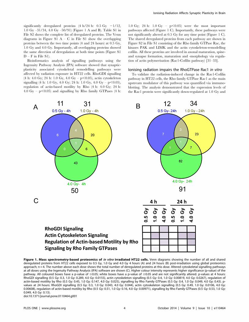
significantly deregulated proteins (4 h/24 h: 0.5 Gy 21/12,
1.0 Gy –31/34, 4.0 Gy –50/91) (Figure 1 A and B). Table S1 in
File S2 shows the complete list of deregulated proteins. The Venn
diagrams in Figure S1 A – C in File S1 show the overlapping
proteins between the two time points (4 and 24 hours) at 0.5 Gy,
1.0 Gy and 4.0 Gy. Importantly, all overlapping proteins showed
the same direction of deregulation at both time points (Figure S1
D – F in File S1).
Bioinformatics analysis of signalling pathways using the
Ingenuity Pathway Analysis (IPA) software showed that synaptic-
plasticity associated cytoskeletal remodelling pathways were
affected by radiation exposure in HT22 cells. RhoGDI signalling
(4 h: 4.0 Gy; 24 h: 1.0 Gy, 4.0 Gy – p,0.05), actin cytoskeleton
signalling (4 h: 1.0 Gy, 4.0 Gy; 24 h: 1.0 Gy, 4.0 Gy – p,0.05),
regulation of actin-based motility by Rho (4 h: 4.0 Gy; 24 h:
4.0 Gy – p,0.05) and signalling by Rho family GTPases (4 h:
1.0 Gy; 24 h: 1.0 Gy – p,0.05) were the most important
pathways affected (Figure 1 C). Importantly, these pathways were
not significantly altered at 0.5 Gy for any time point (Figure 1 C).
The shared deregulated proteins from each pathway are shown in
Figure S2 in File S1 consisting of the Rho family GTPase Rac, the
kinases PAK and LIMK and the actin cytoskeleton-remodelling
cofilin. All these proteins are involved in axonal maturation, spine-
and synapse formation, maturation and -morphology via regula-
tion of actin polymerisation (Rac1-Cofilin pathway) [31–33].
Ionising radiation impairs the RhoGTPase Rac1 in vitroTo validate the radiation-induced change in the Rac1-Cofilin
pathway in HT22 cells, the Rho family GTPase Rac1 as the main
upstream modulator of this pathway was quantified via immuno-
blotting. The analysis demonstrated that the expression levels of
the Rac1 protein were significantly down-regulated at 1.0 Gy and
Figure 1. Mass spectrometry-based proteomics of in vitro irradiated HT22 cells. Venn diagrams showing the number of all and sharedderegulated proteins from HT22 cells exposed to 0.5 Gy, 1.0 Gy and 4.0 Gy 4 hours (A) and 24 hours (B) post-irradiation using global proteomicsapproach; n = 4. The number above each dose shows the total number of deregulated proteins at this dose. Altered cytoskeletal signalling pathwaysat all doses using the Ingenuity Pathway Analysis (IPA) software are shown (C). Higher colour intensity represents higher significance (p-value) of thepathway. All coloured boxes have a p-value of #0.05; white boxes have a p-value of $0.05 and are not significantly altered. p-values at 4 hours:RhoGDI signalling (0.5 Gy: 0.3, 1.0 Gy: 0.289, 4.0 Gy: 0.0155), actin cytoskeleton signalling (0.5 Gy: 0.4, 1.0 Gy: 0.00819, 4.0 Gy: 0.0267), regulation ofactin-based motility by Rho (0.5 Gy: 0.45, 1.0 Gy: 0.147, 4.0 Gy: 0.025), signalling by Rho Family GTPases (0.5 Gy: 0.4, 1.0 Gy: 0.049, 4.0 Gy: 0.43). p-values at 24 hours: RhoGDI signalling (0.5 Gy: 0.3, 1.0 Gy: 0.043, 4.0 Gy: 0.044), actin cytoskeleton signalling (0.5 Gy: 0.49, 1.0 Gy: 0.0106, 4.0 Gy:0.00608), regulation of actin-based motility by Rho (0.5 Gy: 0.51, 1.0 Gy: 0.16, 4.0 Gy: 0.00971), signalling by Rho Family GTPases (0.5 Gy: 0.53, 1.0 Gy:0.049, 4.0 Gy: 0.13).doi:10.1371/journal.pone.0110464.g001
Ionising Radiation Affects Synaptic Plasticity in Brain
PLOS ONE | www.plosone.org 4 October 2014 | Volume 9 | Issue 10 | e110464

4.0 Gy at both time points (Figure 2 A and B). The Rac1 protein
levels at 0.5 Gy were significantly decreased at 4 hours but
returned to control levels after 24 hours post-irradiation (Figure 2 A
and B). These data are in good agreement with the changes in Rac1
expression levels obtained by global proteomics approach at 24 hours
post-irradiation (1.0 Gy: 21.43620.3%; 4.0 Gy: 21.40615.9%)
(Table S1 in File S2).
As miR-132 is involved in the regulation of the Rac1-Cofilin
pathway via blocking of the GTP hydrolysis protein p250GAP
[31], the levels of this microRNA were quantified. The miR-132
levels were significantly decreased at 0.5 Gy but not at higher
doses 4 hours post-irradiation whereas 24 hours post-irradiation
miR-132 levels were still significantly decreased at 0.5 Gy and
were also decreased at the higher doses (Figure 2 C). Importantly,
the changes observed for miR-132 expression originated from the
alterations in the expression of miR-132 and not in the expression
of endogenous standard snoRNA135 (Figure S5 in File S1).
Figures S5 E and S5 F in File S1 show the variation of the n-fold
changes of snoRNA135 used for miRNA normalisation after
4 hours and 24 hours post-irradiation and the DCt values between
miR-132 and snoRNA135 in irradiated HT22 cells, respectively.
Only small variances in the expression of snoRNA135 were
detectable whereas the DCt values were more affected meaning
that miRNA changes originated from radiation-induced differ-
ences in miR-132 expression profile. The error bars were in all
conditions comparable.
Acute effects of ionising radiation on synapticplasticity-associated cytoskeleton signalling pathwaysin vivo
To examine possible radiation-induced effects on spine- and
synapse formation, maturation and morphology, we irradiated
male NMRI mice on postnatal day ten – a developmental stage of
maximal cytoskeleton remodelling in dendritic spines and synapses
within the brain growth spurt period. We irradiated neonates
using a dose of 1.0 Gy and performed experiments 24 hours post-
irradiation as this dose was the lowest showing persistent effect on
the Rac1 expression and signalling pathway alterations in the
irradiated HT22 cells (Figure 1 and 2). The analysis was
performed using both hippocampus and cortex as these two brain
regions are involved in learning and memory formation [34,35].
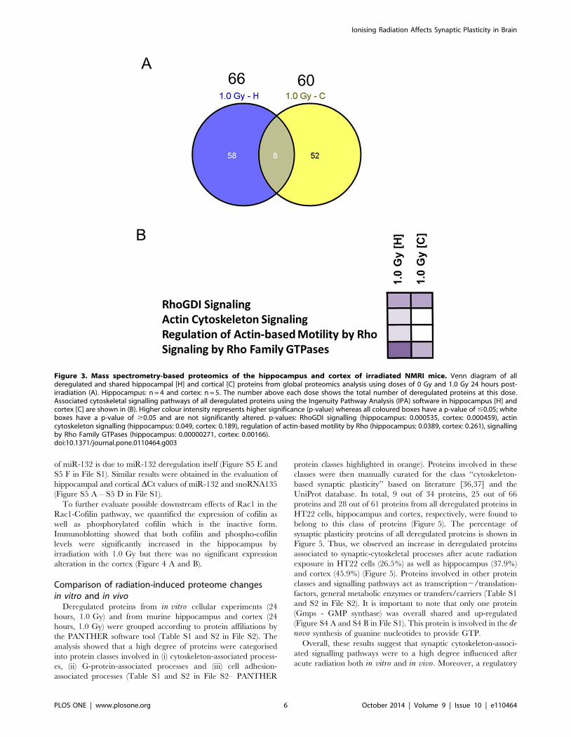
The protein quantification via global proteomics showed that 66
and 60 proteins were deregulated in the hippocampus and cortex
24 hours after exposure to 1.0 Gy, respectively (Figure 3 A). Eight
proteins were found to be shared and up-regulated in these two
brain regions (Figure S3 in File S1). Table S2 in File S2 shows the
complete list of the in vivo deregulated proteins.
Signalling pathway analysis using the IPA software tool
demonstrated that, similar to the in vitro cellular study, synap-
tic-plasticity associated cytoskeletal pathways were affected in vivoby radiation exposure: RhoGDI signalling (hippocampus and
cortex – p,0.05), actin cytoskeleton signalling (hippocampus – p,
0.05), regulation of actin-based motility by Rho (hippocampus –
p,0.05) and signalling by Rho family GTPases (hippocampus and
cortex – p,0.05) (Figure 3 B).
Ionising radiation impairs the RhoGTPase Rac1 and itsdownstream target cofilin in vivo
Quantification of Rac1 protein levels by immunoblotting
confirmed a significant decrease in hippocampus and cortex at
1.0 Gy (Figure 4 A and B) to a similar extent as in the HT22 cells
(Figure 2 A and B). In accordance with decreasing miR-132 levels
in HT22 cells 24 hours post-irradiation with 1.0 Gy (Figure 2 C),
we observed significantly decreased miR-132 levels in both
hippocampus and cortex at 1.0 Gy 24 hours post-irradiation
(Figure 4 C). HT22 data highlighted that the observed deregulation
Figure 2. Immunoblotting and miRNA quantification of using HT22 cells. Data from immunoblotting (A–B) and miRNA quantification (C)associated to the Rac1-Cofilin pathway in HT22 cells irradiated with 0 Gy, 0.5 Gy, 1.0 Gy and 4.0 Gy 4 hours and 24 hours post-irradiation. Thecolumns represent the fold-changes with standard errors of the mean (SEM); n = 3 for immunoblotting, n = 4 for miRNA quantification. Thevisualisation of protein bands shows the representative change from the biological replicates. *p,0.05; **p,0.01; ***p,0.001 (unpaired Student’s t-test). Normalisation was performed against endogenous GAPDH and endogenous snoRNA135 for immunoblotting and miRNA quantification,respectively.doi:10.1371/journal.pone.0110464.g002
Ionising Radiation Affects Synaptic Plasticity in Brain
PLOS ONE | www.plosone.org 5 October 2014 | Volume 9 | Issue 10 | e110464
of miR-132 is due to miR-132 deregulation itself (Figure S5 E and
S5 F in File S1). Similar results were obtained in the evaluation of
hippocampal and cortical DCt values of miR-132 and snoRNA135
(Figure S5 A – S5 D in File S1).
To further evaluate possible downstream effects of Rac1 in the
Rac1-Cofilin pathway, we quantified the expression of cofilin as
well as phosphorylated cofilin which is the inactive form.
Immunoblotting showed that both cofilin and phospho-cofilin
levels were significantly increased in the hippocampus by
irradiation with 1.0 Gy but there was no significant expression
alteration in the cortex (Figure 4 A and B).
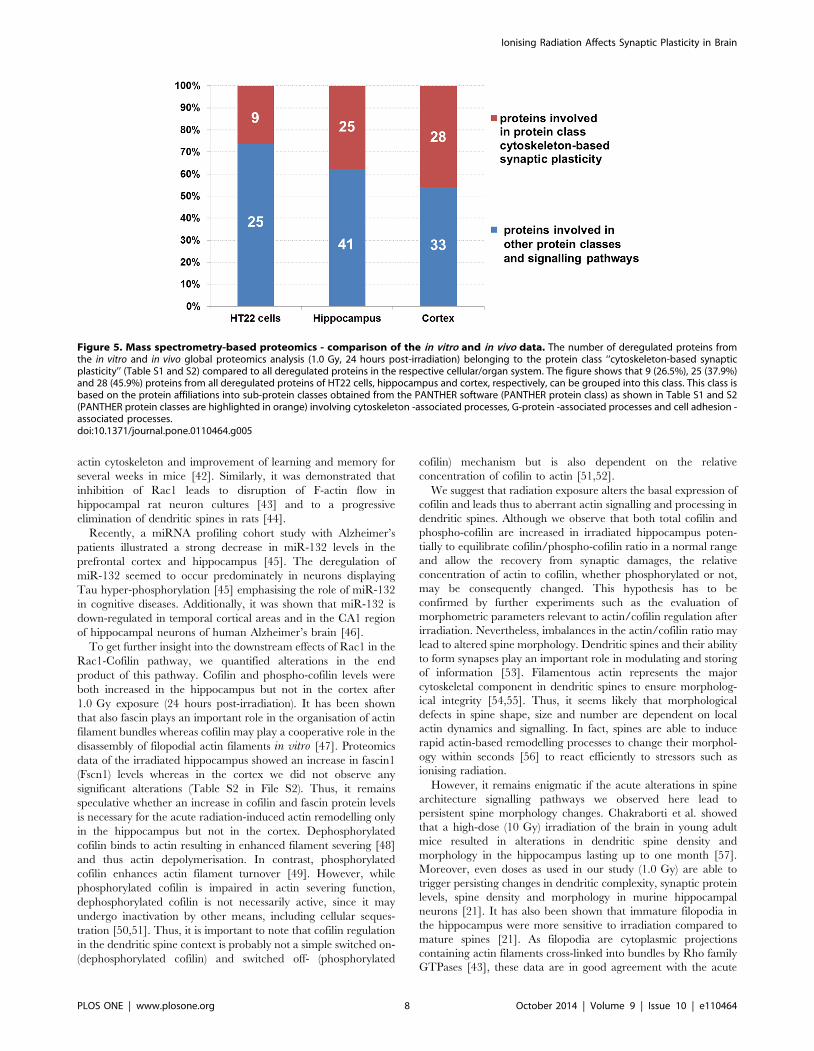
Comparison of radiation-induced proteome changesin vitro and in vivo
Deregulated proteins from in vitro cellular experiments (24
hours, 1.0 Gy) and from murine hippocampus and cortex (24
hours, 1.0 Gy) were grouped according to protein affiliations by
the PANTHER software tool (Table S1 and S2 in File S2). The
analysis showed that a high degree of proteins were categorised
into protein classes involved in (i) cytoskeleton-associated process-
es, (ii) G-protein-associated processes and (iii) cell adhesion-
associated processes (Table S1 and S2 in File S2– PANTHER
protein classes highlighted in orange). Proteins involved in these
classes were then manually curated for the class ‘‘cytoskeleton-
based synaptic plasticity’’ based on literature [36,37] and the
UniProt database. In total, 9 out of 34 proteins, 25 out of 66
proteins and 28 out of 61 proteins from all deregulated proteins in
HT22 cells, hippocampus and cortex, respectively, were found to
belong to this class of proteins (Figure 5). The percentage of
synaptic plasticity proteins of all deregulated proteins is shown in
Figure 5. Thus, we observed an increase in deregulated proteins
associated to synaptic-cytoskeletal processes after acute radiation
exposure in HT22 cells (26.5%) as well as hippocampus (37.9%)
and cortex (45.9%) (Figure 5). Proteins involved in other protein
classes and signalling pathways act as transcription2/translation-
factors, general metabolic enzymes or transfers/carriers (Table S1
and S2 in File S2). It is important to note that only one protein
(Gmps - GMP synthase) was overall shared and up-regulated
(Figure S4 A and S4 B in File S1). This protein is involved in the denovo synthesis of guanine nucleotides to provide GTP.
Overall, these results suggest that synaptic cytoskeleton-associ-
ated signalling pathways were to a high degree influenced after
acute radiation both in vitro and in vivo. Moreover, a regulatory
Figure 3. Mass spectrometry-based proteomics of the hippocampus and cortex of irradiated NMRI mice. Venn diagram of allderegulated and shared hippocampal [H] and cortical [C] proteins from global proteomics analysis using doses of 0 Gy and 1.0 Gy 24 hours post-irradiation (A). Hippocampus: n = 4 and cortex: n = 5. The number above each dose shows the total number of deregulated proteins at this dose.Associated cytoskeletal signalling pathways of all deregulated proteins using the Ingenuity Pathway Analysis (IPA) software in hippocampus [H] andcortex [C] are shown in (B). Higher colour intensity represents higher significance (p-value) whereas all coloured boxes have a p-value of #0.05; whiteboxes have a p-value of $0.05 and are not significantly altered. p-values: RhoGDI signalling (hippocampus: 0.000535, cortex: 0.000459), actincytoskeleton signalling (hippocampus: 0.049, cortex: 0.189), regulation of actin-based motility by Rho (hippocampus: 0.0389, cortex: 0.261), signallingby Rho Family GTPases (hippocampus: 0.00000271, cortex: 0.00166).doi:10.1371/journal.pone.0110464.g003
Ionising Radiation Affects Synaptic Plasticity in Brain
PLOS ONE | www.plosone.org 6 October 2014 | Volume 9 | Issue 10 | e110464

network involving miRNA and GTPases is indicated in this
process.
Discussion
The aim of this study was to elucidate the biological
mechanisms involved in the acute radiation-induced side-effects
on learning and memory as seen in patients treated with
radiotherapy. We first used immortalised primary neurons from
mouse hippocampus (HT22 cells) to get information about
affected signalling pathways. We then validated these data in vivoby using the lowest radiation dose inducing non-transient
signalling pathway alterations in the cell culture system to irradiate
male NMRI mice on postnatal day ten with subsequent analysis of
the affected signalling pathways in the hippocampus and cortex.
Pathway analysis of the in vitro data showed that the doses of
1.0 Gy and 4.0 Gy, but not 0.5 Gy, significantly altered the
expression of proteins functionally involved in RhoGDI signalling,
actin cytoskeleton signalling, regulation of actin-based motility by
Rho as well as signalling by Rho family GTPases.
Rho family GTPases are key regulators of actin cytoskeleton
and are essential for orchestrating spine and synapse morphology
[37]. Their activity is controlled at least in part via RhoGDI
proteins [38]. Overall, these pathways shared several proteins such
as Rac1, PAK, LIMK and cofilin that all are constituents of the
Rac1-Cofilin pathway. Quantification of Rac1 and miR-132 levels
demonstrated a dose- and time-dependent reduction in both only
at doses of 1.0 Gy and 4.0 Gy and 24 hours post irradiation. miR-
132 is known to indirectly positively regulate Rac1 activity by
blocking the GTPase-activating protein p250GAP [39,40]. Thus a
decrease in miR-132 would lead to a decrease in Rac1 activity as
we observed.
Similar signalling pathways were affected in murine hippocam-
pus and cortex at 1.0 Gy 24 hours after the exposure as in the cell
culture system. Also the Rac1 and miR-132 levels were similarly
down-regulated as in vitro at this experimental set-up. Overall, the
decrease in Rac1 expression and activity as observed in vitro and
in vivo may lead to a presumptive aberrant actin remodelling in
dendritic spines.
It has been shown that selective deletion of Rac1 in excitatory
neurons in vivo affects spine structure, impairs synaptic plasticity
and spatial learning [41]. Chemical inactivation of Rac1 impairs
long-term plasticity in the mouse hippocampus [32]. In contrast,
activation of the cerebral Rac1 leads to rearrangement of cerebral
Figure 4. Immunoblotting and miRNA quantification of the in vivo data. Data from immunoblotting (A–B) and miRNA quantification (C)associated to the Rac1-Cofilin pathway in hippocampus [H] and cortex [C] from NMRI mice exposed on postnatal day 10 with doses of 0 Gy and1.0 Gy. The measurement was performed 24 hours post-irradiation. The columns represent the fold-changes with standard errors of the mean (SEM);immunoblotting: n = 4 for Rac1 detection; n = 3 for p-cofilin and cofilin detection; n = 3 for miRNA quantification. The visualisation of protein bandsshows the representative change from the biological replicates. *p,0.05; **p,0.01; ***p,0.001 (unpaired Student’s t-test). Normalisation wasperformed against endogenous GAPDH and endogenous snoRNA135 for immunoblotting and miRNA quantification, respectively.doi:10.1371/journal.pone.0110464.g004
Ionising Radiation Affects Synaptic Plasticity in Brain
PLOS ONE | www.plosone.org 7 October 2014 | Volume 9 | Issue 10 | e110464
actin cytoskeleton and improvement of learning and memory for
several weeks in mice [42]. Similarly, it was demonstrated that
inhibition of Rac1 leads to disruption of F-actin flow in
hippocampal rat neuron cultures [43] and to a progressive
elimination of dendritic spines in rats [44].
Recently, a miRNA profiling cohort study with Alzheimer’s
patients illustrated a strong decrease in miR-132 levels in the
prefrontal cortex and hippocampus [45]. The deregulation of
miR-132 seemed to occur predominately in neurons displaying
Tau hyper-phosphorylation [45] emphasising the role of miR-132
in cognitive diseases. Additionally, it was shown that miR-132 is
down-regulated in temporal cortical areas and in the CA1 region
of hippocampal neurons of human Alzheimer’s brain [46].
To get further insight into the downstream effects of Rac1 in the
Rac1-Cofilin pathway, we quantified alterations in the end
product of this pathway. Cofilin and phospho-cofilin levels were
both increased in the hippocampus but not in the cortex after
1.0 Gy exposure (24 hours post-irradiation). It has been shown
that also fascin plays an important role in the organisation of actin
filament bundles whereas cofilin may play a cooperative role in the
disassembly of filopodial actin filaments in vitro [47]. Proteomics
data of the irradiated hippocampus showed an increase in fascin1
(Fscn1) levels whereas in the cortex we did not observe any
significant alterations (Table S2 in File S2). Thus, it remains
speculative whether an increase in cofilin and fascin protein levels
is necessary for the acute radiation-induced actin remodelling only
in the hippocampus but not in the cortex. Dephosphorylated
cofilin binds to actin resulting in enhanced filament severing [48]
and thus actin depolymerisation. In contrast, phosphorylated
cofilin enhances actin filament turnover [49]. However, while
phosphorylated cofilin is impaired in actin severing function,
dephosphorylated cofilin is not necessarily active, since it may
undergo inactivation by other means, including cellular seques-
tration [50,51]. Thus, it is important to note that cofilin regulation
in the dendritic spine context is probably not a simple switched on-
(dephosphorylated cofilin) and switched off- (phosphorylated
cofilin) mechanism but is also dependent on the relative
concentration of cofilin to actin [51,52].
We suggest that radiation exposure alters the basal expression of
cofilin and leads thus to aberrant actin signalling and processing in
dendritic spines. Although we observe that both total cofilin and
phospho-cofilin are increased in irradiated hippocampus poten-
tially to equilibrate cofilin/phospho-cofilin ratio in a normal range
and allow the recovery from synaptic damages, the relative
concentration of actin to cofilin, whether phosphorylated or not,
may be consequently changed. This hypothesis has to be
confirmed by further experiments such as the evaluation of
morphometric parameters relevant to actin/cofilin regulation after
irradiation. Nevertheless, imbalances in the actin/cofilin ratio may
lead to altered spine morphology. Dendritic spines and their ability
to form synapses play an important role in modulating and storing
of information [53]. Filamentous actin represents the major
cytoskeletal component in dendritic spines to ensure morpholog-
ical integrity [54,55]. Thus, it seems likely that morphological
defects in spine shape, size and number are dependent on local
actin dynamics and signalling. In fact, spines are able to induce
rapid actin-based remodelling processes to change their morphol-
ogy within seconds [56] to react efficiently to stressors such as
ionising radiation.
However, it remains enigmatic if the acute alterations in spine
architecture signalling pathways we observed here lead to
persistent spine morphology changes. Chakraborti et al. showed
that a high-dose (10 Gy) irradiation of the brain in young adult
mice resulted in alterations in dendritic spine density and
morphology in the hippocampus lasting up to one month [57].
Moreover, even doses as used in our study (1.0 Gy) are able to
trigger persisting changes in dendritic complexity, synaptic protein
levels, spine density and morphology in murine hippocampal
neurons [21]. It has also been shown that immature filopodia in
the hippocampus were more sensitive to irradiation compared to
mature spines [21]. As filopodia are cytoplasmic projections
containing actin filaments cross-linked into bundles by Rho family
GTPases [43], these data are in good agreement with the acute
Figure 5. Mass spectrometry-based proteomics - comparison of the in vitro and in vivo data. The number of deregulated proteins fromthe in vitro and in vivo global proteomics analysis (1.0 Gy, 24 hours post-irradiation) belonging to the protein class ‘‘cytoskeleton-based synapticplasticity’’ (Table S1 and S2) compared to all deregulated proteins in the respective cellular/organ system. The figure shows that 9 (26.5%), 25 (37.9%)and 28 (45.9%) proteins from all deregulated proteins of HT22 cells, hippocampus and cortex, respectively, can be grouped into this class. This class isbased on the protein affiliations into sub-protein classes obtained from the PANTHER software (PANTHER protein class) as shown in Table S1 and S2(PANTHER protein classes are highlighted in orange) involving cytoskeleton -associated processes, G-protein -associated processes and cell adhesion -associated processes.doi:10.1371/journal.pone.0110464.g005
Ionising Radiation Affects Synaptic Plasticity in Brain
PLOS ONE | www.plosone.org 8 October 2014 | Volume 9 | Issue 10 | e110464

alterations in the Rac1-Cofilin pathway found in our study.
Importantly, the Rac1 protein regulates the synaptic maturation
and integration of adult born neurons in the hippocampus [58,59].
Deregulation of Rac1 may even play an important role in defects
of hippocampal adult neurogenesis that are observed in several
radiation exposure studies [17–20].
Overall, we show that ionising radiation leads to acute changes
in cytoskeletal signalling pathways associated with spine morphol-
ogy in vitro and in vivo, by altering the molecular players within
the Rac1-Cofilin-pathway. An understanding of the signalling
pathway alterations after acute radiation exposure is essential in
order to prevent immediate side-effects affecting learning and
memory in accidentally exposed persons and patients treated with
radiotherapy against brain tumours.
Supporting Information
File S1 Supporting figures. Figure S1, Mass spectrometry-
based proteomics changes in HT22 cells as a function of time.
Venn diagrams of all and shared deregulated proteins as a function
of time (4 hours vs. 24 hours) from HT22 cells exposed to 0.5 Gy
(A), 1.0 Gy (B) and 4.0 Gy (C) from global proteomics approach;
n = 4 for each radiation dose. Detailed protein information
(protein name, fold-changes, protein variability) of time-dependent
overlapping deregulated proteins is given in D, E and F. Figure S2,
Visualisation of cytoskeletal synaptic-plasticity signalling pathways
and their overlapping proteins. Visualisation of pathways involved
in RhoGDI signalling, regulation of actin-based motility by Rho,
regulation by Rho Family GTPases and actin cytoskeleton
signalling and their overlapping proteins as highlighted in red
boxes. The images were downloaded from Ingenuity Signalling
Pathway Analysis (IPA) software. Figure S3, Mass spectrometry-
based proteomics of in vivo – comparison of brain regions.
Overlapping deregulated proteins in hippocampus [H] and cortex
[C] with protein names, fold-changes, and protein variability
24 hours post-irradiation using global proteomics approach;
hippocampus: n = 4 and cortex: n = 5. Figure S4, Mass spectrom-
etry-based proteomics of in vivo and in vitro – comparison of
overlapping proteins in a similar experimental set-up. Overlapping
deregulated proteins in HT22 cells, hippocampus [H] and cortex
[C] at 1.0 Gy 24 hours post-irradiation are shown (A). The
protein Gmps that was found deregulated in all analysis is shown
with protein name, fold-changes, and protein variability using
global proteomics approach (B); HT22 cells and hippocampus:
n = 4 and cortex: n = 5. Figure S5, Visualisation of the fold
changes of snoRNA135 and DCt values between miRNA-135 and
snoRNA135 in control and irradiated cells and tissues. The
columns represent the fold changes of snoRNA135 in miRNA
normalisation (A, C, E) and DCt values of the differences between
the Ct values of miRNA-135 and snoRNA135 (B, D, and F) with
standard errors of the mean (SEM) in vitro (HT22 cells: n = 4 for
4 hours and 24 hours post-irradiation) and in vivo (hippocampus
[H]: n = 3 for 24 hours post-irradiation; cortex [C]: n = 3 for
24 hours post-irradiation) experimental set-ups.
(PDF)
File S2 Supporting tables. Table S1, Deregulated proteins
found in mass spectrometry-based proteomics in in vitro irradiat-
ed HT22 cells. Complete detailed list of deregulated proteins
(name, unique peptides, n-fold-change, variability and counts per
biological replicate) obtained from HT22 cell experiment 4 hours
and 24 hours post-irradiation at doses of 0.5 Gy, 1.0 Gy and
4.0 Gy from global proteomics analysis. Deregulated proteins at
1.0 Gy 24 hours after radiation exposure were categorised into
protein classes using PANTHER classification system software and
the general annotation from UniProt as indicated by an asterisk.
Table S2, Deregulated proteins found in mass spectrometry-based
proteomics of the hippocampus and cortex of irradiated NMRI
mice. Complete detailed list of deregulated proteins in hippocam-
pus and cortex (name, unique peptides, n-fold-change, variability
and counts per biological replicate) obtained from NMRI mice
experiment 24 hours post-irradiation at doses of 1.0 Gy from
global proteomics analysis. Deregulated proteins at 1.0 Gy
24 hours after radiation exposure were categorised into protein
classes using PANTHER classification system software and the
general annotation from UniProt as indicated by an asterisk.
(XLS)
Acknowledgments
We thank Stefanie Winkler and Sandra Helm for their outstanding
technical assistance. We thank J. Lewerenz for sharing the HT22 cells.
Author Contributions
Contributed reagents/materials/analysis tools: CT SM BS SH PE.
Conceived and designed the experiments: SK PE ST. Performed the
experiments: SK SB CT BS. Analyzed the data: SK. Contributed to the
writing of the manuscript: SK ST MA PE SH SM.
References
1. Hoffman KE, Yock TI (2009) Radiation therapy for pediatric central nervous
system tumors. J Child Neurol 24: 1387–1396.
2. Spiegler BJ, Bouffet E, Greenberg ML, Rutka JT, Mabbott DJ (2004) Change in
neurocognitive functioning after treatment with cranial radiation in childhood.
J Clin Oncol 22: 706–713.
3. Hall P, Adami HO, Trichopoulos D, Pedersen NL, Lagiou P, et al. (2004) Effect
of low doses of ionising radiation in infancy on cognitive function in adulthood:
Swedish population based cohort study. BMJ 328: 19.
4. Ron E, Modan B, Floro S, Harkedar I, Gurewitz R (1982) Mental function
following scalp irradiation during childhood. Am J Epidemiol 116: 149–160.
5. Pearce MS, Salotti JA, Little MP, McHugh K, Lee C, et al. (2012) Radiation
exposure from CT scans in childhood and subsequent risk of leukaemia and
brain tumours: a retrospective cohort study. Lancet 380: 499–505.
6. Fouladi M, Gilger E, Kocak M, Wallace D, Buchanan G, et al. (2005)
Intellectual and functional outcome of children 3 years old or younger who have
CNS malignancies. J Clin Oncol 23: 7152–7160.
7. Kempf SJ, Azimzadeh O, Atkinson MJ, Tapio S (2013) Long-term effects of
ionising radiation on the brain: cause for concern? Radiat Environ Biophys 52:
5–16.
8. Schulz RJ, Albert RE (1968) Follow-up study of patients treated by x-ray
epilation for tinea capitis. 3. Dose to organs of the head from the x-ray treatment
of tinea capitis. Arch Environ Health 17: 935–950.
9. Cipollaro AC, Kallos A, Ruppe JP, Jr. (1959) Measurement of gonadal
radiations during treatment for tinea capitis. N Y State J Med 59: 3033–3040.
10. Omran AR, Shore RE, Markoff RA, Friedhoff A, Albert RE, et al. (1978)
Follow-up study of patients treated by X-ray epilation for tinea capitis:
psychiatric and psychometric evaluation. Am J Public Health 68: 561–567.
11. Dobbing J, Sands J (1979) Comparative aspects of the brain growth spurt. Early
Hum Dev 3: 79–83.
12. Huttenlocher PR, Dabholkar AS (1997) Regional differences in synaptogenesis
in human cerebral cortex. J Comp Neurol 387: 167–178.
13. Eriksson P, Ankarberg E, Fredriksson A (2000) Exposure to nicotine during a
defined period in neonatal life induces permanent changes in brain nicotinic
receptors and in behaviour of adult mice. Brain Res 853: 41–48.
14. Eriksson P (1997) Developmental neurotoxicity of environmental agents in the
neonate. NeuroToxicology 18: 719–726.
15. Eriksson P, Fischer C, Stenerlow B, Fredriksson A, Sundell-Bergman S (2010)
Interaction of gamma-radiation and methyl mercury during a critical phase of
neonatal brain development in mice exacerbates developmental neurobehav-
ioural effects. NeuroToxicology 31: 223–229.
16. Dobbing J, Sands J (1973) Quantitative growth and development of human
brain. Arch Dis Child 48: 757–767.
17. Rola R, Raber J, Rizk A, Otsuka S, VandenBerg SR, et al. (2004) Radiation-
induced impairment of hippocampal neurogenesis is associated with cognitive
deficits in young mice. Experimental Neurology 188: 316–330.
Ionising Radiation Affects Synaptic Plasticity in Brain
PLOS ONE | www.plosone.org 9 October 2014 | Volume 9 | Issue 10 | e110464

18. Allen AR, Eilertson K, Sharma S, Schneider D, Baure J, et al. (2013) Effects of
radiation combined injury on hippocampal function are modulated in micedeficient in chemokine receptor 2 (CCR2). Radiat Res 180: 78–88.
19. Raber J, Rola R, LeFevour A, Morhardt D, Curley J, et al. (2004) Radiation-
induced cognitive impairments are associated with changes in indicators ofhippocampal neurogenesis. Radiat Res 162: 39–47.
20. Mizumatsu S, Monje ML, Morhardt DR, Rola R, Palmer TD, et al. (2003)Extreme sensitivity of adult neurogenesis to low doses of X-irradiation. Cancer
Res 63: 4021–4027.
21. Parihar VK, Limoli CL (2013) Cranial irradiation compromises neuronalarchitecture in the hippocampus. Proc Natl Acad Sci U S A 110: 12822–12827.
22. Tsamis IK, Mytilinaios GD, Njau NS, Fotiou FD, Glaftsi S, et al. (2010)Properties of CA3 dendritic excrescences in Alzheimer’s disease. Curr Alzheimer
Res 7: 84–90.23. Armstrong DD, Dunn K, Antalffy B (1998) Decreased dendritic branching in
frontal, motor and limbic cortex in Rett syndrome compared with trisomy 21.
J Neuropathol Exp Neurol 57: 1013–1017.24. Becker LE, Armstrong DL, Chan F (1986) Dendritic atrophy in children with
Down’s syndrome. Ann Neurol 20: 520–526.25. Sahin M, Saxena A, Joost P, Lewerenz J, Methner A (2006) Induction of Bcl-2
by functional regulation of G-protein coupled receptors protects from oxidative
glutamate toxicity by increasing glutathione. Free Radic Res 40: 1113–1123.26. Merl J, Ueffing M, Hauck SM, von Toerne C (2012) Direct comparison of MS-
based label-free and SILAC quantitative proteome profiling strategies in primaryretinal Muller cells. Proteomics 12: 1902–1911.
27. von Toerne C, Kahle M, Schafer A, Ispiryan R, Blindert M, et al. (2013) Apoe,Mbl2, and Psp plasma protein levels correlate with diabetic phenotype in NZO
mice-an optimized rapid workflow for SRM-based quantification. J Proteome
Res 12: 1331–1343.28. Yentrapalli R, Azimzadeh O, Sriharshan A, Malinowsky K, Merl J, et al. (2013)
The PI3K/Akt/mTOR Pathway Is Implicated in the Premature Senescence ofPrimary Human Endothelial Cells Exposed to Chronic Radiation. PLoS One 8:
e70024.
29. Brosch M, Yu L, Hubbard T, Choudhary J (2009) Accurate and sensitivepeptide identification with Mascot Percolator. J Proteome Res 8: 3176–3181.
30. Shaltiel G, Hanan M, Wolf Y, Barbash S, Kovalev E, et al. (2013) HippocampalmicroRNA-132 mediates stress-inducible cognitive deficits through its acetyl-
cholinesterase target. Brain Struct Funct 218: 59–72.31. Saneyoshi T, Fortin DA, Soderling TR (2010) Regulation of spine and synapse
formation by activity-dependent intracellular signaling pathways. Curr Opin
Neurobiol 20: 108–115.32. Martinez LA, Tejada-Simon MV (2011) Pharmacological inactivation of the
small GTPase Rac1 impairs long-term plasticity in the mouse hippocampus.Neuropharmacology 61: 305–312.
33. Kuhn TB, Meberg PJ, Brown MD, Bernstein BW, Minamide LS, et al. (2000)
Regulating actin dynamics in neuronal growth cones by ADF/cofilin and rhofamily GTPases. J Neurobiol 44: 126–144.
34. Kirwan CB, Wixted JT, Squire LR (2008) Activity in the medial temporal lobepredicts memory strength, whereas activity in the prefrontal cortex predicts
recollection. J Neurosci 28: 10541–10548.35. Clopath C (2012) Synaptic consolidation: an approach to long-term learning.
Cogn Neurodyn 6: 251–257.
36. Fortin DA, Srivastava T, Soderling TR (2012) Structural modulation ofdendritic spines during synaptic plasticity. Neuroscientist 18: 326–341.
37. Tolias KF, Duman JG, Um K (2011) Control of synapse development andplasticity by Rho GTPase regulatory proteins. Prog Neurobiol 94: 133–148.
38. Dovas A, Couchman JR (2005) RhoGDI: multiple functions in the regulation of
Rho family GTPase activities. Biochem J 390: 1–9.
39. Magill ST, Cambronne XA, Luikart BW, Lioy DT, Leighton BH, et al. (2010)
microRNA-132 regulates dendritic growth and arborization of newborn neurons
in the adult hippocampus. Proc Natl Acad Sci U S A 107: 20382–20387.
40. Impey S, Davare M, Lesiak A, Fortin D, Ando H, et al. (2010) An activity-
induced microRNA controls dendritic spine formation by regulating Rac1-PAK
signaling. Mol Cell Neurosci 43: 146–156.
41. Haditsch U, Leone DP, Farinelli M, Chrostek-Grashoff A, Brakebusch C, et al.
(2009) A central role for the small GTPase Rac1 in hippocampal plasticity and
spatial learning and memory. Mol Cell Neurosci 41: 409–419.
42. Diana G, Valentini G, Travaglione S, Falzano L, Pieri M, et al. (2007)
Enhancement of learning and memory after activation of cerebral Rho
GTPases. Proc Natl Acad Sci U S A 104: 636–641.
43. Tatavarty V, Das S, Yu J (2012) Polarization of actin cytoskeleton is reduced in
dendritic protrusions during early spine development in hippocampal neuron.
Mol Biol Cell 23: 3167–3177.
44. Nakayama AY, Harms MB, Luo L (2000) Small GTPases Rac and Rho in the
maintenance of dendritic spines and branches in hippocampal pyramidal
neurons. J Neurosci 20: 5329–5338.
45. Lau P, Bossers K, Janky R, Salta E, Frigerio CS, et al. (2013) Alteration of the
microRNA network during the progression of Alzheimer’s disease. EMBO Mol
Med 5: 1613–1634.
46. Wong HK, Veremeyko T, Patel N, Lemere CA, Walsh DM, et al. (2013) De-
repression of FOXO3a death axis by microRNA-132 and 2212 causes neuronal
apoptosis in Alzheimer’s disease. Hum Mol Genet 22: 3077–3092.
47. Breitsprecher D, Koestler SA, Chizhov I, Nemethova M, Mueller J, et al. (2011)
Cofilin cooperates with fascin to disassemble filopodial actin filaments. J Cell Sci
124: 3305–3318.
48. Pavlov D, Muhlrad A, Cooper J, Wear M, Reisler E (2007) Actin filament
severing by cofilin. J Mol Biol 365: 1350–1358.
49. Maloney MT, Bamburg JR (2007) Cofilin-mediated neurodegeneration in
Alzheimer’s disease and other amyloidopathies. Mol Neurobiol 35: 21–44.
50. van Rheenen J, Condeelis J, Glogauer M (2009) A common cofilin activity cycle
in invasive tumor cells and inflammatory cells. J Cell Sci 122: 305–311.
51. Bamburg JR, Bernstein BW (2010) Roles of ADF/cofilin in actin polymerization
and beyond. F1000 Biol Rep 2: 62.
52. Andrianantoandro E, Pollard TD (2006) Mechanism of actin filament turnover
by severing and nucleation at different concentrations of ADF/cofilin. Mol Cell
24: 13–23.
53. Kasai H, Matsuzaki M, Noguchi J, Yasumatsu N, Nakahara H (2003) Structure-
stability-function relationships of dendritic spines. Trends Neurosci 26: 360–368.
54. Cohen RS, Chung SK, Pfaff DW (1985) Immunocytochemical localization of
actin in dendritic spines of the cerebral cortex using colloidal gold as a probe.
Cell Mol Neurobiol 5: 271–284.
55. Fifkova E, Delay RJ (1982) Cytoplasmic actin in neuronal processes as a possible
mediator of synaptic plasticity. J Cell Biol 95: 345–350.
56. Fischer M, Kaech S, Wagner U, Brinkhaus H, Matus A (2000) Glutamate
receptors regulate actin-based plasticity in dendritic spines. Nat Neurosci 3: 887–
894.
57. Chakraborti A, Allen A, Allen B, Rosi S, Fike JR (2012) Cranial irradiation alters
dendritic spine density and morphology in the hippocampus. PLoS One 7:
e40844.
58. Vadodaria KC, Jessberger S (2013) Maturation and integration of adult born
hippocampal neurons: signal convergence onto small Rho GTPases. Front
Synaptic Neurosci 5: 4.
59. Luo L (2002) Actin cytoskeleton regulation in neuronal morphogenesis and
structural plasticity. Annu Rev Cell Dev Biol 18: 601–635.
Ionising Radiation Affects Synaptic Plasticity in Brain
PLOS ONE | www.plosone.org 10 October 2014 | Volume 9 | Issue 10 | e110464