Best-in-cl - Healthcare-in-Europe
216
RADBOOK 2016 €19.- • IT • CT • MRI • Interventional • Mammo • R / F • Nuc • Displays / Printers • Ultrasound • Injectors • Testing Devices The Radiology Guide to Technology and Informatics in Europe With 270° isocentric rotation, Infinix-i Rite Edition provides unparalleled flexibility and patient access even during the most challenging procedures. Its double c-arm design with 210° coverage and ultrafast rotation of 80°/s enables shorter breath hold times, reduced contrast medium and outstanding 3D imaging from head to toe without the need for moving the patient or the table.
-
Upload
khangminh22 -
Category
Documents
-
view
1 -
download
0
Transcript of Best-in-cl - Healthcare-in-Europe

RAD
BOO
K 20
16 ·
The
Radi
olog
y Gui
de to
Tech
nolo
gy &
Info
rmat
ics in
Euro
pe
RADBOOK 2016 RADBOOK 20161 3
RADBOOK 2016€ 19.-
• IT • CT • MRI • Interventional • Mammo • R / F • Nuc • Displays / Printers• Ultrasound • Injectors • Testing Devices
The Radiology Guide to Technology and Informatics in Europe
With 270° isocentric rotation, Infinix-i Rite Edition provides unparalleled flexibility and patient access even during the most challenging procedures. Its double c-arm design with 210° coverage and ultrafast rotation of 80°/s enables shorter breath hold times,
reduced contrast medium and outstanding 3D imaging from head to toe without the need for moving the patient or the table.
OPEN.PrOvEN.NEW.O-arm® Surgical Imaging System
Find out more in the chapterInterventional Systems
UC
20
16
06
64
8E
N ©
Med
tro
nic,
Inc.
20
16
. All
Rig
hts
Res
erve
d.
Best-in-classEquipped with the largest available FPD at43 x 43 cm and Shimadzu’s digital imagingplatform, the Sonialvision G4 covers thewidest possible range of examinationswith inter-departmental hospital capability.In both functionality and operability, theSonialvision G4 multipurpose R/F table isfar beyond other R/F systems. It provides“Best-in-class” features.
• Smart system architecturesupports outstanding clinical flexibilityfor a wide range of examinations
• Comprehensive dose management packageensures today’s highest safety of patientsand operators
• Excellent image qualityprovided by the advanced “SUREengine”technology enhancing the entire image forclearer details
• Premium application softwaresupporting useful applications, such astomosynthesis for general radiographicimaging
www.shimadzu-medical.eu
Next generation digital R/F system: Sonialvision G4

Management Archiving Image Distribution Workstations
RIS Small Business PACS Enterprise PACS Cardiology PACS Long Term Multimedia Inhouse Teleradiology Portal Solution Cloud Computing Application Multimodality Mammography Orthopedics Cardiology CAD Advanced
VisualizationOrbis RIS Impax Impax Impax for Cardiology ICIS
VNAICISHYDMedia
ICISEnterprise Imaging SuiteXero Viewer
ICISEnterprise Imaging SuiteXero Viewer
ICIS ICIS Impax Clinical Applications Impax Mammography Impax Clinical Applications Impax Clinical Applications Impax Clinical Applications Agfa HealthCareSeptestraat 27 · 2640 Mortsel, Belgiumtel +32 3 444 94 [email protected] · www.agfa.com
Canon Healthcare IT Suite Canon Healthcare IT Suite Canon Healthcare IT Suite Canon Healthcare IT Suite Canon Healthcare IT Suite Canon Healthcare IT Suite Canon Healthcare IT Suite Canon Healthcare IT Suite Canon Healthcare IT Suite Canon PACS Canon PACS CAD for Tuberculosis Canon PACS Canon Europa NVBovenkerkerweg 59 · 1185 XB Amstelveen, The Netherlandstel +31 20 545 8 [email protected] · www.canon-europe.com/medical
CHILI Modality PACS CHILI PACS CHILI PACS CHILI PACS CHILI PACS CHILI/Web CHILI/Web CHILI/Telemedicine Record OmniPACS CHILI Diagnost CHILI Diagnost CHILI Diagnost CHILI Diagnost Partner-Solution Partner-Solution CHILI GmbH Friedrich-Ebert-Str. 2 · 69221 Dossenheim/Heidelberg, Germany tel +49 6221 1 80 79 10 [email protected] · www.chili-radiology.com
Suitestensa RIS Suitestensa PACS Suitestensa PACS Suitestensa CVIS Suitestensa Archive Suitestensa Suitestensa WebSuitestensa Mobile
Suitestensa WebSuitestensa Mobile
Suitestensa Web Suitestensa WebSuitestensa Mobile
Suitestensa Review Suitestensa MG Suitestensa Review Suitestensa Review Cardio Suitestensa 3D Suitestensa Vascular
EBIT S.r.l. – Esaote Group Via Siffredi 58 · 16153 Genoa, Italy tel +39 010 65 47-464 [email protected] · www.esaote.com/healthcare-it
Xplore WebXplore AnalyticsXplore Nuclear medicine
Xplore PACS Solution Xplore PACS Solution Xplore PACS Solution Xplore PACS Solution Xplore Web Xplore Web Xplore Web Xplore Web EDL SAS1031, chemin de la Seyne à Bastian, 83500 La Seyne-sur-mer, Francetel +33 4 94 10 99 [email protected] · www.xplore.eu
Centricity RISi with eRadCockpit
Centricity PACS with Universal Viewer
Centricity PACS with Universal Viewer
Centricity PACS with Universal Viewer
Centricity PACS with Universal Viewer
Centricity Clinical Archive (VNA L1-L4, XDS Repository)
Centricity PACS with Universal Viewer Zero Footprint
Centricity PACS with Universal Viewer Zero Footprint,Centricity 360
Centricity RIS with eRadCockpit,Centricity 360
Centricity 360 Centricity PACS Universal ViewerUniversal Viewer Zero Footprint XDS enabled
Centricity PACS Universal Viewer web client provides Breast Imaging tools powered by IDI
Centricity PACS Universal Viewer with integrated Traumacad by Voyant Health
Centricity Cardio Enterprise Centricity PACS Universal Viewer web client embeds advanced visualization powered by AW
Centricity PACS Universal Viewer web client embeds advanced visualization powered by AW
GE HealthcareLerchenbergstr. 15 · 89160 Dornstadt, Germanytel +49 7348 [email protected] · www.gehealthcare.com
iQ-RIS MED-TAB iQ-SYSTEM PACS MED-TAB MED-TAB MED-TAB MED-TAB iQ-VIEW PRO iQ-VIEW PRO MAMMO TOMO iQ-VIEW PRO OrthoView
iQ-VIEW PRO 4D iQ-VIEW PRO 4D IMAGE Information Systems Europe GmbHLange Str. 16 · 18055 Rostock, Germanytel +49 381 496 58 [email protected] · www.image-systems.biz
RadCentreRadCentre Analytics
RadCentre Multi-PACS Integration
RadCentre Multi-PACS Integration
RadCentre Archiving Solution RadCentre Archiving Solution Health Relations RC Health Relations RC Health Relations RC RadCentre as a Service i-SOLUTIONS Health GmbHAm Exerzierplatz 14 · 68167 Mannheim, Germanytel +49 621 39 [email protected] · www.i-solutions.de
ITZ Hyper.RIS ITZ Hyper.ePACS ITZ Hyper.PACS ITZ Hyper.PACS ITZ Hyper.ARC ITZ Hyper.PACSITZ Hyper.WEB
ITZ Hyper.PACSITZ Hyper.WEB
ITZ Hyper.TELEMED,ITZ Hyper.COMDicom2Mail-Module
ITZ Hyper.WEBITZ Hyper.TELEMEDITZ Hyper.COM ITZ Hyper.UP
ITZ Hyper.PACS Telearchive ITZ Hyper.WEB CloudITZ Hyper.ARC Cloud
ITZ Hyper.PACS ITZ Hyper.PACS ITZ Hyper.PACSHectecRSA-Biomedical Localite
Hyper.PACSPIE-Medical (Esaote)Tomtec
ITZ Hyper.PACS MPR / MIP / 3DTerarecon
ITZ Hyper.PACSIntrasenseTerareconMedian
ITZ Medicom GmbH & Co. KGSiemensring 44 a · 47877 Willich, Germanytel +49 2154 [email protected] · www.itz-medi.com
AciesImagePilot
Acies AciesImagePilot
AciesImagePilot
AciesImagePilot
AciesImagePilot
AciesImagePilot
Acies Acies Acies Acies Konica Minolta Medical & Graphic Imaging Europe B.V.Hoogoorddreef 9 · 1101 BA Amsterdam, The Netherlandstel +31 20 658 41 [email protected] · www.konicaminolta.eu/healthcare
mediCAD Classic mediCAD mobilemediCAD Practice GOmediCAD veterinary
mediCAD Classic mediCAD mobilemediCAD Practice GOmediCAD veterinary
mediCAD Hectec GmbHOpalstr. 54 · 84032 Altdorf, Germanytel +49 871 33 02 [email protected] · www.mediCAD.eu
WinRadiolog RIS ImageBroker XS ImageBroker ImageBroker ImageBroker ImageBroker ImageWeb webConnect PraxisPortal PraxisPortal App ImageVision Diagnost MammoView ImageVision Basic ImageVision Diagnost MammoView CAD ImageVision Diagnost medigration GmbHSchuhstr. 30 · 91052 Erlangen, Germanytel +49 9131 [email protected] · www.medigration.de
IntelliSpace PACS IntelliSpace PACS IntelliSpace cardiovascularXcelera
IntelliSpace PACS IntelliSpace PACS IntelliSpace PACSEnterprise
IntelliSpace PACSRadiology
IntelliSpace PACS IntelliSpace PACSRadiologyIntelliSpace Portal
IntelliSpace PACSIntelliSpace Breast
IntelliSpacecardiovascular
IntelliSpace Portal Philips HealthcareP.O. Box 10.000 · 5680 DA Best, The Netherlandstel +31 40 278 56 [email protected] · www.philips.com/healthcare
CONAXX 2 and PROPAXX PROPAXX and / or CONAXX 2 PROPAXX and / or CONAXX 2 PROPAXX and / or CONAXX 2 PROTEC GmbH & Co. KGIn den Dorfwiesen 14 · 71720 Oberstenfeld, Germanytel +49 7062 [email protected] · www.protec-med.com
syngo.plaza syngo.plaza syngo Dynamics syngo.plaza syngo.plaza syngo.plazasyngo.via WebViewer
syngo.plazasyngo.via WebViewer
syngo.plaza teamplaysyngo.plaza
syngo.viasyngo.plaza
syngo.via syngo.plaza
MediCAD (HECTEC) syngo.viasyngo.plaza
syngo Dynamicssyngo.via
syngo CAD Applicationssyngo.viasyngo.plaza
syngo.via Siemens Healthcare Headquarters · Siemens Healthcare GmbHHenkestr. 127 · 91052 Erlangen, Germanytel +49 9131 [email protected] · www.siemens.com/healthcare
syngo.share and sense syngo.share and sense syngo.share and sense syngo.share and sense syngo.share and sense syngo.share and sense syngo.share and sense ITH icoserve technology for healthcare GmbHInnrain 98 · 6020 Innsbruck, Austriatel +43 512 890 [email protected] · www.ith-icoserve.com · Distributed by Siemens
VioSuite – Image DataManagement (VNA)
Vitrea VitreaView
VitreaVitreaView
VitreaView VitreaView VitreaAdvanced VitreaAdvanced Vitrea Vitrea Vital Images Europe B.V.Zilverstraat 1, 2718 RP Zoetermeer, The Netherlands tel +31 79 206 58 [email protected] · www.vitalimages.com
RADBOOK 2016 RADBOOK 2016
IT-SOLUTIONS IT-SOLUTIONS
Ensure the right person has the right images at
the right time.
VioSuiteTM Data Management and Interoperability
Vitrea® Advanced Visualisation
VitreaView®
Universal Viewer
Vitality SolutionsTM Imaging Intelligence
Unleash the Power of Imaging
www.vitalimages.com
Ensure the right person has the right images at
the right time.
Vitality SolutionsTM Imaging Intelligence
Vitrea® Advanced Visualisation
VitreaView®
Universal Viewer
VioSuiteTM Data Management and Interoperability
Unleash the Power of Imaging
www.vitalimages.com

RAD
BOO
K 20
16 ·
The
Radi
olog
y Gui
de to
Tech
nolo
gy &
Info
rmat
ics in
Euro
pe
RADBOOK 2016 RADBOOK 20161 3
RADBOOK 2016€ 19.-
• IT • CT • MRI • Interventional • Mammo • R / F • Nuc • Displays / Printers• Ultrasound • Injectors • Testing Devices
The Radiology Guide to Technology and Informatics in Europe
With 270° isocentric rotation, Infinix-i Rite Edition provides unparalleled flexibility and patient access even during the most challenging procedures. Its double c-arm design with 210° coverage and ultrafast rotation of 80°/s enables shorter breath hold times,
reduced contrast medium and outstanding 3D imaging from head to toe without the need for moving the patient or the table.
OPEN.PrOvEN.NEW.O-arm® Surgical Imaging System
Find out more in the chapterInterventional Systems
UC
20
16
06
64
8E
N ©
Med
tro
nic,
Inc.
20
16
. All
Rig
hts
Res
erve
d.
Best-in-classEquipped with the largest available FPD at43 x 43 cm and Shimadzu’s digital imagingplatform, the Sonialvision G4 covers thewidest possible range of examinationswith inter-departmental hospital capability.In both functionality and operability, theSonialvision G4 multipurpose R/F table isfar beyond other R/F systems. It provides“Best-in-class” features.
• Smart system architecturesupports outstanding clinical flexibilityfor a wide range of examinations
• Comprehensive dose management packageensures today’s highest safety of patientsand operators
• Excellent image qualityprovided by the advanced “SUREengine”technology enhancing the entire image forclearer details
• Premium application softwaresupporting useful applications, such astomosynthesis for general radiographicimaging
www.shimadzu-medical.eu
Next generation digital R/F system: Sonialvision G4
Best-in-classEquipped with the largest available FPD at43 x 43 cm and Shimadzu’s digital imagingplatform, the Sonialvision G4 covers thewidest possible range of examinationswith inter-departmental hospital capability.In both functionality and operability, theSonialvision G4 multipurpose R/F table isfar beyond other R/F systems. It provides“Best-in-class” features.
• Smart system architecturesupports outstanding clinical flexibilityfor a wide range of examinations
• Comprehensive dose management packageensures today’s highest safety of patientsand operators
• Excellent image qualityprovided by the advanced “SUREengine”technology enhancing the entire image forclearer details
• Premium application softwaresupporting useful applications, such astomosynthesis for general radiographicimaging
www.shimadzu-medical.eu
Next generation digital R/F system: Sonialvision G4

RADBOOK 2016 5
Dear Reader,radiology is and continues to be a fascinating discipline. This is
the tenth edition of RadBook and we are overwhelmed by
the breakneck speed of technological innovation in imaging
systems that produce an ever increasing flow of diagnostically
relevant information.
Progress, however, has its price: today, the species of the
general radiologist who is adept at diagnosing each and every
organ in the body is virtually extinct. It might sound strange,
but the fragmentation of radiology into sub-specialities
triggers the formation of radiology groups in order to be able
to provide physicians with “whole body” diagnostics but also
to be able to survive economically. This paradigm shift that can
be observed throughout Europe might have a major impact
on healthcare delivery.
Collaborative care is the buzzword describing cross-depart-
mental and cross-facility cooperation that aims to bundle
specialist knowledge. This development requires not only
modern hardware but also dedicated software that turns data
into information. We will be tracking the trend towards Big
Data and Deep Learning with great interest.
We should like to thank you for your loyalty in these past ten
years and hope you enjoy this new edition in print and online.
Your editorial team
Daniela Zimmermann and Guido Gebhardt
Mobility at your FingertipsPerformance | Operability | Compact Design
The ARIETTA Prologue provides fast access to diagnostic quality imaging for a broad range of applications.
Hitachi Medical Systems Europe Holding AG, Switzerlandwww.hitachi-medical-systems.com

RADBOOK 20166
CONTENTS
5 Editorial 6 Imprint
Trends & Topics 10 Siemens: How Dual Source technology is revolutionizing computed tomography 14 Toshiba: Ultra-low dose delivers diagnostic quality 18 GE: An outstanding system for emergency centers – the GE Revolution CT 24 Philips: Accustomed workflow, low dose and visibly more precise diagnostics – the IQon Spectral CT 38 GE: One Scan. Six contrasts. Triple Speed. SIGNA Pioneer with MAGiC brings 3.0 T MR to a new clinical setting 54 Toshiba: Treat better, see more, work faster with INFINIX 4DCT 58 Medtronic: Next generation O-arm – Surgical Imaging System 77 EDL: Complex data made simple 92 Hologic: Biopsing areas only seen or better seen with breast tomosynthesis 108 Shimadzu: World-class technologies for healthcare and diagnosis 141 DOTmed: Six ways DOTmed protects buyers and sellers in 253 countries 151 EIZO: Monitor Quality Control Solutions – RadiCS & RadiNET Pro
8 Computed Tomography 9 Dual Source 9 Volume CTs 16 20 to 64 Slices 20 2 to 16 Slices 23 Oncology CT 26 Digital Volume Tomography 26 Accessories / Complementary Systems
30 Magnetic Resonance Imaging 31 7 Tesla 31 3 Tesla 34 1.5 Tesla 40 Open 44 MR-PET 44 MRT Coils 45 Accessories / Complementary Systems
47 Injectors
52 Interventional Systems 53 Hybrid-OPs 53 Bi-Plane 57 Single Plane 64 Surgical II-C-Arms 69 Surgical Flat Panel C-Arms 70 Accessories / Complementary Systems
72 IT Systems 3 IT-Solutions Table – pt1 73 RIS / PACS 78 Advanced Visualization 80 Portal Solution 82 CAD 83 Mammo Workstation 84 Mobile RIS / PACS Viewer 86 Accessories / Complementary Systems 213 IT-Solutions Table – pt2
87 Mammography 88 Tomosynthesis 88 Digital Mammography 94 Film-Screen Mammography 95 Biopsy Tables 96 Accessories / Complementary Systems
98 R / F Film-Screen 99 Bucky 101 Fluoroscopy 103 X-Ray Mobile 105 Accessories / Complementary Systems
106 R / F Digital 107 Conventional 111 Digital 124 DR Retrofit 127 Mobile DR 133 Flatpanel Fluoro 139 Accessories / Complementary Systems
143 Molecular Imaging 144 SPECT 145 SPECT-CT 146 PET-CT 148 PET-MR 148 Accessories / Complementary Systems
149 Displays / Printers 150 Displays – Mammography 152 Displays – Grayscale 153 Displays – Color 158 Displays – Clinical Review 160 Displays – Large Format 161 Printer 162 CD- / DVD-Robot 162 Accessories / Complementary Systems
163 Ultrasound 187 Accessories / Complementary Systems
188 Testing Devices
200 Index of Advertisers 202 Companies / Suppliers
IMPRINTPublished by:
EUROPEAN HOSPITAL Verlags GmbH Theodor-Althoff-Straße 45 · 45133 Essen · Germany · Phone +49-201-87126-851 · www.healthcare-in-europe.com and Radiologieforum Guido Gebhardt · Adalbert-Stifter-Weg 2b · 85661 Forstinning · Germany · Phone +49-8121-907630 · www.radiologieforum.de
Editor-in-Chief: Guido Gebhardt · Daniela Zimmermann · Executive Director: Daniela Zimmermann Advertising: Ralf Mateblowski (D, A, CH) · Eric Jund (E, F, I) · Simon Kramer (B, GB, L, NL) Gavin Hua (China) · Jane Park (Korea) · Hanna Politis (U.S.A, Canada)
Art Directors: Almut & Christoph Muschiol · Agentur für Werbung, Beratung & Organisation · Dorfen · Germany · www.muschiol-online.de · Printing: Weiss-Druck GmbH & Co. KG · Monschau · Germany
Subscription: Janka Hoppe · Subscription rate: € 19.– plus postage
The information and opinions expressed in articles and product entries published in RADBOOK are solely those of the manufacturers / companies, their authors and contributors, for which the publisher holds no responsibility. Errors and omissions excepted.
Disclaimer: All company, brand and product names in this publication are the property of their respective holders. Not all products are available in all European countries. Please note that the manufacturers’ websites may contain further product disclaimers.
© 2016 by EUROPEAN HOSPITAL Verlags GmbH. All rights reserved.

Find out more at: www.philips.com/spectralct
New diagnosis standards with IQon Spectral CTHealthcare is in a state of change. The challenges from a medical and economic standpoint are becoming ever greater. We help � nd the solutions. Our proximity to customers and our deep understanding of their needs enable us to develop important new innovations. For instance, the new Philips IQon Spectral CT. The world’s � rst spectral detector-based CT system uses colour to di erentiate tissue compositions in the CT image, thereby increasing your ability to diagnose without complex pre-planning.
5116112_Sprachadaption_AZ_IQonSpectralCT_DINA4_210x297_RZ.indd 1 11.02.16 12:32

A Division of Philips Healthcare
ComputedTomography
Dual Source
Volume CTs
20 to 64 Slices
2 to 16 Slices
Oncology CT
Digital Volume Tomography
Accessories / Complementary Systems
8

DUAL SOURCE CT
· SOMATOM ForceSiemens
Up to 80 cmGantry boreUp to 737 mm / secScan speedUp to 2,600 mA (2 x 1,300 mA)Power384 (2 x 192)Slices per rotation
Highlights• Kidney-friendly scanning with
significantly reduced contrast media amounts required (low kV imaging)
• Low dose early detection with up to 50 % dose reduction
• “Free-breathing” CT with outstanding native temporal resolution
• Fastest scan mode with the Turbo Flash spiral and a temporal resolution of 66 ms
• Precise dose neutral Energy quantification to add tissue information to morphology
· SOMATOM Definition FlashSiemens
Yes, DSDual Energy75 ms (Full Body)Temporal resolution200 kW (2 x 100 kW)PowerUp to 458 mm/secScan speed
Highlights• FAST CARE technology for
workflow optimization (like FAST DE Results, FAST 3D align etc)
• Stellar detector for optimized low dose imaging and in-creased spatial resolution
• Split-second thorax imaging: avoiding breath hold or sedation in pediatric patients
• Simple low dose – all heart-scanning, without heart rate control, stability or patient size limitations
VOLUME CTS
· Revolution CTGE Healthcare
103 kWPower0.23 mmSpacial resolution512Slices per rotation
Highlights• Gemstone Clarity Detector for 80 or
160 mm detector coverage• Unique image chain hardware with
Volume HD reconstruction • ASiR-V – up to 82 % lower dose*• Best effective temporal resolution
enabled by 0.28-second rotation speed combined with intelligent mo-tion correction for excellent cardiac imaging at any heart rate
• Aorta, heart and lung in just 1 sec* Compared to prior generation
· Revolution HDGE Healthcare
100 kWPower0.23 mmSpacial resolution128 to 256Slices per rotation
HighlightsRevolution HD can reach any part of the body of virtually any patient and perform both generalized and specia-lized clinical applications, including:• Gemstone Spectral Imaging –
quantitative dual-energy CT • Cardiac GSI• Neuro imaging – Revolution HD
ensures ample coverage to perform perfusion studies of the entire brain
• Gemstone detector – highest spatial resolution (0.23 mm)*
• SmartMAR – rawdatabased metal artifact reduction
* Compared to prior generation
Highlights• X-ray tunbe: 7.5 MHU• Minimum scan time for all types of examination: 0.35 seconds• Minimum slice thickness: 0.625 mm• Open design concept with aperture diameter of 750 mm• Unique laterally moving patient table• New algorithms for iterative reconstruction: Intelli IP Advanced• 475 mm wide patient table with weight limit of 230 kg
· SCENARIAHitachi
72 kWPower17.1 Lp / cmSpacial resolution64 / 128Slices per rotation
A Division of Philips Healthcare
ComputedTomography
Dual Source
Volume CTs
20 to 64 Slices
2 to 16 Slices
Oncology CT
Digital Volume Tomography
Accessories / Complementary Systems
RADBOOK 2016 9
COMPUTED TOMOGRAPHY ◼

Since launching SOMATOM De� nition in 2005, Siemens has continued to develop Dual Source technology in order to overcome the remaining challenges in computed tomog-raphy. This signi� cant develop-ment has made it possible to produce diagnostic images of a patient’s beating heart and cor-onary vessels without having to arti� cially lower their heart rate, for example. Scanning speeds that were previously unimaginable are now achievable thanks to the temporal resolution of SOMATOM De� nition Flash and SOMATOM Force Dual Source CT scanners. Increasingly, CT imaging is becoming standard in clin-ical routine for cardiology. A beating heart can now be scanned in fractions of a second with a radiation dose comparable to con-ventional X-ray imaging.
From emergency medicine to pediat-rics, Dual Source computed tomography (DSCT) has sparked signi� cant progress in numerous other � elds of medical imaging – for all patients, regardless of their weight, age, and general state of health.
Technological breakthrough
Siemens built two measuring systems into the CT gantry positioned at 90 degrees from one another in order to achieve high-er temporal resolutions and spectral image information. With two X-ray tubes and two detectors in a single system, the foun-dations for DSCT were laid. The two X-ray tubes and detectors rotate around the patients, acquiring the imaging informa-tion twice as fast as single source scanners.
When two X-ray tubes generate radiation at di� erent energy levels – with the electri-cal voltage of one tube set to 80 kilovolts (kV), and the other to 140 kV, for example – the procedure is called “spectral dual energy imaging”. This technique allows physicians
to di� erentiate between various materials in the body – tissue, bone, implants – with greater precision. It also allows functional parameters, such as the concentration of contrast medium in lungs, heart muscle, or tumors, to be displayed alongside morpho-logical information.
Nowadays, computed tomography has DSCT to thank not only for its signi� cantly higher speed, vastly improved image qual-ity across the entire � eld of measurement, and greatly increased sensitivity and speci-� city. DSCT has also eliminated the need for numerous preparation and follow-up care procedures – including the administration of beta-blockers in cardiac CT or the seda-tion of babies – as well as for breath-hold-ing for thorax imaging. It has enabled perfusion imaging to be successfully inte-grated into clinical routine, and radiation doses to be drastically reduced.
How Dual Source technology is revolutionizing computed tomography
Siemens DSCT scanners allow physicians to perform diagnostic imaging of the thorax, the coronary vessels, and the entire aorta with one scan and a single administration of contrast medium. Flash mode and Turbo Flash mode enable exceptional imaging quality at lower radiation doses than are required using conventional CT scanners. In the case of pulmonary embolisms in particular, DSCT results in a faster diagnosis and treatment start, since it displays not only the cause – the embolus or several smaller emboli – but also their e� ect on perfusion in the lungs. In pediatric cases, DSCT has improved the diagnosis of small and distal pulmonary embolisms thanks to its increased speci� city and sensitivity. [*]
Dynamic perfusion at dose values of conventional scans
In oncology, therapies can be individually tailored to the particular patient. When it comes to the diagnosis, treatment, and monitoring of tumors – such as in the liver and gastrointestinal tract – individ-ualized therapies demand the most detailed information available on parameters such as blood � ow, blood volume, � ow time, and permeability.
With SOMATOM Force, perfusion scans are possible with doses that are no higher than those used for conventional multiphase examinations of the abdomen. The Stellar In� nity detector and the new “Adaptive Dose Shield” dose protection enable up to a 50 per-cent reduction in the radiation dose for 4D imaging in comparison with other modern CT models – from 30 – 40 down to 12 – 15 milli-sievert. SOMATOM Force achieves scan coverage of up to 22 centi-meters, enabling the imaging of entire organs. [*]
Functional information on the e� ciency of the heart muscleCoronary CT angiography (CCTA) is a key non-invasive method for detecting coronary artery diseases. If a patient has moderate lesions, however, information about the hemodynamic signi� cance of cor-onary stenoses is important in deciding whether they would bene-� t from myocardial revascularization. By performing a CT perfusion examination of the myocardium alongside CCTA, a cardiologist can gain information on blood � ow and volume in the heart muscle, and can reliably distinguish between healthy and damaged heart muscle tissue. Following the administration of contrast medium, dynamic CT perfusion imaging acquires several datasets over a period of time in order to precisely determine myocardial perfusion; additional scans or hybrid imaging are then often unnecessary.
Thanks to its high spatial and temporal resolution and large volume coverage, DSCT scanners from Siemens Healthcare is bring-ing dynamic CT perfusion to clinical routine – leading to improved diagnostic procedures and treatment of coronary lesions. [*]
Fast diagnostics in emergency medicineWhen a patient with acute chest pain is brought to the emergency room, time is of the essence. Quick and reliable imaging is key to making a fast and conclusive diag-nosis. In order to improve the outcomes of patient treatment and to use the hospital’s resources most e� ciently, physicians must perform a triple rule-out and eliminate the three most common causes of chest pain: myocardial infarction, pulmonary embo-lism, and aortic dissection. A one-stop diagnostic strategy has signi� cant advan-tages over multiple individual tests and longer monitoring intervals.
For these kinds of trauma cases, where a fast and reliable triple rule-out procedure could prove life-saving, the strengths of DSCT in cardiac and thoracic imaging make a tangible di� erence.
Dynamic perfusion imaging enables full organ coverage of the liver with a radiation dose compa-rable to a conventional multiphase examination delivering additional information about poten-tial tumors that might be relevant for therapy.
The two left images reveal massive bilateral emboli. A 3D-based visualization of the same scan shows the aortic root, pulmonary trunk and coronary arteries and demonstrates an abnormal origin of the right coronary (on the right).
DisclaimerThe products / features (here mentioned) are not commercially available in all countries. Due to regulatory reasons their future availability cannot be guaran-teed. Further details are available from the local Siemens organizations.
The statements by Siemens’ customers described herein are based on results that were achieved in the customer’s unique setting. Since there is no “typical” hospital and many variables exist (e. g., hospital size, case mix, level of IT adoption) there can be no guarantee that other customers will achieve the same results
* For a complete list of references, please visit the Siemens Healthcare website: http://health.siemens.com/CT_applications/YesDS/
Courtesy of University Medical Center Mannheim, Germany
Courtesy of Radiology LMU, Campus Grosshadern, University Hospital Munich, Germany
RADBOOK 201610
◼ COMPUTED TOMOGRAPHY

Since launching SOMATOM De� nition in 2005, Siemens has continued to develop Dual Source technology in order to overcome the remaining challenges in computed tomog-raphy. This signi� cant develop-ment has made it possible to produce diagnostic images of a patient’s beating heart and cor-onary vessels without having to arti� cially lower their heart rate, for example. Scanning speeds that were previously unimaginable are now achievable thanks to the temporal resolution of SOMATOM De� nition Flash and SOMATOM Force Dual Source CT scanners. Increasingly, CT imaging is becoming standard in clin-ical routine for cardiology. A beating heart can now be scanned in fractions of a second with a radiation dose comparable to con-ventional X-ray imaging.
From emergency medicine to pediat-rics, Dual Source computed tomography (DSCT) has sparked signi� cant progress in numerous other � elds of medical imaging – for all patients, regardless of their weight, age, and general state of health.
Technological breakthrough
Siemens built two measuring systems into the CT gantry positioned at 90 degrees from one another in order to achieve high-er temporal resolutions and spectral image information. With two X-ray tubes and two detectors in a single system, the foun-dations for DSCT were laid. The two X-ray tubes and detectors rotate around the patients, acquiring the imaging informa-tion twice as fast as single source scanners.
When two X-ray tubes generate radiation at di� erent energy levels – with the electri-cal voltage of one tube set to 80 kilovolts (kV), and the other to 140 kV, for example – the procedure is called “spectral dual energy imaging”. This technique allows physicians
to di� erentiate between various materials in the body – tissue, bone, implants – with greater precision. It also allows functional parameters, such as the concentration of contrast medium in lungs, heart muscle, or tumors, to be displayed alongside morpho-logical information.
Nowadays, computed tomography has DSCT to thank not only for its signi� cantly higher speed, vastly improved image qual-ity across the entire � eld of measurement, and greatly increased sensitivity and speci-� city. DSCT has also eliminated the need for numerous preparation and follow-up care procedures – including the administration of beta-blockers in cardiac CT or the seda-tion of babies – as well as for breath-hold-ing for thorax imaging. It has enabled perfusion imaging to be successfully inte-grated into clinical routine, and radiation doses to be drastically reduced.
How Dual Source technology is revolutionizing computed tomography
Siemens DSCT scanners allow physicians to perform diagnostic imaging of the thorax, the coronary vessels, and the entire aorta with one scan and a single administration of contrast medium. Flash mode and Turbo Flash mode enable exceptional imaging quality at lower radiation doses than are required using conventional CT scanners. In the case of pulmonary embolisms in particular, DSCT results in a faster diagnosis and treatment start, since it displays not only the cause – the embolus or several smaller emboli – but also their e� ect on perfusion in the lungs. In pediatric cases, DSCT has improved the diagnosis of small and distal pulmonary embolisms thanks to its increased speci� city and sensitivity. [*]
Dynamic perfusion at dose values of conventional scans
In oncology, therapies can be individually tailored to the particular patient. When it comes to the diagnosis, treatment, and monitoring of tumors – such as in the liver and gastrointestinal tract – individ-ualized therapies demand the most detailed information available on parameters such as blood � ow, blood volume, � ow time, and permeability.
With SOMATOM Force, perfusion scans are possible with doses that are no higher than those used for conventional multiphase examinations of the abdomen. The Stellar In� nity detector and the new “Adaptive Dose Shield” dose protection enable up to a 50 per-cent reduction in the radiation dose for 4D imaging in comparison with other modern CT models – from 30 – 40 down to 12 – 15 milli-sievert. SOMATOM Force achieves scan coverage of up to 22 centi-meters, enabling the imaging of entire organs. [*]
Functional information on the e� ciency of the heart muscleCoronary CT angiography (CCTA) is a key non-invasive method for detecting coronary artery diseases. If a patient has moderate lesions, however, information about the hemodynamic signi� cance of cor-onary stenoses is important in deciding whether they would bene-� t from myocardial revascularization. By performing a CT perfusion examination of the myocardium alongside CCTA, a cardiologist can gain information on blood � ow and volume in the heart muscle, and can reliably distinguish between healthy and damaged heart muscle tissue. Following the administration of contrast medium, dynamic CT perfusion imaging acquires several datasets over a period of time in order to precisely determine myocardial perfusion; additional scans or hybrid imaging are then often unnecessary.
Thanks to its high spatial and temporal resolution and large volume coverage, DSCT scanners from Siemens Healthcare is bring-ing dynamic CT perfusion to clinical routine – leading to improved diagnostic procedures and treatment of coronary lesions. [*]
Fast diagnostics in emergency medicineWhen a patient with acute chest pain is brought to the emergency room, time is of the essence. Quick and reliable imaging is key to making a fast and conclusive diag-nosis. In order to improve the outcomes of patient treatment and to use the hospital’s resources most e� ciently, physicians must perform a triple rule-out and eliminate the three most common causes of chest pain: myocardial infarction, pulmonary embo-lism, and aortic dissection. A one-stop diagnostic strategy has signi� cant advan-tages over multiple individual tests and longer monitoring intervals.
For these kinds of trauma cases, where a fast and reliable triple rule-out procedure could prove life-saving, the strengths of DSCT in cardiac and thoracic imaging make a tangible di� erence.
Dynamic perfusion imaging enables full organ coverage of the liver with a radiation dose compa-rable to a conventional multiphase examination delivering additional information about poten-tial tumors that might be relevant for therapy.
The two left images reveal massive bilateral emboli. A 3D-based visualization of the same scan shows the aortic root, pulmonary trunk and coronary arteries and demonstrates an abnormal origin of the right coronary (on the right).
DisclaimerThe products / features (here mentioned) are not commercially available in all countries. Due to regulatory reasons their future availability cannot be guaran-teed. Further details are available from the local Siemens organizations.
The statements by Siemens’ customers described herein are based on results that were achieved in the customer’s unique setting. Since there is no “typical” hospital and many variables exist (e. g., hospital size, case mix, level of IT adoption) there can be no guarantee that other customers will achieve the same results
* For a complete list of references, please visit the Siemens Healthcare website: http://health.siemens.com/CT_applications/YesDS/
Courtesy of University Medical Center Mannheim, Germany
Courtesy of Radiology LMU, Campus Grosshadern, University Hospital Munich, Germany
RADBOOK 2016 11
COMPUTED TOMOGRAPHY ◼

· iCT ElitePhilips
120 kWPower80 mmCoverage256Slices per rotation
Highlights• New Nanopanel Elite Detector –
Enables low dose scanning• iPatient – Consistent image quality
and improved scan time workflow Platform for delivering future CT discoveries like IMR
• Syncright – CT / Injector integration
• IMR – Virtually noise free image quality. 2.7 x improvement in low contrast detectability index
• iDose4 Premium Package
VOLUME CTS
· iCTPhilips
120 kW / 100 kWPower80mm / 40 mmCoverage256 / 128Slices per rotation
Highlights• iPatient – Consistent image quality
and improved scan workflow. • High patient eligibility –
Bariatric and Pediatric• Low energy imaging for a large
number of patients• Low dose coronary CTA for a large
number of patients
• Low-dose brain perfusion• With iDose4 Premium Package –
iDose4 reconstructor including O-MAR• Optional IMR – Iterative Model-based
Reconstruction
· IQon Spectral CT scannerPhilips
Highlights• The world's first and only spectral
detector solution delivering comprehensive, valuable diagnostic and clinical insights.
• Improved tissue characterization and visualization
• Spectral results 100 % of the time, in one scan
• For the most challenging cases, routinely
• Fully integrated with your current workflow, from scanner to PACS
• And at low dose
· Ingenuity ElitePhilips
80 kW (105 kW Effective)Power40 mmCoverage128Slices per rotation
Highlights• New Nanopanel Elite
Detector – Enables low dose scanning• iPatient – Consistent image quality
and improved scan time workflow. Platform for delivering future CT discoveries like IMR
• Syncright – Appropriate contrast dose with CT / Injector integration
• IMR – Virtually noise free image quality. 2.7 x improvement in low contrast detectability index.
• iDose4 Premium Package – iDose4 Reconstructor including O-MAR
· SOMATOM Definition EdgeSiemens
Up to 100 kWPower78 cmGantry bore128Slices per rotationYesDual Energy
Highlights• 0.28 s rotation speed• Revolutionary Stellar detector:
0.50 mm slices for 0.30 mm spatial resolution
• STRATON tube with z-Sharp and 70 kV imaging
• Raw-data based iterative reconstruc-tion (ADMIRE)
• TwinBeam Dual Energy • iMAR (iterative Metal Artifact Reduction)• Dynamic imaging of up to 48 cm
· SOMATOM Definition AS (128-slice AS+ configuration)Siemens
Up to 100 kWPower78 cmGantry bore128Slices per rotationYesDual Energy
Highlights• Rotation time of up to 0.3 s
and 0 MHU STRATON tube with 70 kV
• Workflow optimization for more reliable and reproducible scan-ning with FAST CARE technology
• Automated kV setting with CARE kV• TwinBeam Dual Energy and iMAR
(iterative Metal Artifact Reduction)
• Raw-data based iterative reconstruc-tion (SAFIRE) with up to 20 images / s
• 3D-guided intervention, upgradeable to Stellar detector
RADBOOK 201612
◼ COMPUTED TOMOGRAPHY

RADBOOK 2016 13
COMPUTED TOMOGRAPHY ◼GE Healthcare
Scan Aorta, Heart and Lung in a Single Scan in just 1 Second.Without compromise: The Revolution CT with 160 mm Gemstone Clarity Detector not only delivers outstanding coverage but also off ers maximum temporal and spatial resolution(24 ms/0.23 mm). Heart, aorta and lung can be captured in a single scan in just 1 second – even at very high heart rates, with virtually no breath hold and with a low contrast dose. This allows routine triple-rule-out examinations with reliable diagnostic results even for diffi cult patients.
For more information, visit www.gehealthcare.de
Revolution, not Evolution.
JB37
262D
E

Original FIRST
FIRST: A Model-Based Iterative Reconstruction (MBIR) auto-
matically lowers patient exposure up to 80 % in clinical routine.
The � rst thing to know about FIRST is how easy it is to use. For clinicians the system makes ultra-low-dose iterative reconstruction simple, an automated process that � ts seamlessly into daily work-� ow, Toshiba reports.
“For radiologists who want to look under the hood and study the engine driving this technological break-through, fast will be the � rst word that comes to mind. Toshiba accelerated computational throughput to bring their true iterative reconstruction technique FIRST to the clinic for which extensive reconstruction times are not acceptable.” Available for the Aquilion ONE Family of CT systems, FIRST – Forward projection model-based Iterative Reconstruction SoluTion – visually improves high-contrast spatial resolution while
Ultra-low dose delivers diagnostic quality
ric data set only takes approximately three minutes,” the manufacturer reports.
Blum: “We see an improved image quali-ty with fast reconstruction that’s easy to use, even at two o’clock in the morning. What we also see with FIRST is an opportunity for new protocols and applications, such as ultra low dose chest CT exams for pulmonary embo-lism with frail patients who have renal or
making exams safer for patients by providing ultra-low dose exam-inations, Toshiba explains.
Professor Alain Blum MD, from the University Hospital of Nancy, in France, scanned over 250 patients with the system in the � rst week after installation and was impressed by the speed and image quality. According to Blum it contributes to a signi� cant improve-ment in image detail and it was possible to reduce dose to levels he never saw before. “With the new algorithm we can reduce the dose by a factor three compared to currently state of the art itera-tive reconstrutions, this is very impressive,” he said.
“The new system is integrated in SUREExposure, Toshiba’s AEC tool, to ensure automatic dose reduction of up to 80 % in volume and helical scanning respecting the user-required clinical image quality. Using dedicated hardware the reconstruction of a complex volumet-
FIRST, with forward projection in the raw-data domain and using optical models visually improves spatial and low contrast resolution while making exams safer for patients by automatically providing integrated ultra-low dose settings in clinical routine.
cardiac insu� ciency, for pregnant women or patients in a coma.”
Henk de Vries, Senior Product Manager at Toshiba Medical Systems: “Quite simply our approach is that advanced iterative reconstruction should not be a technologi-cal challenge, but an automated technology that � ts seamlessly into daily clinical prac-tice. FIRST works with forward projection
in the raw data domain using optic models to improve spatial resolution; it is incredibly robust for data with extremely low photon counts and improves image quality. The automated process translates into an easy and fast application to signi� cantly reduce the radiation and improve image quality.’”
www.toshiba-medical.eu
RADBOOK 201614
◼ COMPUTED TOMOGRAPHY

Original FIRST
FIRST: A Model-Based Iterative Reconstruction (MBIR) auto-
matically lowers patient exposure up to 80 % in clinical routine.
The � rst thing to know about FIRST is how easy it is to use. For clinicians the system makes ultra-low-dose iterative reconstruction simple, an automated process that � ts seamlessly into daily work-� ow, Toshiba reports.
“For radiologists who want to look under the hood and study the engine driving this technological break-through, fast will be the � rst word that comes to mind. Toshiba accelerated computational throughput to bring their true iterative reconstruction technique FIRST to the clinic for which extensive reconstruction times are not acceptable.” Available for the Aquilion ONE Family of CT systems, FIRST – Forward projection model-based Iterative Reconstruction SoluTion – visually improves high-contrast spatial resolution while
Ultra-low dose delivers diagnostic quality
ric data set only takes approximately three minutes,” the manufacturer reports.
Blum: “We see an improved image quali-ty with fast reconstruction that’s easy to use, even at two o’clock in the morning. What we also see with FIRST is an opportunity for new protocols and applications, such as ultra low dose chest CT exams for pulmonary embo-lism with frail patients who have renal or
making exams safer for patients by providing ultra-low dose exam-inations, Toshiba explains.
Professor Alain Blum MD, from the University Hospital of Nancy, in France, scanned over 250 patients with the system in the � rst week after installation and was impressed by the speed and image quality. According to Blum it contributes to a signi� cant improve-ment in image detail and it was possible to reduce dose to levels he never saw before. “With the new algorithm we can reduce the dose by a factor three compared to currently state of the art itera-tive reconstrutions, this is very impressive,” he said.
“The new system is integrated in SUREExposure, Toshiba’s AEC tool, to ensure automatic dose reduction of up to 80 % in volume and helical scanning respecting the user-required clinical image quality. Using dedicated hardware the reconstruction of a complex volumet-
FIRST, with forward projection in the raw-data domain and using optical models visually improves spatial and low contrast resolution while making exams safer for patients by automatically providing integrated ultra-low dose settings in clinical routine.
cardiac insu� ciency, for pregnant women or patients in a coma.”
Henk de Vries, Senior Product Manager at Toshiba Medical Systems: “Quite simply our approach is that advanced iterative reconstruction should not be a technologi-cal challenge, but an automated technology that � ts seamlessly into daily clinical prac-tice. FIRST works with forward projection
in the raw data domain using optic models to improve spatial resolution; it is incredibly robust for data with extremely low photon counts and improves image quality. The automated process translates into an easy and fast application to signi� cantly reduce the radiation and improve image quality.’”
www.toshiba-medical.eu
RADBOOK 2016 15
COMPUTED TOMOGRAPHY ◼

VOLUME CTS
· SOMATOM Perspective (64- and 128-slice configuration)Siemens
Only 18.5 m2Installation Area0.39 s equivalent (0.48 s)Rotation speed64 / 128Slices per rotationYesDual Energy
Highlights• Easy user interface with auto-
mated procedures• Efficient daily usage through low
energy consumption, slim gantry design and Illumination Moodlight• Unique eCockpit suite and innovative service for low TCO• Excellent system performance with fast real-time reconstruction and high
image quality at high pitch• iMAR (iterative Metal Artifact Reduction) and fast iterative reconstruction
· Aquilion ONEToshiba
0.35 sRotation speed0.5 mmSlice thickness640Slices per rotation16 cmCoverage per rotation
Highlights• PURE ViSION detector• Upgradeable to 0.275 s / rotation• 78 cm bore• 2 mm @ 3 HU LCR• 300 kg patient load table• Lateral table movement (option)• AIDR 3D Enhanced iterative
reconstruction• FIRST (Model Based IR, option)
• Adaptive Diagnostics• SEMAR (Metal Artifact Reduction)• Sub mSv Cardiac• Arrhythmia scanning• Isophasic organ perfusion• UltraHelical• Dual Energy at 50 cm FOV (option)
· Aquilion ONE ViSION EditionToshiba
0.275 sRotation speed0.5 mmSlice thickness640Slices per rotation16 cmCoverage per rotation
Highlights• PURE ViSION detector• 78 cm bore• 2 mm @ 3HU LCR• 300 kg patient load table• Lateral table movement (option)• AIDR 3D Enhanced iterative
reconstruction• FIRST (Model Based IR, option)• Adaptive Diagnostics
• SEMAR (Metal Artifact Reduction)• Sub mSv Cardiac• Arrhythmia scanning • Isophasic organ perfusion• UltraHelical• Dual Energy at 50 cm FOV (option)
· Aquilion PRIMEToshiba
0.35 sSlice thickness0.5 mmSlices per rotation80 / 160Coverage40Rotation speed
Highlights• PURE ViSION detector• 78 cm bore• 2 mm @ 3 HU LCR• 300 kg patient load table• Lateral table movement (option)• AIDR 3D Enhanced iterative recon-
struction• Iterative bolus tracking• Iterative 3D Fluoro (option)
• Adaptive Diagnostics• SEMAR (Metal Artifact Reduction)• Low dose Cardiac scanning (option)• Dual Energy at 50 cm FOV (option)• 14.8 m2 installation space
· Optima CT660GE Healthcare
0.35 secRotation speed0.31 mmSpacial resolution72 / 100 kWPower64 / 128Slices per rotation
Highlights• Diagnostic power and workflow
efficiency, enabling fast, high-quality acquisitions at optimized dose.
• Intelligent cardaic CT with SnapShot Assist and SnapShot Freeze
• Powered by Smart Technologies• ASiR• SmartMAR - rawdatabased metal
artifact reduction
20 TO 64 SLICES
· Revolution EVOGE Healthcare
0.35 secRotation speed0.28 mmSpacial resolution72 / 400 kWPower64 / 128Slices per rotation
Highlights• Widest variety of patients and ap-
plications, from complex trauma to advanced vascular and perfusion.
• Confidence even when performing advanced procedures such as cardiac and TAVI planning
• High-resolution at low-dose: Clarity
imaging chain with technology inherited from Revolution CT
• ASiR-V – up to 82% lower dose* • SmartMAR – rawdatabased metal
artifact reduction* Compared to prior generation
RADBOOK 201616
◼ COMPUTED TOMOGRAPHY

· SUPRIA 64Hitachi
0.675 mmSlice thickness75 cmGantry bore
13.5 m2System Footprint
64Slices per rotation
Highlights• 5 MHU X Ray tube• Sub second scan time for all examinations• 0.675 mm minimum slice thickness• 75 cm wide gantry bore for improved patient experience• The compact footrpint needs small installation space• New Iterative reconstruction algorithm for low dose examinations • Intuitive GUI design with 24-inch wide monitor
· Ingenuity CorePhilips
80 kW (105 kW Effective)Power40 mmCoverage64Slices per rotation
Highlights• iPatient – Consistent image quality
and improved scan time workflow. Platform for delivering future CT discoveries like IMR
• Syncright • Appropriate contrast dose with CT / Injector integration
• Optional IMR – Virtually noise free image quality. 2.7 x improvement in low contrast detectability index.
• iDose4 Premium Package – iDose4 Reconstructor including O-MAR
· Ingenuity Flex32Philips
60 kWPower24 mmSpacial resolution32Slices per rotation
Highlights• Improvement in z-axis resolution
with 32-slice reconstruction• Wide coverage facilitates fast
acquisitions in routine situations • Now with iDose4 Premium Package • Routine procedures with advanced
capabilities
• Philips DoseWise features help reduce radiation exposure
• Built on proven technology like the fast cooling MRC X-ray tube for high reliability and throughput
· SOMATOM Definition AS (64-slice configuration)Siemens
YesDual EnergyUp to 100 kWPower78 cmGantry bore64Slices per rotation
Highlights• Rotation time of up to 0.3 s
and 0 MHU STRATON tube with 70 kV
• Workflow optimization for more reliable and reproducible scan-ning with FAST CARE technology
• Automated kV setting with CARE kV• 3D-guided intervention
• Raw-data based iterative reconstruc-tion (SAFIRE) with up to 20 images / s
• iMAR (iterative Metal Artifact Reduc-tion) and Dual Energy
· SOMATOM Perspective (16- and 32-slice configuration)Siemens
YesDual EnergySlim design: only 69 cmGantry bore0.39 s equivalent (0.48 s)Rotation speed16 / 32Slices per rotation
Highlights• Easy user interface with auto-
mated procedures• Efficient daily usage through
low energy consumption, low installation area and Illumination Moodlight• Unique eCockpit suite and innovative service for low TCO• Excellent system performance with fast real-time reconstruction and high
image quality at high pitch• iMAR (iterative Metal Artifact Reduction) and fast iterative reconstruction
· Aquilion RXLToshiba
0.5 mmSlice thickness16 / 32Slices per rotation3.2 cmCoverage per rotation0.5 sRotation speed
Highlights• PURE ViSION detector• Upgradeable to 0.4 s rotation• 72 cm bore• 2 mm @ 3 HU LCR• AIDR 3D iterative reconstruction• Dose check and report• SURECardio, low dose cardiac (option)
• CT DSA with SURESubtraction (option)• SUREFluoro for intervention
procedures (option)• SUREXtension, remote access (option)• Reduced energy consumption
RADBOOK 2016 17
COMPUTED TOMOGRAPHY ◼

© U
KJ
◼ COMPUTED TOMOGRAPHY
The University Hospital Jena (UKJ) is the first one in Germany and one of the first in Europe to use the GE Revolution CT for faster diagnostics in an emergency center. Thanks to innovative technology, several steps of the examination can now be done in one single scan, exposing the patient to a quite low radiation dose.
Designed for rapid trauma assessment
The Revolution CT is designed to deliver rapid and comprehensive trauma assess-ment through fast scanning and dedicated scan modes such as the possibility to scan multiple anatomical regions in a single exam. It also contains elaborate review tools such
An outstanding system for emergency centers:
the GE Revolution CTas the real-time image reconstruction for instant access to scan results and new inter-face capabilities to facilitate image review.
„Especially in the emergency unit it is crucial to get a detailed view of the patient in a short period of time, i.e. in case of cor-onary diseases or apolectic stroke. With the new CT we could extend the scope of examinations in radiology at the University Hospital Jena and further improve our patient care” said PD Dr. Jens Maschmann, Medical Director of UKJ.
The comprehensive solution for cardiovascular imaging
The Revolution CT with 160 mm Gemstone Clarity Detector delivers outstanding cover-
age: the whole heart can be captured within a single beat acquisition. In addition it offers maximum temporal and spatial resolution (24 ms / 0.23 mm) which results in diagnostic confidence even in challenging clinical applications such as: quantifying plaque burden to determine the degree of obstruc-tive CAD, assessing stent restenosis and vessel patency or making decision-critical measurements for aortic valve repair.
Prof. Dr. Ulf Teichgräber, Director of Radiology at UKJ, added: „Thanks to this technology, we can now capture coronary vessels, aorta and lung in less than one second. The Revolution CT is a milestone in our diagnostics of patients with coro-nary diseases, apolectic stroke and strongly injured patients from traffic accidents.”
Easy on any patientThe radiology specialist noticed another important advantage: „using this CT, we have the possibility to examine people who experience problems holding their breath, are unable to control their move-ments and behavior sufficiently or have an irregular pulse in just one scan. This also saves us valuable time and multiple examinations can thus be avoided.” Also people who suffer from kidney failure can be examined accurately using the Revolu-tion CT with breathing spaces of less than a second at high and fluctuating heart rates and a low concentration of contrast agent. In addition, thanks to the system's large 80 cm bore, it´s easier for claustrophobic patients to be scanned.
Besides, the necessary radiation dose could be lowered and the noise level could be reduced by nearly 50 % compared to the previous CT systems. This enormously facilitates communication in emergency situations. 33.000 patients per year are being cared for in the emergency unit of the UKJ: „of course and luckily, not all of our patients require a CT scan. But especially in emergency situations a quick and reliable diagnostic investigation showing highly detailed anatomic structures is crucial” said Prof. Dr. Wilhelm Behringer, Director of the medical emergency center of UKJ.
Designed from the ground up
Radiologists and radiographers have to make accurate diagnoses every day under tremendous time pressure. The aim is therefore to continue enhancing efficiency and productivity due to the financial demands of the modern healthcare system. GE Healthcare’s Revolution CT combines the leading technological concepts of computed tomography in one single device and thus represents a revolution from both a technical and clinical point of view. It can be used in cardiology, neu-rology and oncology.
Its uncompromising performance in key areas means that the Revolu-tion CT can even display complicated
multi-phase examinations within a short space of time with a single scan. “An accu-rate diagnosis can be made quickly and reliably even in complex cases with just a single CT scan,” explains Dr. Volker Wetekam, Chairman of the Management Board of GE Healthcare in Germany.
“Time-consuming screening procedures performed by other imaging systems or invasive methods can be omitted most of the time. This provides radiologists and radiographers with a much greater and
more flexible range of applications in the clinical routine.” The underlying technology for this device is the completely redevel-oped imaging chain. All the components such as the detector elements, detector assembly, collimator, tubes, slip ring and mounting, data transmission and image reconstruction were completely redevel-oped as a single function and in interaction with the other components and functions.
www.gehealthcare.com
Faster clinical diagnostics, rapid trauma assessment and better patient care at University Hospital Jena
Heart, aorta and lung in one scan in just a second
RADBOOK 201618

© U
KJ
◼ COMPUTED TOMOGRAPHY
The University Hospital Jena (UKJ) is the first one in Germany and one of the first in Europe to use the GE Revolution CT for faster diagnostics in an emergency center. Thanks to innovative technology, several steps of the examination can now be done in one single scan, exposing the patient to a quite low radiation dose.
Designed for rapid trauma assessment
The Revolution CT is designed to deliver rapid and comprehensive trauma assess-ment through fast scanning and dedicated scan modes such as the possibility to scan multiple anatomical regions in a single exam. It also contains elaborate review tools such
An outstanding system for emergency centers:
the GE Revolution CTas the real-time image reconstruction for instant access to scan results and new inter-face capabilities to facilitate image review.
„Especially in the emergency unit it is crucial to get a detailed view of the patient in a short period of time, i.e. in case of cor-onary diseases or apolectic stroke. With the new CT we could extend the scope of examinations in radiology at the University Hospital Jena and further improve our patient care” said PD Dr. Jens Maschmann, Medical Director of UKJ.
The comprehensive solution for cardiovascular imaging
The Revolution CT with 160 mm Gemstone Clarity Detector delivers outstanding cover-
age: the whole heart can be captured within a single beat acquisition. In addition it offers maximum temporal and spatial resolution (24 ms / 0.23 mm) which results in diagnostic confidence even in challenging clinical applications such as: quantifying plaque burden to determine the degree of obstruc-tive CAD, assessing stent restenosis and vessel patency or making decision-critical measurements for aortic valve repair.
Prof. Dr. Ulf Teichgräber, Director of Radiology at UKJ, added: „Thanks to this technology, we can now capture coronary vessels, aorta and lung in less than one second. The Revolution CT is a milestone in our diagnostics of patients with coro-nary diseases, apolectic stroke and strongly injured patients from traffic accidents.”
Easy on any patientThe radiology specialist noticed another important advantage: „using this CT, we have the possibility to examine people who experience problems holding their breath, are unable to control their move-ments and behavior sufficiently or have an irregular pulse in just one scan. This also saves us valuable time and multiple examinations can thus be avoided.” Also people who suffer from kidney failure can be examined accurately using the Revolu-tion CT with breathing spaces of less than a second at high and fluctuating heart rates and a low concentration of contrast agent. In addition, thanks to the system's large 80 cm bore, it´s easier for claustrophobic patients to be scanned.
Besides, the necessary radiation dose could be lowered and the noise level could be reduced by nearly 50 % compared to the previous CT systems. This enormously facilitates communication in emergency situations. 33.000 patients per year are being cared for in the emergency unit of the UKJ: „of course and luckily, not all of our patients require a CT scan. But especially in emergency situations a quick and reliable diagnostic investigation showing highly detailed anatomic structures is crucial” said Prof. Dr. Wilhelm Behringer, Director of the medical emergency center of UKJ.
Designed from the ground up
Radiologists and radiographers have to make accurate diagnoses every day under tremendous time pressure. The aim is therefore to continue enhancing efficiency and productivity due to the financial demands of the modern healthcare system. GE Healthcare’s Revolution CT combines the leading technological concepts of computed tomography in one single device and thus represents a revolution from both a technical and clinical point of view. It can be used in cardiology, neu-rology and oncology.
Its uncompromising performance in key areas means that the Revolu-tion CT can even display complicated
multi-phase examinations within a short space of time with a single scan. “An accu-rate diagnosis can be made quickly and reliably even in complex cases with just a single CT scan,” explains Dr. Volker Wetekam, Chairman of the Management Board of GE Healthcare in Germany.
“Time-consuming screening procedures performed by other imaging systems or invasive methods can be omitted most of the time. This provides radiologists and radiographers with a much greater and
more flexible range of applications in the clinical routine.” The underlying technology for this device is the completely redevel-oped imaging chain. All the components such as the detector elements, detector assembly, collimator, tubes, slip ring and mounting, data transmission and image reconstruction were completely redevel-oped as a single function and in interaction with the other components and functions.
www.gehealthcare.com
Faster clinical diagnostics, rapid trauma assessment and better patient care at University Hospital Jena
Heart, aorta and lung in one scan in just a second
RADBOOK 2016 19
COMPUTED TOMOGRAPHY ◼

20 TO 64 SLICES
· Astelion Advance EditionToshiba
0.5 mmSlice thickness16 / / 32Slices per rotation2.0 cmCoverage0.75 sRotation speed
Highlights• Upgradeable to 0.6 s rotation• 72 cm bore• 2 mm @ 3 HU LCR• AIDR 3D iterative reconstruction• Navi Mode Operation for fast patient
throughput
• CT DSA with SURESubtraction (option)• SUREFluoro for intervention procedures
(option)• 2.9 ton / year reduction of CO2 emission• Minimized energy consumption• Minimum foot print of 10.4 m2
· Aquilion LightningToshiba
0.5 sRotation speed0.5 mmSlice thickness16 / 32Slices per rotation2.0 cmCoverage per rotation
Highlights• PURE ViSION detector• Upgradeable to 0.5 s fast rotation• 78 cm bore• 2 mm @ 3HU LCR• AIDR 3D Enhanced iterative recon-
struction• Adaptive Diagnostics• vHP (option)
• SEMAR (Metal Artifact Reduction)• Navi Mode Operation for fast patient
throughput• CT DSA with SURESubtraction (option)• SUREFluoro (option)• Minimum foot print of 9.8 m2• 300 kg couch
Highlights• It helps to answer your need for
exceptional clinical results, a steadily increased volume of patient through-put, a focus on patient-centered tasks, and a reduction in unnecessary steps and tedious, time-consuming operations
• Powered by Smart Technologies • ASiR
• Moreover it is designed to provide a reliable CT solution for high quality diagnostic imaging at lower dose in: Oncology / Angiography / Interven-tional / Emergency
· Optima CT540GE Healthcare
0.31 mmSpacial resolution16 / 32Slices per rotation60 / 88 kWPower
2 TO 16 SLICES
· Optima CT520GE Healthcare
0.31 mmSpacial resolution16 / 32Slices per rotation42 / 70kWPower
Highlights• Built on reliable and proven technology, it combines advanced clinical
capacity with economic value • Designed to help healthcare providers deliver the best patient care• High quality diagnostic imaging at low dose with ASiR• Powered by Smart Technologies
· Brivo CT385GE Healthcare
0.35 mmSpacial resolution16 / 32Slices per rotation32 / 40 kWPower
HighlightsBuilt to do more.• Lower-dose exams throughout the
body with ASiR and ODM• High-quality thin-slice images with
IQ Enhance• Higher IQ thanks to HiLight Scintillator
Detector with VolaraDT DAS
• Lower siting costs with smallest 16-slice CT system
• Up to 68 % less annual electricity consumption with GE innovative* energy-saving mode software
* Compared to prior generation
· SUPRIA 16Hitachi
16Slices per rotation
0.675 mmSlice thickness500 mmField of View
75 cmGantry bore
Highlights• 5 MHU X Ray tube• Sub second scan time for all examinations• 0.675 mm minimum slice thickness• 75 cm wide gantry bore for improved patient experience• The compact footrpint needs small installation space• New Iterative reconstruction algorithm for low dose examinations • Intuitive GUI design with 24-inch wide monitor
RADBOOK 201620
◼ COMPUTED TOMOGRAPHY

· Optima CT520GE Healthcare
0.31 mmSpacial resolution16 / 32Slices per rotation42 / 70kWPower
Highlights• Built on reliable and proven technology, it combines advanced clinical
capacity with economic value • Designed to help healthcare providers deliver the best patient care• High quality diagnostic imaging at low dose with ASiR• Powered by Smart Technologies
RADBOOK 2016 21
COMPUTED TOMOGRAPHY ◼
A9
1C
T-9
46
5-A
1-7
60
0 |
© S
iem
ens
Hea
lth
care
Gm
bH
, 2
01
6
Dual Source CT (DSCT) has expanded the potential of computed tomography – in both application range and information quality. This is achieved by facilitating optimum image quality even in the most challenging cases across all medical fields.
An ability to generate diagnostic results regardless of a patient’s age, size, weight, physical condition, and even the surrounding circumstances directly translates into more informed decisions, and, therefore, into improved patient outcomes.
Siemens Dual Source CT has redefined what CT can do – and helped to improve diagnostic confidence in healthcare institutions across the globe.
Improved image quality without additional dose burden? Yes, DS. Increased system sensitivity and specificity? Yes, DS. Lower radiation doses and greatly reduced prep-time? Scan coverage and speed raised to new levels? All new protocols enabled and perfusion CT coming to daily clinical routine? Yes, yes, and yes, DS.
From the precision needed in dynamic perfusion exams in oncology, to the strength and speed necessary for low-dose, whole-body CT without breath-hold. From total reliability in acute care cases, to excellent results and sound treatment support in cardiology and quantitative myocardial perfusion – Dual Source CT is the CT of choice across the disciplines.
Excellent diagnostic imagingfor all patients?Yes, DS. CT without compromises.
siemens.com/YesDS
3638_CT_Anzeige_YES DS_210x297.indd 1 09.02.16 11:54

2 TO 16 SLICES
· Ingenuity FlexPhilips
60 kWPower24 mmCoverage16Slices per rotation
Highlights• Built on proven technology like the
fast cooling MRC X-ray tube for high reliability and throughput
• High image quality with fast acquisition times enabled by 2.4 cm coverage
• Now with iDose4 for improved image quality at low dose
• Philips DoseWise features help reduce radiation exposure
• Access to a full suite of applications to meet your clinical needs
· MX16 EVO2 CT scannerPhilips
50 kWPower24 mmCoverage16Slices per rotation
Highlights• Super image quality
with the EVOEYE algorithm improving LCD and 1,024 matrix
• High patient throughput with reconstruction times up to 20 ips
• Optional iDose4 reconstructor• Enhanced dose management and
long tube life with DoseWise kit
• Full handling of routine procedures such as heads, chest, abdomen and CTA
• Visualization of critical structures with Metal Artifact Reduction
Highlights• Leading image quality from
high-quality UFC detector material and very small focal spot
• Outstanding image quality, at the right dose with CARE Dose4D and iterative reconstruction (IRIS and SAFIRE)
• iMAR (iterative Metal Artifact Reduction) and Dual Energy• Optimized total cost of ownership due to reduced overhead costs and
extended scanner lifetime with eCockpit
· SOMATOM Scope & Scope Power configurationSiemens
12 m2Installation Area8 m2System Footprint16 / 32 (both configurations)Slices per rotation26 / 50 kWPower
Highlights• Easy user interface provides simplicity and a fast learning curve• Outstanding overall system uptime due to robust design and stability• Exceptional patient throughput-to-investment ratio• Low heat dissipation and power consumption• Real-time dose modulation with CARE Dose4D for up 68 % dose reduction• Increased volume coverage with gantry rotation speed of up to 0.8 s
· SOMATOM SpiritSiemens
15.5 Lp / mmSpatial resolution2Slices per rotation
Highlights• Fast anatomical coverage with
0.6 sec rotation and SureView• Installed at nearly 9,000 institutes around the world; famous for its high
versatility and high performance • Famous for its small footprint, extremely low power and air conditioning
requirements
· SOMATOM Emotion Excel EditionSiemens
18 m2Installation Area8 m2System Footprint16Slices per rotation50 kWPower
· SOMATOM Emotion (16-slice configuration)Siemens
18 m2Installation Area8 m2System Footprint16Slices per rotation50 kWPower
Highlights• Runs with the award winning
FAST CARE technology, provid-ing new features such as FAST Planning and FAST Spine
• Fast for diagnosis, with its post- processing capabilities
• Installed at nearly 9,000 institutes around the world; famous for its high versatility and high performance
• Fabulous for its leading image quality, with the great routine spatial resolution and very small focal spot
RADBOOK 201622
◼ COMPUTED TOMOGRAPHY

ONCOLOGY CT
· Discovery CT580 RTGE Healthcare
0.35 mmSpacial resolution16Slices per rotation55 / 100 kWPower
Highlights• Wide bore geometry (80 cm)• All tables TG66 compliant
(225 and 295 kg max)• Up to 40 % dose reduction across
the body with integrated ASiR reconstruction*
• 4D gating reconstruction on the operator console
• Complete and easy to use RT simula-tion planning solution with SIM MD on AW
• smartMAR – rawdatabased metal atrefact reduction
• Deviceless 4D - breath gating• 80 cm max FOV* Compared to prior generation
· Brilliance Big BorePhilips
Highlights• Dedicated RTP features and
applications such as TG66 compliant table (295 kg), simulation and marking tools
• iPatient – Consistent image quality and improved scan time workflow.
• 85 cm gantry opening
• 60 cm true scan FOV and extended 70 cm FOV
• O-MAR metal artifact reduction for orthopedic implants
• 4D respiratory imaging, including phase and/or amplitude binning
• iDose4 reconstruction
· SOMATOM Scope PowerSiemens
8 m2System Footprint12 m2Installation Area16Slices per rotation50 kWPower
Highlights• Leading image quality
resulting from high-quality UFC detector material and iterative reconstruction.
• Improved visualization thanks to iMAR and extended field of view of 70 cm• More efficient examination procedures with the all-in-one workplace• Comprehensive tumor motion management solution• Optimized TCO due to reduced overhead costs and extended scanner lifetime
with eCockpit
· SOMATOM Definition EdgeSiemens
78 cmGantry boreyesDual Energy128Slices per rotationUp to 100 kWPower
Highlights• Advanced evaluation
of therapy response of tumors and tissues properties thanks to Twin Beam Dual Energy and Adaptive 4D Spiral
• Improved visualization thanks to iMAR and extended field of view of 78 cm • Comprehensive tumor motion management solution • Improved process efficiency with a workflow guided RT solution
· SOMATOM Definition AS Open – RT Pro editionSiemens
YesDual EnergyUp to 100 kWPower80 cmGantry bore20 / 64Slices per rotation
Highlights• Leading image quality result-
ing from high-quality UFC detector material and iterative reconstruction
• Improved visualization thanks to iMAR and extended field of view of 80 cm• Comprehensive tumor motion management solution• Ready for new treatment techniques requiring higher accuracy• Improved process efficiency with a workflow guided RT solution
· Aquilion LBToshiba
0.5 mmSlice thickness32Slices per rotation3.2 cmCoverage per rotation0.5 sRotation speed
Highlights• PUREViSION detector• 90 cm bore• 70 cm FOV• 85 cm extended FOV• 2 mm @ 3 HU LCR• 300 kg patient load table• AIDR 3D iterative reconstruction
• SEMAR (Metal Artifact Reduction)• Respiratory gating (option)• Oncology table top (option)• CT DSA with SURESubtraction (option)• SUREFluoro (option)• Reduced energy consumption
RADBOOK 2016 23
COMPUTED TOMOGRAPHY ◼

In spectral imaging, x-ray images are formed in the customary grey scale imag-ing procedure. However different photon energies are used, generating images in different colors. Aside from the acquisition of anatomical information, this measure-ment makes it possible to show different tissue compositions.
For the purposes of additional diagnostic information, spectral imaging utilizes the effect that the weakness and absorption of x-rays are dependent on their energy level and the tissue through which the radiation passes. Herein the specific and quantitative detection of iodine contrast agent is of particular interest. For example, it makes it possible to calculate out the entire back-ground of a region, including bones. The radiologist obtains a better overview of the conditions, significantly improving the determination of findings.
Examples of using the method
In peripheral arterial occlusive disease, the person making the findings can more easily
The IQon Spectral CT from Philips
Accustomed workflow, low dose and visibly more precise diagnostics
recognize and assess the vascular tree. In intracranial aneurysms and arterio-venous malformations, three-dimensional recon-structions and projection images without bones considerably speed up the assess-ment. Last but not least, speed is a major advantage when diagnosing stroke.
The IQon Spectral CT from Philips
The IQon Spectral CT from Philips – a completely new development based on Dual Layer detector technology – has the
ability to detect structures based on tissue composition, and to better differentiate and characterize them. With the Dual Layer detector, which can differentiate between x-ray photons at different high and low energy levels, the IQon Spectral CT opens up new dimensions in CT imag-ing. The Dual Layer detector design allows for perfectly aligned acquisition in time and spatial terms. This results in data that can be fully reconstructed in projection domain, generating unique quantifiable Spectral information. As a result, eg, precise
mono-energetic images can be recon-structed with low noise across the entire range from 40keV to 200keV. Having this at your fingertips offers great value of enhancing Iodine or reducing artifacts.
Use also retrospectively
Another advantage of the Dual Layer detec-tor design is that in the IQon Spectral CT, spectral information is obtained in parallel during normal image acquisition. Aside from conven tional images, every 120kV scan simultane-ously provides Dual Energy data sets – even if the clinical inquiry does not initially indicate it. In other words: The radiologist no longer has to make the decision as to whether use a normal
CT image or a spectral protocol before the scan. Both data sets are available after the examination. If suspicious structures or structures which are difficult to interpret are observed in the “normal” CT scans, addi-tional information about the tissue com-position becomes desirable, the system can immediately provide the spectral information. If required, each scan can also
be shown spectrally without added time requirements due to a second scan and without the additional dose required for a second scan. Users have the security of being able to fall back on a broad range of Spectral information like virtual MonoE and VNC images, using the MagicGlass Window technique as needed.
The MagicGlass tool simultaneously shows spectral data in colour if one guides it over the underlying HU image dataset (HU = Hounsfield units). Whether there is an additional need for tissue characteri-zation, contrast enhancement or artifact reduction, the information is there to provide the support for the diagnosis and make diagnostics more convenient.
www.usa.philips.com/healthcare
Left: Conventional CT image with clearly visible artifacts after embolisation with ONYX. Right: The same image with emphasis on iodine containing elements, leaving out the ONYX embolization material.
Top: Difficult to quantify and characterize material composition based on conventional HU based image. Bottom: Uric acid overlay based on Spectral information proofs the presence of uric acid.
Comparison of conventional and mono-energetic images: Looking at low
mono-energetic images, Iodine can be enhanced. The remaining Iodine in portal phase or late phase scans is still sufficient
to reconstruct cta like images. Low MonoE images need less iodine con-
centration to visualize vessel anatomy.
Due to the MagicGlass Tool, the image can be shown in up to four different display forms simultaneously, each of which emphasizes specific Spectral characteristics such as VNC and Effective Atomic Number. The radiologist receives all information at a glance without having to open additional windows or programs.
RADBOOK 201624
◼ COMPUTED TOMOGRAPHY

In spectral imaging, x-ray images are formed in the customary grey scale imag-ing procedure. However different photon energies are used, generating images in different colors. Aside from the acquisition of anatomical information, this measure-ment makes it possible to show different tissue compositions.
For the purposes of additional diagnostic information, spectral imaging utilizes the effect that the weakness and absorption of x-rays are dependent on their energy level and the tissue through which the radiation passes. Herein the specific and quantitative detection of iodine contrast agent is of particular interest. For example, it makes it possible to calculate out the entire back-ground of a region, including bones. The radiologist obtains a better overview of the conditions, significantly improving the determination of findings.
Examples of using the method
In peripheral arterial occlusive disease, the person making the findings can more easily
The IQon Spectral CT from Philips
Accustomed workflow, low dose and visibly more precise diagnostics
recognize and assess the vascular tree. In intracranial aneurysms and arterio-venous malformations, three-dimensional recon-structions and projection images without bones considerably speed up the assess-ment. Last but not least, speed is a major advantage when diagnosing stroke.
The IQon Spectral CT from Philips
The IQon Spectral CT from Philips – a completely new development based on Dual Layer detector technology – has the
ability to detect structures based on tissue composition, and to better differentiate and characterize them. With the Dual Layer detector, which can differentiate between x-ray photons at different high and low energy levels, the IQon Spectral CT opens up new dimensions in CT imag-ing. The Dual Layer detector design allows for perfectly aligned acquisition in time and spatial terms. This results in data that can be fully reconstructed in projection domain, generating unique quantifiable Spectral information. As a result, eg, precise
mono-energetic images can be recon-structed with low noise across the entire range from 40keV to 200keV. Having this at your fingertips offers great value of enhancing Iodine or reducing artifacts.
Use also retrospectively
Another advantage of the Dual Layer detec-tor design is that in the IQon Spectral CT, spectral information is obtained in parallel during normal image acquisition. Aside from conven tional images, every 120kV scan simultane-ously provides Dual Energy data sets – even if the clinical inquiry does not initially indicate it. In other words: The radiologist no longer has to make the decision as to whether use a normal
CT image or a spectral protocol before the scan. Both data sets are available after the examination. If suspicious structures or structures which are difficult to interpret are observed in the “normal” CT scans, addi-tional information about the tissue com-position becomes desirable, the system can immediately provide the spectral information. If required, each scan can also
be shown spectrally without added time requirements due to a second scan and without the additional dose required for a second scan. Users have the security of being able to fall back on a broad range of Spectral information like virtual MonoE and VNC images, using the MagicGlass Window technique as needed.
The MagicGlass tool simultaneously shows spectral data in colour if one guides it over the underlying HU image dataset (HU = Hounsfield units). Whether there is an additional need for tissue characteri-zation, contrast enhancement or artifact reduction, the information is there to provide the support for the diagnosis and make diagnostics more convenient.
www.usa.philips.com/healthcare
Left: Conventional CT image with clearly visible artifacts after embolisation with ONYX. Right: The same image with emphasis on iodine containing elements, leaving out the ONYX embolization material.
Top: Difficult to quantify and characterize material composition based on conventional HU based image. Bottom: Uric acid overlay based on Spectral information proofs the presence of uric acid.
Comparison of conventional and mono-energetic images: Looking at low
mono-energetic images, Iodine can be enhanced. The remaining Iodine in portal phase or late phase scans is still sufficient
to reconstruct cta like images. Low MonoE images need less iodine con-
centration to visualize vessel anatomy.
Due to the MagicGlass Tool, the image can be shown in up to four different display forms simultaneously, each of which emphasizes specific Spectral characteristics such as VNC and Effective Atomic Number. The radiologist receives all information at a glance without having to open additional windows or programs.
RADBOOK 2016 25
COMPUTED TOMOGRAPHY ◼

Highlights• Cone Beam CT (CBCT) scanner
dedicated to extremity and maxil-lo facial imaging
• kV range 80 – 96 kV• High quality 3D-imaging with
low dose• Compact, mobile, easy to site
• Motorized, soft-surface gantry adapts to the patient
• TearDrop shaped bore with target specific positioning system
• Weight-bearing imaging
DIGITAL VOLUME TOMOGRAPHY
· Planmed VerityPlanmed Oy
18 sScan time0.4 mm, 0.2 mmSpacial resolution16 cm diameter x 13 cm, 16 cm diameter x 7 cmScan volume
· Rotograph Evo 3DVILLA SISTEMI MEDICALI
11.2 s (exposure)Scan time185 μmVoxel sizeMax. 93 x 82 mm (full dentition)Scan volume
Highlights• 3-in-1 dental system with “Cone Beam”
technology: Pan, Ceph, 3D• Pan-3D detector always ready to operate:
no need to switch it from Pan to 3D mode• Optional Evo Xp Examination Module enlarges
the traditional Panoramic views• Accessible to any patient, including ones
on wheelchairs• Selection of reduced FOVs, focused on maxillary
dentition and manibular dentition, for dose reduction
ACCESSORIES / COMPLEMENTARY SYSTEMS
Highlights• Static diagnostic
imaging centers MRI, CT, PET, PET / CT
• Interim services for bridging downtimes
• Regular “routing” services
· Flexible diagnostic imaging servicesAlliance Medical
HighlightsEngineering, rental, sale of modular buildings MRI, CT, PET, PET / CT including or excluding diagnostic equipment
· Modular building solutionsAlliance Medical
Highlights• Replacement tubes for more
manufacturers than any other company in the industry (GE, Siemens, Toshiba, Shimadzu, Philips)
• 24 / 7 – 365 days per year• Tube stocks at major airport hubs throughout the United States, Asia,
Europe and Latin America • Shipment of most popular replacement tubes, typically with same-day or
next-day delivery
· CT Replacement TubesDunlee
Please visit us at
www.healthcare-in-europe.com
RADBOOK 2016
RADBOOK 201626
◼ COMPUTED TOMOGRAPHY

HighlightsSmit Röntgen offers pure Tungsten products made by Direct Metal Laser Sintering. With this unique and patented technology free form parts made out of pure tungsten can be made.
· Smit Röntgen 3D Printed Tungsten PartsDunlee
Applications• Collimators for Molecular Breast
Imaging and SPECT• Dedicated X-ray shieldings and
collimation parts
• CT anti-scatter grids• X-ray tube parts• Breakthrough freedom of design• Eco friendly technology
Highlights• Optimal image quality through high
light output• Extremely low afterglow
Typical values: – 150 ppm after 3 ms – < 5 ppm after 300 ms
• Very high transparency (enabler for high definition)
• Maximum emission at 515 nm• Maximum outer dimensions: 7 x 7 cm2
• Slot width 100 μm• Minimum pixel size: 0.5 x 0.5 mm2
· Smit Röntgen CT Ceramic GOS Scintillator Dunlee
· CIRS PhantomsGCTechnology GmbH
Highlights• Electron density phantom for
calibration• Dynamic Lung phantom• Dynamic Cardiac phantom• CT dose phantoms• Bone analysis CT simulator
• Plastic water and tissue equivalent materials
• Spiral/helical CT phantom• AAPM CT performance phantom • 3D sectional torso Phantom• Head phantom
Highlights• Replacement for GE Scanners: Sytec
6,000 / 8,000 Prospeed, Hispeed Dxi, Fxi, Lxi CT / i Advantage.
• Reloaded in original CT Housing• Careful refurbishing of original casing• Replacing of all wear subject components• Special cathode processing for reliable current
emission• Controlled thickness window for consistent HVL
· RTC 165I.A.E.
Highlights• For CT systems (2-MHU to 4-MHU)• Uses a liquid metal bearing• Supports 0.5 s full scans• Our unique liquid metal bearing technology uses an all-metal target, enabling
high anode heat dissipation with low noise and long bearing life.
· CT Tube assemblyToshiba Electron Tubes & Devices
RADBOOK 2016 27
COMPUTED TOMOGRAPHY ◼

Highlights• Anode end grounded (AEG) replacement tube for GE Lightspeed VCT scanner• Offers lower life cycle costs• Over 30,000 anode end grounded (AEG) tubes sold• Designed with Varian's 20+ years of experience
· MCS 8064 Replacement TubeVarian
Highlights• The new Cardinal CT tube is being designed into new CT equipment• It is also a direct replacement for the Stargate/CTR-2150 tube used in Philips
Brilliance 6 and 16 CT scanners. • The Cardinal has a high heat capacity with excellent image quality and
throughput allowing for quicker imaging which translates into cost savings to the medical facilities.
· Cardinal CT TubeVarian
Highlights• Replacement for GE Performix 6.3 mHU
CT tube• Designed for GE Lightspeed and
Brightspeed family of scanners• Full 12 month replacement warranty• Supports 0.5 second full scans• Long life bearing• Calibrates like the original
· MCS 6074 Replacement TubeVarian
Highlights• Philips Diamond Select provides reliable, like-new refurbished CT imaging
systems at an attractive price.• Diamond Select offers up-to-date technology to expand the variety of
high-quality services available to patients.• All systems undergo a thorough five-step refurbishment process in order to
maintain the high standards set by Philips.
· Refurbished SystemsPhilips
Highlights• Industry-leading low-contrast resolution spec – 2mm @ 0.3% @ 10.4 mGy• Up to 80 % improvement in low contrast & up to 80 % less noise & up to 80 %
lower dose, simultaneously• Virtually noise-free image quality• Majority of reference protocols reconstructed in 3 minutes or less
ACCESSORIES / COMPLEMENTARY SYSTEMS
· IMR Iterative Model ReconstructionPhilips
Highlights• Diagnostic dosemeter
(CE marked, class IIb certified) fully compliant with IEC 61674• Suitable for CTDI measurements acc. to IEC 60601-2-44 using a 100 or 300 mm
CT ion chamber • Data and waveform export to Excel via USB or Bluetooth• Accessories: CTDI head and / or body PHANTOMS (CE marked, class I certified)
· NOMEX Dosemeter PTW
RADBOOK 201628
◼ COMPUTED TOMOGRAPHY
MAM sensor
Light sensor
Survey sensor
Base Unit
R/F sensor
CT sensor
OUR BREADTH OF PRODUCTS We manufacture products and solutions that help our customers avoid unnecessary radiation. Solutions include quality assurance devices for X-ray equipment, a real-time dose monitoring system for medical staff, as well as scatter measuring survey meters and phantoms.
NEW Phantoms
RaySafe X2 – in actionLESS EFFORT. MORE INSIGHT.
Imagine a testing environment where you can plug in your test tool, not worry about settings nor sensor positioning nor other setup complications and get results fast. Then trust that you can do this reliably, measurement after measurement after measurement…
The RaySafe X2 delivers that experience and more. Customer after customer has switched to the RaySafe X2 and come away pleased. Service organizations are standardizing on this device. Physicists too. Why shouldn’t you?
THE RAYSAFE X2 OFFERS
▪ State-of-the-art sensor technology combined witha new user interface that is so simple to use
▪ Precise scatter/leakage measurements in the X-rayenergy range with the new X2 survey sensor
▪ Full range of measurements for R/F, MAM, CT, Light,Survey and mAs applications
www.raysafe.com
Visit us at
ECR 2016 Booth #335
Expo X2

RADBOOK 2016 29
COMPUTED TOMOGRAPHY ◼
MAM sensor
Light sensor
Survey sensor
Base Unit
R/F sensor
CT sensor
OUR BREADTH OF PRODUCTS We manufacture products and solutions that help our customers avoid unnecessary radiation. Solutions include quality assurance devices for X-ray equipment, a real-time dose monitoring system for medical staff, as well as scatter measuring survey meters and phantoms.
NEW Phantoms
RaySafe X2 – in actionLESS EFFORT. MORE INSIGHT.
Imagine a testing environment where you can plug in your test tool, not worry about settings nor sensor positioning nor other setup complications and get results fast. Then trust that you can do this reliably, measurement after measurement after measurement…
The RaySafe X2 delivers that experience and more. Customer after customer has switched to the RaySafe X2 and come away pleased. Service organizations are standardizing on this device. Physicists too. Why shouldn’t you?
THE RAYSAFE X2 OFFERS
▪ State-of-the-art sensor technology combined witha new user interface that is so simple to use
▪ Precise scatter/leakage measurements in the X-rayenergy range with the new X2 survey sensor
▪ Full range of measurements for R/F, MAM, CT, Light,Survey and mAs applications
www.raysafe.com
Visit us at
ECR 2016 Booth #335
Expo X2

Magnetic Resonance Imaging
7 Tesla
3 Tesla
1.5 Tesla
Open
MR-PET
MRT Coils
Accessories / Complementary Systems
30

7 TESLA
· MAGNETOM TerraSiemens
Up to 64Channels200 T/m/sSlew rate80 mT/mGradient7 TField strength
Highlights• Translate 7 T research power into clinical care• 50 % lighter 7 T magnet technology for easier integration into clinical
environments• Double SNR for more precision• 8-channel parallel transmit functionality for higher homogeneity• Submillimeter BOLD fMRI precision for pre-surgical evaluation• Latest applications available with syngo MR E11 softwareThe product is still under development and not commercially available yet. Its future availability cannot be ensured.
Highlights• Exciting diagnostic possibilities thanks to
simultaneous PET / MR acquisition• 3.0 T magnetic resonance (MR) technology integrated with GE’s latest positron
emission tomography (PET) technology• SiPM detector with excellent timing resolution enabling Turbo time-of-flight
(TurboTOF) reconstruction, suitable for ultra short-lived positron emmitters.
· SIGNA PET / MR 3.0 TGE Healthcare
32 / 128 (option)Channels200 T / m / sSlew rate44 mT / mGradient
· SIGNA Pioneer 3.0 TGE Healthcare
97Channels150 T / m / sSlew rate36 mT / mGradient
Highlights• New 3 T wide bore MR
with future oriented technologies in the areas of image quality, produc-tivity, profitability and patient comfort
• MAGiC technology for the acquisition of up to 6 image contrasts in one scan
• SilentScan for examinations as silent as a whisper• 50 % less energy consumption compared to previous systems• Easy installation (compared to 1.5 T systems)
3 TESLA
· Discovery MR750 3.0 TGE Healthcare
32 / 128 (option)Channels200 T / m / sSlew rate50 mT / mGradient
Highlights• Powerfully simple• Express preparation exam• “Can‘t miss” applications and HD coils simply powerful• Shorter TE / TR and faster acquisitions with unique gradients architecture• Faster reconstruction• 27 % more SNR with optical RF technology
· Discovery MR750w 3.0 T & SilentScan / MAGiCGE Healthcare
32 / 128 (option)Channels200 T / m / sSlew rate44 mT / mGradient
Highlights• Patient centric design• 70 cm bore with full 50 x 50 x 50 cm FOV• Geometry Embracing Method (GEM): lightweight and flexible coils,
embedded posterior array, open face head / neck unit, feet first imaging, with a coverage up to 205 cm
• SilentScan for examinations as silent as a whisper• MAGiC technolgoy for up to 6 image contrasts in one MRI scan
· MRgFUS / ExAblate & Discovery MR750w 3.0 TGE Healthcare
32 / 128 (option)Channels200 T / m / sSlew rate44 mT / mGradient
Highlights• Focused, non-invasive thermal ablation therapy, combining highly energetic
focused ultrasound (ExAblate) with MRI imaging. • CE-certified for: Uterine fibroids, bone metastases, facets, essential tremor,
tremor dominant Parkinson’s disease, neuropathic pain. • MRI guidance for therapy planning, targeting and thermal feedback, with
immediate results.
RADBOOK 2016 31
MAGNETIC RESONANCE IMAGING ◼

3 TESLA
· MR Surgical Suite & Discovery MR750w 3.0 TGE Healthcare
32 / 128 (option)Channels200 T / m / sSlew rate44 mT / mGradient
Highlights• Surgical Suite is a solution for enabling pre-operative, intra-operative, and
post-operative MRI imaging for a patient undergoing neurosurgery.• Includes all necessary additional equipment and offers the combination of a
fully equiped Maquet OP table with a state-of-the-art MRI
· Achieva 3.0 T CPhilips
200 mT / m / msSlewrate80 mT / mGradient3.0 TField strength
Highlights• MultiTransmit technology for enhanced speed,
image quality and consistency through patient-adaptive imaging• Productivity and efficency with SmartExam: 1 click for consistent and repro-
ducible MR exams. Available for brain, spine, knee, shoulder and breast• Advanced functionality: high SENSE acceleration capabilities, ultra-fast MR
angiography with 4D-TRAK, cardiac imaging with k-t BLAST, 2k imaging for ultra-high spatial resolution and unique applications like DWIBS, ASL and SENSE spectroscopy
· Achieva 3.0 T X-seriesPhilips
200 mT / m / msSlewrate80 mT / mGradient3.0 TField strength
Highlights• Wide open, patient-friendly, flared short bore design
with 50 cm imaging coverage for comfortable and efficient patient imaging• High productivity and efficiency with SmartExam: 1 click for consistent and
reproducible MR exams. Available for brain, spine, knee and shoulder• Advanced functionality for speed and resolution: high SENSE acceleration
capabilities, ultra-fast MR angiography with 4D-TRAK, cardiac imaging with k-t BLAST, 2k Imaging for ultra-high spatial resolution
· Ingenia 3.0 TPhilips
22 T / m / sSlewrate45 mT / mGradient3.0 TField strength
Highlights• Increase SNR by up to 40 %• Plug-and-play expansion• Largest homogeneous FOV for a 70 cm bore• As much as 30 % improvement in throughput• Significant reduction of routine tasks• Contrast uniformity, speed, consistency MultiTransmit 4D brings the benefits
of MultiTransmit technology to cardiac imaging. It adapts RF signals to each patient, addressing dielectric shading to provide superb image uniformity, contrast and consistency, as well as faster imaging
• The first-ever digital broadband MR system
Highlights• Outstanding image quality and speed with Tim 4G technology • Excellent usability and image consistency with DotGO and Dot Cockpit• Comfortable and easy patient setup with SlideConnect & DirectConnect • Low operating cost through low power consumption and Zero Helium boil off • Fast break-even due to unmatched financial performance• Latest applications available with syngo MR E11 software
· MAGNETOM Spectra, A Tim+Dot SystemSiemens
Up to 24Channels125 T / m / sSlew rate33 mT / mGradient3 TField strength
Highlights• Increased throughput with Tim+Dot• Short, light, and easy to install 3 T system• Greater patient access and comfort with 70 cm Open Bore• TrueForm design for optimized homogeneity volumes matching the true form
of the human body
· MAGNETOM Verio, A Tim+Dot SystemSiemens
Up to 32Channels200 T / m / sSlew rate45 mT / mGradient3 TField strength
RADBOOK 201632
◼ MAGNETIC RESONANCE IMAGING

RADBOOK 2016 33
MAGNETIC RESONANCE IMAGING ◼

Highlights• A unique design for MRI researchers• Unique scannner technology in one package: benchmark 3T magnet;
XR 80 / 200 gradients; advanced parallel transmit technology and Tim4G• Pioneering research applications• The platform for the newest advancements in 3T MRI• Latest applications available with syngo MR E11 software
· MAGNETOM Prisma, A Tim + Dot SystemSiemens
Up to 128Channels200 T / m / sSlew rate80 mT / mGradient3 TField strength
3 TESLA
· MAGNETOM Skyra, A Tim+Dot SystemSiemens
Up to 128Channels200 T / m / sSlew rate45 mT / mGradient3 TChannels
Highlights• Increase patient satisfaction with
complete, quiet neurological and orthopedic exams
• High patient comfort with 70 cm Open Bore, quiet exams, and short system design
• Up to 50 % higher productivity with Tim 4G and Dot*• Top-of-the-line applications and technologies for clinical routine and research• DirectRF – digital in / out for high signal purity and improved stability• Maximizing return due to minimized siting requirements and lower TCO
through increased energy efficiency • Latest applications available with syngo MR E11 software* Case Study Cardiac Dot Engine by: Dr. Russell Bull, Royal Bournemouth Hospital, UK
· Vantage Titan 3 TToshiba
16 or 32 chChannels203 mT / m / msSlew rate30 or 45 mT / mGradient
Highlights• Patient friendly 71 cm open
bore with 50 x 50 x 45 cm cylindrical scan area
• Multi phase transmit with 2 ampl and 4 ports for homogeneous B1
• Pianissimo, acoustic noise reduction system
• Low couchtop of 43 cm for easy patient access
• Next generation of contrast-free angio-graphy FBI, CIA, t-slip, TSA, HOP, FSBB
• Image recon. of up to 12,600 img / s• M-Power intuitive graphical user
interface
1.5 TESLA
· Optima MR450w 1.5 T & SilentScan / MAGiCGE Healthcare
32 / 128 (option)Channels150 T / m / s (XP 200 T / m / s)Slew rate34 mT / m (XP 44 mT / m)Gradient
Highlights• Patient centric design• 70 cm bore with full 50 x 50 x 50 cm FOV• Geometry Embracing Method (GEM):
lightweight and flexible coils, embedded posterior array, open face head / neck unit, feet first imaging, with a coverage up to 205 cm
• SilentScan for examinations as silent as a whisper• MAGiC technolgoy for up to 6 image contrasts in one MRI scan
· MRgFUS / ExAblate & Optima MR450w 1.5 TGE Healthcare
32Channels120 T / m / sSlew rate33 T / m Gradient
Highlights• Focused, non-invasive thermal ablation therapy, combining highly energetic
focused ultrasound (ExAblate) with MRI imaging. • CE-certified for: Uterine fibroids, bone metastases, facets, essential tremor,
tremor dominant Parkinson’s disease, neuropathic pain. • MRI guidance for therapy planning, targeting and thermal feedback, with
immediate results.
Please visit us at
www.healthcare-in-europe.com
RADBOOK 2016
RADBOOK 201634
◼ MAGNETIC RESONANCE IMAGING

RADBOOK 2016 35
MAGNETIC RESONANCE IMAGING ◼
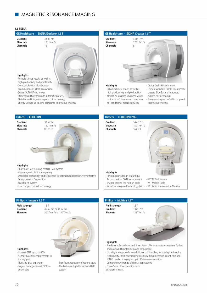
1.5 TESLA
· SIGNA Explorer 1.5 TGE Healthcare
16Channels120 T / m / sSlew rate33 mT / mGradient
Highlights• Reliable clinical results as well as
high productivity and profitability • Compatible with SilentScan for
examinations as silent as a whisper• Digital OpTix RF-technolgy. • Efficient workflow thanks to automatic presets,
Slide Bar and integrated express coil technology.• Energy savings up to 34 % compared to previous systems.
· SIGNA Creator 1.5 TGE Healthcare
8Channels120 T / m / sSlew rate33 mT / mGradient
Highlights• Reliable clinical results as well as
high productivity and profitability• MAVRIC SL enables advanced visual-
ization of soft tissues and bone near MR conditional metallic devices.
• Digital OpTix RF-technolgy. • Efficient workflow thanks to automatic
presets, Slide Bar and integrated express coil technology.
• Energy savings up to 34 % compared to previous systems.
· ECHELON OVALHitachi
16 (32 )Channels150 T / m / sSlew rate34 mT / mGradient
Highlights• Revolutionary design featuring a
74 cm spacious OVAL environment• Shaped around the human body• Workflow Integrated Technology (WIT)
• WIT RF Coil System• WIT Mobile Table• WIT Patient Information Monitor
· ECHELONHitachi
Up to 16Channels150 T / m / sSlew rate33 mT / mGradient
Highlights• Short bore, low running costs HF MRI system• High magnetic field homogeneity• Dedicated technology and sequences for artefacts suppression, very effective
fat suppression / separation• Scalable RF system• Low cryogen boil-off technology
· Ingenia 1.5 TPhilips
200 T / m / s or 120 T / m / sSlewrate45 mT / m or 33 mT / mGradient1.5 TField strength
Highlights• Increase SNR by up to 40 %• As much as 30 % improvement in
throughput• Plug-and-play expansion• Largest homogeneous FOV for a
70 cm bore
• Significant reduction of routine tasks• The first-ever digital broadband MR
system
· Multiva 1.5TPhilips
122 T / m / sSlewrate33 mT / mGradient1.5 TField strength
Highlights• FlexStream, SmartExam and SmartAssist offer an easy-to-use system for fast
and easy workflow for increased throughput• Ultra-light weight coils. No additional coil handling for total spine imaging• High quality, 10-minute routine exams with high channel count coils and
SENSE parallel imaging for up to 16-times acceleration• Comprehensive range of clinical applications• PowerSave – low operation costsNot available in the USA.
RADBOOK 201636
◼ MAGNETIC RESONANCE IMAGING
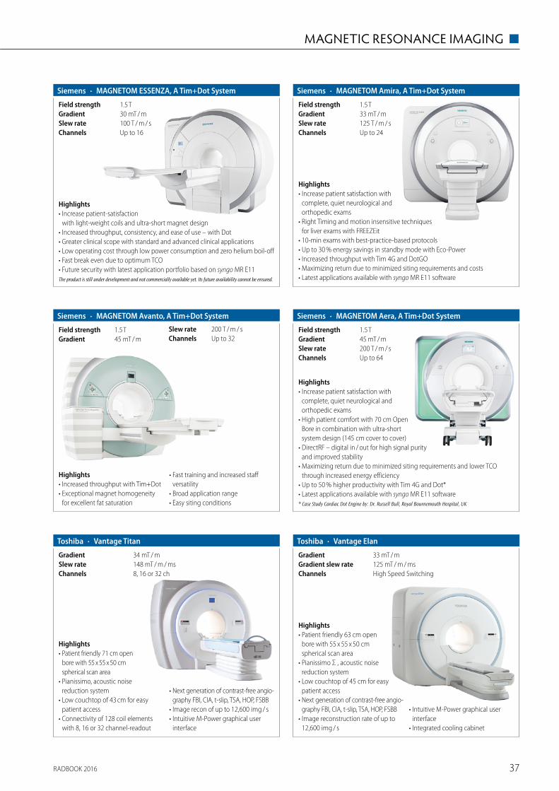
Highlights• Increase patient satisfaction with
complete, quiet neurological and ortho pedic exams
• High patient comfort with 70 cm Open Bore in combination with ultra-short system design (145 cm cover to cover)
• DirectRF – digital in / out for high signal purity and improved stability
• Maximizing return due to minimized siting requirements and lower TCO through increased energy efficiency
• Up to 50 % higher productivity with Tim 4G and Dot* • Latest applications available with syngo MR E11 software* Case Study Cardiac Dot Engine by: Dr. Russell Bull, Royal Bournemouth Hospital, UK
· MAGNETOM Aera, A Tim+Dot SystemSiemens
Up to 64Channels200 T / m / sSlew rate45 mT / mGradient1.5 TField strength
Highlights• Increase patient satisfaction with
complete, quiet neurological and orthopedic exams
• Right Timing and motion insensitive techniques for liver exams with FREEZEit
• 10-min exams with best-practice-based protocols• Up to 30 % energy savings in standby mode with Eco-Power• Increased throughput with Tim 4G and DotGO• Maximizing return due to minimized siting requirements and costs• Latest applications available with syngo MR E11 software
· MAGNETOM Amira, A Tim+Dot SystemSiemens
Up to 24Channels125 T / m / sSlew rate33 mT / mGradient1.5 TField strength
Highlights• Increased throughput with Tim+Dot• Exceptional magnet homogeneity
for excellent fat saturation
• Fast training and increased staff versatility
• Broad application range• Easy siting conditions
· MAGNETOM Avanto, A Tim+Dot SystemSiemens
Up to 32Channels200 T / m / sSlew rate
45 mT / mGradient1.5 TField strength
Highlights• Increase patient-satisfaction
with light-weight coils and ultra-short magnet design• Increased throughput, consistency, and ease of use – with Dot• Greater clinical scope with standard and advanced clinical applications• Low operating cost through low power consumption and zero helium boil-off• Fast break even due to optimum TCO• Future security with latest application portfolio based on syngo MR E11The product is still under development and not commercially available yet. Its future availability cannot be ensured.
· MAGNETOM ESSENZA, A Tim+Dot SystemSiemens
Up to 16Channels100 T / m / sSlew rate30 mT / mGradient1.5 TField strength
· Vantage TitanToshiba
8, 16 or 32 chChannels148 mT / m / msSlew rate34 mT / mGradient
Highlights• Patient friendly 71 cm open
bore with 55 x 55 x 50 cm spherical scan area
• Pianissimo, acoustic noise reduction system
• Low couchtop of 43 cm for easy patient access
• Connectivity of 128 coil elements with 8, 16 or 32 channel-readout
• Next generation of contrast-free angio-graphy FBI, CIA, t-slip, TSA, HOP, FSBB
• Image recon of up to 12,600 img / s• Intuitive M-Power graphical user
interface
· Vantage ElanToshiba
High Speed SwitchingChannels125 mT / m / msGradient slew rate33 mT / mGradient
Highlights• Patient friendly 63 cm open
bore with 55 x 55 x 50 cm spherical scan area
• Pianissimo Σ , acoustic noise reduction system
• Low couchtop of 45 cm for easy patient access
• Next generation of contrast-free angio-graphy FBI, CIA, t-slip, TSA, HOP, FSBB
• Image reconstruction rate of up to 12,600 img / s
• Intuitive M-Power graphical user interface
• Integrated cooling cabinet
RADBOOK 2016 37
MAGNETIC RESONANCE IMAGING ◼

One Scan. Six contrasts. Triple Speed.
Bringing 3.0 T MR to new clinical settingWith up to twice higher signal levels, 3.0 T MR systems improve spatial resolution and therefore provide more information on very fine anatomical structures, com-pared to 1,5 T systems. 3.0 T MRIs are com-mon in academic and research hospitals. Now the SIGNA Pioneer brings the power of 3.0 T for clinical use and accessibility to a broader range of healthcare providers. The SIGNA Pioneer 3.0 T MRI delivers the
ease-of-use and flexibility of a 1.5 T system, with improved image homogeneity and reproducibility for oncology and spine imaging for example. In addition, ultra-high efficiency gradient technology enables the MR system to maintain a high-end perfor-mance, even during demanding clinical applications such as cardiology or oncology.
“In comparison to the 1,5 T MR, our image quality for Brain and MSK clinical cases has improved significantly, now we are able to increase the signal-to-noise ratio (SNR) or resolution or both. We also observe very high cardiac imaging performance with high SNR for perfusion” said Dr. Ahlers, general manager of radiomed. Using equi-valent imaging parameters, physicians
are also able to use the signal gain to shorten patients’ breath-holds for
liver exploration, and even to complete free-breathing body
imaging, offering a much improved patient experience
but also image quality in very challenging situations.
The MAGiC* of complete flexibility for multiple contrast changesThe new MAGiC sequence is a major innovation in neuro imaging. For the first time ever with MAGiC, clinicians have the ability to generate six contrasts in a single scan and in as little as one-third of the total time taken to acquire each contrast separately using conventional techniques. After an acquisition of only five minutes, it is now possible to adjust the image contrast in real time, even after completing the scan by simply moving a dynamic cursor to change conventional MRI acquisition parameters (TE, TR and TI) depending on the disease or the age of the patient. Dr. Ahlers stated: “In addition to all contrasts automatically provided by MAGiC, such as FLAIR, T1, T2, PD, STIR and PSIR, we scan three additional sequences including T2* and DWI to have a comprehen-sive brain routine protocol, all in less than ten minutes.”
This time saved could potentially allow clinicians to scan one more patient per hour, every hour of every day. MAGiC also pro-vides quantitative information (maps T1, T2, and proton density) which has the potential to provide new perspectives for physi-cians to characterize earlier and more precisely small or secondary lesions and moni tor patients with neuro degenerative disorders.
Innovative clinical applications for professional healthcare
In addition to MAGiC, the SIGNA Pioneer offers several impressive advances in imaging with inno-vative applications and thus gives clinicians a high diagnostic confidence: “With SIGNA Pioneer, our goal
was to improve both productivity and image quality simultaneously. We are already seeing results with inno-
vative sequences. For example in body imaging, the new sequences DISCO and FOCUS have changed our pelvic protocol for oncology studies. We now use a FOCUS small FOV diffusion centered on the pelvic area with significantly reduced distortions especially at 3.0 T. DISCO has replaced our previous DCE-MRI sequence since it allows us to achieve very high temporal resolution and very high spatial resolution simultaneously. This has helped to improve assess-ment of lesion enhancements.” He added: “The system is really built for large field of view body and spine imaging. We are able to routinely scan 50 x 45 cm coronal scans with very good image quality. In many cases it minimizes the number of stations we would need for body and spine imaging.”
Improving patient experience in very challenging situations
Patients with neurodegenerative disease, who cannot remain still during the examinations, will also now be able to benefit from MR. With new advanced 3D motion correction software, SIGNA Pioneer will compensate patient movement to provide the same image quality and comfort for diagnosis despite involuntary movements.
An enhanced SilentScan package also considerably reduces scan noise from excess of 110 decibels (dBA) for conventional MRI scans to just three dBA above ambient noise for most head exams. This is a major differentiator for patient comfort and to minimize the risk of deteriorating image quality for the radiologist in anxious patients. “The SilentScan technology has helped us deliver a nearly noise-free neuro exam including Diffusion Weighted imaging. Our patients really appreciate it“, concluded Dr. Ahlers.
www.gehealthcare.com
SIGNATM Pioneer* with MAGiC brings 3.0 T MR to a new clinical setting
SIGNA Pioneer, a new 3.0 T Magnetic
Resonance Imaging (MRI) system,
embodies the exploration and expansion
of modern medical imaging and blazes
a trail to the future of MRI. Dr. Ahlers,
general manager of radiomed, shares
his experience with SIGNA Pioneer
recently installed at radiomed practice
in Wiesbaden, Germany – one of the
first installations worldwide.
3,0 T MRI Signa Pioneer with extra large tunnel and comfortably lowerable patient table offers a wide range of clinical applications.
Conventional: Six scans for six contrasts MAGiC: One scan with six contrasts (based on MAGiC neuro-protocols. Final results may vary based on protocol.)
* Commercially available and cleared for use in Germany, Austria, Switzerland and other select regions of the world.
RADBOOK 201638
◼ MAGNETIC RESONANCE IMAGING

One Scan. Six contrasts. Triple Speed.
Bringing 3.0 T MR to new clinical settingWith up to twice higher signal levels, 3.0 T MR systems improve spatial resolution and therefore provide more information on very fine anatomical structures, com-pared to 1,5 T systems. 3.0 T MRIs are com-mon in academic and research hospitals. Now the SIGNA Pioneer brings the power of 3.0 T for clinical use and accessibility to a broader range of healthcare providers. The SIGNA Pioneer 3.0 T MRI delivers the
ease-of-use and flexibility of a 1.5 T system, with improved image homogeneity and reproducibility for oncology and spine imaging for example. In addition, ultra-high efficiency gradient technology enables the MR system to maintain a high-end perfor-mance, even during demanding clinical applications such as cardiology or oncology.
“In comparison to the 1,5 T MR, our image quality for Brain and MSK clinical cases has improved significantly, now we are able to increase the signal-to-noise ratio (SNR) or resolution or both. We also observe very high cardiac imaging performance with high SNR for perfusion” said Dr. Ahlers, general manager of radiomed. Using equi-valent imaging parameters, physicians
are also able to use the signal gain to shorten patients’ breath-holds for
liver exploration, and even to complete free-breathing body
imaging, offering a much improved patient experience
but also image quality in very challenging situations.
The MAGiC* of complete flexibility for multiple contrast changesThe new MAGiC sequence is a major innovation in neuro imaging. For the first time ever with MAGiC, clinicians have the ability to generate six contrasts in a single scan and in as little as one-third of the total time taken to acquire each contrast separately using conventional techniques. After an acquisition of only five minutes, it is now possible to adjust the image contrast in real time, even after completing the scan by simply moving a dynamic cursor to change conventional MRI acquisition parameters (TE, TR and TI) depending on the disease or the age of the patient. Dr. Ahlers stated: “In addition to all contrasts automatically provided by MAGiC, such as FLAIR, T1, T2, PD, STIR and PSIR, we scan three additional sequences including T2* and DWI to have a comprehen-sive brain routine protocol, all in less than ten minutes.”
This time saved could potentially allow clinicians to scan one more patient per hour, every hour of every day. MAGiC also pro-vides quantitative information (maps T1, T2, and proton density) which has the potential to provide new perspectives for physi-cians to characterize earlier and more precisely small or secondary lesions and moni tor patients with neuro degenerative disorders.
Innovative clinical applications for professional healthcare
In addition to MAGiC, the SIGNA Pioneer offers several impressive advances in imaging with inno-vative applications and thus gives clinicians a high diagnostic confidence: “With SIGNA Pioneer, our goal
was to improve both productivity and image quality simultaneously. We are already seeing results with inno-
vative sequences. For example in body imaging, the new sequences DISCO and FOCUS have changed our pelvic protocol for oncology studies. We now use a FOCUS small FOV diffusion centered on the pelvic area with significantly reduced distortions especially at 3.0 T. DISCO has replaced our previous DCE-MRI sequence since it allows us to achieve very high temporal resolution and very high spatial resolution simultaneously. This has helped to improve assess-ment of lesion enhancements.” He added: “The system is really built for large field of view body and spine imaging. We are able to routinely scan 50 x 45 cm coronal scans with very good image quality. In many cases it minimizes the number of stations we would need for body and spine imaging.”
Improving patient experience in very challenging situations
Patients with neurodegenerative disease, who cannot remain still during the examinations, will also now be able to benefit from MR. With new advanced 3D motion correction software, SIGNA Pioneer will compensate patient movement to provide the same image quality and comfort for diagnosis despite involuntary movements.
An enhanced SilentScan package also considerably reduces scan noise from excess of 110 decibels (dBA) for conventional MRI scans to just three dBA above ambient noise for most head exams. This is a major differentiator for patient comfort and to minimize the risk of deteriorating image quality for the radiologist in anxious patients. “The SilentScan technology has helped us deliver a nearly noise-free neuro exam including Diffusion Weighted imaging. Our patients really appreciate it“, concluded Dr. Ahlers.
www.gehealthcare.com
SIGNATM Pioneer* with MAGiC brings 3.0 T MR to a new clinical setting
SIGNA Pioneer, a new 3.0 T Magnetic
Resonance Imaging (MRI) system,
embodies the exploration and expansion
of modern medical imaging and blazes
a trail to the future of MRI. Dr. Ahlers,
general manager of radiomed, shares
his experience with SIGNA Pioneer
recently installed at radiomed practice
in Wiesbaden, Germany – one of the
first installations worldwide.
3,0 T MRI Signa Pioneer with extra large tunnel and comfortably lowerable patient table offers a wide range of clinical applications.
Conventional: Six scans for six contrasts MAGiC: One scan with six contrasts (based on MAGiC neuro-protocols. Final results may vary based on protocol.)
* Commercially available and cleared for use in Germany, Austria, Switzerland and other select regions of the world.
RADBOOK 2016 39
MAGNETIC RESONANCE IMAGING ◼

Highlights• G-scan Brio eXP is a third generation
of dedicated MRI for MSK imaging in supine and weight-bearing position.
• It provides a complete range of MRI imaging sequences, including the most advanced pulse acquisitions, such as Steady State and Fat & Water separation imaging.
• Weight-Bearing MRI gives an added diagnostic value for those pathologies not clearly defined in conventional MRI.
OPEN
· G-scan Brio eXPEsaote
56 mT / m / msSlew rate20 mT / mGradient0.25 TField strength
Highlights• O-scan eXP is the third
generation of dedicated MRI designed for imaging extremities.
• O-scan provides an out-standing image quality in line with today’s standards.
• O-scan with eXP technology makes the exam time of 15 min per patient.• O-scan break-even figure is only three exams / day thanks to an affordable price
and very low running costs, compatible with the current healthcare's needs.
· O-scan eXPEsaote
100 mT / m / msSlew rate20 mT / mGradient0.31 TField strength
Highlights• S-scan eXP is the third
generation of dedicated MRI for imaging of the spine and extremities.
• S-scan with eXP technology features an outstanding image quality both on spine and joints.
• S-scan is perfectly in line with today’s need for efficient and economic health care, and is a sensible choice for any imaging center with a substantial musculoskeletal workload.
· S-scan eXPEsaote
56 mT / m / msSlew rate20 mT / mGradient0.25 T Field strength
Highlights• Optical RF technology
brings higher SNR and better image quality
• Short bore of 140 cm with a spacious bore diameter of 60 cm
• 8 – 32 channel parallel acquisition achieve higher scanning speed
• Zero helium consumption technolo-gy greatly lower running cost
• iPad / iPhone remote scan control and diagnosis
• Powerful workstation with abundant image processing function
• CE and FDA approved
1.5 TESLA
· i_Magnate 1.5T MRI SystemWandong
8Channels128 T / m / sSlew rate35 mT / mGradient1.5 TField strength
Highlights• Full range of stanning sequences• Best performance at low cost• Matches a wide range of imaging needs in any hospital
· SUPERSCAN 1.5 TXingaoyi (XGY)
100 mT / m / msSlew rate30 mT / mGradient1.5 TField
RADBOOK 201640
◼ MAGNETIC RESONANCE IMAGING

· AIRIS VentoHitachi
55 T / m / sSlew rate22 mT / mGradient0.3 TField strength
Highlights• Comfort class permanent open MRI system, which keeps enhanced capabilities
meeting sophisticated open design• Offers newly developed technologies available at an excellent cost of ownership• High magnetic field homogeneity• Environment friendly: extremely low power consumption and reduced
installation requirements• Low running costs allowing fast return of investment
· APERTO LucentHitachi
55 T / m / sSlew rate25 mT / mGradient0.4 TField strength
Highlights• Wide, 320 degrees open permanent MRI system• Features top field strength – 0.4 T – amongst the permanent MRI systems
presently on the market• Newly developed built-in technologies keep APERTO Lucent delivering image
quality comparable with entry level HF MRI scanner• Fast processing chain allows increasing patient throughput• Reduced running costs allowing fast return of investment
· AIRIS Vento LTHitachi
55 T / m / sSlew rate22 mT / mGradient0.3 TField strength
HighlightsAIRIS Vento LT (0.3 T) – the economic, compact and wide open MR solution.• The open system architecture gives not only a feeling of security but also has
considerable merits when taking care of small children and elderly patients• The floating table allows to fit the system into small spaces while giving the
possibility of placing the patient always in the centre to achieve high image qualitiy
· OASISHitachi
8Channels100 T / m / sGradient slew rate33 mT / mGradient1.2 TField strength
Highlights• World's most powerful
open MRI• 1.2 T vertical field super-
conductive magnet for high SNR• 270° panoramic view, accommo-
dates claustrophobic, paediatric, obese patients
• Fully motorized extra wide 82 cm patient table (up to 300 kg)
• Two-pillar asymmetric design• Soft Sound Technology• Multiple coil connectors with Zenith
solenoid element based, highly sensitive receiver coils
· MagSense 360 MRI SystemMindray Medical
60 T / m / sSlew rate25 mT / mGradient0.36 TField Strength
Highlights• Innovative InScan Technology• Advanced Gradient system Design• Ergonomic Design make you more comfortable• Multi-clinical Applications satisfied doctors requirement• Multiple coils selection make all examination reality
Please visit us at
www.healthcare-in-europe.com
RADBOOK 2016
RADBOOK 2016 41
MAGNETIC RESONANCE IMAGING ◼

Highlights• The first mid-field permanent
magnet MRI system used in clinical applicationworldwide• Full range of scanning sequences, best images• High throughput, shorter scanning time
· OPER 0.5 TXingaoyi (XGY)
70 mT / m / msSlew rate24 mT / mGradient0.5 TField
· Panorama HFOPhilips
120 mT / m / msSlewrate28 mT / mGradient1.0 TField strength
Highlights• Patient friendly: three times larger patient aperture than conventional MR to
handle stressed and claustrophobic patients, children, elderly and large patients• High-field performance comparable to 1.5 T in a truly open configuration• Increased productivity with SmartExam, one-click planning, scanning and
processing• Enables unique applications not possible with cylindrical systems
OPEN
· Panorama HFO Oncology ConfigurationPhilips
HighlightsPhilips Panorama HFO oncology configuration – Imaging that fits your planning.• Imaging in treatment position enabled by the open spacious design of the system• Streamlined clinical workflow with MR images customized for radiation
therapy planning• Quality assurance for geometric deformation
Highlights• Smallest pole diameter (137 cm / 54 inches) for patient comfort• True, multichannel, seamless imaging (up to 100 cm)• No cryogen use and low power consumption • Outstanding image quality at mid-field
· MAGNETOM C!Siemens
55 T / m / msSlew rate24 mT / mGradient0.35 TField strength
Highlights• Two column, large span, super open magnet design• Six-way movement motorized / manual patient table• Automatic laser positioning system with two-LCD touch screen control panel• Four channels digital RF system• Windows based imaging workstation with user friendly interface provides
excellent user experience• CE and FDA approved
· i_Open 0.5T Permanent MRI SystemWandong
80 mT / m / msSlew rate30 mT / mGradient4 channelsChannels0.5 TField strength
· i_Open 0.36T Permanent MRI SystemWandong
95 T / m / sSlew rate28.5 mT / mGradient0.36 TField strength
Highlights• C-Shape permanent magnet• Multi-channel digital RF system• Cross laser positioning system with two-LCD display panel• CE and FDA approved• ACR Accredited• Windows 7 based imaging workstation with user friendly interface provides
excellent user experience• Experienced service team since first overseas installation in the U.S. in 2005
RADBOOK 201642
◼ MAGNETIC RESONANCE IMAGING

Highlights• Higher SNR and larger imaging range with Multi-RF channels• Excellent images and full range of scanning sequences• Low power consumption, low failure rate, high operating ratio• Requires little space for installation
· OPER 0.4 TXingaoyi (XGY)
66 mT / m / msSlew rate20 mT / mGradient0.4 TField
Highlights• Complete function, excellent images, full range of scanning sequences• Clear quick scan image with high slew rate• Extremely low power consumption and very low failure rate
· OPER 0.3 TXingaoyi (XGY)
48 mT / m / msSlew rate15 mT / mGradient0.3 TField strength
Highlights• Excellent images, full range of scanning sequences• Low power consumption, low failure rate• Small installation site
· OPER 0.35 TXingaoyi (XGY)
66 mT / m / msSlew rate19 mT / mGradient0.35 TField
RADBOOK 2016 43

MR-PET
· Biograph mMRSiemens
200 T / m / sSlew rate45 mT / mGradient3 TField strength
Up to 32Channels
Highlights• Largest customer base with over
70 installations worldwide• State-of-the-art 3 T MRI with 2nd
order shim • Comprehensive set of surface coils available for full range of MR-only exams• Not only simultaneous, but synergistic MR-PET: MR-based motion compen-
sation of PET images• Whole-body attenuation MR-based attenuation correction including bones• Up to 10 bed positions with MR-PET• Latest applications available with syngo MR E11 softwaresyngo MR E11 for Biograph mMR is still under development and not commercially available yet. Its future availability cannot be ensured.
Highlights• The NORAS Breast Biopsy 4-Channel Coil Height-Adjustable features for
improved image quality while shortening the acquisition time for MRI breast examinations.
• The Coil ensures optimized patient comfort through the height adjustable patient and head rest. Also it is a flexible solution for both small and large breasts.
• Excellent medial, lateral and craniocaudal access to the breast.
MRT COILS
· Breast Biopsy 4-Channel Coil Height-AdjustableNORAS
SiemensSystem platform4Channels1.5 and 3 TField strength
Highlights• The “Mandibula” is a multi-
element receive array and positioning system for 3D high-resolution dental and maxillomandibular MRI images.
• The coil provides high resolution dedicated MR imaging in dental area and reduces scan times.
• It ensures maximum patient comfort due to its design and accessories like a patient rest pillow, an open-mouth fixation mechanism and a both direction mirror.
· Mandibula 15-Channel Dental CoilNORAS
Siemens Tim SystemsSystem platform15Channels1.5T, 3TField strength
· Neurosurgery Solution FLEXIBILITYNORAS
Siemens and PhilipsSystem platform8Channels1.5 and 3 TField strength
HighlightsThe new NORAS Head Holder Flexibilty consists of a 8-channel iMRI Head Coil for imaging and intervention in a neurosurgical OR environment. Being height adjustable the setup enables optimal positioning in 70 cm bore systems. More-over, the Head Holder is movable along the bore direction, which facilitates flexible patient positioning on the transfer board. Head fixations with 3 up to 5 pins are supported.
· Uni-Lift Prostate Intervention DeviceNORAS
70 cm Bore MR Systems System platformn / a; Compatible with standard MR coil portfolioChannelsn / aField strength
Highlights• The “Uni-Lift” is a MR-compa-
tible Holding Device for for MR-guided interventions of the prostate.• It allows comfortable patient positioning in supine position, which guaran-
tees excellent transperineal access for the performing physician towards the prostate in the MRI system.
• The Uni-Lift device can also be used for therapy of the prostate.
Highlights• The "Variety" is a 16-channel multipurpose flex coil, which has been devel-
oped for high flexibility during examination of challenging anatomic regions. The areas of application of the "Variety" include: diagnosis in orthopedics, pediatrics and veterinary medicine.
• Slim design and optional dedicated positioning aids enable coil placement close to anatomy of interest for optimal image quality.
· Variety 16-Channel Multipurpose Coil with Positioning AidsNORAS
Siemens Tim SystemsSystem platform16 (2 x 8)Channels1.5 and 3 TField strength
RADBOOK 201644
◼ MAGNETIC RESONANCE IMAGING

ACCESSORIES / COMPLEMENTARY SYSTEMS
Highlights• Static diagnostic
imaging centers MRI, CT, PET, PET / CT
• Interim services for bridging downtimes
• Regular “routing” services
· Flexible diagnostic imaging servicesAlliance Medical
HighlightsEngineering, rental, sale of modular buildings MRI, CT, PET, PET / CT including or excluding diagnostic equipment
· Modular building solutionsAlliance Medical
Highlights• Tested at 3 Tesla• Comes with two adjustable side rails• Hydraulic pedal pump
• Adjustable headrest• Choice of 21 upholstery colours• Weight capacity 200 kg
· Adjustable height MRI trolleyallMRI GmbH
Highlights• MRI safe foldable
wheelchair entirely made of 100 % thermo plastic
• Including the ball bearing
• Two swing out ad-justable footrests and armrests
• Also solid rubber tires
· Foldable MRI wheelchairallMRI GmbH
Highlights• Hygiene cart with desinfaction
solution dispenser 0,5 litre and disposable gloves holder
• Three drawers with a lot of room for personal storage equipement
• Width: 700 mm Depth: 410 mm Height: 1.230 mm
· MRI hygiene cartallMRI GmbH
Highlights• New productivity using MR-safe
communications• Doctors can speak with each other,
with their patient, and with techno-logists
• Enables up to eight concurrent dialogs during a scan – five staff members, plus a patient
• Adaptive DSP-based noise reduction filters out EPI gradient noise, ensuring excellent sound quality
• Automatic noise reduction, field- proven reliability
· Wireless MRI communication systemallMRI GmbH
RADBOOK 2016 45
MAGNETIC RESONANCE IMAGING ◼

ACCESSORIES / COMPLEMENTARY SYSTEMS
· CIRS PhantomsGCTechnology
Highlights• MRI-Linac Dynamic Phantom• Main and Large field MRI distortion
phantoms• Triple Modality Abdominal Phantom• Lumbar Training Phantom• Anthropomorphic 3D Skull Phantom
• Multi-Modality Breast Biopsy and Sonographic Trainer
• Multi-Modality Prostate Phantoms• Multi-Modality Pelvic-Phantom• Gillian QA Phantom for distortion
and alignment
HighlightsPhilips Diamond Select pro-vides reliable, like-new refur-bished imaging systems at an attractive price. With the financial challenges in healthcare today, Diamond Select equipment is a simple, economical alternative to purchasing new equipment. Diamond Select offers up-to-date technology to expand the variety of high-quality services available to patients, while helping healthcare providers aim for increased profitability. All systems undergo a thorough five-step refurbishment process in order to maintain the high standards set by Philips. The Philips Diamond Select line of fully configurable refurbished systems is available for the following imaging modal-ities: CT, MR, cardiovascular (CV) Xray, surgical / interventional X-ray, ultrasound and advanced molecular imaging.
· Refurbished SystemsPhilips
HighlightsAmbient Experience is a purposely designed healthcare environment. With a refreshingly creative eye, Ambient Experience integrates tech-nology, spatial design, and workflow improvements to create a more comfortable, stress-reducing environ-ment for both patients and staff.
Every project is a solution tailored to suit individual institutional needs – guided by the four fundamental pillars of Ambient Experience:• Physical and emotional comfort• Patient and staff contact• Experience personalization• Hospital workflow
· Ambient ExperiencePhilips
Highlights• MRI compatible up to 3 Tesla• Parameter: SPO2 and / or NIBP• Mains and battery driven (1.5 hours)• Optimized for day to day application• No installation necessary• HTML printing function• Optimized for adult children
and neonates
· MAGLIFE lightSCHILLER
Highlights• Highest ECG quality even under strongest
gradient influence• MRI compatible up to 3 Tesla• Optical core and skin temperature• Configuration for anaesthesia, cardiac and
intensive care applications• Patented artefact inhibition• Optimized for adult children and neonates• Wireless Data Transmission• Wireless or optically wired Sp02• Mains and battery driven• 12.1" color display
· MAGLIFE SerenitySCHILLER
Please visit us at
www.healthcare-in-europe.com
RADBOOK 2016
RADBOOK 201646
◼ MAGNETIC RESONANCE IMAGING

Injectors
47
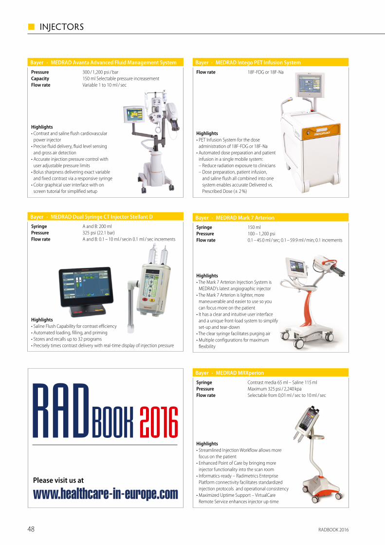
RADBOOK 201648
◼ INJECTORS
· MEDRAD Avanta Advanced Fluid Management SystemBayer
Variable 1 to 10 ml / secFlow rate150 ml Selectable pressure increasementCapacity300 / 1,200 psi / barPressure
Highlights• Contrast and saline flush cardiovascular
power injector• Precise fluid delivery, fluid level sensing
and gross air detection• Accurate injection pressure control with
user adjustable pressure limits• Bolus sharpness delivering exact variable
and fixed contrast via a responsive syringe• Color graphical user interface with on
screen tutorial for simplified setup
· MEDRAD Dual Syringe CT Injector Stellant DBayer
A and B: 0.1 – 10 ml / secin 0.1 ml / sec incrementsFlow rate325 psi (22.1 bar)PressureA and B: 200 mlSyringe
Highlights• Saline Flush Capability for contrast efficiency• Automated loading, filling, and priming• Stores and recalls up to 32 programs• Precisely times contrast delivery with real-time display of injection pressure
· MEDRAD Intego PET Infusion SystemBayer
18F-FDG or 18F-NaFlow rate
Highlights• PET Infusion System for the dose
administration of 18F-FDG or 18F-Na • Automated dose preparation and patient
infusion in a single mobile system: – Reduce radiation exposure to clinicians – Dose preparation, patient infusion,
and saline flush all combined into one system enables accurate Delivered vs. Prescribed Dose (± 2 %)
· MEDRAD Mark 7 ArterionBayer
0.1 – 45.0 ml / sec; 0.1 – 59.9 ml / min; 0.1 incrementsFlow rate100 – 1,200 psiPressure150 mlSyringe
Highlights• The Mark 7 Arterion Injection System is
MEDRAD's latest angiographic injector• The Mark 7 Arterion is lighter, more
maneuverable and easier to use so you can focus more on the patient
• It has a clear and intuitive user interface and a unique front-load system to simplify set-up and tear-down
• The clear syringe facilitates purging air• Multiple configurations for maximum
flexibility
· MEDRAD MRXperionBayer
Selectable from 0,01 ml / sec to 10 ml / secFlow rateMaximum 325 psi / 2,240 kpaPressureContrast media 65 ml – Saline 115 mlSyringe
Highlights• Streamlined Injection Workflow allows more
focus on the patient• Enhanced Point of Care by bringing more
injector functionality into the scan room• Informatics-ready – Radimetrics Enterprise
Platform connectivity facilitates standardized injection protocols and operational consistency
• Maximized Uptime Support – VirtualCare Remote Service enhances injector up-time
Please visit us at
www.healthcare-in-europe.com
RADBOOK 2016

RADBOOK 2016 49
INJECTORS ◼
Contrast medium injectors and consumablesfor CT, MRI and angiography
Hauptstr. 255 · D-66128 SaarbrueckenFor more info: www.medtron.com
The specialist
for Stress-MRI!
Integrated infusion pump!
Wireless & 3-Tesla-capable!
Innovation – Made in Germany!
STRESScan be so relaxing.
Accutron® MR3!
Radbook_1-2_Seite_MR3.qxp_Anzeige 12.02.16 12:37 Seite 1
Please visit us at
www.healthcare-in-europe.com
RADBOOK 2016
· MEDRAD Spectris Solaris EPBayer
Selectable from 0,01 ml / sec to 10 ml / sec
SyringeMaximum 325 psi / 2,240 kpaPressureContrast media 65 ml – Saline 115 ml
Flow rate
Highlights• Flexible power management with battery
operation or continuous battery charging through AC power connection
• Pressure Limit selection from one of six preset maximum pressure limits, and the ability to view pressure during injection on the control room display
• 3 T compatibility • Multiphase injection control with
6 user-programmable phases including PAUSE and HOLD
• Programmable KVO

RADBOOK 201650
◼ INJECTORS
Highlights• Direct injection from contrast media bottles• Air and occlusion detection on fluid channels• Unidirectional flow of fluid• Locking and automatic filling• Digital interface, dual touch screens• DiluJect (optional): contrast media and
saline are injected in rapidly alternating flow through the injector
· CT ExprèsBracco
CTApplication0.5 – 9.9 mL / s in steps of 0.1 mL / sFlow rate9.1 bar maxPressureSyringeless injectorSyringe
Highlights• Tilt sensor / lockout • Arming at the injector • Independently rotating and very
compact injector head (270 degrees) • Integrated electroluminescent display • Modular flexibility of components and
WINDOWS based software allow optimal serviceability and enhanced expandability
• Touch-screen color LCD display and intuitive software
· EmpowerCTA+Bracco
CTApplication
0.1 to 10.0 ml / sec in user-specified increments of 0.1 ml / sec
Flow rate40 to 325 psi in user-specified increments of 1 psiPressure200 ml (CM), 200 ml (NaCl)Syringe
Highlights• Hydraulic injector system • MRI compatible through the use of polymers and
non-ferromagnetic metals • Little contrast media waste due to the very short
distance between injector head and patient • Very lightweight injector head • No active components in the shielded room
(no battery)
· EmpowerMRBracco
MRApplication
0.1 to 10.0 ml / sec in user-specified increments of 0.1 ml / sec
Flow rate40 to 300 psi in user-specified increments of 1 psiPressure100 ml (CM), 100 ml (NaCl)Syringe
· Accutron CTMEDTRON AG
Automatic or manual filling, filling speed 1 – 5 ml / s, optimized tube systems with check valve
Syringe21 bar (304 psi)Max. injection pressure200 ml Easy Loading Syringe (ELS)Capacity0.1 – 10 ml / s, programmable in steps of 0.1 ml / sFlow rate
Highlights• Wireless injector unit, rechargeable batteries• Integrated heated syringe holder with Easy Loading
Syringe (ELS) 200 ml• Touchscreen control panel with different languages• Wireless touchscreen remote control • Secured injection position (built-in sensor) • Aluminium housing• Use of prefilled syringes (as an option)
· Accutron CT-DMEDTRON AG
Automatic or manual filling, filling speed 1 – 5 ml / s, optimized tube systems with check valve
Syringe21 bar (304 psi)Max. injection pressure200 ml (CM), 200 ml (NaCl) Easy Loading Syringe (ELS)Capacity
For both injection units: 0.1 – 10 ml / s, programmable in steps of 0.1 ml / s
Flow rate
Highlights• Wireless injector unit with rechargeable batteries• Integrated heated syringe holder for Easy Loading
Syringe (ELS)• Wireless touchscreen remote control• Use of prefilled syringes (as an option) • Secured injection position (built-in sensor)• Alternatively, display of injection parameters or
pressure graph• Aluminium housing – wall or ceiling suspension• CANopen Interface (as an option)
Please visit us at
www.healthcare-in-europe.com
RADBOOK 2016

RADBOOK 2016 51
INJECTORS ◼
· Accutron HPMEDTRON AG
Automatic or manual filling, filling speed 1 – 4 ml / s, opt. high-pressure tube systems with check valves
Syringe
Angio mode: 83 bar (1,200 psi), CT mode: 21 bar (305 psi), programmable in 1 bar increments
Max. injection pressure200 mlCapacity
Angio mode: 0.1 – 30 ml / s, CT mode: 0.1 – 10 ml / s, programmable in 0,1 ml / s increments
Flow rate
Highlights• Fast high-pressure injections for angiography and
multiphase injection profiles for CT• Wireless injector unit with rechargeable batteries• Wireless touchscreen remote control (option)• Wall or ceiling suspension system• Integrated heated syringe holder for Easy Loading
Syringe (ESL) 200 ml• 120 injection profiles can be stored (60 angio / 60 CT)• Aluminium housing – Interface (option)
· Accutron HP-DMEDTRON AG
Automatic or manual filling, filling speed 1 – 4 ml / s, opt. high-pressure tube systems with check valves
Syringe
Angio mode: 83 bar (1,200 psi), CT mode: 21 bar (305 psi), programmable in 1 bar increments
Max. injection pressure200 ml (CM), 200 ml (NaCl) Easy Loading Syringe (ELS)Capacity
Angio mode: 0.1 – 30 ml / s, CT-mode: 0,1 – 10 ml / s, programmable in 0,1 ml / s increments
Flow rate
Highlights• Wireless unit with rechargeable batteries• Multiphase program controlled injection of CM & NaCl• Single or multi injection mode• Integrated heated syringe holder for Easy Loading
Syringe (ELS)• Touchscreen control panel, multilingual• Up to 3 phases – pressure graph – secured injection
position (built-in sensor)• 60 injection profiles storable• Interface (option) – Aluminium housing
Please visit us at
www.healthcare-in-europe.com
RADBOOK 2016
· Accutron MRMEDTRON AG
Automatic or manual filling, filling speed 1 – 5 ml / s, optimized tube systems with check valve
Syringe21 bar (304 psi)Max. injection pressure
64 ml or 200 ml (CM), 65 ml or 200 ml (NaCl) Easy Loading Syringe (ELS)
Capacity0.1 – 10 ml / sprogrammable in 0.1 ml / s incrementsFlow rate
Highlights• Wireless injector unit with rechargeable batteries• Touchscreen control panel with different languages• Wireless touchscreen remote control• Up to 6 phases – secured injection position • Use of prefilled syringes (as an option)• Alternatively, input of flow rate or phase duration• Pressure graph• Aluminium housing
· Accutron MR3MEDTRON AG
CM: 64 ml (ELS), NaCl: 200 ml (ELS) Infusion pump: 50 ml
Capacity
CM / NaCl: 0,1 – 10 ml / s, programmable in 0,1 ml / s, increments, Infusion pump: 0,001 – 30 ml / min
Flow rate
Automatic or manual filling, filling speed 1 – 4 ml / s,optimized tube systems with check valve
Syringe21 barMax. injection pressure
Highlights• Contrast medium injector with integrated
infusion pump • Wireless injector unit with rechargeable batteries• Touchscreen control panel with different languages• Wireless touchscreen remote control• Up to 6 phases • Alternatively, input of flow rate or phase duration• Aluminium housing

Interventional Systems
Hybrid-OPs
Bi-Plane
Single Plane
Surgical II-C-Arms
Surgical Flat Panel C-Arms
Accessories / Complementary Systems
52

HYBRID-OPS
· MR Surgical Suite & Optima MR450w 1.5 TGE Healthcare
32 up to 128Channels150 T / m / s (XP 200 T / m / s)Slew rate34 mT / m (XP 44 mT / m)Gradient
Highlights• Surgical Suite is a solution for enabling pre-operative, intra-operative, and
post-operative MRI imaging for a patient undergoing neurosurgery.• Includes all necessary additional equipment and offers the combination of a
fully equiped Maquet OP table with a state of the art MRI
· Sonalleve MR-HIFUPhilips
Highlights• A non-invasive treatment stage in
which a high-intensity focused ultra-sound energy beam penetrates through the skin and soft tissue, causing localized high temperatures to coagulate tissue only in the focus area and leaving the skin and intermediate tissue unharmed. The process is monitored and con-trolled by MR imaging and feedback. During treatment, the ultrasound focus is moved electronically over the volume to be ablated
• A therapy verifi cation stage in which contrast-enhanced MRI is used to assess the procedure
Sonalleve MR-HIFU and some of its applications are not available in all countries.
HighlightsPowerful Interplay between Angio and CT delivers state-of-the-art image-guidance• Quickly switching modalities makes routine cases easier• Complex cases become possible with roadmaps free of breathing artifacts • Increase capacity – let one CT serve two rooms • Intra-operatively evaluate response and personalize treatment with
CT-Perfusion and TwinBeam Dual EnergyMIYABI Angio-CT is a customized solution
· MIYABI Angio-CT Siemens
Integration of high-end CT imaging with C-arm angiography system
Design
· Infinix 4DCTToshiba
Integration of High End CT with dedicated Angiography systemDesign
77 %DQE30 x 30 cm or 30 x 40 cm flat panel detectorDetector
100 kWPower
HighlightsThis integrated system combines premium CT and ceiling-mounted angiography technology. The perfect diagnostic and treatment set-up for high-risk procedures in various inter-ventional segments such as:• Interventional Oncology
• Trauma• Neuro / Stroke• General Vascular• Additional or Backup CTThe system is available with 3 different CT configurations: Aquilion ONE – Aquilion PRIME – Aquilion LB
79 %DQE
· Innova IGS 620GE Healthcare
20 x 20 cm frontal / 20 x 20 cm lateralSizeBiplane cardiac systemDetector
Highlights• Smart gantry for optimal C-arm
positioning• High detector DQE and AutoEx for
dose optimization
• Complete integration of intra- vascular-ultrasound, FFR
• InnovaSense patient contouring• Integrated large display monitor
77 %DQE
BI-PLANE
· Innova IGS 630GE Healthcare
30 x 30 cm frontal / 30 x 30 cm lateralSizeBiplane Angio systemDetector
Highlights• Optimal detector size
for dedicated neuro applications
• Innova CT HD, enhanced 3D imaging
• High detector DQE and AutoEx for dose optimization
• Advanced 3D guiding technology• Integrated large display monitor
Interventional Systems
Hybrid-OPs
Bi-Plane
Single Plane
Surgical II-C-Arms
Surgical Flat Panel C-Arms
Accessories / Complementary Systems
RADBOOK 2016 53
INTERVENTIONAL SYSTEMS ◼

Did we get all of the tumor during a cone beam CT? Can the patient hold his breath for several seconds during a CBCT acquisition? There is only one sure way to answer these critical clinical questions. Get the patient to a CT scanner.
In case of serious doubts during a procedure in a conventional angio suite, the procedure could be stopped, and the patient must be transferred to the CT scanner in another room. To reduce this hour-long delay to just a few minutes, to leave the patient on the operating table with the catheters still in place and ready to work again, Interventional radiologists have moved the CT into the angio suite for immediate answers to their questions.
Treat better, see more, work faster with
INFINIX 4DCTAdding a state of the art CT to an angio suite,
the INFINIX 4DCT delivers the big picture in interven-
tional radiology.
can dissipate the full heating effect meant to destroy the tumor.“ This is only possible if you have a CT in the room,” he said.
With a GI bleed it can be very difficult to locate the bleed using the angio view or cone-beam CT due to motion artefacts caused by breathing, “yet with a CT scan-ner in the room, it becomes easier,” he said.INFINIX 4DCT incorporate DoseRite and AIDR3D functions that optimize image quality at low-dose exposures with iterative reconstruction as well as Toshiba’s Adaptive Diagnostics functionalities for bone free contrast enhanced imaging, or metal arte-fact reduction.
“Each intervention holds a risk. Yet if we bring a CT scanner into the room, we have the option to do studies that can give us clear endpoints, to determine whether we have treated the patient’s condition suffi-ciently and not expose the patient to further risk caused by additional interventions.”
“In the future, I believe we will find that adding this hybrid imaging capability can change the way we treat patients,” he said.
www.toshiba-medical.eu
Unique to the INFINIX 4DCT configurations with the 640-slice Aquilion ONE is the 16 cm full anatomical coverage that adds a fourth dimension to 3D full volume scans by show-ing time lapse images in a cine mode, for example the perfusion of the liver.
The INFINIX 4DCT suite also integrates Toshiba’s SURE guidance for rapid, accurate target positioning. From a CT scan data set the clinician selects the reference slice with the best view of the pathology and loads it to the angio system so that the C-arm moves precisely to the target. The C-arm continuously tracks any table movement to remain on target.
SUREGuidance works in the other direc-tion as well with the CT scanner targeting
with precision a position loaded from the angio image. “Having such a system with a CT scanner takes us one step further by acquiring anatomical and functional data simultaneously with high spatial and tem-poral resolution,” said René Degros, Busi-ness Unit Manager X-Ray Europe.
“You can not get this field of view, neither would you have such a level of detail with any other modality,” he said, adding that multiple publications have demonstrated CT perfusion provides the greatest sensitivity.
“With angio, you might think you are covering the entire tumor, yet on CT we can clearly see signs that we should look to oth-er arteries that may be feeding this tumor, and it then becomes easier to locate these extrahepatic feeders and ensure we treat the entire tumor,” he said.
INFINIX 4DCT brings also a new dimension to other procedures, such as gastric-intesti-nal (GI) bleeding, bronchial artery emboli-zation, complex endoleak embolization or complex drainage, he added. During radio frequency ablation of a tumor, it becomes critical to identify vessels adjacent to the tar-geted tumor and block the blood flow that
Imaging morphology and function: Toshiba’s wide detector CT technology with up to 16 cm coverage in a single rotation allows you to acquire anatomical and functional data simultaneously with high spatial and temporal resolution.
Interventional oncology:Advanced perfusion imaging
assists you in imaging and treatment planning of tumors
more accurately and imme-diately verifying the response
to therapy.
Having a full-scale CT available in the same room as the angio-graphy system bring new capabilities to the medical team for plan-ning the intervention, for treatment, and verification of the effects of treatment. Introducing the INFINIX 4DCT, Toshiba adds a new dimension to image guided therapy with a powerful hybrid imag-ing system combining the world’s most flexible angio suite with the most advanced dynamic volume CT.
INFINIX 4DCT systems can be configured with an angiography system and one of four Aquilion CTs, the 32-slice Aquilion Large Bore version, the 160 slice Aquilion PRIME, the 640 slice Aquilion ONE and the 640 slice Aquilion ONE Vision Edition.Toshiba’s signature wide detector CT technology with up to 16 cm coverage in a single rotation provides an outstanding ability to serve a wide range of procedures from interventional oncology to trauma, from neuro to cardiac.
RADBOOK 201654
◼ INTERVENTIONAL SYSTEMS

Did we get all of the tumor during a cone beam CT? Can the patient hold his breath for several seconds during a CBCT acquisition? There is only one sure way to answer these critical clinical questions. Get the patient to a CT scanner.
In case of serious doubts during a procedure in a conventional angio suite, the procedure could be stopped, and the patient must be transferred to the CT scanner in another room. To reduce this hour-long delay to just a few minutes, to leave the patient on the operating table with the catheters still in place and ready to work again, Interventional radiologists have moved the CT into the angio suite for immediate answers to their questions.
Treat better, see more, work faster with
INFINIX 4DCTAdding a state of the art CT to an angio suite,
the INFINIX 4DCT delivers the big picture in interven-
tional radiology.
can dissipate the full heating effect meant to destroy the tumor.“ This is only possible if you have a CT in the room,” he said.
With a GI bleed it can be very difficult to locate the bleed using the angio view or cone-beam CT due to motion artefacts caused by breathing, “yet with a CT scan-ner in the room, it becomes easier,” he said.INFINIX 4DCT incorporate DoseRite and AIDR3D functions that optimize image quality at low-dose exposures with iterative reconstruction as well as Toshiba’s Adaptive Diagnostics functionalities for bone free contrast enhanced imaging, or metal arte-fact reduction.
“Each intervention holds a risk. Yet if we bring a CT scanner into the room, we have the option to do studies that can give us clear endpoints, to determine whether we have treated the patient’s condition suffi-ciently and not expose the patient to further risk caused by additional interventions.”
“In the future, I believe we will find that adding this hybrid imaging capability can change the way we treat patients,” he said.
www.toshiba-medical.eu
Unique to the INFINIX 4DCT configurations with the 640-slice Aquilion ONE is the 16 cm full anatomical coverage that adds a fourth dimension to 3D full volume scans by show-ing time lapse images in a cine mode, for example the perfusion of the liver.
The INFINIX 4DCT suite also integrates Toshiba’s SURE guidance for rapid, accurate target positioning. From a CT scan data set the clinician selects the reference slice with the best view of the pathology and loads it to the angio system so that the C-arm moves precisely to the target. The C-arm continuously tracks any table movement to remain on target.
SUREGuidance works in the other direc-tion as well with the CT scanner targeting
with precision a position loaded from the angio image. “Having such a system with a CT scanner takes us one step further by acquiring anatomical and functional data simultaneously with high spatial and tem-poral resolution,” said René Degros, Busi-ness Unit Manager X-Ray Europe.
“You can not get this field of view, neither would you have such a level of detail with any other modality,” he said, adding that multiple publications have demonstrated CT perfusion provides the greatest sensitivity.
“With angio, you might think you are covering the entire tumor, yet on CT we can clearly see signs that we should look to oth-er arteries that may be feeding this tumor, and it then becomes easier to locate these extrahepatic feeders and ensure we treat the entire tumor,” he said.
INFINIX 4DCT brings also a new dimension to other procedures, such as gastric-intesti-nal (GI) bleeding, bronchial artery emboli-zation, complex endoleak embolization or complex drainage, he added. During radio frequency ablation of a tumor, it becomes critical to identify vessels adjacent to the tar-geted tumor and block the blood flow that
Imaging morphology and function: Toshiba’s wide detector CT technology with up to 16 cm coverage in a single rotation allows you to acquire anatomical and functional data simultaneously with high spatial and temporal resolution.
Interventional oncology:Advanced perfusion imaging
assists you in imaging and treatment planning of tumors
more accurately and imme-diately verifying the response
to therapy.
Having a full-scale CT available in the same room as the angio-graphy system bring new capabilities to the medical team for plan-ning the intervention, for treatment, and verification of the effects of treatment. Introducing the INFINIX 4DCT, Toshiba adds a new dimension to image guided therapy with a powerful hybrid imag-ing system combining the world’s most flexible angio suite with the most advanced dynamic volume CT.
INFINIX 4DCT systems can be configured with an angiography system and one of four Aquilion CTs, the 32-slice Aquilion Large Bore version, the 160 slice Aquilion PRIME, the 640 slice Aquilion ONE and the 640 slice Aquilion ONE Vision Edition.Toshiba’s signature wide detector CT technology with up to 16 cm coverage in a single rotation provides an outstanding ability to serve a wide range of procedures from interventional oncology to trauma, from neuro to cardiac.
RADBOOK 2016 55
INTERVENTIONAL SYSTEMS ◼

· Allura Xper FD20/10 and FD20/20Philips
1,920 x 2,480 pixels, 3.25 lp / mm for Frontal FD 20/10, Frontal and lateral for FD20 / 20 and1,024 x 1,024 pixels, 2.72 lp / mm for thelateral C-arc of FD 20 / 10
Pixel sizea-Si / CsIDetector
Highlights• DoseWise offers low X-ray dose
and excellent image quality• 2 k digital imaging chain provides
crisp, virtually distortion-free visualization of small details and objects for vascular interventions
• Unique Live 3D guidance provides extra insight for complex interven-tional radiology procedures
• Multi-modality information is brought together in your work area.
• Full portfolio of interventional tools, 3D-RA, 3D-Roadmapping, XperCT and XperGuide
BI-PLANE
· AlluraClarity FD20/10 and FD20/20Philips
1,920 x 2,480 pixels, 3.25 lp/mm for Frontal, FD 20 / 10, Frontal and lateral for FD 20 / 20 and 1,024 x 1,024 pixels, 2.72 lp / mm for thelateral C-arc of FD20/10
Pixel sizea-Si / CsIDetector
Highlights• Opens the door to more
interventional procedures• 2 k digital imaging chain provides
crisp, virtually distortion-free visua lization of small details and objects for vascular interventions
• Unique Live 3D guidance provides extra insight for complex interven-tional radiology procedures
• Multi-modality information is brought together in your work area
• Full portfolio of interventional tools, 3D-RA, 3D-Roadmapping, XperCT and XperGuide
Highlights• Wide coverage for smooth operability• SCORE PRO Advance image processing
technology• Unique pioneering imaging technol-
ogy: motion-tolerant SCORE RSM• SCORE StentView+Plus
• SCORE CT• SCORE 3D• SCORE Navi / Navi+Plus• SMART design concept• Comprehensive dose management
package
· Trinias B12 / B8 MiX packageShimadzu
2.58 Lp / mmResolutionDynamic flat panel detector (CsI)Detector12" x 12" (30 x 30 cm) / 8" x 8" (20 x 20 cm)Size
· Artis biplaneSiemens
zen30HDR, hi-res cristalline silicon / CsI, (1,792 x 1,632 pixels), 160 µm
a-Si / CsI, 30 x 40 (1,920 x 2,480 pixels), 154 µma-Si / CsI, 20 x 20 (1,024 x 1,024 pixels), 184 µmDetector100 kWPower
HighlightsBiplane system for interventional imaging. The Artis biplane system offers high performance in inter-ventional imaging combined with high positioning flexibility.• Left-side biplane imaging position
for free head access• Single plane operation with
extended position flexibility enabled by rotated table
• Ergonomic system controls for smooth table-side operation
• 3D acquisition rate up to 75 f / s
· Infinix CF-i Bi-PlaneToshiba
Two 20 x 20 cm flat panel detectorsDetector
100 kWPower77 %DQE
Unique lateral Omega-arm movementDesign
HighlightsCardio intervention demands speed, precision, and optimum performance. The Infinix CF-i Bi-Plane is designed to take advantage of the latest technological innovations to reduce dose for patients and staff. A revolutionary graphic user interface and multi-tasking computer enable the system to fully meet your requirement for high image quality, safety, ease of use, efficiency and improved workflow.
Please visit us at
www.healthcare-in-europe.com
RADBOOK 2016
RADBOOK 201656
◼ INTERVENTIONAL SYSTEMS

· Infinix VF-i Bi-PlaneToshiba
77 %DQE
30 x 30 cm with 30 x 30 cm or 30 x 30 cm with 30 x 40 cm flat panel detectors
Detector
100 kWPower
Unique lateral Omega-arm movementDesign
HighlightsVascular intervention demands speed, precision, and optimum performance. The Infinix VF-i BP is designed to take advantage of the latest technological innovations to reduce dose for patients and staff. A revolutionary graphic user interface and multi-tasking computer enable the system to fully meet your requirement for high image quality, safety, ease of use, efficiency and improved workflow.
· Infinix DP-iToshiba
77 %DQE20 x 20 cm and 30 x 40 cm flat panel detectorsDetector
100kWPower
Dual Plane cardiac and vascular system in single room Design
Highlights• A single room X-ray solution with two C-arms both with dedicated imaging
chains for interventional cardiac and angiography procedures that share a common generator, table, monitors and digital acquisition system. Designed for both diagnostic and interventional examinations.
• Space, time and dose saving technology are key design elements of the dual plane Infinix DP-i.
SINGLE PLANE
· Discovery IGS 740GE Healthcare
41 x 41 cmSizea-SiDetector77 %DQE
Highlights• Laser-guided system• Multiple parking and back-out
positions• Large field of view for big anatomies
coverage• Latest 3D Advanced Applications
• Wide Bore 3D for easier 3D acquisition• Arm trajectories for Interventional
Radiologist• High detector DQE• AutoEx: Dynamic exposure optimization• Integreated large display monitor• Functionalities integration at tableside
77 %DQE
· Discovery IGS 730GE Healthcare
30 x 30 cmSizea-SiDetector
Highlights• Laser-guided system• Multiple parking and back-out
positions• Optimal detector size for hybrid
procedures• Latest 3D Advanced applications• Wide Bore 3D for easier 3D acquisition
• High detector DQE • AutoEx:Dynamic exposure optimization• Integrated large display monitor• Functionalities integration at
tableside
1.5 k a-SiDetector
· Innova IGS 530GE Healthcare
30 x 30 cmSize
77 %DQE
Highlights• Optimal detector size for combo
procedures• Fast gantry with patient contouring
system
• High detector DQE and AutoEx for dose optimization
• Integrated large display monitor• Functionalities integration at
tableside
2 k a-SiDetector
· Innova IGS 540GE Healthcare
41 x 41 cmSize
77 %DQE
Highlights• Large imaging Field of View• High detector DQE and AutoEx for
dose optimization• Latest 3D-guiding solutions
• Integrated large display monitor• Functionalities integration at
tableside
RADBOOK 2016 57
INTERVENTIONAL SYSTEMS ◼

The O-arm Surgical Imaging System is a surgical imaging platform designed for use in spine, cranial, orthopedic, ENT and trauma- related surgeries. It provides real-time, intra-operative imaging of a patient’s anatomy with high quality images and a large � eld-of-view in both two and three dimensions.
Image quality, patient safety, sterility and ease of use in the OR are essential design criteria for the O-arm system. It has been designed to optimally support the surgical work� ow. Its unique patented break-able gantry provides vital lateral patient access, essential for optimal patient positioning and allowing � exibility in the choice of tables used. It also allows to fully prepare the patient before the system is brought into the OR and to create a sterile environ ment, once closed around the patient. The O-arm is fully mobile and can easily been brought from OR to OR for use in concurrent cases and imaging on demand at any time during a procedure.
Through robotic positioning the O-arm System remembers your best views. Programmable memory stores the exact position of the gantry and detector as well as any X-ray technique, in up to four imaging positions. The user can recall the exact image posi-tion at the touch of a button any time during surgery, eliminating time-consuming repositioning and additional X-ray exposure for scouting and minimizing manipulations during surgery.
� e new O-arm® - Best for surgeryThe O-arm Surgical Imaging System, has successfully established as the #1 multi-dimensional intraoperative imaging devise in spine surgery. Surgeons all over the world consider the O-arm their system of choice, convinced by image quality, ease of handling and reliability. Recently the next generation of O-arm® was introduced to the mar-ket. Continuous development and innovation will allow the users to expand their clinical indications for use beyond spine application.
Features of the new generation O-arm system include:
• Multiple Field of View (MFOV) quadrupling the image volume compared to the � rst generation, useful in procedures with stereo tactic frames and pelvic trauma
• Field of View Preview, aiding in the ability to navigate the gantry to patient iso-center with reduced dose
• New Low Dose Mode, decreasing dose by approximately 50% compared to standard mode, providing the clinician an additional alternative to choose from in order to achieve ALARA (As Low As Reasonably Achievable)
• Stereotaxy Mode, which is a dose move used with MFOV to highlight the localizer rods for cranial stereotactic frame-based procedures
� e World leader in intraoperative 2D/3D Spine Imaging
Next generation O-arm® – Surgical Imaging System
When not needed for imaging, the gantry moves to the user- de� ned park position within seconds, allowing surgeon’s patient access while maintaining the integrity of the sterile � eld as the O-arm can remain in the surgical � eld.
This makes the O-arm an integral tool for the surgical team striving for the best patient outcomes. The O-arm System has brought intra-operative imaging to a new level, with superior image quality and large � eld-of-view in both 2D & 3D image sets, providing surgeons with the information they need most, precisely when they need it.
Navigate more e� ciently than ever before
The O-arm System seamlessly integrates with Stealth Station® navigation to reduce X-ray exposure – increasing safety for both OR sta� and patients. Surgical Navigation provides the surgeon with information about the patient’s anatomy while reducing the X-ray exposure to patient, surgeon and sta� . The advanced navigation user Interface is streamlined to � t the work� ow in the OR and to support the surgeon’s work. For spinal surgery, a wide range of dedicated navigated instruments and referencing frame options allow to optimize the use of navigation in each part of the spine.
Automatic data transfer of the patient’s 3D data set and auto-matic anatomical registration on the Stealth Station Navigation System is eliminating the need for lengthy patient registration, allowing the surgeon to navigate within seconds after image acquisition, thereby increasing procedural e� ciency.
With the combination of O-arm and navigation surgeons are able to improve visualization to complete MIS and complex procedures and con� rm the accuracy of advanced surgi-cal procedures before the patient leaves the OR. Additional 3D Datasets may be acquired when-ever needed during surgery, making naviga-tion easy to use, more e� ective and more reliable than ever before.
Because trust ma� ers – Trust through quality and reliability
Since its market launch in 2006, the O-arm has revolutionized intra-operative imaging. Within the last 10 years, the O-arm has become the gold standard in the area of intra-operative 2D / 3D spine imaging. Users from all over the world trust the high product quality, unequalled relia bility and versatility of the O-arm.
Through continuous advancement of the hardware and software, the reliability is at the level of a � x installed diagnostic CT. As a reliable service partner, Medtronic ensures the best possible performance and availability of the O-arm throughout its entire service life.
The experience gained over the years is passed on to new users by supervisors in training centers and by on-site Medtronic sta� . The annual users’ meetings and international special events o� er
the perfect platforms for sharing and exchanging experiences. The Medtronic experts are also happy to assist you as needed during clinical application in the operating theatre.
THE O-arm® in mumbers:900+ O-arm global installations 175+ O-arm installations in Europe 300,000+ Patients bene� ted from O-arm use 2000+ Surgeon user worldwide
Because trust ma� ers – Trust through continuuous advancement
Every day, physicians in neurosurgery, orthopedics, trauma surgery and also in ENT, bene� t from the advantages of the system during countless interventions. Over 170 recognized clinical studies document the bene� ts of the
system, both, during bone surgery as well as during cranial interventions.
Transforming surgical practice
The commitment of Medtronic to the hospital goes beyond equipment. With Medtronic Surgical Synergy™ we o� er you a unique integration of innovative surgical technologies with treatments, implants and therapeutic devices to drive
procedural excellence and optimal patient care. Only Medtronic o� ers the depth and
breadth of experience and technologies that can respond to your needs, no matter the case.
Surgical Synergy™ o� ers hospitals complete solutions for the entire surgery from preoperative planning to the patient closure. We leverage synergies that will help transform the surgical experi-ence, drive better patient outcomes and enhance economic value.
For a listing of indications, contraindications, precautions, warnings, and potential adverse events, please refer to the instructions for use.
www.medtronic.com
RADBOOK 201658
◼ INTERVENTIONAL SYSTEMS

The O-arm Surgical Imaging System is a surgical imaging platform designed for use in spine, cranial, orthopedic, ENT and trauma- related surgeries. It provides real-time, intra-operative imaging of a patient’s anatomy with high quality images and a large � eld-of-view in both two and three dimensions.
Image quality, patient safety, sterility and ease of use in the OR are essential design criteria for the O-arm system. It has been designed to optimally support the surgical work� ow. Its unique patented break-able gantry provides vital lateral patient access, essential for optimal patient positioning and allowing � exibility in the choice of tables used. It also allows to fully prepare the patient before the system is brought into the OR and to create a sterile environ ment, once closed around the patient. The O-arm is fully mobile and can easily been brought from OR to OR for use in concurrent cases and imaging on demand at any time during a procedure.
Through robotic positioning the O-arm System remembers your best views. Programmable memory stores the exact position of the gantry and detector as well as any X-ray technique, in up to four imaging positions. The user can recall the exact image posi-tion at the touch of a button any time during surgery, eliminating time-consuming repositioning and additional X-ray exposure for scouting and minimizing manipulations during surgery.
� e new O-arm® - Best for surgeryThe O-arm Surgical Imaging System, has successfully established as the #1 multi-dimensional intraoperative imaging devise in spine surgery. Surgeons all over the world consider the O-arm their system of choice, convinced by image quality, ease of handling and reliability. Recently the next generation of O-arm® was introduced to the mar-ket. Continuous development and innovation will allow the users to expand their clinical indications for use beyond spine application.
Features of the new generation O-arm system include:
• Multiple Field of View (MFOV) quadrupling the image volume compared to the � rst generation, useful in procedures with stereo tactic frames and pelvic trauma
• Field of View Preview, aiding in the ability to navigate the gantry to patient iso-center with reduced dose
• New Low Dose Mode, decreasing dose by approximately 50% compared to standard mode, providing the clinician an additional alternative to choose from in order to achieve ALARA (As Low As Reasonably Achievable)
• Stereotaxy Mode, which is a dose move used with MFOV to highlight the localizer rods for cranial stereotactic frame-based procedures
� e World leader in intraoperative 2D/3D Spine Imaging
Next generation O-arm® – Surgical Imaging System
When not needed for imaging, the gantry moves to the user- de� ned park position within seconds, allowing surgeon’s patient access while maintaining the integrity of the sterile � eld as the O-arm can remain in the surgical � eld.
This makes the O-arm an integral tool for the surgical team striving for the best patient outcomes. The O-arm System has brought intra-operative imaging to a new level, with superior image quality and large � eld-of-view in both 2D & 3D image sets, providing surgeons with the information they need most, precisely when they need it.
Navigate more e� ciently than ever before
The O-arm System seamlessly integrates with Stealth Station® navigation to reduce X-ray exposure – increasing safety for both OR sta� and patients. Surgical Navigation provides the surgeon with information about the patient’s anatomy while reducing the X-ray exposure to patient, surgeon and sta� . The advanced navigation user Interface is streamlined to � t the work� ow in the OR and to support the surgeon’s work. For spinal surgery, a wide range of dedicated navigated instruments and referencing frame options allow to optimize the use of navigation in each part of the spine.
Automatic data transfer of the patient’s 3D data set and auto-matic anatomical registration on the Stealth Station Navigation System is eliminating the need for lengthy patient registration, allowing the surgeon to navigate within seconds after image acquisition, thereby increasing procedural e� ciency.
With the combination of O-arm and navigation surgeons are able to improve visualization to complete MIS and complex procedures and con� rm the accuracy of advanced surgi-cal procedures before the patient leaves the OR. Additional 3D Datasets may be acquired when-ever needed during surgery, making naviga-tion easy to use, more e� ective and more reliable than ever before.
Because trust ma� ers – Trust through quality and reliability
Since its market launch in 2006, the O-arm has revolutionized intra-operative imaging. Within the last 10 years, the O-arm has become the gold standard in the area of intra-operative 2D / 3D spine imaging. Users from all over the world trust the high product quality, unequalled relia bility and versatility of the O-arm.
Through continuous advancement of the hardware and software, the reliability is at the level of a � x installed diagnostic CT. As a reliable service partner, Medtronic ensures the best possible performance and availability of the O-arm throughout its entire service life.
The experience gained over the years is passed on to new users by supervisors in training centers and by on-site Medtronic sta� . The annual users’ meetings and international special events o� er
the perfect platforms for sharing and exchanging experiences. The Medtronic experts are also happy to assist you as needed during clinical application in the operating theatre.
THE O-arm® in mumbers:900+ O-arm global installations 175+ O-arm installations in Europe 300,000+ Patients bene� ted from O-arm use 2000+ Surgeon user worldwide
Because trust ma� ers – Trust through continuuous advancement
Every day, physicians in neurosurgery, orthopedics, trauma surgery and also in ENT, bene� t from the advantages of the system during countless interventions. Over 170 recognized clinical studies document the bene� ts of the
system, both, during bone surgery as well as during cranial interventions.
Transforming surgical practice
The commitment of Medtronic to the hospital goes beyond equipment. With Medtronic Surgical Synergy™ we o� er you a unique integration of innovative surgical technologies with treatments, implants and therapeutic devices to drive
procedural excellence and optimal patient care. Only Medtronic o� ers the depth and
breadth of experience and technologies that can respond to your needs, no matter the case.
Surgical Synergy™ o� ers hospitals complete solutions for the entire surgery from preoperative planning to the patient closure. We leverage synergies that will help transform the surgical experi-ence, drive better patient outcomes and enhance economic value.
For a listing of indications, contraindications, precautions, warnings, and potential adverse events, please refer to the instructions for use.
www.medtronic.com
RADBOOK 2016 59
INTERVENTIONAL SYSTEMS ◼

1 k a-SiDetector
SINGLE PLANE
· Innova IGS 520GE Healthcare
20 x 20 cmSize
79 %DQE
Highlights• Optimal detector size for cardiac
interventions• A set of advanced clinical tools to
help Plan, Guide, Assess complex procedures
• Fast gantry with patient contouring system
• High detector DQE and AutoEx for dose optimization
• Integrated large display monitor• Easy accessibility to functions at
table side
1.5 k a-SiDetector
· Optima IGS 330GE Healthcare
31 x 31 cmField of View
77 %DQE
Highlights• Optimal detector size for general combo procedures• A set of clinical tools including 3D imaging capabilities to meet the needs of a
wide range of interventional cardiology & interventional radiology procedures
79 %DQE
· Optima IGS 320GE Healthcare
1 k a-SiDetector20 x 20 cmField of View
Highlights• Optimal detector size for general cardiology and electrophysiology procedures• A set of visualization and quantitative analysis tools dedicated to cardiologists needs• Low frame rate to minimize dose even further for electrophysiology procedures
· RADIUS XP 100 CARDIO – CEILING SUSPENDEDINTERMEDICAL
Availbale also with Image Intensifier 9" and 13"II formatDigital Flat Panel Detector 30 x 30 cm / 20 x 20 cmDetector100 kWPower
HighlightsThe new solution for the market demand: higher features at a lower price! Excellent manouvrability with a slim-line design.• Up to 1,000 mA, 100 kW power• Liquid cooled X-ray tube• Suspended LCD screens• Control room screens• E-motion remote control
(all C-arm movements are motorized)• Modular software configurations
suitable for all range of applications
· RADIUS XP 100 CARDIO – FLOOR BASEDINTERMEDICAL
Availbale also with Image Intensifier 9" and 13"II formatDigital Flat Panel Detector 30 x 30 cm / 20 x 20 cmDetector100 kWPower
HighlightsThe new solution for the market demand: higher features at a lower price! Excellent manouvrability with a slim-line design.• Up to 1,000 mA, 100 kW power• Liquid cooled X-ray tube• Suspended LCD screens• Control room screens• E-motion remote control (all C-arm movements are motorized)• Modular software configurations suitable for all range of applications
RADBOOK 201660
◼ INTERVENTIONAL SYSTEMS

· O-arm SystemMedtronic
Digital flat panel detector 30 x 40 cmDetector32 kWPower
HighlightsNew system – Designed for surgery• 13s true 360° 3D scan – Fully mobile • Flexible intra-operative 2D- and
3D-imaging• Large 2D-image size and large
3D scan volume up to 40 cm width• Seamless integration in OR workflow
• Easy in use: All motions motorized, simple control panel
• Position memory remembers four scan positions
• Easy draping of the breakable gantry• Seamless integrating with Stealth-
Station Navigation
· Allura Xper FD20 SeriesPhilips
1,920 x 2,480 pixels, 3.25 lp / mm (30 x 38 cm)Pixel sizea-Si / CsIDetector
Highlights• DoseWise offers low X-ray dose
and excellent image quality• 2 k digital imaging chain provides
crisp, virtually distortion-free visualization of small details and objects for vascular interventions
• Unique Live 3D guidance provides extra insight for complex interven-tional radiology procedures
• Multi-modality information is brought together in your work area
• Full portfolio of interventional tools, 3D-RA, 3D-Roadmapping, XperCT and XperGuide
· AlluraClarity FD20 SeriesPhilips
1,920 x 2,480 pixels, 3.25 lp / mm (30 x 38 cm)Pixel sizea-Si / CsIDetector
Highlights• Opens the door
to more interventional procedures.• 2 k digital imaging chain provides
crisp, virtually distortion-free visualization of small details and objects for vascular interventions
• Unique Live 3D guidance provides extra insight for complex interven-tional radiology procedures
• Multi-modality information is brought together in your work area.
• Full portfolio of interventional tools, 3D-RA, 3D-Roadmapping, XperCT and XperGuide
· BRANSIST alexa C12 MiX packageShimadzu
12" x 12" (30 x 30 cm)SizeDynamic flat panel detector (CsI)Detector2.58 Lp / mmResolution
Highlights• Ceiling-mounted C-arm• Wide coverage of C-arm
(287 cm longitudinal and 160 cm transverse movement)
• Direct Memory offers unsurpassable ease of operation
• Unique pioneering imaging techno-logy – RSM-DSA
• SCORE StentView: precise real-time stent display in fixed position
• SCORE Pro Advance Advance: real-time image enhancement processing technology
· BRANSIST alexa F12 MiX packageShimadzu
12" x 12" (30 x 30 cm)SizeDynamic flat panel detector (CsI)Detector2.58 Lp / mmResolution
Highlights• Floor-mounted C-arm• High sensitive detector technology for outstanding image quality• Six-axis triple-pivot construction for wide body coverage• SCORE Pro Advance: real-time image enhancement processing Technology• Unique pioneering imaging technology – RSM-DSA
Please visit us at
www.healthcare-in-europe.com
RADBOOK 2016
RADBOOK 2016 61
INTERVENTIONAL SYSTEMS ◼

Highlights• Wide coverage for smooth
operability• SCORE PRO Advance image
processing technology• Unique pioneering imaging
technology: motion-tolerant SCORE RSM
• SCORE StentView+Plus• SCORE CT• SCORE 3D• SCORE Navi/Navi+Plus
• SMART design concept• Comprehensive dose management
package
SINGLE PLANE
· Trinias C12 / C8 MiX packageShimadzu
12" x 12" (30 x 30 cm) / 8" x 8" (20 x 20 cm)SizeDynamic flat panel detector (CsI)Detector2.58 Lp / mmResolution
· Trinias F12 / F8 MiX packageShimadzu
12" x 12" (30 x 30 cm) / 8" x 8" (20 x 20 cm)SizeDynamic flatpanel detector (CsI)Detector2.58 Lp / mmResolution
Highlights• Wide coverage for smooth operability• SCORE PRO Advance image processing
technology• Unique pioneering imaging techno-
logy: motion-tolerant SCORE RSM• SCORE StentView+Plus
• SCORE CT• SCORE 3D• SCORE Navi / Navi+Plus• SMART design concept• Comprehensive dose management
package
HighlightsIntelligent operation is enhanced by a configurable head up display, allow-ing you to interact with the system in a completely new, intuitive way.• Small footprint of 25 qm2• Slim-line design for easy patient access
• Ergonomic system controls for smooth table-side operation
• Full patient coverage imaging up to 2.10 m
• Integrated 3D-Imaging and review with acquisition rate up to 66 f / s
· Artis oneSiemens
as30, a-Si / CsI, (1,560 x 1,420 pixels), 184 µmDetector100 kWPower
· Artis floorSiemens
zen30HDR, hi-res cristalline silicon / CsI, (1,792 x 1,632 pixels), 160 µm
a-Si / CsI, 30 x 40 (1,920 x 2,480 pixels), 154 µma-Si / CsI, 20 x 20 (1,024 x 1,024 pixels), 184 µmDetector100 kWPower
HighlightsThe Artis floor-mounted system enables clinicians to care with greater ease, precision and flexibility for small rooms.• Small footprint of 29 qm2• Slim-line design for easy patient access• Ergonomic system controls for smooth
table-side operation• 3D acquisition rate up to 75 f / s• Complete 3D-portfolio including cross-sectional
imaging with syngo DynaCT and syngo 3D Roadmap
· Artis ceilingSiemens
zen30HDR, hi-res cristalline silicon / CsI, (1,792 x 1,632 pixels), 160 µm
a-Si / CsI, 30 x 40 (1,920 x 2,480 pixels), 154 µma-Si / CsI, 20 x 20 (1,024 x 1,024 pixels), 184 µmDetector100 kWPower
HighlightsThe Artis ceiling-mounted system enables clinicians to care with greater ease, precision and flexibility.• Positioning flexibility that supports
any angle• Ergonomic system controls for
smooth table-side operation• 3D acquisition rate up to 75 f / s• Complete 3D-portfolio including
cross-sectional imaging with syngo DynaCT and syngo 3D Roadmap
Please visit us at
www.healthcare-in-europe.com
RADBOOK 2016
RADBOOK 201662
◼ INTERVENTIONAL SYSTEMS

· Artis zeegoSiemens
a-Si with CsI scintillator, 30 x 40 (1,920 x 2,480 pixels), 154 µm
Detector100 kWPower
HighlightsThe Artis zeego takes performance and precision to an unprecedented level• Performance with a new imaging chain
with new applications• Positioning flexibility that supports any
angle• Ergonomic system controls for
smooth table-side operation• 3D acquisition rate up to 75 f / s• Complete 3D-portfolio inclu-
ding cross-sectional imaging with syngo DynaCT and syngo 3D Roadmap
· Artis zee multipurpose SystemSiemens
a-Si / CsI, 30 x 40 (1,920 x 2,480 pixels), 154 µmDetector100 kWPower
HighlightsArtis zee multi-purpose is designed to meet the escalating demands of interventional radio logy, fluoroscopy and interventional cardiology. The system left suspension meets the needs of endoscopic applications in gastroenterology• Ergonomic system controls for smooth table-side operation• 2k imaging with highly practical and user-friendly handling features• 3D acquisition rate up to 60 f / s
· Infinix CC-iToshiba
77 %DQE20 x 20 cm flat panel detectorDetector
100 kWPower
Unique lateral C-arm movementDesign
HighlightsCardio intervention demands speed, precision, and optimum performance. The Infinix CC-i is designed to take advantage of the latest technological innovations to reduce dose for patients and staff. A revolutionary graphic user interface and multi-tasking computer enable the system to fully meet your requirement for high image quality, safety, ease of use, advanced efficiency and improved workflow.
· Infinix CF-iToshiba
77 %DQE20 x 20 cm flat panel detectorDetector
100 kWPower
Left and right side operation without table movementDesign
HighlightsCardio intervention demands speed, precision, and optimum performance. The Infinix CF-i is designed to take advantage of the latest technological innovations to reduce dose for patients and staff. A revolutionary graphic user interface and multi-tasking computer enable the system to fully meet your requirement for high image quality, safety, ease of use, advanced efficiency and improved workflow.
· Infinix HybridToshiba
100 kWPower
30 x 40 cm, 30 x 30 cm, 20 x 20 cm Flat panel detectorDetector77 %DQE
Hybrid OR systemDesign
HighlightsThe combination of the Infinix VC-i with fully integrated dedicated surgical table, e. g. Maquet Magnus, perfectly meets the requirements of the rapidly growing demand for hybrid procedures. The unique lateral C-arm movement allows patient access from all sides which eliminates the need to move table or patient. The system is available in 3 different detector sizes: 20 x 20 cm, 30 x 30 cm and 30 x 40 cm.
· Infinix VC-iToshiba
77 %DQE30 x 30 cm or 30 x 40 cm flat panel detectorDetector
100 kWPower
Unique lateral C-arm movementDesign
HighlightsVascular intervention demands speed, precision, and optimum performance. The Infinix VC-i is designed to take advantage of the latest technological innovations to reduce dose for patients and staff. A revolutionary graphic user interface and multi-tasking computer enable the system to fully meet your requirement for high image quality, safety, ease of use, high efficiency and improved workflow.
RADBOOK 2016 63
INTERVENTIONAL SYSTEMS ◼

· Infinix i Rite EditionToshiba
30 x 40 cmDetector77 %DQE100 kWPower
Dual sliding C-arm for high speed 3D acquisitionDesign
HighlightsNowadays 3D plays a key role in high risk procedures such as aneurysm coiling, AVM / Fistula embolization, endovascular Aortic Aneurysm Repair, etc.As its new flagship, the INFINIX-i Rite Edition incorporates state-of-the-art tech-nologies allowing whole 3D body coverage at 80° / sec covering a range of 210°, from head to toe without any patient or table movement and free head access.
SINGLE PLANE
· Infinix VF-iToshiba
77 %DQE30 x 30 cm or 30 x 40 cm flat panel detectorDetector
100 kWPower
Left and right side operation without table movementDesign
HighlightsVascular intervention demands speed, precision, and optimum performance. The Infinix VF-i is designed to take advantage of the latest technological innovations to reduce dose for patients and staff. A revolutionary graphic user interface and multi-tasking computer enable the system to fully meet your requirement for high image quality, safety, ease of use, high efficiency and improved workflow.
Highlights• 100 kW / 200 kHz generator;• 0.3 / 1.0mm, 2.0 MHU X-ray
tube assembly • Up-to-date flight joystick control,
floor mounted C-arm, large range of movement along with three axes, affiliated with floating movement of cath-table enables all clinic applications
• Cath-Table: floating tabletop, motorized up / down movement
• 40 x 30 cm / 20 x 20 cm FPD, 30 fps image acquisition rate
• InvaRay digital DSA imaging platform, DICOM 3.0 fully support
· CGO-2100 FPD – Angiographic and Cardiac SystemWandong
40 x 30 cm / 20 x 20 cm FPDDetector100 kW / 200 kHzPower
SURGICAL II-C-ARMS
· EVO+ / EVO R+/ EVO R+ 15DMS / APELEM
1,024 x 1,024 pixelsResolution9" or 12"II formatUp to 15 kWPower
Highlights• The range EVO, C-arm
units include a microprocessor controlled high frequency generator and a fixed anode tube for EVO+ version and a rotating anode tube for EVO-R+
• Both systems have “Digital memory systems” and “Digital subtraction angiography” (DSA) and have been conceived for a large range of applications, including traumatology, endoscopy, intensive care and interventional procedures.
· OEC Brivo PlusGE Healthcare
1 k x 1 kResolution9" or 23 cmII format
11 cm, 15 cm or 23 cmField of View
2.2 kWPower
Highlights• 1 k x 1 k high resolution from a fully
digital image processing system• 9” Image Intensifier with high spatial
resolution• Brilliant radiation safety features• Carbon fiber grid• Available Pediatric package
• Intuitive user interface with touch screen
• Advanced connectivity including wire-less DICOM, MPPS and DVI options
• Data protection including a UPS
Please visit us at
www.healthcare-in-europe.com
RADBOOK 2016
RADBOOK 201664
◼ INTERVENTIONAL SYSTEMS

· OEC FluoroStar 7900GE Healthcare
11 cm, 15 cm and 23 cmField of View1 k x 1 kResolution9" or 23 cmII format2.2 kWPower
Highlights• Imaging excellence for
confidence in surgery• Touch screen interface for
simplicity and ease of use• Sleek, high-quality flat panel display• CD / DVD recording device with
PC-based operation• USB port for plug-and-play image
storage
• Available as a Compact configuration with 1 or 2 monitors or with optional monitor cart (Compact2, Compact+ and Series
• Integrated WLAN interface (option)
Highlights• Innovatory mobile C-arm unit
for outstanding performances and superior image quality in surgical imaging application.
• Provided with High Frequency generator and ample C-arm allowing wide and extended mouvements.
• Outstanding flexibility and precision in any type of projection are ensured also by 146° orbital mouvement with 56° overscan.
• 9" to 13" triple field Image Intensifier, 1K CCD
· SYMBOL - Mobile C-arm unit with Image IntensifierGMM
9" / 12" / 13"II formatMobile C-arm unitDesign
5 kWPower
· RADIUS DFGINTERMEDICAL
6.4 Lp / mm (9"); 5.6 Lp / mm (12")Resolution9" and 12"II format
HighlightsModular configurations, from the base one to the top one (DSA Full), even after-sale, just with a USB-key-hardware.• Progressive scan CCD
digital camera 1 k x 1 k• Memory capacity: more than 350,000 images• 40 kHz X-ray monoblock generator, 120 kV, rotating anodeMemory configurations:• DFG Base (15 frames / second); DFG Vascular (30 frames / second)• DICOM 3
· RADIUS XPINTERMEDICAL
6.5 Lp / mm (9"); 6 Lp / mm (13")Resolution9" and 13"II format20 kWPower
Highlights• Large Power reserve of 20 kW• Excellent 1 k x 1 k image quality• Outstanding versatility:
flexible software configu-rations suitable for all the examinations
• 12, 25 or 30 frames / sec. image acquisition depending on the chosen software
• E-motion: all C-arm movements can be motorized
• Dual Cooling System: liquid-to-air heat exchanger
• Dual Power System: power reserve system
· BV Endura 2Philips
31 / 23 / 17 cmII format3.15 kWPower
Highlights• Versatile workhorse designed for
routine and vascular interventions• SmartVision – a fully digital imaging
chain including powerful image processing functions
• High quality images at a low dose, time after time• Extended rotation• Optimally designed mobile view station providing a unique intelligent
viewing concept
· BV Pulsera 2Philips
31 / 23 / 17 cmII format15 kWPower
Highlights• An interventional powerhouse,
covering the widest range of applications, including cardiac interventions• SmartVision – a fully digital imaging chain including powerful image
processing functions• High quality images at a low dose, time after time• Pulsed acquisition 30 pulses / sec• Rotating anode power
RADBOOK 2016 65
INTERVENTIONAL SYSTEMS ◼

SURGICAL II-C-ARMS
· Opescope ActenoShimadzu
2 kWPower23 or 15 cmII formatCCD-Sensor, 1,024 x 1,024 x 12 bitResolution
Highlights• High quality imaging• Easy operation through fully
balanced C-arm• Magnetic locks and all-free buttons• Memory functions support an
efficient workflow• Inside C-arm cabling• Flexible upgradeability
HighlightsCutting-edge mobile imaging with a larger field of view• Large 33 cm (13” ) image intensifier • Powerful 25 kW generator with tube currents of up to 250 mA• 2.57 MHU (Mega Heat Units) heat capacity• EASY (Enhanced Acquisition System) with automatic dose, contrast and
brightness control• Electromagnetical brakes, multifunctional footswitch (option) and remote
user interface (option) for control from within the sterile field
· Arcadis AvanticSiemens
33 cmII format25 kWPower
HighlightsStreamlined workflow and outstand-ing image quality in the OR• EASY (Enhanced Acquisition System)
with automatic dose, contrast and brightness control
• Fully digital 1K2 imaging chain from
acquisition to viewing and archiving• Counterbalanced C-arm design with
optimized free space, immersion depth, and overscan
• 1K² navigation interface NaviLink 2D (option)
· Arcadis VaricSiemens
23 cmII format2.3 kWPower
· Arcadis OrbicSiemens
23 cmII format2.3 kWPower
HighlightsEnhanced precision in the OR• Counterbalanced, isocentric design
C-arm with intelligent color coding for fast and precise positioning
• 190° isocentric orbital rotation• Tube currents of up to 23 mA• EASY (Enhanced Acquisition System)
with automatic dose, contrast and brightness control
· Arcadis Orbic 3DSiemens
23 cmII format2.3 kWPower
HighlightsEnhanced precision in the OR• Isocentric design and 190° orbital
movement optimizing intraoperative 3D imaging
• Streamlined workflow with fast posi-tioning, scan and reconstruction time
• Intraoperative 3D evaluation and revisions reduce rate of second interventions
• Direct connection to navigation systems via NaviLink 3D (option)
Please visit us at
www.healthcare-in-europe.com
RADBOOK 2016
RADBOOK 201666
◼ INTERVENTIONAL SYSTEMS

· Siremobil Compact LSiemens
23 cmII format1.4 kWPower
HighlightsThe compact all-rounder for surgical imaging• Extended fluoro times of more than
50 mins
• Counterbalanced C-arm with a large orbital rotation of 130°
• Ergonomic and space-saving monitor cart
• Consistent digital 1K² imaging chain
HighlightsConnect everyday surgery with reliability• Simplify your fleet management – with a multifunctional C-arm• Optimally balance image quality and dose – with IDEAL• Increase asset utilization – with preventive maintenance and high system
availability
· Cios ConnectSiemens
23 cmII format2.3 kWPower
· Cios SelectSiemens
23 cmII format2.5 kWPower
HighlightsSelect smart surgical imaging• 99.8 % system availability* – reliability in a smart, lean design• Smart system operation – with an intuitive user interface• High image quality – combined with IDEAL dose management• Average system availability over the entire Siemens C-arm installed base
Highlights• Surgery, traumatology,
orthopedics, vascular . . .• Wide range of movements,
large orbital rotation, small footprint
• High resolution CCD camera coupled with Thales Image Intensifier• Collimator with motorised and rotating iris, continuously adjustable• Touch screen user interface• Post-processing software highlight tiny details• Advanced functions: APR, DSA, DICOM connectivity
· OMNISCOP SeriesSTEPHANIX
9" / 12"II formatUp to 15 kWPowerMobile surgical C-armDesign
· TCA6 – high configurationTechnix
23 / 32 cmII formatUp to 15 kWPower9" / 12" surgical C-arm equipped with 1k x 1k cameraDesign
Highlights• Rotating anode• Water cooling• 1k x 1k camera• Image storage: up to 110000• High configuration cart with 19"
monochromatic LCD monitors• Acquisition up to 25 fps• Anatomical programs
• DICOM connectivity (LAN or wireless)• CD / DVD and USB for image exporting• Remote control• Laser for patient centering• Virtual collimators (for dose reduction)• DSA, roadmap, stenosis analysis
RADBOOK 2016 67
INTERVENTIONAL SYSTEMS ◼

SURGICAL II-C-ARMS
· Arcovis 3000 S / RVILLA SISTEMI MEDICALI
48 / 56 / 64 Lp / cm (9" I. I.); 48 / 54 / 62 Lp / cm (12" I. I.)Resolution9" / 12"II format3.5 kW (fixed anode) / up to 15 kW (rotating anode)Power
Highlights• Application in
urology, cardiology, ortho-pedics and general surgery
• Choice between fixed anode (3000 S) or rotating anode (3000 R) versions• Choice between either 9" I. I. (with stationary or rotating anode) or 12" I. I.
(with rotating anode)• Choice of 0.5 x 0.5 k or 1 x 1 k camera and several image storage options to
satisfy all applications• Top version with 15 kW power, 9" I. I., 1 k x 1 k camera
· Arcovis 3000 S CompactVILLA SISTEMI MEDICALI
48 / 56 / 64 Lp / cmResolution9"II format3.5 kWPower
Highlights• Compact C-arm unit available
with 9" I. I. and stationary anode tube
• Equipped with an on-board 17” LCD monitor, not requiring external displays on trolley
• Last Image Hold and storage system based on non-volatile technology• ± 60° rotating control panel for immediate operation even in the most
difficult environment
· XC30Wandong
9 inchII format5 kWPower
HighlightsXC series mobile C-arm system can be used mainly for fluoroscopy and radio-graphy in the operation room, emergency ward, orthopedics and surgical treatment. Apply high frequency conversion tech-nology, greatly improve image quality, shorten exposure time, and reduce the harmful radiation to human body. Ergonomics designed, compact structure, Microcomputer-control, easy to operate, maintain and move.
HighlightsZiehm Solo is the first choice for small operating rooms. The single unit comprises a compact and versatile C-arm, full-size monitor and intuitive touchscreen user interface. All functions required for an optimal image acquisition, processing and archiving are integrated in the C-arm. Ziehm Solo delivers optimal perfor-mance for pain management, orthopedics and lithotripsy.
· SoloZiehm
2 kWPower23 cmII format
21 cm – 2.0 Lp / mm · 16 cm – 2.5 Lp / mm 11.5 cm – 3.1 Lp / mm
Resolution
HighlightsZiehm Vision R is the perfect choice for demanding procedures in neurosurgery, vascular procedures and cardiac applications. The powerful monoblock genera-tor with rotating anode delivers up to 20 kW power, enabling Ziehm Vision R to produce high-quality images with minimal dose exposure. This high-frequency pulse generator operates with a variable pulse width between 4 ms and 50 ms.
· Vision RZiehm
7.5 kWPower23 cm, 31 cmII format
21 cm – 2.0 Lp / mm · 16 cm – 2.5 Lp / mm 11.5 cm – 3.1 Lp / mm · 27.5 cm – 1.6 Lp / mm 21 cm – 2.0 Lp / mm · 16 cm – 2.5 Lp / mm
Resolution
Please visit us at
www.healthcare-in-europe.com
RADBOOK 2016
RADBOOK 201668
◼ INTERVENTIONAL SYSTEMS

Highlights• State-of-the-art flat panel
technology for outstanding performances and superior image quality for any imaging activity in operating room.
• General and vascular surgery, neurosurgery, cardiology, gastroenterology, urology.
• Easy patient positioning thanks to the wide C-arm opening. • Exclusive user interface with LCD touch screen display ensuring complete
management of the operating parameters.
SURGICAL FLAT PANEL C-ARM
· SYMBOL - Mobile C-arm system with DFPDGMM
Amorphous siliconDetector184 µmResolution26 x 30 cmSize
· Fluoroscan InSight-FD Mini C-arm SystemHologic
HighlightsThe Fluoroscan InSight-FD mini C-arm system with flat detector technology offers thin profile and improved workspace access while providing ease of positioning.• Ergonomic flat detector design with ease of
positioning for patient/surgeon access• Forward tube source design offers greater C-arm depth• Flat detector technology with 75 micron array and 2 k x 1.5 k resolution
· RADIUS XP WITH FLAT PANELINTERMEDICAL
20 kWPowerDigital Flat Panel Detector 30 x 30 cm / 20 x 20 cmDetector1,536 x 1,536 pixelsPixel size
Highlights• Large Power reserve of 20 kW• Excellent 1,536 x 1,536 pixels image
quality• Outstanding versatility: flexible
software configurations suitable for all the examinations
• 12 / 30 frames sec. image acquisition
• E-motion: all C-arm movements can be motorized
• Dual Cooling System: liquid-to-air heat exchanger
• Dual Power System: power reserve system
· CYBERBLOC FPPRIMAX International
Chassis of light aluminum alloy for easy positioningDesignNew Flat Panel GenerationDetectorUp to 15 kWPower
Highlights• Large C-arm depth
for maximum accessibility• High sensitivity --> low dose
operation• Smart power management to handle
long procedures• Full touch “smart” user interface
• View station with angle and height adjustments
• Removable grid for paediatric applications
• Image free of any distortion
· Cios AlphaSiemens
20 x 20 cm or optional 30 x 30 cmDetector12 kW or optional 25 kWPower
HighlightsSee the power with Full View FD• Up to 25 % more coverage* even during image rotation – with Full View FD• See and do more – with a powerful 25 kW mobile C-arm• Effortless operability – full table-side control and single-touch positioning
(option)* Compared to today‘s conventional 33 cm image intensifiers
· Cios FusionSiemens
20 x 20 cm or optional 30 x 30 cmDetector2.3 kWPower
HighlightsFuse surgical versatility with Full View FD• 160 % more to see* – with Full View FD• Save time – with advanced table-side control (option)• Drive surgical revenue – with innovative technology* Compared to today‘s conventional 33 cm image intensifiers
RADBOOK 2016 69
INTERVENTIONAL SYSTEMS ◼

SURGICAL FLAT PANEL C-ARM
HighlightsZiehm Vision FD Vario 3D integrates multiplanar reconstructions and 3D volume rendering into a space-saving design. Equipped with flat-panel technology, the system delivers more than 16,000 shades of gray. The crystal-clear and distor-tion-free 3D images provide maximum intraoperative visualization of anatomical structures. The CTlike reconstructions can be combined with navigation systems.
· Vision FD Vario 3DZiehm
194 μmPixel size2 kWPowera-Si; 20 x 20 cmDetector1,024 x 1,024Resolution
HighlightsZiehm Vision RFD offers a viewing experience previously only available with larger stationary imaging systems. With its powerful monoblock generator with a rotating anode and the unique liquid cooling system it is specially designed for extended use in operating theaters, making Ziehm Vision RFD ideal for demanding interventions such as AAA procedures.
· Vision RFDZiehm
194 μmPixel size20 kWPowera-Si; 30 x 30 cm / 20 x 20 cmDetector1,536 x 1,536Resolution
HighlightsZiehm Vision RFD 3D is the only 3D C-arm worldwide with flat-panel technology that provides a 16 cm edge length per scan volume. It combines 2D and 3D functionality to offer maximum ease-of-use. Available with a 30 cm x 30 cm flat-panel, the C-arm offers game-changing 3D imaging and is ideally suited for orthopedics, traumatology and spinal surgery, but also for demanding cardio-vascular hybrid applications.
· Vision RFD 3DZiehm
194 μmPixel size25 kWPowera-Si; 30 x 30 cm / 20 x 20 cmDetector1,536 x 1,536Resolution
HighlightsZiehm Vision RFD Hybrid Edition is the first mobile C-arm offering motorization of all four axes. The movements can be steered with the Position Control Center directly from the sterile field. The newly developed 25 kW generator is one of the most powerful in the market of mobile imaging and delivers crystal-clear images. Outstanding imaging performance is crucial in hybrid room applications.
· Ziehm Vision RFD Hybrid EditionZiehm
194 μmPixel size25 kWPowera-Si; 30 x 30 cm / 20 x 20 cm Detector1,536 x 1,536Resolution
ACCESSORIES / COMPLEMENTARY SYSTEMS
Highlights• Static diagnostic im-
aging centers MRI, CT, PET, PET / CT, Cath Lab
• Interim services for bridging downtimes
• Regular “routing” services
· Flexible diagnostic imaging servicesAlliance Medical
Please visit us at
www.healthcare-in-europe.com
RADBOOK 2016
RADBOOK 201670
◼ INTERVENTIONAL SYSTEMS

· EcholaserEsaote
Solid state Laser at 1,064 nm, 4 sources 7 W Max eachPowerPremium Multi-disciplinary ultrasound system & Laser unitDesign
Highlights• EchoLaser is a complete interven-
tional ultrasound-laser All-in-One system for minimally invasive thermal ablation procedures; it comes with disposable optical fibre kits designed for the various organs together with specific guiding systems.
• The main applications currently involve the reduction of benign thyroid nodules and the destruction of primary and secondary malignant liver lesions.
· CIRS PhantomsGCTechnology
Highlights• Multi modality abdominal biopsy
phantom (for CT, US, MRI)• Multi modality lumbar training
phantom
• Biopsy breast phantom• Thyroid training phantom• Prostate training phantoms family• Kidney training Phantom• Vascular Access Training Phantom
· C30-RTM 70I.A.E.
Highlights• Rotating anode X-Ray tube unit designed for mobile c-arm equipment• Lead lined single piece aluminum body, internal pump for oil circulation,
to improve thermal exchange• Choice of HT cable socket: Parker or Claymount mini• Optional remote water-air heat exchanger increases heat dissipation to 500 W
continuous for demanding interventional applications• Water cooling can be mounted or upgraded on field
Highlights• Suitable for mobile C-arms• Smart design with smooth
surfaces• Excellent performance and high
reliability• Advanced simulation technolo-
gies used in development and production
• Our unique technologies provide a high Gx value, reducing radiation exposure to the patient.
• Environmentally friendly• Compliant with the RoHS directive• Free from hazardous substances
such as hexavalent chromium and cadmium
· X-ray Image IntensifierToshiba Electron Tubes & Devices
For C-ArmDesignOutput image size Ø 20mm , Ø 25mmSizeField size 9 inch, 9 / 6 / 4.5 inchSizeXray Image IntensifierDetector
Highlights• For angiography systems (2.1-MHU to 3-MHU)• Uses a liquid metal bearing• Our unique liquid metal bearing technology provides a long tube life, quiet
operation, continuous high-speed rotation, high stability, and excellent reliability.
· Angio Tube assemblyToshiba Electron Tubes & Devices
80 kW – 102 kWPower2.1-MHU to 3-MHU (Anode Heat Capacity)Power
· CCD camera VP-34509Toshiba Electron Tubes & Devices
60 dBDynamic range30 fpsFlame rate1,024 x 1,024Pixels
Highlights• Superior image quality• Optimal for digital fluoroscopy• Can be used in combination with
TOSHIBA image intensifiers• Simple capture system• Gigabit Ethernet interface• Environmentally friendly
• Compliant with the RoHS directive• Free from hazardous substances
such as hexavalent chromium and cadmium
RADBOOK 2016 71
INTERVENTIONAL SYSTEMS ◼

IT Systems
RIS / PACS
Advanced Visualization
Portal Solution
CAD
Mammo Workstation
Mobile RIS / PACS Viewer
Accessories / Complementary Systems
72

· Enterprise ImagingAgfa
HighlightsBy seamlessly creating a comprehensive medical imaging record, and providing you with the tools to collaborate, exchange, view and manage it, Agfa HealthCare Enterprise Imaging supports you to build a system that will bring you clinical value all along the care continuum.
RIS / PACS
· Enterprise Imaging Radiology SuiteAgfa
HighlightsAgfa HealthCare Enterprise Imaging for Radiology is a unified imaging manage-ment platform that provides PACS, reporting, advanced image processing capabilities and integration of clinical information. The solution offers diagnostic tools and powerful task-based workflow, designed to achieve gains in clinical productivity.
Highlights• Independent of modality• CT, MR, CR, DR, PET, PET-CT, US, AX, …• Mammography• Radio therapy• Powerful hanging protocols
• Independent of OS• Integrated teleradiology• Extensible by other applications• HIS / RIS integration• Consultation functionalities• Teleconferencing
· DiagnostCHILI
Highlights• PACS for foreign data from
CD / tele radiology• Temporary archive in addition to
regular PACS• Manual web-based import • Automatic import with import robotic
• Web-based viewer• Data reconciliation with own IDs• Delivery to regular PACS• Adjustable automatic data removal• DICOM Q / R capable• Works with any other PACS
· Import PACSCHILI
Highlights• Multimedia PACS• One viewer for all areas• Scalable (practice to enterprise)• Multitenancy• Fail over and load balancing• Archiving in existing systems
• Interfaces and synchronisation with HIS / RIS
• Web-based image distribution• Referring physician access• Teleconferencing• Consultation• Portal functionality
· PACSCHILI
Highlights• Vendor-independent protocols• DICOM, DICOM-E-Mail, https, • Rule-based autorouting• Automatic recovery after interruption• Comprehensive security measures• Lossy and lossless compression
• Data encryption• Audit trails• Diagnostic web-viewer• Web-based administration• Compliant to German RöV and
DIN 6868-159• Works with any PACS
· Teleradiology GatewayCHILI
IT Systems
RIS / PACS
Advanced Visualization
Portal Solution
CAD
Mammo Workstation
Mobile RIS / PACS Viewer
Accessories / Complementary Systems
RADBOOK 2016 73
IT SYSTEMS ◼

Highlights• Multi-media (DICOM, jpeg, avi, PDF, …)• Very well suited for teleradiology• Referring physician access • Java technology• User concept with roles and rights
• Central user administration (LDAP, AD)• Security measures• Data compression (lossy & lossless)• Suited for reporting (MPG class IIb)• Works with any PACS
RIS / PACS
· WebCHILI · Xplore RIS / PACSEDL
HighlightsXplore from EDL is a modular RIS solution with a highly adaptability to the needs of all end users:• Conform to IHE standards and compatible with all DICOM PACS• Web-based solution, Citrix XenApp and Microsoft RSD compatible• Ergonomic architecture for an optimized Workflow• All-in one solution for radiologic clinic: scheduling, patient management,
billing, stock management, reporting and speech recognition
Highlights• The newest frontier of mobile PACS
connection anytime- anywhere• Works on modern web browsers,
IOS & Android mobile devices, Laptop-Desktop PC
• Supported OS: Windows, Mac OS• Unparallel security
• Same image simultaneous manag-ment from different access points
• Predefined workspaces and data display as previously assigned to the image modality
• Interactive 2D, 3D & MIP / MPR, 3D Vol Rendering
• Digital slow motion
· SUITESTENSA Mobile PACSEBIT
HighlightsSUITESTENSA is the RIS PACS imaging & information management SW platform bridging RIS PACS and applications. Using web-enabled technology, it exploits DICOM 3.0, HL7 and FDA-XML comm protocols. It implements Structured Report, 3D & 4D for CT / MR / PET and mobile PACS tech-gy. Dedicated to Radio, Nuclear Med, Radiotherapy, Breast Med, Interventional, Ortho, OR, with admin, reporting and post-processing.
· SUITESTENSA RIS PACSEBIT
Highlights• A highly scalable repository• Intelligent image lifecycle manage-
ment capabilities • Flexible tools to help consolidate
and manage a variety of application data across multiple departments, specialties, hospitals and regions
• IHE-XDS support • Intuitive, zero foot-print, non-diag-
nostic clinician viewer• Interfaces with electronic medical
records to provide a single point of access viewing patient’s images and associated clinical doc
· Centricity Clinical ArchiveGE Healthcare
Highlights• Intelligent productivity tools, including smart hanging protocols• Advanced Visualization applications, including oncology; powered by AW• Breast Imaging Workflow, including screening and diagnostic capabilities• A common, streamlined, ergonomic user interface• Access anywhere the Internet is available – web based, zero footprint and
web client access
· Centricity PACS / Centricity PACS IWGE Healthcare
RADBOOK 201674
◼ IT SYSTEMS

HighlightseRadCockpit reporting tool, RIS-i helps you to maximize efficiency by optimizing your workflow, connecting experts, balancing workloads and leveraging your existing infrastructure. • eRadCockpit• e-Order review
• MDT module connecting clinicians outside of radiology with Radiolo-gists running the MDTs
• Embedded XDS consumer• “Lights On, Lights Off” user view to
improve reading comfort in multiple light settings
· Centricity RIS-i 5.0 with eRadCockpitGE Healthcare
HighlightsiQ-SYSTEM PACS is an easily configurable, highly scalable picture archiving and communication system. It is installed in more than 5,000 facilities ranging from small, individual, imaging centers to large multi-modality, multi-site hospital installations across 107 countries. It is full-featured, state-of-the-art, robust and reliable, and available in most major world languages.
· iQ-SYSTEM PACSIMAGE Information Systems
HighlightsRadCentre Analytics offers an integrated solution for specific data analysis and interactive reporting to increase performance in radiology.• Predefined and high performant processing of operating figures• Unlimited analysis options for optimisation of business outcomes• Integrated data warehouse solution
· RadCentre Analyticsi-SOLUTIONS Health
HighlightsRadCentre is a comprehensive process and data management solution for radiology, nuclear medicine and radiotherapy. Based on latest technologies it offers high usability with an innovative user interface (Cockpit) and most efficient reporting with integrated speech recognition.• Latest technology, highest usability• Fast and efficient creation of reports for treatment without delay
· RadCentre Cockpit & Speech Integrationi-SOLUTIONS Health
Highlights• One frontend and one database for all data• Easy to support• ITZ-Parallel-Archiving-Concept; no archiving of errors like with backup-principle• Fast shortterm – and fireproof longterm – archive• Compliance to RöV and MDD Class IIb• Fast, stable, save
· ITZ Hyper.PACS with archiving-system Hyper.ARCITZ Medicom
Please visit us at
www.healthcare-in-europe.com
RADBOOK 2016
RADBOOK 2016 75
IT SYSTEMS ◼

HighlightsOur RIS / PACS solutions are designed for multisite and manufacturer-independent networks. The WinRadiolog RIS product portfolio implies the whole patient management for your medical institution. Our PACS product portfolio comprises a proven DICOM archive, an intuitive operating reporting 3D ImageVision workstation, teleimaging and mobile solutions, patient CD system and DICOM PaperPrint Server.
RIS / PACS
· RIS / PACSmedigration
Highlights• Administrative and assisting functions,
e. g. the integrated interface for reporting the clinical findings or synchronic viewing images
• Detailed 10-bit display of the X-ray images
• Configurable menu with guide access• Individual system size:
single or multiple workstations• Individual system size as multi-user /
multi-client PACS solution• Integrated backup function
· PROPAXXPROTEC · CONAXX 2PROTEC
HighlightsUser-friendly and intuitively operable software for the acquisition of X-ray images and operation of DR-modalities and X-ray generators.• Three clicks only to get your X-ray
image
• Automatic image optimisation• Image diagnose directly in CONAXX
two possible (optional/single work-station solution)
• Compatible with any DICOM PACS• Extraordinary workflow efficiency
Highlightssyngo Dynamics enables efficient and consistent documentation of cardiovas-cular procedures to support clinical and financial results across the enterprise. Improve your clinical and operational efficiency through:• Smart Reporting – high quality structured reports made smarter with
decision support.• Intuitive Interoperability – brings enterprise, EMR integration and
enables external reporting.
· syngo DynamicsSiemens
Highlightssyngo.plaza is the smart PACS workhorse for reading and reporting a large variety all cases - from routine to complex.• It offers robust performance, intuitive operation, and intelligent reading tools.• It boosts routine reading by bringing 3D technology into PACS.• It is a highly scalable PACS solution and its powerful storage capacities enable
vendor-neutral archiving even enterprise-wide.
· syngo.plazaSiemens
Please visit us at
www.healthcare-in-europe.com
RADBOOK 2016
RADBOOK 201676
◼ IT SYSTEMS

Today, radiologists are overwhelmed by the sheer volume of data. Thus, solutions are needed that identify and call up only those that concern the diagnosis at hand. This is where Xplore, the RIS by French manufacturer EDL comes in: it not only provides quick access to all data that are generated and processed in a radio logy department or a radiology o� ce but it also presents only those data that are relevant for the user at this very moment.
Optimized work� ows relieve the administrative burden of physicians and other healthcare professionals to create time for patient-oriented tasks which in turn increases e� ciency. Radio-logists, radiology technicians and o� ce sta� such as secretaries and the typing pool can concentrate on their core competencies and will be able to work more productively in an optimized work� ow.
The heart of any RIS is a well-designed user interface that is intuitive and easy to understand. EDL Xplore uses a web-based architecture and state-of-the-art technology which ensure that the system is platform-neutral and compatible with any kind of hard-ware, from the desktop PC down to the mobile phone.
Complex data made simple –
Due to compatibility with Citrix XenApp and Microsoft RDS Xplore is optimized for di� erent Thin Client solutions. Modular software allows user-de� ned and individual adaptation. Thus EDL Xplore covers a wide range of potential users, be it the single radiology o� ce or the large hospital group. Size doesn’t matter!
Particularly in Germany, the continuing transformation of the hospital landscape, where radiology o� ces increasingly merge into larger units, solutions are required that allow cross-facility data exchange and the presentation of locally � ltered data. Xplore‘s quick response times do away with long waits. Today, a RIS moreover needs to provide cross-site scheduling functionality to improve modality usage particularly when referrals are made to sites where selected diagnostic systems are available that are not present at each site.
A modern RIS is not just a matter of technology and software. The system provider plays a crucial role in the system’s success. The French manufacturer EDL is renowned not only for high-per-formance RIS but also for excellent support and customer orien-tation when new developments or client-speci� c adjustments are required that turn data into valuable information.
the RIS by EDL
The Xplore agenda provides a clear overview of all stations in one day. All patient details are called up with one click, including credentials, allergies, repetitive exams, invoicing & payments and even dose metrics history. Types of examinations can be grouped and the presence of radiologists is recorded.
In radiology, easy access to images and reports triggered exponential growth of data volumes. It is one of the core tasks of any radiology infor mation systems (RIS) to fi lter these data and generate diagnostically relevant information.
For further information and live demos please visit us atECR 2016 · VIENNA, AUSTRIA · 2-6 MARCH · BOOTH NO 565 (LOWER LEVEL)or have a look at www.explore.eu
RADBOOK 2016 77
IT SYSTEMS ◼

RIS / PACS
Highlightssyngo.share is the universal VNA from ITH icoserve technology for healthcare GmbH distributed by Siemens supporting departmental, enterprise-wide or regional patient centered archiving solutions.• Vendor neutral data management and universal web viewer.• Dynamic data allocation for efficient usage of storage.• Multi-site data exchange (IHE XDS / XDS-I).• Supporting tumor boards, research, case collection, thin-slices handling, etc.
· syngo.shareITH icoserve technology for healthcare GmbH
Highlightssense is an eHealth solution from ITH icoserve technology for healthcare GmbH distributed by Siemens to network health institutions, different facilities and patient-related information.• IHE-compliant infrastructure for exchanging medical information in cross-
institutional, regional, and national eHealth structures.• Connects physicians, patients and referrers.• Standardize quality and synchronize care.• Comply to major interoperability standards.
· senseITH icoserve technology for healthcare GmbH
HighlightsVioSuite solutions offer tailored approaches to data management, with VioArchive VNA (vendor neutral archive) providing image consolidation and VioStream technology delivering federated access to images. A combination of products, including Vital’s imaging enterprise solutions, enable customized deployment options.
· VioSuiteVital
HighlightsImaging service providers frequently struggle for access to meaningful data about their practices. Data is often stored in RIS/PACS or resides in disconnected databases, requiring IT resources to create and generate reports. Vitality Solutions are imaging-centric management support tools to continu-ously improve efficiency and quality across the practice.
· Vitality SolutionsVital
Highlights• Pre-operative coronary assessment and post PCI follow-up with CAAS IntraVascular• Analyze your IVUS and OCT data immediately after the pullback has finished or
easily access the data at another convenient time. CAAS IntraVascular facilitates fast data transfer from your PACS or IVUS / OCT console. A dedicated workflow assistant guides you through the analysis. The software automatically fills the report with the available results and screenshots.
· CAAS IVUS OCT – Intravascular SoftwareEBIT
Highlights• Less invasive and more
precise procedures with pre-op analysis
• 3mensio Structural Heart will let you plan aortic and mitral valve procedures and left atrial appendage closures
• The three software packages – LAA (Left Atrial Appendage) & TAVR (Trans catheter Aortic Valve Replace-ment), Aortic Root & TAVI (Trans-catheter Aortic Valve Implantation),
Mitral Valve & TMVI (Transcatheter Mitral Valve Implantation) – work with all major medical imaging formats of US / echo, XA and CTA and can access multiple data stores on the network, CD, DVD, USB or the internet
ADVANCED VISUALIZATION
· 3mensio CT – Structural Heart & EndovascularEBIT
RADBOOK 201678
◼ IT SYSTEMS

HighlightsCAAS MR, Magnetic Resonance Quantitative Analysis for the newest intervention methods:• Left and Right Ventricular Function Infarct Analysis and First Pass Perfusion
Arterial Flow Quantification• CAAS MRV enables cardiologists and radiologists to quickly quantify the
performance of the heartCAAS MR Flow enables the user to perform analysis on Phase-Contrast MR images to quantify pulmonary and aortic blood flow and velocity.
· CAAS MR – Magnetic Resonance Quantitative AnalysisEBIT
HighlightsThe CAAS platform (QCA, QVA, DSA, LVA, RVA, A-Valve, QRA Analysis) offers software packages for Quantitative X-ray Angiography Image Analysis allowing the performing of accurate and reproducible measurements of the dimensions of coronary arteries, peripheral blood vessels as well as left and right ventricles. It is the widest range of post-processing images solutions for cardiologists and radio-logists, for optimized assistance during the intervention and in research settings.
· CAAS XA – Quantitative X-Ray Angiography SoftwareEBIT
HighlightsSUITESTENSA is the CVIS PACS imaging & information management software platform • By encompassing all cardiology specialties into one single platform, it allows
for achieving a better workflow from patient admission to exam execution, reporting, admin and distribution
• SUITESTENSA cardiology folder contains all exams performed (cath-lab, echo, ECG, EP) linkable to other diagnostic examinations
· Suitestensa CVIS PACSEBIT
Highlights• SUITESTENSA RT integrates data and images from all modalities and imaging
departments and covers the needs of physicians and radiotherapists during the chemiotherapic and radiation treatment planning
• It includes the Electronic Patient Record folder, the Review module for advanced image visualization and reporting, and the PACS system for long-term archive and distribution of images related to the oncological patient
• Admission & treatment planning, outpatient visits, financial flows and accounting• Radiotherapy PACS gathering all related images, and talking with all equipments:
centering, treatment planning, simulator etc.
· SUITESTENSA RT – Radiotherapy Information SystemEBIT
Highlights• Solution for all purposes with special hanging protocols• Free selection of postprocessing software for Radiology and Cardiology• One surface for viewing, diagnosis and telemedicine• Viewing-history, session-parking, MRT-space-time-presentation• Real-time viewing. LVA, QCA and 3D-high-end-postprocessing• Unlimited lists for demo, science and presentations
· ITZ Hyper.PACS – Reporting & Advanced VisualizationITZ Medicom
Please visit us at
www.healthcare-in-europe.com
RADBOOK 2016
RADBOOK 2016 79
IT SYSTEMS ◼

Highlights• Easy to use, high performance examination and analysis system for
r adiological routines• Access to all images (including previous images) within seconds• Unique and hierarchical data compression without any loss• Individually configurable hanging protocols• Independent individual scaling of your interfaces
ADVANCED VISUALIZATION
· ImageVisionmedigration
☒ Mammo MR Screening ☐ Calcium scoring ☐ CFA ☒ Curonaries / heart ☒ Lung ☐ EP planning ☒ Functional Imaging ☒ Stroke ☐ Vessel measurement ☐ Virtual colonoscopy
Highlightssyngo.via is Siemens’ software solution for 3D reading and advanced visualization.• syngo.via provides a comprehensive suite of 3D reading applications for key
clinical fields and imaging technologies.• Built on client-server architecture, you can access and process multimodality
images in clinical networks. • It identifies human anatomy for reliable results and enables you to deliver
them in one single report.
· syngo.viaSiemens
· VitreaVital
HighlightsVitrea sets the industry standard in next generation, advanced visualization software. Our software enables the visualization and analysis of 2D, 3D and 4D images of anatomy and physiological functions using CT, MR, PET, and XA scan data.
Available deployment options are:• VitreaWorkstation – workstation• VitreaExtend – multi user access• VitreaAdvanced – client / server
advanced visualization
PORTAL SOLUTION
· Patient and Provider PortalAgfa
HighlightsIntegrated care is becoming a reality, and hospitals need solutions that give them a full overview of the patient, while sharing and collaborating with all stakeholders in the patient care continuum. With the Agfa HealthCare Portal, hospitals can offer care providers, referring physicians and patients “anywhere, anytime” access to the patient’s health information from different sources.
HighlightsA fully integrated Suite of RIS, PACS, Speech, XDS, Quality Management (IQS) and Business Intelligence (Insights)• High performance with high reliability• Ease of use for Clinical and Clerical
professionals
• Strict adherence to open standards, interoperability and vendor neutrality
• Fully web-based and zero footprint• Over 20 years of experience in
realizing excellent performance and reliability
· DelftDI Zillion Healthcare IT SuiteCanon
HighlightsWeb-based platform for the exchange of multimedia documents, e. g. diag-noses, lab results, DICOM-compliant images• Capture, display and administration
of patient data
• Upload and download of DICOM and other images
• Forwarding to referring doctors • Inter-sector exchange of multimedia
patient data• Multicentre studies with DICOM
images
· Telemedicine RecordCHILI
RADBOOK 201680
◼ IT SYSTEMS

Highlights• To connect your referring practices• Efficient and encoded transferral of
image data• Secure, user-defined access control• No elaborate VPN neccessary
• Fast display of images and findings as PDF or SR
• For PC / MAC: Intuitive, web-based tool, to be launched without any installation via any standard browser
· PraxisPortalmedigration · webConnectmedigration
Highlights• Uncomplicated exchange of image
data via the internet• Highly cost effective since only the
actual transferred data is calculated• No VPN connection necessary
• Images and results can be called up within seconds due to intelligent data compression
• Total security by means of 256 bit AES encryption
RADBOOK 2016 81
IT SYSTEMS ◼
Innovative Clinical Solutionsfrom Prevention to Therapy
Service Solutions
Healthcare IT
Ultrasound Systems
Dedicated MRI
Come and visit
us at ECR 2016
Expo X5
Booth n. 9
ADV Esaote A5h 0216.indd 1 10/02/16 11:43

PORTAL SOLUTION
· IntelliSpace PortalPhilips
HighlightsPhilips IntelliSpace Portal turns virtually any networked PC into an advanced multimodality imaging systems workspace. • Rich clinical applications: unlock the full potential of your CT, MR and Advanced
Molecular Imaging systems in order to quickly quantify and diagnose• Multimodality access anywhere: advanced clinical applications, new workflow
and collaboration tools available anywhere• Collaborative workflow: ability to access, create and communicate actionable
information anywhere
Highlightsteamplay the cloud-based network helps you to securely connect, compare, and collaborate.• It provides you with trans-
parent key metrics for your fleet and gives you fast, easy, and secures access.• teamplay’s focus on key metrics helps you to easily identify best-practice
scenarios to standardize both operations and high quality of care.
· teamplaySiemens
· VitreaViewVital
HighlightsVitreaView universal viewer is a diagnostic quality, zero footprint viewer, which provides fast and secure Web-based access to patient information from multiple systems and archives. It helps to integrate images effectively into the primary clinical workflow and improve care coordination by providing a single point of access to DICOM images and multi-media files on a browser, tablet or smartphone.
Highlights• Fully support of CT, X-ray and MRT• NEW 3D preoperative planning• NEW 3D simulation of Range of Motion• NEW distance visualisation of Bone
and implant
• NEW 3D Deformity correction and simulation
• NEW Thieme eRef integration• A-C-S view• Automatic measurements
CAD
· mediCAD 3D HIP – NEW VersionmediCad Hectec
HighlightsThis new module opens up a whole new world for planning doctors. Now you can use CT or MRT images to plan in three dimensions. With fully automated recognition of all present vertebrae and segments, mediCAD 3D provides you with active support while performing a wide variety of measurements. Also available our 3D hip and 3D trauma planning solution.
· mediCAD 3D SPINEmediCad Hectec
Please visit us at
www.healthcare-in-europe.com
RADBOOK 2016
RADBOOK 201682
◼ IT SYSTEMS

HighlightsNEW World of mediCAD offers:• NEW optimized preoperative planning• NEW postoperative control• NEW Thieme eRef Integration
• NEW mediCAD 3D modules 3D HIP, 3D SPINE, 3D ANKLE JOINT
• NEW Ready for individual prosthetics• 85 % faster than conventional planning• More than 500.000 templates included
· mediCAD classic 4.0 – NEW VersionmediCad Hectec
HighlightsmediQR and Query Client PACS connectivity for mediCAD mediCAD works with the DICOM standard. For special adaptations to other digital systems, please contact us. With a range of partners we have already implemented a successful connection.
· mediCAD QueryClient – NEW VersionmediCad Hectec
Highlights• CAD microcalcifications detection and diagnosis support• CAD calculation in the background without separate hardware• Intuitive user interface for identification training• Detected calcifications can be scaled up and viewed individually in sequence
without additional expense
· MammoView CAD-Optionmedigration
☐ Brain ☐ Lung ☒ Mammo ☐ Cardio ☐ Liver ☐ Abdomen ☐ CT ☐ MRI ☐ CR / DR ☐ PET / CT
Highlights• MG and PACS system for Breast Cancer
Screening Programs in integrated HIS, RIS PACS environment
• DICOM IHE interoperability/integra-tion protocols with CR, DR, MG, US, MR, XA, Multislice CT, Elastosono-graphy, Tomosynthesis
• Double-blind reading protocols, auto-matic arbitration, structured reporting performed directly on the images
• Image processing & CAD to auto-matically detect spots of calcifications within dense breast tissue
• Multi-user, -department, -modality, -vendor
MAMMO WORKSTATION
· Suitestensa MG – Mammography Software SystemEBIT
· SecurView Diagnostic WorkstationsHologic
Highlights• Flexible, intuitive image
review capabilities that are tailor made to the radio-logist's specifications
• Interactively and intelligently through information-sharing – fast access to patient images
• Multimodality options allow all DICOM breast images from other imaging modalities such as ultrasound and MRI, improving workflow and efficiency
• Integrated CAD and breast density (Quantra) displays
HighlightsiQ-VIEW PRO MAMMO TOMO is a unique mammography reading solution that incorporates the complete diagnostic imaging and staging process. There is no need to switch between workstations to perform mammography, ultrasound, CT, MRI and tomosynthesis readings anymore. It includes vendor-independent hanging protocol sequences, automatic nipple height alignment, and support of high-resolution displays.
· iQ-VIEW PRO MAMMO TOMOIMAGE Information Systems
RADBOOK 2016 83
IT SYSTEMS ◼

Highlights• Extremely easy to use and manage• Direct findings in the image• CAD support (optional) and a second
view area to examine US and MRT images
• Hanging protocols can be configured individually to automate your routine workflow
• Outstanding image quality (2,048 greyscale)
MAMMO WORKSTATION
· MammoViewmedigration
☒ Default display protocol
☒ Hi-Res displays or mixed setups
☐ Digital dictation integration
☒ Dedicated keypad ☒ WebClient
· IntelliSpace Breast & IntelliSpace Clinical ApplicationsPhilips
Highlights• Seamlessly interfaces advanced viewing and processing capabilities for
mammography, ultrasound and MRI on a single workspace• Improve quality of care – report consistently in compliance to standards
thanks to integrated BI-RADS reporting• Ability to review tomosynthesis mammography data (DICOM standard) and
manually scrolling through the data sets or viewing in cine loop mode• Interoperability with advanced clinical quantitative tools for ultrasound
Highlightssyngo.Breast Care is the advanced solution for state-of-the-art mammography and tomosynthesis reading.• Choose the most suitable solution from a stand-alone workstation to a
multiple-user server.• Customize your automated reading workflow to your personal preferences• Easily include multimodality and 3D ultrasound reading, breast density and
CAD information.
· syngo.Breast CareSiemens
MOBILE RIS/PACS VIEWER
· Enterprise Imaging Universal ViewAgfa
HighlightsPatient-centric image access from across all specialties in the enterprise, with enhanced viewing, collaboration and sharing, on a single web viewer. XERO Viewer provides secure access to imaging data from different departments and multiple sources, in one view, to anyone who needs it. With the mobile device support, you can truly work on the go, capturing and uploading images wherever you are.
Highlights• Mobile image viewer• Teleradiology• PACS administration• Easy integration into HIS / RIS / PACS• Can be integrated into any EPR
• Works without internet shop• Independent of operating system
(iOS, Android, …)• Device independent (Apple, Google, …)• No app – but HTML5!• Works with any PACS
· MobileCHILI
HighlightsCentricity Radiology Mobile Access provides enhanced efficiency for clinicians throughout – and beyond – your facility with the AccessNOW application for qualified Apple iOS and Android mobile devices. Access to images and reports from Centricity PACS and Centricity Clinical Archive, 2D, 3D and MIP / MPR.
· Centricity Radiology Mobile AccessGE Healthcare
RADBOOK 201684
◼ IT SYSTEMS

HighlightsiQ-WEB2GO is a portable viewer of radiology images on iOS, Symbian or Android based mobile, handheld devices including iPhone, iPad, Samsung Galaxy SII or Samsung Galaxy Tab.
• Instant access to any radiology image without running an installer
• Excellent clinical reference solution for referring physicians
• Useful for remote and subspecialty consultation
• Image display in full-screen mode
· iQ-WEB2GOIMAGE Information Systems
HighlightsMED-TAB is the world's first DICOM-calibrated radiology tablet uniquely created for continuous high-quality, incredibly precise image access from any location. It runs on the Android 4.4.4 operating system and is compatible with any zero-foot-print DICOM viewer.
• Large 13.3'' and bright 300 cd / m2 screen
• 2 MP high resolution anti-glare display
• 11-bit DICOM grayscale calibration: a world first
· MED-TAB v.1IMAGE Information Systems
HighlightsiQ-3DVIEW is a zero-footprint viewer for web-based 3D visualization from anywhere. It runs on both tablet and desktop computers without requiring client installation. Feature highlights include curved MPR, Volume Rendering including Cropping and virtual endoscopic view.
· iQ-3DVIEWIMAGE Information Systems
HighlightsiQ-4VIEW is a ground-breaking diagnostic multimodality zero-footprint viewer, suitable for virtually all browsers and operating systems. It runs on almost any device (desktop computer, tablet PC or smartphone) and requires no installa-tion on the client. iQ-4VIEW allows reading, viewing or reviewing any kind of images, structured reports and Encapsulated PDFs.
· iQ-4VIEWIMAGE Information Systems
Highlights• ITZ Hyper.PACS supports all
mobile devices and tablet-PC• The solution is scaleable to your
needs and budgets• Also bidirectional transmission
possible• Secure by encryption and / or
anonymized transmission• Receive your images wherever
you are with high image quality• Different functionalities from
viewing up to diagnosis• Administration from any location
· ITZ Hyper.PACS Mobile SolutionsITZ Medicom
Highlights• To connect your referring practices• Efficient and encoded transferral of
image data• Secure, user-defined access control
• Fast display of images and findings as PDF or SR
• No elaborate VPN neccessary• For iPad / iPhone: Installation and
updates easily via AppStore
· PraxisPortal Appmedigration
RADBOOK 2016 85
IT SYSTEMS ◼

· Enterprise Imaging VNAAgfa
HighlightsA robust solution for enterprise archiving of DICOM and non-DICOM data. As part of the Enterprise Imaging solution, the VNA consolidates all your imaging data, from multiple systems, departments, facilities and vendors, into a central clinical data foundation.Your data ownership, migration and storage costs are reduced, while management is simplified
ACCESSORIES / COMPLEMENTARY SYSTEMS
· Enterprise Imaging Business IntelligenceAgfa
HighlightsEasy access to the information you need through standard and customizable reports. Your Enterprise Imaging solution contains a wealth of information about your healthcare enterprise and its operations. Agfa HeatlhCare Business Intelligence reports are a cornerstone in better understanding operational reali ties, identifying areas for focused improvement and help build efficiency gains.
· Enterprise Imaging ExchangeAgfa
HighlightsFast, secure, reliable transfer of patient studies between hospitals, with no CDs or DVDs. With unlimited inbound and outbound uploading and downloading of images and a web-based way to share images with patients, referring physicians and other hospitals, Agfa HealthCare Imaging Exchange provides the enhanced image sharing you need to improve the delivery of care while decreasing costs.
· DoseWatchGE Healthcare
Highlights• DoseWatch is a web-based dose monitor-
ing software used to capture, track and report radiation dose statistics directly from the imaging device, multi-modality and vendor agnostic. Also from PET/SPECT CTs.
• DoseWatch monitors cumulative dose over time, and helps prevent excessive medical radiation exposure. In addition it supports the management of contrast media.
HighlightsBased on guidelines RadCentre offers Mammography and MRI Prostate work-place profiles for an structured and graphic generation of reports that set new standards in operating comfort and security.• Integrated guidelines for an increase in report quality and comparability• More quality assurance and liability for referring physicians
· RadCentre Mammography & MRI Prostatei-SOLUTIONS Health
HighlightsRadCentre Quality Manager supports the justification and documentation pro-cess. It increases quality assurance, patient safety and efficiency of examinations and offers quick overview of information for doctors to initiate the justification.• Integrated justification process• Overview of non-validated examinations• Easy planning of examinations and specific information for technologists
· RadCentre Quality Manageri-SOLUTIONS Health
RADBOOK 201686
◼ IT SYSTEMS

Mammography
Tomosynthesis
Digital Mammography
Film-Screen Mammography
Biopsy Tables
Accessories / Complementary Systems
87

TOMOSYNTHESIS
Highlights• New DBT system allows superior
clinical results with low dose• DBT scan angle of 30° with 11 exposures• “Step & Shoot” tube motion combined with 85 µm pixel size for the best
visualization of microcalcifications• Fast Iterative Reconstruction Software dedicated for DBT• Multifunctional system: DBT, SINTHETIC VIEW, FFDM, TOMO-Guided or Stereo
Biopsy with the patient in a PRONE or UPRIGHT, CEDM
· GIOTTO CLASS – TomosynthesisIMS
8 kWPower
85 µm (without binning)Pixel sizea-Se 24 x 30 cmResolution
Highlights• The FLEXITABLE in combination with Giotto CLASS allows the operator to
perform Tomo-Guided or Stereo biopsy with the patient in prone position, operating with the same detector used in the DBT clinical investigation. It guarantees to proceed with the same visualization of lesions like in DBT.
• Prone position provides 360° access to the breast with lateral, cranial caudal and inclined approach
· Giotto CLASS FLEXITABLEIMS
Biopsy table for TOMO-Guided or Stereo BIOPSY with the patient in a PRONE position
Technology85 µm (without binning)Pixel sizeAmorphous Selenium latest generation, 24 x 30 cmDetector
Highlights• The Serenys DR Bym, with the
added advantage of an isocentric C-arm including stereotactic biopsy
• The isocentric C-arm can be fully motorized and permits all breast projections without moving the patient and without adjusting the height of the C-arm, making exams faster and more comfortable
• The device is also available in analogic version
DIGITAL MAMMOGRAPHY
· Serenys DR BymDMS / APELEM
FPD 18 x 24 cm or 24 x 30 cmDetector85 µmPixel size20 – 40 kVkV Range
5 kWPower
Highlights• Fast and stream-lined workflow for high
patient troughput• Detector 24 x 31 cm with high quantum
efficiency (DQE)• High picture quality through automatic
optimization of all parameters = AOP• Patented Rh / Mo x-ray tube with matching
Rh / Mo filters• Option: Stereotaxy, Tomosynthesis SenoClaire,
SenoBright (CESM)
· Senographe CareGE Healthcare
100 μmPixel sizea-Silizium, 24 x 31 cmDetector22 – 35 kVkV Range
Highlights• Small, motorized gantry• Fits into small rooms, thank to small footprint• Easy to use interface• For screening and standard diagnostic
· Senographe CrystalGE Healthcare
70 μmPixel sizeCsI CMOS, 23 x 30 cmDetector22 – 35 kVkV Range
Please visit us at
www.healthcare-in-europe.com
RADBOOK 2016
RADBOOK 201688
◼ MAMMOGRAPHY

RADBOOK 2016 89
MAMMOGRAPHY ◼
Affirm™ Prone Biopsy System
ADS-01472-EN-INT-001 © 2016 Hologic, Inc. All rights reserved. Hologic, 3D, Affirm, Multicare, and associated logos are trademarks and/or registered trademarks of Hologic, Inc., and/or its subsidiaries in the United States and/or other countries. This information is intended for medical professionals and is not intended as a product solicitation or promotion where such activities are prohibited. Because Hologic materials are distributed through websites, eBroadcasts and tradeshows, it is not always possible to control where such materials appear. For specific information on what products are available for sale in a particular country, please email [email protected]
Experience exceptional 2D and 3D™ biopsy imaging. Fast and easy access to challenging lesion locations. Significantly improved breast biopsy workflow*.
*Compared to MultiCare® Platinum
To find out more, visit us at ECR on our booth #318

DIGITAL MAMMOGRAPHY
· Senographe Essential GE Healthcare
100 μmPixel sizea-Silizium, 24 x 31 cmDetector22 – 49 kVkV Range
Highlights• Optimized image quality and dose
efficiency• Detector 24 x 31 cm with high
quantum efficiency (DQE)• High picture quality through automatic
optimization of all parameters = AOP• Patented Rh / Mo x-ray tube with
matching Rh / Mo filters• Option: Stereotaxy, Tomosynthesis
SenoClaire, SenoBright (CESM)
Highlights LAMBDA is a completely independent mammography unit allowing clinicians to obtain high quality images while expediting patient throughput. It is suitable both for all the in depth studies of the breast as well as for “screening” programs carried out always with utmost accuracy. LAMBDA series includes analog mammography unit, digital system and digital system with Tomosynthesis.
· LAMBDAGeneral Medical Italy
Amorphous SeleniumTechnology5 kWPower
20 – 35kV Range24 x 30 cmDetector
Amorphous Selenium, 24 x 29 cmDetector
· Selenia Dimensions 2D / 3D Mammography SystemHologic
70 µmPixel size
n / aPower
Highlights• Selenia Dimensions 3D breast
tomosynthesis technology allows doctors to see lesions with a clarity never before possible. Studies show that masses, distortions and asymmetric densities are better visualized and that recall rates are reduced with Hologic’s breast tomosynthesis technology.
• Seamless, instantaneous transition between imaging modes: 2D and 3D acquired in the same compression
Highlights• 3D-movements of the circular arm• Isocentric rotation, prearranged
for stereotactic biopsy and prone biopsy using the same detector
• Very low x-ray dose• High DQE and high MTF• Amorphous selenium detector:
available in 24 x 30 cm
· Giotto Image 3DLIMS
85 µmPixel sizea-Se, 24 x 30 cmDetector8 kWPower
Highlights• Prone biopsy table using the same mammography unit detector, guarantee
that the lesion visualized during the mammography exam will also be visible during biopsy. Reducing the risk to lose hidden lesions.
• The system provides 360° access to the breast with no repositioning of patient. Possibile to choose the best possible approach to the breast: frontal, frontal inclined and lateral.
· Giotto Mammo-bedIMS
85 µmResolutiona-Se, 24 x 30 cm – same of mammographyDetector
Please visit us at
www.healthcare-in-europe.com
RADBOOK 2016
RADBOOK 201690
◼ MAMMOGRAPHY

· MicroDose Mammography SIPhilips
24 x 26 cmSize50 μm, 14 bitResolutionPhoton counting with single-shot spectral imagingTechnology
Highlights• Advanced development of the unique photon
counting technology, performing spectral imaging in one single exposure, making non-invasive spec-tral applications designed for screening possible
• Same excellent image quality at very low dose as with MicroDose • Patient comfort with anatomically curved and warmed breast support,
as well as fast exam time, in under 5 minutes • Robust, stable detector for mobile environment
Highlights• Intelligent Planmed Clarity Flow dual
touch screen user interface that adapts to different imaging modes
• Image post processing that can be tailored to radiologist preferences
• Side access for optimal patient positioning and user ergonomics
• Integrated MaxView breast positioning system for maximal tissue visibility
• Easy field upgrade to Planmed Clarity 3D digital breast tomosynthesis
· Clarity 2DPlanmed Oy
83 µmPixel sizeAmorphous Silicon, 24 x 30 cmDetector23 – 35 kVPower
Highlights• Digital mammography system for conven-
tional 2D imaging, diagnostic imaging, stereotactic biopsies and Digital Breast Tomosynthesis (DBT)
• Continuous Sync-and-Shoot tomosynthesis imaging method with iterative reconstruction and TomoMarker technology to enable sharp and artifact free images
• Intuitive Planmed Clarity Flow touch screen based user interface
· Clarity 3DPlanmed Oy
83 µmPixel sizeAmorphous Silicon, 24 x 30 cmDetector23 – 35 kVPower
Highlights• New mammography system with proven
premium features for everyday screening and diagnostics
• New generation Csl detector technology for higher spatial resolution at low dose
• Proven Tungsten tube technology for dose reduction up to 50 % compared to Mo/Mo
• Personalized OpDose and Adaptive AEC algorithm for individual dose calculation
• Flexible OpView for customized image impression
· Mammomat Fusion Siemens
23 x 30 cmDetector83 μmResolutionW / Rh, CsITechnology
HighlightsPlatform for screening, diagnostics, stereotactic biopsy and tomosynthesis • PRIME Technology: World’s first anti-scatter solution in digital mammography • Combines gridless acquisition and Progressive Reconstruction • Up to 30 % less dose with uncompromised image quality
· Mammomat Inspiration with Prime Technology Siemens
85 μmResolution24 x 30 cmDetectorW / Rh, a-SeTechnology
RADBOOK 2016 91
MAMMOGRAPHY ◼

A tomosynthesis dataset greatly reduces detection challenges associated with
overlapping structures in the breast, which is the primary drawback of con ventional 2D ana log and digital mammo graphy. By the end of 2015, Hologic, the leader in breast tomo -
synthesis¹, had installed approxi-mate ly 3,600 breast tomosynthesis
systems worldwide.² Many sites using the Hologic tomosynthesis system have
purchased Hologic’s C-View™ software which generates a 2D image from the tomo-synthesis dataset, avoiding the need for a sepa-rate 2D exposure. Eliminating the 2D exposure
saves time and makes the dose of a Hologic 3D MAMMOGRAPHY™ exam comparable to the dose
of a conventional 2D exam. Clinical studies, including the landmark JAMA study, “Breast Cancer Screening Using Tomosynthesis in
Combination With Digital Mammography,” found that Hologic breast tomosynthesis exams resulted in a signi� cant increase in Positive Predictive Value (PPV) for biopsy versus conventional 2D mam-mography.³ PPV for biopsy is a widely used meas-ure of the proportion of women having a breast biopsy who are found to have breast cancer.
Unfortunately, suspicious areas found with breast tomosynthesis exams may be occult in other
imaging modalities (conventional 2D mammogra-phy, ultrasound, breast MRI), or better seen with tomo-
synthesis. There clearly is a need for a biopsy system capable of targeting these hard to image areas. That need is answered with the A� rm™ upright and prone biopsy systems from Hologic.
Tomosynthesis targeting capability in an upright biopsy system
In 2014 Hologic introduced the A� rm™ upright breast biopsy guidance system allowing users to target areas only found with 3D MAMMOGRAPHY™ exams. The A� rm upright system with Hologic’s 3D Breast Biopsy o� ered faster targeting, lower dose, and superior performance when compared to conventional stereo tactic biopsy systems.4
The A� rm upright system is an add-on to the Hologic Selenia® Dimensions® mammography system, allowing the same room to be used for screening, diagnostic and biopsy procedures. Because the biopsy system uses the same imaging platform as the screening system, areas of suspicion seen in a mammography exam are quickly and easily targeted.
Tomosynthesis targeting capability in a dedicated prone biopsy system
During 2016 Hologic is introducing the commercial availability of the A� rm™ prone biopsy system, the � rst dedicated prone biopsy system capable of both stereotactic and tomosynthesis-guided breast biopsies. The A� rm prone biopsy system is CE marked and pending 510k clearance in the U.S.
The new system complements Hologic’s Selenia® Dimensions® mammography system and A� rm™ upright biopsy system to ensure that facilities have all the options nec-essary to provide minimally inva-sive breast biopsy to their patients. The A� rm prone system provides enhanced biopsy performance over existing prone systems with:
• Exceptional biopsy imaging capabilities using the same detector technology as the Hologic tomosynthesis mammography system. —> We should not compare but make it absolute
• A streamlined work� ow designed to make using the system fast and easy to use.
• Access to challenging lesion locations with a fully integrated C-arm. The C-arm allows a full 360° access to the breast with both standard and lateral needle approaches – without requiring additional accessory attachments.
The Hologic A� rm™ upright and prone biopsy systems push the boundaries of breast care. With their dual stereotactic and ground-breaking tomosynthesis biopsy capabilities, radiologists can now easily locate and target regions of interest for biopsy, delivering streamlined work� ow, accurate targeting and exceptional images.
www.hologic.com
Biopsing areas only seen or bett er seen with breast tomosynthesis
Tomosynthesis is a breast cancer screening and diag -
nostic modality that acquires images of a breast at multiple
angles during a short scan. The individual images are
then reconstructed into a series of thin, high-resolution
slices typically one mm thick
The lightweight A� rm upright system easily integrates onto the Hologic tomo-synthesis system in under a minute and is pre-programmed to use Hologic’s Eviva® and ATEC® biopsy needles
The ability of the A� rm™ prone system to perform both stereotactic and tomosynthesis-guided breast biopsies, attracted considerable interest when it was shown as a work in progress at the Radiologic Society of North America Congress in 2015.
1 IHS Technology. “Mammography X-ray Equipment-World-2014 – Version 2.” October 2014
2 Hologic 2015 Q4 Earnings Call, November 4, 2015. http://investors.hologic.com/webcastpresentation#past:2016:1(accessed January 20, 2016)
3 Friedewald S, Ra� erty E; Rose S, et. al. “Breast Cancer Screening Using Tomosynthesis in Combination with Digital Mammography.” JAMA. 2014;311(24):2499-2507. doi: 10.1001/jama.2014.6095.
4 Schrading S, Martine D, Dirrichs T, et. al. “Digital Breast Tomosynthesis-guided Vacuum- Assisted Breast Biopsy: Initial Experiences and Comparison with Prone Stereotactic Vacuum-assisted Biopsy.” Radiology. 2015 274:3, 654-662 and Smith A, Sumpkin J, Zuley M, et. Al. “Breast Cancer Screening Using Tomosynthesis in Combination with Digital Mammography,” JAMA. 2014; 311(24): 2499-2507.
ADS-01463-INT-EN, © 2016 Hologic, Inc. Hologic, 3D MAMMOGRAPHY, A� rm, C-View, Dimensions, Selenia, are trademarks and/or registered trademarks of Hologic, Inc., and/or its subsidiaries in the United States and/or other countries. This information is intended for medical professionals and is not intended as a product solicitation or promotion where such activities are prohibited.
Because Hologic materials are distributed through websites, eBroadcasts and tradeshows, it is not always possible to control where such materials appear. For speci� c information on what products are available for sale in a particular country, please contact your local Hologic representative or write to [email protected]
RADBOOK 201692
◼ MAMMOGRAPHY

A tomosynthesis dataset greatly reduces detection challenges associated with
overlapping structures in the breast, which is the primary drawback of con ventional 2D ana log and digital mammo graphy. By the end of 2015, Hologic, the leader in breast tomo -
synthesis¹, had installed approxi-mate ly 3,600 breast tomosynthesis
systems worldwide.² Many sites using the Hologic tomosynthesis system have
purchased Hologic’s C-View™ software which generates a 2D image from the tomo-synthesis dataset, avoiding the need for a sepa-rate 2D exposure. Eliminating the 2D exposure
saves time and makes the dose of a Hologic 3D MAMMOGRAPHY™ exam comparable to the dose
of a conventional 2D exam. Clinical studies, including the landmark JAMA study, “Breast Cancer Screening Using Tomosynthesis in
Combination With Digital Mammography,” found that Hologic breast tomosynthesis exams resulted in a signi� cant increase in Positive Predictive Value (PPV) for biopsy versus conventional 2D mam-mography.³ PPV for biopsy is a widely used meas-ure of the proportion of women having a breast biopsy who are found to have breast cancer.
Unfortunately, suspicious areas found with breast tomosynthesis exams may be occult in other
imaging modalities (conventional 2D mammogra-phy, ultrasound, breast MRI), or better seen with tomo-
synthesis. There clearly is a need for a biopsy system capable of targeting these hard to image areas. That need is answered with the A� rm™ upright and prone biopsy systems from Hologic.
Tomosynthesis targeting capability in an upright biopsy system
In 2014 Hologic introduced the A� rm™ upright breast biopsy guidance system allowing users to target areas only found with 3D MAMMOGRAPHY™ exams. The A� rm upright system with Hologic’s 3D Breast Biopsy o� ered faster targeting, lower dose, and superior performance when compared to conventional stereo tactic biopsy systems.4
The A� rm upright system is an add-on to the Hologic Selenia® Dimensions® mammography system, allowing the same room to be used for screening, diagnostic and biopsy procedures. Because the biopsy system uses the same imaging platform as the screening system, areas of suspicion seen in a mammography exam are quickly and easily targeted.
Tomosynthesis targeting capability in a dedicated prone biopsy system
During 2016 Hologic is introducing the commercial availability of the A� rm™ prone biopsy system, the � rst dedicated prone biopsy system capable of both stereotactic and tomosynthesis-guided breast biopsies. The A� rm prone biopsy system is CE marked and pending 510k clearance in the U.S.
The new system complements Hologic’s Selenia® Dimensions® mammography system and A� rm™ upright biopsy system to ensure that facilities have all the options nec-essary to provide minimally inva-sive breast biopsy to their patients. The A� rm prone system provides enhanced biopsy performance over existing prone systems with:
• Exceptional biopsy imaging capabilities using the same detector technology as the Hologic tomosynthesis mammography system. —> We should not compare but make it absolute
• A streamlined work� ow designed to make using the system fast and easy to use.
• Access to challenging lesion locations with a fully integrated C-arm. The C-arm allows a full 360° access to the breast with both standard and lateral needle approaches – without requiring additional accessory attachments.
The Hologic A� rm™ upright and prone biopsy systems push the boundaries of breast care. With their dual stereotactic and ground-breaking tomosynthesis biopsy capabilities, radiologists can now easily locate and target regions of interest for biopsy, delivering streamlined work� ow, accurate targeting and exceptional images.
www.hologic.com
Biopsing areas only seen or bett er seen with breast tomosynthesis
Tomosynthesis is a breast cancer screening and diag -
nostic modality that acquires images of a breast at multiple
angles during a short scan. The individual images are
then reconstructed into a series of thin, high-resolution
slices typically one mm thick
The lightweight A� rm upright system easily integrates onto the Hologic tomo-synthesis system in under a minute and is pre-programmed to use Hologic’s Eviva® and ATEC® biopsy needles
The ability of the A� rm™ prone system to perform both stereotactic and tomosynthesis-guided breast biopsies, attracted considerable interest when it was shown as a work in progress at the Radiologic Society of North America Congress in 2015.
1 IHS Technology. “Mammography X-ray Equipment-World-2014 – Version 2.” October 2014
2 Hologic 2015 Q4 Earnings Call, November 4, 2015. http://investors.hologic.com/webcastpresentation#past:2016:1(accessed January 20, 2016)
3 Friedewald S, Ra� erty E; Rose S, et. al. “Breast Cancer Screening Using Tomosynthesis in Combination with Digital Mammography.” JAMA. 2014;311(24):2499-2507. doi: 10.1001/jama.2014.6095.
4 Schrading S, Martine D, Dirrichs T, et. al. “Digital Breast Tomosynthesis-guided Vacuum- Assisted Breast Biopsy: Initial Experiences and Comparison with Prone Stereotactic Vacuum-assisted Biopsy.” Radiology. 2015 274:3, 654-662 and Smith A, Sumpkin J, Zuley M, et. Al. “Breast Cancer Screening Using Tomosynthesis in Combination with Digital Mammography,” JAMA. 2014; 311(24): 2499-2507.
ADS-01463-INT-EN, © 2016 Hologic, Inc. Hologic, 3D MAMMOGRAPHY, A� rm, C-View, Dimensions, Selenia, are trademarks and/or registered trademarks of Hologic, Inc., and/or its subsidiaries in the United States and/or other countries. This information is intended for medical professionals and is not intended as a product solicitation or promotion where such activities are prohibited.
Because Hologic materials are distributed through websites, eBroadcasts and tradeshows, it is not always possible to control where such materials appear. For speci� c information on what products are available for sale in a particular country, please contact your local Hologic representative or write to [email protected]
RADBOOK 2016 93
MAMMOGRAPHY ◼

Highlights The widest scan angle of 50° for superior depth resolution • EMPIRE Technology (Enhanced Multiple
Parameter Iterative Reconstruction) for tissue and lesions in unprecedented clarity
• Insight, the first synthetic visualization of tomosynthesis in both 2D and 3D.
• Reduce dose by replacing additional mammograms with Insight 2D
• Gain new depth in reading with Insight 3D
· High Definition Breast Tomosynthesis Siemens
85 μmResolution24 x 30 cmDetectorW / Rh, a-SeTechnology
HighlightsPlatform for screening, diagnostics, stereotactic biopsy and tomosynthesis • Direct-to digital a-Se detector• Personalized OpDose and AEC algorithm for
individual dose reduction• Flexible OpView with 5 different flavors for
customized image impression• Single-touch positioning, and more time
saving features enhanced workflow• Unique MoodLight helping women relax
DIGITAL MAMMOGRAPHY
· Mammomat Inspiration Siemens
85 μmResolution24 x 30 cmDetectorMo / Mo, Mo / Rh, W / Rh, a-SeTechnology
· Melody III dVILLA SISTEMI MEDICALI
85 μmPixel sizea-Selenium, 24 x 30 cmDetector5 kWPower
Highlights• High performance integrated
X-ray generator with wide kV range (20 – 35 kV) and fine adjustment (0.5 kV step)
• AEC with dual modality: PRE in function of effective Breast Density and FAST in function of compressed breast thickness
• Version with isocentric C-arm dedicated for biopsy procedures• Available with digital tomosynthesis for clear and detailed images with low dose
Phoenix Full-field Digital Mammography – DM-1
Beijing Wandong.
Highlights• Programmable positioning greatly speeds up
your work flow• By using unique breast auto examine technology
system will automatically adjust the exposure parameters
• Minimal radiation dose realized with no loss of image quality
• Intelligent compression program and ergonomic designs provide patients with more comfort
4.8 kWPower
20~ 40 kVkV Range
300 x 240 mm / 85 x 85 μmDetector300 kHU 0.1 mm / 0.3 mmAnode
· Phoenix Full-field Digital Mammography – DM-1Wandong
Highlights• Entry level film unit • Optional magnification • Optional stereotactics • Optional CR interface
FILM-SCREEN MAMMOGRAPHY
· Sophie Classic SPlanmed Oy
Mo / RhFilterMoAnode20 – 35 kVPower
RADBOOK 201694
◼ MAMMOGRAPHY

HighlightsDesigned for easy and fast operation:• New analog mammography system
enhanced with smart features• Full access to the exposure controls from
a single compact touch console• Breast thickness and Automatic Exposure
Control (AEC) measurements for achieving optimal image quality at the right dose
· Mammomat Select Siemens
Film ID camera or CR readerInterface(Bucky) 18 x 24 cm or 24 x 30 cmObject TableMo/Mo or Mo / RhFilter
· Melody IIIVILLA SISTEMI MEDICALI
Mo / RhFilterMolybdenumAnode5 kWPower
Highlights• High performance integrated X-ray
generator with wide kV range (20 – 35 kV) and fine adjustment (0.5 kV step)
• AEC with selection of exposure parameters in function of effective breast density
• C-arm with ± 180° rotation• Version with isocentric C-arm dedicated for biopsy procedures• Available with 18 x 24 / 24 x 30 cm bucky or special potter accepting both
cassette sizes
BIOPSY TABLES
· Affirm Breast Biopsy Guidance SystemHologic
HighlightsThe Affirm breast biopsy guidance system is designed to meet the biopsy challenges and needs of today and paves the way for future advances in interventional procedures with its tomosynthesis biopsy option.• For Stereotactic and Tomosyn-
thesis Interventional Procedures • Designed for the Hologic Selenia
Dimensions digital mammography system
• 10° angled biopsy approach for unobstructed view
Highlights• Prone biopsy table using the same mammography unit detector, guarantee
that the lesion visualized during the mammography exam will also be visible during biopsy. Reducing the risk to lose hidden lesions.
• The system provides 360° access to the breast with no repositioning of patient. Possibile to choose the best possible approach to the breast: frontal, frontal inclined and lateral.
· Giotto Mammo-bedIMS
85 µmResolutiona-Se, 24 x 30 cm – same of mammographyDetector
Highlights• The FLEXITABLE in combination with Giotto CLASS allows the operator to
perform Tomo-Guided or Stereo biopsy with the patient in prone position, operating with the same detector used in the DBT clinical investigation. It guarantees to proceed with the same visualization of lesions like in DBT.
• Prone position provides 360° access to the breast with lateral, cranial caudal and inclined approach
· Giotto CLASS FLEXITABLEIMS
Biopsy table for TOMO-Guided or Stereo BIOPSY with the patient in a PRONE position
TechnologyAmorphous Selenium latest generation, 24 x 30 cmDetector85 µm (without binning)Pixel size
Please visit us at
www.healthcare-in-europe.com
RADBOOK 2016
RADBOOK 2016 95
MAMMOGRAPHY ◼

Highlights• Mammography BR3D Phantom
(Tomosynthesis and Breast CT)• Multi-Modality Breast Biopsy and
Sonographic Trainer (CT, US, MR)• Stereotactic needle breast phantom • Mammography test tools
• Mammographic accreditation phan-tom (evaluation of small structures detectability)
• Mammography Phototimer Consist-ency testing slabs
• Digital mammography phantoms• Mammoview markers
ACCESSORIES / COMPLEMENTARY SYSTEMS
· CIRS PhantomsGCTechnology
· ATEC Breast Biopsy and Excision SystemHologic
HighlightsATEC breast biopsy & excising system provides clinicians with easier & more effective access to lesions with a single insertion. • Tissue acquisition occurs every
4.5 seconds • 1 simple console for every modality
• Easily delivers local anesthetic continuously
• Fully closed system & disposable device reduce contamination risk
• No software to program / operate console
• 1 minute set-up and clean-up
· Eviva Breast Biopsy DeviceHologic
HighlightsDesigned to deliver fast, comfortable & accurate procedure. Optimized to reach broad spectrum of patients using prone & upright • Quiet, remote firing• Tissue acquisition time of
4.5 s / sample
• Continuous pain management • Direct control of sampling w / tactile
wheel • High-quality cores, saline lavage &
constant aspiration • Hematoma reduction w / saline lavage• End deploy site marking solution
· Lower Dose Tomo (C-View Software Option)Hologic
HighlightsC-View software generates 2D images from Hologic’s 3D tomosynthesis data without the need for a 2D exposure. C-View software is designed to lower patient radiation dose, making the 3D mammography dose comparable to a 2D only exam while maintaining all the clinical benefits and superior image quality of 3D.
· ImageChecker CADHologic
HighlightsImageChecker CAD software can pro-cess images from most direct capture digital mammography detectors and displays them on a range of workstation environments. The display of digital CAD marks depends upon the viewing solution chosen. Whichever display you choose, basic RightOn CAD marks will appear on all displays.
Please visit us at
www.healthcare-in-europe.com
RADBOOK 2016
RADBOOK 201696
◼ MAMMOGRAPHY

· Quantra Breast Density Assessment SoftwareHologic
HighlightsQuantra volumetric breast density assessment software is a powerful breakthrough technology that estimates a woman's breast density by using details of the x-ray imaging chain to quantify fibroglandular tissue. Quantra aggregates volumetric measurements from each view in a study into a simple, concise assessment for each breast.
Highlights• Rotating anode mammography
X-ray tube, with special bi-angled target, for optimal performances with all techniques
• Two separate focal tracks, small focus on 10° and large focus on 16°, optimal resolution performances
• Reduced thermal stress on the bearings improves tube life duration
• Severe tests during conditioning assure best performances
• Compact light weight structure
· XK1016TI.A.E.
· C340I.A.E.
Highlights• Water cooled mammography tube unit for beam scanning mammography
equipments,high patients throughput screening applications• Brass body lead free X-ray shielding internal pump for oil circulation improves
oil to casing thermal Exchange• Water cooled jacket avoids remote oil circulation• Compact lightweight structure• 800 W continuous dissipation for high energy techniques,
high patients throughput
Highlights• Dosimetry system (CE marked, class
IIb certified) acc. to IEC 61674• Single exposure captures all dose
values, kVp, time, TF, HVL, frequency, pulses and waveforms
• Angular independent for positioning within the beam
• Fully automatic adjustment• Data and waveform export to Excel• Ideal for tomosynthesis measurements• Accessories: NORMI MAM test objects
· NOMEX Multimeter PTW
Highlights• Air Cooled Mammography Housing• Fits with a standard size (three inch)
X-ray tube insert• Digital and tomography applications• 300 Watts of continuous dissipation
with fans
• Increased dissipation rates over standard mammography housings
• 20 % increase without fans• 200 % increase with fans• Two Shroud configurations• Quiet D/C fans; optional A/C fans
· B-121 Mammography HousingVarian
Please visit us at
www.healthcare-in-europe.com
RADBOOK 2016
RADBOOK 2016 97
MAMMOGRAPHY ◼

R / F Film-Screen
Bucky
Fluoroscopy
X-Ray Mobile
Accessories / Complementary Systems
98
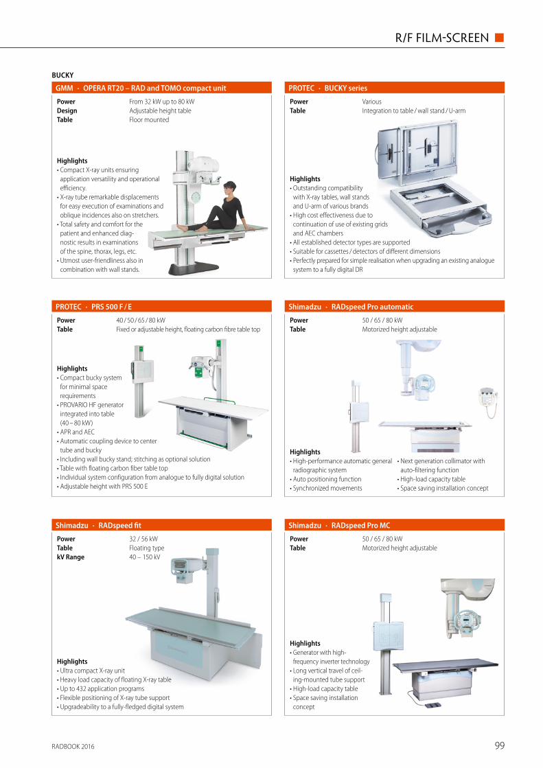
BUCKY
· OPERA RT20 – RAD and TOMO compact unitGMM
Floor mountedTableAdjustable height tableDesignFrom 32 kW up to 80 kWPower
Highlights• Compact X-ray units ensuring
application versatility and operational efficiency.
• X-ray tube remarkable displacements for easy execution of examinations and oblique incidences also on stretchers.
• Total safety and comfort for the patient and enhanced diag-nostic results in examinations of the spine, thorax, legs, etc.
• Utmost user-friendliness also in combination with wall stands.
· BUCKY seriesPROTEC
Integration to table / wall stand / U-armTableVariousPower
Highlights• Outstanding compatibility
with X-ray tables, wall stands and U-arm of various brands
• High cost effectiveness due to continuation of use of existing grids and AEC chambers
• All established detector types are supported • Suitable for cassettes / detectors of different dimensions • Perfectly prepared for simple realisation when upgrading an existing analogue
system to a fully digital DR
Highlights• Compact bucky system
for minimal space requirements
• PROVARIO HF generator integrated into table (40 – 80 kW)
• APR and AEC • Automatic coupling device to center
tube and bucky • Including wall bucky stand; stitching as optional solution • Table with floating carbon fiber table top • Individual system configuration from analogue to fully digital solution• Adjustable height with PRS 500 E
· PRS 500 F / EPROTEC
Fixed or adjustable height, floating carbon fibre table topTable40 / 50 / 65 / 80 kWPower
· RADspeed fitShimadzu
40 – 150 kVkV RangeFloating typeTable32 / 56 kWPower
Highlights• Ultra compact X-ray unit• Heavy load capacity of floating X-ray table• Up to 432 application programs• Flexible positioning of X-ray tube support• Upgradeability to a fully-fledged digital system
· RADspeed Pro automaticShimadzu
Motorized height adjustableTable50 / 65 / 80 kWPower
Highlights• High-performance automatic general
radiographic system• Auto positioning function• Synchronized movements
• Next generation collimator with auto-filtering function
• High-load capacity table• Space saving installation concept
· RADspeed Pro MCShimadzu
Motorized height adjustableTable50 / 65 / 80 kWPower
Highlights• Generator with high-
frequency inverter technology• Long vertical travel of ceil-
ing-mounted tube support• High-load capacity table• Space saving installation
concept
RADBOOK 2016 99
R/F FILM-SCREEN ◼

BUCKY
· RADspeed Pro MFShimadzu
Motorized height adjustableTable50 / 65 / 80 kWPower
Highlights• Generator with high-
frequency inverter technology• Floor-mounted tube support• High-load capacity table• Space saving installation
concept
HighlightsFits your needs. Fits your budget. • Key components adapted
from Ysio like table, tube, bucky wall stand and many more
• Automation – Fast positioning with advanced tube tracking and comfortable maneuvering
• Small space requirements – fits your room and budget
• Prepared for the future – digitize your system whenever you prefer
· Multix Fusion Siemens
55 / 65 / 80 kWPowerFree-floating, height adjustable, up to 300 kgTable
Highlights• Designed for customising to your
application and budgetary considerations
• Multi-functional and digital-ready
• Ergonomically shaped with floating table for easy positioning
• Small space requirement• Wide range of general procedures • Intuitive touch screen generator
with 864 APR available• Fixed or variable height table
• Floor or ceiling tubestand• Tomography
· RAD seriesSTEPHANIX
Up to 80 kWPowerCompact and reliable solutionDesignUpgradable to DRTechnologyCost efficient, multipurposeSystem concept
HighlightsToshiba recommends Radrex compact radio-graphic systems for general-purpose radiography, being highly accurate and efficient. It is possible to expand the original system to meet the particular clinical requirements of the user. When the system is combined with a portable FPD (35 x 43 cm) and digital processor, a wide range of applications can be performed.
· RadrexToshiba
Motorized height adjustable with floating tabletopTable50 kW or 80 kWPower
· Moviplan 800VILLA SISTEMI MEDICALI
From 13 x 18 cm to 35 x 43 cmCassette SizeFixed or elevating tabletopTable32 / 40 / 50 / 65 / 80 kWPower
Highlights• Modular bucky system for general
radio graphic applications, muscoskeletal diagnostic room or emergency ward
• Several configuration options: table available with motorized lift, floor-mounted or ceiling suspended tubestand
• Optional tomographic functionalities• Available with standard or tilting chest stand
RADBOOK 2016100
◼ R/F FILM-SCREEN

FLUOROSCOPY
· OPERA T – Multifunctional remote-controlled tableGMM
I.I. and FPDPowerUniversal remote-controlled tableImage system50 kW up to 80 kWDesign
Highlights• Wide range of advanced, cost-
effective R / F remote-controlled tables.
• Six different configurations available to suit actual operators' needs.
• 90 / 30° or 90 / 90° tilting movement; 210 cm or 240 cm tabletop length; 150 or 180 cm FFD.
• Different combinations with SFD-I.I. / TV chain, DR or RF flat panel detector.• Wide versatility of application enhanced by special accessories.
· Flexavision seriesShimadzu
Digital or analogImage system12 or 9II format50 / 80 kWPower
Highlights• 90 / 30 Digital or analog local R / F table • Flexible configuration • High reliability • Turnable footrest• Meets all requirements for routine R / F exams
Highlights• Complete patient coverage with
8-way tabletop travel and large receptor movements
• Single-handed cassette handling: automatic loading, centering, format sensing and collimation
• Intuitive and fast operation with innovative control console
• Dose-saving fluoroscopy with SUPERVISION (option)
• Bucky wall stand (option) • Excellent price-performance ratio
· Luminos RF Classic Siemens
23 or 33 cmSize1 k x 1 k matrixDetectorRemote-controlled R / F systemDesign
HighlightsLuminos Select – don’t compromise, be select.• Platform concept –
select to match your budget• Common Siemens user interface
for ease of use• Imaging system from Siemens high-end products• Table with excellent patient access from all sides
· Luminos SelectSiemens
33 cmSize1 k x 1 k matrixDetectorDigital remote-controlled R / F systemDesign
Highlights• Complete patient coverage• Smart 8 ways tabletop travel for easy
and comfortable patient transfer
• Column angulation ± 40° on the whole table's length
• Tomography• Fixed or variable height• Video camera for patient positioning
to optimize dose reduction
· EVIDENCESTEPHANIX
Up to 80 kWPowerCompact and reliable solutionDesign
Upgradable to digital with image Intensifier and Flat Panel Detector
TechnologyVersatile and robust remote controlled tableSystem concept
HighlightsThe Toshiba Plessart EX8 is a digital remote control R / F system comprising a R / F diagnostic table with an over-table X-ray tube configuration, an X-ray high-voltage generator, and a digital imaging system. This system is intended for use as a general- purpose system for abdominal angiography, general abdominal radiography, general skeletal radiography, support of endoscopic procedures, etc.
· Plessart EX8Toshiba
1 k x 1 k CCDImage system12"II format80 kWPower
RADBOOK 2016 101
R/F FILM-SCREEN ◼

HighlightsToshiba Plessart VIVO is a remote control R / F system comprising an R / F diag-nostic table with an over-table X-ray tube configuration, an X-ray high-voltage generator, and a digital imaging system. This system is intended for use as a general-purpose system for abdominal angiography, general abdominal radio-graphy, general skeletal radiography, support of endoscopic procedures, etc.
FLUOROSCOPY
· Plessart VIVOToshiba
50 kWPower12" or 9"II format
· ApolloVILLA SISTEMI MEDICALI
Analog or digital with I. I.Image system9" / 12" / 16"II format50 / 65 / 80 kWPower
Highlights• Premium remote controlled
system for full clinical coverage in R / F applications
• Full patient coverage by moving only the tube-receptor assembly, without patient repositioning
• Up to 180 cm Source to Image Distance
• SFD with line and cross divisions• Oblique projections at table edges
and electronic tomography• Automatic grid parking
· Apollo EZVILLA SISTEMI MEDICALI
Analog or digital with I. I.Image system9" / 12"II format50 / 65 / 80 kWPower
Highlights• Compact and cost-effective
system for all the needs of radiographic and R / F imaging
• Available with 2-way or 4-way flat tabletop, plastic or carbon-fiber
• Variable Source to Image Distance: up to 180 cm
• SFD with either line or cross divisions • Oblique projections at table edges
and electronic tomography• Automatic grid parking
· Apollo OpenVILLA SISTEMI MEDICALI
Analog or digital with I.I.Image system9" / 12" / 16"II format50 / 65 / 80 kWPower
Highlights• Premium remote controlled
system with OPEN table-top, allowing 4-side access to the patient
• Full patient coverage by moving only the tube- receptor assembly, without patient repositioning
• SFD with line and cross divisions• Up to 180 cm Source to Image
Distance
• Oblique projections at table edges and electronic tomography
• Standard carbon fiber tabletop • Automatic grid parking
· VisionVILLA SISTEMI MEDICALI
Analog or digital with I.I.Image system9" 12"II format50 / 65 / 80 kWPower
Highlights• Available with 2-way or
4-way tabletop • Powerful SFD with line /
cross divisions • Can mount either 9" or
12" Image Intensifiers • Ready for connection with DIVA digital acquisition system
· HF81 SeriesWandong
CCD 1 k x 1 kImage system12 inchesII format80 kWPower
Highlights• Latest technology
80 kW / 200 kHz generator• Remote-controlled diagnostic
table 90°/–25° or 90° /–45°• SID adjustable 100 / 150 cm• 400 kHU High-speed X-ray tube
Assembly• 9" or 12" three fields I.I.• 1 k x 1 k high resolution with 30 fps
image acquisition rate
• InvaRay digital imaging platform, DICOM 3.0 fully support
• Comprehensive digital imaging processing
RADBOOK 2016102
◼ R/F FILM-SCREEN

· HF51 SeriesWandong
CCD 1 k x 1 kImage system12 inchesII format50 kWPower
Highlights• High frequency 50 kW
generator• Remote tilting table
90° / –25°• Variable SID 100 / 150 cm• 400 kHU High-speed X-ray tube
Assembly• 9" or 12" three fields I.I.
• 1 k x 1 k high resolution with 30 fps image acquisition rate
• InvaRay digital imaging platform• DICOM 3.0 fully support
X-RAY MOBILE
· RAFALE BDMS / APELEM
0.1 to 320 mAsmAs Range40 to 125 kVkV Range32 kWPowerAnalogic upgradable DRImage system
HighlightsThe Rafale B is a battery powered mobile X-ray unit analogic. Its compact size and integrated motor makes the unit move-ment smooth and precise. Thanks to tele-scopic tube arm and swivelling column it is able to easily move even in the hospital's smaller rooms. For precise positionning, motor assisted fine positioning adjustements are possible from the tube head and the entire unit moves millimeter by millimeter.
Highlights• Telescopic arm • Easy positioning • Wide coverage • Compact design
· MobileArt ecoShimadzu
0.32 – 100 (200)mAs Range40 – 125kV Range12.5 kWPower
· MobileArt Evolution EFXShimadzu
0.32 – 320mAs Range40 – 133kV Range32 kWPower
Highlights• Superb image quality • Easy handling • User-friendly operation • Sophisticated radiographic functions• Low noise motorized system• Energy saving collimator with a bright
irradiation field through LEDs• DR ready: Flat panel detector upgradability
· MobileArt Evolution EFXShimadzu
0.32 – 320mAs Range40 – 125kV Range12.5 kWPower
Highlights• Superb image quality • Easy handling • User-friendly operation • Sophisticated radiographic functions • Energy saving collimator with a bright
irradiation field through LEDs• Low noise motorized system
Please visit us at
www.healthcare-in-europe.com
RADBOOK 2016
RADBOOK 2016 103
R/F FILM-SCREEN ◼

HighlightsSimplicity and reliability in mobile X-ray imaging. • High image quality due to high pow-
er output and a minimum exposure time down to 4 ms
• Easy handling and maneuverability based on a lightweight and compact system design
• High reliability• Powerful entry level analog mobile
X-ray system
· Polymobil PlusSiemens
16 kW (optional 20 kW)Power40 – 125kV Range
HighlightsRemarkable user comfort in advanced mobile X-ray imaging. • Excellent image quality due to extremely short exposure times down to 1 ms
(Mobilett XP Eco: 2 ms) and a powerful 30 kW generator (Mobilett XP Eco: 20 kW)• Easy mobility and effortless positioning based on a lightweight and compact design,
and an articulated swivel arm • Remarkable user comfort, supported by self-explaining functionality, to ideally support
the daily routine • Mobilett XP Hybrid can be operated from both battery and mains power and offers the
convenience of motor assisted traveling • Advanced analog mobile X-ray system
Mobilett XP Mobilett XP Hybrid Mobilett XP Eco Siemens ·
40 – 133 40 – 133 40 – 125kV Range30 kW, 450 mA (max.) 30 kW, 450 mA (max.) 20 kW, 400 mA (max.)Power
Highlights• Cost effective solution • Compactness ensures easy
handling• User-friendly interface with
492 customizable anatomical programmes
• Wide range of procedures• X-ray tube with rotating anode• Thin dual focal spots• High heat capacity• Short exposure time
· MOVIX Series E+STEPHANIX
Up to 500 mAsmAs RangeUp to 150 kVpkV RangeCapacitor assisted high frequency generatorTechnologyFrom 16 to 32 kWPower
· Visitor T4VILLA SISTEMI MEDICALI
0.2 – 250mAs Range40 – 110kV Range4 kWPowerNoMotorized
Highlights• Cost-effective mobile unit grant-
ing compactness and ease of use• Suitable for most examinations
performed in plaster rooms, emergency and health screenings contexts
• Compact and lightweight design for easy handling
X-RAY MOBILE
· Multimobil 10 Siemens
40 – 125kV Range10 kWPower
HighlightsThe economical solution in mobile X-ray imaging. • Short exposure times and
a constant imaging power provide a high image quality
• Easy handling and maneu-verability based on a light-weight and compact design
• Entry level analog mobile X-ray system
RADBOOK 2016104
◼ R/F FILM-SCREEN

· Visitor T30CVILLA SISTEMI MEDICALI
0.1 – 220mAs Range40 – 125kV Range32 kWPowerNoMotorized
Highlights• Mobile unit designed for
emergency context as well as orthopedics, pediatric or surgery departments
• Compact and lightweight design for a high maneuverability of the unit
• High performance generator and double focal spot (0.8 / 1.3 mm) tubehead
• APR anatomic mode • User friendly control panel
HighlightsPX series mobile X-ray system can be used mainly for radiography in the operation room, emergency ward, orthopedics and surgical treatment. Apply high frequency conversion technology, greatly improve image quality, shorten exposure time, and reduce the harmful radiation to human body. High frequency generator, Ergonomics designed, Microcomputer- control, easy to operate, maintain and move.
· PX100-CLKWandong
1.6 kWPower0.4 ~ 98 mAsmAs Range40 ~ 100 kVkV Range
Highlights• Pediatric anthropomorphic training
phantom • ATOMMax dental and diagnostic
head phantom
• Radiography fluoroscopy QA phantom
• 3dimensional torso phantom• Test tools
ACCESSORIES / COMPLEMENTARY SYSTEMS
· CIRS PhantomsGCTechnology · PROGNOST XP-seriesPROTEC
Fixed or adjustable height (optional), carbon fiber table topTableLine or batteryPower
Highlights• Mobile patient table to position the patient directly above the corresponding
image receptor • For digital DR detectors or with bucky tray integrated • Fixed table height or elevating with floating carbon fibre table top • Elevating versions with line connection or battery powered
Highlights• Dosimetry system (CE marked, class IIb certified) acc. to IEC 61674• Incl. NOMEX DOSEMETER and MULTIMETER (captures all dose values, time,
kVp, TF, HVL, frequency, pulses, waveforms)• Data and waveform export to Excel via USB or Bluetooth• Accessories: Test objects NORMI RAD/FLU, NORMI DSA, NORMI 3D
(CE marked, class I certified)
· NOMEX System PTW
Highlights• Our German-made anthro-
pomorphic phantoms allow repeated x-ray imaging of specific body regions. They are used in x-ray trainings or for specific equipment tests under life-like conditions.
• The phantoms comprise of real human bones embedded in tissue-equivalent material.
Available phantom versions• Full Body• Head• Hand / Arm• Hip / Spine• Foot / Leg• Special Training Phantoms
· Anthropomorphic X-Ray PhantomsQUART
RADBOOK 2016 105
R/F FILM-SCREEN ◼

106
MECALL
A Division of Philips Healthcare
R / F Digital
Conventional
Digital
DR Retro� t
Mobile DR
Flatpanel Fluoro
Accessories / Complementary Systems

CONVENTIONAL
· CR 10-XAgfa
35 x 43 cmCassette size20 bits / pixelResolution1Slots
Highlights• Affordable CR solution that makes no compromises in image quality• For a convenient and fast workflow• Robust, yet easy to install and maintain• Fits in small spaces and is suited for mobile applications• Networking capabilities deliver seamless integration• Capacity: 34 plates / hour
· CR 15-XAgfa
Single slot cassette feedSlots580 x 700 x 471 mm (w x d x h)SizeAutoranging external power supply (24 V output)Power
Highlights• Affordable for a broad range of applications• Convenient and fast workflow, with usercontrollable speed and resolution• Robust yet easy to install and maintain• Fits in small spaces and is suited for mobile applications• Highly versatile, compact CR 15-X offers an ideal solution fordecentralised
hospital environments, clinics and private practices.
· CR 30-Xm*Agfa
From 15 x 30 cm to 35 x 43 cm, incl. mammographyCassette size10 pixels / mm, 20 pixels / mm for mammographyResolution1Slots
· CR 12-XAgfa
35 x 43 cmCassette sizemax. 200 μm / pixelResolution1Slots
Highlights• Affordable CR system offering high image quality• Customer-chosen optimal workflow• Robust, yet easy to install and maintain• Suited for mobile applications• Networking capabilities deliver seamless integration
Highlights• Tabletop digitizer• Broad range of applications:
mammography, general radiography, orthopaedics, chiropractic, dental and FLFS
• No quality compromises• Horizontal cassette insertion
• Low total cost of ownership• Mobile use• Capacity: up to 82 plates / h*CR 30-XM not available in the US & Canada
HighlightsDX-M : Mixed to perfection• Next-generation CR digitizer• NIP and PIP detectors for general
radiography and mammography• Superb image quality and potential
for dose reduction• Five cassette drop-and-go buffer• Small footprint• Capacity: approx. 83 plates per hour
(35 x 43 cm cassette)• MUSICA Image Processing* DX-M with CR Mammography application is not
available in the US
· DX-M*Agfa
From 15 x 30 cm to 35 x 43 cm, incl. mammographyCassette size6.7 – 20 pixels / mmResolution1 – 5 cassettes: drop and go bufferSlots
Please visit us at
www.healthcare-in-europe.com
RADBOOK 2016
RADBOOK 2016 107
R/F DIGITAL ◼
MECALL
A Division of Philips Healthcare
R / F Digital
Conventional
Digital
DR Retro� t
Mobile DR
Flatpanel Fluoro
Accessories / Complementary Systems

Best-in-class: Sonialvision G4 multifunctional R / F systemThe Sonialvision G4 high performance R/F table provides numerous best-in-class fea-tures significantly increasing its functional-ity and operability. Sonialvision G4 unites the widest possible range of examinations with inter-departmental hospital capabili-ty. The largest available FPD at 43 x 43 cm provides an extensive imaging area. An
additional ceiling-mounted telescopic arm, a Bucky wall stand, and a second mobile FPD, turn the system into a sophisticated multifunctional R/F room. • SUREengine-Advance is a leading-edge digital image processing technology and ensures extremely clear fluoroscopy and radiography images.
• Slot Advance provides high accuracy imag-es with long fields of view, such as for full spine or full leg images, taken with a min-imal X-ray dose. SLOT Advance acquires a series of accurate images of a few centime-tres central slit as the imaging chain moves successively along the patient and allows precise measurements of extremities.
• Tomosynthesis is a proven X-ray imaging technology for higher precision diagnoses. In a simple and quick workflow, and at a low exposure dose, tomosynthesis state-of-the-art imaging technology offers high quality multi-slice images to visualize the
differently sized wireless flat panel detec-tors (FPD), broadens its applications from clinical rounds in hospitals to critical care and applications at disaster sites, as well as operating rooms and neonatal inten-sive care units (NICU). The choice of three detectors (42 x 43, 35 x 43, and 27 x 35 cm) provides superior flexi bility for users. They combine high sensitivity with the lowest possible dose of radiation and provide sharp high-quality images in areas such as radiology, emergency rooms, traumatology, orthopedics, pediatrics, or on the ward.
New features improve safety as well as pro-cessing speed, and save energy: • vibration-resistant DR unit adopting a high-speed solid-state drive (SSD), there-by reducing the risk of data loss
• energy saving collimator with a bright irra-diation field through LEDs
• LCD monitor with a wide viewing angle around the unit
• FPD contributing to improved procedural efficiency.
part which is invisible for conventional plain radiography.
• T-smart provides even clearer tomosyn-thesis images suppressing the artefacts around metal objects even further. This application is of great help in orthope-dics especially for patients with metal implants or fixators as it allows a very exact diagnosis of the status of the boundary between bone and implant.
RADspeed Pro EDGE
The new RADspeed Pro EDGE digital radiog-raphy system adds numerous functionalities
to support diagnostics in clinical appli-cations.
• Tomosynthesis allows to easily obtain multiple digital cross-sec-tion images from a single linear tomography scan.
• Dual-energy subtraction uti-lizes the difference in X-ray absorption levels of bones and soft tissue to gener-ate separate images, which
is useful for diagnoses in the chest area, such as lung cancer.
• Auto-stitching radiography covers the entire lower extremities or entire
spine and links the settings made on the X-ray tube with the Bucky table or Bucky stand with subsequent automatic image stitching.
• Dedicated tomosynthesis workstation allows parallel processing of data with examinations increasing the throughput and reducing the stress on patients.
Mobile X-ray applications: evolving technology with outstanding flexibilityThe new X-ray MobileDaRt Evolution EFX can be moved to any location where radi-ography is required, enabling on-site exami nations and image verification. Capitaliz-ing on the merits of efficien-cy and high throughput, this digital mobile X-ray system, which is equippable with
VISIT US AT ECR 2016 IN VIENNA, AUSTRIA · 2-6 MARCH EXPO X2, STAND 325Further information: Shimadzu Europa www.shimadzu.eu/medical
World-class technologies for healthcare and diagnosisAs a leading global provider of both diagnostic imaging and analytical instrumentation technologies, Shimadzu offers broad expertise in medical imaging and mass spectrometry detection platforms helping to deliver a measurable impact on healthcare and diagnosis. The company is the perfect partner for transformational technologies to acce lerate diagnosis.
Medical Imaging platformsBased on more than 100 years of extensive clinical experience in X-ray technologies, Shimadzu provides a multitude of radiographic and fluoroscopic systems, both either floor- or ceiling mounted, and mobile units. World-leading technology, cutting-edge appli-cations and functionalities enhance healthcare providers’ examina-tion efficiency and safety while reducing the radiation dose.
Laboratory systemsShimadzu’s analytical instrumentation units support healthcare and diagnosis applications through chromatography, mass spectrometry, spectroscopy, and life sciences. Shimadzu’s world-leading quantita-tive mass spectrometry systems are utilized for various applications, e.g. TDM (Therapeutic Drug Monitoring), NBS (Newborn screening), toxicology, steroid and vitamin analysis. The instruments provide high-speed methods and high sensitivity for multi-component simultaneous analysis even at low concentrations.
Passion for detailsNew diagnostic imaging systems and units with best-in-class features and new functionalities provide excellent image quality for a wide range of efficient examinations. Patients benefit from a high level of comfort and low exposure dose, and clinical staff from oper-ability, better patient coverage and throughput.
Trinias MiX package
Shorter treatment times and less contrast media usedThe newly re leased Trinias MiX package (Minimally invasive eXpe-rience) supports less in vasive treatments through a variety of applications. The Trinias MiX package is an exten sion of the Trinias angiography system, which facilitates high-level interventions using a proprietary image processing technology.
The functional enhancements of the Trinias MiX package cover the following applications: • SCORE Navi + Plus 3D Application interlinked with previously acquired CT images imports pre-procedural MDCT images and links them with fluoroscopy images, allowing a seamless workflow from preoperative treatment to perioperative navigation.
• SCORE StentView + Plus PCI Support Application improves device detec tion efficiency displaying stents in a fixed position in real-time even in procedures using multiple devices, thereby shorten-ing treatment times.
• SCORE MAP provides automatic trace mapping for aortic stent grafting au tomatically extracts and displays only the outline of vessel walls providing a map image for easy device guidance.
Sonialvision G4: Multifunctional R/F system
RADspeed Pro EDGE: Extended functionality and comprehensive diagnostic capabilities
Trinias F12 MiX package: Floor-mounted C-arm type pro-vides functional enhancements which achieve shorter treatment times and less con trast media
RADBOOK 2016108
◼ R/F DIGITAL

Best-in-class: Sonialvision G4 multifunctional R / F systemThe Sonialvision G4 high performance R/F table provides numerous best-in-class fea-tures significantly increasing its functional-ity and operability. Sonialvision G4 unites the widest possible range of examinations with inter-departmental hospital capabili-ty. The largest available FPD at 43 x 43 cm provides an extensive imaging area. An
additional ceiling-mounted telescopic arm, a Bucky wall stand, and a second mobile FPD, turn the system into a sophisticated multifunctional R/F room. • SUREengine-Advance is a leading-edge digital image processing technology and ensures extremely clear fluoroscopy and radiography images.
• Slot Advance provides high accuracy imag-es with long fields of view, such as for full spine or full leg images, taken with a min-imal X-ray dose. SLOT Advance acquires a series of accurate images of a few centime-tres central slit as the imaging chain moves successively along the patient and allows precise measurements of extremities.
• Tomosynthesis is a proven X-ray imaging technology for higher precision diagnoses. In a simple and quick workflow, and at a low exposure dose, tomosynthesis state-of-the-art imaging technology offers high quality multi-slice images to visualize the
differently sized wireless flat panel detec-tors (FPD), broadens its applications from clinical rounds in hospitals to critical care and applications at disaster sites, as well as operating rooms and neonatal inten-sive care units (NICU). The choice of three detectors (42 x 43, 35 x 43, and 27 x 35 cm) provides superior flexi bility for users. They combine high sensitivity with the lowest possible dose of radiation and provide sharp high-quality images in areas such as radiology, emergency rooms, traumatology, orthopedics, pediatrics, or on the ward.
New features improve safety as well as pro-cessing speed, and save energy: • vibration-resistant DR unit adopting a high-speed solid-state drive (SSD), there-by reducing the risk of data loss
• energy saving collimator with a bright irra-diation field through LEDs
• LCD monitor with a wide viewing angle around the unit
• FPD contributing to improved procedural efficiency.
part which is invisible for conventional plain radiography.
• T-smart provides even clearer tomosyn-thesis images suppressing the artefacts around metal objects even further. This application is of great help in orthope-dics especially for patients with metal implants or fixators as it allows a very exact diagnosis of the status of the boundary between bone and implant.
RADspeed Pro EDGE
The new RADspeed Pro EDGE digital radiog-raphy system adds numerous functionalities
to support diagnostics in clinical appli-cations.
• Tomosynthesis allows to easily obtain multiple digital cross-sec-tion images from a single linear tomography scan.
• Dual-energy subtraction uti-lizes the difference in X-ray absorption levels of bones and soft tissue to gener-ate separate images, which
is useful for diagnoses in the chest area, such as lung cancer.
• Auto-stitching radiography covers the entire lower extremities or entire
spine and links the settings made on the X-ray tube with the Bucky table or Bucky stand with subsequent automatic image stitching.
• Dedicated tomosynthesis workstation allows parallel processing of data with examinations increasing the throughput and reducing the stress on patients.
Mobile X-ray applications: evolving technology with outstanding flexibilityThe new X-ray MobileDaRt Evolution EFX can be moved to any location where radi-ography is required, enabling on-site exami nations and image verification. Capitaliz-ing on the merits of efficien-cy and high throughput, this digital mobile X-ray system, which is equippable with
VISIT US AT ECR 2016 IN VIENNA, AUSTRIA · 2-6 MARCH EXPO X2, STAND 325Further information: Shimadzu Europa www.shimadzu.eu/medical
World-class technologies for healthcare and diagnosisAs a leading global provider of both diagnostic imaging and analytical instrumentation technologies, Shimadzu offers broad expertise in medical imaging and mass spectrometry detection platforms helping to deliver a measurable impact on healthcare and diagnosis. The company is the perfect partner for transformational technologies to acce lerate diagnosis.
Medical Imaging platformsBased on more than 100 years of extensive clinical experience in X-ray technologies, Shimadzu provides a multitude of radiographic and fluoroscopic systems, both either floor- or ceiling mounted, and mobile units. World-leading technology, cutting-edge appli-cations and functionalities enhance healthcare providers’ examina-tion efficiency and safety while reducing the radiation dose.
Laboratory systemsShimadzu’s analytical instrumentation units support healthcare and diagnosis applications through chromatography, mass spectrometry, spectroscopy, and life sciences. Shimadzu’s world-leading quantita-tive mass spectrometry systems are utilized for various applications, e.g. TDM (Therapeutic Drug Monitoring), NBS (Newborn screening), toxicology, steroid and vitamin analysis. The instruments provide high-speed methods and high sensitivity for multi-component simultaneous analysis even at low concentrations.
Passion for detailsNew diagnostic imaging systems and units with best-in-class features and new functionalities provide excellent image quality for a wide range of efficient examinations. Patients benefit from a high level of comfort and low exposure dose, and clinical staff from oper-ability, better patient coverage and throughput.
Trinias MiX package
Shorter treatment times and less contrast media usedThe newly re leased Trinias MiX package (Minimally invasive eXpe-rience) supports less in vasive treatments through a variety of applications. The Trinias MiX package is an exten sion of the Trinias angiography system, which facilitates high-level interventions using a proprietary image processing technology.
The functional enhancements of the Trinias MiX package cover the following applications: • SCORE Navi + Plus 3D Application interlinked with previously acquired CT images imports pre-procedural MDCT images and links them with fluoroscopy images, allowing a seamless workflow from preoperative treatment to perioperative navigation.
• SCORE StentView + Plus PCI Support Application improves device detec tion efficiency displaying stents in a fixed position in real-time even in procedures using multiple devices, thereby shorten-ing treatment times.
• SCORE MAP provides automatic trace mapping for aortic stent grafting au tomatically extracts and displays only the outline of vessel walls providing a map image for easy device guidance.
Sonialvision G4: Multifunctional R/F system
RADspeed Pro EDGE: Extended functionality and comprehensive diagnostic capabilities
Trinias F12 MiX package: Floor-mounted C-arm type pro-vides functional enhancements which achieve shorter treatment times and less con trast media
RADBOOK 2016 109
R/F DIGITAL ◼

· Regius 110 HQKonica Minolta
From 18 x 24 cm to 35 x 43 cmCassette size3 – 11 Lp / mmResolution1Slots
Highlights• Highy quality mammography read function• Easy to operate and maintain• Powerful compact reader with linear motor technology• Use with standard cassettes and / or mammography cassettes
CONVENTIONAL
· Regius 210Konica Minolta
From 18 x 24 cm to 35 x 43 cmCassette size3 – 11 Lp / mmResolution2Slots
Highlights• High performance dual bay reader• Outstanding image quality in both
general X-ray and mammography• Low dose imaging for paediatric use• Use with standard cassettes and
CsI cassettes (CP-1M, CP-1S)
· Regius Sigma IIKonica Minolta
From 18 x 24 cm to 35 x 43 cmCassette size3 – 6 Lp / mmResolution1Slots
Highlights• Only 28 kg • Foot print only 0.31 m2 • Processes up to 60 plates / hour • Ultra compact: Konica Minolta's smallest and lightest CR reader • Environmentally friendly with an energy consumption of max. 100 VA
Highlights• Single-slot system ideal for normal
to high volume workload and optional high resolution imaging
• Ideal for departments with the decentralized reader-per-room set-up
• Excellent image quality thanks to UNIQUE image processing
• Customizable workflow with the PCR Eleva workspot
• For general applications, including orthopedic and dental applications
• Faster read-out than S reader with high speed mode
• 50 micron scanning option • Orthopedic automatic image
stitching
· PCR Eleva S PlusPhilips
10 pixel / mm, 5 pixel / mm in high-speed mode, 20 pixel / mm with HR cassettes (option)
Resolution
97 plates / h (18 x 24 cm); 94 plates / h (35 x 35 cm in high-speed mode)
Capacity1Slots
· PCR Eleva CompactPhilips
55 plates / hour (18 x 24 cm)Capacity10 pixel / mmResolution1Slots
Highlights• Single-slot system
ideal for smaller facilities with moderate throughput requirements• Excellent image quality thanks to UNIQUE image processing• Customizable workflow with the PCR Eleva workspot• Ideally suitable for X-ray departments with a decentral reader setup• For general applications, including orthopedic and dental applications• Orthopedic automatic image stiching
RADBOOK 2016110
◼ R/F DIGITAL

· DX-D 40 detectorAgfa
CsI and GOSTechnologyAED (Automatic Exposure Detection)Detector384 x 460 mm (outer dimension)SizeAmporhous SiliconDetector
HighlightsThe DX-D 40 Digital Detector with Automatic Exposure Detection (AED) offers a fast and effective way for radiography facilities to benefit from high quality digital imaging using any X-ray equipment: • Improved workflow and exam speed • Cassette-sized detector gives
maximum convenience and ortability • MUSICA processing for excellent
contrast detail
· DX-D 300Agfa
From 0.1 to 500 mAs in 38 stepmAs RangeFrom 40 to 150 kVp in 1 kVp stepkV Range
DIGITAL
Highlights• Universal modality• Single DR detector• MUSICA processing
provides superior contrast detail and consistent, exam-independent image quality
• NX acquisition workstation offers comprehensive functionality for integrated workflow
• Integrated software for generator and positioner interface
• Complete versatility with optional CR / DR combination
• Motorized positioner• Floor mounted
Highlights• Cassette size bucky can rotate
from landscape to portrait• Build-in Dose Area product
meter (optional)• Scalable, flexible and affor dable
modality• Flexible configurations and
options for most needs• Supports CR and DR integration• Requires limited space (4 x 2 m)• MUSICA processing provides superior contrast detail and consistent,
exam-independent image quality• NX acquisition workstation offers comprehensive functionality for integrated
workflow
· DR 400 (floormounted)Agfa
40, 50, 65, 80 kWPower
· DX-D 45 detectorAgfa
Amorphous Silicon with TFTDetectorEffective area: 251.0 x 314.5 mm (10 x 12 inch) SizeCsI (Cesium Iodide) and GOS (Gadolinium oxysulfide)Technology
Highlights• The easiest and most
versatile way to go Direct Digital• High resolution,light-weight and
small cassette-sized• Detector ideal for use in pediatrics, for extremities and special examinations• Automatic Exposure Detection (AED) for seamless use with virtually all X-ray
systems• Excellent connectivity with DICOM compatible SW and imagers• MUSICA processing for excellent contrast detail and exam-independent
· DR 600 (ceiling suspended)Agfa
Highlights• Excellent user-friendly
10 inch tube head display with preview image
• Detector CsI technology with dose reduction potential
• Tilting wallstand bucky with vertical tracking, holders for patient convenience and colli-mator light switch
• High-productivity, top-of-the-line, direct radiography system with motorized auto-positioning.
• MUSICA processing provides superior contrast detail and consistent, exam independent image quality
• NX acquisition workstation offers comprehensive functionality for integrated workflow
• Automatic versions support DR detec-tors in the wall stand and table with optional additional integrated CR
· DelftDI Adora DRiCanon
Motorised carbon fiber, floatig top with 340° rotationTableCanon CXDI-series of high resolution DR detectorsDetectorCeiling-suspended DR systemDesign
HighlightsNext generation High End solution for all radiographic applications• Intelligent workflow for high volume
patient throughput• Easy APR auto-positioning –
up to 1,000 positions• SmartHandle motorized movement,
zero force• Intuitive tube head control • Optional: Integrated image stitching for total spine and total leg, Fluoroscopic
capability, RF, Double tube head for RSA imaging procedures
RADBOOK 2016 111
R/F DIGITAL ◼

DIGITAL
· DelftDI XSense DRCanon
Canon CXDI-series, high resolution DR detectorsDetectorAuto-positioningMotorizedMotorized height adjustable with fixed table topSystem concept
HighlightsA high-end system for radio graphic imaging and beneficial features for your emergency department• Optimized workflow for
high-volume patient throughput• Smart automatic positioning and
detector tracking in all directions• Suitable for orthopedic, trauma and
pediatric procedures with fixed tabletop
• Generator interface on tube head display
• Fully automatic image stitching
· DelftDI Triathlon DRCanon
Motorized height adjustable with floating tabletopTableCanon CXDI-series, high resolution DR detectorsDetectorCeiling-suspended DR systemDesign
HighlightsHigh End solution for all radiographic applications• Optimised workflow for high
volume patient throughput• Smart Automatic Positioning• Fully Automatic Image
Stitching• High efficiency with RIS-integrated workflow• Advanced 6-way Patient Table with motorised adjustment and motorised
detector tracking• Tubehead display allows allows access to a variety of examination information
· DelftDI Trauma DR PLUSCanon
Canon CXDI-series, high resolution DR detectorsDetectorCeiling-suspended U-arm trauma systemDesign
HighlightsVersatile solution for trauma applications:• Fast and efficient workflow• Easy manual positioning with motorized
support for Z-movement• Large open workspace with a fixed focus-
detector distance of 135 cm • Integrated cable management• C-Arm dept of 55 cm• Integrated Dose Area Product Meter (DAP)• Acquisition station with large DICOM calibrated touch screen display
· DelftDI Intuition DRCanon
With floating tableTableCanon CXDI-series, high resolution DR detectorsDetectorCeiling-suspended DR systemDesign
HighlightsVersatile solution for all radiographic applications:• Optimized workflow for high
volume patient throughput• High efficiency with RIS
inte grated workflow• Lightweight manual Alpha,
Beta, X- and Y-movement• Motorized Z-movement,
floating tabletop• Smart Chest and table tracking• Acquisition station with large DICOM calibrated touch screen display• Easy to fit in low ceiling X-ray rooms
· DelftDI Easy DRCanon
Floor mounted X-Ray systemDesignCanon CXDI-series, high resolution DR detectorsDetector
HighlightsVersatile solution for multi purpose examinations• Multipurpose floor mounted
X-Ray system• Suitable for mobile installations
(i. e. truck or container)• Retractable anti-scatter grid• Vertical and horizontal positioning of the U-arm• Acquisition station with DICOM calibrated touch screen display
· Camargue HQ DRDMS / APELEM
36 x 43 cm Wifi / 43 x 43 cm, 41 x 43 cm FixSizeCsI or GadoxDetector50 / 65 / 80 kWPower
HighlightsThe Camargue series was designed to ensure the best radiographic performance.Several model are available:• Manual ceiling suspension• Auto tracking• Fully motorized, 5 axes• Variable height tableDifferent configuration available with:• Only one portable detector wifi for
table & VBS
• One portable detector wifi & flat panel detector
• 2 flat panel detectors
RADBOOK 2016112
◼ R/F DIGITAL

· Discovery XR656 PlusGE Healthcare
200 μmPixel sizea-Si, 41 x 41 cm, FlashPadDetector50 / 65 / 80 kWPower
Highlights• High productivity through complete motorization• Clinical flexibility through wireless FlashPad detector• Possibility of detector sharing• Table with high patient load up to 320 kg• Optimized efficiency and diagnostic confidence through optional Advanced
applications• Advanced applications: VolumeRAD, Dual Energy, AutoPasting
· Optima XR646GE Healthcare
200 μmPixel sizea-Si, 41 x 41 cm, FlashPadDetector50 / 65 / 80 kWPower
Highlights• Universally applicable, with robust table up to 320 kg patient load• Flexible 3D ceiling suspension with tracking• Clinical flexibility through wireless FlashPad detector• Improved efficiency and diagnostic confidence through optional Advanced
applications• Advanced applications: Dual Energy, AutoPasting
· ConnexityGE Healthcare
148 μmPixel sizea-Si, 43 x 43 cmDetector65 / 80 kWPower
Highlights• System design with open, backside system access• FFA variable 115 – 180 cm for max. investigation flexibility• Patient convenience and safety through free patient access from four sides
and height adjustment of the tabletop• 43 x 43 cm flat detector• Options: Wallstand, ceiling suspension with X-Ray tube and others
Highlights• User-friendly solution for direct
digital radiology.• Adjustable height examination ta-
ble floating in the four directions.• X-ray tube column stand sliding
on rails combined with examina-tion table and wall stand.
• Column stand rotation around its vertical axis for an easy and safe execution of lateral projections.
• Advanced digital system for image acquisition and processing.
· CALYPSO F – Multifunctional DR systemGMM
35 x 43 cm and 43 x 43 cmSizeFixed or portableDetectorFloor fixed system with double detectorDesign
· CALYPSO – Multifunctional DR systemGMM
Adjustable heightTableFixed or portable DetectorCeiling suspended-double detector systemDesign
Highlights• Enhanced Direct digital radiology in Trauma,
ER, routine and specialized examinations.• Preset for two flat panel detectors either
fixed or WiFi.• Adjustable height examination table
for easy and safe patient positioning. • Exclusive interlocking technology
ensuring automatic alignment of the X-ray source to the detector movement.
• Advanced digital system with optional stitching
Please visit us at
www.healthcare-in-europe.com
RADBOOK 2016
RADBOOK 2016 113
R/F DIGITAL ◼

DIGITAL
· AeroDR X50Konica Minolta
175 µmPixel sizeAeroDR CsI FPD 14" x 17" / 17" x 17" / 10" x 12"Detector32 – 80 kWPower
Highlights• High image quality, low dose• Compact • Suits small rooms• Optional stitching• AeroDR detector can be used in table, wallstand or outside of bucky
· AeroDR X70Konica Minolta
175 µmPixel sizeAeroDR CsI FPD 14" x 17" / 17" x 17" / 10" x 12"Detector50 – 80 kWPower
Highlights• Multiple configurations possible• Light handling, servo tracking
standard• Excellent workflow in combination
with AeroDR detector• Intuitive CS-7 console• Can be installed in rooms with a
minimal height of 2.5 metres
· AeroDR HQ RangeKonica Minolta
Monocoque carbon fiberDesignCsI scintillator 17 x 17", 14 x 17", 10 x 12"DetectorWLANSystem concept
Highlights• Unique battery technology prevents
overheating• High quality images at a low dose• Two second preview
• Durable design• Lightweight, for light handling:
1.7 kg (10 x 12"), 2.9 kg (14 x 17"), 3.6 kg (17 x 17")
• High DQE CsI detector
Highlights• State-of-the-art system with
single detector and remova-ble auto-focusing grid
• Single end suspended and pivoting tabletop for easy treatment of patients on stretcher
• Full-length patient examination• Advanced ceiling suspension with motorized movements• Auto positioning features driven by anatomical programs• Advanced image processor for perfect images at consistent low dose
· EIDOS 3000 – Single/ Dual FDP DR systemMecall
43 x 43 cm; 35 x 43 cm WiFiSize143 µmResolutionAmorphous silicon Detector
Highlights• Advanced elevating table with
detector floating in the longitudinal and lateral directions
• Automatic alignment of the detector with the X-ray beam
• Useful radiographic area > 2 m including lateral projections
• Auto positioning features driven by anatomical programs
• Advanced image processor fully integrated into ceiling suspension touch screen
· KALOS – Single / Dual / Triple FPD DR systemMecall
43 x 43 cm; 35 x 43 cm Wi-Fi; 24 x 30 cm Wi-FiSize148 µmResolutionAmorphous siliconDetector
· meX+ Softwaremedical ECONET
Including “Patient CD” and “DICOM send”" functionTechnologyModern graphical user interfaceDesignX-ray Image Acquisition SoftwareSystem concept
HighlightsMost sophisticated and user friendly image acquisition software “Made in Germany”.• Simple and perfect images at all time• Integrated automatic image
optimization• Touchscreen function for easy
operating• Fully integrated radiographic
positioning guide• Bones and soft tissues in one image
• Adaption of almost every language possible
• Safe and fast registration of emergency patients
RADBOOK 2016114
◼ R/F DIGITAL

· DigiEye 280 DR SystemMindray Medical
30 kW / 50 kWPowerCesium Iodide ScintillatorDetector
35 x 43 cm; PortableSize140 μmPixel size
Highlights• Integrated high voltage
generator design • The highest frequency generator
460 kHz• Unique LEVELS image post-processing
technology• Limited installation requirement
• Detector auto-tracking Function • Flexible configuration with portable
detector
· DigiEye 560 DR SystemMindray Medical
43 x 43 cmSizeFPDDetectorU-arm DR SystemDesign65 kW / 80 kWPower
Highlights• Compact design less than 14 sqm
for installation• Fully-automatic and intelligent manual
operation • The highest frequency generator 460 kHz• Unique LEVELS image post-processing
technology• One-Key and iKey positioning • Touch screen control panel with all-functional remote control
· DigiEye 760 DR SystemMindray Medical
43 x 43 cmSize
FPDDetectorCeiling suspending DR systemDesign
143 μmPixel Size
Highlights• More Flexible Configuration than
Your Expectation• High Image Quality with Low
X-ray Dose• Fully-automatic and intelligent
manual operation• One-Key and iKey positioning • Two dimensional auto-tracking
and auto-centering • Touch screen control panel with
all-functional remote control• Panoramic Imaging Technology
· DigitalDiagnost – DR high performance roomPhilips
43 x 43 cmSize8.2 Megapixel 2.84 k x 2.88 k image matrix 148 μm pixel sizeResolutionCsI Single plateTechnology
Highlights• Feel the power of the
premium DR room• High patient volume configu-
ration for fast-paced hospitals and trauma environments
• Convenient workflow with the ergonomic moveable vertical stand• Superb application variety with different detector options including wireless
portable detector tray in table• Philips wireless portable detector sharing to achieve improved cost efficiency
and flexibility at the same time
· DigitalDiagnost – DR value roomPhilips
35 x 43 cmSize7.1 Megapixel 3 k x 2.4 k image matrix 144 μm pixel sizeResolutiona-Si, CsI-ScintillatorTechnology
Highlights• Optimize value with
the premium DR room• Attractively priced configuration for a wide variety of applications• Five amplimat chambers for the wireless portable detector may reduce the
risk of incorrect exposure and repeated images• UNIQUE image processing and Eleva user interface provide seamless procedures• Philips wireless portable detector sharing to achieve improved cost efficiency
and flexibility at the same time
· DigitalDiagnost – DR flex roomPhilips
35 x 43 cmSize8.2 Megapixel 2.84 k x 2.88 k image matrix 148 μm pixel sizeResolutionCsI Single plateTechnology
Highlights• Maximize flexibility with the
premium DR room• Single detector solution with
comparable capabilities as multi- detector room set-ups
• Cover all the projections for a medium to high patient load – also in compact rooms
• System works around the patient for increased comfort• Philips wireless portable detector sharing to achieve improved cost efficiency
and flexibility at the same time
RADBOOK 2016 115
R/F DIGITAL ◼

· DigitalDiagnost – wPD chest roomPhilips
35 x 43 cmSize7.1 Megapixel 3 k x 2.4 k image matrix 144 μm pixel sizeResolutiona-Si, CsI-ScintillatorTechnology
Highlights• Chest examinations and
extremity work by tilting the detector in horizontal position
• 14" x 17" (35 x 43 cm) integrated wireless portable detector, five AEC chambers
• All kind of free exposures in the room by extracting the wireless portable detector, plus possibility to share the WPD with other systems
• Automatic geometry movements• Eleva user interface and UNIQUE image processing for optimal image quality
at the lowest dose
DIGITAL
· DigitalDiagnost – DR chest roomPhilips
35 x 43 cmSize8.2 Megapixel 2.84 k x 2.88 k image matrix 148 μm pixel sizeResolutionCsI Single plateTechnology
Highlights• The premium DR room
dedicated for chest• Superb chest image quality to
support diagnostic confidence• Automatic geometry movements reduce physical involvement of technologists• Optional wireless portable detector broadens the application scope• Philips wireless portable detector sharing to achieve improved cost efficiency
and flexibility at the same time
· DigitalDiagnost – DR emergency roomPhilips
35 x 43 cmSize7.1 Megapixel 3 k x 2.4 k image matrix 144 μm pixel sizeResolutiona-Si, CsI-ScintillatorTechnology
Highlights• The premium DR room for emergency care• Lessens interference with ER devices like tubes,
catheters, oxygen• Patients can be X-rayed without repositioning them• Non cable-bound detector is advantageous as
there are no extra cables to trip over• Philips wireless portable detector sharing to achieve
improved cost effi ciency and flexibility at the same time• Optional wall stand with wireless detector tray
· DigitalDiagnost - wireless portable detectorPhilips
35 x 43 cmSize7.1 Megapixel; 3 k x 2.4 k image matrix; 144 μm pixel sizeResolutiona-Si, CsI-ScintillatorTechnology
Highlights• The wireless portable detector is available with trays in
the vertical stand and in the table plus as an additional detector with detector sharing functio nality for all DigitalDiagnost configurations
• More flexibility: the wireless portable detector carries out even the most difficult projections at table, patient bed, wheelchair or trolley
• More efficiency: smooth digital workflow with rapid results at the Eleva workspot
• More freedom: convenient handling thanks to the wireless detector’s cablefree design
· DuraDiagnost CompactPhilips
35 x 43 cm, rotatableSizeMatrix: 1,920 x 2,367ResolutionQuantmaster, High Stability Scintillator (GoS)Technology
Highlights• Affordable digital radiography system with
low cost of ownership• Compact floor-mounted U-arm geometry• Easy to operate manual system, which fits into small radiography rooms• Customizable digital workfl ow with Eleva workspot• Robust and reliable performance with Philips components• Features Philips UNIQUE image processing* Not available in the USA.
· wireless portable detector sharingPhilips
35 x 43 cmSize7.1 Megapixel; 3 k x 2.4 k image matrix; 143 μm pixel sizeResolutiona-Si, CsI-ScintillatorTechnology
Highlights• Philips wireless portable detector sharing allows
sharing detectors between Philips mobile units, premium radiography rooms and fluoroscopy rooms
• Lower initial investment while assuring a high level of fl exibility
• Back-up solution for continuous uptime• Smart starting point for upgrades, i. e. adding more
detectors to hospital departments in the future
RADBOOK 2016116
◼ R/F DIGITAL

· DuraDiagnost Efficiency roomPhilips
35 x 43 cm, rotatableSize1,920 x 2,367Resolution MatrixQuantmaster, High Stability Scintillator (GoS)Technology
Highlights• Outstanding workflow efficiency
in the exam room with two fixed detectors in one room
• Eleva UI and UNIQUE multi-resolution image processing• Philips well proven generator, X-ray tube and detector ensuring diagnostic
confidence• Default SID positions facilitating fast positioning• The “SmartOne” button allows the user to easily execute all related geometry
movements
· DuraDiagnost Focus roomPhilips
35 x 43 cm, rotatableSize1,920 x 2,367Resolution MatrixQuantmaster, High Stability Scintillator (GoS)Technology
Highlights• Cost-effective DR system
providing affordable entry into DR workflow
• Eleva UI and UNIQUE multi- resolution image processing
• Philips well proven generator, X-ray tube and detector ensuring diagnostic confidence
• Dedicated DR room expands departmental flexibility• The “SmartOne” button allows the user to easily execute all related geometry
movements
· RIVIERA DRPRIMAX International
Floor mounted column on railsDesignWireless or fixed flat panelDetectorUp to 80 kWPower
Highlights• Fixed or variable height floating
tabletop• Last generation ultralight wireless
flat panel• Excellent image quality• Easy to install• Full touch interface• Cost effective
RADBOOK 2016 117
R/F DIGITAL ◼
Nothing, with Mobile DR including FreeView technology FreeView technology makes DR Mobile even more maneuverable! Combining full mobility and safer navigation plus Agfa HealthCare’s gold-standard MUSICA image processing, it gives you high quality imaging on the go.
Find out more, visit agfahealthcare.com
WHAt’S bloCkINg yoUR VIeW?

DIGITAL
· PEDS 600 (Touch option)PROTEC
e. g. 127 µmPixel sizeDifferent panel and scintillator versions, max. 43 x 43 cmDetector40 / 50 / 65 / 80 kWPower
Highlights• DR-System with digital flat panel detector• PROVARIO HF generator (40 – 80 kW)• Anatomical programs and AEC• Variable SID 110 – 200 cm• Rotatable U-arm – 30° up to +135°• Rotatable DR-detector• “Touch” version: high-end
solution with integrated image acquisition through touch- display directly at the system (compare: PRS 500 F/E DR Touch)
· PRS 500 F (Touch option)PROTEC
e. g. 127 µmPixel sizeDifferent single or dual panel systems, max. 43 x 43 cmDetector40 / 50 / 65 / 80 kWPower
Highlights• Integrated state-of-the-art touch concept• Radiographic positioning aid directly
at the system• Patient selection, job selection
and generator control at integrated touch-screen
• First preview at the system imme-diately after the exposure
• Outstanding ease of use due to ideal workflow, simple handling and the selection of the patient at the X-ray system directly
· PRS 500 X (Touch option)PROTEC
e. g. 127 µmPixel sizeDifferent panel and scintillator versions, max. 43 x 43 cmDetector40 / 50 / 65 / 80 kWPower
Highlights• Easy system handling
and positioning due to its optimum weight counterbalance concept
• Maximum flexibility and workflow efficiency• Outstanding variability and extensibility in case of changing application
requirements (e. g. upgrading with extended floor-rail)• Fully digital X-ray generator connection by CONAXX image acquisition software• Also available as TOUCH Version (see PRS 500 F / E)
· PRS 500 E (Touch option)PROTEC
e. g. 127 µmPixel sizeDifferent single or dual panel systems, max. 43 x 43 cmDetector40 / 50 / 65 / 80 kWPower
Highlights• PROVARIO HF generator
integrated into the table (40 – 80 kW)
• APR and AEC• Automatic coupling device to
center tube and bucky• Including wall bucky stand;
stitching as optional solution• Floating carbon fibre table top• Adjustable height combined with undertable generator• Fully digital DR-System with flat panel detector technology, different configu-
rations from single to dual detector systems
HighlightsThe X Fit System consist of a Table with floating tabletop, a X-Ray tube column with longitudinal movement and a fixed wall stand. The System integrated power block consisting of generator, collimator and DAP measurement chamber. The X Fit System is prepared for installation of Bucky´s or Flat Panel Detectors and has a mechanical synchronization of the X-ray source and Bucky Table.
· X FitRoesys
50 / 65 kWPower
HighlightsMultifunctional device with two stands for taking images on lying, standing and sitting patients includes an operating unit with all device functions, integrated collimator and X-ray tube. It is equipped with a motorized tracking control for automatic centering of detector and X-Ray-tube unit. In addition, you can adjust the X-ray tube individually by a telescopic extension.
· X TwinRoesys
SID 100 – 200cmMotorizedCsI, 43 x 43 cmDetector65 / 80 kWPower
RADBOOK 2016118
◼ R/F DIGITAL

· GC85ASamsung
43 x 43 cm / 3,040 x 3,036 pixels43 x 35 cm / 3,040 x 2,466 pixels30 x 25 cm / 2,108 x 1,750 pixels
Size Amorphous Silicon TFT / CsI / WirelessDetectorCeiling-suspended DR systemDesign
Highlights• Smart Control for one-touch opera-
tion and flexible positioning• Shared Bucky (S4335-W, S4343-W)
for easy usage on a table• S-Detectors (S4343-W, S4335-W,
S3025-W) with high DQE
• S-Align for precise alignment• S-Vue imaging engine for precise
imaging• S-Share for compatibility of
S-Detectors• Save-power Mode
· GU60ASamsung
43 x 43 cm / 3,040 x 3,036 pixels43 x 35 cm / 3,040 x 2,466 pixels 30 x 25 cm / 2,108 x 1,750 pixels
SizeAmorphous Silicon TFT / CsI / WirelessDetectorUniversial-arm DR System Design
Highlights• Remote Control that enables conven-
ient movement of devices• S-Vue imaging engine • S-Detectors (S4343-W, S4335-W,
S3025-W) with high DQE • Smart Stitching for diagnostic
convenience • Auto Positioning for straightforward
placement
• Collision Avoidance System • Status Color Coding that enables users
to readily view movement status• 4-axis individual blade control that
optimizes radiation dose
· GM60ASamsung
43 x 43 cm / 3,040 x 3,036 pixels43 x 35 cm / 3,040 x 2,466 pixels30 x 25 cm / 2,108 x 1,750 pixels
SizeAmorphous Silicon TFT / CsI / WirelessDetectorMobile DR systemDesign
Highlights• Motorized moving system • S-Vue imaging engine for improved
clarity • S-Detectors (S4343-W, S4335-W,
S3025-W) with high DQE • S-Share compatibility • Fine positioning buttons that enable
handle-free• Quick image acquisition for image
previews
· RADspeed DRShimadzu
160 / 125 µmPixel sizeFlat panel detector (a-Si)Detector50 / 65 / 80 kWPower
Highlights• Flexible choice of different
flat panel detectors• Excellent image quality• Auto-positioning function• Superb dose efficiency• Seamless network integration
• Size: 17" x 17" (43 x 43 cm) 14" x 17" (35 x 43 cm) 9" x 11" (23 x 28 cm)
· RADspeed DR wireless*Shimadzu
125 µmPixel sizeFlat panel detector (a-Si)Detector50 / 65 / 80 kWPower
Highlights• New generation with
wireless flat panel detector
• Excellent image quality • Auto-positioning
function • Superb dose efficiency • Seamless network integration
• Size: 17" x 17" (43 x 42 cm) 14" x 17" (35 x 43 cm) 14" x 11" (35 x 27 cm)
* System configuration available in selected countries only
Please visit us at
www.healthcare-in-europe.com
RADBOOK 2016
RADBOOK 2016 119
R/F DIGITAL ◼

DIGITAL
· RADspeed Pro V4Shimadzu
139 µmPixel size17" x 17", 14" x 17"Detector80 / 65 / 50 kWPower
Highlights• Fully integrated operation system• Flexible and easy to use X-ray tube support• Various FPD line-up: 17" x 17" / 14" x 17" (portable wired, wireless)• Synchronized functions: auto positioning, auto tracking, auto collimation,
speed stitching• Comprehensive dose management
· RADspeed Pro EDGEShimadzu
50 / 65 / 80 kWPower
150 µmPixel size43 x 43 cm integrated, 43 x 35 cm portableDetector
Highlights• High-performance digital
radio graphic system with extended functionality
• Tomosynthesis (Digital multi-slice tomography)
• Auto-stitching (Speed Stitch function)• Dual Energy Subtraction• Auto-positioning
• Innovative flat panel detectors for increased versatility
• Low dose collimator with auto-filtering feature
Highlights• Robust mobile flat detector
to cover the full spectrum of clinical applications
• Imaging system from Siemens’ high-end product line (e. g. Ysio Max, Multix Fusion) enhanced by DiamondView Plus
• Intelligent automation with organ preset programs to speed setup and improve reproducibility
• High system reliability and availability• Economical minimum space requirement of only 11 sqm with an integrated
generator
· Multix Select DRSiemens
35 x 43 cm, 139 μmSizeaSi / GOSDetector55 kWPower
Highlights• High quality key com-
ponents adapted from Ysio Max
• Outstanding images enhanced by DiamondView Plus• Advanced automation - Fast and easy positioning with tube tracking• Flexible system configurations with up to two detectors for your individual needs• GuidedOrtho – easy to use guidance and automation to acquire and com-
pose long leg and long spine images
· Multix FusionSiemens
a-Si / CsI or a-Si / GOSDetectorCsI: 35 x 43 cm (wireless), 139 µm or 43 x 43 cm (fixed), 148 µm; GOS: 43 x 43 cm (fixed), 148 µm
Size
55 / 65 / 80 kWPower
HighlightsYsio Max – the most direct way to the image.• Unique simultaneous FAST
movement in 6 axes • MAXalign: makes free
exams dramatically faster and easier
• MAX wi-D: only 3 kg, just 19 mm thin, image preview within 2 seconds• MAX mini: the right size for orthopedic, pediatric and trauma exams• MAXswap: the right way to share detectors with a safe, quick and easy one-
click registration
· Ysio MaxSiemens
MAX wi-D 43 x 35 cm, MAX mini 24 x 30 cm, MAX static 43 x 43 cm, all 148 µm
Sizea-Si / CsIDetector65 / 80 kWPower
Detector
HighlightsThe world’s first Twin Robotic X-ray scanner enables streamlined clinical pathways while improving diagnostic insights and treatment.• Offers a multitude of
X-rays – in just one room• Lets you see reality with natural Real 3D – for the first time• Let the robots move – not your patients• Defines standards easily – and multiplies your productivity• Is future-proof – with Twin Robotic X-ray
· Multitom RaxSiemens
RAX detector 43 x 43 cm, MAX wi-D 43 x 35 cm, MAX mini 30 x 24 cm
Sizea-Si / CsICeiling-mounted robotic tube and detectorDesign
RADBOOK 2016120
◼ R/F DIGITAL

Highlights• Low footprint for wide range of procedures
at standing, sitting or lying patient• C-arm shaped for cross exams• Autopositioning regarding each
protocol• Automatic and virtual collimation,
additional filtration• User-friendly interface• Wireless remote
· Statif Pro DReamSTEPHANIX
Optional carbon or elevating tabletop, on wheelsTableAutomatic positioning, collimation, filtration, parametersMotorizedFull-field or portable flat panel detectorDetectorUniversal autocentred C-arm DR unitDesign
Highlights• Manual, vertical tracking
or auto positioning version• Single or multi-detectors room• Fixed or tilting wall Bucky• Floating elevating tabletop for patient
weight up to 300 kg• Intuitive user interface with unlimited
preset APR• Possibility to share wireless detectors with different Stephanix modalities
· Xtreme DReamSTEPHANIX
Fixed and wireless solutionsDetectorUp to 3 Flat Panel Detectors, indirect conversionTechnologyCustomizable ceiling RAD roomDesign
Highlights• Manual or vertical tracking
version• Single or multi-detectors
room• Fixed or tilting wall Bucky• Floating elevating tabletop for patient weight up to 250 kg• Intuitive user interface with unlimited preset APR• Possibility to share wireless detectors with different Stephanix modalities
· RAD Series Pro DReamSTEPHANIX
Fixed and wireless solutionsDetectorUp to 3 Flat Panel Detectors, indirect conversionTechnologyCustomizable floor tubestand RAD roomDesign
Highlights• Multipurpose DR solution for small
budgets• It can be dedicated to chest and
extremities examinations• Low footprint for wide range of
procedures at standing, sitting or lying patient
• Manual or motorized (SID and vertical movement)
• User-friendly interface
· Statif DReamSTEPHANIX
Optional carbon or elevating tabletop, on wheelsTableFull-field or portable flat panel detectorDetectorCost-efficient universal autocentred DR unitDesign
· ddRElementSwissray
148 µmPixel sizea-Si CsI, 43 x 43 cmDetector50 / 65 kWPower
Highlights• Space efficient, multifunctional DR
system fits into small X-ray rooms• 43 x 43 cm flat panel detector delivers
superb image quality within seconds• Multiple language capability• Robust and reliable design
• Easy and intuitive to use, includes digital positioning guide
• Off-center and off-detector imaging capability
• Workflow optimization through advanced eXpert and SwissVision user interface
· ddRFormula PlusSwissray
148 µmPixel sizea-Si CsI, 43 x 43 cmDetector65 / 80 kWPower
Highlights• Fully automated Positioning
System (APS) for highest patient throughput
• 1,296 pre-programmed APR’s• Hand held remote control• Superb diagnostic IQ with heigh
contrast details• Single Focus eXpertStitching
function for orthopedic imaging• Multi language capability
• Off-center and off-detector imaging capability
• Integrated video camera to monitor patient and ensure positioning
RADBOOK 2016 121
R/F DIGITAL ◼

DIGITAL
· ddRAura_OTCSwissray
Automated Ceiling suspension DR-SystemSystem concept148 µmPixel sizea-Si CsI, 43 x 43 cm and 35 x 43 cm WIFIDetector50 / 65 / 80kWPower
Highlights• Versatile ceiling suspension DR-System
with height adj. floating table top• Ergonomic handles and multi-
directional lock release buttons• Wall stand optionally tiltable• 9.7" touch-screen console on tube side• Auto tracking, sensing table and wall
stand or fully automated• Rotational bucky with on-board
charging of detector• 5-field AEC
• Patient registration to image storage in just 3 steps
• Stitching up to 5 images
148 µmPixel size
· ddRAura-FMTSSwissray
Multifunctional Bucky-Table System System concept
50 / 65 / 80 kWPowera-Si CsI, 43 x 43 cm and 35 x 43 cm WIFIDetector
Highlights• Floor mounted DR-System with
fixed or height adjustable floating table top
• Tubestand with ergonomic handles and multidirectional lock release buttons
• Wall stand optionally tiltable• 9.7" touch-screen console on tube side• Auto tracking and sensing table and
wall stand
• Rotational bucky with on-board charging of detector
• Patient registration to image storage in just 3 steps
• Manual stitching up to 5 images
Highlights• Wireless type Portable FPD• Incorporates Toshiba’s proven
advanced fine CsI / Tl and direct deposition technologies
• Unique moisture-proof sealing method used for the CsI / Tl screen
• Standard cassette size• Automatic switching between
wireless / tethered mode
• Short cycle time (less than 10 s)• Recharging in tethered mode• Detachable cable connector• Compact and lightweight battery
recharger
· FDX 3543 RPW / FDX 4343 RPWToshiba Electron Tubes & Devices
140 μmPixel sizeCsI / Tl, 43 x 43 cm, 35 x 43 cmDetectorWireless flat panel detectorSystem concept
· FDX 2530 RPWToshiba Electron Tubes & Devices
140 μmPixel size
Wireless flat panel detectorSystem conceptCsI / Tl, 25 x 30 cmDetector
Highlights• Wireless compact FPD• Incorporates Toshiba’s proven
advanced fine CsI / Tl and direct deposition technologies
• Unique moisture-proof sealing method used for the CsI / Tl screen
• Automatic switching between wireless/tethered mode
• Short cycle time (less than 10 s)
• Recharging in tethered mode• Detachable cable connector• Lightweight: 1.7 kg• AED available• Compact and lightweight battery
recharger
· FDX 4343 RToshiba Electron Tubes & Devices
143 μmPixel sizeCsI / Tl, 43 x 43 cmDetectorStatic flat panel detectorSystem concept
Highlights• Toshiba’s proven advanced fine CsI / Tl and direct deposition technologies
provide high DQE and excellent resolution.• The reflective coating in the CsI / Tl screen provides high sensitivity.• Unique moisture-proof sealing method provides an extremely reliable CsI / Tl
screen that is protected from degradation.• Prompt display of preview / full images and short cycle time enable fast image
acquisition.
· Radrex-iToshiba
139 µmPixel sizea-Si / CslDetector80 kWPower
HighlightsThis digital radiography system is a new-concept system that permits radiography to be performed easily and with greater accuracy. In this system, the operating sections for the digital image processor and the X-ray high-voltage generator are integrated, and the use of an integrated panel improves workflow.
RADBOOK 2016122
◼ R/F DIGITAL

· Moviplan iC with floor-mounted columnVILLA SISTEMI MEDICALI
100 μm or 143 μmPixel sizea-Silicon detector with CsI scintillator, 35 x 43 cm or 43 x 43 cmDetector50 / 65 / 80 kWPower
Highlights• Innovative design with no
unsightly cables• Anti-collision system and
reduced thickness rails• Table commands with
distinctive “light barrier”• Touch Screen interface integrated
on tube-head for immediate inputs• No patient limitation thanks to high
weight capacity
• Electronic tomography with free selection of angle
• Available with stitching, auto-posi-tioning, dual energy functions
· Armonicus VILLA SISTEMI MEDICALI
143 μmPixel sizea-Silicon detector with CsI scintillator, 43 x 43 cmDetector50 / 65 / 80 kWPower
Highlights• Cost-effective DR U-arm system for extended
use, including general radiographic and orthopedic studies
• Easy patient positioning via APR functions
• Auto-positioning capabilities according to RIS procedure codes
• Touch screen control panel, secondary keyboard and infrared remote control as standard
• Variable Source to Image Distance up to 180 cm
• On-board parking station for two grids
· Moviplan iC with ceiling suspensionVILLA SISTEMI MEDICALI
100 μm or 143 μmPixel sizea-Silicon detector with CsI scintillator, 35 x 43 cm or 43 x 43 cmDetector50 / 65 / 80 kWPower
Highlights• High-end solution allowing great
application flexibility and high production capacity
• Touch Screen interface integrated on tube-head
• Tiliting chest stand with special horizontal positioning for exams on mobile stretchers
• Rapid and precise system positioning thanks to full auto-tracking and autopositioning
• Available with stitching and dual energy functions
HighlightsNEW ORIENTAL 1000 U-arm DR is a versatile digital X-ray system to meet customer demands of digital diagno-sis. Less dose and faster acquisition.• High frequency 50kW Generator• 600 APR Programs• 17 x 17" FPD
• Compact U-arm structure with motorized rotation and vertical movement is an ideal solution for inadequate installation space
• InvaRay digital acquisition with DICOM 3.0 compliance
· New Oriental 1000 U-arm DRWandong
17 x 17" FPDDetector40 – 150 kVkV Range50 KWPower
Highlights• 80 kW high frequency generator • Advanced FPD detector • Ceiling suspending structure meet all
kinds of clinical needs • 5 axis electric moving and control • Advanced patient protection
technology • More than 600 APR programs, user
definable
• Tube and detector auto-tracking function
• Programmable fast position switch • High acquisition speed• Remote control available
· New Oriental 1000 Fully AutomaticWandong
143 umPixel size43 x 43 cm FPDDetector80 kWPower
· New Oriental 1000 Wandong
3.6 lp / mmResolution43 x 43cm (17 x 17”)Detector40 ~ 150 kVkV Range50 kW Power
Highlights• High frequency 50 kW
generator• Classical mechanical structure
for all needs of clinical application• Large LCD touch screen table-side
control• X-ray tube auto tracking with the
vertical bucky
• 600 APR programs• Fixed or portable 17 x 17" FPD• InvaRay digital imaging platform
with DICOM 3.0 compliance
RADBOOK 2016 123
R/F DIGITAL ◼

DR RETROFIT
· DX-D 30 RetrofitAgfa HealthCare
125 µmPixel sizeCesium Iodide (CsI) detector conversion screenDetectorWirelessSystem concept46 x 38,4 x 1.5 cmSize
Highlights• Detector is the size of a cassette,
for maximum convenience and portability
• MUSICA processing for superior contrast detail and exam-indepen-dent, consistent image quality
• Improves workflow and exam speed• Superior connectivity to PACS,
HIS / RIS and imagers
• Cesium Iodide (CsI) detector con version screen
• Small pixel size gives more image information, for improved diagnostic effectiveness
· CXDI-401C / 701C / 801C WirelessCanon
VariousSize125 µmResolutionCesium Iodide ScintillatorTechnology
HighlightsWireless flat panel detector series• Lighter, high resolution, low dose• Easy upgrade with just two
components• Includes Non-Synchronized exposure• Preview image time in 3 s
• Sophisticated image processing with optional Scatter Correction software
• Detectors are interchangeable between rooms
· Canon DR-Upgrade-within-2-minutesCanon
43 x 42 cm, 35 x 43 cm, 27.4 x 35 cmCassette size125 µmResolution2 componentsDesignDR Upgrade within 2 minutesSystem concept
HighlightsEasy upgrade solution for any X-ray system in two minutes using just two components• No connections or modifications to your existing X-ray system is necessary• With CXDI-401C / 701C / 801C Wireless Flat Panel Detectors• Optional USB DAP-meter for dose registration• DR Upgrade within 2 minutes. Freedom within reach
· EZ2GODMS / APELEM
36 x 43 cm or 24 x 30 cm Detector2 componentsSystem conceptDR upgrade mobile in 2 minutesDesign
Highlights• Connect up to 3 wifi flat panel detectors• Image preview in 2 s and image acquisition in 4 s• 8 h battery / autonomy• The cassette size of the detector allows upgrade everywhere in the hospital• Ideal for control exams for bedridden patients• The lightiest solution of the market (3.9 kg tablet & detector)
Highlights GAMMA retrofit Kit upgrades analog X-ray equipment to Direct Radiography easily and cost-effectively, ensuring all the advantages of a digital system: digital FPDs fitting into standard Bucky devices replace CR cassettes for an outstanding image quality at a low dose while DELUXE acquisition software enables full DR workflow, advanced image processing, export, print and DICOM storage.
· GAMMAGeneral Medical Italy
Deluxe SoftwareImage system36 x 43 WirelessDetector
Please visit us at
www.healthcare-in-europe.com
RADBOOK 2016
RADBOOK 2016124
◼ R/F DIGITAL

· AeroDR PremiumKonica Minolta
CsI scintillatorDetectorWLANTechnology14 x 17" / 35 x 43 cmCassette size
Highlights• Lightweight, only 2.6 kg• Improved cycle time for increased throughput• Robust: surface load of 300 kg• AED – Hybrid detection technology• Waterproof IPX6, this makes the detector suitable for more extreme environments• Konica Minolta's unique capacitor technology: quick charging (30 minutes),
no overheating
· AeroDR 2SKonica Minolta
CsI scintillatorDetector14 x 17" / 35 x 43 cmCassette sizeWLANTechnology
Highlights• Konica Minolta’s lightest 14 x 17” detector on the market at just 2.5 kg• Robust, IPX6 waterproof, carbon monocoque housing • Full image acquisition within four seconds only• Charging time of only 13 minutes • AeroSync
· meX+1717SGC/SCCmedical ECONET
3,328 x 3,328 pixels, 127 μmPixel size460 x 460 x 15.5 mm, 4.4 kgCassette sizeGadolinium Oxysulfide (Gadox) or Cesium-Iodide (Csl) TechnologyWiredDetector
Highlights• Digital retrofit panel for
stationary use• AED function (Automatic
Exposure Detection)• Largest imaging of
430 x 430 mm allows examination of several bodyparts in one shot
• Ultra-flat (15.5 mm) DR detector with same size as film cassette or CR image plate
• Highest image resolution by decreased radiation dose• Low weight and quick acquisition time of 7 seconds
· DR Retrofit-Kit DX | Visionmedigration GmbH
Wireless, portable detector with WLAN and BatterySystem concepta-Si, CsI Pixium, 35 x 43 cmDetector148 µm, 16 bitPixel size
HighlightsYour upgrade to fully digital radiography• Easy integration into an existing X-ray
system• 100 % touch-capable user interface• Cordless and lightweight wireless flat
panel detector• For the use with mobile X-ray systems• Auto-trigger mode (AED function) – No need to synchronise with the
generator• Excellent image quality through an integrated operating program with
HARMONY image processing
· RAPIXX tethered / mobile detectorsPROTEC
e. g. 139 µmPixel size43 x 36 cm (ISO 4090 compliant), different scintillator versionsDetectorPortable, tetheredSystem concept
Highlights• 16 bit dynamic range• Cable connection, lightweight: 3.7 kg• Predestined for simple retrofitting of existing X-ray units due to dimensions
equal to conventional X-ray cassette• High shock tolerance and water resistant portable flatpanel detector• Interface box, power supply and CONAXX 2 image acquisition software
included in standard delivery – fully DICOM compatible for integration to PACS
· RAPIXX fix installed detectorsPROTEC
e.g. 139 µmPixel size43 x 43 cm, different scintillator versionsDetectorStationary, tetheredSystem concept
Highlights• 16 bit dynamic range• Cable connection• Minimal cycle time: 6 s• For integration and upgrade into existing conven-
tional X-ray units / intended for constant mounting in a X-ray unit• Interface box, power supply and CONAXX 2 image acquisition software
included in standard delivery• Fully DICOM compatible for integration to PACS
RADBOOK 2016 125
R/F DIGITAL ◼
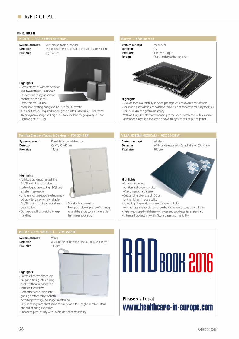
DR RETROFIT
· RAPIXX WiFi detectorsPROTEC
Wireless, portable detectorsSystem concept43 x 36 cm or 43 x 43 cm, different scintillator versionsDetector
Highlights• Complete set of wireless detector
incl. two batteries, CONAXX 2 DR-software (X-ray generator connection as option)
• Detectors are ISO 4090 compliant, existing bucky can be used for DR retrofit
• Just one flatpanel required for integration into bucky table + wall stand• 16-bit dynamic range and high DQE for excellent image quality in 3 sec• Lightweight: < 3.0 kg
e. g. 127 µmPixel size
· X Vision medRoesys
CsIDetectorMobile / fixSystem concept
143 µm / 100 µmPixel sizeDigital radiography upgradeDesign
Highlights• X Vision med is a carefully selected package with hardware and software• For an initial installation or post hoc conversion of conventional X-ray facilities• For use in direct digital radiography• With an X-ray detector corresponding to the needs combined with a suitable
generator, X-ray tube and stand a powerful system can be put together
· FDX 3543 RPToshiba Electron Tubes & Devices
143 μmPixel sizeCsI / Tl, 35 x 43 cmDetectorPortable flat panel detectorSystem concept
Highlights• Toshiba’s proven advanced fine
CsI / Tl and direct deposition technologies provide high DQE and excellent resolution.
• Unique moisture-proof sealing meth-od provides an extremely reliable CsI / Tl screen that is protected from degradation.
• Compact and lightweight for easy handling
• Standard cassette size• Prompt display of preview/full imag-
es and the short cycle time enable fast image acquisition.
· VDX 3543PWVILLA SISTEMI MEDICALI
100 μmPixel sizea-Silicon detector with CsI scintillator, 35 x 43 cmDetectorWirelessSystem concept
Highlights• Complete cordless
positioning freedom, typical of a conventional cassette
• Outstanding pixel size of 100 μm, for the highest image quality
• Auto-triggering mode: the detector automatically synchronizes the acquisition once the X-ray source starts the emission
• System equipped with battery charger and two batteries as standard• Enhanced productivity with Dicom classes compatibility
· VDX 3543TCVILLA SISTEMI MEDICALI
143 μmPixel sizea-Silicon detector with CsI scintillator, 35 x 43 cmDetectorWiredSystem concept
Highlights• Portable lightweight design
flat panel fitting into existing bucky without modification
• Increased workflow• Cost-effective solution, inte-
grating a tether cable for both detector powering and image transferring
• Easy handling from chest stand to bucky table for upright, in-table, lateral and out of bucky exposures
• Enhanced productivity with Dicom classes compatibility
Please visit us at
www.healthcare-in-europe.com
RADBOOK 2016
RADBOOK 2016126
◼ R/F DIGITAL

MOBILE DR
· DX-D 100+ (mobile)Agfa HealthCare
40 to 150 kVpkV Range100 – 500 mA selectablemAs RangeWireless - Amorphous Silicon Detector (a-Si)TechnologyUp to 4 km / hMotorized
Highlights• Easy operation, security and precision
of all patientrelated positioning movements
• MUSICA processing provides superior contrast detail and consistent, exam independent image quality
• Fully motorized, with superior battery capacity due to split battery concept
• NX acquisition workstation offers comprehensive functionality for integrated workflow
• Wireless and tethered detectors available
· DelftDI Mobile DRCanon
Motorized collapsible column support Motorized32 kWPower125 µmResolutionCanon CXDI-series, high resolution DR detectorsDetector
HighlightsSetting a new standard in mobile X-ray• Up to 200 kg lighter and super compact• High power 32 kW IEC guarantees short
exposure times• Advanced new battery technology
“X-tech cell” charging 6 times faster than competitors
• Height and reach adjustable drive handle• Collapsible column is 20 cm lower than competition providing clear forward
visibility• Integrated battery charger for Canon wireless flat panel detectors
HighlightsThe Rafale B is a battery powered mobile X-ray unit featuring the EZ detector and integrated acquisition station which suits a wide range of clinical applications. Its compact size and integrated motor makes the unit movement smooth and precise. Thanks to telescopic tube arm and swivelling column it is able to easily move even in the hospital's smaller rooms.
· RAFALE B EZDMS / APELEM
0.1mAs Range40 to 125 kVkV Range35 x 43 cmDetector32 kWPower
· Optima XR200amxGE Healthcare
0.2 – 630mAs Range50 – 125kV Range15 / 30 kWPowerYesMotorized
Highlights• Investment protection through
upgradeability with wireless detector• Detector choice: GE FlashPad detector
or Konica Minolta AeroDR detector• Motorized variable speed• Easy positioning between the beds –
only 56 cm wide• Imaging possible during charging
· Optima XR220amxGE Healthcare
0.2 – 630mAs Range50 – 125kV Range15 / 30 kWPowerYesMotorized
Highlights• Easy to use with one hand• Easy positioning between the beds –
only 56 cm wide• Wireless FlashPad detector with UWB
connectivity for secure and fast data transmission• Imaging possible during charging
· PI 30General Medical Italy
32 kWPowerDeluxe SoftwareImage system36 x 43 WirelessDetector
Highlights PI is a family of x-ray mobile systems aimed to fullfill a wide range of clinical applica-tions. The compactness and maneuverability of the unit enable the operator to drive it easily and allow accurate positioning between patient beds. PI series includes: battery-powered, analog, also with rotating arm, and digital version, with portable flat panel device and Deluxe digital image acquisition software.
RADBOOK 2016 127
R/F DIGITAL ◼

Highlights• Easy upgrade of existing portable
unit to DR• Improves your workflow• Wireless• Portable CS-7 console for image
checking on the spot• Preview in three seconds• AeroDR detector sharing between
portable unit and X-ray room
· AeroDR Portable SolutionKonica Minolta
175 µmPixel sizeAeroDR CsI FPD 10" x 12" / 14" x 17" / 17" x 17"DetectorWLANSystem concept
MOBILE DR
· AeroDR X30Konica Minolta
CsI ScintillatorDetectorUp to 500 mAsmAs Range40 – 150 kVpkV Range20, 32, 40, 50 kWPower
Highlights• Fully integrated digital
mobile X-ray system• Completely motorized and
very easy to manoeuvre: can be controlled with one hand
• The AeroDR detector can easily be stored and at the same time auto-matically charged in the bin, even during driving
• 100 % wireless communication for effortless usage at patient's bedside
• Retractable, telescopic column • Detector sharing with X-ray rooms
· meX+100medical ECONET
40 – 110 kV in 1 kV stepskV Range0,1 – 100 mAs in 40 stepsmAs Range254 x 225 x 423 mm, 19.6 kgSize5 kW, 110 kV / 100 mAPower
Highlights• High-performance capacitor for
stable and reliable power supply• Equipped with remote control
functions by hand switch• LED display for set up up of kV and mAs• Constant X-ray output without influence of line
power fluctuation• 750 pre-set technique slots (PROM memory)• Automatic line voltage compension
· mex+1012WCAmedical ECONET
2,080 x 2,560 pixels, 127 μmPixel size395 x 337 x 18 mm (without handle), 3.6 kgCassette sizeCesium-Iodide (Csl)TechnologyWirelessDetector
Highlights• WIFI Complete wireless solution with
high performance Li-ion battery• AED function (Automatic Exposure
Detection)• AP MODE Direct wireless
communication between detector and workstation
• Excellent image quality due to Amorphous Silicon with Csl-Scintillator
• Great Advantages for outdoor radiography and mobile applications• Exchangeable positions of handle bar• Acquisition time 7 seconds
· meX+1417WGC/WCCmedical ECONET
2,756 x 3,268 pixels, 127 μmPixel size384 x 460 x 15.4 mm (without handle), 3 kgCassette sizeGadolinium Oxysulfide (Gadox) or Cesium-Iodide (Csl)TechnologyWirelessDetector
Highlights• WIFI Complete wireless solution• AED function (Automatic Exposure
Detection)• AP MODE Direct Wireless commu-
nication between detector and workstation
• Ultra-thin DR flat panel with same size as film cassette or CR image plates
• Available with Gadox or Csl scintillator• Superb-crystal image resolution and fast acquisition time of 7 seconds• Suitable for mobile and stationary usage
· meX+1417PGA/PCAmedical ECONET
3,268 x 2,756 pixels, 127 μmPixel size460 x 417 x 15.9 mm (without handle), 3.6 kgCassette sizeGadolinium Oxysulfide (Gadox) or Cesium-Iodide (Csl)TechnologyWiredDetector
Highlights• AED function (Automatic
Exposure Detection)• Suitable for mobile and
stationary use• Ultra-thin DR flat panel with
same size as film cassette or CR image plates
• Available with Gadox or Csl scintillator• Superb-crystal image resolution and fast acquisition time of 7 seconds• Large imaging area (36 x 43 cm) for all general radiographic examinations
RADBOOK 2016128
◼ R/F DIGITAL

· meX+20medical ECONET
40 – 100 kV/20 mA in 1 kV stepskV Range0,3 – 50 mAs in 22 stepsmAs Range220 x 200 x 352 mm, 9.8 kgSize1,6 kW, 100 kV / 20 mAPower
Highlights• Light weight, compact size and
durable cover• LED collimator light• High frequency technology
enables clean diagnostic images• Equipped with remote control
functions by hand switch• Stable X-ray output with lowest ripple• User-friendly handle bar for outdoor usage• Constant X-ray output without influence of line power fluctuation• Strong body against external shock
· meX+20BT litemedical ECONET
50 – 90 kV in 1 kV stepskV Range0,4 – 20 mAs in 25 stepsmAs Range203 x 174 x 307 mm, 7.2 kgSize1,6 kW, 90 kV / 20 mA (battery powered)Power
Highlights• Hybrid system (device can be operated
by battery or external power supply)• LED collimator light• High performance lithium-ion
polymer battery• Up to 560 exposures by just one charging• Fully charge only within 4 hours• Great advantage for outdoor radiography• Strong body against external shock and contamination• Exclusive remote controller using by hand switch
· meX+40medical ECONET
40 – 100 kV in 1 kV stepskV Range0,4 – 100 mAs in 25 stepsmAs Range250 x 195 x 355 mm, 12.8 kgSize2,4 kW, 100 kV / 35 mAPower
Highlights• Universal unit with high
power for various radiography applications
• LED collimator light• Light weight and compact size• 8 pre-set technique slots (PROM memory)• Exclusive remote control functions by hand switch• Strong body against external shock and contamination• Stable X-ray output with lowest ripple• User-friendly handle bar for outdoor usage
· Mobile X-ray table and bucky standmedical ECONET
Table: 200 x 70 x 72.8 cm, 49 kg Stand: 59 x 45.4 x 206 cm, 10 kg
Size
Highlights• Collapsible X-ray table for maximum
mobility• Integrated bucky and grid holder
moveable• Easy to clean and desinfectant proof• For all cassette sized detectors or
CR plates• Optional: transport case• Collapsible bucky stand for thorax
and all standing X-ray images
• For all cassette sized detectors or CR plates
• Easy to clean and desinfectant proof• Detector holder vertically moveable
· POX-100BTmedical ECONET
40 – 110 kV in 1 kV stepskV Range0,1 – 100 mAs in 40 stepsmAs Range633 x 1,364 x 748 mm, 140 kgSize5 kW, 110 kV / 100 mA / battery poweredPower
Highlights• HYBRID device can be operated by internal
battery or external power supply• Powerful 5 kW and 100 mA• Functional design for mobile application• Smooth movement with dirigible wheels• User-friendly LED-operation panel• 30 pre-set technique slots (PROM memory)• Optional: Mounting kit for tablet PC• Foldable and easy to transport
· PXMS-2010medical ECONET
Gas spring technologyTechnologyMaximum height: 210 cmSizeMobile standSystem concept
Highlights• Mobile stand for meX+100• Easy handling by gas spring technology• Folding and easy to transport• Max. height of 210 cm
RADBOOK 2016 129
R/F DIGITAL ◼

MOBILE DR
· MobiEye 700 Mobile DR SystemMindray Medical
140 μmPixel size30 kW / 50 kWPower40 – 150 kVkV RangeCsI +TFT 35 x 43 cmDetector
Highlights• Marvelous Mobility with
intelligent operation• Bionic design manipulator with
eight high flexible mechanical joints • Superior Power management
technology• Remote motion control and remote
exposure control
• 19 Inch Multiple-touch Screen • Lighter and smaller• High reliability and compatibility • Detector auto-charging
· MobileDiagnost wDRPhilips
0.1 - 500mAs Range40 - 150kV Range20 kW or 40 kWPower
Highlights• Superb quality and full effi ciency of Philips
premium digital radiography rooms all packed into a flexible and easy to maneuver mobile X-ray system
• Access to all hospital and anatomical areas with a flexible system especially suited for acute areas
• Excellent efficiency and workfl ow with intuitive system handling
• Rapid, high-quality images within seconds to facilitate fast diagnoses• Philips wireless portable detector sharing to achieve improved cost efficiency
and flexibility at the same time• Available with a 20 kW or 40 kW generator
Highlights• Light weight unit for easier displacement• Manual or motorized with “dead man”
braking system• Arm rotation around vertical axis• User friendly touchscreen interface• Wireless image transmission• Image export via DICOM CD or USB key• DICOM 3
· RAYBOW DRPRIMAX International
Battery powered, manual or motorized movementSystem conceptWireless ultralight generation flat panelDetector40 kWPower
· PROSLIDE 32-DR TOUCHPROTEC
e. g. 139 µmPixel size
32 kWPowerdifferent panel and scintillator versionsDetector
HighlightsFully digital mobile DR X-ray System, key features:• Optimized for maximum dose reduction with
high end DR-detector• Premium system – 22" Touch Monitor• High quality 32 kW generator allows a
wide range of exposures• Innovative design for ultimate flexibility in
digital X-ray imaging• Complete glass cover with perfect hygienic front
in tablet pc design
· MobileDaRt Evolution EFXShimadzu
125 µmPixel sizeCsIDetector32 kWPower40 – 133 kVkV Range
Highlights• New high-sensitive FPD generation• Dual connectivity of FPD for maximum
efficiency• X-ray images with 2 seconds• WLAN connectivity• Easy and advanced operating functions• mAs range: 0.32 – 320
• Imaging area: 17" x 17" (43 x 42 cm) 17" x 14" (43 x 35 cm) 14" x 11" (35 x 27 cm)
• Energy saving collimator with a bright irradiation field through LEDs
· MobileDaRt Evolution EFX – pediatric versionShimadzu
Pixel sizeDetectorPowerkV Range
Highlights• High sensitive wirelesss FPD type
CXDI-801C (CsI, 14" x 11")• Handling benefit through easy place-
ment, i. g. in standard incubators• X-ray images within 2 seconds• Easy and advanced operating
functions
• Energy saving collimator with a bright irradiation field through LEDs
• Fully DICOM compliant• WLAN connectivity• mAs range: 0.3 – 320
125 µmCsI32 kW40 – 133 kV
RADBOOK 2016130
◼ R/F DIGITAL

HighlightsYour mobile imaging companion.• Flexible to meet your
challenges – exceptional arm range and precise movements
• MAX image quality in every situation – low-weight MAX detectors and high imaging power
• Always ready to assist you – unique charging concept and multiple detector swapping options
• Ready-to-go design (works from mains power even when batteries are empty) • Giraffe design as an option
· Mobilett Mira MaxSiemens
40 – 133kV Range35 kW, 450 mA (max)PowerHigh-end, fully digital mobile X-ray systemDesign
Highlights• Lightweight, less than
100 kg• Design for in /outdoor
operation• Well-suited for applications
at patient bedside, traumatology, paediatrics
• Foldable system easy to store and to transport on field
• Same interface as Stephanix RAD rooms, intuitive with unlimited APR• Secondary generator control console on monoblock tube head• Shareable solution
· MOVIX 4 / 8 DReamSTEPHANIX
Up to 125 kVpkV RangeFoldable and transportable in a dedicated caseDesign4 / 8 kWPower
MOBILE DR
Highlights• New ultra-compact and light design• Motorized up to 5 km / h• Independent from mains, only for batteries
loading• Telescopic column and arm, offering
wide range of movements for easy positioning
• X-ray tube with rotating anode, thin dual focal spots and high heat capacity
• Color LCD touch screen 17"• Same interface as Stephanix RAD rooms, intuitive with unlimited APR• Shareable solution
· MOVIX Series DReamSTEPHANIX
Up to 500 mAsmAs RangeUp to 150 kVpkV RangeBatteries powered high frequency generatorTechnologyFrom 20 to 50 kWPower
· ddRCruze PlusSwissray
2nd workstationSystem concept148 µmPixel sizea-Si CsI, 35 x 43 cm WiFi, 2.8 kgDetector32 / 40 / 50 kWPower
Highlights• Easy to maneuverable motorized mobile
X-ray system with variable speed• 40 to 150 kV and 0.1 to 500 mAs output power• Convenient and fast image acquisition from
the bedside, the OR, ICU or ER room• Includes second monitor for quick image review• Built in navigation-camera to overview the
way you drive• Lightweight WIFI portable detector delivers
superb IQ and maximum workflow efficiency
· TMB 400 / TMB 400 DRTechnix
Tethered or wireless FPD, also in pediatric sizeDetector40 kWPowerYesMotorizedBattery mobile X-ray unitSystem concept
Highlights• Battery-motorized unit for easy maneuvering
and bedside positioning• Battery powered X-ray exposures• Two different versions: analog and digital• X-ray Housing IAE C31• Compact design• Telescopic arm• Swiveling column• Integrated generator• Anatomical programs• 19" touch screen user interface• Full DICOM connectivity+WLAN• Interfaceable with multiple detectors and imaging software
RADBOOK 2016 131
R/F DIGITAL ◼

MOBILE DR
· TMS 320 / TMS 320 DRTechnix
Available in AR and DR configurationImage system32 kWPowerCompact design, lightweightDesignMobile X-ray unitSystem concept
Highlights• Light and maneuverable unit with
small footprint for easy positioning at the patient's bed
• Available in two versions: digital version "TMS 320 DR" and analog version "TMS 320"
• Upgradable to DR configuration directly on the field
• Multiple detectors and imaging software can be interfaced
• High level of detail of X-ray images• 19" touchscreen user interface• Full DICOM connectivity + WLAN
· TMB 320 / TMB 320 DRTechnix
Tethered or wireless FPD, also in pediatric sizeDetectorYesMotorized32 kWPowerBattery mobile X-ray unitSystem concept
Highlights• Battery-motorized unit for easy maneuvering and
bedside positioning• Battery powered X-ray exposures• Two different configurations:
analog and digital version• Compact design• Telescopic arm• Swiveling column• Integrated generator• Anatomical programs• 19" touch screen user interface• Full DICOM connectivity & WLAN• Interfaceable with multiple detectors and imaging software
· TMS 300 DRHTechnix
Analog or digital configuration availableImage systemYesMotorized30 kWPowerMobile X-ray system for home-based radiologySystem concept
Highlights• 30 kW power for performing any kind of
examination• Small footprint for easy maneuvering• Motorized crawler tracks for easy transport
on stairs• Sturdy wheels for moving on long
distances or uneven surfaces• High quality DR images on easy-to-use
tablet PC• Several detectors and imaging software
can be interfaced• Immediate exam review and transmission to the
reference hospital
· MobirexToshiba
40 kWPower
Highlights• Mobile X-ray systems are used
around hospitals regularly to perform radiography on patients who cannot easily get to an X-ray room.
• Toshiba is proud to introduce a new generation mobile X-ray system equipped with a wireless portable flat panel detector (FPD).
· Visitor T30 C-DRVILLA SISTEMI MEDICALI
139 μmPixel sizeWired or wireless flat panel detector, 35 x 43 cmDetector32 kWPowerNoMotorized
Highlights• Compact and lightweight mobile DR unit• High performance X-ray generator, tubehead
with double focal spot (0.8 / 1.3 mm)• 19" touch screen user interface• Complete with post-processing tools and
DICOM classes compatibility• Available with wired or wireless flat panel
detector
· Visitor T30 M-DR VILLA SISTEMI MEDICALI
139 μmPixel sizeWired or wireless flat panel detector, 35 x 43 cmDetector30 kWPowerYesMotorized
Highlights• Motorized DR mobile unit• Exposures possible without connecting
the unit to an external power supply• Compact structure and flexible positioning• ± 320° rotating column with telescopic arm• 35 x 43 cm Flat Panel detector wired
or wireless• 19" LCD touch screen user interface• Full DICOM connectivity
RADBOOK 2016132
◼ R/F DIGITAL

HighlightsPXD series mobile DR system is dedicated designed for clinical applications in the operation room, emergency ward, orthopedics and surgical treatment. Outstanding combination of high frequency technology, Ergonomics and compact struc-ture, 17" Touch-Screen for image acquisition, display and processing. Digital image acquisition with DICOM 3.0 compliance, facilitates transmission to PACS.
· H.F. 30 kW Digital Mobile X-ray Unit – PXD-2000
30 kWPower14 x 17"Detector1 ~ 320 mAsmAs Range40 ~ 125 kVkV Range
Wandong
FLATPANEL FLUORO
· DX-D 800 (Fluoroscopy)*Agfa
HighlightsDynamic 3-in-1 direct radio- graphy system offering real time images for fluorscopy, general radiography and direct exposures.• Single touch, remote-controlled
user-interface and table auto- positioning, improving workflow and maximizing patient comfort
• Wide range of fluoroscopy, general radiography and portable applications, incl. optional full leg/full spine and tomography
* Not available in the US & Canada
· DelftDI Uromat RFCanon
Canon CXDI CsI RF Flat Panel DetectorDetectorFloor mounted RF systemDesign
HighlightsUniversal solution for Urology and Fluoroscopy• Convenient to work with due to easy
ergonomics• Uncompromised direct digital
radiography and fluoroscopy• Isocentric motorized tilting• Optimized working position for
Urologists and nurses• High KUB (Kidney Urether Bladder) FOV
• Highly configurable with modular design
• Multi function footswitch and easy to clean
· DelftDI D2RSCanon
–25 / +90 degreesTableCanon CXDI CsI RF Flat Panel DetectorDetectorRemote controlled digital fluoroscopic systemDesign
HighlightsUnrivalled 3-in-1 solution for radio graphy and fluoroscopy• Uncompromised direct digital radio-
graphy and fluoroscopy• Motorized auto-posi-
tioning, dose reduction features
• Head-to-toe patient coverage• “Smart access” table position for easy
patient transfer• Variable table height, variable SID for all clinical examinations (max. 180 cm)• Customizable pediatric protocols
· OptimaDMS / APELEM
43 x 43 cm, 148 µm, a-Si / CsIDetectorDigital Remote-controlled R / F system fully-motorizedDesign
50 / 65 / 80 kWPowerDRF & AnalogicImage system
HighlightsThe Optima is the latest table designed and developed by DMS APELEM. This solution is designed to be effective and adapt to any type of budget. • SID up to 180 cm• Fully motorized tube rotation• Patient coverage 195 cm with 2 ways and >270 cm with 4-way table top• +90° / –30° motorized tilting table, this table performs all types of R/F examinations• Innovative tilt / shift movement allowing 79 cm fixed height
Please visit us at
www.healthcare-in-europe.com
RADBOOK 2016
RADBOOK 2016 133
R/F DIGITAL ◼

FLATPANEL FLUORO
· Platinum dRFDMS / APELEM
43 x 43 cm, 148 µm, a-Si / CsIDetector50 / 65 / 80 / 100 kWPower
Digital Remote controlled fully motorizedDesign
2,880 x 2,880 pixels, 3.4 lp / mm Resolution
Highlights• True full access all around the
table top for easy patient transfer• 48 cm lowest table height for
optimal patient loading • Excellent image quality
with lowest possible dose (SID 180 cm)
• All movements are motorized and independent for maximum configuration versatility
• Innovative control system based on PC server technology• Constant improvement with new innovations every year• Available DRF & Analogic
· ALPHA EVOGeneral Medical Italy
FFD 195 cmDesign43 x 43 Dynamic Flat PanelDetectorDeluxe SoftwareImage system90 / 90Table
Highlights ALPHA EVO remote controlled table is a completely integrated system for radi-ographic, fluoroscopic and tomographic examinations. Automatic STITCHING function for the reconstruction of the skele-ton and DSA application complete the wide range of its performances. Thanks to dynamic FPD and Deluxe image acquisition software, the system provides excellent image quality and full DRF workflow.
Highlights• Highly integrated system for
enhanced examinations in digital RAD and Fluoro procedures• Extraordinary user-friendliness and operational efficiency in any application:
E.R., digital angiography, Tomosynthesis, column-lower limbs Stitching, ect. • Easy execution of lateral projections and oblique incidences also on stretchers• Exams on tabletop or in direct contact with the detector
· OPERA Swing – Multifunctional system with DFPDGMM
43 x 43 cmSize148 µmPixel sizeAmorphous silicon Detector
· OPERA T90 Sharp – Remote-controlled system with DFPDGMM
43 x 43 cmSize148 µmPixel sizeAmorphous siliconDetector
Highlights• Wide series of R / F remote- controlled
tables with digital flat panel detector• User-friendliness and enhanced
examinations in E.R., trauma, thorax and lungs, skeleton, gastroenterology, urology, digital angiography, etc.
• Reduced distance of the elevating table-top from the floor
• Intelligent user interface inte grating all the controls in a unique advanced touch screen
· EIDOS RF 439 – 90 / 90 Remote-controlled table Mecall
43 x 43 cm; 35 x 43 cm WiFi; 24 x 30 cm WiFi Size148 µmResolutionAmorphous siliconDetector
Highlights• 90 / 90 RF system with
43 x 43 cm flat panel detector and exclusive auto-focusing device
• Single end suspended carbon-fibre patient tabletop for total accessibility from any side
• Elevating tabletop with 50 cm minimum distance from the floor
• Full-length patient examination in both vertical and horizontal position• Full integration with optional ceiling suspension and Wi-Fi detector
Please visit us at
www.healthcare-in-europe.com
RADBOOK 2016
RADBOOK 2016134
◼ R/F DIGITAL

· Juno DRFPhilips
43 x 43 cmCsI-ScintillatorTechnologyDigital remote controlled R / F systemDesign
Highlights• 2-in-1 digital radiography and fluoroscopy system for a wide range of applications• Increased patient throughput and efficiency thanks to smooth digital workflow• Increased room utilization and return on investment thanks to one system for
radiography and fluoroscopy• Patient capacity up to 284 kg in all movements and large table top size allows
for bariatric examinations
· MultiDiagnost Eleva FD with 3D-RX (optional)Philips
Up to 2 k, 14 bitsResolutiona-Si with CsI ScintillatorDetectorMultifunctional C-armDesign
Highlights• Covers a broad range of
applications from RF, ortho-pedics up to interventional and vascular exams
• Easily absorbs changes in application mix• 3D-RX providing excellent anatomical insight and which is unique under
weightbearing conditions• Proven scan principle with C-arm moving around the patients• 180 degree isocentric C-arm rotation increases projection flexibility
Highlights• 2 in 1 system digital
radiology and fluoroscopy
• Patient accessibility from 4 sides• Carbon fibre tabletop• Full patient coverage without table
longitudinal movement• Extractable Auto focus grid (patented)• Automatic stitching function for spine and lower limbs in real time
· NIKAÏA DRFPRIMAX International
+ 90° / – 90° Digital remote controlled tilting tableDesignUp to 80 kWPower43 x 43 cm a-Si dynamic flat panelDetector
Highlights• Portable dynamic FPD for various
studies from head to toe• Outstanding digital image quality• Great flexibility through smart
modular technology• Intensive patient care
· Flexavision F3Shimadzu
160 µmPixel sizeDynamic flat panel detector (a-Si), 14" x 17" (35 x 43 cm)Detector50 / 80 kWPower
· Sonialvision G4Shimadzu
139 µmPixel sizeDynamic flat panel detector (CsI), 17" x 17" (43 x 43 cm), 3.6 Lp / mmDetector80 kW / 65 kWPower
Highlights• Premium R / F system
with dynamic flat panel detector
• 2nd tube option for multi purpose room solution
• Bariatric functionality• SUREengine-Advance: real-time
image enhancement processing technology
• Tomosynthesis and T-smart• Slot radiography• Angiography option (real-time and
motion-tolerant RSM-DSA)• Comprehensive dose management
package
Highlights• 3D-applications• New multi-host imaging system• Right or left side suspension for
endoscopic applications• 2 k-acquisition available• New ergonomic system controls for smooth table-side operation• Undertable / overtable positioning• Full in-room-control (on trolley)• Remote controls for room operation available
· Artis zee multi-purposeSiemens
1,920 x 2,480 pixel, 3.25 Lp / mmResolution2 k a-Si with CsI ScintillatorDetectorMulti-purpose flat detector fluor and angioDesign
RADBOOK 2016 135
R/F DIGITAL ◼

HighlightsTaking 2-in-1 to the MAX in radiography and fluoroscopyThe first 2-in-1 system for:• Safer use – with a 48 cm
minimum table height, full patient access from all sides and SmartTouch touch-sensitive joysticks
• Sharper imaging – MAX image quality with a large 43 x 43 cm MAX dynamic detector
• Stronger synergies – with MAXswap and 2-in-1 efficiency in radiography and fluoroscopy
FLATPANEL FLUORO
· Luminos dRF MaxSiemens
MAX dynamic detector 43 x 43 cm, MAX wi-D 43 x 35 cm, MAX mini 30 x 24 cm
Sizea-Si / CsIDetectorRemote-controlled R / F systemDesign
HighlightsA more RADical way in fluoroscopy. The first patient-side system to offer:• Safer use with a height-adjus table
table• Sharper imaging with a large MAX
dynamic flat detector• Stronger synergies with MAX dual
use in R/F
Ysio Max options:• Ceiling-suspended tube with bucky
tracking • MAX wi-D and MAX mini detectors
with MAXswap• SmartOrtho: long leg and full spine
imaging
· Luminos Agile MaxSiemens
MAX dynamic detector 43 x 43 cm, MAX wi-D 43 x 35 cm, MAX mini 30 x 24 cm
Sizea-Si / CsIDetectorPatient-side controlled R / F systemDesign
HighlightsThe 2-in-1 system that fits your needs and fits your budget• MAX image quality in R / F
(FD version only) • Technology from high-end
MAX systems• Easy access for fast and easy
patient positioning• Touch-sensitive joysticks • Outstanding dose reduction with CARE• Wide range of options and applications• 2-in-1 efficiency: flexibility and high utilization saves space and costs
· Luminos FusionSiemens
43 x 43 cmSizea-Si / CsIDetectorRemote-controlled R / F systemDesign
HighlightsThe world’s first Twin Robotic X-ray scanner enables streamlined clinical pathways while improving diagnostic insights and treatment.• Offers a multitude of X-rays – in just one room• Lets you see reality with natural Real 3D – for the first time• Let the robots move – not your patients• Defines standards easily – and multiplies your productivity• Is future-proof – with Twin Robotic X-ray
· Multitom RaxSiemens
RAX detector 43 x 43 cm, MAX wi-D 43 x 35 cm, MAX mini 30 x 24 cm
Sizea-Si / CsIDetectorCeiling-mounted robotic tube and detectorDesign
Highlights• Unmatched patient coverage• Patient weight up to 310 kg• Autopositioning regarding each
protocol• Smart access for secure patient
transfer• Dose optimization with virtual
collimation, additional filtration, video camera . . .
• Intuitive user interface• Wireless remote• Secondary console• DSA
• Stitching • Tomosynthesis• Second tubestand and additional
detectors
· D²RSSTEPHANIX
Automatic positioning, collimation, filtration, parametersMotorizedCompact, lightweight and robustDesignHigh-end remote controlled tableSystem conceptDynamic flat panel detectorTechnology
Highlights• Head-to-toe exploration• Smart 8 ways tabletop travel for easy
patient displacement• Column angulation
± 40° on the whole table's length
• Tomography• Fixed or variable
height• Radiation-free for patient
positioning with video camera• Stitching • Second tubestand and additional detectors
· Evidence DReamSTEPHANIX
Fixed and wireless solutionsDetectorIndirect conversion Flat Panel DetectorsTechnology3-in-1 cost-effective remote controlled tableSystem concept
RADBOOK 2016136
◼ R/F DIGITAL

· ddRImpulseSwissray
Mobile C-Arm with Flat PanelSystem concept184 µmPixel sizea-Si CsI, 26 x 30 cmDetector20 kWPower
Highlights• Easy to maneuver C-Arm for
orthopedic, pain-management or general fluro application
• Extra wide C-Arm design for leading flexibility
• Swiveling touchscreen interface• Intuitive and user-friendly one
touch – one function software• High resolution 26 x 30cm dynamic
Flat Panel for impeccable image quality
• Advanced Fluoroscopic and DR modes• Add. filters for noise reduction• High-res Dual 21" monitors• Low system weight of 320 kg
· FDX3334RFToshiba Electron Tubes & Devices
143 μmPixel sizeCsI / Tl, 33 x 34 cmDetectorDynamic flat panel detectorSystem concept
Highlights• Toshiba’s proven advanced fine CsI / Tl and direct deposition technologies
provide high DQE and excellent resolution.• The reflective coating in the CsI / Tl screen provides high sensitivity.• Unique moisture-proof sealing method provides an extremely reliable CsI / Tl
screen that is protected from degradation.• High-speed real-time image processing is used to produce high-quality
fluoroscopic images.
· Ultimax-iToshiba
148 µmPixel size3 k x 3 k high resolution 43 x 43 cm flat panel detectorDetector80 kWPower
Highlights• The Ultimax-i system provides
a multipurpose digital X-ray system with a tilting C-arm table for multipurpose diagnostic applications and interventional radiology.
• An additional ceiling mounted X-ray tube can be combined. This system can be used for a wide variety of clinical applications.
· XantaraToshiba
3 k x 3 k high resolution 43 x 43 cm flat panel detectorDetector148 µmPixel size
Highlights• The Xantara system was
designed to provide maximum flexibility for all types of exam rooms and for all types of exams.
• From the clean, sleek lines of the design, to the simplified all-in-one control console, to the mechanical ergonomics and elegance, the Xantara is the remote controlled table solution like you’ve never seen before.
• Source-to-Imager Distance 180 cm. • Four-way movement of tabletop.• Optional second X-ray tube, vertical Bucky stand and wireless FPD.
· Zexira / FPDToshiba
148 µmPixel size3 k x 3 k high resolution 43 x 43 cm flat panel detectorDetector80 kWPower
Highlights• General radiography
abdominal / skeletal).• Non-vascular contrast-enhanced
studies of the spine, intervertebral disks, joint cavities, biliary tract, nerve block procedures, etc.
• Non-vascular IVR (ERCP, PTC, biopsy, ileus tube, etc.).
• Angiography (abdomen, shoulders, upper / slower trunk and cervical spine, etc.).
• Vascular IVR (simple angioplasty, maintaining the dialysis paths, etc.).
Please visit us at
www.healthcare-in-europe.com
RADBOOK 2016
RADBOOK 2016 137
R/F DIGITAL ◼

· Apollo DRFVILLA SISTEMI MEDICALI
148 μmPixel sizeDynamic flat panel detector, 43 x 43 cmDetector65 – 80 kWPower
Highlights• Premium digital remote controlled
system for full clinical coverage in R / F applications
• Full patient coverage by moving only the tube-receptor assembly, without patient repositioning
• Standard Auto Grid Selection function
• Oblique projections at table edges and electronic tomography
• 180 cm Source to Image Distance (SID)• Available with DSA and stitching
options
· Apollo EZ DRFVILLA SISTEMI MEDICALI
148 μmPixel sizeDynamic flat panel detector, 43 x 43 cmDetector65 – 80 kWPower
• Compact and cost-effective digital system for all the needs of radio-graphic and R / F imaging
• Available with 2-way or 4-way flat tabletop, plastic or carbon-fiber
• Standard Auto Grid Selection function• Variable Source to Image Distance
(SID): up to 180 cm
• Oblique projections at table edges and electronic tomography
• Available with DSA and stitching options
FLATPANEL FLUORO
· Apollo Open DRFVILLA SISTEMI MEDICALI
148 μmPixel sizeDynamic flat panel detector, 43 x 43 cmDetector65 – 80 kWPower
Highlights• Premium digital remote
controlled system with OPEN tabletop, allowing 4-side access to the patient
• Standard carbon fiber tabletop
• Full patient coverage by moving only the tube- receptor assembly, without patient repositioning
• Standard Auto Grid Selection function• Oblique projections at table edges
and electronic tomography• 180 cm SID• Available with DSA and stitching
options
· DRF SeriesWandong
194 umPixel size43 x 43 cm / 40 x 30 cm FPDDetector
80 kW Dynamic FPD digital radiography and fluoroscopy system
System concept
Highlights• Advanced FPD detector • Latest technology 80kW / 200kHz
generator • Large size detector brings larger
Field of View• High Definition image acquisition
without distortion• High acquisition rate• Variable SID • Outstanding user experience
• Powerful InvaRay digital imaging platform providing centralized system control and image acquisition and processing
Highlights• XGY-Gemini-DRF-4343 goes beyond the
separation between radiography and fluoroscopy
• The large 43 x 43cm active area and the image resolution more than 3.5 lp / mm
• One room, one detector and one imaging platform an extensive range of applications that typically require multiple devices when based on legacy equipment
• Operation System: Microsoft Windows XP / Dual-core processor Memory ≥ 2GB / Monitor: 1,024 x 768 pixel
· Gemini-DRF-4343Xingaoyi (XGY)
148 x 148 μmPixel size
Photography voltage: 40 ~ 150 kV Fluoroscopy voltage: 40 ~ 125 kV
Image system
Photography electric current: 10 ~ 800 mA Fluoroscopy electric current: 0.5 ~ 6 mA
mAs Range
Please visit us at
www.healthcare-in-europe.com
RADBOOK 2016
RADBOOK 2016138
◼ R/F DIGITAL

· StratosDMS / APELEM
Digital fast beam, the fastest on the marketTechnology
Highlights• The complete solution for an optimal
fracture risk diagnosis in routine• Full options including peadiatric and
orthopedic software • Exams can be performed in only
60 seconds per site• Powerful easy-to-use software
• Compatible with 3D-DXA technolgy that allows cortical thickness analysis and volumic BMD
• Body composition application for weight management, tracking fat and lean tissue
· 3D DXADMS / APELEM
Breakthourgh Technology to complete fracture risk assessment
Technology
Highlights3D-DXA is a 3D modelization of the hip performed with DMS DXA systems. Detailing information such as:• Color mapping of cortical thickness • Mean cortical thickness on relevant
regions
• vBMD (volumic BMD) trabecular, cortical and global (total femur, femoral neck, intertrochanteric, greater trochanter)
• Femoral Neck Axis Length in 3D• Femoral Neck Shaft Angle in 3D
· Stratos DRDMS / APELEM
256 elements, highest image resolutionDetector2D-Fan BeamTechnology
Highlights• Complete solution for an optimal
fracture risk diagnosis• Full options including peadiatric and
orthopedic software • Exams can be performed in only
30 seconds in routine mode• Powerful easy-to-use software
• Compatible with 3D-DXA technolgy that allows cortical thickness analysis and volumic BMD
• Body composition application for weight management, tracking fat and lean tissue
ACCESSORIES / COMPLEMENTARY SYSTEMS
· BIOMOD 3SDMS / APELEM
HighlightsBiomod 3S is a sophisticated software platform that uses several algorithms to combine simultaneously taken optic and radiographic (front and profile full spine) acquisitions. These two sources of infor mation (optic and radiographic) taken together make it possible to generate a 3D model of the vertebral column, called a stereo-radiographic acquisition.• No additional radiation dose
Highlights• Standard grids, mammography grids and grids designed for special applications• Low absorption because of the fiber interspacer• Higher SNR with detectors in digital applications and a significant dose
advantage over aluminum interspaced grids• Any focal distance between 70 cm and 300 cm – Less weight than aluminum
interspaced grids, up to 1 / 3
· Smit Röntgen GridsDunlee
Highlights• Tubes for RAD, CV and RF• Tubes from Dunlee offer optimal performance, high-heat dissipation
capabilities, and unique ball-bearing construction. Most new tube units include a trunnion ring assembly.
· Radiographic TubesDunlee
RADBOOK 2016 139
R/F DIGITAL ◼

ACCESSORIES / COMPLEMENTARY SYSTEMS
· Aloka AOS-100E EggQus Hitachi
Battery / AC adaptorPower(SOS) Speed of SoundMeasurement item32 x 38 x 18 cm (W x D x H) / weight ~4 kgSize
Highlights• Designed for maximum portability• Compact and handy compared to
conventional quantitative ultrasound systems
• The large integral handle facilitates in-hospital rounds and house visits
• Powered by rechargeable batteries, AC-adaptor available for long contin-uous measurement
• Measurement using Speed of Sound • Approx. three Seconds Measurement
Time (measurement performed on a PC)
· Aloka AOS-100SA Hitachi
ACPower
OSI (Osteo Sono Assessment Index), BUA (Broadband Ultrasound Attenuation), TI (Transmission Index), SOS (Speed of Sound)
Measurement item32 x 53 x 27cm (W x D x H) / weight ~ 14 kgSize
Highlights• Reliability under all circum-
stances – from routine checkup to screening of the elderly and children
• Color touch panel LCD, printer for direct measurement output, data memory, all included in single unit
• OSI (Osteo Sono Assessment Index) works as a comprehensive index reflecting Speed of Sound and wave band
• Short measurement time (~ 2 seconds) for rapid handling of elderly and other patients
· Horizon DEXA (fan beam) Bone Densitometer Hologic
HighlightsThe Horizon bone densito meter platform for osteoporosis, cardiovascular disease, and obesity assessment is designed for fast and precise exams.• Less than 15 sec for Hip and Spine BMD, 20 sec for Vertebral Fractures Assessment,
3 min Whole Body and 20 sec atypical femur detection • High resolution imaging with ceramic detectors• A Dynamic Calibration for greater long-term measurement stability
Highlights• Rotating anode X-ray tube unit for
mobile x-ray equipment with film and digital detectors.
• Lead lined aluminium body.• H.T. cable sockets: type MINI75 4 pin.
• Storage and shipment temperature range –10° C / +80° C.
• Optional mounting plate for tilting brackets.
· C31-RTM 72I.A.E.
300 kHU (Anode heat capacity) 500 W (Anode heat dissipation)
Capacity30 kW / 75 kWPower0.6 / 1.2Size
· RTC 600I.A.E.
Highlights• Rotating anode graphite XRay tube,
specifically designed for remote controlled table and digital systems
• Enhanced anode heat dissi pation, provided by high emittance coating and target design
• Severe tests during conditioning assure reliable performances
• High anode heat storage for repeat-ed loading
• Ground glass window for consistent HVL
• Variety of housings allows flexible systems configurations
· AeroDR Auto-Stitching SystemKonica Minolta
AeroDR 14" x 17"Detector
Highlights• Unique and easy to use• Can be used with any X-ray system• AeroDR CsI FPD 14" x 17"• Effective image size after stitching:
up to 35 x 120 cm• No markers required
RADBOOK 2016140
◼ R/F DIGITAL

Over the last 18 years, almost exclusively by word of mouth, DOTmed has become one of the busiest websites in healthcare. The services that DOTmed o� ers enables Buyers and Sellers of equipment and parts – as well as providers looking for service partners – to � nd exactly what they’re looking for.
Over 250,000 people around the world have registered on DOTmed and more than 22,000 people visit the website every day. Our news organization, DOTmed HealthCare Business News, has a
Six ways DOTmed protects buyers and sellers in 253 countries
1 DOTmed is free: Anybody can search the listings on DOTmed. Those listings feature equip-ment and parts either wanted or for sale. In addi-tion, visitors can browse service companies in our Services Directory. Our Users can communicate with each other at zero cost because DOTmed is driven by advertising. Therefore, throughout your visit, our site will display advertisements relevant to your speci� c interests.
2 Comprehensive: With over 450,000 active listings on most days, including 150,000 di� erent parts for sale, our 250,000 registered Users visit our site more than 22,000 times daily because they know – if it is available anywhere in the world – they will � nd it on DOTmed.
3 Easy to use: Whatever you are looking for, it is easy to � nd it on DOTmed because the site is so intuitive. There are several ways to search for things on whichever type of interface you prefer. We even o� er a free mobile app.
4 Users are protected: We review all regis-trations and postings every day, every four hours. There is someone on duty around the clock. More importantly, our Users police our site as well. We have a rating system and many of our Users apply for and receive DOTmed Certi� cation (more than 1,000 � rms). In our Honest / Dishonest Dealings Forum, our Users expose those � rms that are less than honorable and the worst of those � rms are Blacklisted from DOTmed. While you can never be 100 % sure about anything, there is no other web-
site that o� ers this level of protection for Buyers and Sellers of medical equipment and services.
5 Service forums: End Users ask thousands of service-related questions and the service com-munity on DOTmed always helps to answer them.
6 Industry news: DOTmed HealthCare Busi-ness News has nine journalists covering health-care around the world and report on a daily basis. Many of our Users visit dotmed.com/news every day to stay on top of the business dealings that matter most to health professionals. More than 50,000 people have signed up for our weekly news digest, which brings the most interesting and important headlines directly to their e-mail inbox at no charge.
WHY
monthly magazine with a circulation exceeding 30,000, and every week 50,000 people receive our weekly online news digest.
All of these features combined have resulted in the continued success of DOTmed over the last 10 years. Whether you are a Buyer, a Seller, in need of service or spare parts, or if you just want to stay informed about the latest healthcare news – DOTmed is a free service that you should take advantage of.
www.dotmed.com
© S
tuar
t Mile
s/Sh
utte
rsto
ck.co
m
R/F DIGITAL ◼

Highlights• Dosimetry system (CE marked, class IIb certified) acc. to IEC 61674• Incl. NOMEX DOSEMETER and MULTIMETER (captures all dose values, time,
kVp, TF, HVL, frequency, pulses, waveforms)• Data and waveform export to Excel via USB or Bluetooth• Accessories: Test objects NORMI RAD/FLU, NORMI DSA, NORMI 3D
(CE marked, class I certified)
ACCESSORIES / COMPLEMENTARY SYSTEMS
· NOMEX System PTW
Highlights• Mobile patient table with single side
suspended, floating carbon tabletop and electromagnetic lock. Motorized height adjustment for optimal patient positioning
• Also available with a fix table height
• Floating tabletop for optimum access to patient and large radiolucent exposure area
• High mobility of the table due to swivel castors and a rechargeable battery for height adjustment
· X Mobil / X Mobil QRoesys
62 – 102 cmMotorized
Highlights• 3 inch ROTANODE X-ray tube assembly for RF systems• High power input: 78 kW / 32 kW (0.1 s)• Advanced simulation technologies are used in development and manufacture
to produce tubes with excellent performance and reliability and a long tube life.
· XRR-3331 XToshiba Electron Tubes & Devices
300 kHU (Anode heat capacity)870 W (Anode heat dissipation)
Capacity78 kW / 32 kW (Input Power)Power1.2 / 0.6 (Focal Spot)Size
Highlights• 4 inch ROTANODE X-ray tube assembly
for RF systems• 20 % smaller housing than previous
model
• Can be used as a replacement part for similar models
• High power input: 100 kW / 40 kW (0.1 s)
• High cooling rate provided by housing
· XRR-4631GToshiba Electron Tubes & Devices
400 kHU (Anode heat capacity) 1,200 W (Anode heat dissipation)
Capacity100 kW / 40 kW (Max Rating)Power1.2 / 0.6 (Focal Spot)Size
· OrthoMod3DToshiba
Optical orthopedic image acquisition & fusionImage system
Highlights• One platform, one software application.• 3D reconstruction & spine analysis in
weight-bearing position.• Based on optical & X-ray data fusion.• Innovating and unique combination of
the spine with the back surface.
Please visit us at
www.healthcare-in-europe.com
RADBOOK 2016
RADBOOK 2016142
◼ R/F DIGITAL

SPECT
SPECT-CT
PET-CT
PET-MR
Accessories / Complementary Systems
Molecular Imaging
143

· Discovery NM 630GE Healthcare
540 x 400 mmField of View9.8 %Energy resolution (NEMA)270 cpm / μCiSystem sensitivity
HighlightsPremium, all-purpose, dual detector free geometry integrated nuclear imaging system, featuring:• Excellent image quality based on advanced Elite NXT detectors• Slim-profile, wide-bore, fast and fl exible robotic gantry design for
exceptional clinical versatility• Upgradeability path to SPECT / CT: Optima NM / CT 640
or Discovery NM / CT 670 (subject to appropriate site preparation)
· Discovery NM 750bGE Healthcare
160 x 240 mmField of View6.5 %Energy resolution (NEMA)–System sensitivity
HighlightsCZT based gamma camera dedicated to imaging of breast cancer as adjunct to mammography• High-resolution, direct conversion,
solid-state CZT semiconductor detectors• For dense breast, MBI technology outperformed
mammography in early detection and in finding more cancers
• Tracers with indication for breast cancer diagnosis
• Powered by Xeleris 3 advanced tools and optional packages
SPECT
· Brivo NM 615GE Healthcare
540 x 400 mmField of View9.8 %Energy resolution (NEMA)270 cpm / μCiSystem sensitivity
Highlights• Excellent image quality based on advanced Elite NXT detectors• Exceptional productivity enabled through evolution ½ time planar and
SPECT scans options• Fast and flexible robotic gantry motions for exceptional clinical versatility• Investment protection enabled through upgradeability path to Discovery
NM 630 and even to SPECT / CT: Optima NM / CT 640 or Discovery NM / CT 670
· Discovery NM 530cGE Healthcare
–Field of View6.2 %Energy resolution (NEMA)1,300 cpm / μCiSystem sensitivity
HighlightsAlcyone Technology:• Solid State CZT Detectors• Pin hole focused collimation• Stationary acquisition
• 3D reconstruction• Higher sensitivity; Flexibility to
manage dose more efficiently• Scans as fast as 3 minutes
· BrightViewPhilips
40.6 x 54 cmField of View277 cpm / μm Ci (LEGP)Sensitivity3.3 mm, FWHM intrinsicResolution
Highlights• Patient focus for an open experience with all patients and sizes• Maximized image quality with CloseUp technologies• Improved workflow efficiency, BodyGuard automatic contouring• Rich in capability yet compact in design• Scalable to match the capabilities with practice
SPECT
· BrightView XPhilips
40.6 x 54 cmField of View2.77 cpm / μm Ci (LEGP)Sensitivity3.3 mm, FWHM intrinsicResolution
Highlights• Fully featured variable-angle camera that is field-upgradeable to BrightView
XCT without any increase in room size or power requirements• Fast and easy to use with exceptional image quality
RADBOOK 2016144
◼ MOLECULAR IMAGING

· Symbia Evo*Siemens
533 x 387 mmField of View≤ 3.8 mm FWHM in CFOVIntrinsic spatial resolution202 cpm/μCi (LEHR 3 / 8" at 10 cm)System sensitivity
Highlights• Save up to 50 %** more time and
potentially double patient throughput with automated quality control and col- li-mator exchange, as well as ultra-fast cardiac imaging
• Image every patient*** and improve patient comfort with a larger bore; a high-capacity, low-height patient bed; and hospital bed imaging capabilities
• Industry-leading** image quality delivers accurate and reproducible clinical information to support physicians’ diagnostic confidence
* Symbia Evo is not commercially available in all countries. Due to regulatory reasons, its future availability cannot be guaranteed. Please contact your local Siemens organization for further details.
** Based on competitive literature available at time of publication. Data on file. *** Patients up to 227 kg.
· Symbia SSiemens
533 x 387 mmField of View≤ 3.8 mm FWHM in CFOVIntrinsic spatial resolution202 cpm / μCi (LEHR 3 / 8" at 10 cm)System sensitivity
Highlights• Siemens AUTOFORM, a
unique collimator design that allows for up to 26 %* higher sensitivity
• IQ•SPECT ultra-fast cardiac solution provides a complete cardiac work-up in only 5 minutes
• Automated Quality Control saves time and reduces radiation exposure• Automated Collimator Changer increases workflow efficiency* Based on competitive literature available at time of publication. Data on file.
· Symbia Evo Excel*Siemens
533 x 387 mmField of View≤ 3.8 mm FWHM in CFOVIntrinsic spatial resolution202 cpm / μCi (LEHR 3 / 8" at 10 cm)System sensitivity
Highlights• Smallest** room size in
its class, reducing costs as- so- ci-ated with room remodeling and expansion
• Ability to image every patient*** and improve patient com-fort with a larger bore; a high-capacity, low-height patient bed; and hospital bed imaging capabilities
• Industry-leading** image quality delivers accurate and reproducible clinical information to support physicians’ diagnostic confidence
* Symbia Evo Excel is not commercially available in all countries. Due to regulatory reasons, their future availability cannot be guaranteed. Please contact your local Siemens organization for further details.
** Based on competitive literature available at time of publication. Data on file. *** Patients up to 227 kg.
· Symbia Intevo Excel*Siemens
533 x 387 mmField of View≤ 3.8 mm FWHM in CFOVIntrinsic spatial resolution202 cpm / μCi (LEHR 3 / 8" at 10 cm)System sensitivity
Highlights• SPECT with integrated CT for attenuation
correction and anatomical localization• Flash 3D enables up to 45 % higher recon-
structed resolution** than conventional SPECT 3D iterative reconstruction
• Largest CT field-of-view** enables physicians to more accurately localize lesions
• IQ•SPECT enables up to 80 % lower injected dose** or shorter imaging time, increasing patient comfort and satisfaction
* Symbia Intevo Excel is not commercially available in all countries. Due to regulatory reasons, its future availability cannot be guaranteed. Please contact your local Siemens organization for further details.
** Based on competitive literature available at time of publication. Data on file.
· Symbia Intevo*Siemens
533 x 387 mmField of View≤ 3.8 mm FWHM in CFOVIntrinsic spatial resolution202 cpm / μCi (LEHR 3 / 8" at 10 cm)System sensitivity
Highlights• Higher image resolution
enables physicians to distinguish between degenerative disease and cancer
• The first and only system offering accurate and reproducible SPECT quantification
• Up to 68 % lower CT dose** with CARE Dose4D and up to 80 % lower injected dose** with IQ•SPECT to reduce patient radiation risk
• Productivity tools and IQ•SPECT save time and can double patient throughput* Symbia Intevo is not commercially available in all countries. Due to regulatory reasons, their future
availability cannot be guaranteed. Please contact your local Siemens organization for further details.** Based on competitive literature available at time of publication. Data on file.
SPECT-CT
· Discovery NM / CT 670 ESGE Healthcare
540 x 400 mmField of View9.8 %Energy resolution (NEMA)270 cpm / μCiSystem sensitivity
HighlightsAll great capabilities of Discovery NM 680 plus:• Full diagnostic Optima 540
8 slice CT for localization and diagnostic CT studies
• Designed to enable 16 min Whole body + Hybrid SPECT / CT scan
• CT Dose management with ASiR• IQE3 enables more coverage
w/ fewer artifacts
• CT Calcium Scoring and Angio functionality
• Expanded NM dose management Evolution Toolkit
• SUV Quantification for every radionuclide
MOLECULAR IMAGING ◼
RADBOOK 2016 145

SPECT-CT
· Optima NM / CT 640GE Healthcare
540 x 400 mmField of View9.8 %Energy resolution (NEMA)270 cpm / μCiSystem sensitivity
HighlightsAll great capabilities of Discovery NM 630 plus:• SPECT / CT low-dose imaging without compromise• Low total cost of ownership, with a technology continuum for upgradability• Acquisition speed that drives efficiency• Designed to enable 16 min Whole body & Hybrid SPECT / CT scan• Simplified hybrid scan setup
· BrightView XCTPhilips
40.6 x 54 cmField of View2.77 cpm / μm Ci (LEGP)Sensitivity3.3 mm, FWHM intrinsicResolution
Highlights• Flat panel CT allows acquisition of the entire heart volume in just one rotation
to aid in cardiac studies• Concurrent imaging allows for shorter exams and smarter assessments.• Full Iterative Technology (FIT) now available on the BrightView XCT uses
advanced algorithms for the truest picture possible
· Symbia T SeriesSiemens
≤ 3.8 mm FWHM in CFOVIntrinsic spatial resolution533 x 387 mmField of View
202 cpm / μCi (LEHR 3 / 8" at 10 cm)System sensitivity
Highlights• SPECT / CT with integrated diagnostic stand-alone CT• IQ•SPECT ultra-fast cardiac solution provides a complete cardiac work-up in
only 5 minutes• Reduce exposure and improve workflow with Automated Quality Control and
Automated Collimator Exchange• Offers 2-, 6- or 16-slice spiral CT
· Biograph mCT 20 ExcelSiemens
Up to 164 mm (axial)Field of View95 mm3Volumetric Resolution78 cmGantry Opening
Highlights• Affordable performance• Industry-leading PET resolution*
of 95 mm³ for visualization of small tumors
• Accurate SUV quantification and full HD lesion detection with frozen-motion images
• One-click gating integrated in daily routine• Image virtually all patients** with unique 78 cm wide bore and short tunnel• Increase referral base for bariatric and radiation therapy patients* Based on volumetric resolution available in competitive literature for systems greater than 70 cm bore size.
Data on file. ** Patients up to 227 kg.
· Discovery NM / CT 670 ProGE Healthcare
540 x 400 mmField of View9.8 %Energy resolution (NEMA)270 cpm / μCiSystem sensitivity
HighlightsAll great capabilities of Discovery NM 680 plus:• Full diagnostic Optima 540
16 slice CT for localization and diagnostic CT studies
• Designed to enable 16 min Whole body & Hybrid SPECT / CT scan
• CT Dose management with ASiR• IQE3 enables more coverage
w/ fewer artifacts
• CT Calcium Scoring and Angio functionality
• Expanded NM dose management Evolution Toolkit
• SUV Quantification for every radio-nuclide
PET-CT
· Discovery PET / CT 710GE Healthcare
70 cmField of View
7.5 cps / kBqSystem sensitivity2 mm (w.SharpIR)Energy resolution (NEMA)
HighlightsLeading edge technology for advanced applications and demanding academicpractices• Designed for short-lived tracers –
high count rate capability• Treatment assessment and
quantitative consistency with Q.Suite• VUE Point HD – 3D iterative
reconstruction with Time of flight capability
• Optimized for complex research protocols
• CT flexibility• LBS detector design
RADBOOK 2016146
◼ MOLECULAR IMAGING

· Discovery IQ PET/CTGE Healthcare
2 mm (w.SharpIR)Energy resolution (NEMA)22 cps / kBq (5 rings)System sensitivity
70 cmField of View
HighlightsNew LightBurst PET detector and New Image Reconstruction Technologies• Up to five detector rings –
26 cm axial PET coverage• Up to 22 cps/kBq NEMA sensitivity• VUE Point HD – 3 D iterative
reconstruction with Time of flight capability
• On-site upgrade capability• Modern Optima 540 CT with 16 slices• Q.Clear – Full convergence PET
reconstruction
· GEMINI TF Big Bore PET/CTPhilips
16-sliceCT Configuration4.3 mmSpatial Resolution90 kcpsPeak NECR
Highlights• Optimized for radiation oncology• Brilliance CT Big Bore simulator subsystem and exclusive PET Astonish
TF time-of-flight technology• 85 cm bore aperture for reproducible patient positioning between imaging
and therapy• Compliant with the AAPM TG-66 standards for positional accuracy• State of the art diagnostic image quality
· GEMINI TF PET/CTPhilips
16-slice or 64-channelCT Configuration4.3 mmSpatial Resolution110 kcpsPeak NECR
Highlights• Philips proprietary Astonish TF time-of-flight technology• Fast scans (10 min) with low dose• Premium Brilliance CT image quality and applications• 190 cm PET / CT scan length• Exclusive open-view gantry design
· Ingenuity TF PET/CTPhilips
128-sliceCT Configuration4.3 mmSpatial Resolution110 kcpsPeak NECR
Highlights• Astonish TF allows fast TOF scans, low dose, and excellent image quality• Increase diagnostic confidence with up to 30 % improved contrast and
reconstruction as fast as 30 seconds per bed• Manage both PET and CT dose better
· TruFlight SelectPhilips
16-sliceCT Configuration4.7 mmSpatial Resolution65 kcpsPeak NECR
Highlights• Full fidelity TOF reconstruction with Astonish TF technology• Up to 50 % contrast improvement facilitates improved lesion detectability• Up to 5 x higher sensitivity than non TOF• Full diagnostic CT capabilities
78 cmGantry Opening
· Biograph mCTSiemens
Up to 221 mm (axial)Field of View95 mm3Volumetric Resolution
Highlights• Molecular CT – quantification redefined• Increased confidence in quantitative results with automatic daily quality
control with normalization• Superb visualization, particularly of small tumors with industry-leading
volumetric resolution* of 95 mm³• Whole-body PET scans in only 5 minutes or with 5 mCi injected dose**• Increase revenue with a 78 cm bore for radiation therapy planning* Based on volumetric resolution available in competitive literature for systems greater than 70 cm bore size.
Data on file. ** With TrueV.
MOLECULAR IMAGING ◼
RADBOOK 2016 147

Highlights• Only PET / CT where planning
and scanning are based on a single continuous table motion
• Finest detail in every organ with industry’s highest resolution** of 95 mm³
• Up to 25 % less scan time per patient with single scan protocol using motion management
• Whole-body PET scan in 5 minutes***• Accurate and reproducible quantification in all dimensions enables a more
confident interpretation* Biograph mCT Flow is not commercially available in all countries. Due to regulatory reasons, its future
availability cannot be guaranteed. Please contact your local Siemens organization for further details.** Based on volumetric resolution available in competitive literature for systems greater than 70 cm bore size.
Data on file. *** With TrueV.
78 cmGantry Opening
PET-CT
· Biograph mCT Flow *Siemens
Up to 221mm (axial)Field of View95 mm3Volumetric Resolution
· Biograph Horizon *Siemens
Up to 221 mm (axial)Field of View87 mm3Volumetric Resolution70 cmGantry Opening
Highlights• Designed with technologies that set the stand-
ard in PET / CT, Biograph Horizon brings you premium performance at an attractive level of investment.
• More accurately stage disease by identifying small lesions early with Biograph Horizon’s 4 mm, high resolution LSO crystals and Time of Flight.
• Leverage automated tasks and protocols to free up your staff’s time, so they can focus on what matters most, your patients.
• Reduce your capital investment and keep overhead expenses under control with minimal upfront infrastructure requirements and low operating costs.
* Biograph Horizon is not commercially available in all countries. Due to regulatory reasons, its future availability cannot be guaranteed. Please contact your local Siemens organization for further details.
PET-MR
· Ingenuity TF PET/MRPhilips
4.7Spatial Resolution110 kcpsPeak NECR
Highlights• Groundbreaking system with a multitude of opportunities• Astonish TF time-of-flight technology combined with the exceptional soft
tissue imaging of Achieva 3.0 T MRI in a whole body footprint• Allows you to bring personalized medicine to your patients• Allows advanced clinical research via whole-body molecular imaging across a
wide range of validated applications
· Biograph mMRSiemens
258 mm (axial)Field of View4.4 mm transverse FWHM @ 1 cm, typicalVolumetric Resolution13.2 cps/kBq at 430 keVSystem sensitivity
Highlights• World’s first
simultaneous, whole-body molecular MR
• One fully integrated MR and PET system for simultaneous data acquisition from both modalities
• Motion-compensated PET for new patient groups• Cutting-edge technology for advanced research and successful institution’s
business
ACCESSORIES / COMPLEMENTARY SYSTEMS
Highlights• Static diagnostic im-
aging centers MRI, CT, PET, PET / CT, Cath Lab
• Interim services for bridging downtimes
• Regular “routing” services
· Flexible diagnostic imaging servicesAlliance Medical
HighlightsEngineering, rental, sale of modular buildings MRI, CT, PET, PET / CT including or excluding diagnostic equipment.
· Modular building solutionsAlliance Medical
RADBOOK 2016148
◼ MOLECULAR IMAGING

Displays / Printers
Displays – Mammo
Displays – Color
Displays - Grayscale
Displays – Clinical Review
Displays – Large Format
Printers
CD- / DVD-Robot
Accessories / Complementary Systems
149

DISPLAYS - MAMMO
· Nio 5MPBarco
1,200 cd / m2Max. luminance5 MP (2,048 x 2,560)Resolution21''Panel size
Highlights• 600 cd / m2 – to increase detection of the smallest details• I-Luminate button to temporarily boast brightness for detailed inspection • Renders more JNDs to help you see more shades of gray• Pixel-perfect diagnostic precision for constant DICOM-compliance• 5-year warranty incl. front sensor
· Coronis 5MPBarco
1,600 / 2,100 cd / m2Max. luminance5 MP (2,048 x 2,560)Resolution21"Panel size
Highlights• 600 cd/m2 – to increase detection of the smallest details• I-Luminate button to temporarily boast brightness for detailed inspection • Renders more JNDs to help you see more shades of gray• Pixel-perfect diagnostic precision for constant DICOM-compliance• 5-year warranty incl. front sensor
· RadiForce GX540EIZO
1,200 cd / m2Max. luminance21.3"Panel size5 MPPixel matrix
Highlights• Consistency with DICOM part 14 calibration• Easy calibration with integrated front sensor• Quick brightness stabilization for instant viewing• Brightness uniformity for a steadier image across the screen• Light sensor for measuring the ambient light conditions of the working
environment• Presence sensor for immediately activating the screen upon your return
Highlights• Cleared for digital mammography
thanks to 5MP resolution• High contrast ratio of 1200:1 • Up to 1,024 simultanious shades of
gray out of a palette of 12,277• Maximum 1,200 cd/m² luminance
for a very long lifetime • Front sensor and LED backlight
supporting the long lifetime
· Grayscale Diagnostic Display MD211G5NEC
21“Panel size2,048 x 2,560Resolution5 MPPixel matrix
Highlights• LED Backlight• 900 : 1 contrast ratio• True 11 Bit grayscale• ISD Support• Front and ambient light sensor• Remote management and calibration• Integrated power supply• DVI and DisplayPort interface• Optional AR coating
· MS55i2TOTOKU
1,200 cd / mMax. luminance21.3"Panel size2,048 x 2,560 / 2,048 x 7,680 (with ISD)Pixel matrix
Highlights• 1,000 cd / m2 brightness• 900 : 1 contrast ratio• True 11 Bit grayscale• ISD Support• Front and ambient light sensor• Remote management and calibration• Integrated power supply• DVI and DisplayPort interface• Optional AR coating
· MS53i2TOTOKU
21.3"Panel size2,048 x 2,560 / 2,048 x 7,680 (with ISD)Resolution
IPSPanel Technology
RADBOOK 2016150
◼ DISPLAYS / PRINTERS

Server Room
Reading Room
Clinic
Administrator
Clinic
Reading Room
Server Room
RadiCSRadiCS
RadiCS
RadiCS RadiNET Pro
A monitor’s display of color and brightness changes over time with use. Having a monitor that lasts long and is capable of maintaining quality control with regular adjustments is important. RadiForce monitors are equipped with various features and functions for stabilizing and adjusting monitor brightness to meet stand-ard viewing requirements. They also have built-in sensors for easily maintaining quality control. EIZO’s con� dence in its product quality extends to brightness stability which is also covered by a warranty during the recommended usage time.
With � lmless imaging spreading in med-icine, maintaining the quality of monitors for medical imaging is becoming increasingly important. With the know-how and experi-ence as a specialist in monitor manufacturing, EIZO o� ers monitor quality control solutions for diagnostic precision and comprehensive management to contribute to the improve-ment of the quality of medical care.
Ensure Precise Quality ControlRadiCS quality control software provides total support for the quality maintenance and con-
trol of client monitors, covering everything from calibration to acceptance and constancy tests, calibration asset, and historical manage-ment. Complying with AAPM, DIN, IEC, and other international QC standards, RadiCS ena-bles precise QC with intuitive, easy-to-follow procedures.
Advanced User-Interface and Enhanced OperabilityGraphical design and icons are arranged next to the text making it easy to comprehend the functions visually and intuitively. A compen-dium list also enables users to check the condi-tion of monitors instantaneously. Furthermore, RadiCS simpli� es operability such as gaining access to necessary information with just one click of a mouse.
DICOM Part 14 CalibrationThe built-in backlight sensor enables simpli-� ed calibration com pliant with the DICOM Part 14 standard to correct the grayscale tones and brightness of the monitor. Furthermore, the use of an Integrated Front Sensor or bundled UX1 Sensor enables higher calibration perfor-mance.
Flexible Schedule Se� ingThe timing of when to perform QC tasks such as daily tests or constancy tests can be set according to the needs of your institute. For example, when turning the PC on or just after a speci� c application is opened.
Warning Icon for Swi� QCA warning icon appears on the desktop task-bar when the monitor fails a QC check such as a constancy test. This enables prompt detec-tion and correction of the monitor quality.
Mobile Control Made EasyMonitor administrators can access the QC server anytime, from any location where their web-en-abled mobile device has Internet connectivity. This helps administration personnel to work remotely saving both the time and expense of on-site visits and improves the speed of the QC work � ow.
Keep Monitor Management OrganizedRadiNET Pro network QC management soft-ware enables centralized management of cali-bration tasks, data history of multiple RadiCS clients via a network, and remote QC functions, signi� cantly saving on costs related to compli-cated QC management.
Carving out the smallest details is essential in medical practiceOnly people who can obtain a clear picture, and only those who can separate what is important from what is not, get clear results in medicine. Exceptional image quality, a perfectly coordinated network, support software, and excellent customer service are some of the rea-sons why EIZO RadiForce medical solutions are found in leading hospitals around the world.
www.eizoglobal.com
EIZO Monitor Quality Control Solutions
RadiCS & RadiNET Pro
RADBOOK 2016 151
DISPLAYS / PRINTERS ◼
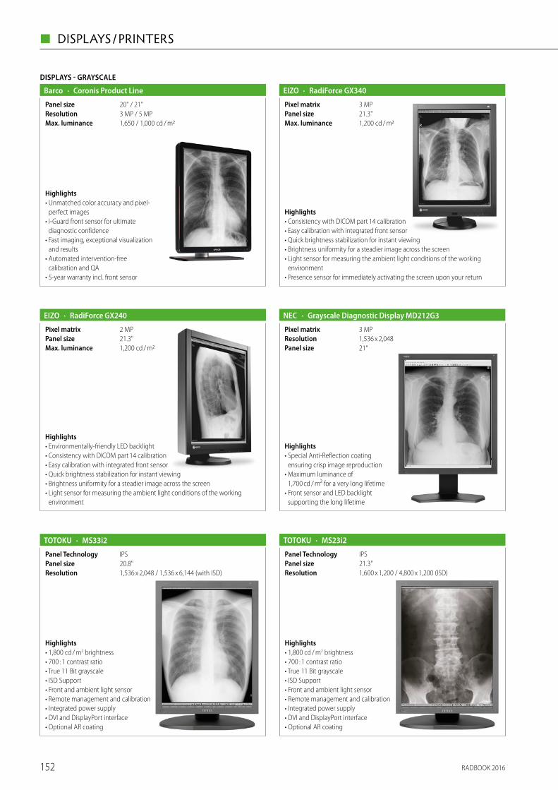
DISPLAYS - GRAYSCALE
· Coronis Product LineBarco
1,650 / 1,000 cd / m2Max. luminance3 MP / 5 MPResolution20" / 21"Panel size
Highlights• Unmatched color accuracy and pixel-
perfect images• I-Guard front sensor for ultimate
diagnostic confidence• Fast imaging, exceptional visualization
and results• Automated intervention-free
calibration and QA• 5-year warranty incl. front sensor
· RadiForce GX340EIZO
1,200 cd / m2Max. luminance21.3"Panel size3 MPPixel matrix
Highlights• Consistency with DICOM part 14 calibration• Easy calibration with integrated front sensor• Quick brightness stabilization for instant viewing• Brightness uniformity for a steadier image across the screen• Light sensor for measuring the ambient light conditions of the working
environment• Presence sensor for immediately activating the screen upon your return
· RadiForce GX240EIZO
1,200 cd / m2Max. luminance21.3"Panel size2 MPPixel matrix
Highlights• Environmentally-friendly LED backlight• Consistency with DICOM part 14 calibration• Easy calibration with integrated front sensor• Quick brightness stabilization for instant viewing• Brightness uniformity for a steadier image across the screen• Light sensor for measuring the ambient light conditions of the working
environment
Highlights• 1,800 cd / m2 brightness• 700 : 1 contrast ratio• True 11 Bit grayscale• ISD Support• Front and ambient light sensor• Remote management and calibration• Integrated power supply• DVI and DisplayPort interface• Optional AR coating
· MS23i2TOTOKU
1,600 x 1,200 / 4,800 x 1,200 (ISD)Resolution21.3"Panel sizeIPSPanel Technology
Highlights• 1,800 cd / m2 brightness• 700 : 1 contrast ratio• True 11 Bit grayscale• ISD Support• Front and ambient light sensor• Remote management and calibration• Integrated power supply• DVI and DisplayPort interface• Optional AR coating
· MS33i2TOTOKU
1,536 x 2,048 / 1,536 x 6,144 (with ISD)Resolution20.8"Panel sizeIPSPanel Technology
Highlights• Special Anti-Reflection coating
ensuring crisp image reproduction• Maximum luminance of
1,700 cd / m² for a very long lifetime • Front sensor and LED backlight
supporting the long lifetime
· Grayscale Diagnostic Display MD212G3NEC
21“Panel size1,536 x 2,048Resolution3 MPPixel matrix
RADBOOK 2016152
◼ DISPLAYS / PRINTERS

DISPLAYS - COLOR
· Coronis Fusion Product LineBarco
1,000 / 800 cd / m2Max. luminance30"Panel size4 MP / 6 MPPixel matrix
Highlights• Bezel-free 30-inch multi-modality PACS imaging desktop• Unmatched viewing characteristics and image quality • High-performance medical-grade image processing• Automated intervention-free calibration and QA• 5-year warranty incl. front sensor
· Nio Product LineBarco
750 / 800 / 1,100 cd / m2Max. luminance20" / 21"Panel size2 MP / 3 MP / 5 MPPixel matrix
Highlights• Excellent brightness, contrast along with a wide viewing• Proven technology for long-term image confidence• High-speed image processing for maximum productivity• Fully transparent calibration and QA• 5-year warranty incl. front sensor
Highlights• 1,500 cd / m2 brightness• 1,000 : 1 contrast ratio• Brightness stabilization• DVI and Video input to connect modality systems
· ME193TOTOKU
19.1"Panel size1,280 x 1,024ResolutionIPSPanel Technology
· Barco Coronis UnitiBarco
> 2100 cd / m²Max. luminanceColor and grayscale LCDTechnology12 MegaPixel (4,200 x 2,800)Resolution33 inchPanel size
Highlights• Approved for PACS, FFDM, DBT, breast MRI & US• Proven 10 % higher detection when scrolling DBT IMAGES Proven 10 – 15 %
higher detection probability compared to other FFDM displays• 2 x the lifetime and 2 x the brightness of other PACs and FFDM displays • 5-year warranty incl. front sensor
· RadiForce RX850EIZO
850 cd / m2Max. luminance31.1"Pixel matrix8 MPPanel size
Highlights• LCD module with 8 megapixel resolution
and LED backlight for a reliably high and constantly stable brightness• Dual-screen display (4 x 4 MP) on one monitor• Consistency with DICOM part 14 calibration• Monochrome and color images on one monitor• Brightness uniformity for a steadier image across the screen• Light sensor for measuring the ambient light conditions of the working
environment
· RadiForce RX650EIZO
800 cd / m2Max. luminance30"Panel size6 MPResolution
Highlights• LCD module with 6 megapixel resolution
and LED backlight for a reliably high and constantly stable brightness• Dual-screen display (3 x 3 MP) on one monitor• Consistency with DICOM part 14 calibration• Monochrome and color images on one monitor• Brightness uniformity for a steadier image across the screen• Light sensor for measuring the ambient light conditions of the working
environment
RADBOOK 2016 153
DISPLAYS / PRINTERS ◼

· RadiForce EX270WEIZO
600 cd / m2Max. luminance27"Panel size1,920 x 1,080Pixel Matrix
Highlights• Powerful LED backlight for an optimal presentation of critical images• Five factory calibrated look-up tables for quick and easy adaptation to diverse
application and viewing environments• Modular concept for targeted integration into current and future systems• Sleek, encapsulated design with laminated safety glass and an unsurpassed
IP rating ideally suited to the OR environment
· RadiForce RX350EIZO
1,000 cd / m2Max. luminance21.3"Panel size3 MPPixel matrix
Highlights• Consistency with DICOM part 14
calibration• Sharpness recovery function
(MTF increases by approx. 52 %)• Brightness uniformity for a steadier
image across the Screen• Quick brightness stabilization for
instant viewing• Light sensor for measuring the ambient light conditions of the working
Environment• Presence sensor for immediately activating the screen upon your return
· RadiForce RX240EIZO
760 cd / m2Max. luminance21.3"Panel size2 MPPixel matrix
Highlights• Consistency with DICOM part 14 calibration• Monochrome and color images on one
monitor• Brightness uniformity for a steadier image
across the screen• Quick brightness stabilization for instant
viewing• Light sensor for measuring the ambient light conditions of the working
environment• Presence sensor for immediately activating the screen upon your return
DISPLAYS - COLOR
· RadiForce RX440EIZO
750 cd / m2Max. luminance29.8"Panel size4 MPPixel matrix
Highlights• LCD module with 4 megapixel resolution
for a reliably high and constantly stable brightness• Dual-screen display (2 x 2 MP) on one monitor• Consistency with DICOM part 14 calibration• Monochrome and color images on one monitor• Brightness uniformity for a steadier image across the screen• Light sensor for measuring the ambient light conditions of the working
environment
Highlights• Multiple interface connectors to show 4 x 2MP on one screen, or 2 x 4MP• Built-in backlight sensor to provide stabilized image quality• Built-in power supply unit eliminating the need for an external AC adapter
· Colour Diagnostic Display MD322C8NEC
32‘‘Panel size3,840 x 2,160; Dual 1,920 x 2,160Resolution8 MPPixel matrix
Highlights• Special Anti-Reflection coating ensuring crisp image reproduction• Flexible hanging protocols possible by connecting one video input with 6MP
or dual configuration with 3MP each• Integrated front sensor and LED backlight supporting the long lifetime
· Colour Diagnostic Display MD302C6NEC
30“Panel size3,280 x 2,048; Dual 1,640 x 2,048Resolution6 MPPixel matrix
RADBOOK 2016154
◼ DISPLAYS / PRINTERS

RADBOOK 2016 155
DISPLAYS / PRINTERS ◼
EIZO RadiForce® ― Intelligent Solutions for Medical Imaging.
EIZO’s professional medical monitors provide doctors around the world with the perfect conditions for highly accurate diagnosis at PACS stations and modalities. All the models in the RadiForce series boast outstanding image quality that is permanently secured by EIZO’s own software solutions RadiCS and RadiNET Pro.
Place your trust in EIZO’s top quality and benefi t from our excellent customer service and maximum security of investment. Because most models in the RadiForce series come with an unusually long warranty of fi ve years. For more information, go to www.eizoglobal.com
EIZO — The Visual Technology Company
EIZO ECR Anzeige •Radbook •210x297mm + 3mm Beschnitt•IC300•DU: 08.02.16
Visit us at the ECR in Vienna, March 3–6, 2016, Hall X5, Booth 10.
eizME5052_Anzeige_ECR_A4_ICv2_Radbook_EN_1ml.indd 1 03.02.16 09:36

Highlights• Multiple interface connectors to show one 4MP screen or a dual configuration
of 2 x 2MP• QuickScreenQA for fast DICOM-compliance tests• Integrated front sensor and LED backlight supporting the long lifetime
DISPLAYS - COLOR
· Colour Diagnostic Display MD302C4NEC
30“Panel size2,560 x 1,600; Dual 1,280 x 1,600Resolution4 MPPixel matrix
Highlights• Anti-Glare coating layer minimizing
ambient light reflections • Native 10bit panel supporting up to
1,074 bio total colors• QuickScreenQA for fast DICOM-com-
pliance tests• Integrated front sensor and LED back-
light supporting the long lifetime
· Colour Diagnostic Display MD211C3NEC
21“Panel size1,536 x 2,048Resolution3 MPPixel matrix
Highlights• Multiple interface connectors to show one 2MP screen or a dual configuration
of 2 x 1MP• QuickScreenQA for fast DICOM-compliance tests• Integrated front sensor and LED backlight supporting the long lifetime
· Colour Diagnostic Display MD242C2NEC
24“Panel size1,920 x 1,200Resolution2 MPPixel matrix
Highlights• Anti-Glare coating layer minimizing
ambient light reflections • Native 10bit panel supporting up to
1,074 bio total colors• integrated front sensor and LED
backlight supporting the long lifetime
• QuickScreenQA for fast DICOM- compliance tests
· Colour Diagnostic Display MD211C2NEC
21“Panel size1,200 x 1,600Resolution2 MPPixel matrix
Highlights• 800 cd / m2 brightness• 1400 : 1 contrast ratio• Front and ambient light sensor• Remote management and calibration• Integrated power supply• DVI and DisplayPort interface• Optional AR coating
· CCL358i2TOTOKU
IPSPanel Technology21.3"Panel size2,048 x 1,536Resolution800 cd / m2Max. luminance
Highlights• 800 cd / m2 brightness• 1,000 : 1 contrast ratio• Brightness stabilization system• Remote management• Integrated power supply• Dual DVI / DisplayPort Input
· CCL650i2TOTOKU
3,280 x 2,048Resolution800 cd / m2Max. luminance
IPSPanel Technology30“Panel size
RADBOOK 2016156
◼ DISPLAYS / PRINTERS

IPSPanel Technology
Highlights• 900 cd / m2 brightness• 1400 : 1 contrast ratio• Front and ambient light sensor• Remote management and calibration• Integrated power supply• DVI and DisplayPort interface• Optional AR coating
· CCL258i2TOTOKU
900Max. luminance
21.3"Panel size1,600 x 1,200Resolution
Highlights• 400 cd / m2 brightness• 1,000 : 1 contrast ratio• Brightness stabilization system• Remote management
• Integrated power supply• Optional AR coating
· CCL240TOTOKU
1,920 x 1,200Resolution24.1"Panel sizeIPSPanel Technology
Highlights• 950 cd / m2 brightness• 900 : 1 contrast ratio• Front and ambient light sensor• Remote management and calibration• Integrated power supply• DVI and DisplayPort interface• Optional AR coating
· CCL256i2TOTOKU
1,600 x 1,200Resolution21.3"Panel sizeIPSPanel Technology
Highlights• 700 cd / m2 brightness• 1000 : 1 contrast ratio• Brightness stabilization system• Remote management• Integrated power supply
· CCL230TOTOKU
700 cd / m2Max. luminance1,600 x 1,200Resolution19.6"Panel sizeIPSPanel Technology
Highlights• 800 cd / m2 brightness• 750 : 1 contrast ratio• Front and ambient light sensor• Remote management and calibration• Integrated power supply• DVI and DisplayPort interface• Optional AR coating
· CCL356i2TOTOKU
2,048 x 1,536Resolution21.3"Panel sizeIPSPanel Technology
RADBOOK 2016 157
DISPLAYS / PRINTERS ◼

DISPLAYS - CLINICAL REVIEW
· Eonis FamilyBarco
250 / 300 cd / m²Max. luminance2 MP (1,920 x 1,080)Resolution22“ / 24‘‘Panel size
Highlights• Protective toughened, scratch proof glass cover• 100 % cleanable (70 % alcohol) design supports hospital infection control initiatives• IEC 60601-1 for use within 1m of patients• Desk or cart-mounted for ultimate flexibility• QA management and asset management• 3-year warranty incl. front sensor
· MDRC Product LineBarco
400 cd / m2Max. luminance19"Panel size1 MPPixel matrix
Highlights• Providing consistent DICOM images anywhere, anytime• Professional LCD quality with exceptional image quality• Approved for medical use anywhere in the hospital• Backlight output stabilization and long-life time• User-friendly calibration and QA• 3-year warranty
· RadiForce MX242WEIZO
350 cd / m2Max. luminance24.1"Panel size2.3 MPPixel matrix
Highlights• View more with widescreen and wide viewing angles• DICOM part 14 compliant, simplified calibration• Brightness stabilization• Brightness uniformity for a steadier image across the screen• Customer assurance with medical standards
· RadiForce MX215EIZO
420 cd / m2Max. luminance21.3"Panel size2 MPPixel matrix
Highlights• DICOM part 14 compliant plus simplifi ed calibration• Brightness stabilization• Selection for optimum viewing• Customer assurance with medical standards
· RadiForce MX191EIZO
300 cd / m2Max. luminance19"Panel size1.3 MPPixel matrix
Highlights• DICOM part 14 compliant plus simplified calibration• Brightness stabilization• Mode selection for optimum viewing• Customer assurance with medical standards
Highlights• DICOM preset and hardware-calibration option for medical image viewing• 14bit LUT supporting DICOM conformance• Digital Uniformity Control for homogeneous grayscales
· Clinical Review Display MDview272NEC
27"Panel size2,560 x 1,440ResolutionAH-IPSPanel Technology
RADBOOK 2016158
◼ DISPLAYS / PRINTERS

RADBOOK 2016 159
DISPLAYS / PRINTERS ◼
The NEC MDT Room Solution is a complete solution including everything needed to provide the latest medical meeting
room infrastructure. The Medical Desktop and Large Format Display – both 8MP – can be cloned on a pixel to pixel level
to provide outstanding image quality without any loss of data or visual detail. This solution helps to establish efficient
reviewing processes and diagnostic investigations as well as providing hospitals a future-proof investment in a state of the
art technology and quality.
www.medical.nec-display-solutions.com
NEC 8 MEGAPIXEL MDT ROOM SOLUTION SETS NEW STANDARD
FOR RADIOLOGICAL PRACTICE
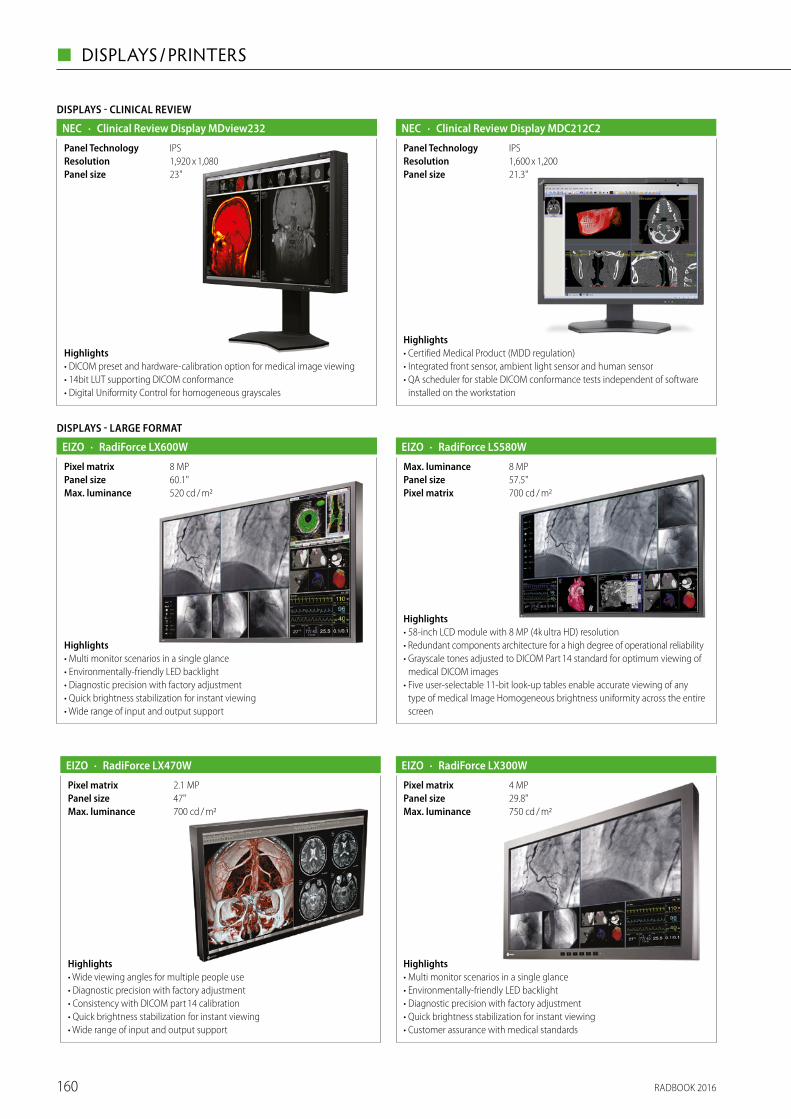
Highlights• Certified Medical Product (MDD regulation)• Integrated front sensor, ambient light sensor and human sensor • QA scheduler for stable DICOM conformance tests independent of software
installed on the workstation
· Clinical Review Display MDC212C2NEC
21.3"Panel size1,600 x 1,200ResolutionIPSPanel Technology
Highlights• DICOM preset and hardware-calibration option for medical image viewing• 14bit LUT supporting DICOM conformance• Digital Uniformity Control for homogeneous grayscales
DISPLAYS - CLINICAL REVIEW
· Clinical Review Display MDview232NEC
23"Panel size1,920 x 1,080ResolutionIPSPanel Technology
· RadiForce LS580WEIZO
700 cd / m2Pixel matrix57.5"Panel size8 MPMax. luminance
Highlights• 58-inch LCD module with 8 MP (4k ultra HD) resolution • Redundant components architecture for a high degree of operational reliability• Grayscale tones adjusted to DICOM Part 14 standard for optimum viewing of
medical DICOM images• Five user-selectable 11-bit look-up tables enable accurate viewing of any
type of medical Image Homogeneous brightness uniformity across the entire screen
DISPLAYS - LARGE FORMAT
· RadiForce LX600WEIZO
520 cd / m2Max. luminance60.1"Panel size8 MPPixel matrix
Highlights• Multi monitor scenarios in a single glance• Environmentally-friendly LED backlight• Diagnostic precision with factory adjustment• Quick brightness stabilization for instant viewing• Wide range of input and output support
· RadiForce LX470WEIZO
700 cd / m2Max. luminance47"Panel size2.1 MPPixel matrix
Highlights• Wide viewing angles for multiple people use• Diagnostic precision with factory adjustment• Consistency with DICOM part 14 calibration• Quick brightness stabilization for instant viewing• Wide range of input and output support
· RadiForce LX300WEIZO
750 cd / m2Max. luminance29.8"Panel size4 MPPixel matrix
Highlights• Multi monitor scenarios in a single glance• Environmentally-friendly LED backlight• Diagnostic precision with factory adjustment• Quick brightness stabilization for instant viewing• Customer assurance with medical standards
RADBOOK 2016160
◼ DISPLAYS / PRINTERS

Highlights• IP55-proteced 46” • DICOM preset and hardware-calibration option for medical image viewing• Multiple interface connectors available for any medical equipment in the OR• OPS slot for easy integration of Slot-in PC (Intel Core i7 4 x 2,4 GHz)
· Operating Room Display MD462ORNEC
46"Panel size1,920 x 1,080ResolutionS-PVAPanel Technology
· DRYSTAR 5300Agfa
320 dpiResolution70 films / h (14 x 17)CapacityDirect digital imagingTechnology
AGF-DryStar5300.psd• Tabletop, next-to-application Direct Digital Imager• Suitable for all applications and ideal for CT / MR• Reliable, low maintenance printer• A#Sharp image enhancement for excellent quality• Very short access time ensures fast printing of small print jobs
· DRYSTAR 5302Agfa
320 dpiResolution75 films / h (14 x 17)CapacityDirect digital imagingTechnology
AGF-DryStar5302.psd• Suitable for all applications and ideal for CR / DR• A#Sharp technology for optimized image quality• Convenient imaging with two media sizes on-line (multi-format)• Very short access time ensures fast printing of small print jobs
PRINTER
· DRYSTAR 5503Agfa
508 dpi / 50 μm pixelsizeResolution100 films / h (14 x 17)CapacityDirect digital imagingTechnology
AGF-DryStar5503.psd• Multi-modality, high throughput imager
with film sorter• Ideal for centralized workflow, can easily
be connected to the network• Integrated A#Sharp technology for
optimized image quality• Three multi-format trays, each supporting
different film sizes and types• Suitable for CT, MRI, DSA, digital R / F,
CR, DR and optional mammography applications
· DRYSTAR AXYSAgfa
508 dpi / 50 μm pixelsizeResolution75 films / h (14 x 17)CapacityDirect digital imagingTechnology
AGF-DryStarAXYS.psd• Flexible, tabletop imager delivering mammography-quality images• Multi-application hardcopy solution, including digital mammography• Integrated A#Sharp technology for optimized image quality• Two multi-format trays, each supporting different film sizes and types• Very short access time for extremely fast delivery of first four prints
· DICOM PaperPrintmedigration
1.200 x 2.400 dpi (print), 600 x 600 dpi (copy)ResolutionUp to 120 paper prints / hCapacityDIN A3, 11 x 17 inchFormat
Highlights• Supports all DICOM 3.0 modalities
(e. g. CT, MRT, CR, DR, US, NUK, etc)• Supports one or more PostScript printers
within the network• General licence package
(no restrictions on how many DICOM modalities are connected)
• Image header and footer customizable incl. physician logo
• Separate LUT (Look Up Table) for each printing system
• GSDF calibration according IHE
RADBOOK 2016 161
DISPLAYS / PRINTERS ◼

CD- / DVD-ROBOT
· Burn GatewayCHILI
Highlights• Receives data by DICOM C-Store• Burns data on one or more CD / DVDs• Optional reports• Individual label printing• Client enabled (different logos)• CHILI viewer in report quality• Alternative presentation as
HTML / jpeg
• Certified by OFFIS and DRG• Works with any PACS• External output tray!
· Import RobotCHILI
Highlights• Automatic import robot• Import of patient CD / DVD• 2, 5 or 10 drives• 2 import trays (regular / express)• 2 output trays (ok, failed)• Optional virus scan
• Correction of foreign data• Automatic DICOM transfer• Works with any PACS
Highlights• Fully automatic compact system for creating DICOM patient CDs or DVDs• Highly compatible with all digital DICOM modalities (multimodality)• Individual labeling (practice / clinic logo)• Easy integration of DICOM patient data• Extremely cost effective due to quick printing times and low link consumption
· CD-Imagermedigration
2 x 50 pcsMagazine size30 CDs / h or 15 DVDs / h (burn and print)CapacityCD-R, DVD-R, DVD+R, DVD-R DL, DVD+R DLFormat
· RadiNET Pro – Network QC Management for DisplaysEIZO
Highlights• Centralized Management of up to 8,000 Monitors• Instant Notification for Immediate Maintenance• Time Saving Remote Calibration• Easy-to-Use Web-Based Application
ACCESSORIES / COMPLEMENTRAY SYSTEMS
· RadiCS – Quality Control Software for DisplaysEIZO
Highlights• Acceptance and Constancy Testing in Easy Steps• DICOM Part 14 Calibration• History Recording and Report Generator• Flexible Schedule Setting• Intelligent Hands-Off Check
Please visit us at
www.healthcare-in-europe.com
RADBOOK 2016
RADBOOK 2016162
◼ DISPLAYS / PRINTERS

RADBOOK 2016 163
ULTRASOUND ◼

Ultrasound
164

· EBitChison
Convex, linear, phased array, micro-convexScan format
B, C, CPA/DPD, PW/CW, TDI, Color M Mode2Transducer inputs
Highlights• Breakthrough new technologies: THI,
Space Compound Imaging, SRA, FHI, X-contrast, Q-flow, Q-beam, Q-image
• Built in battery ≥ 2 hours (option)• 30 degree rotatable LED screen• Full Screen Mode• Advanced Cardiovascular Packages:
Steering M, Color M, CW, TDI, Auto IMT
• About 7.5kg (with battery), convert-ible design
• Wide Range of transducers• 18 MHz High Frequency Linear Probe
· QBit 7Chison
Convex, linear, transvaginal, phased array, 4D volume, micro-convex
Scan format
CW, TDI, Free M mode, Color M mode, ECGMode4Transducer inputs
Highlights• Versatile diagnostic solutions.• Intelligent workflow, simplified keyboard• Advanced 4D technologies: Virtual HD, Depth View• Breakthrough new technologies : FHI, X-contrast,
Q-flow, Q-beam, Q-image• Built in battery 80 min (option)• CW, TDI, Free M mode, Color M mode, ECG
· QBit 9Chison
Convex, linear, transvaginal, phased array, 4D volume, micro-convex
Scan format
CW, TDI, Free M mode, Color M mode, ECGMode4Transducer inputs
Highlights• Hassle-Free maintenance (Hero Kit)• Breakthrough new technologies :
FHI,X-contrast, Q-flow, Q-beam, Q-image• Stress Echo• Elastography• Advanced 4D technologies: Virtual HD,
Depth View• High definition digital output ports: DVI• Built in battery 80 min (option)
• 19" high definition LED monitor with 270° rotation angle
• 10.4" touch screen for more user friendly workflow • Integrated gel warmer • 2.5 MHz – 18 MHz operating frequency range• THI, SRA, Fusion harmonic • Universal Compound Imaging • i-Image / 2D Steer / Curved Panoramic Imaging • IMT / Elastography / Super needle • Advanced 4D technologies: 4D probe and
display package, Virtual HD, Depth view • Professional Cardiac packages
· i9Chison
B, 2B, 4B, B / M, 2D Steer BC, CFM PW, HPRF, CW PD, Directional PD Instant Triplex, Duplex, Quadplex Trapezoidal Curved Panoramic Imaging(option) 4D (option) Chroma B / M / PW / CW ECG (option) Free Steering M (option) Color M (option)
Mode
4Transducer inputs
Highlights • 19" high definition LCD Monitor, 4 probe connectors • Advanced 4D technologies: 4D probe and
display package, Virtual HD, Depth View • Advanced Imaging Technologies:
THI, SRA, Compounding, i-Image, Quardplex • Elastography, Super Needle, 2D Steer • Shared Service: Cardiac, Vascular, AB, OB / GYN, MSK,
Small Parts, Urology and Pediatric • Professional Cardiac package
Mode B, 2B, 4B, B / M, 2D Steer BC, CFM PW, HPRF, CW PD, Directional PD Instant Triplex, Duplex, Quadplex Trapezoidal Curved Panoramic Imaging(option) 4D (option) Chroma B / M / PW / CW ECG (option) Free Steering M (option) Color M (option)Convex,Linear,Transvaginal,Phased array,4D Volu-me,Micro-Convex
Scan format
· i8Chison
4Transducer inputs
B, 2B, 4B, B / M, M CFM PW Mode Power Doppler / Directional PD Trapezoidal Real-time 4D (Option) Chroma B / PW
Mode
Highlights • 19” LCD, 4 probe connectors • Advanced 4D technology • Superb image: Compound imaging, SRA, i-Image • Comprehensive OB & GYN package • Streamlined workflow • EasyView archive system • DICOM 3.0, PC & Video printer • Great value for OB & GYN, General imaging
· i3Chison
4Transducer inputs
Convex probe Linear probe Linear probe (60mm) Transvaginal probe Micro-Convex probe 4D Volume probe Wideband, Multi-frequency
Scan format
Ultrasound
RADBOOK 2016 165
ULTRASOUND ◼

B, CFM, PW,M,2B,4BMode
Highlights • Touch screen, icon-driven,
easy to use • Quick boot within 30 seconds • Long battery life up to 2.5 hours • Compact, durable, water proof
(from panel) • High resolution LED screen• Portable stand with adjustable
viewing angles• Versatile imaging functions and
report management software
• USB and DICOM 3.0 • Super Needle • B, CFM, PW, M, 2B, 4B
· SonoTouch 30Chison
7 kgWeight1 for main unit, 3 with cart (option)Transducer inputs
B, 2B, 4B, B / M, 2D Steer BC, CFM PW, HPRF, CW PD, Directional PD Instant Triplex, Duplex, Quadplex Trapezoidal Curved Panoramic Imaging(option) 4D (option) Chroma B / M / PW / CW ECG (option) Free Steering M (option) Color M (option) TDI (option)
Mode
Highlights • 15" high definition LCD Monitor• Dual probe connectors• Advanced 4D technologies: 4D probe and
display package, Virtual HD, Depth View• Advanced Imaging Technologies:
THI, SRA, Compounding, i-Image, Quardplex• Elastography, Super Needle• Shared Service : Cardiac, Vascular, ABD, OB / GYN, MSK, Small Parts, Breast,
Urology and Pediatric
· Q9Chison
2Transducer inputsConvex, Linear, Phased array, Volume, Micro convexScan format
Mode B, 2B, 4B, B / M, M CFM PW Mode Power Dopp-ler / Directional PD Trapezoidal Real-time 4D (Option) Chroma B / PW
Highlights • 15" LCD monitor• Advanced 4D technology: 4T
(Fast, Light, Quiet, Smart)• Professional OB report package• B, CFM, PW, Power Doppler and
Directional Power Doppler• Trapezoidal Mode• Streamlined workflow• Dual probe connectors
• Advanced technologies: SRA, Compound Imaging, THI, i-Image
· Q5Chison
Transducer inputs
Scan format
2
Convex, Linear, Transvaginal, Transvaginal, Volume, Micro-Convex
6.5 kg (with built-in battery)Weight2Transducer inputsB, B / B, 4B, M, B / M, CFM, PW, TrapezoidalScan format
Highlights • Ultra-portable color
ultrasound system• PW Doppler with auto-trace• Additional phased array probe• Wide viewing angle (0 – 180°),
from left to right• Built-in battery (> 2 hours)• 12 inch rotatable LED monitor (0 – 30°)
• One key to full screen• 8G memory card
· ECO5Chison
B, C, M, PWMode
Highlights • Advanced image technologies:
THI, SRA, i-Image, Compound imaging• 8G memory card• Dual probe connectors• Trapezoidal• High resolution LED monitor
30° rotatable• One key to full screen• Anti-water keyboard cover• Chroma
· ECO1Chison
6.5 kg (with built-in battery)Weight2Transducer inputsConvex, Linear, Micro-Convex, TransvaginalScan formatB, B / B, 4B, M, B / MMode
• Better solution for accessories: Carry case & CartTR9000
· ECO 3 EXPERTChison
Convex, linear, transvaginal , micro-convexScan format
B, 2B, 4B, B / M, M, PWMode2Transducer inputs
Highlights• 30 degree rotatable LED screen:
better resolution & economy • Advanced image techonology:
THI, SRA, i-Image, Compound imaging• Long battery life: >2 hours• Professional accessories:
Carry case (BG-100)
• Better solution with Cart TR 9000, Anti-water keyboard cover
• User-friendly and modern design• Streamlined workflow(6-one-key step)• Chroma
RADBOOK 2016166
◼ ULTRASOUND

RADBOOK 2016 167
ULTRASOUND ◼
Visit us at ECR 2016 for a live demonstration: Expo X5, booth #3
Sonimage HS1 is Konica Minolta’s powerful and portable Ultrasound System for use in a variety of clinical environments. It is a true point-of-care system with an intuitive interface for easy operation in the most demanding situations. Whether you are an orthopedist, pain management specialist or anesthesiologist, high definition imaging is always key. Sonimage HS1 brings you crystal clear image quality wherever and whenever you need it.
Sonimage HS1 Ultrasound System for crystal clear imaging
KONICA MINOLTA MEDICAL & GRAPHIC IMAGING EUROPE B.V.Hoogoorddreef 9 - 1101BA - Amsterdam - The Netherlands - [email protected] - www.konicaminolta.eu/healthcare
P O W E R F U L & P O R T A B L E

· MyLabAlphaEsaote
2 on board, 4 with cartTransducer inputs
Convex, Microconvex, Linear, Phased Array, Extended, Panoramic and Volumetric
Scan format
2D, 3D, 4D, M, CMM, TVM, CFM, PW, CW, PWD, XFlow and others
Mode
Highlights• MyLabAlpha is a premium
portable system, designed to deliver top performance for both imaging and ergonomics in small size and weight
• Portable ultrasound system for Radiology, Cardiovascular, MSK, Rheuma, OB-Gyn, POC as well as Surgery and Interventional Radiology
• Advanced technologies available such as ElaXto, CEUS, XStrain4D, QIMT and QAS Arterial Stiffness tool, 3D / 4D
· Esaote MyLabSix ChristaLineEsaote
3 probe connectorsTransducer inputs
Convex, Microconvex, Linear, Phased Array, Extended, Panoramic and Volumetric
Scan format2D, 3D, 4D, M, CMM, CFM, TVM, PW, CW, PWD and othersMode
Highlights• MyLabSix offers high level Image Quality in a
Compact Design• eDesign product to maximize user comfort
and diagnostic confidence• 19” Wide screen monitor, Touch Screen and easy
workflow• Extended transducer range, offering also Hockey
Stick High Frequency, Biopsy dedicated convex transducer, TEE, Endocavity, Surgery and Laparo-scopic transducers
• Ultra-low power consumption: Esaote Eco Efficiency engine• Advanced technologies available such as, QIMT, 3D / 4D
· Esaote MyLab Twice eHD ChrystaLineEsaote
Convex, Linear, Phased Array, Extended, 3D Panoramic and Volumetric
Scan format
4 & 1 probe connectorsTransducer inputs
2D, 3D, 4D, M, CMM, CFM, PWD, XFlow, PW, CW, CnTI and others
Mode
Highlights• Premium system with Point-of-Care portable
ultrasound unit optionally integrated• High level Ergonomics with intuitive Touch
Screen panel, user friendly workflow and App based MyLabRemote tool for remote control though Smartphone or Tablet
• Superb Imaging, Color and Spectral Doppler with Advanced Technologies (ElaXto, CnTI, Virtual Navigator Fusion Imaging) applicable to different types of transducer and to extensive range of clinical applications
· MyLabClassCEsaote
4 & 1 probe connectorsTransducer inputs
Convex, Microconvex, Linear, Phased Array, Extended, 3D Panoramic and Volumetric
Scan format
2D, 3D, 4D, M, CMM, TVM, CFM, PW, CW, PWD, XFlow CnTI and others
Mode
Highlights• High-end System, perfect choice for high
performance combined with excellent ergonomics (OptiLight and MyLabRemote) and user friendly workflow
• Superb Imaging, Color and Spectral Doppler with Advanced Technologies (ElaXto, Low MI CEUS, Fusion Imaging, 3D & 4D, QIMT, QAS, XFlow, HD CFM, Frequency range up to 22 MHz)
• Multidisciplinary Digital Platform for General Imaging, Women’s Health, Cardiovascular, MSK
· Esaote MyLab SevenEsaote
4 probe connectorsTransducer inputs
Convex, Linear, Phased Array, Extended, Panoramic and Volumetric
Scan format2D, 3D, 4D, M, CMM, CFM, PW, CW, PWD, XFlow and othersMode
Highlights• Confident diagnosis in an innovative
system design to deliver high-class imaging performance in compact size
• Touch-screen centered user interface allows automatic workflow features: eTouch, Protocols, SmarTouch
• Fully customizable user interface to have always the best workflow in any clinical application and setting
• Advanced features available, i. e.: ElaXto, CEUS, 3D / 4D, QIMT, QAS RF-based arterial stiffness, XStrain4D
· Esaote MyLab EightEsaote
2D, 3D, 4D, M, CMM, CFM, PWD, XFlow, SWE, PW, CW, CnTI and others
Mode
Convex, Linear, Phased Array, Extended, 3D Panoramic and Volumetric
Scan format
4 probe connectorsTransducer inputs
Highlights• Premium system with MPowered
beam forming to optimize high-density and Single Crystal transducers
• State of the art visualization with WideView technology for crispy details, deep image contrast and extensive image size
• Superb Imaging, hemodynamics and tissue stiffness quantification with an extensive package of Advanced Tech nologie (QElaXto Shear Wave Elastography, XFlow, CnTI, Virtual Navigator Fusion Imaging)
RADBOOK 2016168
◼ ULTRASOUND

· MyLabGammaEsaote
2 on board, 4 with cartTransducer inputs
Convex, Microconvex, Linear, Phased Array, Extended, Panoramic and Volumetric
Scan format2D, 3D, 4D, M, CMM, TVM, CFM, PW, CW, PWD and othersMode
Highlights• MyLab Gamma sets ultrasound free
bringing superb quality imaging and fast, confident diagnosis to the Point-of-Care in any situation – wherever and whenever
• Incorporating high resolution imaging, advanced technologies, and supporting a range of probes it is an optimal solution for Cardiovascular, General Imaging, MSK, OB-Gyn, Emergency
• Esaote Eco Efficiency product with ultra-low power consumption; eDesign advanced ergonomics solutions for system and transducers.
• Advanced technologies available such as, QIMT, 3D / 4D
· MyLabOneEsaote
Convex, Linear, Phased Array and ExtendedScan format1 on board, 3 on roll standTransducer inputs2D, M, CFM, PWD, PW and othersMode
Highlights• Dedicated solution for Point Of Care • Intuitive user interface, fully touch screen • Wireless connectivity • Fast workflow / Easy to clean / On-board MyLibrary • Remote controls integrated on the transducers • NNE technology for enhancement of needle
visibility • XHF technology: Frequency up to 22 MHz • QIMT and QAS tools, for accurate and easy
assessment of IMT and arterial stiffness, based on RF technology
· EDGE IIFUJIFILM SonoSite
1 for main unit, 3 with TTC optionTransducer inputs
Linear, curved and phased array, multiplane TEE and micro-convex
Scan format
B mode, M mode, Tissue Harmonic Imaging, Velocity Color Doppler, Color Power Doppler, PW, PW Tissue Doppler, CW
Mode
3.85 kgWeight
HighlightsThe Edge II offers you enhanced imaging experience through industry-first transducer innovations like DirectClear and Armored Cable Technology. Because it’s a SonoSite, the Edge II stays true to our design pillars: durability, reliability & ease of use. It offers a compact clamshell design that exceeds expectations for infection control and featuring enhanced cardiac & abdominal imaging experience.
· iVizFUJIFILM SonoSite
1Transducer inputs
2D, M-Mode, Colour Doppler and THI, with multiple optimisation setting
Mode
520 gWeight
Broadband and Multifrequency Phased ArrayScan format
HighlightsiViz augments the value of ultrasound for clinical users from hospitals to clinics in remote villages with the ability to perform ultrasound when and where it’s needed. It delivers fast and improved patient care with superior clarity, mobility, and unprecedented connectivity. Users can easily access patient records, store exams, submit reports, and consult with remote providers for assessments.
· M-TurboFUJIFILM SonoSite
1 for main unit, 3 with TTC optionTransducer inputs
Linear, curved and phased array, multiplane TEE and micro-convex
Scan format
B mode, M mode, Tissue Harmonic Imaging, Velocity Color Doppler, Color Power Doppler, PW, PW Tissue Doppler, CW
Mode
3.4 kgWeight
HighlightsThe M-Turbo’s engineered for striking image quality, durability and ease of use. It lets you visualise detail, improving your ability to differentiate structures, vessels and pathology. The M-Turbo ultrasound system offers an advanced set of features with a wide array of connectivity options that seam-lessly connects you to hospital information networks and your own PC.
2.7 kg
· NanoMaxxFUJIFILM SonoSite
1Transducer inputsLinear, curved and phased arrayScan formatB mode, M mode, Color Doppler, Color Power DopplerMode
Weight
HighlightsWith its unique one-button control, high-quality diagnostic imaging, and full-color flow map-ping, the NanoMaxx ultrasound system is designed to address the needs of physicians making key clinical decisions or guiding interventional procedures. It’s portable & incredibly tough, has an easy to disinfect splash resistant touch screen interface and combines performance with affordability and simplicity.
RADBOOK 2016 169
ULTRASOUND ◼

· S SeriesFUJIFILM SonoSite
1Transducer inputs
Linear, curved and phased array, multiplane TEE and micro-convex
Scan format
B mode, M mode, Tissue Harmonic Imaging, Color Doppler, Color Power Doppler, PW, PW Tissue Doppler, CW
Mode
3.8 kgWeight
HighlightsThe S Series ultrasound systems are designed to be mounted to a wall, ceiling or cart and is custom designed for your prac-tice. It has simplified controls that let you focus in on your target areas in a matter of seconds. High-resolution images help you see exactly where to perform procedures and allow for accurate diagnoses when treating patients.
· X-PorteFUJIFILM SonoSite
3Transducer inputs
Linear, curved and phased array, multiplane TEE and micro-convex
Scan format
2D Broadband imaging, Tissue Harmonic Imaging, Pulse Inversion Harmonic Imaging, M Mode (update and simul-taneous), Velocity Colour Doppler, Colour Power Doppler, Pulsed Wave Doppler, Pulsed Wave Tissue Doppler, Continuous Wave Doppler, ECG
Mode
HighlightsX-Porte represents a new approach to clinical ultrasound. At the sweep of your hand, it responds quickly and intelligently to your imaging needs. Its self-explanatory control panel makes system navigation easy and its sealed touch screen has no buttons for pathogens to hide behind. X-Porte’s slender profile makes it easy to maneuver along-side beds and exam tables for visualization and procedures.
· LOGIQ E9 XDclear 2.0GE Healthcare
4Transducer inputs
Linear, convex, microconvex, sector phased array, trapezoid, 3D/4D
Scan format
B-mode, M-mode, CFM-mode, HiRes Contrast, TVI, stressecho, Auto-IMT, Doppler, shear wave elastography, LogiqView, realtime 4D, volume navigation, needle tracking, color-coded B-Flow, parametic imaging
Modus
Highlights• Extraordinary images: agile ultrasound beamformers
with acoustic models, Matrix Array transducer technology, single crystal, CrossXBeam, SRI
• Expert tools: contrast imaging with new HiRes + amplitude modulation settings, elastography + PDI with quantification, realtime 4D in CEUS mode, volume navigation with fusion GPS + needle tracking
• Easy workflow: scan assistant, raw data imaging, Q&R with multimodality imaging navigation
· LOGIQ S8 XDclearGE Healthcare
4 active ports + 1 parking slotTransducer inputs
Linear, convex, microconvex, sector phased array, biopsy convex, Biplane TRT and TEE
Scan format
B-mode, M-mode, CFM-mode, Doppler, B-flow, contrast, TVI, stressecho, Auto-IMT, elastography, LogiqView, parametric imaging, volume navigati-on, needle tracking, Quick Start
Modus
Highlights• Superb imaging: S-Agile ultrasound beamformers,
matrix array transducer technology, single crystal, contrast imaging with amplitude modulation settings, elastography with quantification, B-flow imaging
• Simplified workflow: slim and light console, fully flexible configuration
• Scalable to your needs: wide applications coverage to maximize scan productivity.
• Scan assistant, raw data imaging
· LOGIQ P7 / P9GE Healthcare
3+1 (optional)Transducer inputsLinear, convex, microconvex, sector phased arrayScan format
B-mode, M-mode, CFM-mode, Doppler, B-flow, contrast (LP9), TVI, stressecho, Auto-IMT, Logiq-View, elastography, Quick Start
Modus
Highlights• Touchscreen concept allows an intuitive
and quick operation• Numerous innovative assistance
functions with an extended range of applications support a confident diagnosis
• LOGIQView (panoramic imaging)• Modern and high-resolution wides-
creen monitor
4Transducer inputs
· LOGIQ S7 V2GE Healthcare
Linear, convex, microconvex, sector phased array, Biplane TRT and TEE
Scan format
B-mode, M-mode, CFM-mode, Doppler, B-flow, contrast, TVI, stressecho, Auto-IMT, elastography, LogiqView
Modus
Highlights• Farbtriplex system• Flexible system concept allows tailoring to
individual requirements• Facilitated with innovations such as
B-Flow, elastography, etc.• Beyond standards in terms of ergonomics
RADBOOK 2016170
◼ ULTRASOUND

· Venue 50GE Healthcare
1 (expandable to 3 with Cart)Transducer inputsLinear, convex, phased arrayScan format
Black and white mode for displaying anatomy in real-time, Color-coded overlay for real-time blood flow imaging
Modus
Highlights• No buttons, no knobs, no keyboard - easy to use at
the point of care• Concurrent acquisition technology provides fast,
high-resolution imaging to easily visualize anatomy and needle placement
• Pre-configured settings for different applications• Can be flexibly taken from place to place when
using a universal dock system or a table station
· LOGIQ F8GE Healthcare
3 (4 optional)Transducer inputs
Convex, linear, microconvex, sector phased array, realtime 4D volume
Scan format
B-mode, tissue harmonics, M-mode, Color-M-mo-de, CFM, Power Doppler Imaging (PDI), directional PDI, PW-Doppler with High-PRF, scan assistant, scan coach; optional: anatomical M-mode, CW-Doppler, LogiqView, TVI Mode, 3D/4D
Modus
Highlights• Outstanding display properties as well as numerous
innovative assistance functions support a confidant diagnosis
• Compatible with a wide range of transducers and different software packages
• Can be used in nearly all medical disciplines
· LOGIQ P6 GE Healthcare
3Transducer inputs
Linear, convex, microconvex, sector phased array, trapezoid
Scan format
B-mode, M-mode, CFM-mode, Doppler, B-flow color, coded contrast harmonic, stressecho, EKG, anatomical M-mode, 3D/4D
Modus
Highlights• Compact shared service system • B-flow color (digitally substraction technique)• CrossXBeam realtime compound and speckle
reduction imaging• LOGIQView (panoramic imaging)• Auto optimize (For B-mode, color, Doppler)• Digital archive with RawData support• Matrix array transducer support• Elastography
· LOGIQ e R7GE Healthcare
1 (expandable to 3 with Cart)Transducer inputs
Linear, convex, microconvex, sector phased array, trapezoid, TEE
Scan formatB-mode, M-mode, CFM-mode, DopplerModus
Highlights• Portable premium system with
shared service capabilities• Hockey stick probe for interventional• Needle recognition feature for a better
needle imaging• CrossXBeam, B-steer and SRI imaging• LOGIQ view (panoramic imaging)• High frequency imaging up to 18 MHz for vascular and musculoskeletal exams• Musculoskeletal suite with 2D PDI quantification and patient follow up settings
· VscanGE Healthcare
390 g (unit and probe)Weight
Field-of-View for black and white imaging: up to 75 degrees with maximum depth of 25 cm, the color flow sector represents blood flow within an angle of 30 degrees
Scan format
Black and white mode for displaying anatomy in real-time, Color-coded overlay for real-time blood flow imaging
Modus
Highlights• The size of a smart phone: Vscan
ultrasound is helping redefine the speed and depth of patient care
• Patient imaging – immediately and non-invasively – during the physical exam
• Visually validate what you feel and hear• Small and lightweight, Vscan slips easily into a
lab coat pocket• The ample battery capacity provides over one hour of scanning on a single charge
· Vscan Dual ProbeGE Healthcare
400 gWeightlinear, phased arrayScan format
Black and white mode for displaying anatomy in real-time, Color-coded overlay for real-time blood flow imaging
Modus
Highlights• The size of a smart phone –
one-hand operation• Patient imaging – immediately
and non-invasively – during the physical exam
• System is equipped with a completely redesigned transducer (linear array and phased array transducer)
• Small and lightweight• Harmonic Imaging and Color Doppler – able to differentiate between stationary
and flowing liquids
RADBOOK 2016 171
ULTRASOUND ◼

Highlights• Award-winning, ergonomic design• Graphical user interface incorporating smart tab menus, image
thumbnails and touchscreen panel for image optimisation• Advanced signal processing for all-round high perfor-
mance imaging• Optional expert modalities such as strain elastography,
CEUS and multi-modality fusion imaging• Supports leading edge technologies such as Shear Wave
Measurement and 4D-elastography
· HI VISION AscendusHitachi
4 active portsTransducer inputs
Sector, linear and convex array, 360° electronic radial scanning, trapezoid, B-steer, dual imaging, WideView pan-oramic, HI-Definition Zoom, pan Zoom; Picture in Picture
Scan format
B & M-mode; omnidirectional M-mode; PW and CW Doppler; Dual Gate Doppler; color and power Dopp-ler; FineFlow-mode; triplex; TDI; shear wave and strain elastography; contrast harmonic imaging; freehand 3D; 4D; Real-time Virtual Sonography; Real-time Bi-plane
Mode
Highlights• Unique ergonomic design for wide applications range• AutoIMT, NT, eTracking and WI, contrast analysis• Hi-Freq compound probe for MSK and SmallPart• New eFlow morphological tool for high sensitivity
microvascular map• eTracking / Wave Intensity for easy artery stiffness
assessment• Full 3D / 4D capabilities in a variety of application
including MSK, Small Parts and Cardiac with 3DTEE probe
· ProSound F75Hitachi
4 active portsTransducer inputsSector, linear, convex, trapezoid, ext. Field of ViewScan format
B & M-mode; free angle M-mode; PW and CW Doppler; color and power Doppler; eFlow-Flow Emphasis; trip-lex-mode; TDI and 2DTT; RT-Elasto; BbH tissue & contrast; RT-3D-tissue and contrast; freehand 3D
Mode
Highlights• Multi-disciplinary platform, ergonomic design• Symphonic Technologies underpin high quality of
diagnostic images• High quality 21" IPS-PRO high contrast monitor• Wide range of transducers for interventional guidance,
urology and TEE applications• Advanced modalities: SWM, Real-time Elastography,
CEUS, RVS Fusion• Advanced analysis: Time Intensity Curve, eTracking/Wave Intensity, 2D Tissue
· ARIETTA V70Hitachi
B & M-mode; free angle M-mode; PW and CW Doppler; Triplex; Dual Gate Doppler; TDI; color and power Doppler; eFlow-Flow Emphasis; SWM and strain Elastography; Contrast Harmonic Imaging; Free Hand 3D; 4D; Real-time Virtual Sonography
Mode
Sector, linear and convex array, 360° electronic radial scanning, trapezoid, B-steer, dual imaging, Dual Slow-Motion Display, Wideview panoramic, HI-Definiti-on Zoom, pan Zoom; Picture in Picture
Scan format
4 active portsTransducer inputsHighlights• Lightweight compact multi-disciplinary platform with
ergonomic design• Symphonic Technologies underpin outstanding
image quality• High quality 17 inch IPS-PRO LCD• Wide range of transducers include interventional
guidance, urology and TEE applications• Advanced modalities & analysis: Strain Elastography,
CEUS, Time Intensity Curve, eTracking
· ARIETTA V60Hitachi
Sector, linear and convex array, 360° electronic radial scanning, trapezoid, B-steer, dual imaging, Dual Slow-Motion Display, Wideview panoramic, HI-Definiti-on Zoom, pan Zoom; Picture in Picture
Scan format
B & M-mode; free angle M-mode; PW and CW Doppler; Triplex; Dual Gate Doppler; TDI; color and power Doppler; eFlow-Flow Emphasis; Elastography; Contrast Harmonic Imaging; Free Hand 3D; 4D
Mode
3 active portsTransducer inputs
Highlights• For surgical use, full range of transducers• High image quality - uses same advanced image
processing technologies as high-end systems• 21.5 inch monitor incorporates a full touch panel• Tablet-style remote allowing a flexible layout in
the OR• Simple and intuitive to use with automatic image
optimisation and presets• All parts fully compatible with commonly-used
disinfectant procedures
· ARIETTA PrecisionHitachi
3 active portsTransducer inputs
Sector, linear and convex array, trapezoid, panoramic field of view, 360° FOV
Scan format
B, Dual (DDD, DSD), Quad, B / M, B / PW, B / CW, Triplex, M, Free angular M, PW, CW, Colour Flow, Power Doppler, eFlow, TDI
Mode
Total components approx. 30 kgWeight
Highlights• For POC use• Compact design, high mobility,
in-built battery for portable use• Simple and intuitive to use, tablet-
style with touch screen control• Hand carry, can be used with probe tray or cart• Ethernet, Wi-Fi, Bluetooth network connections• Option of 9 transducers, offers high quality imaging for a broad range of appli-
cations including MSK, rheumatology, emergency medicine, anaesthesiology
· ARIETTA PrologueHitachi
1 smart connectorTransducer inputs
Sector, linear and convex array, trapezoid, Extended Field of View
Scan format
B, B-Zoom, Dual (DDD, DSD), Quad, B / M, B / PW, B / CW, Triplex, M, Free angular M, PW, CW, Colour Flow, Power Doppler, eFlow, TDI, Needle Emphasis
Mode
4.5 kgWeight
RADBOOK 2016172
◼ ULTRASOUND

Highlights• Three types tissue harmonic imaging (choice of frequencies)• Award-winning, unique ergonomic design gives increased
system flexibility• Tissue adaptive filtering, HI Rez+ (8 levels) for speckle
and noise reduction• Compound imaging, HI Com (from multiple directions
and different frequencies)• Graphical user interface incorporating smart tab menus,
image thumbnails and touchscreen panel for image optimisation
· HI VISION PreirusHitachi
3 active portsTransducer inputs
Sector, linear and convex array, 360° electronic radial scanning, trapezoid, B-steer, dual imaging, WideView pan-oramic, HI-Definition Zoom, pan Zoom; Picture in Picture
Scan format
B & M-mode; omnidirectional M-mode; PW and CW Dopp-ler; Dual Gate Doppler; color and power Doppler; FineFlow mode; triplex; TDI; real-time tissue elastography; contrast harmonic imaging; freehand 3D; 4D; Real-time Virtual Sono-graphy; realtime Bi-plane
Mode
Highlights• Three types tissue harmonic imaging
(choice of frequencies)• Tissue adaptive filtering, HI Rez+ (8 levels) for speckle
and noise reduction• Compound imaging, HI Com (from multiple directions
and different frequencies)• Graphical user interface incorporating smart tab
menus, image thumbnails for image optimisation• PSS, patient specific scanning selector
· HI VISION AviusHitachi
3 active portsTransducer inputs
Sector (phased), linear and convex array, 360° electronic radial scanning, trapezoid, B-steer, dual imaging, WideView panoramic, HI-Definition Zoom, pan Zoom; Picture in Picture
Scan format
B & M-mode; omnidirectional M-mode; PW and CW Doppler; color and power Doppler; FineFlow-mode; trip-lex; TDI; real-time tissue elastography; contrast harmonic imaging; freehand 3D; 4D; simultaneous Bi-plane
Mode
RADBOOK 2016 173
ULTRASOUND ◼

Highlights• Easy and compact for wide applications range• Spatial Compound Imaging• Trapezoid scan• Adaptive Image Processing (AIP)• Dynamic Slow-Motion Display • Automated measurement for IMT, NT,
Free Angle M-mode• DICOM SR and Raw Data
· F31Hitachi
3 active portsTransducer inputs
B & M-mode; free angle M-mode; PW and CW Doppler; color and power Doppler; eFlow; DDD; triplex-mode; TDI; Broadband tissue Harmonic; freehand 3D, Freehand Color 3D
Mode
Sector, linear, convex, trapezoid, compound, AIP, ext. Field of View
Scan format
Highlights• Powerful, friendly and compact for wide range
applications• Automated measurement for IMT, NT, eTracking
and WI, contrast analysis• Full control of sound velocity for a perfect
ocused imaging• Wide range of features for Women's Health and
perinatal imaging• eTracking / Wave Intensity for easy artery stiffness
assessment• Full 3D / 4D capabilities for a variety of applications
· ProSound Alpha 6Hitachi
3 active portsTransducer inputsSector, linear and convex array, trapezoid, ext. Field of ViewScan format
B & M-mode; free angle M-mode; PW and CW Doppler; color and power Doppler; eFlow; DDD; triplex-mode; TDI; broadband tissue & contrast harmonic; RT-3D; freehand 3D
Mode
Highlights• Uses high-end technology migrated from HI VISION platforms• Wide range of compatible transducers for many different
clinical applications• Premium image quality and advanced functions• Flexibly designed in the form of a laptop PC with optional cart• Unique space-saving design• Tilt and swivel monitor• Smart Touch feature for parameter adjustment by
direct touch on image screen
· NoblusHitachi
Sector, linear and convex array, 360° electronic radial scanning, trapezoid, B-steer, dual imaging, WideView panoramic, HI-Definition Zoom, pan Zoom
Scan format
Up to 3 active portsTransducer inputs
B & M-mode; omnidirectional M-mode; PW and CW Doppler; color and power Doppler; FineFlow mode; triplex; TDI; real-time tissue elastography; contrast har-monic imaging; Freehand 3D; 4D; simultaneous Bi-plane
Mode
· F37Hitachi
3 active portsTransducer inputs
Sector, linear, convex, trapezoid, compound, AIP, ext. Field of View
Scan format
B & M-mode; free angle M-mode; PW and CW Doppler; color and power Doppler; eFlow; DDD; triplex-mode; TDI; Broadband tissue Harmonic; RT-3D; freehand 3D, Freehand Color 3D
Mode
Highlights• Easy and compact for
wide applications range• 4D Shading• Spatial Compound
Imaging• Trapezoid scan• Adaptive Image Processing
(AIP)
• Silky Image Processing (SIP)
• Needle Emphasis• Dynamic Slow-Motion
Display• Automated measurement
for IMT, NT, Free Angle M-mode
• DICOM SR and Raw Data
· iVu SOFIA – Automated Whole Breast Ultrasound SystemHitachi
92 mm linear transducer, frequency range 5 – 13 MHzTransducer inputsReview using radial 2D, 3D, and MPR imagesModeRadial scanningScan format
Highlights• Rapid automated bilateral whole breast
image acquisition (< 1 min/breast) • Compatible with Noblus and 92 mm Broad Band Linear Transducer• Adjunct to mammography for dense breast patients• Whole breast imaging for patients where mammography is contraindicated• Identification of bilateral and multi-focal disease• Comfortable exam in prone position, radial image acquisition
Highlights• Triad Tissue Harmonic Imaging (3 THI)• SNV – Simple Needle Visualization• Newly developed multi-frequency probes
up to 18 Mhz• Portable system with built-in battery• Start-up from standby within 15 seconds• Excellent for MSK / orthopaedic, nerve,
vascular and anaesthesia• Rotatable and tiltable 15 inch touchscreen
· Sonimage HS1Konica Minolta
7.8 kgWeightLinear, convex, sectorScan formatB, M, Colour Flow, Power D, PW, CWMode
RADBOOK 2016174
◼ ULTRASOUND

· DC-7Mindray Medical
TEE, linear, convex, phased array, micro-convex, endo-cavity, 4D-volume
Scan format
B-mode, M-mode, color-mode, power-mode, PW / CW Doppler-mode
Mode
2 – 15 MHzTransducer inputs
Highlights• Touchscreen• Free Xros M-mode: anatomic M-mode• Stress Echo• TDI and QA• Free Xros CM: curved anatomic M-mode• IMT• 3D / 4D-imaging
· DC-70Mindray Medical
2 – 14 MHzTransducer inputs
Convex, Phased Array, Linear,endo-cavity, convex volume, endo-cavity volume, Pedoff
Scan formatB, C, M, PW, CW, Power (DirPower),TDI, CM (Color M), 4DMode
Highlights• 10.4" Gesture sensitive touch screen designed
to improve workflow• Quality exams guaranteed by 3T transducer
technology and Echo-enriched beamformer • Obatain realistic view of the fetus via iLive
technology• MedSight, interactive app to transfer clinical
images via iOS or android powered smart device• Range of application specific auto measure-
ment packages to improve productivity
· DC-8Mindray Medical
linear, convex, phased array, micro-convex, endo-cavity, 4D-volume
Scan format
B-mode, M-mode, color-mode, power-mode, PW / CW Doppler-mode
Mode
2 – 15 MHzTransducer inputs
Highlights• Touchscreen• Elastography• Free Xros M-mode: anatomic M-mode• TDI• IMT• iNeedle: needle visualization enhancement• 3D / 4D-imaging• iWorks: auto workflow protocol
· DC-8 ExpMindray Medical
1 – 16 MHzTransducer inputs
Single Crystal Convex, Single Crystal Phased Array, Matrix Linear, Phased array, convex, Linear, endo-cavity, convex volume, endo-cavity volume, Pedoff, TEE
Scan formatB, C, M, PW, CW, Power (DirPower), TDI, CM (Color M), 4DMode
Highlights• Brand new imaging architecture for more
powerful and intelligent processing • Advanced transducer series for maxi-
mised penetration • Encompass a comprehensive range of clinical
exams including abdominal, OB / GYN and small parts• Intelligent auto optimisation to achieve best imaging setting in one keystroke• Standard workflow protocol to improve exam consistency and efficiency
· DC-N3Mindray Medical
2 – 14 MHzTransducer inputs
Convex, Phased Array, Linear, convex volume, endo-cavity, Pedoff
Scan formatB, C, M, PW, CW, Power (DirPower), TDI, CM (Color M), 4DMode
Highlights• Exceptional image quality to enhance
diagnostic confidence• 4D capability with various rendering modes and
iPage (multi-slice imaging)• Auto Intima-Media Thickness measurement,
to deliver a reliable carotid analysis• Tissue Doppler Imaging and Free Xros CM for
comprehensive cardiac diagnosis• iPower, iRoam and full DICOM compatibility
providing you with state of the art connectivity
· DC-T6Mindray Medical
2 – 15 MHzTransducer inputsConvex, Linear, endo-cavity, convex volumeScan formatB / 2B / 4B, B / M, B / C, B / C / PWMode
Highlights• 3T transducer technology• Octal beam formation, phase shift THI• 4D-imaging with iPage function• iNeedle: needle visualization enhancement• TDI with quantitative analysis• Free Xros CM: curved anatomic M-mode• iPower: intelligent power solution with built-in battery• iTouch: intelligent image optimization for
B-, color- and PW-mode• iZoom: automatically expand the image to full screen
RADBOOK 2016 175
ULTRASOUND ◼

· M7Mindray Medical
2 – 16 MHzTransducer inputs
TEE, linear, convex, phased array, micro-convex, endo-cavity, 4D-volume
Scan format
B-mode, M-mode, color-mode, power-mode, PW / CW Doppler-mode
Mode
Highlights• 15" LCD monitor• Free Xros M-mode: anatomic M-mode• Anatomic M-mode• Stress Echo• TDI and QA• Free Xros CM: curved anatomic M-mode• IMT• iNeedle: needle visualization enhancement• 3D / 4D-imaging
· M9Mindray Medical
1 – 16 MHzTransducer inputs
Single Crystal Phased Array, Linear, Phased array, con-vex,endo-cavity, Pedoff, TEE
Scan formatB, C, M, PW, CW, Power(DirPower), TDI, CM (Color M)Mode
Highlights• Advanced premium level laptop style color
Doppler offering easy handling and mobility• Rich in technology such as 3T transducer with
single crystal and high dynamic range flow• Ideal shared-service solution suitable to be used
within muptiple clinical settings• Intelligent workflow with iTouch
(one key image optimisation)• User-defined operation to improve work efficiency
· TE7Mindray Medical
2.5 kgWeight2 – 16 MHzTransducer inputs
Convex, Phased array, Linear, endo-cavity, endo-cavity volume, Pedoff, TEE
Scan formatB, C, M, PW, CW, Power (DirPower), CM (Color M)Mode
Highlights• Touch enabled repsonse providing simple control
and setting optimization• Touch-screen gestures such as pinch to zoom in
or out• Three second boot up from standby and swift
touch response of settings• Equipped with efficiency-boosting features
iNeedle, iZoom, iTouch and Smart Track• Easy to transport and store, can be mounted on trolley,
desktop table or wall
· DP-50Mindray Medical
2 – 15 MHzTransducer inputs
Linear, micro-convex, convex, trans-vaginal, trans-rectal, bi-plane
Scan formatB-mode, B / B-mode, 4B-mode, M-mode, B / M-modeMode
Highlights• Sleek, streamlined,
compact shape• High resolution, wide-angle
15" LCD with tilt functionality for better viewing
• iBeam spatial compounding imaging• Phase shift harmonic imaging
• iTouch auto optimization• IMT auto measurement• iStation patient information manage-
ment system
· Z.One PROMindray Medical
B, C, M, PW, CW, Power (DirPower), TDIModePhased array, convex, Linear, endo-cavity, TEE, PedoffScan format1 – 14 MHzTransducer inputs66 kgWeight
Highlights• ZONE Sonography Technology (ZST) featured• Focused image across the full field of view• Faster acoustic acquisition• Patient specific imaging• Novel Techniques• Mobile system with battery
· ZS3Mindray Medical
1 – 20 MHzTransducer inputsPhased array, convex, Linear, endo-cavity, TEE, PedoffScan formatB, C, M, PW, CW, Power (DirPower), TDIMode
66 kgWeight
Highlights• ZONE Sonography Technology (ZST) featured• Focused image across the full field of view• Faster acoustic acquisition• Patient specific imaging• Novel Techniques• Mobile system with battery• High frequency linear transducer• Contrast enhanced ultrasound imaging
RADBOOK 2016176
◼ ULTRASOUND

· Resona 7Mindray Medical
Single Crystal Convex, Single Crystal Phased Array, Matrix Linear, Phased array, convex, Linear, endo-cavity, convex volume, endo-cavity volume
Scan format
B, C, M, PW, CW, Power (DirPower), TDI, CM (Color M), 4D, V Flow(Vector Flow)
Mode
1 – 20 MHzTransducer inputs
Highlights• Powered by ZST+ platform, the next generation
ZONE Sonography Technology based on Channel Domain Software processing.
• A premium ultrasound system that helps customers to see more.
• Faster and more accurate images.• Complete functionality for Radiology and
clinical research• Multi-modality diagnosis with Fusion
· H60Samsung
4Transducer inputsConvex, Linear, Phased, 3D / 4D, PencilScan format
2D, M, Color, PD, S-Flow, PW / CW, Color M, Anatomical M , 3D / 4D
Mode
Highlights:• Slim and compact design for better
use of space• Superb image quality with hybrid
imaging engine and S-Vue transducer• Advanced imaging function
(ClearVision, S-Flow)• Convenient 3D functions (XI-STIC, 3D XI)• Needle guidance technology
(Needle Mate, Beam Steer)• Semi-automated bodymark tool
(e-Motion Marker)• 21.5" LED monitor / 10.1" touch screen /
Digital TGC preset
· RS80A with PrestigeSamsung
4Transducer inputsConvex, Linear, Phased, 3D / 4D, PencilScan format
2D, M, Color, PD, S-Flow, PW / CW, TDI / TDW, Color M, Anatomical M, 3D / 4D
Mode
Highlights:• Superb image quality through S-Vision
architecture and S-Vue transducer• Diagnostic guidance tool
(S-Detect for Breast/Thyroid)• Multi-modality fusion (S-Fusion)• Contrast enhanced ultrasound (CEUS+)• Shearwave with quantification
(S-Shearwave)• Needle guidance technology
(Clear Track, Virtual Track, Needle Mate)• Advanced arterial analysis tool• 23" LED monitor / 13.3" tilting touch screen
· WS80A with EliteSamsung
4Transducer inputsConvex, Linear, Phased, 3D / 4DScan format
2D, M, Color, PD, S-Flow, PW / CW, Color M, Anatomical M, 3D / 4D
Mode
Highlights:• Superb image quality through enhanced
3D imaging engine and S-Vue transducer • Efficient diagnosis with 5D solutions
(5D Heart Color, 5D CNS+, 5D Follicle, 5D NT, 5D Limb Vol.)
• Innovative volume rendering (Crystal Vue)• Advanced imaging functions
(S-Harmonic, ClearVision)• Feature for sending ultrasound images
to smartphone (Hello Mom)• Elastography for breast with strain ratio
(E-Breast)
· HS70ASamsung
4Transducer inputsConvex, Linear, Phased, 3D / 4D, PencilScan format
2D, M, Color, PD, S-Flow, PW / CW, TDI / TDW, Color M, Anatomical M, 3D / 4D
Mode
Highlights:• Superb image quality through S-Vision
imaging engine and S-Vue transducer• Advanced imaging function
(S-Harmonic, ClearVision)• Diagnostic guidance tool
(S-Detect for Breast)• Elastography for breast with strain ratio
(E-Breast)• Cardiac measurement solutions
(Strain+, Stress Echo)• Contrast enhanced ultrasound (CEUS+)• Advanced arterial analysis tool• 23" LED monitor / 10.1" touch screen
RADBOOK 2016 177
ULTRASOUND ◼

· HM70A with PlusSamsung
3Transducer inputsConvex, Linear, Phased, 3D / 4D, PencilScan format2D, M, Color, PD, S-Flow, PW / CW, 3D / 4DMode
Highlights:• Laptop design to suit various diagnostic
environments• Advanced imaging functions
(ClearVision, HDVI, S-Flow, SFVI)• Elastography for cervix, breast (ElastoScan)• Convenient 3D functions (3DXI, SFVI,
MagiCut)• Needle guidance technology (Needle Mate)• Fast booting within 20 sec • Full screen mode• 15" LED monitor / Optional cart
(3 transducer ports / extended battery)
· PT60ASamsung
1Transducer inputsConvex, Linear, PhasedScan format2D, M, Color, PD, PWMode
Highlights:• Improved point-of-care usability with
tablet design• Advanced imaging functions (ClearVision)• Needle guidance technology
(Needle Mate)• Semi-automated measurement of
intima-media thickness (Auto IMT)• 10.1" LED full touch screen monitor /
Lightweight (3.6 kg) / Long battery time (80 Min)
• Optional cart (height-adjustable / 3 transducer ports / printer space)
2D, M, Color, PD, DPDI, PW / CW, TDI / TDW, Color M, Anatomical M , 3D / 4D
Mode
· Accuvix A35Samsung
4Transducer inputsConvex, Linear, Phased, 3D / 4D, PencilScan format
Highlights:• Superb image quality with hybrid
imaging engine and S-Vue transducer• Advanced imaging functions
(DMR+, HDVI, DPDI)• Elastography for cervix, breast, thyroid
(ElastoScan)• Elasticity contrast index calculation tool
for thyroid (E-Thyroid)• Convenient 3D functions
(FRV, FAD, SFVI, SmoothCut)• Contrast enhanced ultrasound (Low-MI)• 23" LED monitor / 9" touch screen
· Accuvix XGSamsung
3Transducer inputsConvex, Linear, Phased, 3D / 4D, PencilScan format
2D, M, Color, PD, DPDI, PW / CW, TDI / TDW, Color M, Anatomical M, 3D / 4D
Mode
Highlights:• Improved image quality with multi-
beamforming and S-Vue transducer• Advanced imaging functions
(DMR+, HDVI, DPDI)• Elastography for cervix, breast,
prostate (ElastoScan)• Semi-automated fetal NT & IT measurement
(Volume NT & IT)• Convenient 3D functions
(VSI, FAD, SFVI, Multi Volume Slice, Mirror View, Multi-OVIX, 3D OH)
• 19" monitor / 9" touch screen
· Sonoace R7Samsung
3Transducer inputsConvex, Linear, Phased, 3D / 4D, PencilScan format
2D, M, Color, PD, DPDI, PW / CW, TDI / TDW, Color M, Anatomical M , 3D / 4D
Mode
Highlights:• Slim and compact design for better use of space• Improved image quality with multi-beamforming• Advanced imaging functions (DMR+, DPDI)• Elastography for cervix and breast (ElastoScan)• Various live 3D / 4D ultrasound features (3D XI)• Cardiac measurement solutions
(Strain, Stress Echo)• Semi-automated measurement of intima- media
thickness (Auto IMT)• 19" monitor
· EKO 7Samsung
3Transducer inputsConvex, Linear, Phased, PencilScan format
2D, M, Color, PD, DPDI, PW / CW, TDI / TDW, Color M, Anatomical M
Mode
Highlights:• Improved image quality with multi-
beamforming and S-Vue transducer• Advanced imaging functions
(DMR+, DPDI)• Features that meet the essential
cardiovascular imaging needs (Strain 2.0 with bull's eye, Stress Echo)
• 4-way motorized TEE transducer• Semi-automated measurement of
intima-media thickness (Auto IMT) • 19" monitor / LCD display on control panel
RADBOOK 2016178
◼ ULTRASOUND

· Sonoace R3Samsung
2Transducer inputsConvex, LinearScan format2D, M, Color, PD, PW, Color MMode
Highlights:• Portability combined with essential imaging
capabilities for various applications• Advanced imaging functions (FSI, SRF)• Workflow improving tool
(QuickScan, shortcut key)• Wide dynamic range• 15" LED monitor• Optional cart
(height-adjustable / transducer holders / printer space)
· Sonoace R5Samsung
3Transducer inputsConvex, LinearScan format2D, M, Color, PD, PW, Color MMode
Highlights:• Slim and compact design for better use of
space • Lifting control panel, front and rear handles• Advanced imaging functions (FSI, SRF) • Workflow improving tool (QuickScan)• Wide dynamic range• 15” LED monitor
· Apogee 5300SIUI
4Transducer inputs
4D volume, Linear, Convex, Phased array, Micro-convex, Trans-vaginal, Trans-rectal, Bi-plane
Scan format
B-mode, M-mode, Color / CPA / DPA / TDI-mode, PWD-mode, CW-mode, TDI-mode, 3D & 4D mode, Elastography-mode
Mode
Highlights• 18.5" medical LCD monitor/ 10.4" touch screen• Distinct control panel with intuitive layout• Technology: XBeam / Nanoview / Fusion-
Freq / Panoscope / Fusion Tissue / Harmonic (Fusion THI) / Auto-Fit
• 4D Pro: nSlice, Q-Cut, Opti-4D • Smart Elastography for breast exams• Tissue Doppler Image and Continuous Wave
Doppler for cardiology
SIUI · Apogee 5500
4Transducer inputs
4D volume, Linear, Convex, Phased array, Micro-convex, Trans-vaginal, Trans-rectal, Bi-plane
Scan format
B-mode, M-mode, Color / CPA / DPA / TDI-mode, PWD-mode, CW-mode, 3D & 4D-mode, Elastography-mode
Mode
Highlights• 19" medical LCD monitor /
10.4" touch screen• Detachable heating cup for gel,
temperature controllable• Probe socket with hook• Ultracloud • Technology: MFI / Wideband-beam Emission
Technology / VS-Flow / XBeam• New 4D imaging tools: nSlice / Q-Cut / Opti-4D
· Apogee 5800SIUI
6Transducer inputs
4D volume, Linear, Convex, Phased array, Micro-convex, Trans-vaginal, Trans-rectal, Bi-plane
Scan format
B-mode, M-mode, Color / CPA / DPA / TDI-mode, PWD-mode, CW-mode, 3D & 4D mode, Elastography-mode
Mode
Highlights• 19" Medical LCD monitor / 10.4" touch screen• Detachable heating cup for gel, temperature
controllable• Control panel up and down, left and right moveable• Integrated control panel with keyboard• Probe socket with hook• Ultracloud • Technology: MFI / VS-Flow / XBeam / Nanoview• Imaging Solution: 4D Pro / Elastography (Option) /
Panoscope
SIUI · Apogee 1000
Bulit-in: 1 / Optional: external up to 2 or 4Transducer inputs
Linear, Convex, Phased array, Micro-convex, Trans-vaginal, Trans-rectal, Bi-plane
Scan format
B-mode, M-mode, Color / CPA / DPA / TDI-mode, PWD-mode, CW-mode, Elastography-Mode
Mode
5 kg (without battery)Weight
Highlights• Monitor 90° left and right rotatable,
multi-angle field of view• 15" LCD• Track ball: easy to use, precise operation• Duplex built-in battery, standby time up to
1.5 hours• Operation navigation guidance• Ultracloud • Superb Technology: Nanoview / VS-Flow• Comprehensive Diagnostic Tools:
TDI / Continuous Wave Doppler / Simpson auto tracing
RADBOOK 2016 179
ULTRASOUND ◼

· Apogee 2000SIUI
4 kgWeightBulit-in: 2 / Optional: external up to 2 or 4Transducer inputs
Linear, Convex, Phased array, Micro-convex, Trans-vaginal, Trans-rectal, Bi-plane
Scan format
B-mode, M-mode, Color / CPA / DPA / TDI-mode, PWD-mode, CW-mode, Elastography-Mode
Mode
Highlights• 15" tablet touch screen• Unique parameter settings, simplify the operation
at utmost• Wall hanging, portable, sustainable (multi-angle)• Wireless remote control operation • Duplex built-in battery, service time up to 1.5 hours• Ultracloud • Operation Advantage: Operating room / Emergency
department / ICU• Application: Neurosurgery / MSK / Abdomen / Cardiology
· Apogee 3300SIUI
4Transducer inputs
4D volume, Linear, Convex, Phased array, Micro-convex, Trans-vaginal, Trans-rectal, Bi-plane
Scan format
B-mode, M-mode, Color / CPA / DPA-mode, PWD-mode, CW-mode, 3D & 4D-mode, Elastography-Mode
Mode
Highlights• Complete measurement and calculation software
for obstetrics and gynecology application• Advanced 4D probe technology (option)• 4D scanning with multiple rendering modes • 4D convex probe and 4D vaginal probe available• CW, ECG and anatomic M mode is optional to support
cardiac measurement• User-friendly design with touch screen control and
distinct panel layout
· Apogee 3500SIUI
4Transducer inputs
4D volume, Linear, Convex, Phased array, Micro-convex, Trans-vaginal, Trans-rectal, Bi-plane
Scan format
B-mode, M-mode, Color / CPA / DPA / TDI-mode, PWD-mode, CW-mode, 3D &1804D-mode, Elastography-mode
Mode
Highlights• 18.5" LCD monitor / 8.4" touch screen• Streamlined workflow with touch screen control• Four active probe connectors• Professional imaging technology for image
enhancement• Comprehensive application package for
women’s health• Complete exam modes for OB / GYN• 4D imaging (option)• Revolutionary Elastography for breast exam
(option)• CW for cardiovascular solution
· Apogee 3800 SIUI
4Transducer inputs
4D volume, Linear, Convex, Phased array, Micro-convex, Trans-vaginal, Trans-rectal, Bi-plane
Scan format
B-mode, M-mode, Color / CPA / DPA / TDI-mode, PWD-mode, CW-mode, 3D & 4D-mode, Elastography-mode
Mode
Highlights• Ergonomic appearance• Swivel keyboard • High resolution color monitor• 19" LCD monitor / 10.4" touch screen• Four active probe connectors• Complete 4D clinical solution (option)• Equipped with 4D convex probe• Easy use with compact design of
volumetric probes • Comprehensive and efficient rendering
modes in 4D imaging• Continues wave Doppler for cardiovascular
solution
· Apogee 1200SIUI
Bulit-in: 2 / Optional: external up to 4Transducer inputs
4D volume, Linear, Convex, Phased array, Micro-convex, Trans-vaginal, Trans-rectal, Bi-plane
Scan format
B-mode, M-mode, Color / CPA / DPA / TDI-mode, PWD-mode, CW-mode, 3D & 4D-mode, Elastography-Mode
Mode
Highlights• Compact design• 15" medical LCD• Two active probe connectors• Clinical application for general imaging:
CW / ECG / TDI / Auto IMT / Pano ramic image for small part and musculoskeletal
• Complete 4D clinical solution (option): Equipped with 4D convex probe / Easy use with compact design of volumetric probes / Comprehensive and efficient rendering modes in 4D imaging
HighlightsEconomic color Doppler with basic application• Advance imaging technology: Speckle reduction technology / Trapezoidal
imaging / Smart one key optimization• Value-added clinical solutions: Compound Image (Option) / Smart 3D imaging
(Option) / 4D Lite (Option) / Elastography (Option)
· CTS-8800 Plus ColorSIUI
2Transducer inputs
4D volume, Linear, Convex, Micro-convex, Trans-vaginal, Trans-rectral, Bi-plane
Scan format
B-mode, M-mode, Color / CPA / DPA-mode, PWD-mode, 3D & 4D-mode, Elastography-mode
Mode
RADBOOK 2016180
◼ ULTRASOUND

· CTS-4000SIUI
3Transducer inputs
Linear, Convex, Micro-convex, Trans-vaginal, Trans-rectal, Bi-plane, 4D volume
Scan formatB-mode, M-mode, PWD-mode, 3D & 4D-mode, THIMode
HighlightsMobile ultrasound system with high- precision digital imaging technology• 15" high resolution medical LCD
color monitor• Speckle reduction technology • Tissue harmonic imaging • Pulsed wave Doppler• Excellent 4D effect with simple and quick
operation (Option)• Elastography (Option)
· CTS-8800 Plus SIUI
B-mode, M-mode, PWD-mode, 3D & 4D ModeMode4D volume, Linear, Convex, Micro-convex, Trans-vaginal, Trans-rectal, Bi-plane
Scan format
2Transducer inputs
Highlights• OB / GYN 4D performance
(option)• Excellent 4D imaging compared
favorably with color Doppler mode• Multi-rendering modes
(includes surface, X-Ray and Max modes)• Auto 3D imaging• Functionally versatile: B / W & PW / Spatial compound imaging (option) /
Upgradable CFM function (option)• Compact design: 15" medical LCD / Built-in lithium battery (option) /
Trolley for mobile use (option)
Please visit us at
www.healthcare-in-europe.com
RADBOOK 2016
RADBOOK 2016 181
All essentials covered.One clear picture.
SONOSITE and the SONOSITE logo are trademarks and registered trademarks of FUJIFILM SonoSite, Inc. in various jurisdictions. FUJIFILM is a trademark and registered trademark of FUJIFILM Corporation in various jurisdictions. All other trademarks are the property of their respective owners.
Copyright © 2016 FUJIFILM SonoSite, Inc. All rights reserved. Subject to change. MKT02772 01/16
SonoSite’s latest innovations continue to influence the future of medical ultrasound.
iVizHelps deliver fast and improved patient care with superior clarity,
mobility, and unprecedented connectivity.
Edge IIEnhanced imaging experience through industry-first transducer innovations like DirectClear™ and Armored Cable Technology.
X-PorteHigh-resolution imaging, features, and educational resources are fluidly brought together in a convenient, all-in-one kiosk design.
See More:
Email: [email protected]

· CTS-6600SIUI
B-mode, M-mode, THIModeLinear, Convex, Micro-convex, Trans-vaginal, Trans-rectalScan format2Transducer inputs
HighlightsB / W ultrasound with complete applications• 15" resolution medical LCD • Scanning depth up to 252 mm• Probe frequency range from
2.0 to 12.0 MHz• Tissue Harmonic Imaging
• With or without built-in lithium battery in two version
• Comprehensive measurement package
SIUI · CTS-5500 Plus
2Transducer inputsLinear, Convex, Micro-convex, Trans-vaginalScan formatB-mode, M-mode, THIMode
HighlightsPortable Digital B / W Ultrasound System• Monitor: 10" LCD monitor• Powerful digital beamforming
technology
• Unique high-definition zooming function
• IP one-key optimization• Two probe conncectors as standard• 2 USB ports
· CTS-900 NeoSIUI
Bulit-in: 1 / Optional: external up to 2 Transducer inputsLinear, Convex, Micro-convex, Trans-vaginal, Trans-rectalScan formatB-mode, M-mode, THIMode
HighlightsNew generation laptop design B / W ultrasound system• 10.4" 1024 x 768 high resolution LCD
monitor• Built-in battery for 2 hours’ operation• 3.8 kg only
• Crystal-clear image quality updated from CTS-900
• Supporting maximum 12 MHz linear probe
• 2 USB Ports• DICOM 3.0 (Option)
· CTS-5500SIUI
2Transducer inputsLinear, Convex, Micro-convex, Trans-vaginalScan formatB-mode, M-mode, THIMode
HighlightsCost-effective ultrasound sys-tem beyond your expectation• 10.4" monitor• Powerful digital beamform-
ing technology• Unique high-definition zooming
function• IP one-key optimization
• Complete clinical document management system
• 2 USB Ports• Three probe connectors as standard• DICOM 3.0 (Option)
· Apogee 5300V NeoSIUI
4Transducer inputs
Linear, Convex, Phased array, Micro-convex, T rans-rectal, 4D volume
Scan format
B-mode, M-mode, Color / CPA / DPA-mo-de, PWD-mode, CW mode, 3D & 4D-mode, Elastography-mode
Mode
Highlights• Ergonomic design with 10.4" touch screen• Detachable heating cup for gel (optional)• Advanced technology: MF / Nanoview / XBeam /
FusionFreq / Fusion THI etc• Innovative diagnostic tools: ECG / TDI / CW /
VS Flow / Smarchive• Complete application: abdomen, reproductive
systems, cardiology, etc• Powerful data management including report,
hard disk, DICOM 3.0, USB prots and DVD-RW
· Apogee 1000VSIUI
Bulit-in: 1 / Optional: external up to 2 or 4Transducer inputs
Linear, Convex, Phased array, Micro-convex, Trans-rectal, Bi-plane
Scan format
B-mode, M-mode, Color / CPA / DPA / TDI-mode, PWD-mode, CW-mode, Elastography-Mode
Mode
5 kg (without battery)Weight
Highlights• 15 “ Monitor 90° left and right rotatable• Touch panel and rolling ball: easy to use,
precise operation• Duplex built-in battery, standby time up
to 1.5 hours• New technology: MFI, Nanoview / XBeam,
FusionFreq / Fusion THI and so on• Innovative diagnostic tools:
ECG / TDI/ CW / VS Flow / Smarchive / Ultracloud• General application: canine / feline / bovine / equine / ovine / porcine
RADBOOK 2016182
◼ ULTRASOUND

· Apogee 1200V NeoSIUI
Bulit-in: 2 / Optional: external up to 4Transducer inputs
Linear, Convex, Phased array, Micro-convex, Trans-rectal, 4D volume
Scan format
B-mode, M-mode, Color / CPA / DPA / TDI-mode, PWD-mode, CW-mode, 3D & 4D-mode, Elastography-mode
Mode
Highlights• 15" high resolution monitor • Advanced processing:
MFI / Nanoview / XBeam / Smart GSC / Foco Tracing etc
• Innovative diagnostic tools: ECG / TDI / CW / VS Flow / Smarchive
• General application: canine, feline, bovine, equine, ovine and porcine• High frequency phased-array probe and ECG module for cardiology solution• Data management including report, hard disk, DICOM 3.0 and USB ports
· CTS-8800V PlusSIUI
2Transducer inputsLinear, Convex, Micro-convex, Trans-rectal, 4D volumeScan format
B-mode, M-mode, Color / CPA / DPA-mode, PWD mode, 3D & 4D mode, Elastography-mode
Mode
Highlights• 15" LCD monitor • Built-in lithium battery (option)• Color Doppler (option)• Scanning depth up to 300 mm• Probe frequency range from 2 MHz to 12 MHz• User-programmable presetting for personal preference• Advanced Speckle Reduction Technology with multiple sets• Ports like USB, video out and HDMI for signal transfer• Storage media: large capacity hard disk, USB disk and DICOM 3.0
· CTS-900V NeoSIUI
Bulit-in 1, Optional external up to 2 Transducer inputsLinear, Convex, Micro-convex, Trans-rectal, Linear (back fat)Scan formatB mode, M mode, THIMode
HighlightsLightweight system with superior image quality• As compact as 3.8 kg• 10.4" high resolution
LCD monitor• Built-in battery for 2-hour operating
time• Display mode includes B, 2B, 4B, M
and B / M mode
• B mode cine loop playback up to 256 frames
• 4G CF card for image and cine storage• Probes with five frequency variation
· CTS-800SIUI
1Transducer inputsLinear, Convex, Micro-convex, Trans-rectal, Linear (back fat)Scan formatB mode, M modeMode
0.8 kgWeight
HighlightsHandheld ultrasound scanner for farm animals• 7" WVGA LCD monitor• Environmental rating: IP54 (main unit)
and IP67 (probe head)• Battery can last three hours for operating• Software and report for reproductive system• Gravity sensor for layout change
(transverse / vertical)• Measurement for distance, area,
circumference, volume, angle, heart rate• Video glasses (option)
· CTS-7700V PlusSIUI
2Transducer inputsLinear, Convex, Micro-convex, Trans-rectalScan formatB-mode, M-mode, PWD modeMode
HighlightsB / W ultrasound with Pulsed wave Doppler • 12" high resolution medical LCD • Built-in lithium battery (option)• Two probe connectors as standard• Probe frequency range from 2 MHz to 12 MHz• General application: canine / feline / bovine / equine / ovine / porcine• 2 USB Ports
· CTS-5500V PlusSIUI
2Transducer inputsLinear, Convex, Micro-convex, Trans-rectalScan formatB-mode, M-mode, THIMode
HighlightsCost-effective ultrasound system beyond your expectation• 10.4" LCD monitor• Powerful digital beamforming
technology• Tissue harmonic imaging
• IP one-key optimization• Two probe connectors as standard• General application: canine / feline /
bovine / equine / ovine / porcine
RADBOOK 2016 183
ULTRASOUND ◼

· ACUSON S2000 HELX Evolution with Touch Control Siemens
3 ports for micro-pinless transducers, 1 parking, 1 pencilTransducer inputsLinear, curved/convex, phased array, endo-cavity, pencilScan format
2D and Native tissue harmonic imaging (THI), 3D / 4D imaging, color Doppler velocity, color Doppler energy, M-mode and tissue harmonic imaging (THI), M-mode and color Doppler velocity, anatomical M-mode, PW and CW Doppler, elastography / ARFI, CEUS
Mode
Highlights• Superior imaging performance in General Imaging
and Women’s Health with next generation HD transducer technology
• Advanced applications to expand clinical capabilities: Automated Breast Volume Scanning (ABVS), ARFI shear wave and manual elastography, contrast-enhanced ultrasound
• Intuitive, user-centric workflow design with simpli-fied control panel and eSieScan workflow protocols
· ACUSON S3000 HELX Evolution with Touch Control Siemens
3 ports for micro-pinless transducers, 1 parking, 1 pencilTransducer inputsLinear, curved / convex, phased array, endo-cavity, pencilScan format
2D and Native tissue harmonic imaging (THI), 3D / 4D imaging, color Doppler velocity, color Doppler energy, color M-mode and tissue harmonic imaging (THI), M-mode and color Doppler velocity, anatomical M-mo-de, PW and CW Doppler, elastography / ARFI, CEUS
Mode
Highlights• Superior imaging performance in General
Imaging and Interventional Radiology with next generation HD transducer technology
• Advanced applications to expand clinical capa-bilities: eSieFusion multi-modality imaging, ARFI shear wave & manual elastography, contrast- enhanced ultrasound
• Intuitive, user-centric workflow design with simplified control panel to eliminate unnecessary keystrokes
· ACUSON S2000 Automated Breast Volume Scanner Siemens
2D and Native tissue harmonic imaging (THI), 3D / 4D imaging, color Doppler velocity, color Doppler energy, M-mode and tissue harmonic imaging (THI), M-mode and color Doppler velocity, anatomical M-mode, PW and CW Doppler, elastography / ARFI
Mode
Linear, ABVS module (15.4 x 16.8 cm)Scan format3 micro-pinless transducer ports, 1 parking, Transducer inputs
Highlights• Automated volume acquisition for operator-
independent, standardized 3D imaging to enable consistent, reproducible results improving the quality of breast imaging
• Excellent 2D imaging capabilities using hand-held high-frequency HD transducers
• Advanced technologies: manual and shear wave elastography
• syngo.Ultrasound Breast Analysis reviewing and reporting workstation software with comprehensive BI-RADS capabilities
· ACUSON S1000 HELX Evolution with Touch Control Siemens
3 micro-pinless transducer ports, 1 parking, 1 pencilTransducer inputsLinear, curved / convex, phased array, endo-cavity, pencilScan format
2D and Native tissue harmonic imaging (THI), 3D / 4D imaging, color Doppler velocity, color Doppler energy, M-mode and tissue harmonic imaging (THI), M-mode and color Doppler velocity, anatomical M-mode, PW and CW Doppler, elastography, CEUS
Mode
Highlights• Excellent imaging performance in Vascular and Shared
Service with next generation HD transducer technology • Advanced technologies to expand clinical capabil-
ities: Manual elastography, multi-modality review, contrast-enhanced ultrasound
• Efficient workflow design with intuitive, user- centric interface, simplified control panel to reduce repetitive hand movements and eSieScan workflow protocols
· ACUSON X700 Siemens
Supports micro-pinless and DL type connectors
Transducer inputsCurved, phased & linear array, endo-cavity, 3D / 4D imagingScan format
B-mode, phased and filtered THI, color, color velocity mode, Power Doppler, bi-directional power Doppler, pulsed wave spectral Doppler mode (PW), continuous wave spectral Doppler (CW), duplex, triplex, M-mode incl. color and anatomical l M-mode
Mode
Highlights• Excellent clinical performance with advanced
imaging technologies• Straightforward workflow features enable faster
exams• Innovative design and ergonomics facilitate
improved user comfort and usability
· ACUSON X600 Siemens
3 DL (260) type connectorsTransducer inputsHigh density phased array, curved array and linear array, 2DScan format
B-mode, phased and filtered THI, color, color velocity mode, power Doppler, bidirectional power Doppler, pulsed wave spectral Doppler mode (PW), continuous wave spectral Doppler (CW), duplex, triplex, M-mode incl. color and anatomical l M-mode
Mode
Highlights• Dynamic TCE tissue contrast enhancement
reduces speckle• TGO tissue grayscale optimization automatically
adjusts image brightness and equalizes image gain• SieClear multi-view spatial compounding to
increase contrast resolution and improve tissue differentiation
• 20" LED monitor supports advanced imaging• QuikStart Rapid Boot to enhance efficiency before,
during and after procedures
RADBOOK 2016184
◼ ULTRASOUND

· ACUSON X300 Premium Edition Siemens
3Transducer inputs
Curved array, phased array, linear, endo-cavity, 3D / 4D imaging
Scan format
B-mode, color M-mode, M-mode, color Doppler velocity mode, Power Doppler mode, pulsed wave spectral Doppler mode (PW), continuous wave spectral Doppler mode (CW), duplex mode, triplex mode
Mode
Highlights• Excellent imaging performance through excellent
detail and contrast resolution• High temporal resolution in 2D• TGO tissue grayscale optimization technology for
more consistent image quality• High quality 4D imaging through Advanced FourSight
technology• Exceptional clinical performance across a variety of
applications and patient body types• Easy-to-use ErgoDynamic imaging system design
· ACUSON X300 Siemens
3Transducer inputsPhased array, curved array, endo-cavity, linear arrayScan format
B-mode, color M-mode, M-mode, color Doppler velo-city mode, Power Doppler mode, pulsed wave (PW), spectral Doppler mode, CW continuous wave spectral Doppler mode
Mode
Highlights• Hanafy lens transducer technology• Tissue harmonic imaging (THI)• DTI Doppler tissue imaging capability• Multi-beam formation technology• Streamlined clinical workflow with integrated DIMAQ-
IP workstation, a user customizable control panel, and TGO tissue grayscale optimization technology
• ErgoDynamic imaging system design with flat panel display and articulating arm
· ACUSON SC2000 ultrasound system PRIME edition Siemens
3 universal ports supporting micro-pinless transducers
Transducer inputsLinear, curved, matrix, vectorScan format
2D, volume B-mode, M-mode with Native tissue harmonic imaging (THI), color Doppler (CDV, DTV, DTE), spectral Doppler (PW, CW, Tissue, HPRF, Auxiliary CW), Contrast Agent Imaging (3D volume, 2D thin volume LVO), full volume imaging (with TEE)
Mode
Highlights• One-click automated aortic and mitral valve
modeling and measurements within seconds with eSie Valves advanced analysis package
• 2D and 3D transthoracic (TTE), transesophageal (TEE), and intracardiac echocardiography (ICE)
• Clinical applications: eSie Measure Workflow Acceler-ation Package, eSie LVA volume LV analysis, Volume Right Ventricular Analysis (RVA), Volume ICE and more
· ACUSON Freestyle Siemens
WirelessTransducer inputsCurved array, linear arrayScan formatB-mode, velocity color Doppler, Power color Doppler Mode
Highlights• Fully submersible
wireless transducers can be disinfected, sterilized or covered in a sterile bag for optimized infection control
• Enhanced needle visualization and Pixelformer image processing archi-tecture on an expanded image display improve procedural confidence in interventional settings
• Empowered workflow with zero cable-drag and single user operation via integrated scanning controls
· ACUSON X150 Siemens
2 + 1 optionalTransducer inputsArray, endo-cavity, linear arrayScan format
B-mode, M-mode, color Doppler velocity mode, Power Doppler mode, pulsed wave (PW) spectral Doppler mode, duplex mode, triplex mode, phased array, curved
Mode
Highlights• Top diagnostic performance and scalability• Superior 2D-mode imaging• Color imaging option• Cardiac screening option and phased array
transducer fully integrate 3-Scape real-time 3D imaging during freehand acquisition
· ACUSON P500 Frosk Edition Siemens
3 microCase transducer ports Transducer inputsLinear, curved, phased array, endo-cavityScan format
2D with phased, alternative and filtered tissue harmonic imaging (THI), velocity color Doppler, Power color Doppler, spectral PW Doppler, spectral steerable CW Doppler, spectral duplex and triplex Doppler, M-mode, B-mode
Mode
Highlights• Intuitive dual traditional and infrared touch con-
trol panels offer improved accuracy and flexible gesturing (fingers, gloves, stylus) for enhanced user experience
• Unprecedented image quality with Dynamic Persistence and Auto Flash Artifact Suppression work together to detect movements and reduce noise
• Advanced Technologies: Dynamic TCE, Advanced SieClear Spatial Compounding, TEQ technology
RADBOOK 2016 185
ULTRASOUND ◼

· ACUSON NX3 Siemens
Up to 4 active transducer ports, (3 standard) that support phased array, curved array and linear array transducers
Transducer inputsLinear, curved / convex, phased array, endo-cavity, wobblerScan format
B-Mode, phased and filtered THI, color Doppler, Power Doppler, color velocity mode, spectral Doppler, M-mode, PW, SCW, 3D / 4D imaging, pulsed wave spectral Dopp-ler mode (PW), continuous wave spectral Doppler (CW), duplex, triplex, M-mode incl. color and anatomical
Mode
Highlights• Powerful platform driven by efficiency and built for
performance. • Intuitive user interface with up to 28 % fewer
keystrokes and 3 x more user-defined keys • 21.5" HD display provides expanded field of view • 10.4 inch touch display with swipe motion• Transducer compatibility with existing and legacy
Siemens systems
· ACUSON NX3 Elite Siemens
4 active transducer ports that support phased array, curved array and linear array transducers
Transducer inputsLinear, curved / convex, phased array, endo-cavity, pencilScan format
B-Mode, Phased and filtered THI, color Doppler, Power Doppler, color velocity mode spectral Doppler, M-mode, PW, SCW, 3D / 4D imaging, pulse wave spectral Doppler mode (PW), continuous wave spectral Doppler (CW), duplex, triplex, M-mode incl. color and anatomical
Mode
Highlights• Powerful platform driven by efficiency and built
for performance. • Intuitive user interface with up to 28 % fewer
keystrokes and 3x more user-defined keys• 21.5" HD display and 220° endo-cavity transducer
provides expanded field of view • 10.4 inch touch display with swipe motion• Transducer compatibility with existing and legacy
Siemens systems
Highlights• Impeccable Image Quality• Next-generation software-based UltraFast
beamformer (20,000 fr / sec)• Real-time Quantitative ShearWave Elastography
in a full High-Res 2D area. Optimized on a wide range of probes and applications
• UltraFast Doppler: Full retrospective spectral analysis of multiple PW sample volumes simultaneously
• Outstanding ergonomics. Fast, reproducible, cost effective workflow
· AIXPLORERSuperSonic Imagine
4 Ports, over 100 Clinically Optimized PresetsTransducer inputs
Linear, Convex, Endocavity, Micro-convex, Phased, Compact-linear, 3D, Panoramic, Dual, CEUS
Scan format
B-mode, Color Flow, Power, Directional Power, PW Doppler, M-mode, Contrast, ShearWave Elastogra-phy (SWE), 3D B-mode, 3D SWE, UltraFast Doppler
Mode
· Aplio 400Toshiba
4 & 1 (pencil)Transducer inputs
Linear, convex and phased arrays; biopsy and 4D-volume probes, motorized TEE, endocavitary and pencil probes
Scan format
2D-, 3D-, 4D-, M-mode, PW / CW Doppler, high PRF, color / power Doppler, ADF, SMI
Mode
Highlights• High Density Beamformer, Precision Imaging,
ApliPure+, Differential THI, TSO, ADF, Superb Micro Vascular Imaging
• Whole body 4D-imaging, CEUS; surface, MPR, MultiView, Luminance
• Realtime elastography, MicroPure, Auto IMT, Wall Motion Tracking, advanced CEUS contrast imaging incl. VRI and MicroFlow imaging
• iStyle+ with fully customizable console, Quick Start, Quick Scan and Quick Assist
· Aplio 500Toshiba
4 & 1 (pencil)Transducer inputs
Linear, convex, matrix and phased arrays; biopsy and 4D-volume probes, motorized TEE, endocavitary and pencil probes
Scan format
2D-, 3D-, 4D-, M-mode, PW / CW Doppler, high PRF, color / power Doppler, ADF, SMI
Mode
Highlights• High Density Beamformer, Precision Imaging,
ApliPure+, Differential THI, TSO, ADF, Superb Micro Vascular Imaging
• 4D, CEUS; surface, MPR, MultiView, Luminance• FlyThru virtual endoscopy, Smart Fusion,
RT and Shearwave elastography, Acoustic Structure Quantification, MicroPure, Auto IMT, AUTO NT, Wall Motion Tracking
• Advanced CEUS incl. VRI, MicroFlow imaging and CEUS quantification
Please visit us at
www.healthcare-in-europe.com
RADBOOK 2016
RADBOOK 2016186
◼ ULTRASOUND

· Aplio 300Toshiba
4 & 1 (pencil)Transducer inputs
Linear, convex and phased arrays; biopsy and 4D-volume probes, motorized TEE, endocavitary and pencil probes
Scan format
2D-, 3D-, 4D-, M-mode, PW / CW Doppler, high PRF, color / power Doppler, ADF, SMI
Mode
Highlights• High Density Beamformer, Precision Imaging,
ApliPure+, Differential THI, Tissue Enhance-ment, Advanced Dynamic Flow, Superb Microvascular Imaging
• Whole body 4D-imaging; surface rendering, MPR, MultiView, Luminance
• Realtime elastography, Auto IMT, Auto NT, Wall Motion Tracking, CEUS contrast imaging
• iStyle+ productivity suite with fully custo-mizable console, Quick Start, Quick Scan and Quick Assist
· Xario 200Toshiba
3 & 1 (pencil)Transducer inputs
Linear, convex and phased arrays; biopsy and 4D-volume probes, motorized TEE, endocavitary and pencil probes, Smart 3D (Freehand 3D)
Scan format
2D-, 3D-, 4D-, M-mode, PW / CW Doppler, high PRF, color / power Doppler, ADF
Mode
Highlights• High Density Beamformer, Precision Imaging,
ApliPure+, Differential THI, Tissue Enhancement, Advanced Dynamic Flow
• 4D-imaging; surface rendering, MPR, MultiView, Freehand 3D
• Realtime elasto, Auto IMT, Stress Echo, CEUS contrast imaging
• iStyle+ productivity suite with fully custo mizable panel, agile housing, height adjustable console, panel swivel, Quick Start, Quick Scan & Quick Assist
· Xario 100Toshiba
3 & 1 (pencil)Transducer inputs
Linear, convex and phased arrays; biopsy and 4D-volume probes, motorized TEE, endocavitary and pencil probes
Scan format
2D-, 3D-, 4D-, M-mode, PW/CW Doppler, high PRF, color / power Doppler, ADF
Mode
Highlights• High Density Beamformer, Precision Imaging,
ApliPure+, Differential THI, Tissue Enhancement, Advanced Dynamic Flow
• 4D-imaging; surface rendering, MPR, MultiView,• Realtime elastography, Auto IMT, Panoramic View,
Trapezoid Scan• iStyle+ productivity suite with fully customizable
panel, agile housing, height presettable console, Quick Start, Quick Scan and Quick Assist
· Xario 100MXToshiba
70 kgWeight2 (3rd is optional)Transducer inputsLinear, convex, and endocavitary probesScan format
2D-, M-mode, PW Doppler, high PRF, color / power Doppler, ADF
Mode
Highlights• High Density Beamformer, Precision Imaging,
Tissue Enhancement, Advanced Dynamic Flow• iStyle+ productivity suite with fully customizable
panel, agile housing, height presettable console, Quick Start, Quick Scan & Quick Assist
· VIAMOToshiba
2Transducer inputsLinear, convex and phased arraysScan format
2D-, M-mode, spectral Doppler, high PRF, color / power Doppler, ADF
Mode
Highlights• Portable ultrasound system• Swivel touch screen, Tablet
mode possible• Single transducer input,
expandable to 2 transducers• Battery and AC operation, fast boot
time (< 10 s from standby to scanning)• High color sensitivity, exceptional image quality• Highly programmable Touch Screen, few buttons, easy to operate
Highlights• Fetal ultrasound phantom family• Doppler Flow Phantom• Quality assurance test phantoms • Ultrasound Accreditation Phantoms• Male and female ultrasound pelvic
phantoms
• Prostate phantom family – Breast phantom family
• Thyroid ultrasound training phantom• Kidney training phantom• Vascular access training phantom kit• Shear Wave Liver Fibrosis Phantoms• Elastography Phantoms
· CIRS PhantomsGCTechnology
RADBOOK 2016 187
ULTRASOUND ◼

From Radiation to Information
Testing Devices
188

· DoseWatchGE Healthcare
Highlights• DoseWatch is a web-based dose monitor-
ing software used to capture, track and report radiation dose statistics directly from the imaging device, multi-modality and vendor agnostic. Also from PET/SPECT CTs.
• DoseWatch monitors cumulative dose over time, and helps prevent excessive medical radiation exposure. In addition it supports the management of contrast media.
Highlights• Adult Head and Body /
Pediatric Body VD1003110• Phantom for CTDI measurements according to IEC 60601-2-44,
IEC 61223-3-5, IEC 61223-2-6• Consisting of:
– 1 adult head- / pediatric body phantom, 16 cm diameter, 5 holes – 1 adult body annulus, 32 cm diameter, 4 holes – 9 acrylic rods for plugging all the phantom holes
· 2-part PMMA CT-PhantomIBA Dosimetry
HighlightsInnovative 3-part nested phantom• Simulation of head and /or body
scans for adults as well as pediatric• Determination of the dose delivered
to the patient for a given series of CT scans
• Application flexibility with any CT scanner
• Specification– pediatric head, 10cm diameter– adult head, 16cm diameter– adult body, 32cm diameter– 3 acrylic rods for CT phantom
· 3-part PMMA CT-PhantomIBA Dosimetry
HighlightsPTP-approved single channel dosimeter according to IEC 61674, designed for acceptance tests and for quality checks at radiographic, fluoroscopic, dental and mammographic X-ray units
· Dosimax plus AIBA Dosimetry
HighlightsPTB-approved single channel dosimeter with internal high voltage supply according to IEC 61674 for use with ionization chamber DCT10-RS. Designed for measurements at CT
· Dosimax plus A HVIBA Dosimetry
HighlightsDual-channel dosimeter especially for constancy tests at radiographic and fluoroscopic X-ray units with sandwich detector DE2DX. Entrance and exit dose / dose rate measurement with one single exposure
· Dosimax plus Duo incl. Sandwich Detector DE2DXIBA Dosimetry
RADBOOK 2016 189
TESTING DEVICES ◼

HighlightsMeasurement kit for QA at Image• Display Devices according to
DIN 6868-157, IEC 61223-2-5, AAPM TG18
• Distance and contact measurement• Easy targeting with a built-in camera • Luminance Meter; 0.05 to 10,000 cd / m2• Distance and Imaging Sensor for
Measurements
• V(λ)-Uncertainty ≤ 3%• External Illuminance Detector;
0.1 bis 10,000 lx• RS 232 and USB Interfaces• Li-ION Battery
· IBAcan KitIBA Dosimetry
Highlights• Duo-channel multifunctional dosimeter dedicated to measure DAP or DAP
rate or exposure time in patient dose monitoring.• One rectangular, transparent ionization chamber with integrated electronics
and “Dual Line Display D” with two very bright LED display lines indicating either the DAP / DAP rate or exposure time
• The system provides two RS 232 interfaces (RIS / HIS and printer connection)
· KermaX plus DDP “Single” – 120-DDP SIBA Dosimetry
· DSA Test Device incl. Carrying CaseIBA Dosimetry
Highlights• For quality tests in digital subtraction
angiography (IEC 61223-3-3, DIN 6868-4, 2007 and DIN 6868-150, 2013)
• Test Parameters:– Dynamic range– DSA contrast sensitivity– Artifacts– Logarythmic check
Highlights• Ideal solution for a quick and convenient retrofit installation to measure DAP
and DAP rate for patient dose monitoring.• Rectangular, transparent ionization chamber with integrated electronics and a
10-digit internal background lighting LCD display; optional RS 232 / RS 485 for computer or printer interface
• Suitable for measurements in pediatric applications with a resolution of 0.01 μGym2
· KermaX plus IDP – 120-IDPIBA Dosimetry
Highlights• Easy to install standard dosimeter dedicated to measure DAP and DAP rate
for patient dose monitoring.• Rectangular, transparent ionization chamber with integrated electronics and
a separate 10-digit background lighting LCD Single Line Display providing an RS 232 PC / Printer interface
• Suitable for measurements in pediatric applications due to its digital resolution of 0.01 μGym2
· KermaX plus SDP – 120-SDPIBA Dosimetry
HighlightsRectangular, transparent ionization chamber with integrated electronics, a 10-digit internal background lighting LC-Display, interface optionally
· KermaX plus TinO IDP – 120-TinO-IDPIBA Dosimetry
RADBOOK 2016190
◼ TESTING DEVICES
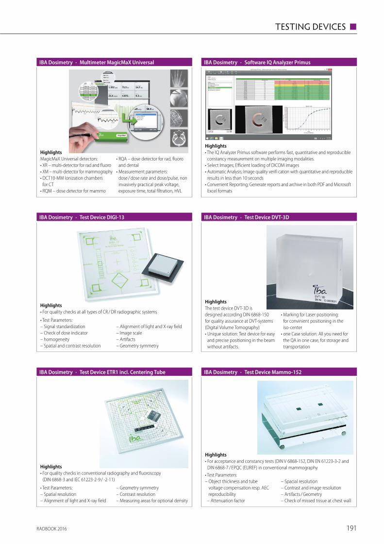
HighlightsMagicMaX Universal detectors:• XR – multi-detector for rad and fluoro• XM – multi-detector for mammography• DCT10-MM Ionization chambers
for CT• RQM – dose detector for mammo
• RQA – dose detector for rad, fluoro and dental
• Measurement parameters: dose / dose rate and dose/pulse, non invasively practical peak voltage, exposure time, total filtration, HVL
· Multimeter MagicMaX UniversalIBA Dosimetry
Highlights• The IQ Analyzer Primus software performs fast, quantitative and reproducible
constancy measurement on multiple imaging modalities.• Select Images; Efficient loading of DICOM images• Automatic Analysis; Image quality verifi cation with quantitative and reproducible
results in less than 10 seconds• Convenient Reporting; Generate reports and archive in both PDF and Microsoft
Excel formats
· Software IQ Analyzer PrimusIBA Dosimetry
HighlightsThe test device DVT-3D is designed according DIN 6868-150 for quality assurance at DVT-systems (Digital Volume Tomography) • Unique solution: Test device for easy
and precise positioning in the beam without artifacts.
• Marking for Laser positioning: for convinient positioning in the iso-center
• one Case solution: All you need for the QA in one case, for storage and transportation
· Test Device DVT-3DIBA Dosimetry · Test Device DIGI-13IBA Dosimetry
Highlights• For quality checks at all types of CR / DR radiographic systems
• Test Parameters:– Signal standardization – Check of dose indicator– homogeneity – Spatial and contrast resolution
– Alignment of light and X-ray field – Image scale– Artifacts – Geometry symmetry
· Test Device ETR1 incl. Centering TubeIBA Dosimetry
Highlights• For quality checks in conventional radiography and fluoroscopy
(DIN 6868-3 and IEC 61223-2-9 / -2-11)
• Test Parameters:– Spatial resolution– Alignment of light and X-ray field
– Geometry symmetry– Contrast resolution– Measuring areas for optional density
Highlights• For acceptance and constancy tests (DIN V 6868-152, DIN EN 61223-3-2 and
DIN 6868-7 / EPQC (EUREF) in conventional mammography• Test Parameters:– Object thickness and tube
voltage compensation resp. AEC reprodu cibility
– Attenuation factor
– Spacial resolution– Contrast and image resolution – Artifacts / Geometry – Check of missed tissue at chest wall
· Test Device Mammo-152IBA Dosimetry
RADBOOK 2016 191
TESTING DEVICES ◼

Highlights• 40 mm basic body with integrated Al step wedge with 14 steps from 0 to 5.2 mm• 6 mm structural plate with recess for test inserts, 2 rows of steel balls with
integrated turnable resolution test in line groups of 5, 6, 7, 8 and 10 Lp / mm• Attenuation body 3 x 20 / 1 x 10 / 1 x 6 mm PMMA• Various test inserts• Carrying case
· Test Device PASMAM 1054 A / CIBA Dosimetry
Highlights• 40 mm base plate with integrated Al step wedge with 14 steps from 0 to 5.2 mm
and 2 rows of steel balls for checking the image limitations towards the thorax side• 6 mm structural plate with recess for test inserts, 2 rows of steel balls with
integrated turnable resolution test in line groups of 5, 6, 7, 8 and 10 Lp / mm• Various test inserts• Carrying case – Attenuation body 2 x 20 / 2 x 10 mm
· Test Device PASMAM 1054 CIBA Dosimetry
HighlightsTest Device Primus A is designed according DIN 6868-150 for Radiography and Fluoroscopy systems.• Universal: Phantom for Radiograpic and Fluoroscopic systems• Internal Grid: For easy visual test of distortion and Light-/Beam-field coincidence• Two in One: Testarea for Dynamic verification and Low contrast sensitivity• Smart design: Die to the frame, the Phantom is very convinient in handling
· Test Device Primus AIBA Dosimetry
Highlights• For quality checks at digital / conventional radiographic and fluoroscopic
X-ray units (according to DIN 6868-4, 2007)• Test Parameters:– Spatial resolution – Verification of used kV-range– Contrast resolution
– Alignment of light and X-ray field– Geometry symmetry – Image scale,
Dimensions: 300 x 300 x 18.5 mm
· Test Device Primus LIBA Dosimetry
Highlights• Diagnostic dosemeter
(CE marked, class IIb certified) fully compliant with IEC 61674• Suitable for CTDI measurements acc. to IEC 60601-2-44 using a 100 or 300 mm
CT ion chamber • Data and waveform export to Excel via USB or Bluetooth• Accessories: CTDI head and / or body PHANTOMS (CE marked, class I certified)
· NOMEX Dosemeter PTW
Highlights• Dosimetry system (CE marked, class IIb certified) acc. to IEC 61674• Incl. NOMEX DOSEMETER and MULTIMETER (captures all dose values, time,
kVp, TF, HVL, frequency, pulses, waveforms)• Data and waveform export to Excel via USB or Bluetooth• Accessories: Test objects NORMI RAD/FLU, NORMI DSA, NORMI 3D
(CE marked, class I certified)
· NOMEX System PTW
RADBOOK 2016192
◼ TESTING DEVICES

RADBOOK 2016 193
TESTING DEVICES ◼
| visit us onfollow us onEuropean Hospital Verlags GmbH | Theodor-Althoff-Straße 45 | 45133 Essen
www.healthcare-in-europe.com
Trends & innovations in healthcare.
25 YEARSof Europeanhealthcare
communication:print & online.
Online Portal | Newspaper | Product Guides | Newsletter
Staytunedon allchannels.
.artu
ndw
ork
desi
gnbü
ro
Hitachi Medical Systems Europe Holding AG · Sumpfstrasse 13 · CH-6300 Zug
www.hitachi-medical-systems.eu
Embracing lifethrough better medical
imaging solutions.
Better medical imaging solutions for the needs of our customers
We have been committed to delivering outstanding medical imaging solutions over decades. Original solutions,
that have grown out of the groups' expertise and technical know-how, providing innovations that meet equally
the Zeitgeist and the needs of our customers and their patients.
Shimadzu_RADbook_2013.qxd 0
7.02.2013 13:33 Uhr Seite
1
RADBOO
K 20
12 ·
The
Radi
olog
y Gu
ide
to T
echn
olog
y &
Info
rmat
ics
in E
urop
e
W IT W CTW MRI W InterventionalW MammoW R / FW NucW Displays / Printers
W UltrasoundW InjectorsW Testing Devices
The Radiology Guide to Technology & Informatics in Europe
T 19.-
2013
The world’s most advanced dynamic volume CT just got even better. The new Aquilion ONE ViSION Edition provides you and
your patients robust clinical solutions when you need them most. This innovative Dynamic Volume CT enables all patients a
successful examination, with the lowest possible radiation exposure and the highest quality diagnostic outcomes.
For more information please turn to page 12 or visit www.aquilionvision.com.
RADBOOK 2014The Radiology Guide to Technology and Informatics in Europe
T 19.-
.artu
ndw
ork
desi
gnbü
ro
Hitachi Medical Systems Europe Holding AG · Sumpfstrasse 13 · CH-6300 Zug
www.hitachi-medical-systems.eu
Embracing lifethrough better medical
imaging solutions.
Better medical imaging solutions for the needs of our customers
We have been committed to delivering outstanding medical imaging solutions over decades. Original solutions,
that have grown out of the groups' expertise and technical know-how, providing innovations that meet equally
the Zeitgeist and the needs of our customers and their patients.
Shimadzu_RADbook_2013.qxd 0
7.02.2013 13:33 Uhr Seite
1
RADBOO
K 20
12 ·
The
Radi
olog
y Gu
ide
to T
echn
olog
y &
Info
rmat
ics
in E
urop
e
W IT W CTW MRI W InterventionalW MammoW R / FW NucW Displays / Printers
W UltrasoundW InjectorsW Testing Devices
The Radiology Guide to Technology & Informatics in Europe
T 19.-
2013
The world’s most advanced dynamic volume CT just got even better. The new Aquilion ONE ViSION Edition provides you and
your patients robust clinical solutions when you need them most. This innovative Dynamic Volume CT enables all patients a
successful examination, with the lowest possible radiation exposure and the highest quality diagnostic outcomes.
For more information please turn to page 12 or visit www.aquilionvision.com.
ITCTMRIInterventionalMammoR/FNucDisplays/PrintersUltrasoundInjectorsTesting Devices
Villa Sistemi Medicali’s new general radiographic system, the Moviplan iC, has been conceived for every diagnostic need.
It is available in a wide range of configurations, from basic analog versions up to fully digital and automatized rooms.
T H E E U R O P E A N F O R U M F O R T H O S E I N T H E B U S I N E S S O F M A K I N G H E A L T H C A R E W O R K
CONTENTS
NEWS & MANAGEMENT 1-3
ONCOLOGY 4-5
RADIOLOGY 6-8
MEDICA PRODUCTS 9
INFECTION CONTROL 10
LABORATORY 11-16
www.european-hospital.com
V O L 2 3 I S S U E 6 / 1 4 • D E C E M B E R 2 0 1 4
Report: Michael Krassnitzer
By definition, an emerging infec-
tious disease is one that has newly
appeared in a population or has
been known for some time but is
rapidly increasing in incidence or
spreading to new geographic areas.
At the International Meeting on
Emerging Diseases and Surveillance
(IMED 2014), in Vienna, this year’s
focus was on one particular emerg-
ing infectious disease: Ebola.
According to the World Health
Organisation (WHO) in the recent
outbreak 5,400 people have died
of this dangerous disease and, as
of 20 November 2014, more than
15,000 cases were reported in eight
countries.
‘This most serious Ebola outbreak
ever was caused by social, geo-
graphic and political factors in the
regions affected,’ says Belgian physi-
cian Hilde De Clerck MD, who works
for the aid organisation Médecins
sans frontières in the crisis regions.
All severely affected countries are
poor, have underdeveloped health
systems, were involved in wars or
armed conflicts in the past few
years, and the people tend to mis-
trust government agencies and the
countries mistrust each other.
De Clerck: ‘The epidemic start-
ed off slowly and spread quick-
ly, affecting people from all walks
of life. Lack of awareness about
the disease, insufficient protection
measures and the high degree of
mobility of the people between
three of the countries concerned –
be it for business or family purposes
– contributed to Ebola being able
acute Ebola infection cannot travel.
During the severe acute respira-
tory syndrome (SARS) pandemic,
in 2002/2003, 45 million travellers
were screened at airports – with
only a single SARS case being iden-
tified.
It is much more important,
according to the experts, to use the
money for aid programmes right in
the affected regions. ‘It is crucial
to provide help where the epi-
demic broke out, that is in Western
Africa,’ underlines Jack Woodall
MA PhD, an epidemiologist from
Brazil and vice-editor of ProMED-
mail, the web-based Programme
for Monitoring Emerging Diseases
of the International Society for
Infectious Diseases (ISID). He is
confident: ‘We can stop the disease
from spreading if we manage to
break the chain of infection.’
to spread.’ There is, however, good
news: Oyewale Tomori DVM, PhD,
President of the Nigerian Academy
of Science, describes how Ebola
was contained in his country. On
20 July 2014 a person from Liberia
with acute Ebola symptoms arrived
at Lagos International Airport. The
preliminary diagnosis – Ebola – was
confirmed by a private hospital. This
index case had had contact with 72
people at the airport and in the hos-
pital, who potentially were exposed
to the virus.
The Ministry of Health and the
Nigeria Centre for Disease Control
(NCDC) declared an Ebola emer-
gency. On 23 July the Ministry of
Health, the regional government
of Lagos and international partners
set up an Ebola crisis intervention
centre.
The index case died on 25 July 20.
Subsequent Ebola infections were
reported in Nigeria. All of them
could be traced back to the index
case. Close to 900 identified con-
tact persons were monitored; eight
patients died. On 20 October 2014,
WHO officially declared Nigeria
Ebola-free.
‘It is to a large extent due to
the quickly established crisis inter-
vention centre that we were able
to successfully fight the disease,’
said Tomori. ‘We not only suf-
fer from a real epidemic in West
Africa, we are also suffering from
an epidemic of fear that’s spread-
ing all over the globe,’ said Dr
Pamela Rendi-Wagner, Director of
the Department of Public Health
Services and Medical Affairs at the
Austrian Federal Ministry of Health
and adds, ‘Despite concerted efforts
by all public health authorities it is
difficult to counter the rising public
panic’. In Austria it was reported
that several health professionals quit
their jobs because they feared hav-
ing to care for Ebola patients. ‘It
is our main task to listen to these
fear-driven concerns and to inform
and communicate widely, openly,
early and in a transparent fashion
in order to create trust,’ the health
official emphasised.
Experts agree on one issue: they
consider screening of in-coming
travellers at international airports
for Ebola to be useless, inter alia
because usually people with an
Ebola: Reports of
panic among medicsExperts confirm the disease can be contained by stopping the chain of infection
Médecins Sans Frontières medics: a vital aid organisation in crisis zones
So
urc
e: M
SF;
Julien R
ey
RADIOLOGY 6-8
• Precision radiotherapy
with 4-D imaging
• MorphMatch technology
for 3-T scans
LABORATORY 11-16
• Breakthrough in hepatitis
C research
• Stain-free 3-D digital pathology
• Recycling blood lost during surgery

Highlights• Dosimetry system (CE marked, class
IIb certified) acc. to IEC 61674• Single exposure captures all dose
values, kVp, time, TF, HVL, frequency, pulses and waveforms
• Angular independent for positioning within the beam
• Fully automatic adjustment• Data and waveform export to Excel• Ideal for tomosynthesis measurements• Accessories: NORMI MAM test objects
· NOMEX Multimeter PTW
Highlights• QUART dent /digitest 2D dental test
phantoms are designed to assess x-ray imaging parameters according DIN and IEC QA / QC requirements.
• Features patient equivalent filtration and test objects to perform full-scale x-ray image quality analyses.
Parameters• Spatial resolution• High-contrast resolution• Low-contrast resolution• Homogeneity / artefacts• Radiation field/tube alignment
· dent / digitest Dental QA / QC Test PhantomQUART
Highlights• The QUART didoEASY meters are designed for quick measurements of dose,
dose rate and exposure time in X-ray QA / QC and service. • didoEASY meters automatically compensate all radiation qualities in their
area of application. Three meter versions are available: for R / F and Dental (40 – 160 kV), for Mammography (25 – 40 kV), and one for the full diagnostic range (25 – 160 kV).
· didoEASY Diagnostic X-Ray MetersQUART
· didoSVM Precision Survey MeterQUART
Highlights• The QUART didoSVM Medical survey meter is designed to detect beta,
gamma and x-ray sources of very low intensity around diagnostic x-ray equip-ment as well as in radiation therapy environments. Excellent energy response to measure radiation rate and dose.
• Its detection technology is based on solid-state components, enabling measure ments with high sensitivity and very quick response.
· dido2000 Series Diagnostic X-Ray MetersQUART
HighlightsThe QUART dido2000 series diagnostic x-ray dosemeters are used for QA and service in Radiography, (Pulsed) Fluoros-copy, DSA, Dental, 3D (CBCT), and Mammo graphy.• Compact multi-functional
state-of-the-art solid state detector
• Enable measurements in spots with limited space
• Measurements behind scatter radiation grids
• Direct measurement of DWP in dental panoramic applications
Highlights• The QUART didoCT pencil-shaped ion chamber meter is designed for easy
and precise dose-width product measurements. • The meter does not require any pre-setting procedure for direct reading of
DWP, rate and time.• As an optional feature, the QUART didoCT can be supplied with free-in-air kV
(eff.) measurement capability covering RQT 8-10 and further ranges.
· didoCT Pencil Chamber MeterQUART
RADBOOK 2016194
◼ TESTING DEVICES

Highlights• The QUART DSA phantom features longitudinal sliding technique to minimise
structural movement artefacts in the test image. It complies with DIN 6868-4, 6868-150 and IEC 61223-3-3.
• A special characteristic of the phantom is that it realistically reproduces the injection procedure of the contrast agent into vessels with different atte-nuation properties.
· DSA Test PhantomQUART
Highlights• The QUART DVT 150 phantom is designed to meet the requirements of the
German DIN 6868-150 x-ray imaging acceptance test standard.• Handling and positioning of the phantom is easy and straight-forward. It en-
ables quick and simple contrast resolution tests for 3D, ENT and angiography x-ray applications.
· DVT 150 CBCT IQ Test PhantomQUART
Highlights• The QUART DVT AP phantom is designed for QA / QC at Cone Beam CT
(CBCT), Dental Volume Tomography (DVT) and 3D imaging equipment.• It is to be used in dental 3D imaging (according DIN 6868-161 requirements)
as well as angiography in C-arm x-ray applications (manufacturer-specific applications). Based on latest research, the solution can also be utilised for standard CT IQ tests.
· DVT AP Cone-Beam CT Test PhantomQUART
Highlights• The QUART mam / digi phantom is designed to be used as universal tool for
QA / QC routine testing in Digital and Analog Mammography. The phantom creates a link between technical and clinical image quality. It can also be used as QA tool for Digital Tomosynthesis.
• The phantom incorporates QUART's unique Landolt ring objects. They serve to verify low-contrast and perceptibility limits.
· mam / digi Mammography IQ PhantomQUART
Highlights• The QUART nonius is a sophisticated, fully electronic x-ray ruler to verify size
and geometrical properties of x-ray fields in Radiography and Mammography. It can also be used to analyse fanned CT or dental OPG x-ray beams.
• Its resolution capabilities and precision go down into to the nonius range of 0.1 mm!
• Take only 3 steps to obtain the test result: Position – Expose – Evaluate.
· nonius Electronic X-Ray RulerQUART
Highlights• The QUART SP_dl phantom enables assessment of digital x-ray equipment
according to the German DIN 6868-150 and DIN 6868-4.• The phantom is available with a unique kV test object to assess radiation
quality and generator performance on a routinely basis.• For ease of use, a frame / extension is provided as well as a wire-mount system
for use with wall stand units.
· SP dl R / F IQ PhantomQUART
RADBOOK 2016 195
TESTING DEVICES ◼

· ACCU-GOLD+Radcal
Highlights• Extensive Sensor Selection• Both Solid State and Gold Standard Ion Chamber Technology• Rapid Simultaneous Measurements• The Smallest Footprint Solid State Sensor• Customizable Software• Replaces first generation Accu-Gold Diagnostic System• WiFi available using Nugget device
· ACCU-DOSE+Radcal
Highlights• The newest member of the Accu-Gold family• Dose Measurement System with WiFi• Gold Standard Ion Chamber Sensors & Solid-state Dose Diode Sensors• Excellent Solution for Radiography, Fluoroscopy, Mammography,
CT & Dental applications• Dose-oriented set of functionality including Dose, Dose Rate, Waveform,
Pulse, dose / pulse & Exposure time• Several display options & customizable software
· DAP AnalyzersRadcal
Highlights• PDC (Patient Dose Calibrator)• Use to calibrate DAP (Dose Area Product) meters• Measures and displays DAP / Rate, Dose / Rate• Optical and radiographic alignment markers• Simple to use with optional computer control
· RAPID-GOLD+Radcal
Highlights• Accu-Gold+ Technology• Uses only Solid State Sensors for Diagnostic, Dental and Mammography X-Ray• Optional mA / mAs invasive or Non-invasive measurement sensors• Replaces first generation Rapid-Gold• WiFi available using Nugget device
HighlightsThe RTI Black Piranha brings quickness and power to your X-ray QA work. The Black Piranha includes what you would expect in a multifunction meter. Connection to various accessories, tablet and PC. The Quick Check feature identifies the probes you insert and selects the optimum settings for your measurements. One-shot HVL for Mammography, Radiography, CT and Dental. Optimized for X-ray equipment from a large number of manufacturers.
· Black PiranhaRTI
HighlightsCobia Smart is a straightforward and simple-to-use instrument for checking that the output from an X-ray tube is correct. Place it beneath the X-ray tube, make an X-ray exposure, and rapidly get an accurate reading. The measured values can be read directly from Cobia Smart’s large and clear display, even from a distance. No adjustments are required, making it exceptionally easy to use. Easy to position, no position dependence.
· Cobia SmartRTI
RADBOOK 2016196
◼ TESTING DEVICES
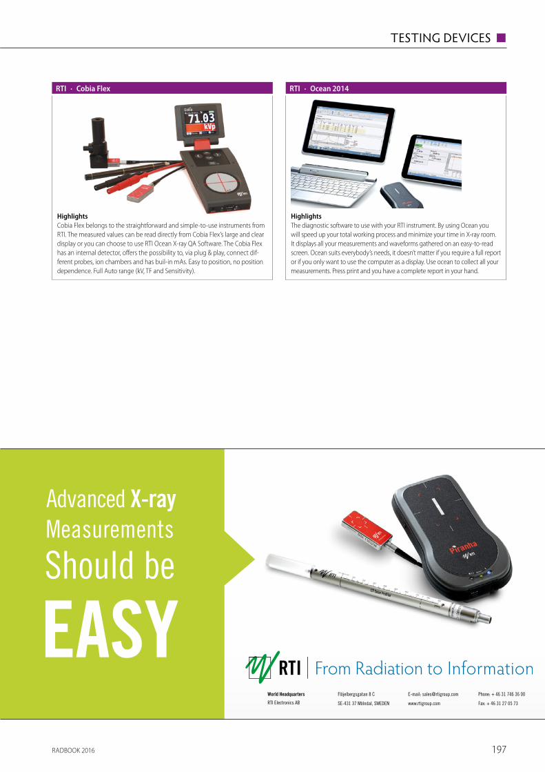
HighlightsCobia Flex belongs to the straightforward and simple-to-use instruments from RTI. The measured values can be read directly from Cobia Flex’s large and clear display or you can choose to use RTI Ocean X-ray QA Software. The Cobia Flex has an internal detector, offers the possibility to, via plug & play, connect dif-ferent probes, ion chambers and has buil-in mAs. Easy to position, no position dependence. Full Auto range (kV, TF and Sensitivity).
· Cobia FlexRTI
HighlightsThe diagnostic software to use with your RTI instrument. By using Ocean you will speed up your total working process and minimize your time in X-ray room. It displays all your measurements and waveforms gathered on an easy-to-read screen. Ocean suits everybody’s needs, it doesn’t matter if you require a full report or if you only want to use the computer as a display. Use ocean to collect all your measurements. Press print and you have a complete report in your hand.
· Ocean 2014RTI
RADBOOK 2016 197
TESTING DEVICES ◼
Advanced X-ray Measurements
Should be
EASYWorld Headquarters
RTI Electronics AB
Flöjelbergsgatan 8 C
SE-431 37 Mölndal, SWEDEN
E-mail: [email protected]
www.rtigroup.com
Phone: + 46 31 746 36 00
Fax: + 46 31 27 05 73

· RaySafe i2Unfors
HighlightsThe RaySafe i2 is an active dosimetry system providing real-time insights around personal radiation exposure. Operators can pre-set limits on dose to keep exposure below legal limits. The i2’s color-coded indicators provide at-a-glance access to exposure levels. Mirroring the latest personal electronics, the i2 dosimetry system offers touchscreen technology.
· RaySafe Solo CTUnfors
HighlightsThe RaySafe Solo CT shares the same patented technology as the rest of the RaySafe line. Designed to rapidly measure CT applications, the Solo CT requires less than one minute to take the first exposure, including dose and dose length.
· RaySafe Solo DENTUnfors
HighlightsThe RaySafe Solo DENT shares the same patented technology as the rest of the RaySafe line. Designed to rapidly measure dental x-ray equipment, the Solo DENT requires less than one minute for set up.
· RaySafe Solo DOSEUnfors
HighlightsThe RaySafe Solo Dose measures dose, dose rate, time and pulses on both radiographic and fluoroscopic X-ray machines, sharing the same patented technology as the rest of the RaySafe line.
· RaySafe Solo MAMUnfors
HighlightsThe RaySafe Solo MAM shares the same patented technology as the rest of the RaySafe line. Designed to rapidly measure MAM applications, the Solo MAM requires less than one minute to take the first exposure.
· RaySafe ThinXUnfors
HighlightsThe RaySafe ThinX is a compact tool for quick, easy results across multiple parameters. Its fully automatic interface makes it the easiest tool to use – turning itself on when radiation is detected! Featuring patented technology, the ThinX automatically corrects itself for beam filtration.
RADBOOK 2016198
◼ TESTING DEVICES

· RaySafe X2Unfors
HighlightsThe RaySafe X2 is a single device that offers full range of measurements, an intuitive interface, and simplicity. Our advanced, groundbreaking sensor technology is ready to take exposures in one minute with no menus or set-tings. Designed to register R/F, MAM, CT, survey, and light applications, the X2 requires little or no manual operation.
· RaySafe XiUnfors
HighlightsThe RaySafe Xi is a modular system. Whether you need one modality or multiple ones, it can be modified based on need. Its two key operation inter-face makes it easy to use and quick to set up for the first exposure. Compact yet powerful, the Xi is preferred by leading manufacturers of x-ray equipment.
HighlightsThe VacuDAP family provides a wide range of DAP and Dose measuring solutions for most of the diagnostic X-ray systems in the market.
Technical specs• Resolution DAP: 0,01 µGym2• Resolution Dose: 0,003 mGy• Interface: RS485, RS232, Bluetooth, CAN• Active area:
(123 x 123) mm / (147 x 147) mm
· VacuDAP / VacuDAP duoVacuTec
Highlights• VacuDAP chamber is now available
with Wi-Fi or Bluetooth technology.• Perfect suitable for DR upgrades and
mobile X-ray units.• The battery ensures simplest instal-
lation ever.
Technical specs• Resolution DAP: 0,01 µGym2• Active area:
(123 x 123) mm / (147 x 147) mm • Battery operation time: about 12 h
· VacuDAP wireless / VacuDAP BluetoothVacuTec
HighlightsThe VacuDAP-C systems for measure-ment of DAP and Dose are basically integrated in interventional devices with customized calibration settings.
Technical specs• Resolution DAP: 0,01 µGym2• Resolution Dose: 0,005 mGy• Interface: RS485, RS232, Bluetooth, CAN• Active area: Ø (8 …100) mm
· VacuDAP-C / VacuDAP-C duoVacuTec
HighlightsDigital interface ensures EMC stable signal transmis-sion and provides an open dose working range.
Technical specs• Tube voltage: 40 kV …150 kV• Dose rate range: 0.5 …1,000 µGy/s• Aluminium equivalent: 0.75 mm Al• Analog interface:
ramp voltage 0 – 10 V
• Digital interface: differential pulses (RS422)
• Resolution: 0.025 µGy• Pulse width: 2 µs
· VacuTec AEC SensorVacuTec
RADBOOK 2016 199
TESTING DEVICES ◼

RADBOOK 2016200
INDEX OF ADVERTISERS
117
43
163
205
155
81
181
13
5
89
167
49
back cover
159
7
29
From Radiation to Information 197
back cover
front cover
21
173
title
front & back cover
33
35
page

RADBOOK 2016 201
COMPANIES & SUPPLIERS
| visit us onfollow us onEuropean Hospital Verlags GmbH | Theodor-Althoff-Straße 45 | 45133 Essen
www.healthcare-in-europe.com
Trends & innovations in healthcare.
Subscribe to our Newsletter on healthcare-in-europe.com.
Onl ine Portal | Newspaper | Product Guides | Newsletter
phot
ogra
phy
by R
ober
t Hile
25 YEARSof Europeanhealthcare
communication:print & online. Subscribe to our Newsletter on healthcare-in-europe.com.
Staytunedon allchannels.

■ R
IS
■ P
ACS
■ W
orks
tatio
ns
■ C
T
■ M
RI
■ I
njec
tors
■ I
nter
vent
iona
l
■ I
T Sy
stem
s
■ M
amm
ogra
phy
■ R
/ F F
ilm-S
cree
n
■ R
/ F D
igita
l
■ M
olec
ular
Imag
ing
■ D
ispla
ys
■ P
rinte
rs
■ U
ltras
ound
■ T
estin
g D
evic
es
Agfa HealthCareSeptestraat 272640 Mortsel, Belgiumtel +32 3 444 94 [email protected]
3 3 4
213 214
73808486
107111124127133
161
Alliance Medical Interim SolutionsIceni Centre, Warwick Technology Park, Warwick, CV34 6DA, UK tel +44 192 648 20 [email protected]
26 45 70 148
allMRI GmbHSüdstr. 2374226 Nordheim, Germanytel +49 71 33 2 37 02 [email protected]
45
Barco N.V.President Kennedypark 358500 Kortrijk, Belgiumtel +32 56 23 32 [email protected]
150152153158
Bayer Medical Care B.V.Avenue Céramique 276221 KV Maastricht, The Netherlandstel +31 43 358 56 01www.radiology.bayer.com
4849
Bracco Injeneering S.A.Avenue de Sévelin 461004 Lausanne, Switzerlandtel +41 21 621 74 [email protected]
50
Canon Europa N.V.Bovenkerkerweg 591185 XB Amstelveen, The Netherlandstel +31 20 545 8 [email protected]/medical
3 34
213 214 80
111112124127133
CHILI GmbH Friedrich-Ebert-Str. 2 69221 Dossenheim / Heidelberg, Germany tel +49 62 21 180 79 [email protected]
34
213 214
73748084
162
CHISON Medical Imaging Co., Ltd.No. 9 Xin Hui Huan Road, New District,Wuxi, Jiang Su Province, China 214028tel +086 510 85 31 05 93 [email protected]
165166
DMS APELEM393 rue Charles Lindbergh34130 Mauguio, Francetel +33 4 67 50 49 00www.dms.com
64 88 103
112124127133134139
Dunlee Medical ComponentsEuropean Customer Service CenterVeenpluis 65684 PC Best, The Netherlandstel +31 40 276 25 [email protected]
A Division of Philips Healthcare
2627 139
EBIT S.r.l. – Esaote Group Via Siffredi 58 16153 Genoa, Italy tel +39 010 65 [email protected] www.esaote.com/healthcare-it
3 34
213 214
74787983
RADBOOK 2016202
COMPANIES & SUPPLIERS

■ R
IS
■ P
ACS
■ W
orks
tatio
ns
■ C
T
■ M
RI
■ I
njec
tors
■ I
nter
vent
iona
l
■ I
T Sy
stem
s
■ M
amm
ogra
phy
■ R
/ F F
ilm-S
cree
n
■ R
/ F D
igita
l
■ M
olec
ular
Imag
ing
■ D
ispla
ys
■ P
rinte
rs
■ U
ltras
ound
■ T
estin
g D
evic
es
EDL SAS1031, chemin de la Seyne à Bastian83500 La Seyne-sur-mer, Francetel +33 4 94 10 99 [email protected]
3 34 74
EIZO Europe GmbHHelmut-Grashoff-Str. 1841179 Moenchengladbach, Germanytel +49 21 61 82 [email protected]
150152153154158160162
Esaote S.p.A.Via Siffredi 5816153 Genoa, Italytel +39 010 65 [email protected]
40 71 168169
SonoSite, Inc.21919 30th Drive SEBothell, Washington 98021-3904, USAtel +31 20 462 00 [email protected]
169170
GCTechnology GmbHFreidling 1284172 Buch am Erlbach, Germanytel +49 87 06 94 15 [email protected]
27 46 71 96 105 187
GE Healthcare 283 Rue de la Minière78533 Buc Cedex, Francetel +33 130 70 40 [email protected]
3 34
213 214
9162023
31323436
5357606465
74758486
8890
113127
144145146147
170171 189
GENERAL MEDICAL ITALIA S.R.L.Via Nazionale snc83030 Montefredane (AV), Italytel +39 08 25 60 72 [email protected]
90124127134
GENERAL MEDICAL MERATE S.p.A.Via Partigiani, 2524068 Seriate (BG), Italytel +39 035 45 25 [email protected]
6569
99101
113134
Giotto / IMS Internazionale Medico SciSagittario, 540037 Sasso Marconi (BO), Italytel +39 051 84 68 [email protected]
889095
Hitachi Medical Systems Europe (Holding) AG Sumpfstrasse 136300 Zug, Switzerlandtel +41 41 748 63 33welcome@hitachi-medical-systems.comwww.hitachi-medical-systems.com
91720
3641 140
172173174
Hologic Europe N.V. Leuvensesteenweg 250A1800 Vilvoorde, Belgiumtel +32 2 711 46 [email protected]
69 83
90959697
140
I.A.E. S.P.A.Via Fabio Filzi, 53 20032 Cormano (MI), Italytel +39 02 66 30 32 [email protected]
27 71 97 140
RADBOOK 2016 203
COMPANIES & SUPPLIERS

■ R
IS
■ P
ACS
■ W
orks
tatio
ns
■ C
T
■ M
RI
■ I
njec
tors
■ I
nter
vent
iona
l
■ I
T Sy
stem
s
■ M
amm
ogra
phy
■ R
/ F F
ilm-S
cree
n
■ R
/ F D
igita
l
■ M
olec
ular
Imag
ing
■ D
ispla
ys
■ P
rinte
rs
■ U
ltras
ound
■ T
estin
g D
evic
es
IBA Dosimetry GmbHBahnhofstr. 590592 Schwarzenbruck, Germanytel +49 91 28 [email protected]
189190191192
IMAGE Information Systems Europe GmbHLange Str. 1618055 Rostock, Germanytel +49 381 496 58 20 [email protected]
3 34
213 214
758385
IMS Internazionale Medico SciSagittario, 540037 Sasso Marconi (BO), Italytel +39 051 84 68 [email protected]
889095
INTERMEDICAL SRLE. Fermi, 2624050 Grassobbio (BG), Italytel +39 035 659 48 [email protected]
606569
i-SOLUTIONS Health GmbHAm Exerzierplatz 1468167 Mannheim, Germanytel +49 621 39 [email protected]
3 34
7586
ITH icoserve technology for healthcare GmbHInnrain 986020 Innsbruck, Austriatel +43 512 890 [email protected]
34 78
ITZ Medicom GmbH & Co. KGSiemensring 44 a47877 Willich, Germanytel +49 21 54 49 79 [email protected]
3 34
213 214
757985
KONICA MINOLTA Medical & Graphic Imaging Europe B.V.Hoogoorddreef 91101 BA Amsterdam, The Netherlandstel +31 20 658 41 [email protected]/healthcare
34
213 214
110114125128140
174
MECALL S.R.L.Via Negrelli, 5520851 Lissone (MB), Italytel +39 039 24 31 [email protected]
MECALL
114134
mediCAD Hectec GmbHOpalstr. 5484032 Altdorf, Germany tel +49 871 33 02 [email protected] www.mediCAD.eu
213 214
8283
Medical ECONET GmbHIm Erlengrund 2046149 Oberhausen, Germanytel +49 208 37 78 [email protected]
114125128129
medigration GmbHSchuhstr. 3091052 Erlangen, Germanytel +49 91 31 690 [email protected]
3 34
213 214
768081838485
125 161162
RADBOOK 2016204
COMPANIES & SUPPLIERS

RADBOOK 2016 205
COMPANIES & SUPPLIERS
Afghanistan 190, Albania 380, Algeria 313, American Samoa 39, Andorra 60, Angola 44, Anguilla 20, Antarctica 16, Antigua 4, Antigua and Barbuda 140, Argentina 1542, Armenia 202, Aruba 39, Australia 1685, Austria 332, Azerbaijan
133, Bahamas 72, Bahrain 95, Bangladesh 271, Barbados 32, Belarus 81, Belgium 442, Belize 58, Benin 55, Bermuda 34, Bhutan 24, Bolivia 460, Bonaire 2, Bosnia and Herzegovina 160, Botswana 46, Brazil 3068, British Indian Ocean Territory 16, Brunei Darussalam 37, Bulgaria 590, Burkina Faso 40, Burundi 18, Cambodia 112, Cameroon 200, Canada 4583, Canary Islands 2, Cape Verde 31, Cape Verde Islands 1, Caroline Islands 2, Côte D’Ivoire 1, Cayman
Islands 38, Central African Republic 495, Chad 17, Chile 708, China 4339, Christmas Island 10, Cocos(Keeling)Island 1, Colombia 1317, Comoros 18, Congo 7, Congo, Democratic Republic of the 65, Cook Islands 17, Costa Rica 234, Côte D’Ivoire 139, Croatia 191, Cuba 33, Curacao 4, Cyprus 365, Czech Republic 196, Denmark 252, Djibouti 26,
Dominica 22, Dominican Republic 502, Ecuador 620, Egypt 1487, El Salvador 247, Equatorial Guinea 28, Eritrea 17, Estonia 98, Ethiopia 132, Faeroe Islands 1, Falkland Islands 1, Faroe Islands 1, Fiji 34, Finland 124, France 1034,
French Guiana 18, French Polynesia 21, Gabon 23, Gambia 31, Georgia 478, Germany 1782, Ghana 365, Gibraltar 27, Greece 915, Greenland 23, Grenada 25, Guadeloupe 18, Guam 71, Guatemala 366, Guinea 14, Guinea-Bissau
943, Guyana 30, Haiti 121, Honduras 266, Hong Kong 383, Hungary 367, Iceland 49, India 7123, Indonesia 1096, Iran 1085, Iraq 545, Ireland 301, Isle of Man 1, Israel 753, Italy 1926, Jamaica 158, Japan 450, Jordan 356, Kazakhstan
123, Kenya 327, Kiribati 9, Kuwait 162, Kyrgyz Republic 70, Lao People’s Democratic Republic 30, Latvia 118, Lebanon 494, Lesotho 20, Liberia 19, Libyan Arab Jamahiriya 247, Liechtenstein 12, Lithuania 123, Luxembourg 38, Macao 28, Macedonia 218, Madagascar 62, Malawi 43, Malaysia 692, Maldives 33, Mali 35, Malta 56, Marshall Islands
23, Martinique 22, Mauritania 32, Mauritius 66, Mayotte 13, Mexico 5172, Micronesia 19, Moldova 126, Monaco 26, Mongolia 144, Montenegro 31, Montserrat 9, Morocco 426, Mozambique 43, Myanmar 65, Namibia 35, Nauru 15,
Nepal 127, Netherlands 699, Netherlands Antilles 34, Nevis 1, New Caledonia 18, New Zealand 271, Nicaragua 168, Niger 20, Nigeria 1369, Niue 12, Norfolk Island 21, Northern Mariana Islands 32, North Korea 15, Norway 129, Oman
88, Pakistan 3289, Palau 8, Palestinian Territory 8, Panama 224, Papua New Guinea 42, Paraguay 243, Peru 865, Philippines 1459, Pitcairn Island 1, Poland 1111, Portugal 461, Puerto Rico 740, Qatar 83, Reunion 16, Romania 955,
Russia 1304, Rwanda 40, Saint Barthelemy 2, Saint Helena 13, Saint Kitts 2, Saint Kitts and Nevis 20, Saint Lucia 46, Saint Maarten 4, Saint Pierre and Miquelon 11, Saint Vincent 1, Saint Vincent and the Grenadines 21, Samoa
13, San Marino 12, Sao Tome and Principe 2, Saudi Arabia 1025, Senegal 101, Serbia 348, Serbia and Montenegro 79, Seychelles 19, Sierra Leone 31, Singapore 392, Slovakia 115, Slovenia 115, Solomon Islands 13, Somalia 40, South Africa 684, South Korea 674, Spain 1056, Sri Lanka 160, Sudan 212, Suriname 50, Swaziland 11, Sweden
242, Switzerland 293, Syria 477, Tahiti 2, Taiwan 558, Tajikistan 57, Tanzania 159, Thailand 422, Timor-Leste 18, Togo 50, Tokelau 4, Tonga 13, Trinidad and Tobago 151, Tunisia 120, Turkey 1780, Turkmenistan 20, Turks and Caicos
Islands 22, Tuvalu 13, Uganda 234, Ukraine 895, United Arab Emirates 960, United Kingdom 3660, Uruguay 149, USA 130148, USVI 1, Uzbekistan 137, Vanuatu 17, Venezuela 1187, Vietnam 905, Virgin Islands (U.K.) 5, Virgin Islands (U.S.) 21, Wallis and Futuna Islands 11, Western Sahara 1, Western Samoa 2, Yemen 305, Yugoslavia 112, Zambia 80,
Zimbabwe 120,
So they can buy & sell medical equipment and parts, find a service partner, browse over 425,000 listings, and read
HealthCare Business News online. Register for free and join them.
The World’s Leading Medical Equipment Marketplace
www.dotmed.com
Why are more than a quarter million health care professionals registered on DOTmed?

■ R
IS
■ P
ACS
■ W
orks
tatio
ns
■ C
T
■ M
RI
■ I
njec
tors
■ I
nter
vent
iona
l
■ I
T Sy
stem
s
■ M
amm
ogra
phy
■ R
/ F F
ilm-S
cree
n
■ R
/ F D
igita
l
■ M
olec
ular
Imag
ing
■ D
ispla
ys
■ P
rinte
rs
■ U
ltras
ound
■ T
estin
g D
evic
es
MEDTRON AGHauptstr. 25566128 Saarbruecken, Germanytel +49 681 970 [email protected]
5051
Medtronic International Trading SàrlRoute du Molliau 311131 Tolochenaz, Switzerlandtel +41 21 802 70 00www.oarm.com/#contactwww.medtronic.com
61
SHENZHEN MINDRAY BIO-MEDICAL ELECTRONICS CO., LTD.Mindray Building, Keji 12th Road SouthNanshan, Shenzhen 518057, Chinatel +86 755 81 88 89 98 [email protected]
41 115130
175176177
NEC Display Solutions Europe GmbHLandshuter Allee 12 – 1480637 Munich, Germanytel +49 89 996 [email protected] www.medical.nec-display-solutions.com
150152154156158160161
NORAS MRI products GmbHLeibnizstr. 497204 Hoechberg, Germanytel +49 931 29 92 [email protected]
44
Philips HealthcareP. O. Box 10.0005680 DA Best, The Netherlandstel +31 40 278 56 [email protected]/healthcare
213 214
1217222328
32364246
53566165
8284 91
110115116117130135
144146147148
Planmed OySorvaajankatu 700880 Helsinki, Finlandtel +358 20 77 95 [email protected]
26 9194
PRIMAX International"Le Minotaure"30 – 34 Avenue Henri Matisse06200 Nice, Francetel +33 492 29 23 [email protected]
69117130135
PROTEC GmbH & Co. KGIn den Dorfwiesen 1471720 Oberstenfeld, Germanytel +49 70 62 925 [email protected]
4 213 214 76 99
105
118125126130
PTW-FreiburgPhysikalisch-Technische Werkstaetten Dr. Pychlau GmbHLoerracher Str. 779115 Freiburg, Germanytel +49 761 490 [email protected]
28 97 105 142 192194
QUART GmbHKirchenweg 785604 Zorneding, Germanytel +49 81 06 24 91 [email protected]
105 194195
Radcal Corporation426 West Duarte RoadMonrovia, CA 91016, USAtel +1 626 357 [email protected]
196
RADBOOK 2016206
COMPANIES & SUPPLIERS

■ R
IS
■ P
ACS
■ W
orks
tatio
ns
■ C
T
■ M
RI
■ I
njec
tors
■ I
nter
vent
iona
l
■ I
T Sy
stem
s
■ M
amm
ogra
phy
■ R
/ F F
ilm-S
cree
n
■ R
/ F D
igita
l
■ M
olec
ular
Imag
ing
■ D
ispla
ys
■ P
rinte
rs
■ U
ltras
ound
■ T
estin
g D
evic
es
Roesys GmbHDr.-Max-Ilgner-Str. 232339 Espelkamp, Germanytel +49 57 72 915 55 [email protected]
118126142
RTI ElectronicsFloejelbergsgatan 8C43137 Moelndal, Swedentel +46 31 746 36 [email protected]
From Radiation to Information
196197
SAMSUNG MEDISON CO., LTD.42, Teheran-ro 108-gil, Gangnam-gu, Seoul, Koreatel +82 2 21 94 14 [email protected]
119177178179
SCHILLER AGAltgasse 68, P. O. Box 10 526341 Baar, Switzerlandtel +41 41 766 42 [email protected]
46
Shimadzu Europa GmbHMedical Systems DivisionAlbert-Hahn-Str. 6 – 1047269 Duisburg, Germanytel +49 203 76 [email protected]
56616266
99100101103
119120130135
Siemens AG, Healthcare SectorHenkestr. 12791052 Erlangen, Germanytel +49 91 31 [email protected]/healthcare
34
213 214
91216172223
313234374244
53566263666769
76808284
919495
100101104
120131135136
145146147148
184185186
Shantou Institute of Ultrasonic Instr. Co., Ltd.#77, Jinsha Road515041 Shantou, Chinatel +86 754 88 25 01 [email protected]
179180181182183
STEPHANIX10, Rue Jean Moulin42150 La Ricamarie, Francetel +33 4 77 47 81 [email protected]
67100101104
121131136
SuperSonic ImagineLes Jardins de la Duranne, Bât E & F510, Rue René Descartes13857 Aix-en-Provence, Francetel +33 442 99 24 [email protected]
186
Swissray Medical AGTurbistr. 25 – 276280 Hochdorf, Switzerlandtel +41 41 914 12 [email protected]
121122131137
Technix S.p.A.Via Fermi 45 24050 Grassobbio (BG), Italytel +39 035 384 66 [email protected]
67 131132
Toshiba Medical Systems EuropeZilverstraat 12718 RP Zoetermeer, The Netherlandstel +31 79 368 92 [email protected]
162023
3437
5356576364
100101102
122132137142
186187
RADBOOK 2016 207
COMPANIES & SUPPLIERS

■ R
IS
■ P
ACS
■ W
orks
tatio
ns
■ C
T
■ M
RI
■ I
njec
tors
■ I
nter
vent
iona
l
■ I
T Sy
stem
s
■ M
amm
ogra
phy
■ R
/ F F
ilm-S
cree
n
■ R
/ F D
igita
l
■ M
olec
ular
Imag
ing
■ D
ispla
ys
■ P
rinte
rs
■ U
ltras
ound
■ T
estin
g D
evic
es
Toshiba Electronics Europe GmbHHansaallee 18140549 Duesseldorf, Germanytel +49 211 52 [email protected]
27 71122126137
142
TOTOKU Europe GmbHJakob-Krebs-Str. 12447877 Willich, Germanytel +49 21 56 49 68 [email protected]
150152153156157
Unfors RaySafe ABUggledalsvaegen 2942740 Billdal, Swedentel +46 31 719 97 [email protected]
198199
VacuTec Meßtechnik GmbHDornbluethstr. 1401277 Dresden, Germanytel +49 351 317 [email protected]
199
Varian Medical Systems GmbHKarl-Arnold-Str. 1247877 Willich, Germany +49 21 54 92 49 [email protected]/xray
28 97
VILLA SISTEMI MEDICALI s.p.a.Via delle Azalee, 320090 Buccinasco (MI), Italytel +39 02 48 85 [email protected]
26 68 9495
100102104105
123126132138
Vital Images Europe B.V.Zilverstraat 1 2718 RP Zoetermeer, The Netherlands tel +31 79 206 58 [email protected]
4 213 214
788082
China Resources Wandong Medical Equipment Co., Ltd.Bld.3, No. 9 Jiuxianqiaodong Road Chaoyang District100015 Beijing, Chinatel +86 10 845 757 [email protected]
4042
6468 94
102103105
123133138
Ningbo Xingaoyi Medical Instruments Co. Ltd(XGY Medical)777 West Tanjialing Rd.315400 Yuyao, Chinatel +86 574 627 308 [email protected]
404243
138
Ziehm Imaging GmbHDonaustr. 3190451 Nuremberg, Germanytel +49 911 21 [email protected]
6870
RADBOOK 2016208
COMPANIES & SUPPLIERS

RADBOOK 2016 209
NOTES

RADBOOK 2016210
NOTES

RADBOOK 2016 211
NOTES

RADBOOK 2016212
NOTES

RAD
BOO
K 20
16 ·
The
Radi
olog
y Gui
de to
Tech
nolo
gy &
Info
rmat
ics in
Euro
pe
RADBOOK 2016 RADBOOK 20161 3
RADBOOK 2016€ 19.-
• IT • CT • MRI • Interventional • Mammo • R / F • Nuc • Displays / Printers• Ultrasound • Injectors • Testing Devices
The Radiology Guide to Technology and Informatics in Europe
With 270° isocentric rotation, Infinix-i Rite Edition provides unparalleled flexibility and patient access even during the most challenging procedures. Its double c-arm design with 210° coverage and ultrafast rotation of 80°/s enables shorter breath hold times,
reduced contrast medium and outstanding 3D imaging from head to toe without the need for moving the patient or the table.
OPEN.PrOvEN.NEW.O-arm® Surgical Imaging System
Find out more in the chapterInterventional Systems
UC
20
16
06
64
8E
N ©
Med
tro
nic,
Inc.
20
16
. All
Rig
hts
Res
erve
d.
Best-in-classEquipped with the largest available FPD at43 x 43 cm and Shimadzu’s digital imagingplatform, the Sonialvision G4 covers thewidest possible range of examinationswith inter-departmental hospital capability.In both functionality and operability, theSonialvision G4 multipurpose R/F table isfar beyond other R/F systems. It provides“Best-in-class” features.
• Smart system architecturesupports outstanding clinical flexibilityfor a wide range of examinations
• Comprehensive dose management packageensures today’s highest safety of patientsand operators
• Excellent image qualityprovided by the advanced “SUREengine”technology enhancing the entire image forclearer details
• Premium application softwaresupporting useful applications, such astomosynthesis for general radiographicimaging
www.shimadzu-medical.eu
Next generation digital R/F system: Sonialvision G4
Best-in-classEquipped with the largest available FPD at43 x 43 cm and Shimadzu’s digital imagingplatform, the Sonialvision G4 covers thewidest possible range of examinationswith inter-departmental hospital capability.In both functionality and operability, theSonialvision G4 multipurpose R/F table isfar beyond other R/F systems. It provides“Best-in-class” features.
• Smart system architecturesupports outstanding clinical flexibilityfor a wide range of examinations
• Comprehensive dose management packageensures today’s highest safety of patientsand operators
• Excellent image qualityprovided by the advanced “SUREengine”technology enhancing the entire image forclearer details
• Premium application softwaresupporting useful applications, such astomosynthesis for general radiographicimaging
www.shimadzu-medical.eu
Next generation digital R/F system: Sonialvision G4

Management Archiving Image Distribution Workstations
RIS Small Business PACS Enterprise PACS Cardiology PACS Long Term Multimedia Inhouse Teleradiology Portal Solution Cloud Computing Application Multimodality Mammography Orthopedics Cardiology CAD Advanced
VisualizationOrbis RIS Impax Impax Impax for Cardiology ICIS
VNAICISHYDMedia
ICISEnterprise Imaging SuiteXero Viewer
ICISEnterprise Imaging SuiteXero Viewer
ICIS ICIS Impax Clinical Applications Impax Mammography Impax Clinical Applications Impax Clinical Applications Impax Clinical Applications Agfa HealthCareSeptestraat 27 · 2640 Mortsel, Belgiumtel +32 3 444 94 [email protected] · www.agfa.com
Canon Healthcare IT Suite Canon Healthcare IT Suite Canon Healthcare IT Suite Canon Healthcare IT Suite Canon Healthcare IT Suite Canon Healthcare IT Suite Canon Healthcare IT Suite Canon Healthcare IT Suite Canon Healthcare IT Suite Canon PACS Canon PACS CAD for Tuberculosis Canon PACS Canon Europa NVBovenkerkerweg 59 · 1185 XB Amstelveen, The Netherlandstel +31 20 545 8 [email protected] · www.canon-europe.com/medical
CHILI Modality PACS CHILI PACS CHILI PACS CHILI PACS CHILI PACS CHILI/Web CHILI/Web CHILI/Telemedicine Record OmniPACS CHILI Diagnost CHILI Diagnost CHILI Diagnost CHILI Diagnost Partner-Solution Partner-Solution CHILI GmbH Friedrich-Ebert-Str. 2 · 69221 Dossenheim/Heidelberg, Germany tel +49 6221 1 80 79 10 [email protected] · www.chili-radiology.com
Suitestensa RIS Suitestensa PACS Suitestensa PACS Suitestensa CVIS Suitestensa Archive Suitestensa Suitestensa WebSuitestensa Mobile
Suitestensa WebSuitestensa Mobile
Suitestensa Web Suitestensa WebSuitestensa Mobile
Suitestensa Review Suitestensa MG Suitestensa Review Suitestensa Review Cardio Suitestensa 3D Suitestensa Vascular
EBIT S.r.l. – Esaote Group Via Siffredi 58 · 16153 Genoa, Italy tel +39 010 65 47-464 [email protected] · www.esaote.com/healthcare-it
Xplore WebXplore AnalyticsXplore Nuclear medicine
Xplore PACS Solution Xplore PACS Solution Xplore PACS Solution Xplore PACS Solution Xplore Web Xplore Web Xplore Web Xplore Web EDL SAS1031, chemin de la Seyne à Bastian, 83500 La Seyne-sur-mer, Francetel +33 4 94 10 99 [email protected] · www.xplore.eu
Centricity RISi with eRadCockpit
Centricity PACS with Universal Viewer
Centricity PACS with Universal Viewer
Centricity PACS with Universal Viewer
Centricity PACS with Universal Viewer
Centricity Clinical Archive (VNA L1-L4, XDS Repository)
Centricity PACS with Universal Viewer Zero Footprint
Centricity PACS with Universal Viewer Zero Footprint,Centricity 360
Centricity RIS with eRadCockpit,Centricity 360
Centricity 360 Centricity PACS Universal ViewerUniversal Viewer Zero Footprint XDS enabled
Centricity PACS Universal Viewer web client provides Breast Imaging tools powered by IDI
Centricity PACS Universal Viewer with integrated Traumacad by Voyant Health
Centricity Cardio Enterprise Centricity PACS Universal Viewer web client embeds advanced visualization powered by AW
Centricity PACS Universal Viewer web client embeds advanced visualization powered by AW
GE HealthcareLerchenbergstr. 15 · 89160 Dornstadt, Germanytel +49 7348 [email protected] · www.gehealthcare.com
iQ-RIS MED-TAB iQ-SYSTEM PACS MED-TAB MED-TAB MED-TAB MED-TAB iQ-VIEW PRO iQ-VIEW PRO MAMMO TOMO iQ-VIEW PRO OrthoView
iQ-VIEW PRO 4D iQ-VIEW PRO 4D IMAGE Information Systems Europe GmbHLange Str. 16 · 18055 Rostock, Germanytel +49 381 496 58 [email protected] · www.image-systems.biz
RadCentreRadCentre Analytics
RadCentre Multi-PACS Integration
RadCentre Multi-PACS Integration
RadCentre Archiving Solution RadCentre Archiving Solution Health Relations RC Health Relations RC Health Relations RC RadCentre as a Service i-SOLUTIONS Health GmbHAm Exerzierplatz 14 · 68167 Mannheim, Germanytel +49 621 39 [email protected] · www.i-solutions.de
ITZ Hyper.RIS ITZ Hyper.ePACS ITZ Hyper.PACS ITZ Hyper.PACS ITZ Hyper.ARC ITZ Hyper.PACSITZ Hyper.WEB
ITZ Hyper.PACSITZ Hyper.WEB
ITZ Hyper.TELEMED,ITZ Hyper.COMDicom2Mail-Module
ITZ Hyper.WEBITZ Hyper.TELEMEDITZ Hyper.COM ITZ Hyper.UP
ITZ Hyper.PACS Telearchive ITZ Hyper.WEB CloudITZ Hyper.ARC Cloud
ITZ Hyper.PACS ITZ Hyper.PACS ITZ Hyper.PACSHectecRSA-Biomedical Localite
Hyper.PACSPIE-Medical (Esaote)Tomtec
ITZ Hyper.PACS MPR / MIP / 3DTerarecon
ITZ Hyper.PACSIntrasenseTerareconMedian
ITZ Medicom GmbH & Co. KGSiemensring 44 a · 47877 Willich, Germanytel +49 2154 [email protected] · www.itz-medi.com
AciesImagePilot
Acies AciesImagePilot
AciesImagePilot
AciesImagePilot
AciesImagePilot
AciesImagePilot
Acies Acies Acies Acies Konica Minolta Medical & Graphic Imaging Europe B.V.Hoogoorddreef 9 · 1101 BA Amsterdam, The Netherlandstel +31 20 658 41 [email protected] · www.konicaminolta.eu/healthcare
mediCAD Classic mediCAD mobilemediCAD Practice GOmediCAD veterinary
mediCAD Classic mediCAD mobilemediCAD Practice GOmediCAD veterinary
mediCAD Hectec GmbHOpalstr. 54 · 84032 Altdorf, Germanytel +49 871 33 02 [email protected] · www.mediCAD.eu
WinRadiolog RIS ImageBroker XS ImageBroker ImageBroker ImageBroker ImageBroker ImageWeb webConnect PraxisPortal PraxisPortal App ImageVision Diagnost MammoView ImageVision Basic ImageVision Diagnost MammoView CAD ImageVision Diagnost medigration GmbHSchuhstr. 30 · 91052 Erlangen, Germanytel +49 9131 [email protected] · www.medigration.de
IntelliSpace PACS IntelliSpace PACS IntelliSpace cardiovascularXcelera
IntelliSpace PACS IntelliSpace PACS IntelliSpace PACSEnterprise
IntelliSpace PACSRadiology
IntelliSpace PACS IntelliSpace PACSRadiologyIntelliSpace Portal
IntelliSpace PACSIntelliSpace Breast
IntelliSpacecardiovascular
IntelliSpace Portal Philips HealthcareP.O. Box 10.000 · 5680 DA Best, The Netherlandstel +31 40 278 56 [email protected] · www.philips.com/healthcare
CONAXX 2 and PROPAXX PROPAXX and / or CONAXX 2 PROPAXX and / or CONAXX 2 PROPAXX and / or CONAXX 2 PROTEC GmbH & Co. KGIn den Dorfwiesen 14 · 71720 Oberstenfeld, Germanytel +49 7062 [email protected] · www.protec-med.com
syngo.plaza syngo.plaza syngo Dynamics syngo.plaza syngo.plaza syngo.plazasyngo.via WebViewer
syngo.plazasyngo.via WebViewer
syngo.plaza teamplaysyngo.plaza
syngo.viasyngo.plaza
syngo.via syngo.plaza
MediCAD (HECTEC) syngo.viasyngo.plaza
syngo Dynamicssyngo.via
syngo CAD Applicationssyngo.viasyngo.plaza
syngo.via Siemens Healthcare Headquarters · Siemens Healthcare GmbHHenkestr. 127 · 91052 Erlangen, Germanytel +49 9131 [email protected] · www.siemens.com/healthcare
syngo.share and sense syngo.share and sense syngo.share and sense syngo.share and sense syngo.share and sense syngo.share and sense syngo.share and sense ITH icoserve technology for healthcare GmbHInnrain 98 · 6020 Innsbruck, Austriatel +43 512 890 [email protected] · www.ith-icoserve.com · Distributed by Siemens
VioSuite – Image DataManagement (VNA)
Vitrea VitreaView
VitreaVitreaView
VitreaView VitreaView VitreaAdvanced VitreaAdvanced Vitrea Vitrea Vital Images Europe B.V.Zilverstraat 1, 2718 RP Zoetermeer, The Netherlands tel +31 79 206 58 [email protected] · www.vitalimages.com
RADBOOK 2016 RADBOOK 2016
IT-SOLUTIONS IT-SOLUTIONS
Ensure the right person has the right images at
the right time.
VioSuiteTM Data Management and Interoperability
Vitrea® Advanced Visualisation
VitreaView®
Universal Viewer
Vitality SolutionsTM Imaging Intelligence
Unleash the Power of Imaging
www.vitalimages.com
Ensure the right person has the right images at
the right time.
Vitality SolutionsTM Imaging Intelligence
Vitrea® Advanced Visualisation
VitreaView®
Universal Viewer
VioSuiteTM Data Management and Interoperability
Unleash the Power of Imaging
www.vitalimages.com

RAD
BOO
K 20
16 ·
The
Radi
olog
y Gui
de to
Tech
nolo
gy &
Info
rmat
ics in
Euro
pe
RADBOOK 2016 RADBOOK 20161 3
RADBOOK 2016€ 19.-
• IT • CT • MRI • Interventional • Mammo • R / F • Nuc • Displays / Printers• Ultrasound • Injectors • Testing Devices
The Radiology Guide to Technology and Informatics in Europe
With 270° isocentric rotation, Infinix-i Rite Edition provides unparalleled flexibility and patient access even during the most challenging procedures. Its double c-arm design with 210° coverage and ultrafast rotation of 80°/s enables shorter breath hold times,
reduced contrast medium and outstanding 3D imaging from head to toe without the need for moving the patient or the table.
OPEN.PrOvEN.NEW.O-arm® Surgical Imaging System
Find out more in the chapterInterventional Systems
UC
20
16
06
64
8E
N ©
Med
tro
nic,
Inc.
20
16
. All
Rig
hts
Res
erve
d.
Best-in-classEquipped with the largest available FPD at43 x 43 cm and Shimadzu’s digital imagingplatform, the Sonialvision G4 covers thewidest possible range of examinationswith inter-departmental hospital capability.In both functionality and operability, theSonialvision G4 multipurpose R/F table isfar beyond other R/F systems. It provides“Best-in-class” features.
• Smart system architecturesupports outstanding clinical flexibilityfor a wide range of examinations
• Comprehensive dose management packageensures today’s highest safety of patientsand operators
• Excellent image qualityprovided by the advanced “SUREengine”technology enhancing the entire image forclearer details
• Premium application softwaresupporting useful applications, such astomosynthesis for general radiographicimaging
www.shimadzu-medical.eu
Next generation digital R/F system: Sonialvision G4
Best-in-classEquipped with the largest available FPD at43 x 43 cm and Shimadzu’s digital imagingplatform, the Sonialvision G4 covers thewidest possible range of examinationswith inter-departmental hospital capability.In both functionality and operability, theSonialvision G4 multipurpose R/F table isfar beyond other R/F systems. It provides“Best-in-class” features.
• Smart system architecturesupports outstanding clinical flexibilityfor a wide range of examinations
• Comprehensive dose management packageensures today’s highest safety of patientsand operators
• Excellent image qualityprovided by the advanced “SUREengine”technology enhancing the entire image forclearer details
• Premium application softwaresupporting useful applications, such astomosynthesis for general radiographicimaging
www.shimadzu-medical.eu
Next generation digital R/F system: Sonialvision G4