YESDISC_Thesis_SeoulNational... - HBE HealthCare
11
International Journal of Pain 15 INTRODUCTION Discogenic low back pain (LBP) from internal disc disruption (IDD) is referred to the pain reproduced by inflammatory materials from the nucleus pulposus which irritate the sinuvertebral nerves [1] through the annular tear. Although all IDD results in discogenic LBP, it has been reported that 39% of patients with chronic LBP is associated with IDD [2]. However, a diagnosis with discogenic LBP is known to be difficult because most patients show no characteristic changes in a disc on lumbar x-ray images, and/or computed tomography (CT). Black disc(s) with or without high intensity zone can be noted on magnetic resonance imaging (MRI) [3]. Therefore, provocation discography followed by CT-discography (CTD) has been applied to diagnose discogenic LBP [4]. To diagnose discogenic International Journal of Pain 2018;9:15-25 pISSN 2233-4793 / eISSN 2233-4807 Percutaneous Lumbar Nucleoplasty in the Management of Lumbar Discogenic Pain Yongjae Yoo 1 , Hyungul Song 1 , Seoyeong Park 1 , Jee Youn Moon 1,2 , Yong-Chul Kim 1 1 Department of Anesthesiology and Pain Medicine, Seoul National University Hospital College of Medicine, Seoul, 2 Integrated Cancer Management Center, Seoul National University Cancer Hospital, Seoul, Korea Background: Percutaneous lumbar nucleoplasty (PLN) is an effective treatment for internal disc disruption (IDD). In this retrospective study, we evaluated the effectiveness of PLN to manage discogenic low back pain (LBP) and predictive factors associated with the successful outcome of PLN. Methods: PLN guided by fluoroscopy was conducted for discogenic LBP by one pain physician. Successful outcome was defined as more than 50% pain relief on the numerical rating scale (NRS) pain score, no increase in analgesics, and no additional treatment during the 6-month follow-up period. The relationship between outcomes and independent variables, including patient demographics, comorbid diseases, pain duration, numbers and level of the affected disc, the Modified Dallas Discogram Scale, and preoperative MRI findings were investigated using multivariate analyses. Results: Of 80 patients, 56 experienced a successful outcome after PLN. Higher Modified Dallas Discogram Scale and the L3/L4 level treatment were related with the positive outcome. There were no other statistically significant between-group differences in the other factors. No serious complications related to PLN occurred. Conclusions: In this study, 70% of the included patients showed more than 50% pain reduction without any complications during the 6-month follow-up period. The high-grade Modified Dallas Discogram Scale and L3/L4 level treatment were positive predictors for successful PLN. Key Words: intervertebral disc degeneration, low back pain, minimally invasive procedure, outcome assessment, predictive value. Received: November 17, 2018 Accepted: December 3, 2018 IJP International Journal of Pain ORIGINAL ARTICLE Correspondence to: Yong-Chul Kim, Department of Anesthesiology and Pain Medicine, Seoul National University Hospital College of Medicine, 110 Daehak-ro, Jongno-gu, Seoul 03080, Korea. Tel: +82-2-2072 3289, Fax: +82-2-763-9390, E-mail: [email protected]
-
Upload
khangminh22 -
Category
Documents
-
view
1 -
download
0
Transcript of YESDISC_Thesis_SeoulNational... - HBE HealthCare
International Journal of Pain 15
INTRODUCTION
Discogenic low back pain (LBP) from internal disc disruption (IDD) is referred to the pain reproduced by inflammatory materials
from the nucleus pulposus which irritate the sinuvertebral nerves [1] through the annular tear. Although all IDD results in discogenic
LBP, it has been reported that 39% of patients with chronic LBP is associated with IDD [2]. However, a diagnosis with discogenic LBP
is known to be difficult because most patients show no characteristic changes in a disc on lumbar x-ray images, and/or computed
tomography (CT). Black disc(s) with or without high intensity zone can be noted on magnetic resonance imaging (MRI) [3]. Therefore,
provocation discography followed by CT-discography (CTD) has been applied to diagnose discogenic LBP [4]. To diagnose discogenic
International Journal of Pain 2018;9:15-25pISSN 2233-4793 / eISSN 2233-4807
Percutaneous Lumbar Nucleoplasty in the Management of Lumbar Discogenic Pain
Yongjae Yoo1, Hyungul Song1, Seoyeong Park1, Jee Youn Moon1,2, Yong-Chul Kim1
1Department of Anesthesiology and Pain Medicine, Seoul National University Hospital College of Medicine, Seoul, 2Integrated Cancer Management
Center, Seoul National University Cancer Hospital, Seoul, Korea
Background: Percutaneous lumbar nucleoplasty (PLN) is an effective treatment for internal disc disruption (IDD). In this retrospective
study, we evaluated the effectiveness of PLN to manage discogenic low back pain (LBP) and predictive factors associated with the
successful outcome of PLN.
Methods: PLN guided by fluoroscopy was conducted for discogenic LBP by one pain physician. Successful outcome was defined as more
than 50% pain relief on the numerical rating scale (NRS) pain score, no increase in analgesics, and no additional treatment during the
6-month follow-up period. The relationship between outcomes and independent variables, including patient demographics, comorbid
diseases, pain duration, numbers and level of the affected disc, the Modified Dallas Discogram Scale, and preoperative MRI findings were
investigated using multivariate analyses.
Results: Of 80 patients, 56 experienced a successful outcome after PLN. Higher Modified Dallas Discogram Scale and the L3/L4 level
treatment were related with the positive outcome. There were no other statistically significant between-group differences in the other
factors. No serious complications related to PLN occurred.
Conclusions: In this study, 70% of the included patients showed more than 50% pain reduction without any complications during
the 6-month follow-up period. The high-grade Modified Dallas Discogram Scale and L3/L4 level treatment were positive predictors for
successful PLN.
Key Words: intervertebral disc degeneration, low back pain, minimally invasive procedure, outcome assessment, predictive value.
Received: November 17, 2018Accepted: December 3, 2018
IJPInternational Journal of Pain
ORIGINAL ARTICLE
Correspondence to: Yong-Chul Kim, Department of Anesthesiology and Pain Medicine, Seoul National University Hospital College of Medicine,
110 Daehak-ro, Jongno-gu, Seoul 03080, Korea. Tel: +82-2-2072 3289, Fax: +82-2-763-9390, E-mail: [email protected]
ORIGINAL ARTICLE
Percutaneous Lumbar Nuceloplasty in Low Back PainYongjae Yoo, et al
International Journal of Pain16 International Journal of Pain
LBP, a concordant pain should be provocated in the relevant discs during the discography. Then, annular disruption of the target disc
is finally verified by the CTD. Using International Association for Study of Pain (IASP) standard, the false-positive rate of discography
to diagnose DBP is 6% based on meta-analysis [5].
Current treatment options for DBP varies from pharmacologic treatment to invasive surgical procedures [6]. However, the efficacy
of these treatments is controversial [7]. NSAIDs [8] or physical therapy [9] has resulted in only minimal or temporary pain relief
compared to their placebo in the management of discogenic LBP. Spine fusion surgery may be considered for chronic discogenic
LBP, however, it is invasive, accompanied with rare but severe complications [10]. As a minimal invasive procedure, intradiscal
electrothermal therapy (IDET) was introduced to manage discogenic LBP [11]. IDET may modify the collagen fibers of the disc and
destroy the nociceptors by thermal energy delivered through the percutaneous threading of a flexible catheter [12]. Although a
systematic review [13] reported the evidence of IDET to be "fair" for managing DBP, there might be drawbacks, such as technical
difficulty and risk of complications. As the temperature of the catheter tip is almost 90°C, it has been suggested that IDET weakens
the posterior annular wall predisposing to disc prolapse. There have been three reports of cauda equina syndrome following IDET [14]
and one report of a giant herniated disc following IDET [15]. And Kim et al. [16] reported thermal-induced osteonecrosis of adjacent
vertebrae after IDET.
As an alternative minimally invasive procedure for discogenic LBP, percutaneous lumbar nucleoplasty (PLN) [17,18] has been
suggested since 2000. PLN supplied by Perc-DLE Spine WandⓇ (ArthroCare, Sunnyvale, CA, USA) utilizes coblation (controlled
ablation) technology to dissolve nucleus pulposus, leading to a decrease in intradiscal pressure [19]. But PLN of this system may
not make lesioning at the sinuvertebral nerves which sprouted into the disc through annular tear because the lesioning should
be performed on the ipsilateral side and the introducer of electrode should be placed into the nucleus pulposus, not in annulus
where the sprouting of sinuvertebral nerves occurs. To compensate this problem of conventional PLN, a curved-tip technique with
contralateral approach (YES DISCⓇ, Mcarekorea, Seongnam-si, Republic of Korea) was devised for PLN. It is possible to rotate the
curved-tip 360 degrees within a 5 cm range, which may contribute precise location at the target(s) during PLN. The practitioner
can easily control the catheter tip in any directions, moving forward and backward, straight or curved. Also, this ablation produces
the plasma which has a minimal damage on the body tissues, thereby, reduces the risk of accelerating disc degeneration which is
a common adverse effect of disc-involving minimally invasive procedures [20]. In addition, lesioning of sinuvertebral nerves can
be made using a contralateral approach technique. With those advantages, PLN using this device can be regarded as a safe and
effective tool for the management of pain from herniated disc [21].
Previous studies have focused on effectiveness of PLN in the management of radicular LBP from disc herniation [17,22], which may
stem from indirect decompression of intradiscal pressure during PLN. There have been a few studies of PLN to manage discogenic
LBP [23]. However, diagnosis with discogenic LBP depended on only MRI and physical findings without CTD in those studies.
Although a recent retrospective study reported that discography results before PLN could contribute to an outcome prediction [24],
it was only a single provocative discography without following CT confirmation.
Despite multiple advantages of PLN, there are not enough literature investigating the clinical effects of PLN on IDD. Moreover, the
predictive factors, which can affect successful PLN outcomes, have not yet been investigated. Therefore, the aim of this study was to
investigate the effectiveness of PLN to manage discogenic LBP which was diagnosed by CTD and explore clinical factors associated
with the successful procedure.
MATERIALS AND METHODS
1. PatientsThis retrospective study was approved by the institutional review board (IRB) of Seoul National University Hospital, Seoul (SNUH

Percutaneous Lumbar Nuceloplasty in Low Back PainYongjae Yoo, et al
International Journal of Pain 17 International Journal of Pain
IRB No.1805-128-948), Republic of Korea, a university-based tertiary civilian hospital. The manuscript adheres to the applicable
STROBE (Strengthening the Reporting of Observational Studies in Epidemiology) guidelines [25]. The need for obtaining informed
consent from the patients was waived because of the retrospective nature of the study.
After obtaining IRB approval, the medical records of the consecutive patients who underwent lumbar discography between Jan.
1, 2012, and December 31, 2017 were reviewed. Patients with axial LBP with moderate to severe degenerative disc disease without
severe herniation in MRI were suspicious of lumbar discogenic pain and underwent discography. To confirm discogenic pain,
provocative discography and CT-discography (CTD) were mandatorily performed. If concordant pain was reproduced in the relevant
disc by the discography, the patient was underwent additional CTD to confirm internal annular disruption of the pain-provoked disc
level. Then, the patients who fulfilled positive discography and CTD were selected as candidates for PLN.
Inclusion criteria in this study were as follows; (1) patients who underwent PLN using YES DISCⓇ (Mcarekorea, Seongnam-
si, Republic of Korea) by one pain physician (YCK) due to discogenic LBP after selection of the corresponding level of disc(s) by
provocation discography and CT-discography (CTD); (2) aged 20-80 years; (3) pain duration > 3 months; (4) predominant LBP rather
than radicular or referred leg pain; (5) 11-point numerical-rating-scale (NRS) pain score [26] of 4 or higher after receiving at least
3-month conservative treatment, including oral medication, physical therapy, and epidural steroid injections, etc.
Exclusion criteria were as follows (1) a large contained or sequestered herniation disc or a loss of more than 50% of disc height
at the target level in lumbar MRI; (2) neurogenic claudication due to lumbar spinal stenosis; (3) spondylolisthesis at a symptomatic
disc level; (4) absence of CTD; (5) absence of lumbar spinal MRI one month prior to PLN; (6) surgical procedure in the lumbar spine
prior to PLN; (7) psychological disorders that may affect treatment outcome; (8) concurrent medical condition that could interfere
with follow-up care or evaluation; and (9) pregnant women, the patients with coagulation disorder, general infection, fever, or local
infection at the puncture site.
All patients treated between Jan. 1, 2012, and December 31, 2017 who met the above-noted criteria were included in our analysis.
2. Percutaneous lumbar nucleoplasty All patients received an intravenous injection of prophylactic antibiotics (1 g of cefazolin in 100 ml of normal saline) 1 hour
before PLN after confirmation of a negative skin test result and administration of 500 mL of Hartman’s Solution. After entering the
procedure room, patients were placed in a prone position with a pillow under the abdomen to decrease the lumbar lordosis, and
electrocardiography, heart rate, noninvasive blood pressure, and peripheral oxygen saturation were monitored. All procedures were
conducted in a strict, sterile manner using local anesthetics. After the skin preparation with betadine soap and betadine solution,
sterilized drapes were applied to the target area. Throughout the PLN, C-arm fluoroscopy was used in the anteroposterior and lateral
images to confirm the target disc, align the endplates of the vertebra body, and direct needle placement onto the disc surface.
First, the interventionist inserted a 14-gauge introducer needle contra-laterally in the intervertebral disc to be treated using a
posterolateral approach via under the C-arm fluoroscopy. The needle tip was placed into the margin of the annulus fibrosus and
nucleus pulposus. Then, the stylet within the introducer was removed and YES DISCⓇ (Mcarekorea, Seongnam-si, Republic of Korea)
catheter was inserted into the introducer, which resulted in the placement of 5 cm coblation tip in the nucleus pulposus. Guided by
the fluoroscopy, the 2 mm active tip was advanced circumferentially around the inner surface of the posterior annular tear lesion
which was detected in CTD using specially designed moving tip by forward/backward and straight/bending wheel. Then the device
was connected the backside of connection on Generator, MK-5000 (Mcarekorea, Seongnam-si, Republic of Korea), for YES DISCⓇ
cable. After confirming no stimulation of nerve root by checking paresthesia radiated to the leg with 50 Hz of test stimulation, the
lesion area was created handling foot plate of YES DISCⓇ generator. The practitioner usually lesioned the tear site which confirmed
on the CTD 5-6 times for 10-15 seconds at 15-30 watt (Fig. 1). If the tear site was broad, the practitioner moved the catheter
slightly backward and repeat the lesioning until covering all the affected area. After lesioning, the catheter was removed, and 1.0 ml

Percutaneous Lumbar Nuceloplasty in Low Back PainYongjae Yoo, et al
International Journal of Pain18 International Journal of Pain
of 0.125% levobupivacaine and 5 mg of cefazolin mixture was injected at the lesioned disc and along the needle trajectory during
the removal of the needle. There was no need of any suture of skin incision site. After the procedure, patients were observed for 4
hours for checking any complications related to the procedure such as motor or sensory deficit; they were asked to have a bedrest in
the supine position for 24 hours. Patients were advised to apply a waist protector for 2 weeks and to avoid vigorous activities for 4
weeks. Patients were discharged the next day after confirming that there was no pain or sensory and motor abnormality related to
the procedure. And they visited the outpatient clinic after 1 and 6 months for the evaluation.
3. Data collectionPre-, intra-, and post-procedure information were collected. For preoperative information, we collected age, sex, body mass
index (BMI) using height and weight, comorbid psychiatric conditions (depression and mood disorder), litigation status, disease
duration, the number of the operated disc, the level of the operated disc, the Modified Dallas Discogram Scale checked by CTD,
preoperative MRI findings, and current using analgesic (none, nonsteroidal anti-inflammatory drugs, and weak and strong opioids).
Postprocedural variables including any changes in analgesics and incidence of additional treatment for discogenic pain (disc
injection or rami-communicans block) for 6 months after PLN were also recorded.
The primary outcome measure was pain as measured on an 11-point numerical rating-scale (NRS) pain score with 0 as the lowest
score (no pain at all) and 10 as the highest score (unbearable pain). Before undergoing PLN, the patients were asked to rate their
average daily level of back pain, and were reassessed two-four weeks after the procedure, and six months after the procedure using
the same pain scale. The reduction of NRS pain score by ≥ 50% at the patient’s 6-month follow up, no increase in analgesics, and no
additional treatment of discogenic LBP over the entire follow-up period were considered effective.
Any complications that occurred after the procedure (e.g. lower extremity motor weakness, sensory changes, discitis, hematoma,
infection) were reviewed within the 6 months following the procedure.
4. Statistical analysisPatients were classified into either the positive or negative outcome group, according to the suggested success criteria. Between-
group comparisons of patients’ characteristics were made using Student t-tests (continuous variables) and χ2 or Fisher’s exact tests
Fig. 1. Lesions created by the YES DISCⓇ device during percutaneous lumbar
nucleoplasty.
Percutaneous Lumbar Nuceloplasty in Low Back PainYongjae Yoo, et al
International Journal of Pain 19 International Journal of Pain
(descriptive variables). P values less than 0.05 were considered statistically significant.
To quantify the relationship between positive outcomes and patients’ clinical and demographic characteristics, binary logistic
regression techniques were used. To determine independent positive prognostic factors of PLN, multivariate logistic regression
analyses were performed with variables showing a statistical significance (P < 0.2) via univariate analyses, using a stepwise method.
Baseline reference characteristics were male sex, no litigation status, no concurrent psychiatric disease, single disc lesion, low
Modified Dallas Discogram Scale, absence of herniated disc, and no analgesic use.
All parametric data are presented as mean ± standard deviation (SD) or median (interquartile range), and nonparametric data as
numbers and proportions (%). Odds ratios (ORs) and 95% confidence intervals (CIs) were also calculated as required. All statistical
analyses were performed using SPSS Statistics version 23.0 (IBM, Armonk, New York, USA).
RESULTS
We reviewed the medical records of 126 patients who underwent PLN between Jan. 1, 2012, and December 31, 2017. Of these,
patients were excluded from our analyses for the following reasons: (1) underwent lumbar discectomy with nucleoplasty (n = 36); (2)
absence of CTD before the lumbar nucleoplasty (n = 2); (3) procedure was done by another device (n = 2); (4) absence of 6-month
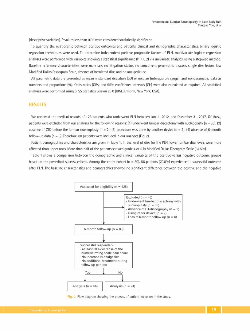
follow-up data (n = 6). Therefore, 80 patients were included in our analyses (Fig. 2).
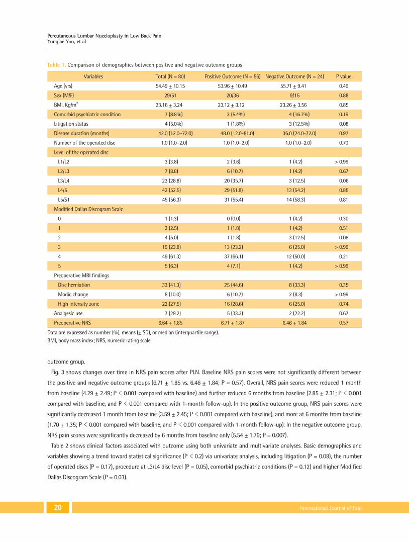
Patient demographics and characteristics are given in Table 1. In the level of disc for the PLN, lower lumbar disc levels were more
affected than upper ones. More than half of the patients showed grade 4 or 5 in Modified Dallas Discogram Scale (67.5%).
Table 1 shows a comparison between the demographic and clinical variables of the positive versus negative outcome groups
based on the prescribed success criteria. Among the entire cohort (n = 80), 56 patients (70.0%) experienced a successful outcome
after PLN. The baseline characteristics and demographics showed no significant difference between the positive and the negative
Assessed for eligibility (n = 126)
6-month follow-up (n = 80)
Successful responder?
- At least 50% decrease of the
numeric rating scale pain score
- No increase in analgesics
- No additional treatment during
follow-up periods
Analysis (n = 56) Analysis (n = 24)
Excluded (n = 46)
- Underwent lumbar discectomy with
nucleoplasty (n = 36)
- Absence of CT-discography (n = 2)
- Using other device (n = 2)
- Loss of 6-month follow-up (n = 6)
Yes No
Fig. 2. Flow diagram showing the process of patient inclusion in the study.
Percutaneous Lumbar Nuceloplasty in Low Back PainYongjae Yoo, et al
International Journal of Pain20 International Journal of Pain
outcome group.
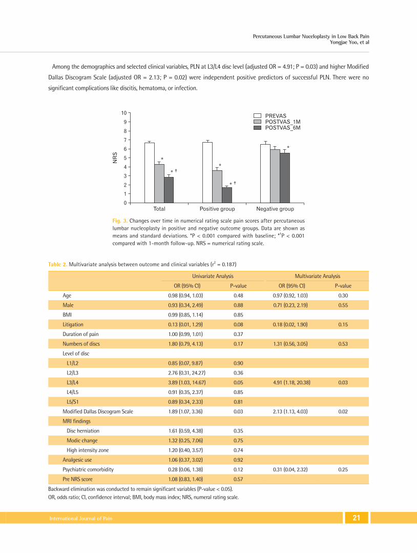
Fig. 3 shows changes over time in NRS pain scores after PLN. Baseline NRS pain scores were not significantly different between
the positive and negative outcome groups (6.71 ± 1.85 vs. 6.46 ± 1.84; P = 0.57). Overall, NRS pain scores were reduced 1 month
from baseline (4.29 ± 2.49; P < 0.001 compared with baseline) and further reduced 6 months from baseline (2.85 ± 2.31; P < 0.001
compared with baseline, and P < 0.001 compared with 1-month follow-up). In the positive outcome group, NRS pain scores were
significantly decreased 1 month from baseline (3.59 ± 2.45; P < 0.001 compared with baseline), and more at 6 months from baseline
(1.70 ± 1.35; P < 0.001 compared with baseline, and P < 0.001 compared with 1-month follow-up). In the negative outcome group,
NRS pain scores were significantly decreased by 6 months from baseline only (5.54 ± 1.79; P = 0.007).
Table 2 shows clinical factors associated with outcome using both univariate and multivariate analyses. Basic demographics and
variables showing a trend toward statistical significance (P < 0.2) via univariate analysis, including litigation (P = 0.08), the number
of operated discs (P = 0.17), procedure at L3/L4 disc level (P = 0.05), comorbid psychiatric conditions (P = 0.12) and higher Modified
Dallas Discogram Scale (P = 0.03).
Table 1. Comparison of demographics between positive and negative outcome groups
Variables Total (N = 80) Positive Outcome (N = 56) Negative Outcome (N = 24) P value
Age (yrs) 54.49 ± 10.15 53.96 ± 10.49 55.71 ± 9.41 0.49
Sex (M/F) 29/51 20/36 9/15 0.88
BMI, Kg/m2 23.16 ± 3.24 23.12 ± 3.12 23.26 ± 3.56 0.85
Comorbid psychiatric condition 7 (8.8%) 3 (5.4%) 4 (16.7%) 0.19
Litigation status 4 (5.0%) 1 (1.8%) 3 (12.5%) 0.08
Disease duration (months) 42.0 (12.0–72.0) 48.0 (12.0-81.0) 36.0 (24.0-72.0) 0.97
Number of the operated disc 1.0 (1.0–2.0) 1.0 (1.0–2.0) 1.0 (1.0–2.0) 0.70
Level of the operated disc
L1/L2 3 (3.8) 2 (3.6) 1 (4.2) > 0.99
L2/L3 7 (8.8) 6 (10.7) 1 (4.2) 0.67
L3/L4 23 (28.8) 20 (35.7) 3 (12.5) 0.06
L4/5 42 (52.5) 29 (51.8) 13 (54.2) 0.85
L5/S1 45 (56.3) 31 (55.4) 14 (58.3) 0.81
Modified Dallas Discogram Scale
0 1 (1.3) 0 (0.0) 1 (4.2) 0.30
1 2 (2.5) 1 (1.8) 1 (4.2) 0.51
2 4 (5.0) 1 (1.8) 3 (12.5) 0.08
3 19 (23.8) 13 (23.2) 6 (25.0) > 0.99
4 49 (61.3) 37 (66.1) 12 (50.0) 0.21
5 5 (6.3) 4 (7.1) 1 (4.2) > 0.99
Preoperative MRI findings
Disc herniation 33 (41.3) 25 (44.6) 8 (33.3) 0.35
Modic change 8 (10.0) 6 (10.7) 2 (8.3) > 0.99
High intensity zone 22 (27.5) 16 (28.6) 6 (25.0) 0.74
Analgesic use 7 (29.2) 5 (33.3) 2 (22.2) 0.67
Preoperative NRS 6.64 ± 1.85 6.71 ± 1.87 6.46 ± 1.84 0.57
Data are expressed as number (%), means (± SD), or median (interquartile range).BMI, body mass index; NRS, numeric rating scale.
Percutaneous Lumbar Nuceloplasty in Low Back PainYongjae Yoo, et al
International Journal of Pain 21 International Journal of Pain
Among the demographics and selected clinical variables, PLN at L3/L4 disc level (adjusted OR = 4.91; P = 0.03) and higher Modified
Dallas Discogram Scale (adjusted OR = 2.13; P = 0.02) were independent positive predictors of successful PLN. There were no
significant complications like discitis, hematoma, or infection.
Table 2. Multivariate analysis between outcome and clinical variables (r2 = 0.187)
Univariate Analysis Multivariate Analysis
OR (95% CI) P-value OR (95% CI) P-value
Age 0.98 (0.94, 1.03) 0.48 0.97 (0.92, 1.03) 0.30
Male 0.93 (0.34, 2.49) 0.88 0.71 (0.23, 2.19) 0.55
BMI 0.99 (0.85, 1.14) 0.85
Litigation 0.13 (0.01, 1.29) 0.08 0.18 (0.02, 1.90) 0.15
Duration of pain 1.00 (0.99, 1.01) 0.37
Numbers of discs 1.80 (0.79, 4.13) 0.17 1.31 (0.56, 3.05) 0.53
Level of disc
L1/L2 0.85 (0.07, 9.87) 0.90
L2/L3 2.76 (0.31, 24.27) 0.36
L3/L4 3.89 (1.03, 14.67) 0.05 4.91 (1.18, 20.38) 0.03
L4/L5 0.91 (0.35, 2.37) 0.85
L5/S1 0.89 (0.34, 2.33) 0.81
Modified Dallas Discogram Scale 1.89 (1.07, 3.36) 0.03 2.13 (1.13, 4.03) 0.02
MRI findings
Disc herniation 1.61 (0.59, 4.38) 0.35
Modic change 1.32 (0.25, 7.06) 0.75
High intensity zone 1.20 (0.40, 3.57) 0.74
Analgesic use 1.06 (0.37, 3.02) 0.92
Psychiatric comorbidity 0.28 (0.06, 1.38) 0.12 0.31 (0.04, 2.32) 0.25
Pre NRS score 1.08 (0.83, 1.40) 0.57
Backward elimination was conducted to remain significant variables (P-value < 0.05).OR, odds ratio; CI, confidence interval; BMI, body mass index; NRS, numeral rating scale.
Total
10
9
8
7
6
5
4
3
2
1
NR
S
0
Positive group Negative group
PREVAS
POSTVAS_1M
POSTVAS_6M
*
*
*
*
*
Fig. 3. Changes over time in numerical rating scale pain scores after percutaneous lumbar nucleoplasty in positive and negative outcome groups. Data are shown as means and standard deviations. *P < 0.001 compared with baseline; *†P < 0.001 compared with 1-month follow-up. NRS = numerical rating scale.

Percutaneous Lumbar Nuceloplasty in Low Back PainYongjae Yoo, et al
International Journal of Pain22 International Journal of Pain
DISCUSSION
In this study, we found that the overall NRS pain scores of patients who received PLN were decreased significantly at 1- and
6-month follow-up. The pain scores were decreased continuously during 6 month follow-up period. In the positive outcome group,
the Modified Dallas Discogram Scale was higher. Moreover, the L3/L4 level YES DISCⓇ procedure was more effective than other
lesions. Multivariate analyses show that a procedure at L3/L4 level disc, a high Modified Dallas Discogram Scale at CTD were the
clinical factors related with the successful outcome of PLN.
Previous studies have reported variable outcomes of lumbar nucleoplasty, like success rate of 82% in patients with no prior surgical
intervention [27] and the patient satisfaction rate from 73% in the early postoperative period to 61% at 6 months postoperatively
[28]. Using the predefined successful outcome criteria, the success rate of YES DISCⓇ procedure in our study was 70% in patients
with IDD, which is similar to previous studies. In addition, our result is focused on the treatment of IDD, which confirmed by
provocation discography and CTD. Percutaneous nucleoplasty using coblation technique was approved for the treatment of
LBP in July 2000 [29]. This is accomplished by applying radiofrequency (RF) energy to a conductive medium, thereby inducing a
plasma field to form around the charged electrodes [19]. The ionized particles that are created possess sufficient energy to break
molecular bonds within tissue (i.e., nucleus pulposus), thus forming a coblation technique involves the use of radiofrequency energy
which created a 1-nm thick region of precisely focused plasma at the tip of the wand [29]. But the decompression mechanism by
nucleoplasty could not explain that the patients with a full-thickness tear of the annulus fibrosus experienced improvement of
symptoms after nucleoplasty [30]. It was introduced that the mechanism of percutaneous nucleoplasty involves the reduction of
the release of inflammatory mediators [31]. Moreover, in some animal study [32], the coblation technique reduced phospholipase A2
activity in degenerated intervertebral discs, suggesting that the effects of coblation energy on inflammatory factors may be one of
the mechanisms of the treatment [33].
In Our study, we did the lumbar discography for all patients as a diagnostic test for IDD. The lumbar provocation discography
performed according to IASP criteria might be a useful tool for evaluating chronic lumbar discogenic pain [34]. And we evaluated
CTD and checked the Modified Dallas Discogram Scale by experienced radiologist. The Dallas Discogram Classification system was
first described in 1980 and modified by Aprill and Bogduk et al. [3] in 1992, and then finally modified in 1996. Vanharanta et al.
reported an association between the tear expansion in the annulus fibrosus and the degree of pain elicited during discography [35].
With this provocation discography and CTD protocol, we found the painful disc and exact annular tear or fissure area of the disc.
The results obtained in our study indicate the coblation treatment at target lesion is related with the successful outcome.
Mechanical causes of LBP include degenerative process, herniated disc, spinal stenosis, spondylosis, or discogenic pain [36]. It is
presumed that patients with more lumbar spinal diseases show less effectiveness with percutaneous lumbar nucleoplasty because
multiple factors are related to their pain.
Interestingly, PLN at L3/4 level was more effective than other levels in our study. The possible reason for this difference is that the
patient with L3/4 lesion showed only one level disc lesion. Another possibility may be L3/4 disc was less related to other comorbid
spinal conditions like spinal stenosis or disc herniation. On the other hand, the possibility that there was extraordinary characteristic
in our patient's cohort cannot be excluded.
Previous study with IDET showed that clinical improvements can be realized after IDET in select surgical candidates with mild
disc degeneration and high intensity zone (HIZ) in MRI, discography, and low-grade Dallas Discogram Scale, with more effective
treatment results [37]. On the other hand, PLN was more successful in the presence of high-grade Modified Dallas Discogram
Scale in our study and shows no significant relationship with HIZ in MRI. This suggests that contra-lateral approach technique and
navigable catheter ablation during YES DISCⓇ procedure can reduce pain by treating severe annular tear than conventional IDET
regardless of MRI findings.
Percutaneous Lumbar Nuceloplasty in Low Back PainYongjae Yoo, et al
International Journal of Pain 23 International Journal of Pain
This study has inherent limitations. First of all, the retrospective nature is an obvious weakness. We didn’t control the additional
procedure after PLN and medications. We also may be criticized for not checking other parameters such as Oswestry Disability
Index (ODI) or patient satisfaction score. Nonetheless, as we included only patients with IDD confirmed by IASP guideline and
CTD, our results could suggest the clinical factors associated with the positive outcome of PLN using YES DISCⓇ in IDD patients. If
conservative management was not helpful in IDD patients, especially with high degree annular teat, physicians should consider PLN
as the following treatment method to avoid invasive surgery. This is the first study that statistically evaluated predictors of successful
percutaneous lumbar nucleoplasty in patients with IDD. Further prospective studies will be required to ascertain our results.
In this study, PLN was found to be safe and effective for the management of discogenic LBP from IDD. 70% of the included
patients showed more than 50% pain reduction, without any complications during the 6-month follow-up period. The high-grade
Modified Dallas Discogram Scale and L3/L4 level treatment were positive predictors for successful PLN.
ACKNOWLEDGEMENTS
Declaration of Conflicting Interests: The authors declare no potential conflicts of interests with respect to the research,
authorship, and/or publication of this article.
Funding: This research did not receive any specific grant from funding agencies in the public, commercial, or not-for-profit
sectors.
REFERENCES
1. Schliessbach J, Siegenthaler A, Heini P, Bogduk N, Curatolo M: Blockade of the sinuvertebral nerve for the diagnosis of lumbar
diskogenic pain: an exploratory study. Anesth Analg 2010;111:204-6.
2. Schwarzer AC, Aprill CN, Derby R, Fortin J, Kine G, Bogduk N: The prevalence and clinical features of internal disc disruption in
patients with chronic low back pain. Spine 1995;20:1878-83.
3. Aprill C, Bogduk N: High-intensity zone: a diagnostic sign of painful lumbar disc on magnetic resonance imaging. Br J Radiol
1992;65:361-9.
4. Lim CH, Jee WH, Son BC, Kim DH, Ha KY, Park CK: Discogenic lumbar pain: association with MR imaging and CT discography. Eur
J Radiol 2005;54:431-7.
5. Wolfer LR, Derby R, Lee JE, Lee SH: Systematic review of lumbar provocation discography in asymptomatic subjects with a meta-
analysis of false-positive rates. Pain Physician 2008;11:513-38.
6. Raj PP: Intervertebral disc: anatomy-physiology-pathophysiology-treatment. Pain Pract 2008;8:18-44.
7. Carragee EJ, Tanner CM, Khurana S, Hayward C, Welsh J, Date E, et al.: The rates of false-positive lumbar discography in select
patients without low back symptoms. Spine 2000;25:1373-80.
8. Enthoven WTM, Roelofs PD, Koes BW: NSAIDs for Chronic Low Back Pain. Jama 2017;317:2327-8.
9. van Middelkoop M, Rubinstein SM, Kuijpers T, Verhagen AP, Ostelo R, Koes BW, et al.: A systematic review on the effectiveness
of physical and rehabilitation interventions for chronic non-specific low back pain. Eur Spine J 2011;20:19-39.
10. Manchikanti L, Staats PS, Nampiaparampil DE, Hirsch JA: What is the Role of Epidural Injections in the Treatment of Lumbar
Discogenic Pain: A Systematic Review of Comparative Analysis with Fusion. Korean J Pain 2015;28:75-87.
11. Hsia AW, Isaac K, Katz JS: Cauda equina syndrome from intradiscal electrothermal therapy. Neurology 2000;55:320.
12. Helm Ii S, Simopoulos TT, Stojanovic M, Abdi S, El Terany MA: Effectiveness of Thermal Annular Procedures in Treating Discogenic
Low Back Pain. Pain Physician 2017;20:447-70.
Percutaneous Lumbar Nuceloplasty in Low Back PainYongjae Yoo, et al
International Journal of Pain24 International Journal of Pain
13. Helm S, Hayek SM, Benyamin RM, Manchikanti L: Systematic review of the effectiveness of thermal annular procedures in
treating discogenic low back pain. Pain Physician 2009;12:207-32.
14. Wetzel FT: Cauda equina syndrome from intradiscal electrothermal therapy. Neurology 2001;56:1607.
15. Cohen SP, Larkin T, Polly DW Jr: A giant herniated disc following intradiscal electrothermal therapy. J Spinal Disord Tech
2002;15:537-41.
16. Kim S, Lee SH, Kim ES, Eoh W: Thermal-Induced Osteonecrosis of Adjacent Vertebra after Intradiscal Electrothermal Therapy. J
Korean Neurosurg Soc 2017;60:114-7.
17. Eichen PM, Achilles N, Konig V, Mosges R, Hellmich M, Himpe B, et al.: Nucleoplasty, a minimally invasive procedure for disc
decompression: a systematic review and meta-analysis of published clinical studies. Pain Physician 2014;17:E149-73.
18. Singh V, Piryani C, Liao K, Nieschulz S: Percutaneous disc decompression using coblation (nucleoplasty) in the treatment of
chronic discogenic pain. Pain Physician 2002;5:250-9.
19. Cohen SP, Williams S, Kurihara C, Griffith S, Larkin TM: Nucleoplasty with or without intradiscal electrothermal therapy (IDET) as
a treatment for lumbar herniated disc. J Spinal Disord Tech 2005;18 Suppl:S119-24.
20. Cuellar VG, Cuellar JM, Vaccaro AR, Carragee EJ, Scuderi GJ: Accelerated degeneration after failed cervical and lumbar
nucleoplasty. J Spinal Disord Tech 2010;23(8):521-4,O'Neill CW, Liu JJ, Leibenberg E, Hu SS, Deviren V, Tay BK, et al.: Percutaneous
plasma decompression alters cytokine expression in injured porcine intervertebral discs. Spine J 2004;4:88-98.
21. Kim MK, Sim SE, Kim YC, Kim JS, Kwon SM, Yoo Y, et al.: Predictive Factors of Successful Percutaneous Cervical Nucleoplasty for
the Treatment of Pain with Cervical Herniated Disk. World Neurosurg 2018;114:e654-e62.
22. Manchikanti L, Falco FJ, Benyamin RM, Caraway DL, Deer TR, Singh V, et al.: An update of the systematic assessment of
mechanical lumbar disc decompression with nucleoplasty. Pain Physician 2013;16(2 Suppl):Se25-54.
23. Kumar NS, Shah SM, Tan BW, Juned S, Yao K: Discogenic axial back pain: is there a role for nucleoplasty? Asian Spine J
2013;7:314-21.
24. Liliang PC, Lu K, Liang CL, Chen YW, Tsai YD, Tu YK: Nucleoplasty for treating lumbar disk degenerative low back pain: an
outcome prediction analysis. J Pain Res 2016;9:893-8.
25. von Elm E, Altman DG, Egger M, Pocock SJ, Gotzsche PC, Vandenbroucke JP: The Strengthening the Reporting of Observational
Studies in Epidemiology (STROBE) statement: guidelines for reporting observational studies. Lancet 2007;370:1453-7.
26. Breivik H, Borchgrevink PC, Allen SM, Rosseland LA, Romundstad L, Hals EK, et al.: Assessment of pain. Br J Anaesth 2008;101:17-
24.
27. Sharps LS, Isaac Z: Percutaneous disc decompression using nucleoplasty. Pain Physician 2002;5:121-6.
28. Al-Zain F, Lemcke J, Killeen T, Meier U, Eisenschenk A: Minimally invasive spinal surgery using nucleoplasty: a 1-year follow-up
study. Acta Neurochir 2008;150:1257-62.
29. Masala S, Massari F, Fabiano S, Ursone A, Fiori R, Pastore F, et al.: Nucleoplasty in the treatment of lumbar diskogenic back pain:
one year follow-up. Cardiovasc Intervent Radiol 2007;30:426-32.
30. Ren DJ, Liu XM, Du SY, Sun TS, Zhang ZC, Li F: Percutaneous Nucleoplasty Using Coblation Technique for the Treatment of
Chronic Nonspecific Low Back Pain: 5-year Follow-up Results. Chin Med J 2015;128:1893-7.
31. Yakovlev A, Tamimi MA, Liang H, Eristavi M: Outcomes of percutaneous disc decompression utilizing nucleoplasty for the
treatment of chronic discogenic pain. Pain Physician 2007;10:319-28.
32. Kuelling FA, Foley KT, Liu JJ, Liebenberg E, Sin AH, Matsukawa A, et al.: The anabolic effect of plasma-mediated ablation on the
intervertebral disc: stimulation of proteoglycan and interleukin-8 production. Spine J 2014;14:2479-87.
33. Ren D, Zhang Z, Sun T, Li F: Effect of percutaneous nucleoplasty with coblation on phospholipase A2 activity in the intervertebral
disks of an animal model of intervertebral disk degeneration: a randomized controlled trial. J Orthop Surg Res 2015;10:38.
Percutaneous Lumbar Nuceloplasty in Low Back PainYongjae Yoo, et al
International Journal of Pain 25 International Journal of Pain
34. Manchikanti L, Hirsch JA: An update on the management of chronic lumbar discogenic pain. Pain Manag 2015;5:373-86.
35. Vanharanta H, Sachs BL, Ohnmeiss DD, Aprill C, Spivey M, Guyer RD, et al.: Pain provocation and disc deterioration by age. A CT/
discography study in a low-back pain population. Spine 1989;14:420-3.
36. Deyo RA, Weinstein JN: Low back pain. N Engl J Med 2001;344:363-70.
37. Kircelli A, Coven I, Cansever T, Sonmez E, Yilmaz C: Patient Selection and Efficacy of Intradiscal Electrothermal Therapy with
Respect to the Dallas Discogram Score. Turk Neurosurg 2017;27:623-30.