V, D and J Gene Rearrangements on the Unexpressed Igh ...
247
Loyola University Chicago Loyola University Chicago Loyola eCommons Loyola eCommons Dissertations Theses and Dissertations 1996 V, D and J Gene Rearrangements on the Unexpressed Igh Allele in V, D and J Gene Rearrangements on the Unexpressed Igh Allele in Rabbit B Cells Rabbit B Cells Chainarong Tunyaplin Loyola University Chicago Follow this and additional works at: https://ecommons.luc.edu/luc_diss Part of the Microbiology Commons Recommended Citation Recommended Citation Tunyaplin, Chainarong, "V, D and J Gene Rearrangements on the Unexpressed Igh Allele in Rabbit B Cells" (1996). Dissertations. 3400. https://ecommons.luc.edu/luc_diss/3400 This Dissertation is brought to you for free and open access by the Theses and Dissertations at Loyola eCommons. It has been accepted for inclusion in Dissertations by an authorized administrator of Loyola eCommons. For more information, please contact [email protected]. This work is licensed under a Creative Commons Attribution-Noncommercial-No Derivative Works 3.0 License. Copyright © 1996 Chainarong Tunyaplin
-
Upload
khangminh22 -
Category
Documents
-
view
0 -
download
0
Transcript of V, D and J Gene Rearrangements on the Unexpressed Igh ...

Loyola University Chicago Loyola University Chicago
Loyola eCommons Loyola eCommons
Dissertations Theses and Dissertations
1996
V, D and J Gene Rearrangements on the Unexpressed Igh Allele in V, D and J Gene Rearrangements on the Unexpressed Igh Allele in
Rabbit B Cells Rabbit B Cells
Chainarong Tunyaplin Loyola University Chicago
Follow this and additional works at: https://ecommons.luc.edu/luc_diss
Part of the Microbiology Commons
Recommended Citation Recommended Citation Tunyaplin, Chainarong, "V, D and J Gene Rearrangements on the Unexpressed Igh Allele in Rabbit B Cells" (1996). Dissertations. 3400. https://ecommons.luc.edu/luc_diss/3400
This Dissertation is brought to you for free and open access by the Theses and Dissertations at Loyola eCommons. It has been accepted for inclusion in Dissertations by an authorized administrator of Loyola eCommons. For more information, please contact [email protected].
This work is licensed under a Creative Commons Attribution-Noncommercial-No Derivative Works 3.0 License. Copyright © 1996 Chainarong Tunyaplin
LOYOLA UNIVERSITY CHICAGO
V, D AND J GENE REARRANGEMENTS ON THE
UNEXPRESSED IGH ALLELE IN RABBIT B CELLS
A DISSERTATION SUBMITTED TO
THE FACULTY OF THE GRADUATE SCHOOL
IN PARTIAL FULFULLMENT OF THE REQUIREMENT OF THE DEGREE OF
DOCTOR OF PHILOSOPHY
DEPARTMENT OF MICROBIOLOGY AND IMMUNOLOGY
BY
CHAINARONG TUNY APLIN
CHICAGO, ILLINIOS
JANUARY, 1996
LOYOLA UNIVERSITY MEDICAL CENTER LIBRARY
Copyright by Chainarong Tunyaplin, 1995
All rights reserved.
ii
ACKNOWLEDGEMENTS
I am greatly indebted to Dr. Katherine L. Knight, my mentor, for the best training
one can ever have in their graduate education and for all the supports in the past several
years. Despite her extremely busy schedule, she is always available to students when they
need her. Her dedication to science and educating young scientists is exemplary.
I also like to thank my committee, Dr. C. F. Lange, Dr. P. M. Witte, Dr. P. T. Le,
Dr. S. C. Baker and Dr. H. M. Jack, for all the guidance and encouragement, and for
enduring with the everchanging focus of my project over the years. I thank my best friend,
Martha L. Friedman, who helped me tremendously when I first came to the United States
and continues to do so after all these years. I also thank Helga Spieker-Polet and Pi-Chen
Yam for their rabbit hybridomas and Shi-Kang Zhai for all the sequencing reactions.
Lastly, I would like to acknowledge all the members of Dr. Knight's laboratory and the
Department of Microbiology and Immunology, who together make this department such
an ideal workplace.
iii
TABLE OF CONTENTS
ACKNOWLEDGEMENTS...................................................................................... iii
LIST OF TABLES ... ...... ...... ...... ...... ...... ............ ............................... ... ..... ... ... ..... ..... VI
LIST OF FIGURES................................................................................................... vii
LIST OF ABBREVIATIONS................................................................................... xi
ABSTRACT.............................................................................................................. xiii
INTRODUCTION..................................................................................................... 1
Chapter
I. LITERATURE REVIEWS....................................................................... 3
Basic Structure of Ab Molecules ...................................... ........ ........ 4
Discovery of Somatic Gene Rearrangement .. .. .. .. .. .. .. .. .. .. .. .. .. .. .. .. .. .. . 5
V H• D and JH Genes Organization..................................................... 9
IgH Gene Rearrangement.................................................................. 24
Allelic Exclusion............................................................................... 36
V H Gene Utilization in B-lineage Cells............................................. 49
II. MATERIALS AND METHODS............................................................. 68
Animals............................................................................................. 68
Chemicals.......................................................................................... 68
Purification of Rabbit B Cells........................................................... 68
Hybridoma Cell Culture.................................................................... 70
Collection of Rabbit Fetuses . . . . . . . . . . . . . . . . . . . . . . . . . . . . . . . . . . . . . . . . . . . . . . . . . . . . . . . . . . . . . 71
Genomic DNA Purification ............................................................ ,.. 72
iv
Genomic Blot Analysis . ............. ........................ .......... ... ........ ..... ... ... 73
PCR Amplifications and Cloning of VDJ, DJ and VD Genes.......... 75
Nucleotide Sequence Analysis of V H Promoters .............................. 82
ID. RESULTS................................................................................................. 84
Quantification of Germline JH and VHJ in Polyclonal Rabbit B Cells............................................................................ 84
Quantification of Germline JH in Polyclonal Rabbit B Cells .. .. .. .. .. .. ..................................................... 84
Quantification of Germline V HJ in Polyclonal Rabbit B Cells .. .. .. .. .. .. .... .. .. .. .. .. .. .. .. .. .. .. .. .. .. .. .. .. .. .. .. .... .. .. . 91
Rearrangements of VHJ and JH in the B Cell Hybridomas................ 105
IgH Gene Rearrangement in Rabbit T Cell Lines .. .. .. .. .. .. .. .. .. .. .. .. .. .. . 140
Intermediates of lgH Gene Rearrangement in Polyclonal B Cells.... 145
Fetal VDJ Genes............................................................................... 155
VH Gene Usage in Fetal VDJ Gene Repertoire................................. 156
V H Promoter Regions........................................................................ 169
IV. DISCUSSION........................................................................................... 176
REFERENCES ......................................................................................................... 200
VITA......................................................................................................................... 229
v
LIST OFT ABLES
Table Page
I. Probes used in this study and their origins.................................................... 7 4
2. PCR primers used in this study ..................................................................... 76
3. The hybridization patterns of probe A and pJ5 probe for each possible status of IgH gene on the unexpressed allele . . . . . . . . . . . . . . . . . . . . . . . . . . . . . . . . . . . . . . . . . . 106
4. Summary of Southern analysis of B cell hybridomas derived from rabbit 363Hl #2 ....................................................................................... 113
5. Summary of Southern analysis of B cell hybridomas derived from rabbit 27Hl ............................................................................................. 116
6. Summary of Southern analysis of B cell hybridomas derived from rabbit 196G2 . . . . . . . . . . . . . . . . . . . . . . . . . . . . . . . . . . . . . . . . . . . . . . . . . . . . . . . . . . . . . . . . . . . . . . . . . . . . . . . . . . . . . . . . . . . 120
7. Summary of Southern analysis of B cell hybridomas derived from rabbit 6412 ............................................................................................... 123
8. Distribution of DJ, VDJ genes and germline VH, D and JH gene segments on the unexpressed allele of 29 rabbit hybridomas ................................. 133
9. Frequency of V H gene usage in fetal VDJ gene repertoire............................ 170
vi
Figure
1.
2.
3.
4.
5.
6.
7.
8.
9.
10.
LIST OF FIGURES
Page
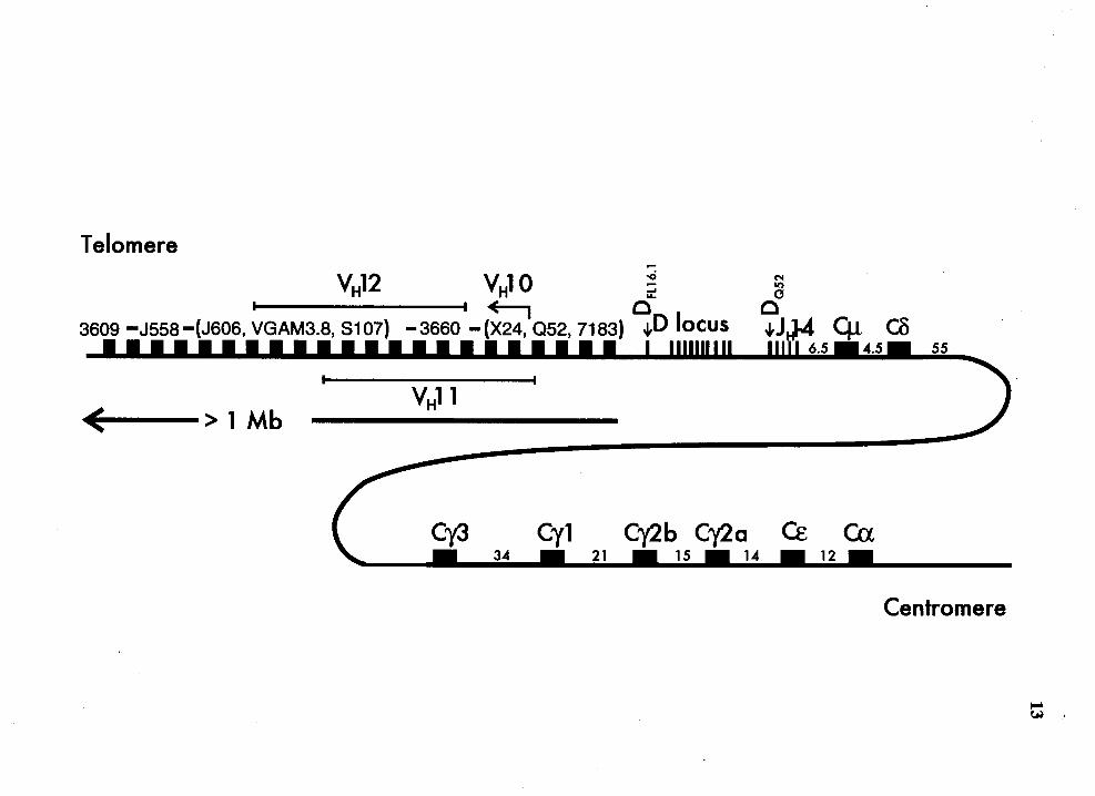
Maps of the mouse IgH locus........................................................................ 12
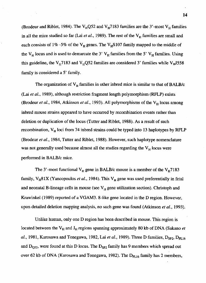
Map of human IgH locus on chromosome 14 ....... .. .......... .............. .. ............ 16
Maps of the rabbit IgH locus......................................................................... 22
Diagram of IgH gene rearrangement products ........... ..... .. . .. . .......... ........ ...... 26
Schematic representation of the location of PCR primers used in the nested-primer PCR .................................................................................. 79
A partial map of the V, D and 1 chromosomal region from the al (C haplotype), a2 (E haplotype) and a3 (G haplotype) allotype rabbits....... 86
Quantitative Southern analysis of EcoRI/Hindill-restricted DNA for germline 1H in al+ B cells of rabbit number 35G ................................... 88
Quantitative Southern analysis of EcoRI/HindIII-restricted DNA for germline 1H in al+ B cells of rabbit number 13806 ............................... 92
Quantitative Southern analysis of BamHI-restricted DNA for probe A region in al+ B cells from rabbit number 14715 .................................... 96
Quantitative Southern analysis of BamHI-restricted DNA for probe A region in al+ B cells from rabbit number 14716 .................................... 99
vii

Figure Page
11. Quantitative Southern analysis of Hindill-restricted DNA for probe A region in slg+ B cells from rabbit number 64Hl .................................... 102
12. Genomic analysis for the status of IgH genes in the fusion partner using probe A and pJ5 probe ............................................................................ 108
13. Genomic analysis for the status of IgH genes in the hybridomas generated from rabbit# 363Hl using pJ5 probe ..................................................... 111
14. Genomic analysis for the status of IgH genes in the hybridomas generated from rabbit# 27Hl using pJ5 probe ....................................................... 114
15. Genomic analysis for the status of IgH genes in the hybridomas generated from rabbit # 196G2 using pJ 5 probe ..................................................... 117
16. Genomic analysis for the status of IgH genes in the hybridomas generated from rabbit# 64J2 using pJ5 probe ......................................................... 121
17. Genomic analysis for the status of IgH genes in the hybridomas generated from rabbit# 363Hl using probe A ........................................................ 124
18. Genomic analysis for the status of IgH genes in the hybridomas generated from rabbit # 27H 1 using probe A .......................................................... 126
19. Genomic analysis for the status of IgH genes in the hybridomas generated from rabbit# 196G2 using probe A ........................................................ 128
20. Genomic analysis for the status of IgH genes in the hybridomas generated from rabbit# 64J2 using probe A. .......................................................... 131 ·
21. Nucleotide sequence of the region immediately upstream of JH locus ......... 135
viii
Figure Page
22. PAGE analysis of the PCR products from PCR amplification for DJ genes from the indicated hybridomas ................................................................ 138
23. Comparison of the nucleotide sequences of four PCR-amplified DJ genes with those of germline D and JH genes . .. . . . . .. .. . . . . . . . . . . . . . . . . . . . . . . . . . . . . . . . . . . . . . . . 141
24. Genomic analysis for the status of IgH genes in the 14 rabbit T cell lines using D 1 probe . . .. . . . . . . . . . . . . . . . . . . . . . . . . . . . . . . . . . . . . . . . . . . . . . . . . . . . . . . . . . . . . . . . . . . . . . . . . . . . . . . . . . . . . 143
25. Genomic analysis for the status of IgH genes in the 14 rabbit T cell lines using pJ 5 probe . . . . . . . . . . . . . . . . .. . . . . . . . . . . . . . . . . . . . . . . . . . . . . . . . . . . . . . . . . . . . . . . . . . . . . . . . . . . . . . . . . . . . . . 146
26. PAGE analysis of the PCR products from PCR amplification for D2bJ and VD2b genes from rabbit splenic DNA .................................................... 149
27. Comparison of the nucleotide sequences of the DJ genes isolated from splenic DNA with those of germline D2b and JH genes ........................ 151
28. Comparison of the nucleotide sequences of the VD2b genes isolated from splenic DNA with those of germline VHJ-a3 and D2b genes ................. 153
29. PAGE analysis of nested-primer PCR for fetal VDJ gene ............................ 157
30. Comparison of nucleotide sequence of VDJ genes (FR3 through JH) from day 14 fetuses with germline V HJ -a3 or V H2-a3. .. . . . .. . . . . . . . . . . . . . . . . . . . . . . . . . . . . . 159
31. Comparison of nucleotide sequence of VDJ genes (FR3 through JH) from day 15 fetuses with germline V HJ -a2 . . . . . . . . . . . . . . . . . . . . . . . . . . . . . . . . . . . . . . . . . . . . . . . . . . . . . . 161 .
32. Comparison of nucleotide sequence of VDJ genes (FR3 through JH) from day 21 fetuses with germline V HJ -a3 or V H2-a3 ..................................... 163
ix
Figure Page
33. Comparison of nucleotide sequence of VDJ genes (FR3 through JH) from day 24 fetuses with germ.line V HJ -al or V HJ -a3 . . . . . . . . . . . . . . . . . . . . . . . . . . . . . . . . . . . . . 165
34. Comparison of nucleotide sequence of VDJ genes (FR3 through JH) from day 28 fetuses with germ.line VH1-a3 or VH2-a3 ..................................... 167
35. Comparison of the nucleotide sequences of the promoter region of five germ.line V H genes ................................................................................... 172
36. Schematic representation of the "narrow window for rearrangement" model ....................................................................................................... 181
37. Schematic representation of the "inefficient double-strand breaks repair" model ....................................................................................................... 184
38. Schematic representation of the "suppression and desuppression" model ... 187
39. Schematic representation of the "inactivation of one IgH allele" model ...... 191
40. Comparison of the RSSs of all several functional germ.line V H genes ......... 195
x

Ab
Ag
AMuLV
bp
CD40L
D
EBV
g
HAT
HBSS
hr
lg
lgH
LIST OF ABBREVIATIONS
antibody
antigen
Abelson Murine Leukemia Virus
base pair
CD40ligand
D gene of heavy chain gene
Ebstein-Barr Virus
gram
Hypoxanthine-Aminopterin-Thymidine
Hank's balance salt solution
hour
Immunoglobulin
Heavy chain of immunoglobulin
xi
JH J gene of heavy chain gene
kb kilo base
LTBM Long term bone marrow culture
µg microgram
mg milligram
µl micro liter
ml milliliter
mAb monoclonal antibody
MLN Mesenteric lymph node
mM millimolar
PCR Polymerase chain reaction
RAG2 Recombination Activating Gene 2
TCR T cell receptor
VH V gene of heavy chain gene
xii
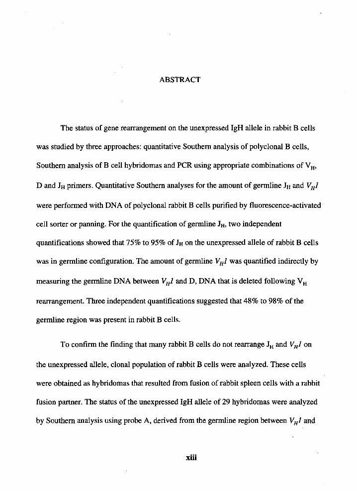
ABSTRACT
The status of gene rearrangement on the unexpressed IgH allele in rabbit B cells
was studied by three approaches: quantitative Southern analysis of polyclonal B cells,
Southern analysis of B cell hybridomas and PCR using appropriate combinations of V H•
D and JH primers. Quantitative Southern analyses for the amount of germline JH and VHJ
were performed with DNA of polyclonal rabbit B cells purified by fluorescence-activated
cell sorter or panning. For the quantification of germline JH, two independent
quantifications showed that 75% to 95% of JH on the unexpressed allele of rabbit B cells
was in germline configuration. The amount of germline V HJ was quantified indirectly by
measuring the germline DNA between VHJ and D, DNA that is deleted following VH
rearrangement. Three independent quantifications suggested that 48% to 98% of the
germline region was present in rabbit B cells.
To confirm the finding that many rabbit B cells do not rearrange JH and V HJ on
the unexpressed allele, clonal population of rabbit B cells were analyzed. These cells
were obtained as hybridomas that resulted from fusion of rabbit spleen cells with a rabbit
fusion partner. The status of the unexpressed IgH allele of 29 hybridomas were analyzed
by Southern analysis using probe A, derived from the germline region between V HJ and
xiii
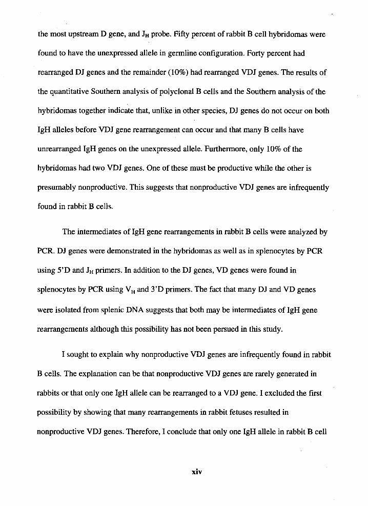
the most upstream D gene, and JH probe. Fifty percent of rabbit B cell hybridomas were
found to have the unexpressed allele in germline configuration. Forty percent had
rearranged DJ genes and the remainder ( 10%) had rearranged VDJ genes. The results of
the quantitative Southern analysis of polyclonal B cells and the Southern analysis of the
hybridomas together indicate that, unlike in other species, DJ genes do not occur on both
IgH alleles before VDJ gene rearrangement can occur and that many B cells have
unrearranged IgH genes on the unexpressed allele. Furthermore, only 10% of the
hybridomas had two VDJ genes. One of these must be productive while the other is
presumably nonproductive. This suggests that nonproductive VDJ genes are infrequently
found in rabbit B cells.
The intermediates of lgH gene rearrangements in rabbit B cells were analyzed by
PCR. DJ genes were demonstrated in the hybridomas as well as in splenocytes by PCR
using 5'D and JH primers. In addition to the DJ genes, VD genes were found in
splenocytes by PCR using VH and 3'D primers. The fact that many DJ and VD genes
were isolated from splenic DNA suggests that both may be intermediates of IgH gene
rearrangements although this possibility has not been persued in this study.
I sought to explain why nonproductive VDJ genes are infrequently found in rabbit
B cells. The explanation can be that nonproductive VDJ genes are rarely generated in
rabbits or that only one lgH allele can be rearranged to a VDJ gene. I excluded the first
possibility by showing that many rearrangements in rabbit fetuses resulted in
nonproductive VDJ genes. Therefore, I conclude that only one lgH allele in rabbit B cell
xiv
progenitors can be rearranged to a VDJ gene.
The analysis of V H gene usage in the nonproductive VDJ genes showed that most
of them had utilized V HJ. This result indicates that the preferential utilization of V HJ
resulted from its preferential rearrangement. I tested for the possibility that the promoter
of V HJ may be responsible for its preferential rearrangement by searching for any
prominent difference between the promoter of V HJ and those of other V H genes. I found
that the core transcriptional factor motifs were similar in all V H promoters examined and
therefore, the activity of the VHJ promoter probably does not explain the preferential
rearrangement of VHJ.
xv
INTRODUCTION
Much of our knowledge of lg structure and genetics was obtained from studies of
rabbit lg. In the 1960s, Todd (1963) and Feinstein (1963) reported the study of the
inheritance and molecular localization of the allotypic specificities in the V H and CH
regions of rabbit lg, the study which led to the revolutionary idea that defied the "one
gene one polypeptide" dogma (Dreyer and Bennett, 1965, Lennox and Cohn, 1967).
Furthermore, allelic exclusion in plasma cells was first discovered in rabbit by Pemis et
al. ( 1965) and Cebra et al. ( 1966). Other studies of rabbit lg which contributed to our
knowledge of lg gene structure include the linkage of genes encoding the V and C regions
(Dubiski, 1969, Mandy and Todd, 1970), the cis-association of V and C region genes
(Landucci-Tosi et al., 1970, Kindt et al., 1970) and the high frequency of germline
recombination which occurs between VH and CH genes (Mage et al., 1971, Kindt and
Mandy, 1972, Mage et al., 1982, Kelus and Steinberg, 1991).
Rabbit immunoglobulin (lg) is unique in that it has allotypic specificities not only
in the constant (C) regions but also in the variable (V) regions of the heavy chains. For
many years the allelic inheritance of the allotypes in the V H region, V Hal, V Ha2 and V Ha3
allotypes, was the most perplexing aspect of rabbit lg. Because 80%-90% of serum lg
molecules in rabbits bear the V Ha allotypes and because the rabbit genome contains more .
than 100 V H genes, most of the germline V H genes must encode the V Ha allotypic
specificities assuming that many of these germline V H genes are used to generate the VDJ
gene repertoire. The question was then how was the rabbit V H chromosomal region
protected from meiotic recombination which would shuffle the V H genes encoding the

different allotypes, such that the allelic inheritance of the allotypes would be lost. The
answer to this puzzle is rather simple. Knight and Becker (Knight and Becker, 1990)
found that 80%-90% of rabbit B cells preferentially utilized only one V H gene, V H 1, the
gene which encodes a prototypic V Ha molecule. Therefore, the recombination event
would not be detected phenotypically even if meiotic recombination occurred within the
Vtt locus of different alleles, which most likely it had. Up until now, the molecular
mechanism for the preferential utilization of V H 1 is unknown.
2
Although rabbit lg seized the early attention of immunochernists, whose
motivation led to many studies on rabbit lg which contributed significantly to the advance
in our knowledge of lg structure and genetics, the molecular study of rabbit IgH gene lags
behind those of mouse and human. In this study, two aspects of IgH gene rearrangement
in rabbit B cells were studied. The first aspect is how rabbit B cell rearranges its lgH
gene. I approached this issue by examining the status of the IgH gene on the unexpressed
allele in polyclonal and monoclonal rabbit B cells. The second aspect is whether the
preferential utilization of V H 1 resulted from preferential rearrangement. This issue was
approached by analyzing the V H gene usage in nonproductive VDJ genes. Here, I report
the findings.

CHAPTER I
LITERATURE REVIEW
The humoral immune system, mediated by the B cell compartment, plays an
important part in immune response by producing antibodies (Abs) against foreign
antigens (Ags). The hallmark of this response is that Abs can be generated against a
practically infinite number of antigenic determinants. This raised a unique genetic
mystery about how B cells generate such a large repertoire using the genome with limited
coding capacity. Thanks to the discovery of restriction enzymes and the advent of
molecular biology techniques, this mystery was solved in mid 1970s representing a major
achievement in immunogenetics and marking the beginning of molecular B cell biology.
Because Ab is the effector of the humoral immunity, understanding how the Ab
repertoire develops is crucial to our understanding of the humoral immune system. The
discovery of somatic gene rearrangement, the process by which B cells generate the Ab
repertoire, has raised many questions, for example, what is the enzymology of the
recombinase, how the recombinase performs the gene rearrangement, what is the pattern
of gene usage in the Ab repertoire and what is the mechanism that contributes to the
tissue specificity of the rearrangement process. In the 20 years since the discovery of the
somatic gene rearrangement, we have been accumulating information regarding the gene
rearrangement process, some of which will be presented here. This literature review will
summarize the basic structure of Ab molecules and the discovery of somatic gene
rearrangement. Then I will discuss the organization of IgH gene organization, the process
3
of lgH gene rearrangement and the V H gene utilization in three species, mouse, human
and rabbit.
Basic Structure of Ab Molecules
4
Each Ab or immunoglobulin (lg) monomer is made of four polypeptide subunits:
two identical heavy chains (H-chains) and two identical light chains (L-chains) (reviewed
in Cohen and Milstein, 1967, Potter and Lieberman, 1967). Much of the data regarding
the basic structure of lg molecules were also obtained from studying the homogeneous lg
secreted by plasmacytomas, the tumors of Ab secreting cells. Some of these proteins were
of H-chain type and some were of L-chain type. From extensive analysis of the amino
acid sequences of these myeloma proteins, it became clear that the amino-terminal
regions of both H- and L-chains were highly variable while the carboxy-terminal regions
were relatively constant. Thus each H-chain and L-chain could be divided into two
regions, variable (V) and constant (C) regions (reviewed in Kabat et al., 1991).
The variable regions, as the name implied, are different among different H-chains
or L-chains. No two identical V regions have ever been found among the myeloma
proteins. The constant regions of H-chains can be typed into five major isotypes, µ, o, y, E
and a. For L-chain, two major C regions were described, K and A.. Some major isotypes
could be further divided into subisotypes. For example, the Cy region in mouse could be
divided into 4 subisotypes, Cyl, Cy2a, Cy2b and C'{3.
At the end of 1960' s, even before the studies of myeloma protein, papain and
pepsin digestion studies of polyclonal Ab have provided the scientists the first glimpse of
how an Ab molecule is organized (Porter, 1958, Nisonoff and Woernley, 1959, Nisonoff
et al., 1960). From these studies, an Ab molecule can be divided into two parts. One part
is responsible for the Ag binding. This part is subsequently found that it consists ·of the
5
variable region and part of constant region (reviewed in Cohen and Milstein, 1967, Capra
and Kehoe, 1975). The other part does not bind Ag but bears the antigenic specificity
found in all lg molecules and consists of only the constant region. (reviewed in Cohen
and Milstein, 1967, Capra and Kehoe, 1975).
Discovery of Somatic Gene Rearrangement
Generation of Ab Diversity via V(D)J Gene Rearrangement
Because myeloma proteins with different V regions could be found in association
with identical C region, the question was raised as how the H- and L-chains were encoded
in the germline. According to the "one gene, one polypeptide" dogma, there was one gene
in the germline for each different H- and L-chains. Because many different H- and L
chains must be generated to form the Ab repertoire, the genome required to encode all the
possible H and L chain would be much larger than the genome of any organism. A
compromising theory between these two facts was proposed in 1965 by Dryer and
Bennett (1965).
Dryer and Bennett proposed that the V and C regions were encoded by two
separate genes and that there were many V genes but only one gene for each of the C
regions. During B cell genesis, one of the "pre-made" V genes in the germline V gene
repertoire was selected and rearranged to the C gene to form an expressible gene. This
proposal was revolutionary and remained hypothetical until a decade later when the first
evidence of such somatic gene rearrangement was reported.
In mid 1970s, the discovery of restriction enzymes permitted the examination of
lg genes directly. Hozumi and Tonegawa (1976) tested whether the 1C gene underwent
gene rearrangement in 1C producing myeloma. They performed solution hybridization of
BamHI-digested genomic DNA from either a K-producing plasmacytoma cell line or

BALB/c embryo using K mRNA as probe. They found that in the embryonic
configuration, the VK and CK genes were located on two different BamHI fragments but
in MOPC321, both VK and CK genes were rearranged such that they were found on the
same BamHI fragment. This experiment described for the first time the evidence to
support the somatic gene rearrangement theory of Dryer and Bennett. What Dryer and
Bennett had not predicted was that the V regions were not pre-made and encoded in the
germline. But rather, they were generated by the gene rearrangement itself.
At the end of the 1970s, gene cloning techniques were available and many
germline V and C genes of H-, K- and A.- loci were cloned (Tonegawa et al., 1977,
Tonegawa et al., 1978, Seidman et al., 1978, Seidman and Leder, 1978, Brack et al.,
1978, Early et al., 1980, Sakano et al., 1980). It was then apparent that part of the V
region as defined by the amino acid sequence analysis of the myeloma proteins was not
encoded in the cloned germline VH and VL genes (Seidman et al., 1978). The repetitive
occurrence of the same missing sequence in different myeloma proteins suggested that
they were encoded by another germline gene separated from the V genes. By gene
cloning, these missing parts were shown to be encoded by germline genes, D (for
diversity) and J (for joining). For L-chain, only J genes encoded for the missing parts
(Seidman et al., 1978, Sakano et al., 1979, Max et al., 1979). In the case of H-chain, the
missing parts were encoded by both D and J genes (Early et al., 1980, Sakano et al.,
6
1980, Sakano et al., 1981). During B cell development then, one of each of the V, (D) and
J genes rearranged to generate a functional expressible V H and V L genes. To generate a
V L gene, only one rearrangement is required, V to J, while the generation of V H gene
requires two separate rearrangements, V to D and D to J.

7
Recombination Signal Sequence
The comparison of nucleotide sequences of several germline V, D and J genes
revealed a conserved DNA motif adjacent to all genes, at every locus known to undergo
rearrangement (Tonegawa et al., 1977, Tonegawa et al., 1978, Seidman et al., 1978,
Seidman and Leder, 1978, Brack et al., 1978, Sakano et al., 1979, Max et al., 1979, Early
et al., 1980, Sakano et al., 1980, Ramsden et al., 1994). This DNA motif was named
Recombination Signal Sequence (RSS) because of its potential involvement in the
rearrangement process. The motif consists of two stretches of conserved sequences
separated by non-conserved sequence of either 12-bp or 23-bp in length (Tonegawa et al.,
1977, Tonegawa et al., 1978, Seidman et al., 1978, Seidman and Leder, 1978, Brack et
al., 1978, Max et al., 1979, Early et al., 1980, Sakano et al., 1980, Ramsden et al., 1994).
The first conserved stretch is a heptamer, whose consensus sequence is a dyad symmetric
CACAGTG and is found immediately adjacent to the coding elements. The second
conserved stretch is a nonamer whose consensus sequence is ACAAAAACC. This
nonamer is less-conserved than the heptamer. The consensus sequence of RSS and the
frequency of occurrence of the consensus sequence at each position in the heptamer and
nonamer are shown below (Ramsden et al., 1994, Hesse et al., 1989).
Heptamer No namer
c A c A G T G A c A A A A A c c
100 99 99 87 82 85 76 72 86 83 73 91 97 87 84 76
Blackwell et al. ( 1984) and Lewis et al. ( 1984) made a major contribution to the
studies of V(D)J gene rearrangement when they showed that pre-B cell lines could
rearrange artificial rearrangement substrates. This finding permitted the analysis of factors
or sequences which influence somatic rearrangement simply by testing the rearrangement
ability of artificial substrates containing these factors in pre-B cell lines. Akira et al.
8
( 1987) and Hesse et al. ( 1987) directly tested the involvement of RSS in the
rearrangement process. They constructed several rearrangement substrates, each
contained different combinations of RSS and found that the rearrangement could occur in
the substrate with only one pair of RSSs. No sequence from the coding region was
required. This result suggested that, indeed, the RSS was important in the rearrangement
process. Upon examination of the arrangement of the RSS in germline V, D and J genes,
Early et al. ( 1980) proposed that the rearrangement could occur only between the gene
with a 12-bp RSS and the gene with a 23-bp RSS. Indeed, Aldra et al. (1987) and Hesse
et al. ( 1987) showed that rearrangement of the artificial substrates would only occur
between the 12-bp RSS and 23-bp RSS.
The nucleotides in the RSS important for mediating the rearrangement have been
studied by determining the ability of artificial rearrangement substrates with mutated RSS
to undergo rearrangement in pre-B cell lines. Two different approaches were taken to
measure the extent of rearrangement of the substrates with essentially the same result.
One approach employed an in vitro recombination assay in which the rearrangement
substrate was transiently transfected into pre-B cell line. After 48-76 hr, the
rearrangement substrate was then recovered and the frequency of rearrangement was
determined (Hesse et al., 1989). The other approach was to determine the frequency of
rearrangement of a stably integrated rearrangement substrate mediated by a retroviral
vector in pre-B cells (Akamatsu et al., 1994). Both approaches showed that mutations in
the heptamer sequence were less tolerable than mutations in the nonamer. The most
important nucleotides were the first three nucleotides of the heptamer where any single
mutation resulted in abrogation of rearrangement. The most important nucleotides in the
nonamer were the fifth and sixth adenine residues but rearrangement substrates
containing single mutations at one of these two bases still underwent rearrangement at
appreciable frequency. These results complemented the finding that the first three bases
9
of the heptamer and at the fourth, fifth and sixth bases of the nonamer are most conserved
(Ramsden et al., 1994, Hesse et al., 1989).
v.!:::!. D and J:!:I Genes Organization
This section of the review will summarize how the V H• D and JH genes are
organized in the lgH locus of three species, mouse, human and rabbit. The organization of
IgH locus in mouse, human and rabbit is similar in that the V H• D, JH and CH genes are
linked on the same chromosome. The mos.t upstream region of the lgH locus is the V H
locus which generally contains >100 genes. The next regions in the 5' to 3' order are the
D, JH and CH loci. The D locus contains as few as 11 D genes to as many as 30 D genes.
Downstream of the D locus in all three species is the JH locus which resides less than 90
kb downstream of VH regions and contains 4 to 9 JH genes. The CH region is the most
downstream region of the IgH locus residing 3' to the JH locus and contains between 8-16
CH genes.
The germline V H genes are generally encoded in two exons. The 5' exon encodes
for most of the leader region. The 3' exon is approximately 350 bp long with 23-bp RSS
at the 3' end and encodes for the rest of the Vu gene. The V H genes with more than 80%
similarity are placed in the same Vu families. Initially, the categorization was based on
the similarity in amino acid sequences of many myeloma proteins. Later, advances in
molecular biology made the nucleotide sequence easily obtainable and therefore, the
categorization is now based on the similarity of the nucleotide sequences between V 8
genes (Brodeur et al., 1988, Dildrop, 1984, Kabat et al., 1991). It is also possible to place
an unknown V H gene to a described V H family by its ability to cross-hybridize with a V H
family specific probe. This type of grouping provides a fast method to place unknown V H
genes in a particular Vu family.
10
The categorization of V H genes into several families has an implication on the
study of VH gene evolution (Tutter and Riblet, 1989, Schroeder et al., 1990). On the basis
of cross-species hybridization with family-specific VH probes, the human VH3 family, the
mouse V H 7183 and S 107 families, and the only rabbit V H family, were found to be
analogous (Tutter and Riblet, 1989). These families of VH genes are thought to represent
the primordial v H gene because they are the only V H families found in every species
examined so far (Tutter and Riblet, 1989). In fact, the rabbit VH genes are so similar to
the primordial V H gene that antibodies to the allotypic determinants in the variable region
of rabbit lg cross-react with V H regions of lg from several species, including human,
shark, and toad, which are evolutionary distant from rabbit (Knight et al., 1975,
Rosenshein and Marchalonis, 1985).
Germline D genes are characterized by a 10-30 bp coding region flanked by two
12-bp RSS's on both 5' and 3' ends. The D genes could also be grouped into families
based on cross-hybridization with germline D genes. Three to seven D families in mouse,
human and rabbit were described based on such hybridization. In human, non
conventional D genes were also identified. These D genes, named DIR genes, are
characterized by a rather long coding region (>120 bp long) and are flanked on both sides
with multiple RSS 's. Six DIR genes were described in human so far.
The JH genes are approximately 30 bp long with one 23-bp RSS on the 5' end.
Four to nine JH genes were described in mouse, human and rabbit. Downstream of JH
region, eight to 16 CH genes were identified. The CH locus in mouse, human and rabbit all .
spans over 200 kb of DNA.
The VH, D and JH Genes Organization in Mouse
Mouse IgH locus mapped to chromosome 12 (Hengartner et al., 1978) in_the
telomere-V ~D-J~Cir--eentromere orientation (Erikson et al., 1985, Brueckner et

11
al., 1989). The locus contains approximately 100-1,000 germline VH genes (Brodeur and
Riblet, 1984, Livant et al., 1986), 12 D genes (Sakano et al., 1981, Kurosawa and
Tonegawa, 1982, Wood and Tonegawa, 1983), 4 JH genes (Gough and Bernard, 1981, Lai
et al., 1989) and 8 CH genes (Tucker et al., 1980, Roeder et al., 1981, Shimizu et al.,
1981, Nishida et al., 1981, Shimizu et al., 1982). Most of the studies oflgH gene
organization were performed in the BALB/c strain and its IgH locus is presented in
Figure 1. The following discussion refers to the findings in BALB/c mice except where
noted.
The V H locus spans over 1 Mb of DNA (Tutter and Riblet, 1989) on chromosome
12 (Hengartner et al., 1978). Unlike human, no VH gene located outside this IgH locus
has been described. From published germline V H genes, approximately 30% of them are
pseudogenes (Lai et al., 1989). These VH genes could be divided into 13 VH families
based on their similarity. These families are J558, VGAM3. 8, 3660, Q52, 7183, X24,
1606, S107, 3609, VHlO, VHl 1, VH12 and VH13 (Dildrop, 1984, Brodeur and Riblet,
1984, Brodeur et al., 1984, Winter et al., 1985, Kroemer et al., 1987, Reininger et al.,
1988, Kofler, 1988, Lai et al., 1989, Pennell et al., 1989, Hardy et al., 1989, Kirkham et
al., 1992). Most of the V H families were named after the plasmacytomas or hybridomas
from which the Vtt genes were first described. Only recently, beginning with the VttlO
family, the VH families were named numerically reflecting the order of their discovery.
Each VH family has different degree of complexity (namely, number ofVH genes
in the family) as determined by Southern hybridization of genomic DNA with VH-family
specific probes. The VttJ558 family is the most complex family consisting of 45% of the
germline VH genes (Brodeur and Riblet, 1984) and is located at the 5' end of the VH locus
Brodeur et al., 1988). The VH3609, VHQ52 and VH7183 families are the next most
complex families, each consisting of approximately 10% of the germline V H genes
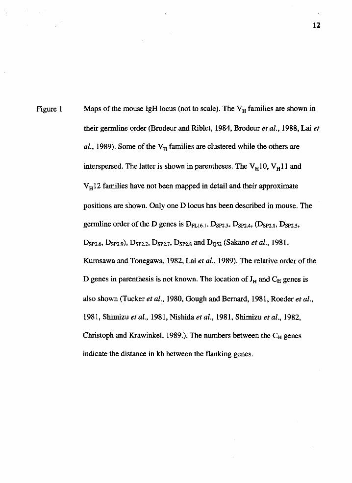
Figure 1
12
Maps of the mouse IgH locus (not to scale). The VH families are shown in
their germline order (Brodeur and Riblet, 1984, Brodeur et al., 1988, Lai et
al., 1989). Some of the VH families are clustered while the others are
interspersed. The latter is shown in parentheses. The V H 10, V H 11 and
VH12 families have not been mapped in detail and their approximate
positions are shown. Only one D locus has been described in mouse. The
germline order of the D genes is DFL16.1. DsP2.3. DsP2.4. (DsP2.1. DsP2.s,
DsP2.6. DsP2.9), DsP2.2, DsP2.1. DsP2.s and DQs2 (Sakano et al., 1981,
Kurosawa and Tonegawa, 1982, Lai et al., 1989). The relative order of the
D genes in parenthesis is not known. The location of JH and CH genes is
also shown (Tucker et al., 1980, Gough and Bernard, 1981, Roeder et al.,
1981, Shimizu et al., 1981, Nishida et al., 1981, Shimizu et al., 1982,
Christoph and Krawinkel, 1989.). The numbers between the CH genes
indicate the distance in kb between the flanking genes.
Telomere -
VH12 VHl 0 ~ ~ 0
3609 -J558-(J606, VGAM3.8, 8107) -3660 - (X24, 052, 7183) -!-D locus
--(---> 1 Mb
Cy3 Cyl 3.4
Centromere
14
(Brodeur and Riblet, 1984). The VHQ52 and VH7183 families are the 3'-most VH families
in all the mice studied so far (Lai et al., 1989). The rest of the V H families are small and
each consists of 1 % -5% of the VH genes. The VHS107 family mapped to the middle of
the VH locus and is used to demarcate the 3' VH families from the 5' VH families. Using
this guideline, the VH7183 and VHQ52 families are considered 3' families while VHJ558
family is considered a 5' family.
The organization of V H families in other inbred mice is similar to that of BALB/c
(Lai et al., 1989), although restriction fragment length polymorphism (RFLP) exists
(Brodeur et al., 1984, Atkinson et al., 1993). All polymorphisms of the V H locus among
inbred mouse strains appeared to have occurred by recombination events rather than
deletion or duplication of the locus (Tutter and Riblet, 1988). As a result of such
recombination, VH loci from 74 inbred strains could be typed into 13 haplotypes by RFLP
(Brodeur et al., 1984, Tutter and Riblet, 1988). However, such haplotype nomenclature
was not generally used because almost all the studies regarding the V H locus were
performed in BALB/c mice.
The 3' -most functional V H gene in BALB/c mouse is a member of the V H 7183
family, VH81X (Yancopoulos et al., 1984). This VH gene was used preferentially in fetal
and neonatal B-lineage cells in mouse (see VH gene utilization section). Christoph and
Krawinkel (1989) reported of a VGAM3. 8-like gene located in the D region. However,
upon detailed deletion mapping analysis, no such gene was found (Atkinson et al., 1993).
Unlike human, only one D region has been described in mouse. This region is
located between the V H and JH regions spanning approximately 80 kb of DNA (Sakano et
al., 1981, Kurosawa and Tonegawa, 1982, Lai et al., 1989). Three D families, Dsp2, DFL16
and DQ52, were found at this D locus. The Dsp2 family has 9 members which spread out
over 62 kb of DNA (Kurosawa and Tonegawa, 1982). The DFL16 family has 2 members,
15
one of which is the 5'-most D gene (Kurosawa and Tonegawa, 1982). The last D family,
DQ52, has only one member and is located only 700 bp 5' of the JH.
The JH region is located 90 kb 3' of the VH locus (Christoph and Krawinkel, 1989)
and 6. 5 kb 5' of Cµ gene (Gough and Bernard, 1981). Four functional JH genes were
found in this region, JHl-4, with the most upstream JH being JHl. The CH locus spans over
150 kb of DNA and contains eight CH genes: Cµ, CC>, Cyl, Cy2a, C"(2b, Cy3, CE and Ca
(Tucker et al., 1980, Roeder et al., 1981, Shimizu et al., 1981, Nishida et al., 1981,
Shimizu et al., 1982). The order of the CH genes and distance between them is shown in
Figure 1.
The VH, D and JH Gene Organization in Human
The human IgH locus maps to chromosome 14q32. 33 (Kirsch et al., 1982,
McBride et al., 1982) in the same orientation in mouse, i.e. telomere-Vir-D-Jir
Cir-eentromere (Erikson et al., 1982). The locus contains 100-200 VH, over 25 D, 9 JH,
9 CH and 2 CH pseudogenes (Pascual and Capra, 1991). The organization of human VH,
D, JH and CH loci is presented in Figure 2. Because IgH locus in human is polymorphic
(Benger and Cox, 1989, Sasso et al., 1990, Bottaro et al., 1991, Pascual and Capra, 1991,
van Dijk et al., 1991, Rubinstein et al., 1994), this organization may vary between
individuals.
The major V H locus spans 2. 5-3 Mb of DNA (Berman et al., 1988, Matsuda et
al., 1988, Walter et al., 1990) and contains an estimated 100-200 germline VH genes.
From published V H sequences, approximately 50% of them are functional (Kodaira et al.,
1986, Matsuda et al., 1993). The VH genes in human could be categorized into 7 families,
V H 1-7, based on the order of discovery. The V H 1-3 families were described initially from
amino acid sequences of several myeloma proteins (Capra and Kehoe, 1975). Since 1987,
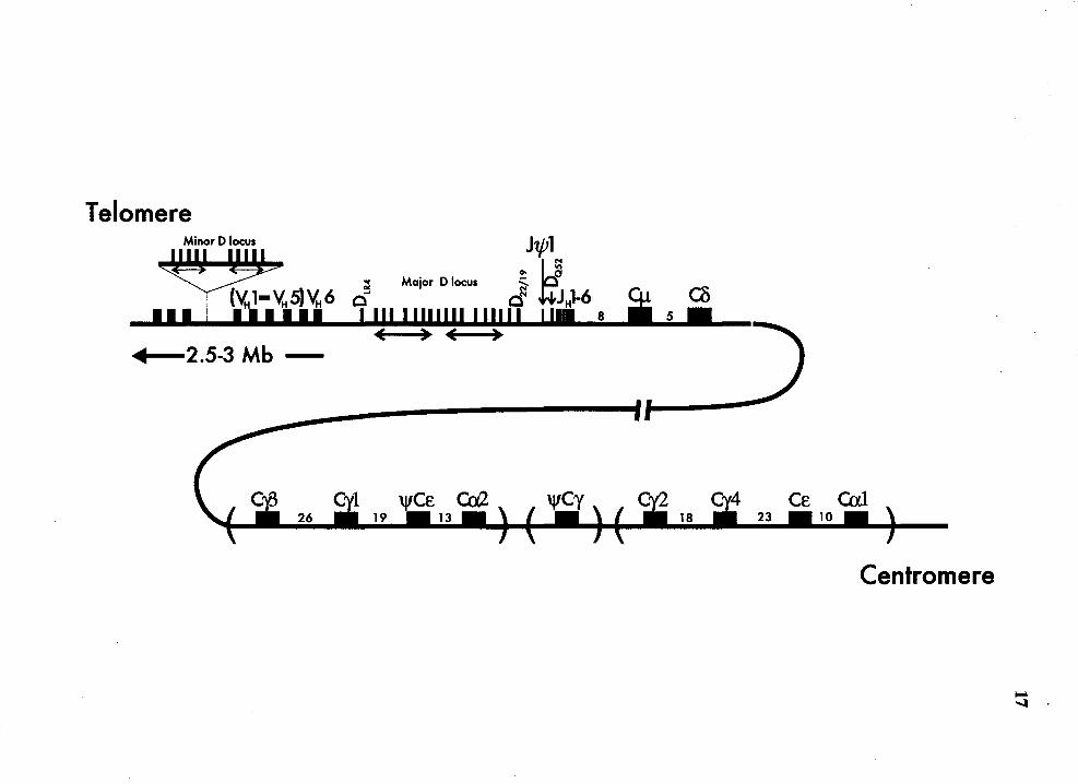
Figure 2 Map of human IgH locus on chromosome 14 (not to scale). The 3'-most
VH gene is the only member of the VH6 family (Schroeder et al., 1988).
16
Upstream of it are members of the V H 1-V H5 families which are highly
interspersed (Berman et al., 1988). The major and minor D loci are shown.
The repeating units within both D loci are indicated by double-headed
arrows. In the major D locus, the order of the D genes from 5' to 3' is
DIR2, DM2, DLR2. DLR3· D22119 and DQs2 (Matsuda et al., 1988, Buluwela et
al., 1988, lchihara et al., 1988, Ravetch et al., 1981). In the minor D locus,
the order of the D genes is DMsa. DLRsa. DXPsa. DAsa. DK5a. DMsb, DLRsb,
DXPsb. DAsb and DKsb (Matsuda et al., 1988, Matsuda et al., 1990). The 5'
and 3' orientation of the D genes in the minor D lcus is not known. Nine
JH genes were found in the following order: J\j/l, JHl, JH2, J\j/2, JH3, Jtt4,
JH5, J\j/3 and JH6 (Schroeder et al., 1988). The DQs2 gene is located
between the J\j/l and JH 1 genes. The relative germline order of Ctt genes is
shown. The germline order of all the Ctt genes is as shown although the Ctt
genes in brackets are not yet physically linked (Ravetch et al., 1981,
Flanagan and Rabbitts, 1982, Lefranc et al., 1982, Chaabani et al., 1985,
Bottaro et al., 1991, Pascual and Capra, 1991). The numbers between the
Ctt genes indicate the distance in kb between the flanking genes.
Telomere Minor D locus
Major D locus
11111 2.5-3 Mb -
Centromere
18
VH genes with less than 80% similarity to VHl-3 at the nucleotide level were isolated and
they were designated V tt4-7 (Berman et al., 1988, Pascual and Capra, 1991, Rubinstein et
al., 1994). Unlike mouse Vu families, the VH families in human are highly interspersed
among each other (Berman et al., 1988). The largest Vu family is the V u3 family
comprising 38% of the germline Vu genes (Berman et al., 1988, Walter et al., 1990).
However, this family is larger than the VHl family by only a few genes (Berman et al.,
1988, Walter et al., 1990). The other Vu families are small, comprising 1 % to 10% of the
germline Vu genes. The smallest family is the VH6 family having only one member,
identified as the 3'-most Vu gene (Schroeder et al., 1988). Most of the VH genes in
human are polymorphic, as demonstrated by nucleotide sequences of the allelic V H genes
and RFLP (Sasso et al .• 1990, Pascual and Capra, 1991, van Dijk et al., 1991, Rubinstein
et al., 1994). In contrast to the polymorphism in the mouse VH locus which were
generated mostly by recombination events (Tutter and Riblet, 1988), the polymorphisms
in the human V H locus appeared to have occurred through gene duplication and deletion
events (Sasso et al .• 1990, Pascual and Capra, 1991, van Dijk et al., 1991, Rubinstein et
al., 1994). However, the single-member VH6 family appeared to be non-polymorphic
(Schroeder et al., 1988). Honjo' s laboratory had analyzed the 3' -most 0.8 Mb region of
the Vu locus. They identified 64 germline V H genes (Kodaira et al., 1986, Matsuda et al.,
1993, Matsumura et al., 1994) and confirmed the previous finding that the Vu genes from
different families were highly interspersed (Berman et al., 1988, Buluwela et al., 1988).
Not all VH genes in human are located on chromosome 14. Matsuda et al. (1990)
had performed Southern analysis of DNA from a panel of mouse-human hybrids and
found VH genes on human chromosome 15. They named these Vu genes orphons. Cherif
and Berger ( 1990) had identified additional orphons on human chromosome 16 by in situ
hybridization. Four orphon V H genes on chromosome 15 were cloned and were (ound to
belong to Vttl and Vtt3 families (Matsuda et al., 1990). The members of the Vtt3 family
19
appeared functional and could potentially be rearranged to generate VDJ genes. However,
whether these orphons are associated with their own D, J and CH genes and whether they
undergo gene rearrangement is currently unknown.
In contrast to mouse and rabbit, D genes in human are found in at least 2 loci,
major and minor D loci. The major D locus in human is located between V H and JH
region on the chromosome 14q32. There are at least 17 D genes identified in this region.
These D genes are divided into 7 families, Dxp, DA, DK, DN, DM, DLR and DQ52 (Matsuda
et al., 1988, Buluwela et al., 1988, Ichihara et al., 1988, Ravetch et al., 1981). DQ52 is the
3 '-most D gene located only 25 bp upstream of JH 1 (Ravetch et al., 1981 ). DLR4 gene is
the 5' most D gene separated from the most downstream VH by 20 kb (Matsuda et al.,
1988). The other 15 D genes reside between DQ52 and DLR4 genes. Except for DLR3 and
D2119 genes, located at the 3' end of the D locus, the other 13 D genes were arranged in
two repeating units of 9 kb in length (Buluwela et al., 1988, Ichihara et al., 1988), each
unit having 6-7 D genes. These repeating units are shown in Figure 2 as double-headed
arrows.
The minor D region was found to be in the V H region (Matsuda et al., 1988,
Matsuda et al., 1990). As in the major D region, the minor D region also exists as two
repeating units of approximately 9 kb in length. Each unit contains 5 D genes which
belong to the Dxp, DA, DK, . DM, and DLR families. These two repeating units are
physically linked to a VH pseudogene on the chromosome 14 (Matsuda et al., 1990). The
exact location of this minor D region is unknown.
In the course of analyzing the D region, Ichihara et al. (lchihara et al., 1988) had
identified two non-conventional D genes in the major D region, DIRl and DIR2 genes.
These genes are located between DN and DM in the two 9 kb repeats at the major _D region
(lchihara et al., 1988) and are flanked on both sides with multiple 12-bp and 23-bp RSSs.
20
The coding regions of the DIR genes are rather long, ranging from 130 to 150 bp
depending on which RSSs were used. Additional DIR genes, DIR 3-6, are described (Shin
et al., 1993, Sanz et al., 1994) but the location of these genes is unknown.
The entire JH locus is located on a 3 kb region 77 kb downstream of V H6
(Schroeder et al., 1988), the 3' -most V H gene, and 8 kb upstream of Cµ gene (Ravetch et
al., 1981). In this 3 kb region, nine JH genes were found. six of which are functional. As
in other species, JH 1 was assigned to the most upstream functional JH gene and JH6 was
assigned to the most downstream functional JH gene.
The CH locus on chromosome 14 spans approximately 200 kb (Bottaro et al.,
1991 ). This locus contains 9 CH genes and 2 pseudogenes (Pascual and Capra, 1991 ). The
organization of CH genes in human is somewhat similar to that in mouse in that the Cµ
and Co genes were found at the 5' end of the locus and the CE and Ca genes were found
at the 3' end (Ravetch et al., 1981, Flanagan and Rabbitts, 1982). However, there is a
large segment containing multiple CH genes which has been duplicated in the middle of
the CH locus (Flanagan and Rabbitts, 1982). One cluster contains CY3- Cyl -'lfCEl -
Cal genes (Lefranc et al., 1982) is found upstream of the second cluster which contains
Cy4-C'"{2-CE-Ca2 genes (Lefranc et al., 1982). One 'lfCywas described and
mapped to between Cal and C'"{2 (Chaabani et al., 1985). The human CH region has yet
to be linked by cosmid clones but at least we now know that the entire CH locus spans
less than 210 kb (Bottaro et al., 1991). The diagram of CH locus is presented in Figure 2.
The VH, D and JH Genes Organization in Rabbit
Rabbit IgH locus was reported to be on chromosome 16 (Medrano and Dutrillaux,
1984). The locus contains more than 100 VH genes (Gallarda et al., 1985, Currier et al.,
1988), at least 11 D genes (Becker et al., 1990, Friedman et al., 1994), 6 JH genes (Becker
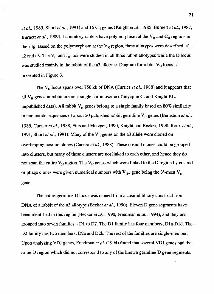
21
et al., 1989, Short et al., 1991) and 16 CH genes (Knight et al., 1985, Burnett et al., 1987,
Burnett et al., 1989). Laboratory rabbits have polymorphism at the VH and CH regions in
their lg. Based on the polymorphism at the V H region, three allotypes were described, al,
a2 and a3. The VH and JH loci were studied in all three rabbit allotypes while the D locus
was studied mainly in the rabbit of the a3 allotype. Diagram for rabbit V H locus is
presented in Figure 3.
The V H locus spans over 750 kb of DNA (Currier et al., 1988) and it appears that
all V H genes in rabbit are on a single chromosome (Tunyaplin C. and Knight KL.
unpublished data). All rabbit V H genes belong to a single family based on 80% similarity
in nucleotide sequences of about 50 published rabbit germline VH genes (Bernstein et al.,
1985, Currier et al., 1988, Fitts and Metzger, 1990, Knight and Becker, 1990, Roux et al.,
1991, Short et al., 1991). Many of the VH genes on the a3 allele were cloned on
overlapping cosmid clones (Currier et al., 1988). These cosmid clones could be grouped
into clusters, but many of these clusters are not linked to each other, and hence they do
not span the entire V H region. The V H genes which were linked to the D region by cosmid
or phage clones were given numerical numbers with V H 1 gene being the 3 '-most V H
gene.
The entire germline D locus was cloned from a cosmid library construct from
DNA of a rabbit of the a3 allotype (Becker et al., 1990). Eleven D gene segments have
been identified in this region (Becker et al., 1990, Friedman et al., 1994), and they are
grouped into seven families-DI to D7. The Dl family has four members, Dla-Dld. The
D2 family has two members, D2a and D2b. The rest of the families are single-member.
Upon analyzing VDJ genes, Friedman et al. (1994) found that several VDJ genes had the
same D region which did not correspond to any of the known germline D gene segments.

Figure 3
22
Maps of the rabbit IgH locs (not to scale). The 3' -most V H gene is V H 1
(Knight and Becker, 1990). Only one D locus has been described in rabbit.
The germline order of the D genes is D3, D 1 a, D4, D 1 b, D6, D7, D2a,
Dlc, D2b, Dld and D5 (Becker et al., 1990, Friedman et al., 1994). The
location of JH and Ctt genes is also shown (Knight et al., 1985, Burnett et
al., 1987, Becker et al., 1989, Burnett et al., 1989). The 13 Ca genes are
shown in five unlinked clusters. The Ca4, Ca5 and Ca6 genes have been
linked to each other and to the the rest of the IgH locus, while the rest of
the Ca genes have not been linked. Within each unlinked cluster, the Ca
genes are shown in their germline order. However, the germline order of
these unlinked clusters is not yet known.
5'
( >750kb -
MD . Lt') C region C 11111111 II
(£ Ca4 Ca5 Ca6
24
This suggested that these D regions represented germline D genes that have not yet been
identified.
The JH region is contained within 2 kb of DNA located 63 kb downstream of VHl
(Becker et al., 1989) and 8.kb upstream of Cµ gene (Knight et al., 1985). The nucleotide
sequence of the entire region was determined and six JH genes were identified, JH 1-JH6
(Becker et al., 1989, Short et al., 1991). The JHl was assigned to the 5'-most JH gene. All
but JH 1 are functional genes.
The rabbit CH chromosomal locus spans over 200 kb of DNA (Knight et al., 1985,
Burnett et al., 1989, Burnett et al., 1987). Sixteen CH genes were identified: Cµ, Cy, Ce
and Cal-Ca13 genes. In contrast to mouse and human, rabbit CB gene has not been
identified. Not all CH genes were physically linked and those which were not linked are
shown in brackets in Figure 3. Unlike mouse in which the order of its unlinked CH genes
was known for certain, the order of the unlinked CH genes rabbit is not yet known.
lgH Gene Rearrangement
Combinatorial Rearrangement and Its Product
Combinatorial V(D)J gene rearrangement can generate a large number of unique
variable regions from a finite number of genes. During B-cell development, one of the V,
(D) and J genes are rearranged to form a functional V genes. If we consider that there are
200 VH, 12 D and 6 JH genes in the germline, the combinatorial rearrangement of these
genes alone can generate many different VDJ genes. An even larger repertoire is possible
following junctional diversity and random nuleotide insertion (N region). The Ab
repertoire was further expanded after the rearrangement by random combination of V H
and V L, and somatic diversification such as somatic gene conversion and somatic point
mutation. The following section will discuss the generation of Ab repertoire during the
V (D )J gene rearrangement.
25
After the gene rearrangement, two joints are formed, coding and signal joints
(Max et al., 1979, Sakano et al., 1979, Hochtl et al., 1982, Van et al., 1982, Tonegawa,
1983, Desiderio et al., 1984, Selsing et al., 1984, Feddersen and Van, 1985, Lewis et al.,
1985, Yancopoulos and Alt, 1986,). The formation of the coding joints resulted from
fusion of the coding region of the V, D and J genes into one functional V region. The
signal joints are formed by joining the RSSs in a head-to-head fashion and believed to be
by-products with no biological significance. Gene rearrangement can be either deletional
or inversional depending on the orientation of the rearranging RSSs. If the two RSSs are
directed toward each other, the intervening DNA between the two genes will be deleted
(Figure 4a) (Cory and Adams, 1980, Yamagishi et al., 1983, Okazaki et al., 1987,
Fujimoto and Yamagishi, 1987, Hirama et al., 1991, Shimizu and Yamagishi, 1992).
After the signal joint formation, this deleted by-product persists as circular DNA. If the
two RSSs are in the same orientation, the intervening DNA between the two genes will be
inverted and retained in the chromosome (Figure 4a) (Hochtl et al., 1982, Lewis et al.,
1982, Hochtl and Zachau, 1983, Feddersen and Van, 1985, Malissen et al., 1986). The
characteristic of coding and signal joints will be discussed in the next section.
Most of the rearrangement has been shown to occur via deletion rather than
inversion (Meek et al., 1989, Gauss and Lieber, 1992). The underlying mechanism for
this preference is currently unknown. It was originally thought that the bias for deletion is
due to chromosomal topology. However, Gauss and Lieber (1992) showed by in vitro
recombination assay using artificial rearrangement substrates which contained the mouse
DFL16 and JH genes that this may not be the case. Most of rearrangements which occurred
between the DFL16 and JH genes in the lgH locus preferentially rearranged the 3'-RSS of
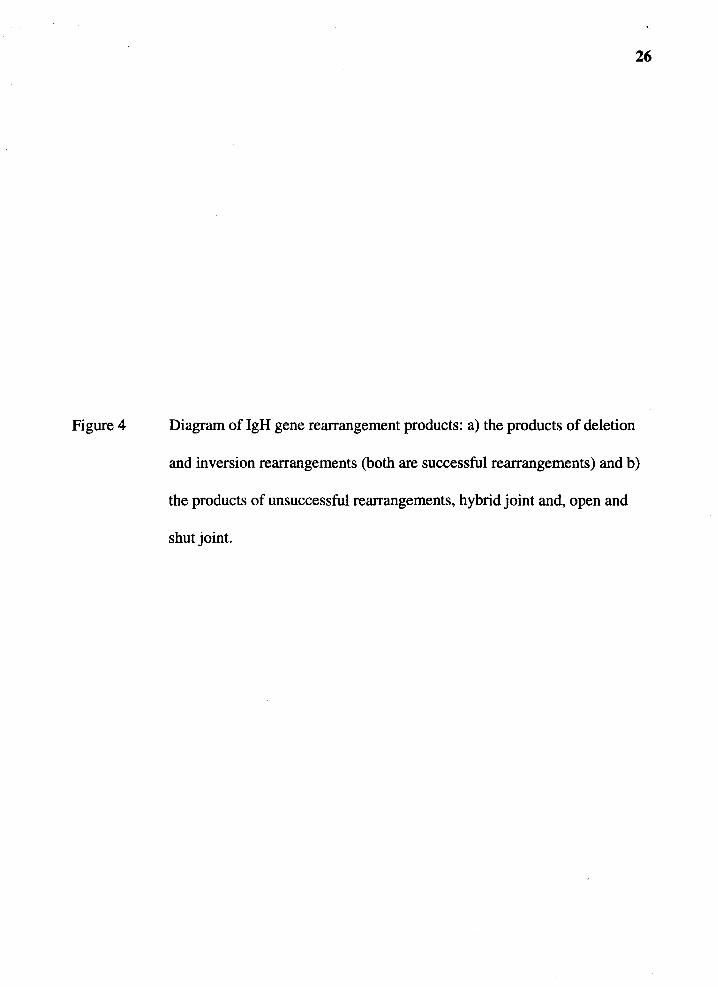
Figure 4
26
Diagram of lgH gene rearrangement products: a) the products of deletion
and inversion rearrangements (both are successful rearrangements) and b)
the products of unsuccessful rearrangements, hybrid joint and, open and
shut joint.

a)
Deletion
b)
Hybrid joint
Inversion
-1 __ _
;
Open and shut joint
D=coding region
<l = 12-bp RSS ...-=23-bp RSS
D =coding region
<]=12-bp RSS ...-=23-bp RSS
28
the DFL16 to the RSS 5' of 1tt resulting in deletional rearrangements despite the fact that
the 5'-RSS of DFL16 could also be rearranged to 1tt. In the in vitro rearrangement assay,
when the DFL16 was placed in the artificial rearrangement substrate in an inverted
orientation such that the preferentially used 3'-RSS became a 5'-RSS, the authors found
that most of the rearrangements still preferentially used that particular RSS (which is now
a 5' -RSS) resulting high frequency of inversional rearrangements. This result suggested
that the high frequency of the deletional rearrangements resulted from the specific
recognition of the recombinase for the RSS, regardless of the 5' or 3' location of the RSS,
rather than from topological preference. No qualitative differences between the RSSs
which confer the deletional bias and those which confer the inversional bias has been
studied.
Not all V, D, and J gene rearrangements are successful. Two unusual
rearrangement products have been identified: a hybrid joint, and an open and shut joint
(Lewis et al., 1988, Morzycka et al., 1988, Lewis and Gellert, 1989). The hybrid joint is
characterized by the exchange of RSSs between the two rearranging genes (Figure 4b)
(Lewis et al., 1988, Morzycka et al., 1988, Lewis and Gellert, 1989). The open and shut
joint involves the disconnection and rejoining of the RSSs and their flanking sequences
without any rearrangement (Figure 4b). These two unusual joints were identified in the
rearrangement of artificial rearrangement substrates. We do not know whether these two
joints also occur in the rearrangement of endogenous genes.
It has long been known that the coding joints are imprecise (Bernard et al., 1978,
Max et al., 1979, Sakano et al., 1979, Alt, 1987,). Sakano et al. (1980) reported, after
they compared sequences of VDJ genes with the germline V Hand JH genes, that the exact
boundary of the V H and JH were unfixed between different VDJ genes. This imprecise
joining generally resulted in loss of nucleotides encoded by the germline genes (Honjo,
29
1983, Tonegawa, 1983, Lewis et al., 1985, Alt, 1987). It is thought that after the
recombinase recognized the rearranging genes and made the excision, the coding ends
were exposed to exonuclease activity before they were joined. Such flexibility allows an
even larger Ab repertoire to be generated than from combinatorial rearrangement alone.
However, the drawback is that the rearrangement could result in out-of-frame coding
region (nonproductive gene) which, in theory, could be as many as two-thirds of the
rearrangements. In support of this, many of the rearrangement products were found to be
nonproductive genes (Max et al., 1979, Altenburger et al., 1980, Perry et al., 1980). The
signal joint formation, in contrast to the coding joint formation, is precise (Lewis et al.,
1985, Okazaki et al., 1987, Fujimoto and Yamagishi, 1987, Hirama et al., 1991, Shimizu
and Y amagishi, 1992) and, in general, the heptamer of the two RSS' s are joined head-to
head with no base loss.
Many of the VDJ genes had extra-nucleotides at the V ID and D/J junctions which
were not part of the V, D or J genes. These nucleotide additions could be categorized into
two groups. The first group is N-segment, a non-templated nucleotide addition. Although
the sequences added are template independent, there is a bias toward the additon of G and
C (Kurosawaet al., 1981, Alt and Baltimore, 1982, Kurosawa and Tonegawa, 1982). The
second group of nucleotide addition is P-elements which are templated addition. The
sequence added is palindromic to 1-2 bp of the end of the coding region (McCormack et
al., 1985, Lafaille et al., 1989).
In mouse, the capability to add N-segment to the juction is acquired after birth.
This was determined by comparing the VDJ and DJ sequences at the lgH locus from B
cells of fetal, newborn and adult mice and in general, the sequences derived from
newborn mice do not have N-region. Only 10% or less of VDJ genes from fetal liver or
newborn spleen have N-segments (Feeney, 1990). For those which do have N-segments,
30
they are shorter than 4 bp and presented only at one of the V ID or D/J junctions (Feeney,
1990, Gu et al., 1990). In contrast, more than 85% of adult VDJ genes have N-segments.
In most cases, N-segments are found at both VID and D/J junctions. The N-segements at
both junctions are generally longer than those found in VDJ genes from fetus and
neonates, and the N-segments at the VID junctions are longer on average than those at the
DIJ junctions (Gu et al., 1990).
In human, it appears that N-segments are found in VDJ genes derived from liver
of fetus as young as 105 days (Schroeder, 1987, Schroeder and Wang, 1990). More than
90% of the VDJ genes isolated from fetal liver and cord blood cDNA libraries contain N
segments, usually at both the VID and D/J junctions (Schroeder, 1987, Schroeder and
Wang, 1990, Mortari et al., 1992). The average length of these N-segments in the VDJ
genes from fetal or neonatal tissues is, however, shorter than that of adult (Schroeder,
1987, Schroeder and Wang, 1990, Sanz, 1991).
Signal joints were also shown to contain N-segments. Shimizu and Yamashigi
(Shimizu and Yamagishi, 1992) isolated circular DNA generated from deletional
rearrangements of D to JH and V H to D from mouse fetal liver or adult spleen and
analyzed the signal joints in these circular DNA. They found that N-segments were also
present in signal joints in the circular DNA generated from both V H to D and D to JH
rearrangements. Like the N-segment at the coding joints in mouse VDJ genes, the N
segments of the D/JH signal joints were sparse while those of V HID signal joint were
abundant. The addition of N-segment at the signal joint was also demonstrated in the
rearrangement of artificial rearrangement substrate in in vitro rearrangement assay (Lieber
et al., 1988).
P-elements, in contrast to N-segment, are templated nucleotide insertion initially
described at the V/J junction of chicken A chain gene (McCormack et al., 1985) These
31
nucleotide insertions are 1-2 bp long and are palindromic to the very 1-2 bp of the ends of
the coding region. The P-elements were not specific to chicken lg gene and are
subsequently identified in lg genes of many species including mouse, human and rabbit
(Feeney, 1990, Gu et al., 1990, Chen and Alt, 1993, Pascual, 1993). Since P-elements are
found only at junctions with no junctional diversity, it is thought that the addition of P
elements precedes the exposure of the coding end to the endonuclease.
Recombinase
The enzyme system that performs the V(D)J gene rearrangement is collectively
called VDJ-recombinase. Although the products of the V(D)J gene rearrangement are
now well-characterized, the exact nature of the recombinase is still obscure, although
several genes that play a role in VDJ gene rearrangements have been identified. It is
surprising to find that the expression of some of these genes is not lymphoid-specific. In
fact most of the components of recombinase are probably expre_ssed in many cell types, if
not ubiquitously expressed (Schatz et al., 1989, Oettinger et al., 1990, Pergola et al.,
1993, Taccioli et al., 1994). Only three components of recombinase, RAG-1, RAG-2 and
Terminal deoxynucleotidyl transferase (TdT), have been found to be lymphoid specific
thus far.
Two essential components of the recombinase are RAG-1 and RAG-2 (Schatz et
al., 1989, Oettinger et al., 1990). The expression of these two genes alone in a mouse
fibroblast cell line, which by itself is rearrangement-incompetent, conferred the cell line
the rearrangement capability suggesting that RAG-1 and RAG-2 were indispensable
components of the recombinase. The result also suggested that all other components of
recombinase were expressed in the fibroblast cell line. Both RAG-1 and RAG-2 genes are
conserved through out evolution and are expressed only in lymphoid tissue, pre-Band
pre-T cell lines (Schatz et al., 1989, Oettinger et al., 1990). Mice deficient in either RAG-
32
1 or RAG2 have no mature B and T cells were found (Mombaerts et al., 1992, Shinkai et
al., 1992) and AMuLV-transformed B cell lines generated from these knock-out mice
showed no sign of lg or TCR gene rearrangements. These data suggested that the whole
process of lgH gene rearrangement was halted in the absence of RAG-1 or RAG-2,
confirming the importance of RAG-1 and RAG-2 in the rearrangement process.
The function of RAG-1 and RAG-2 proteins in the rearrangement process is still
obscure. In order for DNA rearrangement to occur, one would expect the recombinase to
bind DNA, cleave it via endonuclease activity and then ligate the DNA via ligase activity.
RAG-1 and RAG-2 have none of these expected properties. Because RAG-1 contained a
topoisomerase-1 domain, which was found to be important for its function, the possibility
that RAG-1 protein might be responsible for cleaving DNA during the rearrangement was
proposed (Kallenbach and Rougeon, 1992, Kallenbach et al., 1993, Silver et al., 1993,
Sadofsky et al., 1994a, Sadofsky et al., 1994b). However, since a mutation at the active
conserved tyrosine in the topoisomerase I domain did not abolish the rearrangement
activity of RAG-1, this domain must not function in this manner. Therefore, if RAG-1 is
responsible for cleaving the DNA during the rearrangement process, the mechanism must
be different from the conventional mechanism used by other topoisomerases of type I.
Unlike RAG-1, RAG-2 does not have any recognizable functional domains (Oettinger et
al., 1990). The only prominent feature of RAG-2 is an acidic-residue rich domain which
was found to be dispensable for its activity (Silver et al., 1993, Sadofsky et al., 1994a,
Sadofsky et al., 1994b ). It is disappointing that scientists have been attempting to study
the role of RAG-1 and RAG-2 in the rearrangement process since they were cloned, but
yet this goal has not been realized. Now that monoclonal Abs to RAG-1 and RAG-2
proteins have been developed (Lin and Desiderio, 1993, Silver et al., 1993), the protein
chemistry and enzymology studies of RAG-1 and RAG-2 should appear shortly.
33
Another lymphoid-specific component of recombinase is the TdT enzyme. TdT is
a DNA polymerase which catalyzes the polymerization of dNTPs to the 3' -end of DNA
molecule in a template-independent manner (Bollum, 1974). Although any nucleotide
could be incorporated, the TdT is biased toward incorporating dGTP and dCTP. The level
of TdT expression in a pre-B cell line correlated well with the extent to which N
segments are found (Desiderio et al., 1984, Landau et al., 1987). Conclusive evidence
that TdT is responsible for the N-segment addition was reported by Komori et al. (1993)
and Gilfillan et al. (1993) who generated TdT knock-out mice. The VDJ genes from these
mice showed minimal N-segment addition, with most of the VDJ genes having no N
segment. Therefore, TdT is the enzyme responsible for most of N-segment additions in
the V(D)J gene rearrangements. The fact that V(D)J gene rearrangement occurs normally
in TdT-deficient mice suggests that, unlike the other components of the recombinase,
TdT is dispensable.
The rest of the recombinase enzyme components have not yet been studied.
Studies which identified these components could be divided into two groups. One group
of studies is based on the assumption that after the cleavage of DNA, the DNA repair
machinery would be required to repair the DNA lesion. Four mutants of chinese hamster
ovary cell line defective in DNA-repair were found to be incapable of performing V(D)J
gene rearrangement even after RAG-1 and RAG-2 were supplied suggesting that the
defective genes in these four mutants were essential component of the recombinase.
These genes are xrs-6, XR-1, V3 and XR-V9B (Pergola et al., 1993, Taccioli et al.,
1993). Mouse SCID mutant was also defective in DNA-repair pathway and was found to
be defective in V(D)J gene rearrangement (Bosma and Carroll, 1991). The SCID gene
was found to be a homolog of the V3 gene in chinese hamster (Taccioli et al., 1994).
Taken together, these results suggested that four genes involved in DNA-repair pathway,
xrs-6, XR-1, V3 or SCID and XR-V9B, are components of the recombinase enzyme. Of

34
these four genes, only xrs-6 gene has been cloned. It was shown to encode the 80kb
subunit of the DNA-dependent protein kinase or Ku Ag (Taccioli et al., 1994) but how it
participates as part of the recombinase is unknown.
The other group of studies approached the identification of recombinase
components based on the assumption that there should be DNA binding proteins which
bind specifically to the RSS and assist the recombinase to recognize the V, D and J genes.
Several RSS-binding proteins have been described in nuclear lysate of lymphocytes, some
of which have been cloned (Aguilera et al., 1987, Halligan and Desiderio, 1987,
Hamaguchi et al., 1989, Matsunami et al., 1989, Shirakata et al., 1991). Not much is yet
known about their proposed function in the recombinase complex except that these
proteins bind the RSS.
It is apparent that the V(D)J gene rearrangement is not a trivial process, illustrated
by the complexity of the recombinase enzyme. Although many of the its components
have been identified, their enzymology and how they function as a recombiase has been
only minimaly studied. Since monoclonal Abs to many of these components are now
available and many components of the recombinase have been described, the time has
arrived for scientists to start deciphering these intriguing aspects of the recombinase.
Order of lgH Gene Rearrangement
The V region of H-chain is encoded in the germline by three non-contiguous
genes, VH, D and JH. To generate a V region, two separate rearrangements, VH to D and D
to JH, are required. The assembly process occurs in an orderly fashion and at least one
rearrangement occurs at each IgH allele. The order of IgH gene rearrangement has been
established in mouse by Alt et al. (1984), and their finding has been regarded as dogma
for the order of lgH gene rearrangement in all species.
35
The order of IgH gene rearrangement in mouse could be established primarily due
to the ability of AMuL V to transform mouse B cells, especially immature B cells or pre-B
cells (Rosenberg et al., 1975, Rosenberg and Baltimore, 1976). Furthermore, the
AMuLV-transformed fetal liver pre-B cells continued to rearrange their IgH genes in vitro
(Alt et al., 1981). Alt et al. found that most fetal liver derived AMuLV-transformants
contained rearranged DJH on both alleles. These cell lines then rearranged VH to the DJ to
form VDJ genes. None of the cell lines rearranged VDJ genes without first rearranging
DJ genes on both alleles. These results suggested that DJ genes are the intermediates in
the IgH gene rearrangement process and that the first step of IgH gene rearrangement is
DJ on both alleles. Neither VD gene nor D-D fusion were found in these cell lines
suggesting that DJ gene was the major, if not exclusive, intermediate. This conclusion
was supported by the finding that AMuLV-transformed B cell lines from JH gene knock
out mice showed no sign of the lgH gene rearrangement (Chen et al., 1993).
The order of IgH gene rearrangement in mouse B cells was established based on
the finding that JH on both alleles were rearranged to DJ or VDJ genes. Such analysis in
human B cells has not been determined due to several factors. First, human equivalent of
AMuL V -transformed cell in mouse is not available. Therefore, the sequential progress of
the lgH gene rearrangement in human B cells can not be followed in vitro. Second, the
lgH gene rearrangement in human appears to be more complex than mouse. Shin et al.
(1993) had isolated a genomic phage clone containing a VD gene rearrangement
suggesting that the order of IgH gene rearrangement in human may occur via an
alternative intermediate not found in mouse. Third, many VDJ genes from human B cells
contained D-D fusions suggesting that D-D fusion occurred rather frequently in human.
Finally, human DQ52 gene is located only 25 bp 5' of JHl. The DQ52Jttl rearrangement
would result in 25 bp deletion and therefore, this rearrangement is difficult to distinguish
from germline JH on Southern analysis. These factors, in addition to the polymorphism of
36
human IgH locus, made the evaluation of the status of IgH gene on the unexpressed allele
in human B cells complex and therefore, the order of lgH gene rearrangement in human
has not been conclusively determined.
Allelic Exclusion
The term allelic exclusion describes a phenomenon in which the lg and TCR
proteins are made from only one of the two homologous chromosomes in individual B
and T cells. In the mid 1960' s, several groups of investigators reported that each
individual lg producing plasma cell from rabbits and mice, which are heterozygous at the
heavy (H) or light (L) chain genes, produced Hand L chains from only one of the two
alleles (Cebra et al., 1965, Weiler, 1965, Pemis et al., 1965). After the IgH and IgL genes
were cloned, the evidence of allelic exclusion of IgH and lgL genes accumulated at both
serological and molecular levels (Coleclough et al., 1981, Gathings et al., 1982, Alt et al.,
1984). After the TCR genes were cloned, they too were assumed to be under allelic
exclusion control. But, so far, only TCR~ genes were conclusively shown to be allelically
excluded (Goverman et al., 1985, Chou et al., 1986, Dembic et al., 1986, Leiden et al.,
1986, Malissen et al., 1988, Uematsu et al., 1988).
The reason why B and T cells develop allelic exclusion mechanism is not known.
One may speculate that, to minimize the error associated with the VDJ gene
rearrangement, any unnecessary rearrangements are prohibited after a productive VDJ
gene was generated. Not only could non-functional genes result from the rearrangement
process, but much evidence suggest that chromosomal translocations could also occur
(Tsujimoto et al., 1985a, Tsujimoto et al., 1985b, Finger et al., 1986). These
translocations may cause tumors and may explain why chromosomal translocations are
prevalent in tumors of lymphoid origin (Foon and Piro, 1992, Hagemeijer, 1992; Mirro,
1992). Another possible purpose of allelic exclusion is to prevent autoreactive Band T

37
cells from escaping clonal deletion. In one H-chain transgenic mouse model in which the
heavy chain transgene was derived from an autoreactive hybridoma, autoreactive B cells
could be generated if the transgenic H-chain was paired with a certain VL gene (to be
derived from endogenous rearrangement) (Iliev et al., 1994). B cells which paired the H
chain transgene with other VL genes would be non-autoreactive. Iliev et al. (1994) found
that autoAb producing B cells in these mice always co-expressed non-autoreactive
endogenous IgM while the non-autoreactive B cells did not. This result suggested that the
expression of endogenous IgM allowed the autoreactive B cells, which normally would
have been clonally deleted (Nossal, 1994), to escape the clonal deletion. These
speculations of the significance of allelic exclusion are still hypothetical and remain to be
proven. To gain insight into the significance of allelic exclusion, it is desirable to be able
to disrupt the allelic exclusion in normal mice and observe the outcome. As a
steppingstone toward that goal, many investigators have studied how the allelic exclusion
is regulated.
The allelic exclusion can possibly be regulated at two, not mutually exclusive,
steps. One controlling step is at the rearrangement (or genetic) level by inhibiting the
rearrangement of the second allele so that it would remain in the un-expressible state.
Otherwise, allelic exclusion can be implicated post-rearrangement at the level of gene
expression and/or protein assembly ,or at the cellular level by negative selection for B
cells which simultaneously express H-chains from both IgH alleles. This review will
present the studies regarding allelic exclusion as two groups: studies which addressed the
genetic level and those which addressed the post-rearrangement control.
Allelic Exclusion at the Rearrangement (Genetic) Level
Although there are seven lg and TCR loci which undergo somatic rearrangement
and are theoretically subjected to allelic exclusion phenomenon, the allelic exclusion at
38
the rearrangement level was studied virtually in four of the seven loci: lgH, lgK, TCR~
and TCRa.. These studies will be discussed in the following section. The allelic exclusion
at the rearrangement of the lgH and TCR~ genes will be discussed together because their
rearrangements bear similar characteristics. These characteristics are that the lgH and
TCR~ genes are the first gene to rearrange in pre-Band pre-T cells (Siden et al., 1981,
Raulet et al., 1985, Snodgrass et al., 1985a, Snodgrass et al., 1985b) and that they
required two separate rearrangements, D to J and V to DJ, to generate a functionally
rearranged gene. Then, the allelic exclusion at the rearrangement level of lgK and TCRa.
genes will be discussed.
Two theories were proposed to explain the allelic exclusion at the rearrangement
step: stochastic and regulated theories. The stochastic theory assumes that the
rearrangement of lg and TCR genes is so prone to error that many rearrangements result
in non-productive genes and, therefore, the probability of having two functional genes in
one cell is remote (Coleclough et al., 1981). The regulated theory explains that the
rearrangement of lg and TCR genes on the second allele are inhibited when a productive
gene is achieved (Bernard et al., 1981, Kwan et al., 1981, Alt et al., 1984). Many
experiments support the idea that the allelic exclusion is better explained by the regulated
theory.
The first evidence to support the regulated theory was obtained from the study of
lgH genes. In AMuLV-transformed pre-B cell lines which actively rearranged their lgH
genes, the rearrangement of the unexpressed lgH allele ceased after a functional VDJ
gene was made (Alt et al., 1984). Furthermore, Reth et al. (1987) had transfected a VDJ
Cµ gene into two AMuLV-transformed pre-B cell lines and the active ongoing
rearrangement of the endogenous lgH genes was halted. It appeared as if once a µ protein
was made, the rearrangement of the lgH locus was turned off and the unexpressed allele
39
stably existed as either DJ or non-functional VDJ genes. At the TCRP locus, the allelic
exclusion appeared to be regulated in a similar fashion because only DJ or non-functional
VDJ genes were found on the unexpressed TCRP allele (Tsukamoto et al., 1984,
Malissen et al., 1992). These results suggested that the rearrangement of the unexpressed
IgH and TCRP alleles were inhibited, presumably by the µ and TCRP chains.
To directly test the assumption that a productive rearrangement turns off the
rearrangement at each locus, transgenic mice were generated with functionally rearranged
µand TCRP transgenes (Grosschedl et al., 1984, Rusconi and Kohler, 1985, Weaver et
al., 1985, Fenton et al., 1988, Imanishi-Kari et al., 1988, Manz et al., 1988, Nussenzweig
et al., 1988, Uematsu et al., 1988, van Meerwijk et al., 1991, Rothe et al., 1993). As
predicted, rearrangement of the endogenous lgH genes was inhibited in the µ transgenic
mice and rearrangement of the endogenous TCRP genes was inhibited in the TCRP
transgenic mice. Only the constant regions of the µ and TCRP genes were required to
mediate allelic exclusion because endogenous gene rearrangement was also inhibited in
transgenic mice generated from only the constant region portions (Krimpenfort et al.,
1989, Corcos et al., 1991). The ability of other heavy chain genes to inhibit the
rearrangement was also tested using transgenic mice. Allelic exclusion of the endogenous
lgH gene was found only in o but not ila or ilb transgenic mice (Iglesias et al., 1987,
Offen et al., 1992, Tsao et al., 1992) suggesting that the o chain could also mediate the
inhibition of rearrangement.
The fact that both the functionally rearranged µ and o, but not ila or ilb,
transgenes could mediate allelic exclusion of lgH genes suggested that the regulatory
region responsible for the allelic exclusion should be present in both the µ and o genes.
Subsequently, several studies have shown that the membrane exon (µm) of theµ gene,
which shares high homology with the membrane exon of the o gene, was essentilll for
40
allelic exclusion of endogenous IgH genes (Manz et al., 1988, Nussenzweig et al., 1988,
Kitamura and Rajewsky, 1992). Transgenic mice with a VDJ-Cµ gene lacking the µm
exon (Manz et al., 1988, Nussenzweig et al., 1988) or mice in which the µm exon has
been disrupted (Kitamura and Rajewsky, 1992) showed high incidence of double-lg
producers that expressed IgH from more than one allele simultaneously. Furthermore,
Reth et al. (1987) showed that the ability of the transfected functionally rearrangedµ
gene to inhibit the rearrangement of the endogenous IgH gene depended on the presence
of µm exon in the transfected gene. These results suggested that the µm exon was
important for the regulation of allelic exclusion at the IgH locus. Similar studies have not
been performed for the TCR~ gene.
Allelic exclusion at the rearrangement level of IgH and TCRJ} loci was proposed
to be mediated at the V to DJ gene rearrangement step (Alt et al., 1984, Malissen et al.,
1992). If this were the case, it would be expected that D to J rearrangement of the
endogenous IgH and TCR~ genes could occur normally in the IgH and TCR~ transgenic
mice but the V to DJ rearrangement would be blocked. Indeed, analysis of T cell lines or
thymocytes from the TCR~ transgenic mice showed that all rearrangements of the
endogenous TCR~ genes were DJ gene rearrangements (Uematsu et al., 1988,
Krimpenfort et al., 1989, van Meerwijk et al., 1991). No germline J~ or VDJJ} genes
were found. One interpretation of these results is that allelic exclusion of TCRJ} gene by
the functional TCRJ} protein was mediated solely at the V to DJ rearrangement step.
Analysis of lgH genes in hybridomas generated from µ and B transgenic mice also
revealed DJ gene rearrangements (Rusconi and Kohler, 1985, Weaver et al., 1985,
Iglesias et al., 1987, Muller et al., 1989, Iacomini et al., 1991). However, in contrast to
the TCR~ transgenic mice model, complete endogenous VDJ genes as well as
unrearranged D and JH genes were found. The VDJ genes represented 15%-33% _of the
endogenous IgH alleles while the unrearranged JH genes were found in 10%-20% of the
41
alleles (Rusconi and Kohler, 1985, Weaver et al., 1985, Iglesias et al., 1987). These
results explain why µ and o transgenic mice expressed endogenous lg in their serum
(Grosschedl et al., 1984, Rusconi and Kohler, 1985, Weaver et al., 1985, Stall et al.,
1988, Rath et al., 1989, Forni, 1990, Vos and Hodes, 1992). They also suggested that,
unlike TCRP genes, both D to J as well as V to DJ gene rearrangements at the IgH locus
could be inhibited by allelic exclusion process.
Since endogenous lgH genes in the µ and o transgenic mice could be rearranged to
functional VDJ genes, this raised the possibility that allelic exclusion was not complete
and that the endogenous VDJ genes and the transgene were co-expressed in the same B
cell. In fact, mixed-lgM molecules, which containedµ chains from both the transgene and
the endogenous gene, were found in the serum of the µtransgenic mice (Stall et al., 1988,
Rath et al., 1989, Vos and Hodes, 1992). Furthermore, some hybridomas derived from the
µ transgenic mice produced and secreted both transgenic and endogenous µ chains (Manz
et al., 1988, Kenny et al., 1989, Muller et al., 1989). In addition, B cells from these mice
expressed on the surface both transgenic and endogenous µ chains on the surface (Kenny
et al., 1989, Lamers et al., 1989, Forni, 1990, Grandien et al., 1990). However, another
group of investigators reported that they could never find such double-producers (Rusconi
and Kohler, 1985, Weaver et al., 1985, Nussenzweig et al., 1988, lmanishi-Kari et al.,
1993). These contradictory results were not due to the differences in the transgenic lines
because some were in common among the studies (Rusconi and Kohler, 1985, Weaver et
al., 1985, Stall et al., 1988, Forni, 1990, Grandien et al., 1990, Imanishi-Kari et al.,
1993). Imanishi-Kari and co-workers suggested that the double-producers were
experimental artifacts caused by anti-allotypes specific for lg from the endogenous genes
shown to cross-react with the variable region of the transgenicµ protein (lmanishi-Kari et
al., 1993). In this way, it would appear in studies which used these Abs that the ~ells
which expressed only transgenic µ chain also expressed the endogenous lgH gene
42
(Rusconi and Kohler, 1985, Stall et al., 1988, Rath et al., 1989, Forni, 1990, Grandien et
al., 1990). This proposal remains to be tested.
One possible mechanism which mediates the allelic exclusion effect of functional
IgH and TCRP gene rearrangements is that the expression recombinase is down
regulated. It is an unlikely mechanism, however, because the pre-Band pre-T cells which
had successfully rearranged the IgH and TCRP genes have yet to rearrange IgL and TCRa
loci. An alternative explanation would be that after a successful VDJ gene rearrangement
at the lgH or TCRP loci, the rearrangement at the respective locus is suppressed. This
explanation is attractive for the following reasons. First, it has been shown that a constant
production of µ chain is required to maintain the inhibition of rearrangement at the IgH
locus in pre-B cells. Maeda et al. (1989) had identified a subclone of a pre-B cell line
which stopped rearranging its IgH genes after it had functionally rearranged a VDJ gene
and expressed the µ chain. However, the rearrangement of the lgH gene was reinitiated in
the subclones which lost the functional VDJ allele. Similar results were obtained by
Beck-Engeser et al. (1987) who reported that IgH gene rearrangements were re-initiated
in a pre-B cell line after it switched from expressingµ chain to expressing "(lb chain. The
inhibition of rearrangement at the TCRP locus also depended on the expression of TCRP
protein. Pircher et al. (1990) examined the expression of the endogenous TCRP gene in
several TCRP transgenic lines and found that the expression of the endogenous gene was
inhibited only in the lines which expressed high level of the trans gene. This result
suggested that the high level of transgenic TCRP chain could inhibit the rearrangement of
the endogenous TCRP gene. Taken together, these data suggest that inhibition of lgH and
TCRP gene rearrangement is an active process which requires the production ofµ and
TCRP proteins.
43
The biochemical basis of the inhibitory effect on the rearrangement process by µ
and TCRP chains is currently unknown but some evidence suggests that p56lck may be
involved. Transgenic mice which over-expressed p56lck proteins showed a reduced level
of VDJP rearrangement while the level of DJP rearrangement was normal (Anderson et
al., 1992). Furthermore, by interfering with the function of p56lck in TCRP transgenic
mice, using a dominant negative mutant of p561ck, vp to DJP gene rearrangements were
no longer inhibited (Anderson et al., 1993). In both experiments, the rearrangement of
TCRa. occurred at levels equivalent to normal mice. These results suggest that p56lck is
involved in delivering an allelic exclusion signal specific for the vp to DJP gene
rearrangements. Whether p561ck exerts its inhibitory effect via the same pathway it uses in
the activation of peripheral T cells remains to be determined. Because p56lck is expressed
in B-lineage cells and involved in B-cell activation pathway (Gold et al., 1994), it is
possible that p56lck is also involved in µ-chain mediating allelic exclusion of the IgH
gene. To answer this question, it is necessary to examine the effect of p56lck over
expression as well as that of the dominant negative mutant of p56lck on the allelic
exclusion by µ gene and to determine the level of p56lck expression at different stages of
B cell development.
Susceptibility of endogenous IgH and TCRP genes to the allelic exclusion process
may also be dependent on chromosomal location. In µ transgenic mice, more endogenous
VDJ gene rearrangements involve VH genes in the 3' VH families than those in the 5' VH
families (Iacomini et al., 1991, Costa et al., 1992). In mice double-transgenic for a
functionally rearranged TCRP gene and a germline vp, op, JP and cp minilocus, van
Meerwijk et al. ( 1991) found that the expression of the functionally rearranged TCRP
transgene could inhibit the rearrangement of the germline TCRP minilocus. However, the
inhibition of the rearrangement of the minilocus occurred in only one of the five
transgenic lines they have generated. The fact that the five double-transgenic mice all had

44
the functionally rearranged TCR~ gene integrated at the same chromosomal location but
had the minilocus integrated at different location suggested that the location where the
minilocus transgene integrated into the chromosome determined the susceptibility of the
minilocus to the allelic exclusion machinery. It is possible that the reason why TCR~
transgene can completely inhibit the rearrangement of V/3 to DJ{3 (Fenton et al., 1988,
Uematsu et al., 1988, van Meerwijk et al., 1991, Rothe et al., 1993) is that the
endogenous TCR~ gene is located in the chromosomal region which is highly susceptible
to allelic exclusion machinery. One way to test this hypothesis is by generating a mouse
in which the TCR~ locus is shuffled to the IgH locus and observe the allelic exclusion
effect of the TCR~ transgene in the rearrangement of TCR~ gene in its new location.
However, such an experiment is not feasible with current technology and awaits the
development of the technique to shuffle genetic loci.
Allelic exclusion at the Igx: locus is less complicated than those at IgH and TCR~
loci. Many plasmacytomas rearranged both K alleles, only one of which was functional
(Perry et al., 1980, Coleclough et al., 1981, Kwan et al., 1981) so that it appeared that
allelic exclusion at the K locus could be explained by a stochastic theory. However,
subsequent studies in polyclonal B cells showed that a substantial amount of K gene in B
cells was not rearranged (Nottenburg and Weissman, 1981, Tsukamoto et al., 1984). This
result suggested that the rearrangement of the unexpressed Igx: allele may also be
inhibited. The analysis of the K locus in the functionally rearranged K transgenic mice
supports this idea. Ritchie et al. ( 1984) analyzed trans gene-expressing hybridomas
derived from transgenic mice and found that their endogenous K genes were in germline
configuration. This inhibition was not found in the hybridomas which did not express the
transgene or the µ gene suggesting that the inhibitory process depended on both K and µ
chains. These results, together with the finding that µm exon of the µ gene was important
for the allelic exclusion, suggest that the inhibitory process is mediated by slgM
45
molecules rather than the K chains alone. Because the expression of RAG-1 and RAG-2
genes was terminated after the expression of slgM molecules (Ma et al., 1992), it is
unclear whether lgK rearrangement is actively suppressed, or simply due to the lack of
recombinase enzyme.
The expression of TCRa was assumed to be allelically excluded, similar to lg and
TCR~ genes. However, this idea is challenged by recent findings. Approximately 30% of
T cell lines have been found to functionally rearrange VJa on both alleles (Champagne et
al., 1988, Furutani et al., 1989, Casanova et al., 1991, Couez et al., 1991, Kuida et al.,
1991, Malissen et al., 1992, Heath and Miller, 1993), both of which could be transcribed
(Marche and Kindt, 1986, Casanova et al., 1991, Couez et al., 1991, Kuida et al., 1991,
Hu et al., 1993). Padovan et al. (1993) used Ab to Va2, Va12 and Va24 to show that
one in 103 to 104 human peripheral blood T cells express two different Vas on the
surface. T cell clones established from these cells stably expressed the two different Vas
and were shown to contain two functional VJa genes. T cells expressing two different
VJ a genes were further demonstrated in mice transgenic for a functionally rearranged
TCRa gene (Borgulya et al., 1992). These results together suggest that neither the
expression nor the rearrangement of TCRa gene were allelically excluded. However, in
vivo, approximately 70% of T cell clones contain a non-functional rearrangement on the
unexpressed allele (Malissen et al., 1992) and these T cells would thus express only one
species of T cell receptor. The TCRa genes probably continue to rearrange until the
expression of RAG-I and RAG-2 genes is down-regulated after the cells are positively
selected in the thymus (Turka et al., 1991).
The lack of allelic exclusion at the TCRa locus raised a question whether the
allelic exclusion at lgK locus was an active suppression. Because the allelic exclusion at
the K locus is mediated by slgM (Ritchie et al., 1984) and because the expression of
46
RAG-1 and RAG-2 genes is down-regulated after the cross-linking of slgM (Ma et al.,
1992), it is possible that the inhibition of the rearrangement of the unexpressed Igx: allele
is simply due to the lack of recombinase enzyme. Evidence from bcl-2 transgenic mice
supports this view (Rolink et al., 1993). slgM+ B cell lines derived from these transgenic
mice continue to express RAG-1 and RAG-2 genes and also continue to rearrange their
light chain genes. This suggests that there was no active suppression of K gene
rearrangement following the expression of slgM. Rather, lgx: gene rearrangement
continues as long as recombinase enzyme is expressed.
Although we currently do not know for certain, the supposition that there is no
active suppression of the rearrangement at the lgK and TCRa. loci makes the allelic
exclusion of lg and TCR genes fairly straight forward to implement in B and T cells.
After pre-Band pre-T cells have a functionally rearranged VDJ gene at the IgH and
TCRP genes, further rearrangement at these loci are inhibited by an active inhibitory
process which requires the expression of theµ or TCRP chain and p56Ick. The
rearrangements at these loci remain suppressed while the pre-Band pre-T cells rearrange
their Igx: and TCRa. genes, respectively. Then lgx: and TCRa. genes continue to rearrange
until the expression of RAG-1 and RAG-2 genes is turned off. In B cells, the expression
of RAG-1 and RAG-2 genes could be turned off rapidly after the expression of slgM.
Therefore, the chance of having a second rearrangement of K gene is small. In T cells,
RAG-1 and RAG-2 genes continue to be expressed until the cells undergo positive
selection. Therefore, both TCRa. alleles are usually rearranged. This scenario, although
simple, is my hypothetical view and remains to be tested.
47
Post-Rearrangement Control of Allelic Exclusion
The idea that the allelic exclusion can be mediated through post-rearrangement
control originated from the .findings that some B and T cells had more than one functional
VDJ or VJ genes but only one of the two functional genes was expressed. For example,
Nottenburg et al. (1987) showed that some mouse peripheral B cells have two functional
VDJ genes yet only one was expressed on the surface. Bernard et al. (1981) found
plasmacytoma cell lines with functional VJ genes on both K alleles, both of which were
transcribed but only one assembled into lg molecule. T cell clones which had two
functionally rearranged TCRa. genes but only one Va. on the surface have also been
reported (Malissen et al., 1988, Furutani et al., 1989, Couez et al., 1991, Kuida et al.,
1991). All these results suggest that there must be some post-rearrangement control of
allelic exclusion.
Most of the post-rearrangement control has been studied at the level of protein
assembly. It was found that some Kand TCRa. chains fail to properly pair with the heavy
and TCR~ chains (Bernard et al., 1981, Cherepakhin et al., 1987, Malissen et al., 1988,
Berinstein et al., 1989, Furutani et al., 1989) thus, these Kand TCRa. chains were not
expressed on the surface of the cells or secreted. However, we do not know how
frequently these improper pairings between heavy and light chains and between TCRa.
and TCR~ chains occur in vivo.
One potential step of post-rearrangement control is at the level of transcription.
However, this possibility has been addressed only to a limited extent. Gerondakis et al.
(1984) showed that the fusion of a mouse pre-B cell line with a mouse plasmacytoma cell
line could induce the expression of a previously silent functional VDJ gene and this event
48
correlated with the demethylation of the lgH allele. In liver cells which do not express lg
genes, the IgH and IgL loci are hypermethylated and DNasel hyposensitive. Both loci
became increasingly demethylated and acquire DNasel hypersensitivity as B cells mature
(Storb et al., 1981, Storb and Arp, 1983). These results raise the possibility that, in cells
which had functionally rearranged two genes, one allele might be hypermethylated and is
therefore not expressed. However, the methylation status of lg or TCR loci has not been
examined in cells which contained two functional genes.
The allelic exclusion can also be explained by cellular selection theory (W abl and
Steinberg, 1982). The theory proposes that when B cells simultaneouly produce heavy
chains from both IgH alleles, they are overloaded by the extra heavy chain proteins being
produced from the second allele. This, in turn, results in free heavy chain proteins in the
cells and the cells die from toxicity of the free heavy chain proteins. The theory was based
on the observation that, while it is relatively easy to isolate a variant of hybridoma which
produces only light chain, rarely a variant which produces only heavy chain can be
isolated (Kohler, 1980). At the present time, the extent to which this cellular selection
contributes to the allelic exclusion has not been conclusively determined.
In summary, allelic exclusion can be regulated at both the rearrangement (genetic)
and post-rearrangement levels. At the rearrangement level, the expression of the µand
TCR~ chains, in turn, inhibits further rearrangement of the respective loci. It appears that
the rearrangement of lgK locus was also inhibited by the 1C chain. However, at present it is .
still unclear whether the inhibition of lgK rearrangement is an active inhibitory process.
TCRa locus is different from all the mentioned genes in that it was not allelically
excluded. While many experiments studied the allelic exclusion at the rearrangement
level, studies of allelic exclusion at the post-rearrangement level have been limited. The

49
best characterized mechanism is the failure of the Kand TCRa chains to pair with the H
chain and TCRf3 chain. At present, the phenomenon of allelic exclusion has been well
described and characterized. Although the involved biochemical pathway is still largely
unknown, pS6Ick has been shown to be involved in the allelic exclusion at the TCRf3
locus. Ultimately, we would like to understand the biochemistry of the allelic exclusion.
The study of the involvement of pS61ck in the allelic exclusion process should bring us
toward that goal.
V H Gene Utilization in 8-lineage Cells
In many species, Ab diversity is generated primarily through combinatorial
rearrangement of V, D and J gene segments. In the adult mouse, scientists have found that
all the 13 described VH gene families contribute to the diversity of the VDJ gene
repertoire. However, before the adult pattern of V H gene usage is reached, B cells in fetal
and neonatal mice preferentially utilized the 3' V H gene families (defined as the V H gene
families that are 3' to the VHS107 family). The shift to the adult pattern of VH gene usage
occurs gradually after birth via an unknown mechanism. In human, the Ab diversity is
also generated largely via the combinatorial rearrangement. Unlike mouse, however, the
pattern of V H gene usage does not change from as early as an 8-week fetus through
adulthood. The characteristic of this V H gene usage is that V H families are utilized
independently of their germline location. For example, the V H3 family, located at the S'
end of the IgH locus, is used frequently as are the VHS, V H6 and V H 7, which are located
toward the 3' end of the locus. The V H3 family is the biggest V H family and is used most
frequently. The VHS, VH6 and VH7 are small VH families and are used less frequently than
the VH3 family. At present, it has not been determined whether the VH gene usage would
be different in fetus earlier than 8-weeks of gestation.

50
In some species, the combinatorial rearrangement does not appear to contribute
much to the Ab diversity. For example, in rabbit, the entire VDJ gene repertoire is
generated from as few as four V H genes in spite of the presence of more than one hundred
V H genes in the germline, half of which appear to be functional. Each of the four utilized
VH genes are used at different frequencies. One of them, VHl, is the 3'-most VH gene and
is utilized in 70%-80% of the VDJ genes. Unlike the preferential utilization of the 3' VH
families in fetal and neonatal mouse, the preferential utilization of these V H genes in
rabbit starts from as early as neonatal period and continues through adulthood. Another
species which does not use combinatorial rearrangement to generate Ab diversity is
chicken. Chicken has only one functional V H gene and therefore, the generation of Ab
diversity through combinatorial rearrangement of different V H genes is not possible.
The following review will present our current knowledge regarding the V H gene
usage in B-lineage cells of three species: mouse, human and rabbit. The V H gene usage in
chicken, which is limited to the one and only functional germline V H gene, will not be
discussed. The VH gene usage in mouse pre-Band B cells will be presented separately.
The V H gene usage in human and rabbit will be discussed as V H gene usage in B-lineage
cells because the studies of V H gene usage in human and rabbit were generally performed
in B-lineage cells, not in separate pre-Band B cell populations.
VH Gene Utilization in Mouse Pre-B Cells
Several approaches have been employed to examine the V H gene usage in newly
generated mouse pre-B cells. These approaches are directed toward examining the pre-B
cells in fetal liver or adult bone marrow at monoclonal or polyclonal levels. At the
monoclonal level, V H gene usage has been determined in individual untransformed pre-B
cells (Blackwell, 1984, Malynn, 1987, Jeong, 1988, Yancopoulos, 1988, Freitas et al.,
1989, Freitas et al., 1990, Jeong and Teale, 1990, Malynn et al., 1990), pre-B cells
51
generated in long-term bone marrow (LTBM) culture (Reth, 1986, Yoshida, 1987, Jeong
and Teale, 1989), transformed pre-B cell lines (Yancopoulos et al., 1984, Lawler et al.,
1987, Kleinfield and Weigert, 1989) and pre-B cell hybridomas (Yancopoulos et al.,
1984, Perlmutter, 1985) using in situ hybridization (Jeong, 1988, Freitas et al., 1989,
Jeong and Teale, 1989, Jeong and Teale, 1990, Freitas et al., 1990), northern analysis
(Blackwell, 1984, Perlmutter, 1985, Reth, 1986, Lawler et al., 1987, Malynn, 1987,
Yoshida, 1987, Yancopoulos, 1988, Kleinfield and Weigert, 1989, Malynn et al., 1990)
and Southern analysis (Yancopoulos et al., 1984, Yoshida, 1987). At the polyclonal level,
V H gene usage in polyclonal pre-B cells was determined using quantitative northern
analysis (Blackwell, 1984, Yancopoulos, 1988, Kleinfield and Weigert, 1989). The study
at the monoclonal level informs us how frequently each V H family is used in fetus and
adult, but the number of clones that can be assayed is limited. On the other hand, the
study at the polyclonal level allows the examination of many clones but it only informs us
of the levels to which various V H families are used in fetus relative to those in adult or
vice versa. Using these approaches, the fetal and neonatal pre-B cells are found to be
biased toward the usage of the 3' V H gene families. In adult pre-B cells, however, the
studies in different laboratories have yielded contradictory results so no definite
conclusion can be drawn.
~ Gene Utilization in Fetal and Neonatal Pre-B Cells
Preferential Vu gene usage in fetal pre-B cells was first reported by Y ancopoulos
et al. (Yancopoulos et al., 1984) who studied VH gene usage in fetal liver pre-B cell
hybridomas and in AMuL V-transformed fetal liver pre-B cell lines, which rearranged
their IgH genes in vitro. They found that these fetal pre-B cells preferentially rearranged
V H genes in the V H 7183 family, one of the 3' V H gene families, and concluded that the
preferential usage of 3' Vu gene family is characteristic of fetal pre-B cells. Since then,
52
many studies regarding the preferential VH gene usage in fetal pre-B cells have been
reported. Like the study of Yancopoulos et al. (1984), most studies were performed in
BALB/c fetuses, although the VH gene usage in fetuses from C57Bl/6, NFS/N, Columbia
inbred mice and NIH/Swiss outbred mouse has also been examined (Blackwell, 1984,
Yancopoulos et al., 1984, Perlmutter, 1985, Reth, 1986, Lawler et al., 1987, Jeong, 1988,
Yancopoulos, 1988, Freitas et al., 1989, Jeong and Teale, 1989, Jeong and Teale, 1990).
All the studies showed that fetal pre-B cells preferentially utilize the two 3'-most VH
families, V H 7183 and V HQ52 but the frequency at which each V H family was used varied
among different studies.
One difficulty of drawing any firm conclusion regarding how often each V H
family was used is that different studies and experimental approaches resulted in different
levels of usage. For example, Y ancopoulos et al. ( 1984) examined the V H gene usage by
northern analysis in AMuL V-transformed fetal pre-B cell lines and reported that 92 % ( 11
of 12) of the lines used the VH7183 family. But when they examined the hybridomas
generated from fetal pre-B cells, only 40% (2 of 5) of the lines used VH7183 family. Yet
in another study, Jeong and Teale (Jeong and Teale, 1989) found by in situ hybridization
that only 30% of fetal pre-B cells generated in L TBM culture used V H 7183 family. The
differences in these numbers are not due to the difference in mouse strain used in the
studies because all of these studies were performed with BALB/c mice. It is evident in all
the studies mentioned above that different experimental approaches, and even the same
approach performed in different laboratories, resulted in different frequency of V H gene
usage.
The studies mentioned above showed that members of the VH7183 and VHQ52 are
used preferentially in the fetal pre-B cells of all mouse strains examined. However, both
V H families are utilized at a different level in different mouse strains. The majority of
53
AMuL V-transformed fetal liver pre-B cell lines derived from BALB/c prefer to use the
germline VH7183 gene family (Yancopoulos et al., 1984). On the other hand, most pre-B
cell lines derived from NFS/N mouse (Kleinfield and Weigert, 1989) and NIH/Swiss
(Reth, 1986) preferred to use the VttQ52 gene family. Currently, we do not know whether
the differences in VH gene usage among mouse strains are factual because, as discussed
earlier, it is difficult to compare data from different experimental approaches used in
these studies.
In addition to the studies mentioned above which determined V H gene usage in
monoclonal pre-B cells, VH gene usage in polyclonal fetal pre-B cells has also been
studied by quantitative RNA blot analysis of µm mRNA. In these experiments, RNA
blots of fetal liver and adult spleen RNA were sequentially hybridized with V H -family
specific and Cµ probes. The amount of radioactivity was measured and the relative
utilization of each V H gene family in µm mRNA was calculated in each RNA sample.
This approach is based on the fact that µm mRNA is produced by pre-Band B cells and
that the B-lineage cells in fetal liver are mostly pre-B cells. Therefore, the VH gene usage
in fetal liver µm mRNA would reflect the VH gene usage in fetal pre-B cells. Using this
approach, Yancopoulos et al. (1988) and Blackwell et al. (1984) reported that the relative
utilization of the VH7183 family in fetal liver RNA from BALB/c and C57BV6 mice is
much higher than that of adult spleen RNA. In a similar experiment performed in NFS/N
mice, Kleinfield & Weigert (1989) found that neonatal mice used V8 Q52 at a much
higher level than adult mice. These results from polyclonal pre-B cells in fetus and
neonate are similar to those of monoclonal pre-B cells in that V 8 7183 is preferentially
utilized in fetuses of BALB/C and C57BV6, and V HQ52 is preferentially utilized in
neonates of NFS/N mice.
54
One genetic mutation that abrogates the preferential usage of 3' V H genes in fetal
liver pre-B cells has been reported. Mice with xid (X-linked immune deficiency) mutation
have a partial defect of B cell compartment. They have a decreased number of B cells, a
decreased serum IgM level and a failure to respond to type II thymus-independent
polysaccharide antigen (Sher, 1982, Rawlings, 1994). Using homozygous xid mice
(phenotypically xid), Osman et al. (1992) derived fetal liver pre-B cell lines and
examined the V H gene usage in these cell lines. The authors found that these cell lines do
not display the biased usage of 3' VH genes. Rather, these cell lines used the 5' VH gene
families, VHJ606 and VHJ558, at a high level. When the authors derived pre-B cell lines
from fetal liver of heterozygous female mice (phenotypically normal), these cell lines
displayed the normal bias for the 3' V H gene families. These results suggested that the xid
or closely linked locus can influence the VH gene usage in fetal pre-B cells. Xid gene in
mouse was shown to be the homologue of human Btk (Bruton' s tyrosine kinase) gene
(Rawlings, 1994). Because Btk protein is involved in B cell developmental and activation
pathways, it is likely that xid gene product affects the V H gene usage in fetal pre-B cells
by being involved in proper development of pre-B and B cells rather than in the
rearrangement process per se.
Currently we do not yet understand the mechanism of preferential usage of the 3'
VH families in fetal pre-B cells in mouse. Several hypotheses have been put forward to
explain this observation (Wood and Tonegawa, 1983, Alt, 1987, Malynn, 1987,
Perlmutter, 1987, Wu et al., 1990, Cancro et al., 1991, Chukwuocha and Feeney, 1993,
Grandien, 1994). The position-dependent hypothesis, suggests that these genes are easily
accessible to recombinase because of their 3' location and, therefore, they can be readily
rearranged. Several lines of evidence suggested that this hypothesis might not be the
explanation. Atkinson et al. (1993) mapped the location of the 3' VH families and found
that the preferentially utilized VH genes were not always the 3'most genes, suggesting that
55
the 3' location is irrelevant to preferential utilization. The irrelevance of the 3' location is
further supported by the finding that preferentially utilized V K genes in fetal liver in
mouse were dispersed throughout the K locus (Kaushik et al., 1989, Lawler et al., 1989).
and that the two V H genes in V H5 family that were equally over-represented in human
fetal B cell repertoire were located 500 kb apart in the genome (Pascual, 1993). In
addition, Guass and Lieber (1992) showed in an in vitro recombination assay of an
artificial recombination substrate containing several recombination signal sequences that
the more 5' recombination signal sequence was used at the same frequency as the 3' one.
Taken together, these data do not support "position-dependent hypothesis. " The second
hypothesis suggests that the preferential usage of VH7183 and VHQ52 in fetus and
neonates is associated with CD5 B cells, the predominant B cell population during fetal
and neonatal period. After birth, the CD5 B cell population continues to decline resulting
in a decreased usage of VH7183 and VHQ52 gene families. It is unlikely that this
hypothesis is the explanation because the CD5 B cells preferentially utilize neither
VH7183 nor VHQ52 families (Hardy, 1994) and because the preferential usage of VH7183
family in neonate was shown to be associated with the CD5- B cell subpopulation (Jeong
and Teale, 1990). Another hypothesis explains that the Ab specificity encoded by the 3'
V H genes are required to initiate the immuno-regulatory network and that the interaction
of this network ultimately leads to the development of the adult-pattern of V H gene usage.
This hypothesis is supported by the finding that many poly-specific natural Abs secreted
by newborn B cell hybridomas utilized VH7183 and VHQ52 gene families (Holmberg,
1987). Furthermore, the V H 7183 family is conserved in different mammalian species.
This conservation of V H genes suggested that there might be a special function for these
genes besides encoding for the variable region. To test the immuno-regulatory hypothesis,
we need to examine the VDJ gene repertoire in a mutant mouse that lacks both VH7183
and V HQ52 families but such mutant mouse has not yet been identified (Tutter and Riblet,
1988). A recently proposed hypothesis is based on the finding that most of the
rearrangement of V H81X, a member of V H 7183 family, in fetus resulted in productive
56
VDJ genes (Chukwuocha and Feeney, 1993). This is believed to be due to homology
directed rearrangement, a process in which the homologous sequences at the 3' end of V H
and the 5' end of D gene segments target the rearrangement such that most of the
junctions would occur at the overlapping sites. From nucleotide sequence analysis, such
homology directed rearrangement of V H 7183 and V HQ52 families with the D gene
segments would result in productive VDJ genes. The higher yield of productive VDJ
genes then appears as if these genes are preferentially utilized. This hypothesis is
attractive, but requires further investigation. Other hypotheses proposed to explain the
preferential usage of 3' V H genes include the variation in recombination signal sequences,
the properties of recombinase and selection. Because the knowledge of how VDJ gene
repertoire develops is important to our understanding of the humoral immune response,
the mechanism of the preferential V H gene usage will attract many investigations for quite
sometime.
V H Gene Utilization in Adult Pre-B Cells
The V H gene utilization in newly generated pre-B cells in adult has been studied
although no firm conclusion can be drawn from these data. The findings, from several
laboratories, can be divided into 2 groups. One group of findings shows that, unlike fetal
pre-B cells, adult pre-B cells do not preferentially use the 3' VH families. Rather, they use
the VHJ558, the 5'-most VH family more frequently. Yancopoulos et al. (1984) showed
that most AMuLV-transformed adult pre-B cell lines did not utilize the Vtt7183 family,
but the authors did not further characterize which V H genes were utilized. In subsequent
studies, Jeong et al. (1989) and Yoshida et al. (1987) cultured bone marrow from adult
mice in L TBM cultures and found that the majority of pre-B cells generated in this in
If.'
57
vitro system use the VHJ558 family. In contrast to the first group of findings, the second
group showed that the V H gene usage in adult pre-B cells was similar to that of fetal pre
B cells. Freitas et al. (1990) performed an almost identical experiment to that of Jeong et
al. (1989) and Yoshida et al. (1987) and found an opposite result in that the pre-B cells
generated in vitro still preferred to use the VH7183 family. Likewise, Lawler et al. (1987)
examined the VH gene usage in adult pre-B cell hybridomas and found that the VH7183
family was preferentially used. Furthermore, Malynn et al. (1990) showed by quantitative
northern analysis that adult bone marrow RNA contained a higher level of µm mRNA
that hybridized to a VH7183 family-specific probe than adult spleen RNA. These results
suggested that B-lineage cells in adult bone marrow, presumably newly generated pre-B
cells, still preferentially utilized the VH7183 family. The conclusive experiment, in my
opinion, would be to examine slg-, Cµ+ bone marrow cells for the VH gene usage by in
situ hybridization or single-cell PCR. However, this approach presents a technical
challenge and, so far, has not been undertaken.
VH Gene Utilization in Mouse Resting B Cells
Several methods have been used to study V H gene usage in resting slg+ B cells.
These studies were performed with peripheral B cells, especially those in spleen. Since
plasma cells are found in spleen and the inclusion of plasma cells in the evaluation could
significantly skew the V H gene usage due to the fact that V H gene usage in plasma cells is
antigen-dependent, care must be taken to exclude these cells from the analysis. The most
well-characterized and accurate method to evaluate the V H gene usage in resting B cells
was to examine LPS-stimulated B cells. The interference of contaminating plasma cells
can be greatly reduced after the LPS stimulation because only B cells proliferate and
produce µ mRNA at high level in response to LPS (Wu et al., 1990). The V H gene usage
in stimulated cells can then be determined directly by using in situ hybridization (Dildrop,
58
1985, Schulze, 1987, Jeong, 1988, Wu, 1988, Freitas et al., 1989, Jeong and Teale, 1989,
Feng and Stein, 1991, Viale et al., 1992, Ravichandran et al., 1994). VH gene usage in
these LPS-stimulated cells was also determined indirectly, by RN A blot analysis of the B
cell hybridomas generated from the stimulated splenocytes (Perlmutter, 1985, Holmberg,
1987, Jeong, 1988, Bos and Meeuwsen, 1989, Osman et al., 1992) or by constructing a
cDNA library from LPS-stimulated B cells and examining the V H gene usage of all µm
cDNAs represented in the library (Sheehan and Brodeur, 1989, Sheehan et al., 1993). The
advantage of these approaches is that they study the V H gene usage at the monoclonal B
cell level. The V H gene usage in resting B cells has also been examined at the polyclonal
level by quantitative northern analysis (Yoshida, 1987, Lawler et al., 1987, Yancopoulos,
1988, Malynn et al., 1990). This approach was based on the fact that resting B cells
produce mostly µm mRNA while plasma cells produce little or no µm mRNA. By
examining the V H gene usage in µm mRNA, we are able to determine the V H gene usage
in resting B cells.
VH Gene Utilization in Fetal and Neonatal Resting B Cells
V H gene usage in fetal and neonatal resting B cells has not been examined as
intensely as that in fetal pre-B cells. The results from limited studies have been shown
that resting B cells in fetal and neonatal mice, like pre-B cells, also preferentially utilized
the 3' Vtt families, the VH7183 and VHQ52. Among fetal B cell hybridomas, Perlmutter et
al. (1985) found that 66% of them used the Vtt7183 family while Jeong and Teale (1988)
and Holmberg et al. (1987) found that the VH7183 and VHQ52 families were used at the
same level, approximately in 25% of the hybridomas. Subsequently, Jeong and Teale
confirmed their finding in normal resting B cells using in situ hybridization (Jeong and
Teale, 1989). The differences in the number of V H 7183 utilizing hybridomas may reflect
the difference in the mouse strains used in the studies. In Perlmutter' s study, the fetal B

59
cell hybridomas were derived from several mouse strains while in Jeong's and
Holmberg's studies, the hybridomas were derived from only BALB/c. More experiments
must be conducted for a firm conclusion to be drawn.
~ Gene Utilization in Adult Resting B Cells
The V H gene usage in resting B cells in adults was examined in several mouse
strains by several laboratories and the findings varied among the reports. Although the
frequency that each V H family being utilized differed among the studies, it is clear that
VttJ558 family was the most utilized VH family in adult resting B cells (Dildrop, 1985,
Lawler et al., 1987, Schulze, 1987, Yoshida, 1987, Jeong, 1988, Wu, 1988, Yancopoulos,
1988, Bos and Meeuwsen, 1989, Freitas et al., 1989, Jeong and Teale, 1989, Sheehan and
Brodeur, 1989, Malynnet al., 1990, Feng and Stein, 1991,0sman et al., 1992, Viale et al.,
1992, Sheehan et al., 1993, Ravichandran et al., 1994). The VH families that were also
utilized frequently were the Ytt7183 and YttQ52, the two preferentially utilized VH
families in fetal VDJ gene. At birth, the VH7183 and VHQ52 families were preferentially
utilized. After birth, the VHJ558 family was increasingly utilized and by 1-2 weeks of age,
the adult pattern of V H gene usage was reached (Perlmutter, 1985, Malynn et al., 1990).
The frequent usage of VttJ558 in adult resting B cells led to the suggestion that the VH
gene usage in adults is "normalized" such that it reflects the complexity of each V H gene
family in the genome. Although this may appear so for the large V H families, the usage of
VH families other than the Ytt J558, Vtt7183 and YttQ52 may not reflect their complexity.
For example, in BALB/c mice, 7%-10% of resting B cells used V ttX24 family which
comprises of only 1-2 genes while Ytt3660 family, which has 10 genes, was used at lower
level, 2%-6% (Freitas et al., 1989). However, the frequency of small V H families being
utilized is usually low and statistical analysis of the results is required.
60
Although the usage of large V H families is normalized in adults, each V H family is
used to a different extent in resting B cells in different mouse strains (Schulze, 1987,
Jeong, 1988, Yancopoulos, 1988, Wu, 1988, Kastner et al., 1989, Sheehan and Brodeur,
1989, Viale et al., 1992). The most apparent differences were in the frequency at which
the two 3'-most VH families and the 5'-most VH family were used in resting B cells. The
two 3 '-most V H families, the V H 7183 and V HQ52, were used more frequently in BALB/c
and C.AL20 mice than in C57BV6, CBA and B.AL20 mice. The 5'-most VH family,
VHJ558, was used more frequently in C57BV6 and B10.D2 than in BALB/c, CBA and
C.AL20 mice (Jeong, 1988, Wu, 1988, Sheehan and Brodeur, 1989, Viale et al., 1992).
This hierarchy of V H gene usage is highly reproducible. It may simply reflect differences
in the number of functional V H genes among mouse strains or may be the result of an
actively controlled process governed by environmental or genetic factors. The fact that
germline V H gene repertoires in inbred mice are similar (Brodeur and Riblet, 1984, Riblet
et al., 1986) makes the first possibility less attractive. However, all germline VH genes
must be characterized to exclude this possibility. Regarding the latter proposal, at present,
there is evidence to suggest that the different level of each V H family being utilized in
different mouse strains may be due to genetic factors. Wu and Paige (1988) showed that
the preference ofresting B cells in BALB/c for VH7183 and those in C57BV6 for VHJ558
is controlled by a genetic locus different from the lgH locus suggesting that the
mechanism mediating the V H gene usage is encoded in the genome. The influence of
genetic loci outside the IgH locus on the VH gene usage in BALB/c and C57BV6 mice has
been verified by Viale et al. (1992). No further characterization of this locus has been
reported.
Why do B cells in adult mice switch to use the more 5' VH families, especially
VHJ558? One could argue that it is because the usage of a large VH family would_ ensure a
larger size VDJ gene repertoire. Although this correlation seems apparent, a large
61
germline V H family may not be necessary for generating a diverse VDJ gene repertoire.
Wagner et al. (1994) reported that transgenic mice, generated with a human IgH
minilocus containing 2 to 6 germline VH, D and JH genes plus the Cµ gene, were capable
of mounting human Ab response to two irrelevant Ags, phenyl-oxazolone and hepatitis
core protein Ag. This result suggested that the D and JH gene segments, and other
mechanisms, such as junctional diversity, may be sufficient to generate a large VDJ gene
repertoire. Another possibility to explain the switching to utilize the V HJ558 family is that
rearrangement of the 3' and 5' VH families are regulated by different mechanisms. This is
supported by the finding in VDJ-Cµ transgenic mice (Iacomini et al., 1991, Atkinson et
al., 1993). Even with the expression of theµ transgene, the 3' VH families were
rearranged and expressed in B cells from transgenic mice, but not the 5' V H families
(Iacomini et al., 1991, Atkinson et al., 1993). This result suggested that rearrangement of
the 5' VH families is under an allelic exclusion mechanism mediated byµ chain while
rearrangement of the 3' V H families is not. Other explanations to explain switching to use
5' V H families in adult mice include chromosomal topology and competence of
recombinase, although we do not yet know how these two factors affect rearrangement.
Our current knowledge about how VDJ gene repertoire develops is still in its infancy. As
new experimental tools are developed and the mutant mice generated by gene targeting
are at hand, the experiments that probe into a more mechanistic view of how VDJ gene
repertoire develops are now possible.
VH Gene Utilization in human B-lineage cells
Most studies of V H gene usage in the human VDJ gene repertoire do not
distinguish usage in pre-B from that in B cells. Therefore, this review will present these
results altogether as V H gene usage in B-lineage cells. All experimental approaches used
to examine VH gene usage in human B-lineage cells were similar to those that were

62
employed in the mouse studies. These approaches include northern analysis (Cuisinier et
al., 1989, Logtenberg et al., 1989a, Logtenberg et al., 1989b, Nickerson et al., 1989,
Cuisinier et al., 1990, Guigou et al., 1990, Timmers et al., 1991), PCR (Huang et al.,
1992, Huang and Stollar, 1993, Pascual, 1993), cDNA library construction (Schroeder,
1987, Perlmutter, 1988, Mortari et al., 1992) and in situ hybridization (Guigou et al.,
1990, Zouali and Theze, 1991) in both untransformed cells (Schroeder, 1987, Perlmutter,
1988, Cuisinier et al., 1989, Guigou et al., 1990, Zouali and Theze, 1991, Huang et al.,
1992, Mortari et al., 1992, Huang and Stollar, 1993, Pascual, 1993) and transformed cell
lines(Logtenberg et al., 1989a, Logtenberg et al., 1989b, Nickerson et al., 1989, Cuisinier
et al., 1990, Guigou et al., 1990, Timmers et al., 1991).
VH gene usage in fetal B-lineage cells in human was studied to some extent.
Cuisinier et al. (1989) examined the VH gene usage of pre-B cells in 49-day human fetal
liver using RNA dot-blot analysis. At this early stage of development, B-lymphopoiesis
has just begun and only pre-B cells can be detected in fetal liver (Gathings et al., 1977).
The authors found that only the V H5 and V tt6 families were expressed in 49-day fetal
liver. Because the study did not distinguish theµ mRNA from the germline VH transcript,
we cannot conclude that these VH genes were rearranged in pre-B cells. Several
subsequent studies of V H gene usage in fetal liver or cord blood cells analyzed µm
mRNA, but did not examine pre-Band B cells separately. The fetal liver cells were from
fetuses ranging from 56-day to 130-day (Schroeder, 1987, Perlmutter, 1988, Logtenberg
et al., 1989a, Logtenberg et al., 1989b, Nickerson et al., 1989, Berman and Alt, 1990,
Pascual, 1993). and the cord blood was from a full-term infant (Mortari et al., 1992). All
these studies consistently showed that V H3 family was used most frequently in human
fetal B-lineage cells. The V H 3 family was also utilized most often in fetal resting B cells
as shown in the majority of IgM-secreting mature B cell lines derived from fetal liver
(Logtenberg et al., 1989b, Cuisinier et al., 1990).
63
In the human fetal VDJ gene repertoire, certain V H families are over-represented.
The VH3 family, although it is the largest VH family, has a complexity of only 38%
(Berman et al., 1988), but it was found in to be utilized in over 50% of fetal VDJ genes
(Schroeder, 1987, Perlmutter, 1988, Logtenberg et al., 1989a, Logtenberg et al., 1989b,
Nickerson et al., 1989, Berman and Alt, 1990, Mortari et al., 1992, Pascual, 1993). In
some studies, the VH5 family (Cuisinier et al., 1990, Pascual, 1993), the VH6 family
(Schroeder, 1987, Schroeder et al., 1988, Schroeder and Wang, 1990) or the VH7 family
(Mortari et al., 1992) were also over-represented. Except for the VH3 family, different VH
families were over-represented in different studies. The differences, again, may result
from different experimental approaches, from variation between experiments or from
differences in the number of functional V H genes between individuals.
Only adult resting B cells have been examined for their V H gene usage
(Logtenberg et al., 1989, Logtenberg et al., 1989, Guigou et al., 1990, Timmers et al.,
1991, Zouali and Theze, 1991, Huang et al., 1992,Huang and Stollar, 1993). Most of
studies were performed with peripheral blood mononuclear cells using in situ
hybridization. All studies showed that V H3, V H5 and V H6 families were over-represented.
Only the report from one group of investigators found that V H gene utilization correlated
with the complexity of each VH family in the genome (Logtenberg et al., 1989a,
Logtenberg et al., 1989b). Even though the approach was the same as in other
publications, no obvious explanation was given as to why the finding in this group of
reports was different from all the others.
In summary, it appears that VH gene usage in B-lineage cells of human fetus is
similar to that of adult. The V H3 family is consistently utilized more often than other
families and is over-represented in both the fetal and adult VDJ gene repertoire. Three
other families, VH5, VH6 and VH7, are also over-represented in some studies. Thi'.s pattern
of V H gene usage starts early during fetal development, as early as day 56 of gestation
(Nickerson et al., 1989). Although the exclusive expression of V H5 and V H6 families in
49-day fetal liver (Cuisinier et al., 1989) may suggest that the 3' VH families may be
preferentially utilized in early human fetus, experiments with different experimental
approachs are required to draw such a conclusion.
V H Gene Utilization in Rabbit B-lineage Cells
64
The study of V H gene usage in rabbit is more difficult than in other species due to
the fact that all rabbit VH genes belong to a single VH family (Gallarda et al., 1985,
Currier et al., 1988). This, in effect, prohibits the determination of V H gene usage based
only on hybridization. To assign the utilized germline V H genes, the nucleotide sequences
of VDJ genes must be determined. Two methods have generally been used: examining
VDJ genes in genomic library (Knight and Becker, 1990) and PCR (Short et al., 1991,
Becker and Knight, 1990, Friedman et al., 1994). For PCR, analysis has been performed
using DNA from leukemic B cell lines (Becker et al., 1990), mouse X rabbit B cell
heterohybridomas (Raman et al., 1994), and normal bone marrow cells and splenocytes
(Knight and Becker, 1990). In the neonatal rabbit, the VDJ genes are undiversified (Short
et al., 1991, Friedman et al., 1994) and the utilized germline VH gene can be determined
from the nucleotide sequence analysis of the rearranged VDJ, usually obtained by PCR.
In adult rabbit, the VDJ genes are highly diversified and the utilized germline V H genes
cannot be assigned with certainty by the nucleotide sequences of the VDJ genes. To
determine the V H gene usage in adult rabbit B cells, the examinination of the promoter
region of the VDJ genes is required. Studies regarding V H gene usage in rabbit do not
distinguish the V H gene usage in pre-B, B cells and plasma cells. Therefore, in the
following discussion, no attempt will be made to distinguish these possibilities.

65
The analysis of rabbit VDJ genes showed that the 3'-most VH gene, VHJ, was
preferentially utilized. Analysis of the VDJ genes cloned from genomic libraries of
leukemic B cell lines (Becker et al., 1990, Knight and Becker, 1990), splenocytes (Becker
and Knight, 1990) and mouse X rabbit heterohybridomas (Raman et al., 1994) showed
that most of them utilized V HJ. Since the splenocytes used to generate heterohybridomas
or genomic library were from adult rabbits (Becker and Knight, 1990, DiPietro et al.,
1990), it appears that V HJ continues to be preferentially utilized during rabbit adulthood.
Not only was the discovery of the preferential usage of VHJ important to
understanding the development of rabbit VDJ gene repertoire, it had solved the puzzle
regarding the allelic inheritance of the V Ha allotypes of rabbit lg. Serum lg in rabbit can
be grouped serologically into 2 groups: V Ha+ and V Ha- molecules, using Abs directed
against the V Ha allotypes. The a+ molecules account for 70-80% of serum lg (Dray et al.,
1963, Kim and Dray, 1973). The a allotypes are polymorphic and they are inherited as if
controlled by allelic genes (Dray et al., 1963, Kim and Dray, 1973). The finding by
Knight and Becker (1990) that VHJ is preferentially utilized simplifies the explanation.
Further, the authors showed that V HJ on all three a alleles, a1, a2 and a3 encodes the
prototypic V Ha molecules of the respective allotype and the deletion of V HJ on the a2
allele (V HJ-a2) correlated with the loss of the expression of a+ molecules in a mutant
Alicia rabbit (Knight and Becker, 1990, Allegrucci et al., 1991).
The preferential VH gene usage was also found among the a- molecules. The a
molecules can be grouped serologically into 2 groups, x and y, each of which is present at
an equivalent level in the serum (Kim and Dray, 1973). The x and y allotypic
specificities, like the a specificities, are polymorphic and are inherited by allelic genes.
The molecular characterization of VDJ genes which encode the a- molecules was mostly
performed in a2 and mutant Alicia rabbits (DiPietro et al., 1990, Short et al., 1991, Chen
et al., 1993, Friedman et al., 1994), both of which expressed lg bearing x32 and y33
determinants. The results showed that as few as three VH genes, V~32, VHy33 and VHz,
66
are preferentially utilized (DiPietro et al., 1990, Short et al., 1991, Chen et al., 1993,
Friedman et al., 1994). The frequency that V~32 and VHy33 were found in the VDJ
genes correlated well with the serum levels of x32 and y33 molecules. V ~ has only been
identified by molecular cloning and, currently, we do not know whether V Hz encodes any
allotypic determinants. A limited study of the a- molecules in a3 rabbits has also been
reported (DiPietro et al., 1992). In the a3 rabbit, the x group of molecules bore the x32
allotypic determinant, but the y group did not have the y33 determinant (Kim and Dray,
1973). From molecular cloning, the VDJ genes encoding these a- molecules in a3 rabbits
can also be divided into 2 groups. One group had sequence very similar to the V ~32
suggesting that they may encode the x32 molecules in the serum. The nucleotide
sequences of the other group were similar to each other, but not to any utilized genes
previously reported.
The study of the V H gene usage in a- molecules further undermines the position
dependent hypothesis that was proposed to explain the preferential utilization of the 3' V H
families in mouse fetus. In normal neonatal a2 rabbits, although V HJ was preferentially
used in 70%-80% of the VDJ genes, V ~32, V Hy33 and V ~that were used to generate the
rest of the repertoire (Friedman et al., 1994) are at least 50 kb upstream of VHJ (Knight
and Becker, 1990). In Alicia rabbits, that do not have VHJ, instead of generating the VDJ
gene repertoire from VJll, the next upstream functional VH gene, they used V~32 and
VHy33 to a high level. These data suggest that the preferential VH gene usage in rabbit did
not depend on their location at the 3' end of the IgH locus. At present, the mechanism
governing the preferential V H gene usage in rabbit is still unknown.
It is interesting how all three species discussed above have evolved to use
combinatorial rearrangement differently. Many observations regarding VH gene
67
utilization were made and many questions are still unanswered. We do not yet know what
the purpose is for the 3' V H families being utilized at the beginning of mouse
development just to be replaced by a repertoire which use the 5' VH families later on. We
do not know why rabbit preferentially rearranges as few as four V H genes while the
germline contains many more functional V H genes which can be used to generate the Ab
repertoire. These are examples of the problems which we expect to solve in the future. In
perspective, we made a considerable progress toward the understanding of humoral
immunity since the first gene rearrangement in mammals was discovered, and yet we still
have much to learn.
CHAPTER II
MATERIAL AND METHOD
Animals
Colony rabbits of known V Ha allotypes were maintained at the animal facility of
Loyola University Chicago Medical Center, Maywood, IL 60153.
Chemicals
All chemicals used in this study were purchased from Sigma (St. Louis, MO)
unless indicated otherwise.
Purification of Rabbit B Cells
Fluorescence-Activated Cell Sorting: Single cell suspension was prepared from mesenteric
lymph node (MLN) and spleen from four al/a2 heterozygous rabbits, 13806, 35G, 147J5
and 147J6. Cells were released into suspension by pressing the tissues between the
frosted end of two glass slides. For splenocytes preparation, cells were spun down at 250
g for 5 minutes and red blood cells were lysed by hypotonic PBS solution before they
were stained. The splenocytes were resuspended in 1 ml of lX PBS, then 9 ml of
deionized water was added such that the final concentration of PBS is 0. lX. One
milliliter of lOX PBS was added after the cells were incubated in O.lX PBS for 15
seconds. The cells were washed once in lX PBS before staining. For MLN cell
preparation, the cells were used directly without hypotonic lysis. For staining, 2-:3 x 10⁸
68
69
splenocytes and MLN cells were resuspended in 10 ml of lX HBSS (Hank's Balance Salt
Solution) (Biologos, Naperville IL) and stained with biotinylated anti-al antibody at a
final concentration of 1 µg/ml. The incubation was done at 4°C for 90 minutes, the cells
were washed twice in 10 ml of cold lX HBSS. The cells were resuspended in 10 ml of
lX HBSS and stained with avidin-Phycoerythrin at 1 µg of avidin-Phycoerythrin per 1 x
106 cells and incubated at 4 °C for 90 minutes. Then the cells were washed twice in 10 ml
of cold lX HBSS and resuspended in 3 ml of buffered HEPES solution ( lX HEPES
buffered with saline, lmM Ca2+, 0. 5 mM Mg2+, 10 mg/ml BSA). Ten million al+ B cells
were sorted by FACS to a purity of more than 95%. In the sorting ofMLN cells from
14716, propidium iodide was added before the sorting (final concentration 50 µglml) to
exclude non-viable cells which had taken up the fluorescent dye non-specifically.
Panning: Single cell suspension was prepared from MLN of rabbit 64Hl and kept in
Modified RPMI-1640 (see hybridoma cell culture section) overnight at 4°C. Three plastic
petri-dishes (100 x 20 mm) as well as 5 small plastic pieces were coated with 1 mg/ml of
goat anti-rabbit light chain in lX PBS overnight at 4°C. The morning after, the petri
dishes and the plastic pieces were rinsed extensively with lX PBS before use. The slg +
cells were positively selected by incubating 1 x 10⁸ MLN cells with each anti-light chain
coated petri-dish. After the incubation at room tempearture for 60 minutes, non-adhering
cells were removed. Cells adhering non-specifically were dislodged from the petri-dishes
by vigorous swirling with large volume of lX HBSS. Cells that adhered to the small
plastic pieces were stained with 9AE10 (monoclonal anti-rabbit T cells) and 2C4
(monoclonal anti-rabbit MHC class IT) to check for purity. All panned cells were positive
for 2C4 but negative for 9AE10 as determined by fluorescent microscope suggesting that
most, if not all, the panned cells were B cells.
70
Hybridoma Cell Culture
Generation of B cell hybridomas: B cell hybridomas were generated by fusing splenocytes of
four rabbits, 363Hl-2, 27Hl, 196G2 and 6412, with the rabbit fusion partner, 240El-1, as
described (Spieker-Polet H. et al. submitted). The hybridomas were cultured in modified
RPMI supplemented with 15% fetal calf serum (FCS). The content of modified RPMI is
as follows: lX RPMI-1640 (Biologos), lmM HEPES, 0. 3 mg/ml L-Glutamine, 110
µglml Sodium pyruvate, 50 units/ml Penicillin, 50 µglml Streptomycin, 0.5 µglml
Fungizone® (GibcoBRL, Gaithersburg MA), 30 µglml Gentamicin and 5 mM 2-ME. In
addition, the following solution mixtures were added to a concentration of lX: Vitamin
supplement (GibcoBRL), MEM-essential amino acid supplement (GibcoBRL), and
MEM-nonessential amino acid supplement (GibcoBRL).
Cloning and culturing of B cell hybridomas: B cell hybridomas were cloned by limiting dilution
at the cell density of 0. 5 to 1 celVwell in modified RPMI medium supplemented with
15% FCS. Fifty to 100 hybridoma cells were mixed with 50 ml of 5 x 10⁵ cells/ml of
HAT-sensitive 240El-1 cell line (used as feeder cells). Then 0. 5 ml of the cell
suspension mixture was seeded into each well of two 48-well tissue culture plates. After 2
days of incubation at 37°C 6% C02, the feeder cells were selected against by adding 0. 5
ml of modified RPMI containing 15% FCS and 2X HAT (GibcoBRL) to each well such
that the final concentration of HAT was lX. The medium was changed with modified
RPMI containing 15% FCS and lX HAT every 4-5 days until the hybridomas clones were
visible, which generally took 2-3 weeks from the time when the limiting dilution was
performed. Generally, less than 30% of the wells contained hybridomas after the cloning
suggesting that they are monoclonal. The supemate was then collected and tested for the
presence of rabbit lg bearing the V Ha allotypes of the spleen donors by ELISA. Two Ig-
secreting subclones were selected from each parental hybridoma and grown up to 1-2 x
71
107 cells for genomic DNA isolation. Some of the clonings yielded only one subclone. In
this instance, only one subclone was analyzed.
Collection of Rabbit Fetuses
Isolation of fetal liver DNA: Rabbits of our V Ha allotype-defined colony maintained at Loyola
University Chicago were mated, and fetal liver was obtained from 12- to 28-day-old
fetuses. The 12- and 24-day-old fetuses were offspring from matings between al and
al/a3 allotype rabbits; the 13-, 14-, 21- and 28-day-old fetuses were offspring from
matings between a3 allotype rabbits; and the 15-day-old fetuses were offspring from
mating between a2 allotype rabbits.
At days 12 to 15, 21, 24 and 28 after mating, the female rabbits were sacrificed
and the uteri were retrieved. To collect the fetuses from the uterus, a small incision was
made to uterine wall on the opposite side from the site of implantation. The incision was
then extended to expose the amnion which was then cut open and the fetus was collected.
The use of stereoscope facilitates the collection of each fetus. Fetuses from day-12 and -
13 were not easily seen. To collect fetus at these early gestation stages, the uterus was
immersed in a large volume of RPMI-1640 medium to detach the fetus from the placenta,
after which the fetus could be easily identified. After the fetuses were collected, they were
rinsed extensively in RPMI-1640 to remove any trace of maternal blood. The fetal livers
were then collected. The liver in 21-, 24- and 28-day-old fetuses could be readily
identified. At early stages of fetal development (day 14 and 15), the livers are identified
as the largest organ in the peritoneal cavity. The liver in fetuses younger than 14-day-old
could not be identified and therefore, the middle-third of the fetal bodies were collected
in place of the livers. All livers from the same litter were pooled; from them genomic
DNA was prepared as outline below.
72
Genomic DNA Purification
Nuclei preparation: Genomic DNA was prepared from several tissue sources (spleen,
kidney, liver and fetal liver), FACS-purified B cells, B cells purified by panning and B
cell hybridomas. For DNA preparation from tissue sources, 0. 2 to 0. 4 gm of tissue were
homogenized using tissue homogenizer (Polytron ®) in 5 ml of sterile isotonic Saline
solution containing TE buffer at a final concentration of 10 mM Tris•HCl pH 7. 4 and 1
mM EDT A. For fetal liver DNA preparation, all fetal liver from each litter of fetuses
were pooled and homogenized as above. For DNA preparation of cells in suspension,
cells were used directly without homogenization. The homogenated tissues or the B cell
suspensions were then spun at 250g for 5 minute and the supernate was decanted. The
cell pellet was resuspended by tapping and then 10 ml of cold nuclei lysis buffer (0. 32M
Sucrose, 0. OlM Tris•HCl pH 7. 6, 5mM MgCh and 1% Triton X-100) was added. The
nuclei were harvested by centrifugation at 1,500g for 15 minutes. For DNA preparation
from panned B cells, the cells were directly lysed on the petri-dishes by adding 3 ml of
cold nuclei lysis buffer onto each petri-dish. The nuclei were harvested by scraping the
cells off the dishes using cell scraper and pelleted as outlined above.
Genomic DNA extraction: Nuclei were resuspended in 300 µl of proteinase K buffer (0. 025
M EDTA, 0. 15 M NaCl and 0. 04 M Tris•HCl pH 7. 6). Subsequently, proteinase Kand
SDS were added to a concentration of 200 µg/ml and 0. 05% respectively. The proteinase
K digestion was carried out overnight at 37°C. After the overnight digestion with
proteinase K, the RNA in the samples was digested with RNaseA at a final concentration
of 75µg/ml for 4 hrs at 37°C. The samples were then extracted twice with Phenol, twice
with 1: 1 mixture of Phenol:Sevag (24: 1 mixture of Chloroform:Isoamyl alcohol (Sevag,
1935)) and twice with Sevag. The purified genomic DNA was then dialyzed against large
volume of lX TE overnight at room temperature. All the DNA used in this study_ was
prepared in Dr. Knight's laboratory except for the genomic DNA of rabbit T cell lines
which was kindly provided by Dr. Thomas J. Kindt (NIAID, Bethesda MA).
Genomic Blot Analysis
Five to ten micrograms of genomic DNA were restricted with appropriate
enzymes(s) at 2-3 units of enzyme(s)/µg of DNA at 37°C for 2 hrs to overnight. The
completion of the digestion was analyzed on a mini-agarose gel. The completely
restricted DNA was electrophoresed in 1 % agarose gel in lX E buffer at lV/cm
overnight, transferred overnight onto 0. 2 µm pore-size nitrocellulose membrane by
73
standard capillary force method using 20X SSC and fixed to the nitrocellulose membrane
by baking at 80°C for 1-2 hrs. The blot was prehybridized in genomic hybridization
solution (40% deionized formamide, 4X SSC, 6 mM Tris•HCl pH 7. 5, 0. 8X Denhardt's
solution and lOµg/ml of sheared salmon-sperm DNA) at 42°C for at least 6 hrs before 10
ng of 32P-dCTP labelled probe was added (the probes used in this study are listed in Table
1 and were labelled by standard random priming to a specific activity of at least 1 X 10⁸
cpm/µg). After overnight hybridization, the blot was washed in lX blot wash solution
( lX SSC, 0. 1 % SDS and 0. 1 % Sodium pyrophosphate) at 68°C and autoradiography
was performed. For quantitative genomic analysis (quantification for the amount of
germline JH and probe A region in polyclonal B cells), the radioactivity of the hybridized
probe was measured on the phosphoimager (Betagen ®) overnight before autoradiography
was performed.
In quantitative Southern analysis experiments, the radioactive probe A and pJ5
probe were removed from the blots before the blots were rehybridized with RAG2 or
CD40L probes. This was done by pouring boiling O. lX blot wash over the Southern blot.
After the blot wash cooled to room temperature, the procedure was repeated one more
time. Then the blots were rehybridized as outlined above.

Table 1 Probes used in this study and their origins
Probe Germline origin Derived as References
p181 FR2 (codon 7-66) of pRV14, 181 bp BamHl/Pstl fragment Garlarda J L. et al., 1985 a l/J isolated from of p 181 plasmid Currier S J. et al., 1988
an a3 allotype rabbit
pJ5 germline JH region 1.2 kb BamHl/Pstl fragment of Becker RS. et al., 1989 encompassing JH4-JH6 pJ5 plasmid
probe A germline region located between 400 bp HindIII/Xbal fragment Becker RS. et al., 1989 VHl and D3, the 5'-most D of pD 13 plasmid
Dl probe germline D 1 b 800 bp EcoRI/Xbal fragment Knight KL., unpublished data of pD7 plasmid
RAG2 germline RAG2 gene 1.5 kb PstJ/HindIII fragment of Short J A. and Knight K L., unpublished data RAG2 plasmid
CD40L CD40 ligand cDNA 1.4 kb EcoRI fragment of Boonthum A. and Knight K L., unpublished data CD40L cDNA plasmid
...:a ~
75
PCR Amplification and Cloning of VDJ. DJ and VD Genes
DJ from the hybridomas: DJ genes were PCR-amplified from DNA of the hybridomas in
which the unexpressed allele was rearranged to a DJ gene as indicated by Southern
analyses. Because the D gene used in the rearrangements was not known, a degenerate
primer which recognizes all known D genes, o5'panD (Table 2), was used as 5' -primer in
combination with JttprB, which was used as 3' -primer. The PCR reaction was performed,
using 0.5 to 1.0 µg of genomic DNA from hybridomas as templates, in lX PCR buffer
(Boehringer Mannheim, Indianapolis IN), 0.2 µM o5'panD primer, 0.2 µM 1ttprB primer,
50 µMeach of dATP, dCTP, dTTP and dGTP, and 25 units/ml of Taq polymerase. The
volume of the reaction was adjusted to 50 µl and the reaction was carried out in thin wall
PCR tubes for 30 cycles. Each cycle of the reaction consists of incubation at 94 ° for 45
seconds, at 60° for 45 seconds and at 72° for 45 seconds. Taq polymerase was not added
to the reaction until the temperature of the reaction reached 80° during the first cycle.
After the completion of the PCR reaction, 10 µl of the PCR products were analyzed on
5% or 12% PAGE. As negative controls, DNA from hybridomas which have unexpressed
IgH gene in germline configuration (Hyb 11-1, Hyb 15-3, Hyb 21-1 and Hyb 68-1) or
from hybridomas which have 2 VDJ genes (Hyb 31-11 and Hyb 2-1) was used. No PCR
product was observed in these negative controls. The amplified DJ genes from Hyb 25-1,
Hyb 36-1, Hyb 23-18 and Hyb 37-1 were cloned into pGEM-T vector (Promega, Madison
WI). JH probe-hybridizing clones were selected by standard dot-blot or standard colony
lift hybridization using the pJ5 probe. The hybridization was performed overnight in 3X
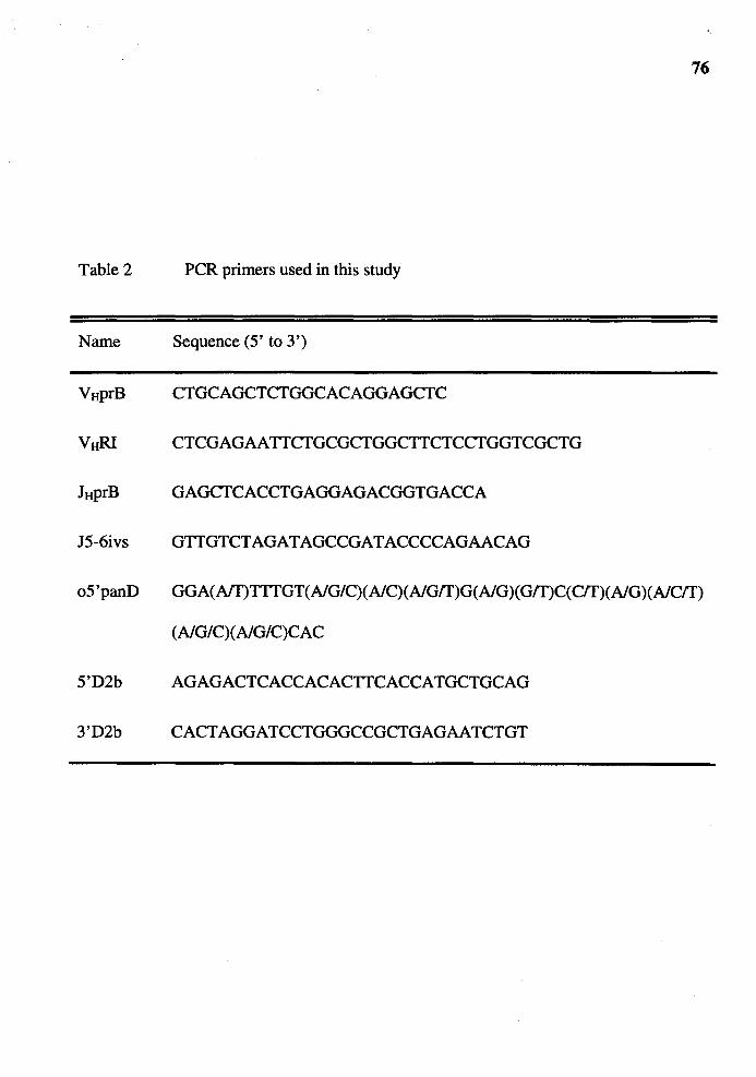
76
Table 2 PCR primers used in this study
Name Sequence (5' to 3')
VHprB CTGCAGCTCTGGCACAGGAGCTC
VHRI CTCGAGAATTCTGCGCTGGCTTCTCCTGGTCGCTG
JHprB GAGCTCACCTGAGGAGACGGTGACCA
J5-6ivs GTTGTCTAGATAGCCGATACCCCAGAACAG
o5'panD GGA(Aff)TTTGT(A/G/C)(A/C)(A/G/T)G(A/G)(G/T)C(C/T)(A/G)(A/C/T)
(A/G/C)(A/G/C)CAC
5'D2b AGAGACTCACCACACTTCACCATGCTGCAG
3'D2b CACTAGGATCCTGGGCCGCTGAGAATCTGT
77
hybridization solution (3X SSC, 5X Denhardt's, O. I% SDS and IOO µg/ml of salmon
sperm DNA) at 68°C and washed in lX blot wash at 68°C. Nucleotide sequencing of pJ5-
hybridizing clones was performed by using Sequenase (USB, Cleveland OH) and SP6
promoter primer.
DJ and VD genes from splenocytes: The D2bJ and VD2b genes were amplified by PCR
from 0.5 to 1.0 µg of splenic DNA prepared from an a3 allotype rabbit (#357D2). For
D2bJ gene amplification, 5'D2b and JHprB primers (Table 2) were used. For VD2b gene
amplification, VHRI and 3'D2b primers (Table 2) were used. The PCR reaction was
performed in IX PCR buffer (Perkin-Elmer, Norwalk CT), 0.2 µM of 5'D2b or V HRI, 0.2
µM of 1ttprB or 3'D2b, 50 µMeach of dATP, dCTP, dTTP and dGTP, and 25 units/ml of
Taq polymerase. The reaction volume was adjusted 50 µland carried out in regular PCR
tube for 30 cycles. Each cycle consists of incubation at 94 ° for I minute, at 60° for 2
minutes and at 72° for I minute. The PCR products were analyzed on 5% PAGE. The
amplified D2bJ genes were resticted with Pstl/Sacl and cloned into M13mpI8 vector. JH
probe-hybridizing clones were selected by standard dot-blot hybridization using the pJ5
probe. The hybridization was performed overnight in 3X hybridization solution (3X SSC,
5X Denhardt's, O.I % SDS and IOO µglml of salmon-sperm DNA) at 68°C and washed in
IX blot wash at 68°C. Nucleotide sequencing of Jwhybridizing clones was performed as
above using M13 universal primer. The amplified VD2b genes were resticted with
BamHl/EcoRI and cloned into MI3mpI8 vector. VH probe-hybridizing clones were
selected by standard dot-blot hybridization using the pl SI probe. The hybridization was

78
performed overnight in 3X hybridization solution (3X SSC, 5X Denhardt's, 0.1 % SOS
and 100 µg/ml of salmon-sperm DNA) at 68°C and washed in lX blot wash at 68°C.
Nuclotide sequencing of V H probe-hybridizing clones was performed as above using M13
universal primer.
VDJ genes from fetuses: The VDJ genes in fetal liver were PCR-amplified by nested
primer PCR (except for 18-day-old fetus, see below) using 0.5 to 1.0 µg of fetal liver
DNA template. The rationale for the nested-primer PCR is depicted in Figure 5. The
primary PCR was performed with VHprB and J5-6ivs primers. The final concentration of
each component in the primary PCR was as followed: lX PCR buffer (Boehringer
Mannheim), 0.2µM VttPrB, 0.2µM J5-6ivs, 50µM each of dATP, dCTP, dTTP and
dGTP, and 25 units/ml of Taq polymerase. The reaction volume was adjusted 50 µland
carried out in thin wall PCR tube for 30 cycles. Each cycle consists of incubation at 94°
for 45 seconds, at 60° for 45 seconds and at 72° for 45 seconds. Taq polymerase was not
added to the reaction until the temperature of the reaction reached 80° during the first
cycle. Ten microliters of the products from the primary PCR were used directly in the
nested-primer PCR, which was performed in an essentially identical manner as the
primary PCR except that the products from the primary PCR were used in place of fetal
liver DNA, and that V HRI and 1ttprB primers were used in place of V ttPrB and J5-6ivs
primers. Ten microliters of the products from nested-primer PCR were analyzed on 5%
PAGE. The PCR-amplified VDJ genes were either restricted with EcoRI/Sacl and cloned
into M13mp18 vector, or cloned directly into pGEM-T vector (Promega). VH probe-
Figure 5
79
Schematic representation of the location of PCR primers used in the
nested-primer PCR. The 1tt4-utilizing VDJ gene is used in the diagram but
the rationale is applicable to VDJ gene which had utilized any ltt genes.
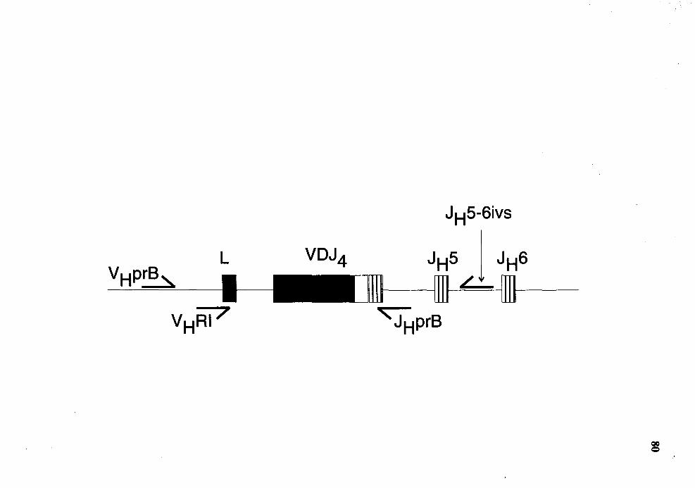
L
IHI!
JH5-6ivs
JHS 1 JH6 / ---
81
hybridizing clones were selected by standard dot-blot or standard colony lift hybridization
using the p181 probe. The hybridization was performed overnight in 3X hybridization
solution (3X SSC, 5X Denhardt's, 0.1 % SDS and 100 µg/ml of salmon-sperm DNA) at
68°C and washed in lX blot wash at 68°C.
The VDJ genes in 28-day-old fetal liver were PCR-amplified only by primary
PCR reaction without nested-primer amplification using V HPrB and JttprB as primers.
The PCR-amplified VDJ genes were then cloned directly into pGEM-T vector (Promega,
Madison WI). V H probe-hybridizing clones were selected by standard colony lift
hybridization using the p181 probe. The hybridization was performed overnight in 3X
hybridization solution (3X SSC, 5X Denhardt's, 0.1 % SDS and 100 µg/ml of salmon
sperm DNA) at 68°C and washed in lX blot wash at 68°C.
The V H probe-hybridizing clones were then subjected to nucleotide sequencing
reactions. The nucleotide sequences of all VDJ clones in this study was partial and was
determined in one orientation. The sequences obtained were all in anti-sense orientation
(read from Jtt upward to V H) and therefore, the VD and DJ junctions in each VDJ genes
can be determined with certainty. Any VDJ gene with identical VD and DJ junctions
were considered as one clone. Five of the VDJ clones were randomly chosen for complete
nucleotide sequence analysis (see legends for Figures 30, 31and34). Nonproductive VDJ
genes were defined as the genes which 1) V H and Jtt are in different reading frames, 2) a

82
stop codon is present in the D region or N-segment and 3) a pseudogene was used in the
rearrangement.
Nucleotide Sequence Analysis of VH Promoters
VHI and VJl4 promoters: the 1. 2 kb HindlIJ/Pstl fragments upstream of germline VH1-a3
and V Jt4-a3 were isolated from plasmids containing germline V HJ -a3 and V Jt4-a3
previously described (Knight and Becker, 1990). The fragment from each V H gene was
further restricted with Hpal to generate two subfragments of 700 bp and 500 bp in size.
Each of these fragments were cloned into M13mp18 and M13mp19 vectors. The clones
which hybridized to the insert were selected by standard dot-blot hybridization. The
hybridization was performed overnight in 3X hybridization solution (3X SSC, 5X
Denhardt's, 0.1 % SDS and 100 µg/ml of salmon-sperm DNA) at 68°C and washed in lX
blot wash at 68°C.
V Hy33 promoter: because no Hpal site analogous to that found in the promoters of V Hl
a3 and V Jt4-a3 was found in the VHy33 promoter, an alternative strategy was employed.
The plasmid containing germline VHy33 promoter (20Al clone) was restricted with Accl.
After the restriction was completed, the 5' overhangs generated by Accl were filled with
standard K.lenow reaction. The Klenow was heat-inactivated by incubation at 68° for 15
minutes. The DNA was then further digested with Sacl to generate several small
fragments. The 200 bp, 350 bp and 550 bp fragments were gel-purified and cloned into
M13 vector. The 200 bp fragment was cloned into M13mpl9 vector while the 350 bp and

83
550 bp fragments were cloned into both M13mp18 and M13mp19 vectors. The clones
which hybridized to the insert under standard dot-blot condition were selected. The
hybridization was performed overnight in 3X hybridization solution (3X SSC, 5X
Denhardt's, 0.1 % SDS and 100 µg/ml of salmon-sperm DNA) at 68°C and washed in lX
blot wash at 68°C.
The promoter sequences were compared using Pileup module of GCG package
(Genetics Computer Group, Inc. , Madison WI).

CHAPTER III
RESULTS
Quantification of Germline JH and V!:f 1 in
Polyclonal Rabbit 8 Cells
The order of IgH gene rearrangement during B cell development was established
in mouse (Alt et al., 1984). This order of "DJ on both lgH alleles before V to DJ" has
since been regarded as dogma of how lgH genes are rearranged in every species.
However, two findings regarding rabbit IgH gene suggested that rabbit may rearrange its
IgH genes differently. First, Southern analysis of DNA of purified rabbit B cells showed
that some of the JH genes were in germline configuration (Allegrucci et al., 1991 ). This
finding is unusual in that similar study in mouse showed no JH remained in germline
configuration (Alt et al., 1984). Further, Becker et al. found a VD gene rearrangement in
a rabbit B cell line (Becker R. and Knight KL. unpublished data). This VD gene
rearrangement has thus far never been found in mouse. To gain more insight into how
rabbit B cells rearrange their IgH genes, I performed quantitative Southern analysis for
the amount of JH and V H 1 remaining in germline configuration in rabbit B cells.
Quantification of Germline JH in Polyclonal Rabbit 8 Cells
To quantify the amount of germline JH in polyclonal rabbit B cells, I purified al+
B cells from mesenteric lymph nodes of two al/a2 heterozygous rabbits (#35G and
13806, C/E haplotype) by FACS and prepared genomic DNA from the sorted al+ B cells.
84
85
The al+ B DNA as well as DNA prepared from livers of the two rabbits were then
restricted with Hindlll/EcoRI and subjected to quantitative Southern analysis using the
JR-specific probe, pJ5 and a probe from the non-rearranging gene, RAG2. I exploited the
polymorphic EcoRI site in the JH locus of C and E haplotype rabbits (Figure 6) so that I
can distinguish the germline JH on the al (expressed) allele from that on the a2
(unexpressed) allele.
Hybridization of the Southern blot of liver DNA of the 35G rabbit with pJ5
resulted in two hybridizing bands, a 3. 5 kb and a 5 kb fragment which corresponded to
the germline JH on al and a2 alleles, respectively (Figure 7a). The hybridization of DNA
from purified al+ B cells showed that the 3. 5 kb fragment (al-associated band) was
absent (Figure 7a) while the 5 kb fragment (corresponding to the germline JH gene on the
unexpressed a2 allele) was present (Figure 7a). The amount of JH on the a2 allele in both
liver and purified al+ B cells DNA was quantified by measuring the radioactivity in the 5
kb hybridizing bands on a phosphoimager (Betagen ®). The radioactivity measured in liver
DNA represented the amount of JH which should be present in al+ B cells if there had
been no rearrangement of JH on the a2 allele in these cells. Before comparing the amount
of germline JH on the a2 allele of purified al+ B cells with that in liver, I normalized the
amount of DNA loaded in each lane by rehybridizing the Southern blot with RAG2 probe
and comparing the amount of RAG2 hybridization of DNA from the al+ B cells with that
of liver. The radioactivity in these samples was determined on the Betagen ®. Because the
RAG2 gene does not undergo rearrangement in either liver or B cells, the amount of
RAG2 can be used as reference for the amount of DNA in each lane. From the amount of
the radioactivity of pJ5 and RAG2 (Figure 7a), I calculated the amount of germline JH on
the unexpressed a2 allele of al+ B cells as shown in Figure 7b. I found that 75% of the
theoretical amount of JH on the a2 allele was in germline configuration in al+ B c"ells
suggesting that 75% of al+ B cells did not rearrange JH on their unexpressed IgH allele.
: ~
Figure 6 A partial map of the V, D and J chromosomal region from the al (C
haplotype ), a2 (E haplotype) and a3 (G haplotype) allotype rabbits. The
regions from which probe A and JH probes are derived are designated by
brackets. The expected restriction fragments that hybridize with probe A
and JH probes for each haplotye are shown with double-headed arrows.
The expected sizes are in kb.
86

V region D region J region
VH3 VH2 VH1 H H H H
!+:j!j 11111111111 I c:i·4m a1[C) ~ I ~5~ 8.1 =+I B B E - -prtileA JHprtile
VH3 VH2 VH1 H H H 64 H 11111111111 I );iif a2[E) 3.5
E - -prtileA JHprtile
a3[G)
H H 11111111111 I
H a.4 H C-1111ij1 -JI-I prOOe
-prtileA
Figure 7
88
Quantitative Southern analysis of EcoRI/Hindlll-restricted DNA for
germline ltt in al+ B cells of rabbit number 35G. a) The autoradiogram of
pJ5 (upper panel) and RAG2 (lower panel) hybridization. The hybridizing
fragments associated with the germline ltt on the al and a2 alleles are
indicated. The numbers in the diagrams next to the autoradiogram denote
the amount of radioactivity of the indicated fragments (total counts). b)
The calculation for the amount of germline ltt on the a2 allele of al+ B
cells.
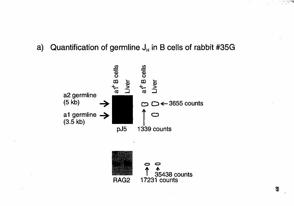
a) Quantification of germline JH in B cells of rabbit #35G
a2 germline (5 kb) ~
a1 germline ~ (3.5 kb)
en -Q) ()
co """ -i;.... ~
ca ~
c:> D ~ 3655 counts
j a
pJ5 1339 counts
0 0
t :5438 counts RAG2 17231 counts

b) Quantification for the amount of germline J" in al+ B cells from 35G
pJ5:RAG2 ratio in Liver
Maximum ratio of pJ5:RAG2 in B cells
Observed ratio of pJ5:RAG2 in B cells
=
=
3655 I 35438
1339 I 17231
= 0.103
= 0.103
= 0.078
The percentage of remaining pJ5:RAG2 ratio to the maximum pJ5:RAG2 ratio
= (0.078 I 0.103) x 100 = 75%
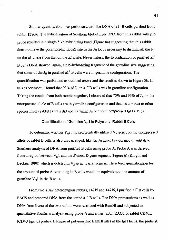
91
Similar quantification was performed with the DNA of al+ B cells purified from
rabbit 13806. The hybridization of Southern blot of liver DNA from this rabbit with p15
probe resulted in a single 5 kb hybridizing band (Figure Sa) suggesting that this rabbit
does not have the polymorphic EcoRI site in the 1H locus necessary to distinguish the 1H
on the al allele from that on the a2 allele. Nevertheless, the hybridization of purified al+
B cells DNA showed, again, a p15-hybridizing fragment of the germline size suggesting
that some of the 1H in purified al+ B cells were in germline configuration. The
quantification was performed as outlined above and the result is shown in Figure Sb. In
this experiment, I found that 93% of 1H in al+ B cells was in germline configuration.
Taking the results from both rabbits together, I observed that 75% and 93% of 1H on the
unexpressed allele of B cells are in germline configuration and that, in contrast to other
species, many rabbit B cells did not rearrange 1H on their unexpressed IgH alleles.
Quantification of Germline VH 1 in Polyclonal Rabbit B Cells
To determine whether V HJ, the preferentially utilized V H gene, on the unexpressed
allele of rabbit B cells is also unrearranged, like the 1H gene, I performed quantitative
Southern analysis of DNA from purified B cells using probe A. Probe A was derived
from a region between VHl and the 5'-most D gene segment (Figure 6) (Knight and
Becker, 1990) which is deleted in VH gene rearrangement. Therefore, quantification for
the amount of probe A remaining in B cells would be equivalent to the amount of
germline V H 1 in the B cells.
From two al/a2 heterozygous rabbits, 14715 and 14716, I purified al+ B cells by
FACS and prepared DNA from the sorted al+ B cells. The DNA preparations as well as
DNA from livers of the two rabbits were restricted with BamHI and subjected to
quantitative Southern analysis using probe A and either rabbit RAG2 or rabbit CD40L
(CD40 ligand) probes. Because of polymorphic BamHI sites in the IgH locus, the probe A
Figure 8
92
Quantitative Southern analysis of EcoRI/HindIII-restricted DNA for
germline Jtt in al+ B cells of rabbit number 13806. a) The autoradiogram
of pJ5 (upper panel) and RAG2 (lower panel) hybridization. The
hybridizing fragment associated with the germline Jtt is indicated. The
numbers in the diagrams next to the autoradiogram denote the amount of
radioactivity of the indicated fragments (total counts). b) The calculation
for the amount of germline JH on the unexpressed allele of al+ B cells.
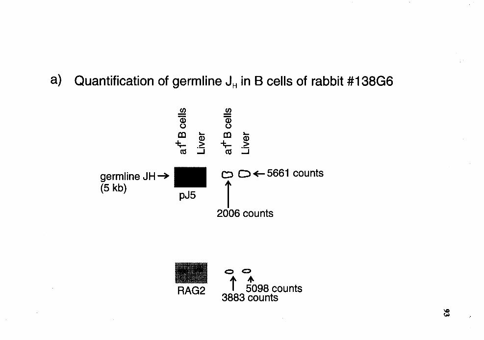
a) Quantification of germline JH in B cells of rabbit #138G6
germline JH-+ (5 kb)
pJ5
RAG2
c:> D ~ 5661 counts
i 2006 counts
0 0
t toga counts 3883 counts
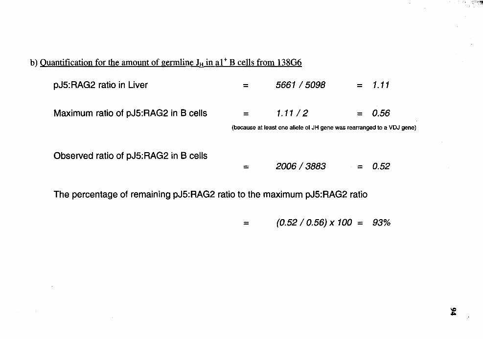
b) Quantification for the amount of germline 1tt in al+ B cells from 13806
pJ5: RAG2 ratio in Liver = 5661I5098 = 1.11
Maximum ratio of pJ5:RAG2 in B cells = 1.1112 = 0.56 (because at least one allele of JH gene was rearranged to a VDJ gene)
Observed ratio of pJ5: RAG2 in B cells = 2006 I 3883 = 0.52
The percentage of remaining pJ5:RAG2 ratio to the maximum pJ5:RAG2 ratio
= (0.52 I 0.56) x 100 = 93%
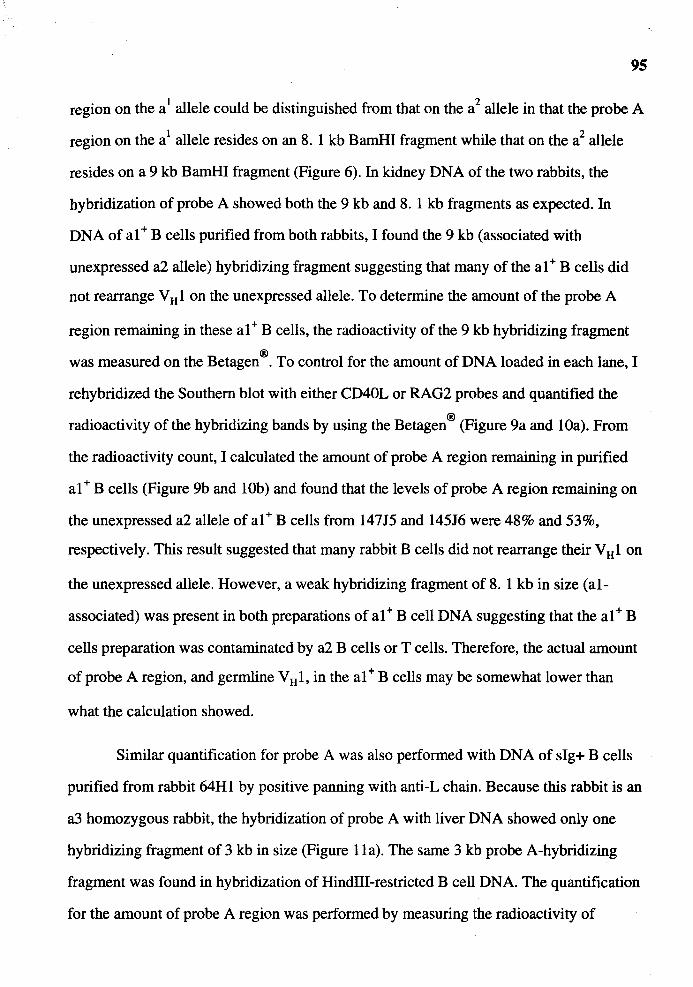
95
region on the a1 allele could be distinguished from that on the a2 allele in that the probe A
region on the a1 allele resides on an 8. 1 kb BamHI fragment while that on the a2 allele
resides on a 9 kb BamHI fragment (Figure 6). In kidney DNA of the two rabbits, the
hybridization of probe A showed both the 9 kb and 8. 1 kb fragments as expected. In
DNA of al+ B cells purified from both rabbits, I found the 9 kb (associated with
unexpressed a2 allele) hybridizing fragment suggesting that many of the al+ B cells did
not rearrange V H 1 on the unexpressed allele. To determine the amount of the probe A
region remaining in these al+ B cells, the radioactivity of the 9 kb hybridizing fragment
was measured on the Betagen®. To control for the amount of DNA loaded in each lane, I
rehybridized the Southern blot with either CD40L or RAG2 probes and quantified the
radioactivity of the hybridizing bands by using the Betagen ® (Figure 9a and lOa). From
the radioactivity count, I calculated the amount of probe A region remaining in purified
al+ B cells (Figure 9b and lOb) and found that the levels of probe A region remaining on
the unexpressed a2 allele of al+ B cells from 14715 and 14516 were 48% and 53%,
respectively. This result suggested that many rabbit B cells did not rearrange their VHl on
the unexpressed allele. However, a weak hybridizing fragment of 8. 1 kb in size (al
associated) was present in both preparations of al+ B cell DNA suggesting that the al+ B
cells preparation was contaminated by a2 B cells or T cells. Therefore, the actual amount
of probe A region, and germline V H 1, in the al+ B cells may be somewhat lower than
what the calculation showed.
Similar quantification for probe A was also performed with DNA of slg+ B cells
purified from rabbit 64Hl by positive panning with anti-L chain. Because this rabbit is an
a3 homozygous rabbit, the hybridization of probe A with liver DNA showed only one
hybridizing fragment of 3 kb in size (Figure l la). The same 3 kb probe A-hybridizing
fragment was found in hybridization of Hindlll-restricted B cell DNA. The quantification
for the amount of probe A region was performed by measuring the radioactivity of
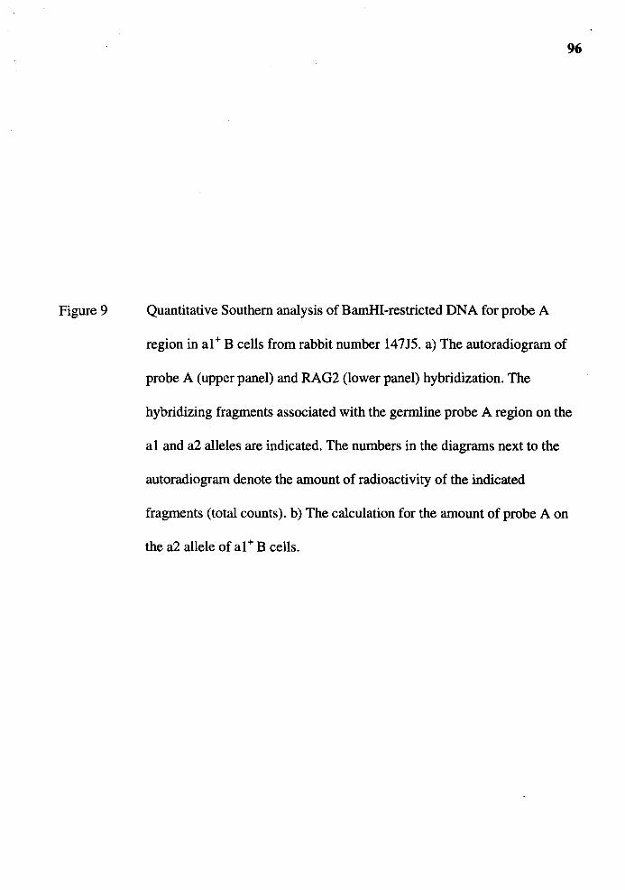
Figure 9
96
Quantitative Southern analysis of BamHI-restricted DNA for probe A
region in al+ B cells from rabbit number 14715. a) The autoradiogram of
probe A (upper panel) and RAG2 (lower panel) hybridization. The
hybridizing fragments associated with the germline probe A region on the
al and a2 alleles are indicated. The numbers in the diagrams next to the
autoradiogram denote the amount of radioactivity of the indicated
fragments (total counts). b) The calculation for the amount of probe A on
the a2 allele of al+ B cells.
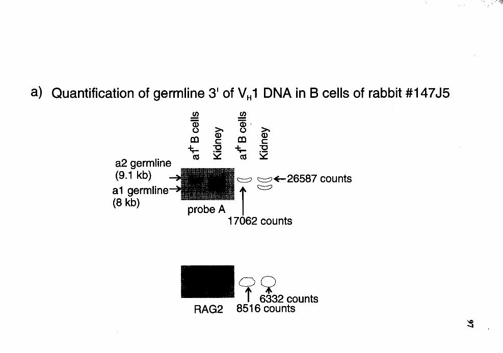
a) Quantification of germ line 3' of V H 1 DNA in B cells of rabbit #14 7 JS
a2 germline (9.1 kb)
a1 germline (8 kb)
J!l C/J
Q) >.
Q) . >. 0 0
al Q)
al Q)
c: c: -f;... "O -f;... "O
as ~ as ~
c:::::? c:::::?+-26587 counts
probe A l c:::::?
17062 counts
C)9 t 6332 counts
RAG2 8516 counts
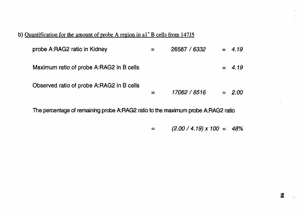
b) Quantification for the amount of probe A region in al+ B cells from 147J5
probe A:RAG2 ratio in Kidney
Maximum ratio of probe A: RAG2 in B cells
Observed ratio of probe A:RAG2 in B cells
=
=
26587 I 6332
17062 I 8516
= 4.19
= 4.19
= 2.00
The percentage of remaining probe A:RAG2 ratio to the maximum probe A:RAG2 ratio
= (2.00 I 4.19) x 100 = 48%
Figure 10
99
Quantitative Southern analysis of BamHI-restricted DNA for probe A
region in al+ B cells from rabbit number l 47J6. a) The autoradiogram of
probe A (upper panel) and CD40L (lower panel) hybridization. The
hybridizing fragments associated with the germline probe A region on the
al and a2 alleles are indicated. The numbers in the diagrams next to the
autoradiogram denote the amount of radioactivity of the indicated
fragments (total counts). b) The calculation for the amount of probe A on
the a2 allele of al+ B cells.
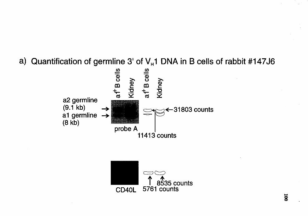
a) Quantification of germline 31 of VH 1 DNA in B cells of rabbit #147J6
a2 germline (9.1 kb) __.. a 1 germ line __,. (8 kb)
"' Q) ~ (.)
CD Q) c
-f;.... "'C
ctS ~
probe A
.!fl. Q)
~ (.)
CD Q) c
-f;.... "'C ctS ~
l~+-31803 counts
-===- eJ
11413 counts
c=J <:::::::)
t 0t35 counts CD40L 5761 counts
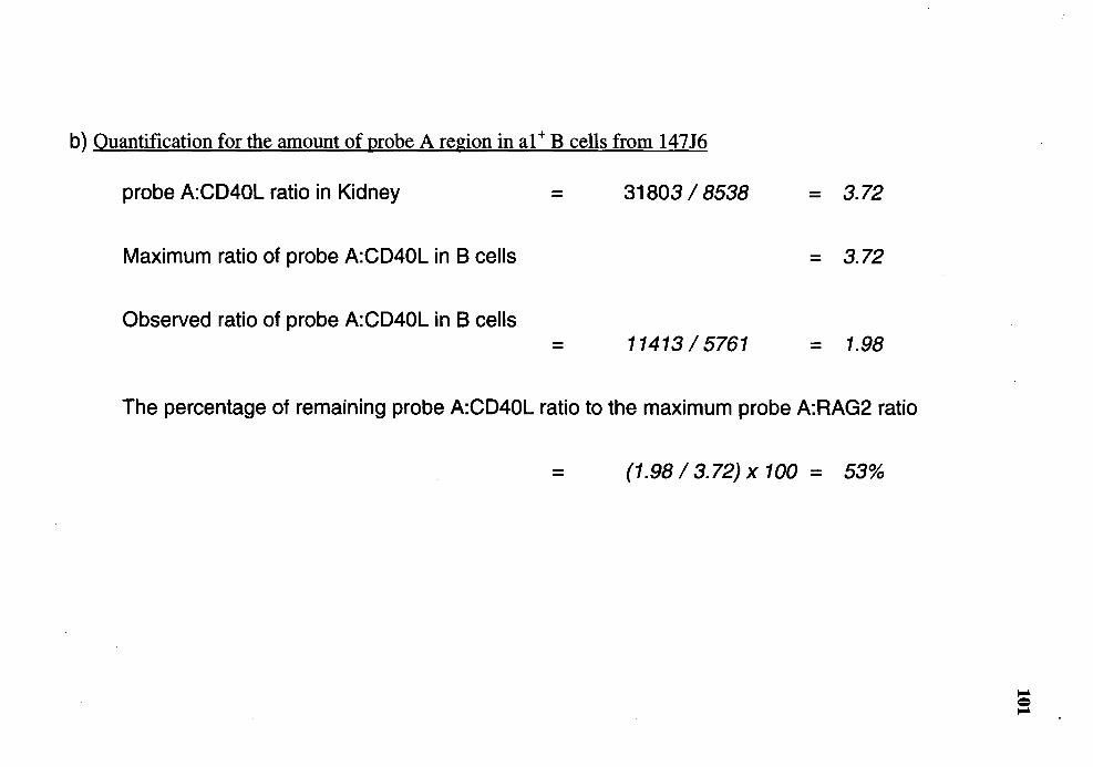
b) Quantification for the amount of probe A region in al+ B cells from 147J6
probe A:CD40L ratio in Kidney = 31803 I 8538 = 3.72
Maximum ratio of probe A:CD40L in B cells = 3.72
Observed ratio of probe A:CD40L in B cells 11413 I 5761 = 1.98
The percentage of remaining probe A:CD40L ratio to the maximum probe A:RAG2 ratio
(1.98 I 3.72) x 100 = 53%
Figure 11
102
Quantitative Southern analysis of Hindill-restricted DNA for probe A
region in slg+ B cells from rabbit number 64Hl. a) The autoradiogram of
probe A (upper panel) and RAG2 (lower panel) hybridization. The
hybridizing fragment associated with the germline probe A region (3 kb) is
indicated. The numbers in the diagrams next to the autoradiogram denote
the amount ofradioactivity of the indicated fragments (total counts). b)
The calculation for the amount of probe A on the unexpressed allele of
slg+ B cells.
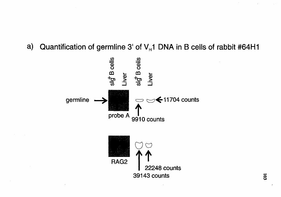
a) Quantification of germline 3' of VH 1 DNA in B cells of rabbit #64H1
germline
en .!!). -Cl) Cl) 0 0 cc - cc -~
Cl) ~
Cl)
> > ~ - ~ en en
) • f <::::::?~ 11704 counts
probe A 9910 counts
uv RAG2 t t
22248 counts 39143 counts
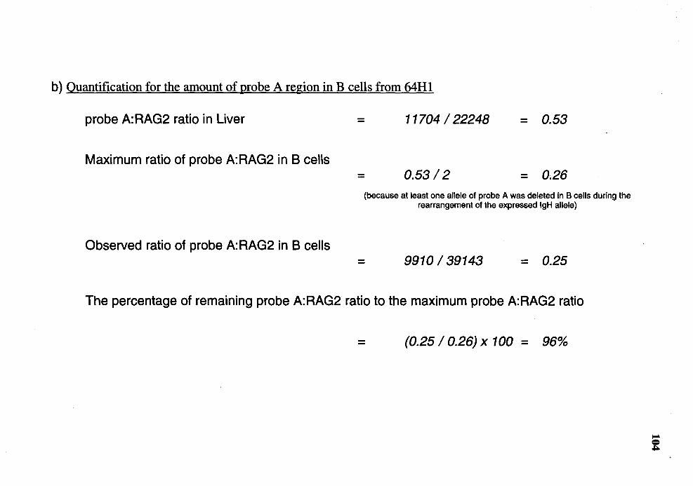
b) Quantification for the amount of probe A region in B cells from 64Hl
probe A:RAG2 ratio in Liver
Maximum ratio of probe A:RAG2 in B cells
Observed ratio of probe A: RAG2 in B cells
= 11704 I 22248 = 0.53
= 0.53 I 2 = 0.26 (because at least one allele of probe A was deleted in B cells during the
rearrangement of the expressed lgH allele)
= 9910 I 39143 = 0.25
The percentage of remaining probe A:RAG2 ratio to the maximum probe A:RAG2 ratio
= (0.25 I 0.26) x 100 = 96%
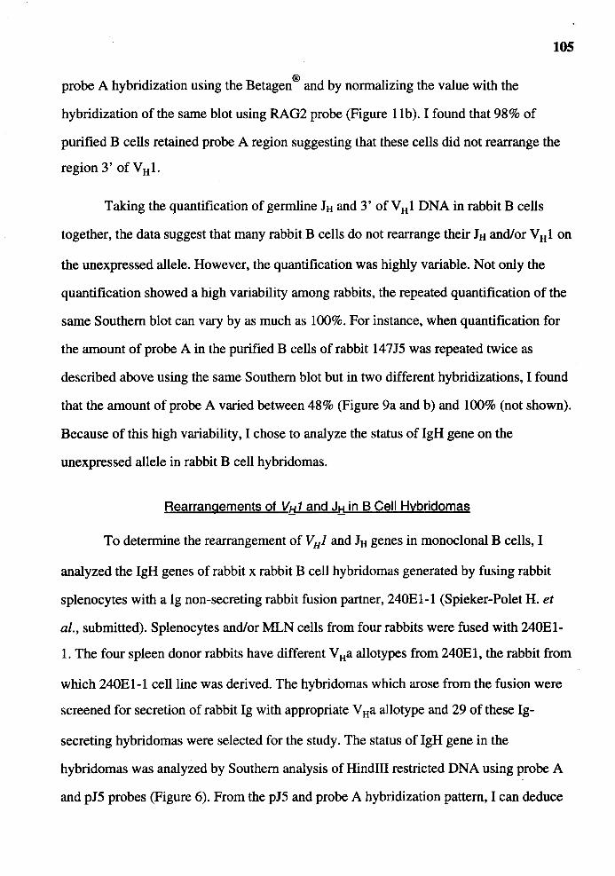
105
probe A hybridization using the Betagen ® and by normalizing the value with the
hybridization of the same blot using RAG2 probe (Figure 1 lb). I found that 98% of
purified B cells retained probe A region suggesting that these cells did not rearrange the
region 3' of VHl.
Taking the quantification of germline JH and 3' of V H 1 DNA in rabbit B cells
together, the data suggest that many rabbit B cells do not rearrange their JH and/or V H 1 on
the unexpressed allele. However, the quantification was highly variable. Not only the
quantification showed a high variability among rabbits, the repeated quantification of the
same Southern blot can vary by as much as 100%. For instance, when quantification for
the amount of probe A in the purified B cells of rabbit 14715 was repeated twice as
described above using the same Southern blot but in two different hybridizations, I found
that the amount of probe A varied between 48% (Figure 9a and b) and 100% (not shown).
Because of this high variability, I chose to analyze the status of lgH gene on the
unexpressed allele in rabbit B cell hybridomas.
Rearrangements of VH1 and JH in B Cell Hybridomas
To determine the rearrangement of V HJ and JH genes in monoclonal B cells, I
analyzed the lgH genes of rabbit x rabbit B cell hybridomas generated by fusing rabbit
splenocytes with a lg non-secreting rabbit fusion partner, 240El-1 (Spieker-Polet H. et
al., submitted). Splenocytes and/or MLN cells from four rabbits were fused with 240El-
1. The four spleen donor rabbits have different V Ha allotypes from 240E 1, the rabbit from
which 240El-1 cell line was derived. The hybridomas which arose from the fusion were
screened for secretion of rabbit lg with appropriate V Ha allotype and 29 of these lg-
secreting hybridomas were selected for the study. The status of lgH gene in the
hybridomas was analyzed by Southern analysis of Hindlll restricted DNA using probe A
and pJ5 probes (Figure 6). From the pJ5 and probe A hybridization pattern, I can deduce
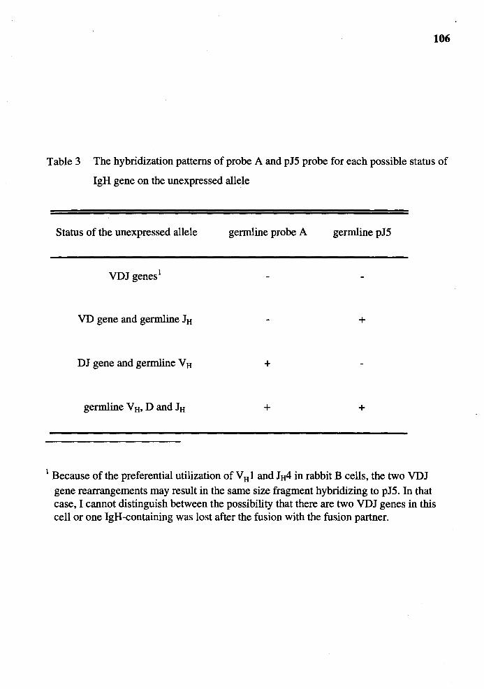
106
Table 3 The hybridization patterns of probe A and pJ5 probe for each possible status of
IgH gene on the unexpressed allele
Status of the unexpressed allele germline probe A germline pJ 5
VDJ genes1
VD gene and germline JH +
DJ gene and germline V H +
germline V H• D and JH + +
1 Because of the preferential utilization of V H 1 and 1tt4 in rabbit B cells, the two VDJ gene rearrangements may result in the same size fragment hybridizing to pJ5. In that case, I cannot distinguish between the possibility that there are two VDJ genes in this cell or one IgH-containing was lost after the fusion with the fusion partner.
107
the status of the unexpressed IgH allele of the splenocyte as shown in Table 3. The status
of the IgH gene in Table 3 represents theoretical status. In B cells of mouse, human and
chicken (Alt et al., 1984, Nickerson et al., 1989, Reynaud et al., 1987, Barth and
Humphries, 1988, Benatar et al., 1992) only two of these possible rearrangements, DJ and
VDJ genes, were generally found on the second IgH allele.
First the status of IgH gene in the rabbit fusion partner was examined by Southern
analysis of DNA from the fusion partner cell line as well as from liver DNA of rabbit
#240El, the rabbit from which the fusion partner was derived. The Southern blot of
HindIII-restricted DNA was hybridized sequentially with the pJ5 probe and probe A. For
the hybridization with the pJ5 probe, two hybridizing bands were detected in the rabbit
fusion partner. One hybridizing fragment is 6. 2 kb in size (Figure 12), the same size as
the germline JH fragment seen in liver DNA, suggesting that the JH on this unexpressed
allele of the fusion partner was not rearrranged. Another pJ5-hybridizing fragment is a
rearranged JH of 4. 1 kb in size which must be a VDJ gene because the fusion partner
initially secreted lg in culture. The status of IgH gene on the unexpressed IgH allele was
further examined by hybridization using probe A. The hybridization showed a hybridizing
6. 5 kb fragment, the germline size for probe A region as seen in DNA from liver of
rabbit #240El (Figure 12). This result suggests that VHl on the unexpressed allele was
not rearranged. From these results, I conclude that one IgH allele of the fusion partner
was in germline configuration and the other allele was a VDJ gene.
Because the fusion partner retained its IgH gene in germline configuration on the
unexpressed allele, the pattern of hybridization in the hybridomas is complex.
Nevertheless, because of HindIII polymorphism in the the lgH locus, the IgH gene (both
JH and probe A regions) of the splenocytes can be distinguished from that of the fusion
partner. The 29 hybridomas I chose to analyze were cloned by limiting dilution and

Figure 12
108
Southern analysis for the status of IgH genes in the fusio~ partner. DNA of
fusion partner (240El-1) was restricted by Hindill, Southern blotted and
hybridized with either probe A (left panel) or pJ5 probe (right panel).
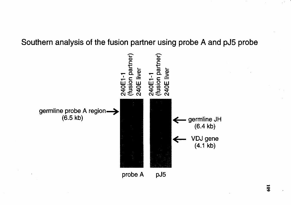
Southern analysis of the fusion partner using probe A and pJ5 probe
germline probe A region~ (6.5 kb)
-I... Q) c: t CtS I...
T'9 a. ~ I C: ·-T- -
w.o w o en o v :::::J v C\I !!::::- C\I
'I:" Q) c: t CtS I...
T'9 a. ~ I C: ·-T- -
w.o w o en o v :::::J v C\I ~ C\I
probe A pJ5
~ germline JH (6.4 kb)
~ VDJgene (4.1 kb)
110
therefore, are monoclonal. Two subclones from each hybridoma were analyzed, if more
than one subclone was obtained from the limiting dilution. In general, the two subclones
derived from the same parental hybridomas gave the same result. From the hybridization
with the pJ5 probe, each hybridoma had two or more hybridizing fragments, two of which
were derived from the fusion partner. Because these hybridomas were selected for the
secretion of lg bearing V Ha allotypic determinant, each of them must have at least one
productive VDJ gene derived from the splenocytes. Because rabbit B cells preferentially
utilized VHl and JH4 genes (Knight and Becker, 1990, Friedman et al., 1994) in their VDJ
gene rearrangements, it is likely that the splenocytes utilized V H 1 and Jtt4 which resulted
in a rearranged 4. 1 kb Hindlll fragment and the resulting VDJ gene would be
indistinguishable from the VDJ gene of the fusion partner. The hybridization with the pJ5
probe confirmed this prediction (Figures 13-16). For example, Hyb 21-1 (Figure 13) did
not have any rearranged JH genes except for the 4. 1 kb Hindlll. The hybridization was
informative regarding the status of the JH gene on the unexpressed allele, however.
Fifteen of the 29 hybridoma clones examined had a 6. 4 kb (germline) JH-hybridizing
fragment suggesting that JH genes on the unexpressed allele were in germline
configuration (Figures 13-16 and Tables 4-7). The remaining 14 hybridomas did not have
the germline-sized band that hybridized to the JH probe suggesting that these hybridomas
had rearranged a DJ or VDJ gene rearrangement on the second allele (Figure 13-16 and
Tables 4-7).
To examine the rearrangement of V H 1 on the second IgH allele, I removed the pJ5
probe from the genomic blot by using boiled-0. lX blot wash and rehybridized the blot
with probe A. The 6. 5 kb probe A hybridizing fragment of the fusion partner was found
in all the hybridomas I examined. In addition to this 6. 5 kb fragment, I found that in 26
of the 29 hybridomas, probe A hybridized to a fragment of 4. 5 kb or 2. 4 kb in s~ze
(Figures 17-20 and Tables 4-8). Because the 4. 5 and 2. 4 kb fragments which hybridized
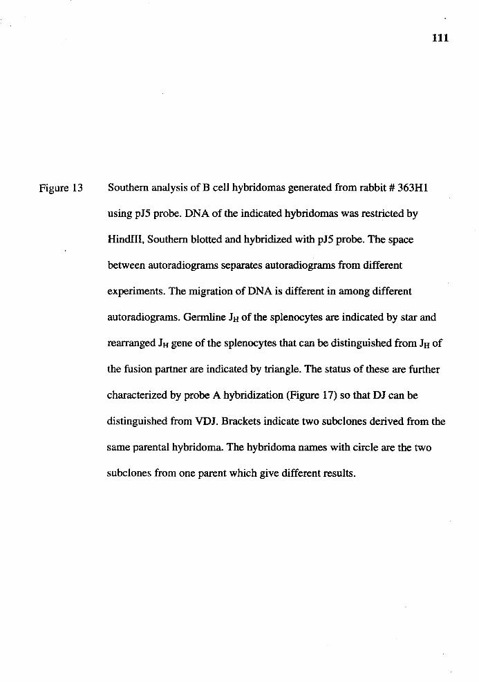
Figure 13
111
Southern analysis of B cell hybridomas generated from rabbit # 363Hl
using pJ5 probe. DNA of the indicated hybridomas was restricted by
Hindlll, Southern blotted and hybridized with pJ5 probe. The space
between autoradiograms separates autoradiograms from different
experiments. The migration of DNA is different in among different
autoradiograms. Germline JH of the splenocytes are indicated by star and
rearranged JH gene of the splenocytes that can be distinguished from JH of
the fusion partner are indicated by triangle. The status of these are further
characterized by probe A hybridization (Figure 17) so that DJ can be
distinguished from VDJ. Brackets indicate two subclones derived from the
same parental hybridoma. The hybridoma names with circle are the two
subclones from one parent which give different results.
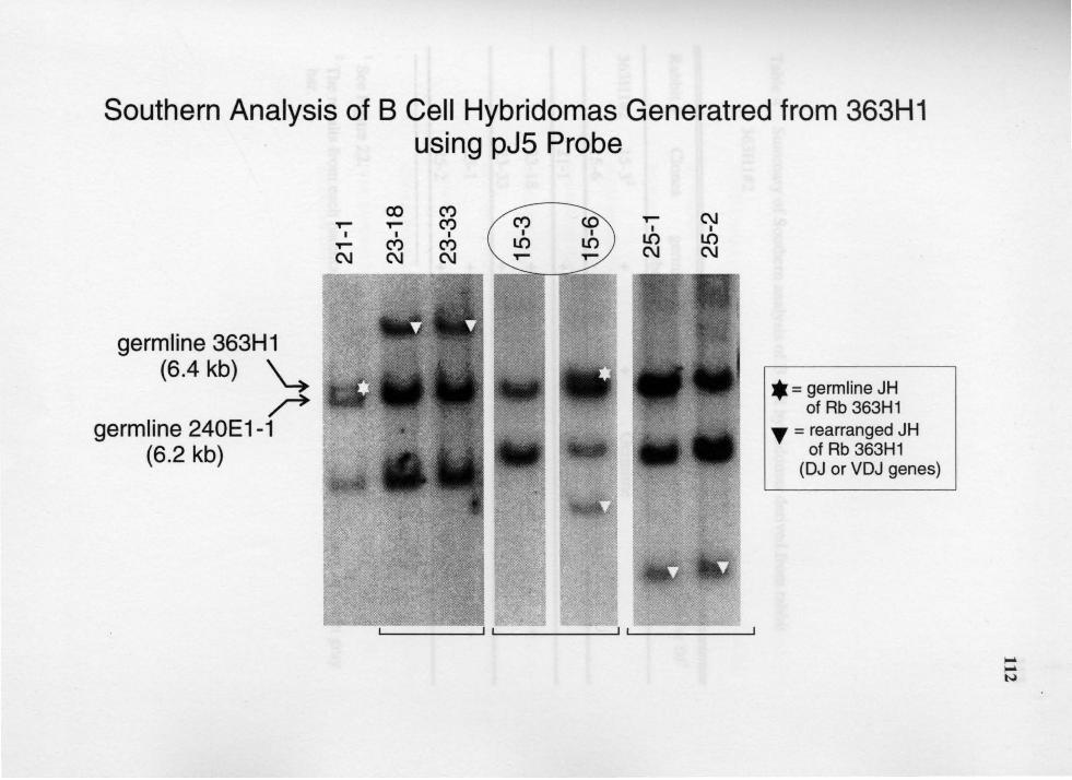
Southern Analysis of B Cell Hybridomas Generatred from 363H1 using pJ5 Probe
germline 363H1 (6.4 kb) ~
r germline 240E1-1
(6.2 kb)
co C') <O
I I
LO LO
'r" C\J I I
LO LO C\J C\J
* = germline JH of Rb 363H1
T = rearranged JH of Rb 363H1
(DJ or VDJ genes)
Table 4 Summary of Southern analysis of B cell hybridomas derived from rabbit
363H1#2
113
Rabbit# Clones germline germline JH Conclusion PCRforDJ1
robe A
363H1#2 15-32 + + Germline
15-6 excluded (see text)
21-1 + + Germline
23-18 + DJ gene +
23-33 + DJ sene
25-1 + DJ gene + 25-2 + DJ sene
1 See Figure 22. 2 The results from each parental hybridoma are separated from each other by a thin gray
bar.
Figure 14
114
Southern analysis of B cell hybridomas hybridomas generated from rabbit
# 27Hl using pJ5 probe. DNA of the indicated hybridomas was restricted
by Hindlll, Southern blotted and hybridized with pJ5 probe. The space
between autoradiograms separates autoradiograms from different
experiments. The migration of DNA is different in among different
autoradiograms. Germline JH of the splenocytes are indicated by star and
rearranged JH gene of the splenocytes that can be distinguished from JH of
the fusion partner are indicated by triangle. The status of these are further
characterized by probe A hybridization (Figure 18) so that DJ can be
distinguished from VDJ. Brackets indicate two subclones derived from the
same parental hybridoma. The hybridoma names with circle are the two
subclones from one parent which give different results.
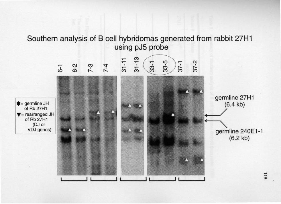
Southern analysis of B cell hybridomas generated from rabbit 27H1 using pJ5 probe
•= germline JH of Rb 27H1
T= rearranged JH of Rb 27H1
(DJ or VDJ genes)
,... C") ,... ,... ,... C\J C") ~
I I I I
I I ,... ,... c.o c.o """' """' C") C")
C\J I
"""' C")
___ ,, -___ , _, ___ , _, ___ , , ___ _
116
Table 5 Summary of Southern analysis of B cell hybridomas derived from rabbit 27Hl
Rabbit# Clones germline germline JH Conclusion PCR forDJ1
robe A
27Hl 6-12 + DJ gene + 6-2 + DJ sene
7-3 + DJ gene + 7-4 + DJ sene
31-11 VDJ gene
31-13 VDJ sene
33-1 excluded (see text)
33-5 + + Germline
37-1 + DJ gene + 37-2 + DJ ~ene
1 See Figure 22. 2 The results from each parental hybridoma are separated from each other by a thin gray
bar.
Figure 15
117
Southern analysis of B cell hybridomas generated from rabbit # 196G2
using pJ5 probe. DNA of the indicated hybridomas was restricted by
HindIII, Southern blotted and hybridized with pJ5 probe. The space
between autoradiograms separates autoradiograms from different
experiments. The migration of DNA is different in among different
autoradiograms. Germline J8 of the splenocytes are indicated by star and
rearranged JH gene of the splenocytes that can be distinguished from J8 of
the fusion partner are indicated by triangle. The status of these are further
characterized by probe A hybridization (Figure 19) so that DJ can be
distinguished from VDJ. Brackets indicate two subclones derived from the
same parental hybridoma. Hyb 11-1 does not have 4.1 kb pJ5-hybridizing
fragment suggesting that it had lost the VDJ gene of the fusion partner.

Southern analysis of B cell hybridomas generated from rabbit 196G2 using pJ5 probe
T""" T""" C\J I I I
T""" C\J T""" C\J C\J I I I I
I Q Q T""" T""" C\J ('I) ('I) LO T""" T""" T""" T"""
germ line 196G2 (6.4 kb)
germ line .,__ __ 240E1-1
(6.2 kb)
* = germline JH of Rb 196G2
T =rearranged JH of Rb 196G2
(DJ or VDJ genes)
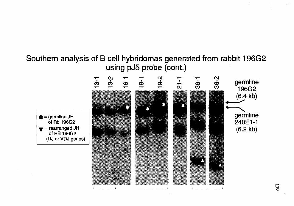
Southern analysis of B cell hybridomas generated from rabbit 196G2 using pJ5 probe (cont.)
$ = germline JH of Rb 196G2
~=rearranged JH of RB 196G2
(DJ or VDJ genes)
T""" C\J I I
C") C") T""" T"""
T""" T""" C\J I I I co m m
T""" T""" T"""
T""" T""" C\J I I I germline T""" co co
C\J C") C") 196G2 (6.4 kb)
E /
~ germ line 240E1-1 (6.2 kb)
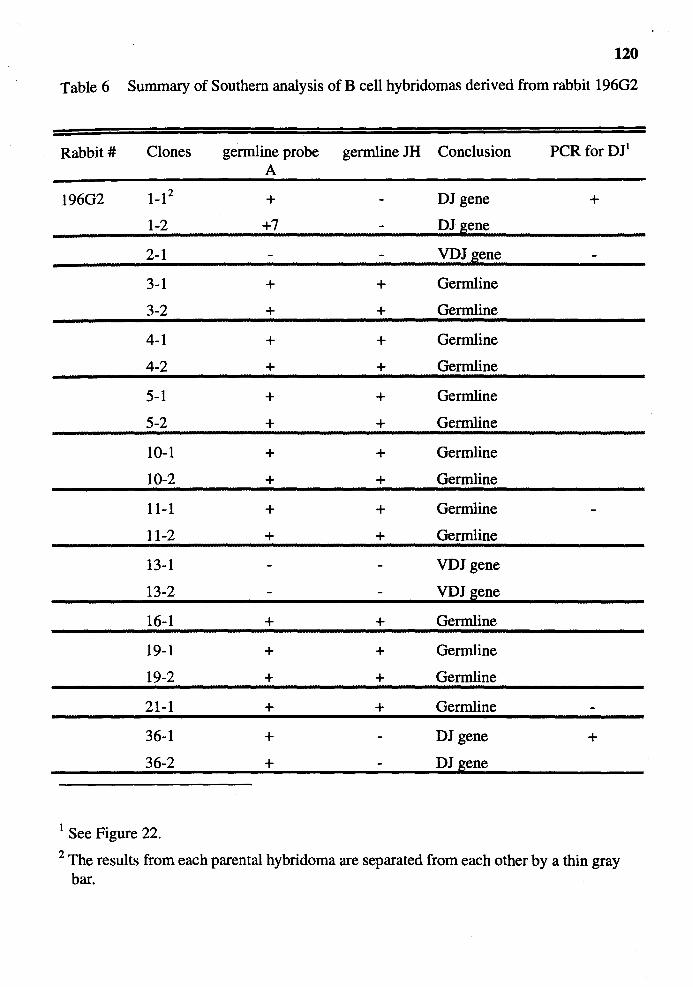
120
Table 6 Summary of Southern analysis ofB cell hybridomas derived from rabbit 19602
Rabbit# Clones germline probe germline JH Conclusion PCRforDJ1
A
19602 1-12 + DJ gene +
1-2 +7 DJ gene
2-1 VDJ gene
3-1 + + Oermline
3-2 + + Oermline
4-1 + + Oermline
4-2 + + Oermline
5-1 + + Germline
5-2 + + Oermline
10-1 + + Oermline
10-2 + + Oermline
11-1 + + Oermline
11-2 + + Germline
13-1 VDJ gene
13-2 VDJ ~ene
16-1 + + Oermline
19-1 + + Oermline
19-2 + + Oermline
21-1 + + Oermline
36-1 + DJ gene +
36-2 + DJ sene
1 See Figure 22. 2 The results from each parental hybridoma are separated from each other by a thin gray
bar.
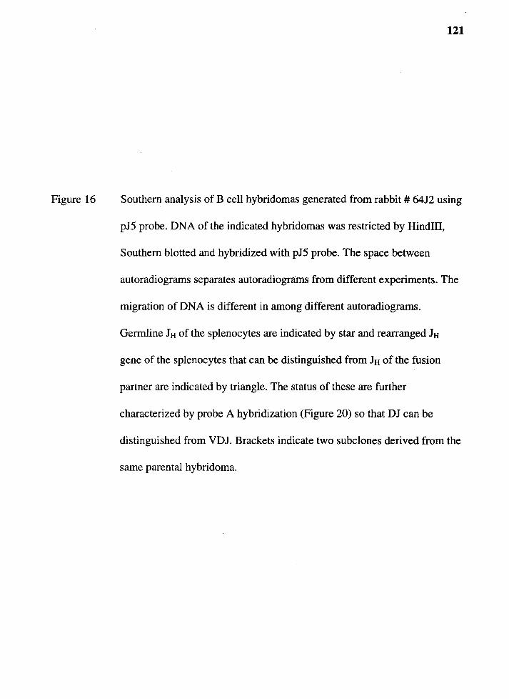
Figure 16
121
Southern analysis of B cell hybridomas generated from rabbit # 6412 using
pJ5 probe. DNA of the indicated hybridomas was restricted by Hindill,
Southern blotted and hybridized with pJ5 probe. The space between
autoradiograms separates autoradiograms from different experiments. The
migration of DNA is different in among different autoradiograms.
Germline Itt of the splenocytes are indicated by star and rearranged Itt
gene of the splenocytes that can be distinguished from Itt of the fusion
partner are indicated by triangle. The status of these are further
characterized by probe A hybridization (Figure 20) so that DJ can be
distinguished from VDJ. Brackets indicate two subclones derived from the
same parental hybridoma.
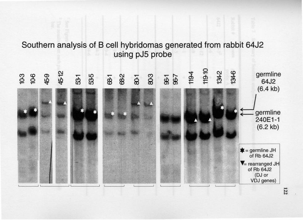
Southern analysis of B cell hybridomas generated from rabbit 64J2 using pJ5 probe
germ line 64J2
(6.4 kb)
~germline 240E1-1 (6.2 kb)
* = germline JH of Rb 64J2
T= rearranged J H of Rb 64J2
(DJ or VDJ genes)
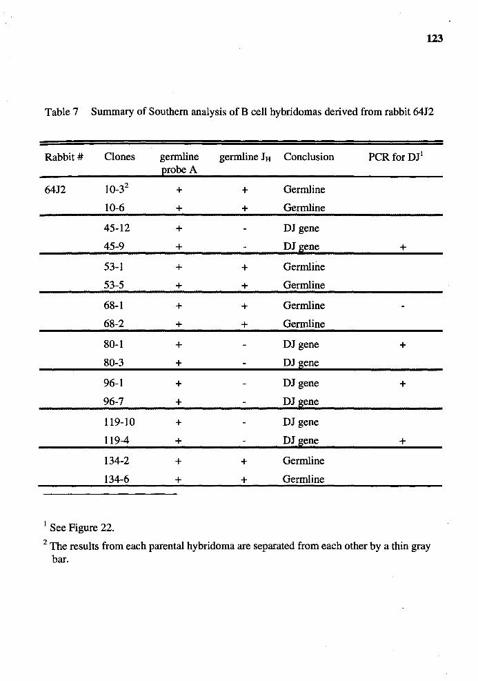
123
Table 7 Summary of Southern analysis of B cell hybridomas derived from rabbit 64J2
Rabbit# Clones germline germline JH Conclusion PCR forDJ1
robe A
64J2 10-32 + + Germline
10-6 + + Germline
45-12 + DJ gene
45-9 + DJ gene +
53-1 + + Germline
53-5 + + Germline
68-1 + + Germline
68-2 + + Germline
80-1 + DJ gene + 80-3 + DJ gene
96-1 + DJ gene + 96-7 + DJ gene
119-10 + DJ gene
119-4 + DJ gene +
134-2 + + Germline
134-6 + + Germline
1 See Figure 22. 2 The results from each parental hybridoma are separated from each other by a thin gray
bar.
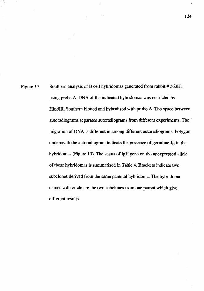
Figure 17
124
Southern analysis of B cell hybridomas generated from rabbit# 363Hl
using probe A. DNA of the indicated hybridomas was restricted by
Hindlll, Southern blotted and hybridized with probe A. The space between
autoradiograms separates autoradiograms from different experiments. The
migration of DNA is different in among different autoradiograms. Polygon
underneath the autoradiogram indicate the presence of germline JH in the
hybridomas (Figure 13). The status oflgH gene on the unexpressed allele
of these hybridomas is summarized in Table 4. Brackets indicate two
subclones derived from the same parental hybridoma. The hybridoma
names with circle are the two subclones from one parent which give
different results.
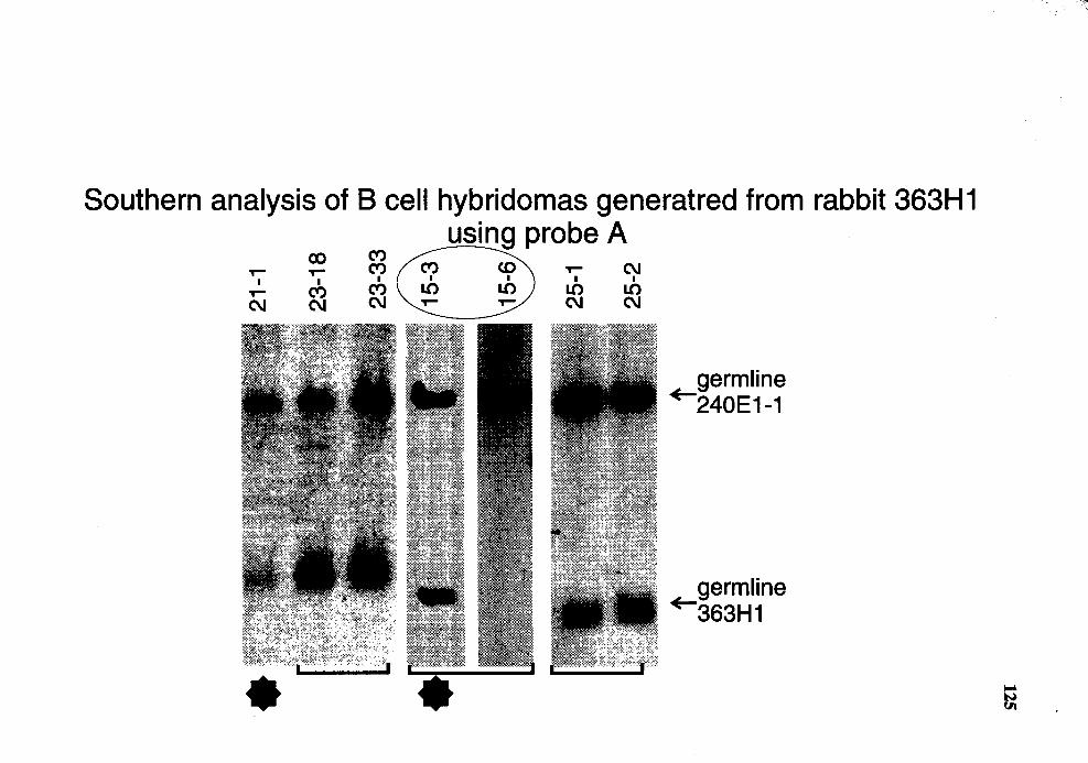
Southern analysis of B cell hybridomas generatred from rabbit 363H 1
T""" T""" I I
T""" ct) C\I C\I
using probe A T""" C\I
I I
LO LO C\I C\I
germ line +-240E1-1
· germline +-363H1

Figure 18
126
Genomic analysis for the status of lgH genes in the hybridomas generated
from rabbit# 27Hl using probe A. DNA of the indicated hybridomas was
restricted by HindID, Southern blotted and hybridized with probe A. The
space between autoradiograms separates autoradiograms from different
experiments. The migration of DNA is different in among different
autoradiograms. Polygon underneath the autoradiogram indicate the
presence of germline JH in the hybridomas (Figure 14). The status of IgH
gene on the unexpressed allele of these hybridomas is summarized in
Table 5. Brackets indicate two subclones derived from the same parental
hybridoma. The hybridoma names with circle are the two subclones from
one parent which give different results.
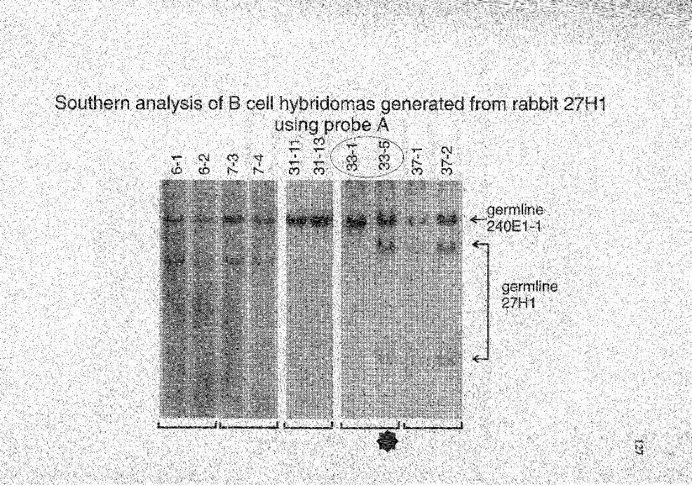
1
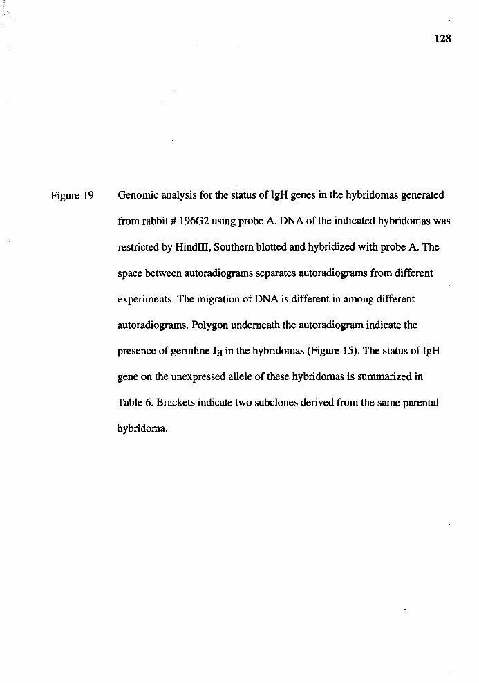
Figure 19
128
Genomic analysis for the status of lgH genes in the hybridomas generated
from rabbit# 196G2 using probe A. DNA of the indicated hybridomas was
restricted by HindIII, Southern blotted and hybridized with probe A. The
space between autoradiograms separates autoradiograms from different
experiments. The migration of DNA is different in among different
autoradiograms. Polygon underneath the autoradiogram indicate the
presence of germline JH in the hybridomas (Figure 15). The status of IgH
gene on the unexpressed allele of these hybridomas is summarized in
Table 6. Brackets indicate two subclones derived from the same parental
hybridoma.

,.... ,.... ,.... ,.... ,....
I ,.... ,.... ,.... ,....
1
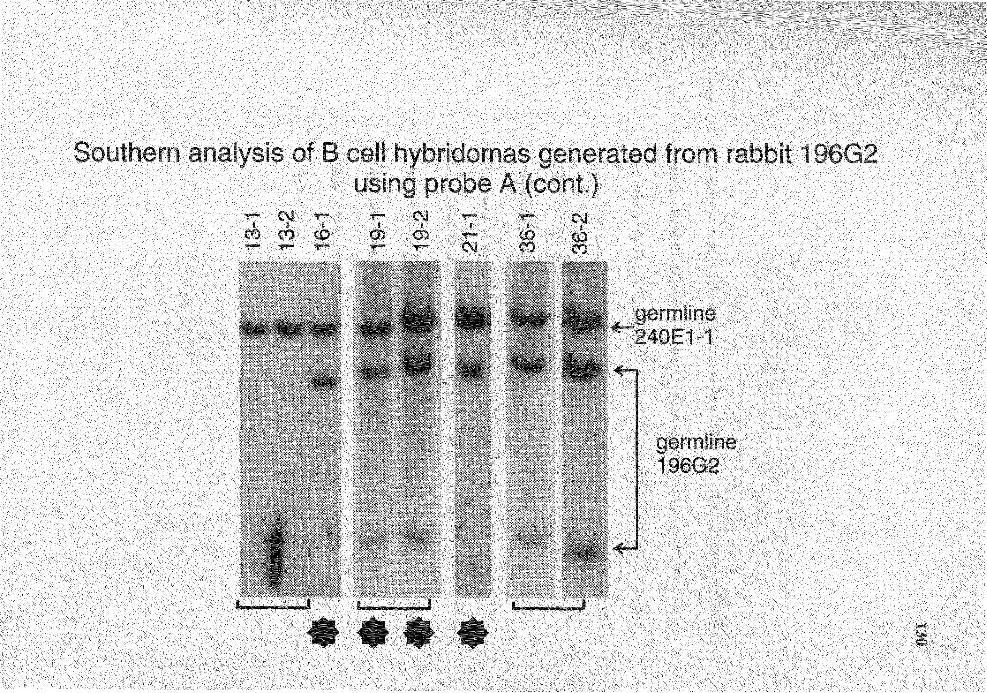
1
,_ ,_ ,_ ,_ ,_

Figure 20
131
Genomic analysis for the status of IgH genes in the hybridomas generated
from rabbit # 6412 using probe A. DNA of the indicated hybridomas was
restricted by HindIII, Southern blotted and hybridized with probe A. The
space between autoradiograms separates autoradiograms from different
experiments. The migration of DNA is different in among different
autoradiograms. Polygon underneath the autoradiogram indicate the
presence of germline ltt in the hybridomas (Figure 16). The hybridizing
signals of Hyb 45-9 and 45-12, Hyb 68-1 and 68-2, and Hyb 80-1 and 80-3
are weak but is conclusively identifiable. The status of lgH gene on the
unexpressed allele of these hybridomas is summarized in Table 7.
Brackets indicate two subclones derived from the same parental
hybridoma.
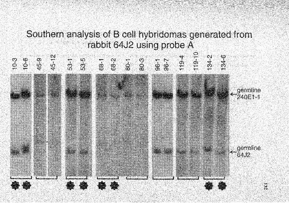
II • n en
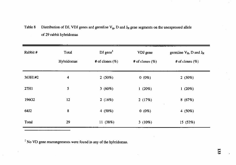
Table 8 Distribution of DJ, VDJ genes and germline V H• D and JH gene segments on the unexpressed allele
of 29 rabbit hybridomas
Rabbit# Total DJ gene1 VDJ gene germline V H. D and JH
Hybridomas # of clones (%) #of clones(%) # of clones (%)
363Hl#2 4 2 (50%) 0 (0%) 2 (50%)
27Hl 5 3 (60%) 1 (20%) 1 (20%)
196G2 12 2 (16%) 2 (17%) 8 (67%)
6412 8 4 (50%) 0 (0%) 4 (50%)
Total 29 11 (38%) 3 (10%) 15 (52%)
r No VD gene rearrangements were found in any of the hybridomas.
to probe A represent the germline probe A region of the rabbits which the splenocytes
were taken from, I conclude that these 27 hybridomas did not rearrange V H 1 on their
134
unexpressed IgH alleles. The remaining three hybridomas (Hyb 31-11and31-13; Hyb 2-
1; Hyb 13-1and13-2) did not have the germline probe A region from the splenocytes
(Figure 18 and 19, Table 8) suggesting that the VHl on the unexpressed allele was
rearranged.
It is possible that the germline JH I found in polyclonal and monoc~onal B cells is
actually DJ genes that had rearranged a D gene located in close proximity to a JH gene
such that the DJ gene is indistinguishable from germline JH on Southern analysis. This
possibility was ruled out when I examined a 340-bp region immediately upstream of JH
and found no indication of a D gene (Figure 21 ).
Taking the results of pJ5 and probe A hybridization together, I found that the 15
hybridomas which had the JH gene in germline configuration also had germline probe A
region (Tables 4-8). Therefore, these hybridomas had the IgH gene on the unexpressed
allele in germline configuration, like that of the fusion partner. Eleven hybridomas had no
germline JH of the spleen donors but still had probe A region in germline configuration
and hence, these hybridomas had DJ genes on the unexpressed IgH allele (Tables 4-8).
The remaining three hybridomas had neither germline JH and probe A region suggesting
that IgH gene on both alleles were rearranged to VDJ genes (Tables 4-8).
From the cloning of the 29 hybridomas by limiting dilution, I obtained more than
one subclone for 25 of them. For each of these 25 hybridomas, two subclones were
analyzed for the status of the IgH gene on the unexpressed allele. The result I obtained
from analyzing two subclones was generally identical suggesting that the hybridomas
were already monoclonal before the cloning. Only two pairs of monoclonal hybridomas
gave different results (Hyb 15-3 and 15-6 (Figure 13); Hyb 33-1 and 33-5 (Figure 14)). In
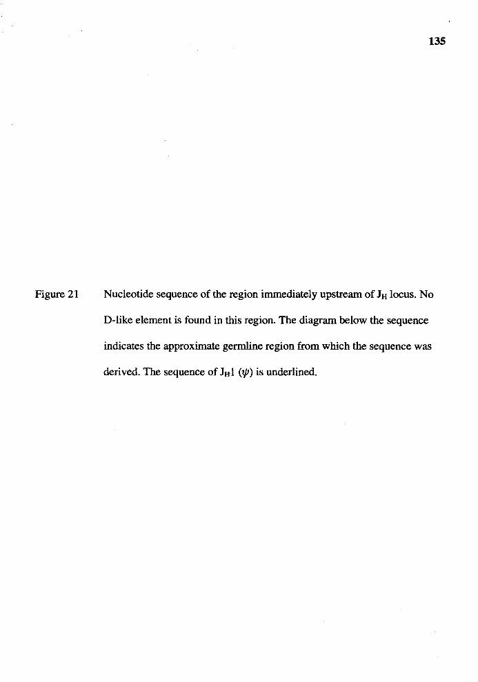
Figure 21
135
Nucleotide sequence of the region immediately upstream of JH locus. No
D-like element is found in this region. The diagram below the sequence
indicates the approximate germline region from which the sequence was
derived. The sequence of lttl (tp) is underlined.
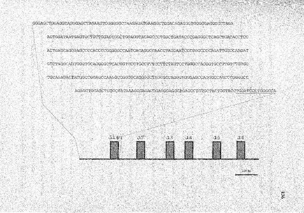
AGTGGATAATGAGTGCTGTTGGACTGGCTGGAGGTACAGCCCTGGCTGATACCCGAGGGCTCAGCTGACACCTCC
ACTGAGCAGGGAGCCCCACCCCGGGGGCCAATGAGAGGCCAACCTAGGAATCCCGGGCCCCAGATTGGCCAAGAT
GTCTAGGCAGTGGGTGCAGGCGCTCACGGTGCCTGCCTTTCCTTCTAGTCCTGGGCCAGGGTGCCTTGTCTGTGC
TGCAGAGACTATGGCCGGAGCCAAGGCCGGGGCAGGGGCTAGGGCCAGGGTGGGAGCCAGGGCCAGCCCGGGGCC
250 bp

137
both pairs, one subclone (Hyb 15-3 and Hyb 33-5) showed that the unexpressed IgH allele
was in germline configuration while the other subclone in the pair (Hyb 15-6 and Hyb 33-
1) showed that a VDJ gene was on the unexpressed allele. There are two possible
explanations. First, it is possible that the difference between the two subclones was due to
the polyclonality of the parental hybridomas and the two subclones are derived from
different clones. In this case, both the parental hybridomas (Hyb 15 and Hyb 33) must
contain one population of cells which have germline IgH gene on the unexpressed allele
and another population which have VDJ gene. However, because VDJ gene on the
unexpressed IgH allele was infrequently found in rabbit B cells (3 of 29 hybridomas), it is
rather unlikely that both of the only two potentially "polyclonal" hybridomas in this
experiment would coincidentally contain a subpopulation of cells which have VDJ gene
on the unexpressed allele. I think that an alternative explanation that the Hyb 15-6 and
Hyb 33-1 had lost their unexpressed IgH allele is more likely the explanation. Because of
this reason, I have excluded these two hybridomas from this analysis.
To confirm the result of Southern analysis that 11 hybridomas had rearranged DJ
genes on their unexpressed IgH alleles, I attempted to PCR-amplify the DJ genes in these
hybridomas using a degenerate PCR primer specific for all known D genes (Table 2) and
a JHprB primer specific for all known JH genes (Becker et al., 1989). From PAGE
analysis of the PCR products, an amplified product of approximately 100 bp , the size
expected for a product of DJ gene, were observed in all 11 hybridomas (Figure 22). As a
negative control, I used DNA from two hybridomas which had unexpressed IgH genes in
germline configuration (Hyb 15-3; Hyb 11-1) and from two hybridomas which had VDJ
genes on both lgH alleles (Hyb 31-11; Hyb 2-1) in the PCR reaction. No PCR-product
was observed in these negative control DNA. To confirm that the PCR products from the
11 hybridomas are DJ genes, I chose at random the products from four of the 11
hybridomas (Hyb 37-1; Hyb 25-1; Hyb 36-1;Hyb23-18), cloned and determined their
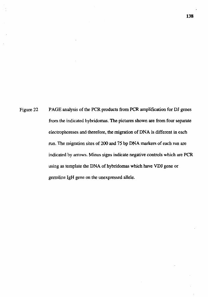
Figure 22
138
PAGE analysis of the PCR products from PCR amplification for DJ genes
from the indicated hybridomas. The pictures shown are from four separate
electrophoreses and therefore, the migration of DNA is different in each
run. The migration sites of 200 and 75 bp DNA markers of each run are
indicated by arrows. Minus signs indicate negative controls which are PCR
using as template the DNA of hybridomas which have VDJ gene or
germline lgH gene on the unexpressed allele.
140
nucleotide sequences. The nucleotide sequence analysis showed that they are, indeed, DJ
genes (Figure 23). I conclude that the PCR products from these four hybridomas as well
as the remaining seven hybridomas were generated from amplification of DJ genes.
lgH Gene Rearrangement in Rabbit T Cell Lines
From the findings described above, it appears that DJ gene rearrangement occurs
less frequently in rabbit than in mouse, human and chicken. Half of the rabbit B cell
hybridomas I examined have not rearranged their unexpressed lgH alleles and for those
that do, most rearrange DJ genes. Further, rabbit B cells rearrange VD genes, which was
not regularly found in B cells of other species (see following section). Before we had the
technology to generate rabbit x rabbit B cell hybridomas, I examined rabbit T cell lines
for the IgH gene rearrangement with the goal of gaining insight into the IgH gene
rearrangement in rabbit. Specifically what I asked is whether I can find DJ or VD gene
rearrangement in rabbit T cell lines.
DNA from 14 rabbit T cell lines was kindly provided by Dr. Thomas Kindt
(NIAID, Bethesda MD). These cell lines were derived from five rabbits: K30, K34, 6600,
6516 and RLS. The Southern blot of BamHI-restricted DNA from these 14 cell lines was
probed with a probe specific for D genes in the Dl family (Table 1). Four members of the
Dl gene family are scattered throughout the D region and therefore, most of the
rearrangement of D gene segments, if not all, result in a non-germline fragment
hybridizing to D 1 probe. The Southern hybridization of T cell line DNA showed that T
cell lines derived from the same rabbit had identical hybridization patterns (Figure 24).
Although I did not include, as control for germline configuration, the liver DNA of
rabbits from which the T cell lines were derived, the identical hybridization patterns in
the T cell lines which were derived from the same rabbit suggests that the hybridization
patterns represent the germline configuration. The alternative explanation for the identical

Figure 23
141
Comparison of the nucleotide sequences of four PCR-amplified DJ genes
with those of germline D and JH genes. Dots denote the similarity to the
germline genes; the N regions are boxed; the degenerate PCR-primers
specific for D genes are underlined.
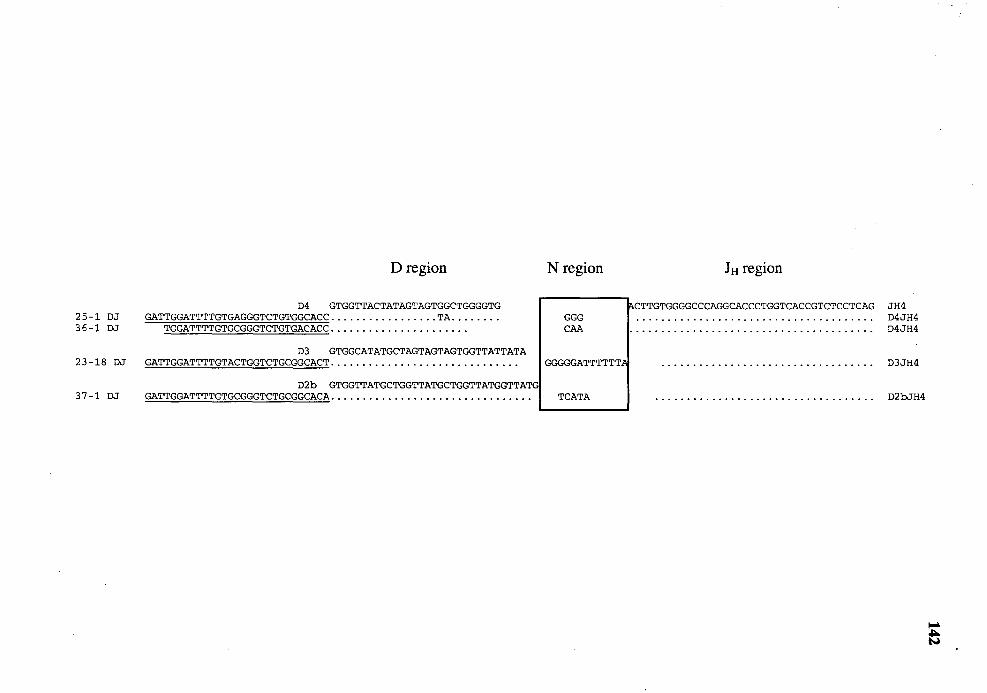
25-1 DJ 36-1 DJ
23-18 DJ
37-1 DJ
D region
D4 GTGGTTACTATAGTAGTGGCTGGGGTG GATTGGATTTTGTGAGGGTCTGTGGCACC ................. TA ....... .
TGGATTTTGTGCGGGTCTGTGACACC ..................... .
D3 GTGGCATATGCTAGTAGTAGTGGTTATTATA GATTGGATTTTGTACTGGTCTGCGGCACT ............................. .
D2b GTGGTTATGCTGGTTATGCTGGTTATGGTTATG GATTGGATTTTGTGCGGGTCTGCGGCACA ............................... .
N region
GGG CAA
GGGGGATTTTTT~
TCATA
JH region
~CTTGTGGGGCCCAGGCACCCTGGTCACCGTCTCCTCAG JH4 D4JH4 D4JH4
D3JH4
D2bJH4
Figure 24
143
Genomic analysis for the status of lgH genes in the 14 rabbit T cell lines
using D 1 probe, a probe specific for all four D gene in the D 1 family. The
rabbits from which the T cell lines were derived is indicated above the
clone numbers.
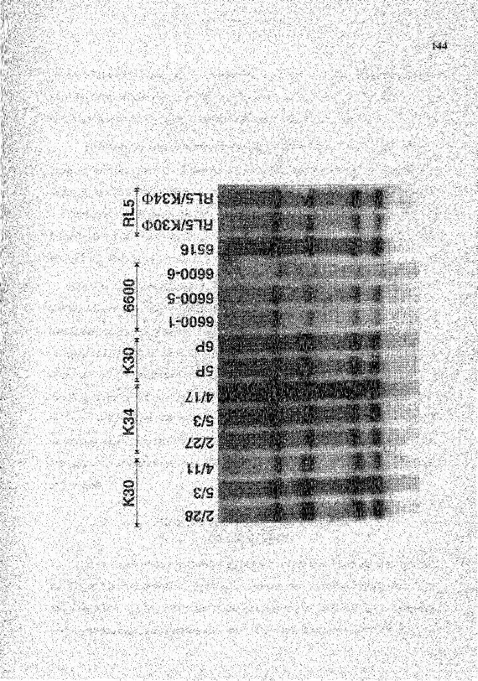
145
pattern of hybridization that all T cell lines had rearranged the same D genes resulting in
identical hybridization pattern is highly unlikely. I conclude that there was no
rearrangement of the D region of IgH gene in any of the 14 T cell lines.
To further investigate whether the JH gene was rearranged, the Dl probe was
removed by using boiled-0. lX blot wash and the blot was rehybridized with the pJ5
probe (Figure 25). The hybridization showed a non-polymorphic 2 kb BamHl-fragment
hybridizing to the pJ5 probe. No non-germline fragment was found. Taking the result of
Dl and pJ5 hybridization together, I conclude that the IgH gene was not rearranged in all
the 14 T cell lines I examined.
There was a submolar band of approximately 3 kb in size hybridizing to the pJ5
probe in clones 2/28, 5/3, 5P and 6P (Figure 25). The submolar appearance of JH
hybridizing fragments in Southern analysis is characteristic of cell lines capable of
rearranging lgH gene in vitro (Alt et al., 1981). The submolar characteristic was a result
of the utilization of different D and J genes in different rearrangements and therefore, the
resulting daughter cells are polyclonal. However, I do not think that the submolar band in
the four T cell lines are rearranged JH genes because it is unlikely that T cell lines would
be actively rearranging IgH gene in vitro. I conclude that these submolar bands resulted
from either incomplete digestion of DNA of these cell lines or non-specific hybridization
of pJ5 probe.
Intermediates of lgH Gene Rearrangement in
Polyclonal B Cells
The analysis of the unexpressed lgH gene of rabbit B cell hybridomas suggests
that DJ gene is an intermediate of IgH gene rearrangement in rabbit. Further, Becker R.
and Knight K. L. analyzed the status of the IgH gene in a rabbit B cell line and identified
a VD gene rearrangement (unpublished data). This finding suggests that a VD gene may
Figure 25
146
Genomic analysis for the status of IgH genes in the 14 rabbit T cell lines
using pJ5 probe. The rabbits from which the T cell lines derived from are
indicated above the clone numbers. The position of germline JH (2 kb) was
indicated. " * " indicates the 4.3 band which is superimposable to one of
the D 1 hybridizing fragment. The signal was likely from the D 1 probe
which was not completely removed."**" indicates the possible submolar
band (see text).
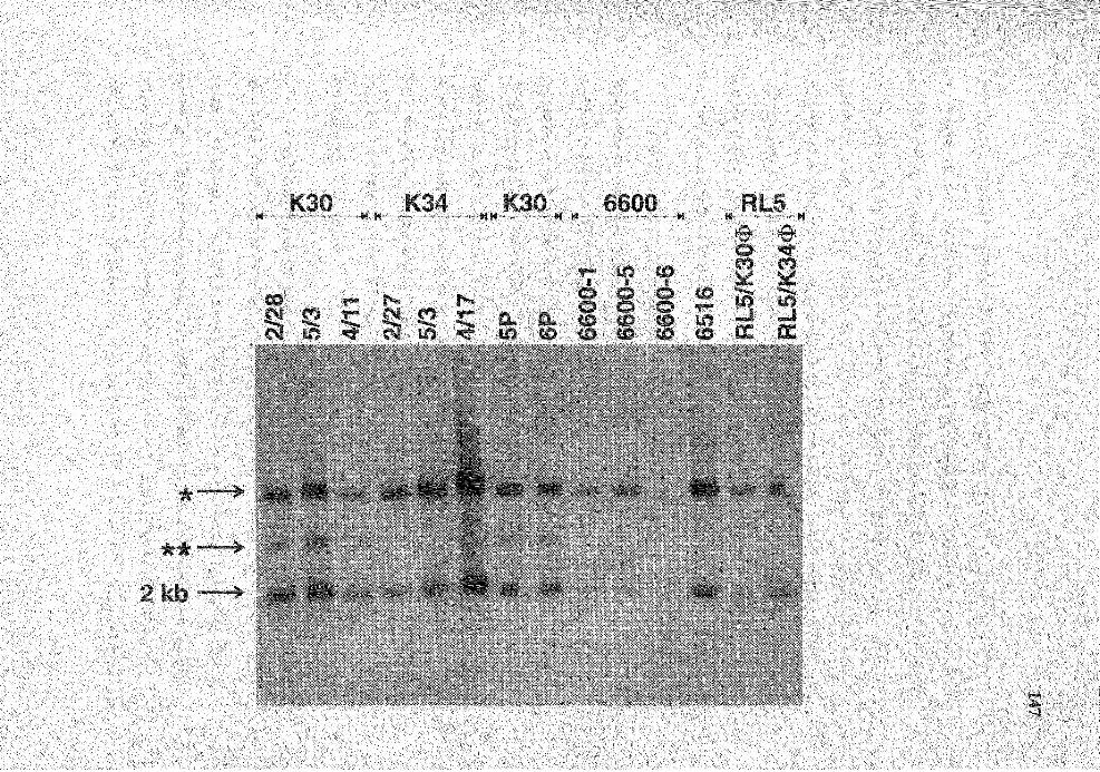
148
also be an alternative intermediate for lgH gene rearrangement in rabbit B cells. To
determine whether the DJ and VD genes could be found in normal rabbit B cells, I PCR
amplified both DJ and VD genes from normal rabbit splenic DNA. In this experiment,
instead of using the degenerate PCR-primer specific for all known D genes, I used D2
specific primers (Table 1). To amplify D2bJ gene rearrangements, 5'D2b and JJIPrB
primers were used whereas VHprB and 3'D2b primers were used to amplify VD2b gene
rearrangements. I chose to examine VD and DJ genes which utilized D2 gene family
simply because the members of D2 gene family, especially D2b, were utilized most
frequently among the 11 known D genes.
For the D2bJ gene rearrangements, I PCR-amplified two splenic DNA
preparations prepared from an a3 allotype rabbit and an al/a2 allotype rabbit using 5'D2b
and JHprB primers. The PAGE analysis of PCR products from both genomic DNA
preparations showed a diffuse band of approximately 120 bp in size (Figure 26). The
diffuse appearance of the PCR product is probably due to the addition of N-segment
between the DJ junction resulting in DJ genes of varying length. These PCR products
were cloned into M13mp18 for sequencing. The nucleotide sequence analysis of the
PCR-amplified DJ genes showed that they are authentic DJ genes and that their RSSs
located 5' of D2b are intact (Figure 27) suggesting that these DJ genes potentially can be
rearranged to VDJ gene.
I PCR-amplified VD gene rearrangements from the a3 splenic DNA, which was
also used in the amplification for DJ genes described above, using V HprB and 3 'D2b
primers. A product of approximately 500 bp in size was observed (Figure 26). The
amplified VD genes were cloned into Ml3mp18 and sequenced. From the nucleotide
sequences, I found that the amplified products are VD genes (Figure 28). All of them had
utilized V H 1-a3 and D2b in the rearrangements. The RSSs located 3' of D2b in these VD
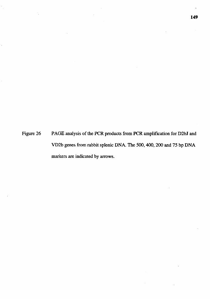
Figure 26
149
PAGE analysis of the PCR products from PCR amplification for D2bJ and
VD2b genes from rabbit splenic DNA. The 500, 400, 200 and 75 bp DNA
markers are indicated by arrows.

Figure 27 Comparison of the nucleotide sequences of the DJ genes isolated from
splenic DNA with those of germline D2b and JH genes. Dots denote the
similarity to the germline genes; the N regions are boxed.
151
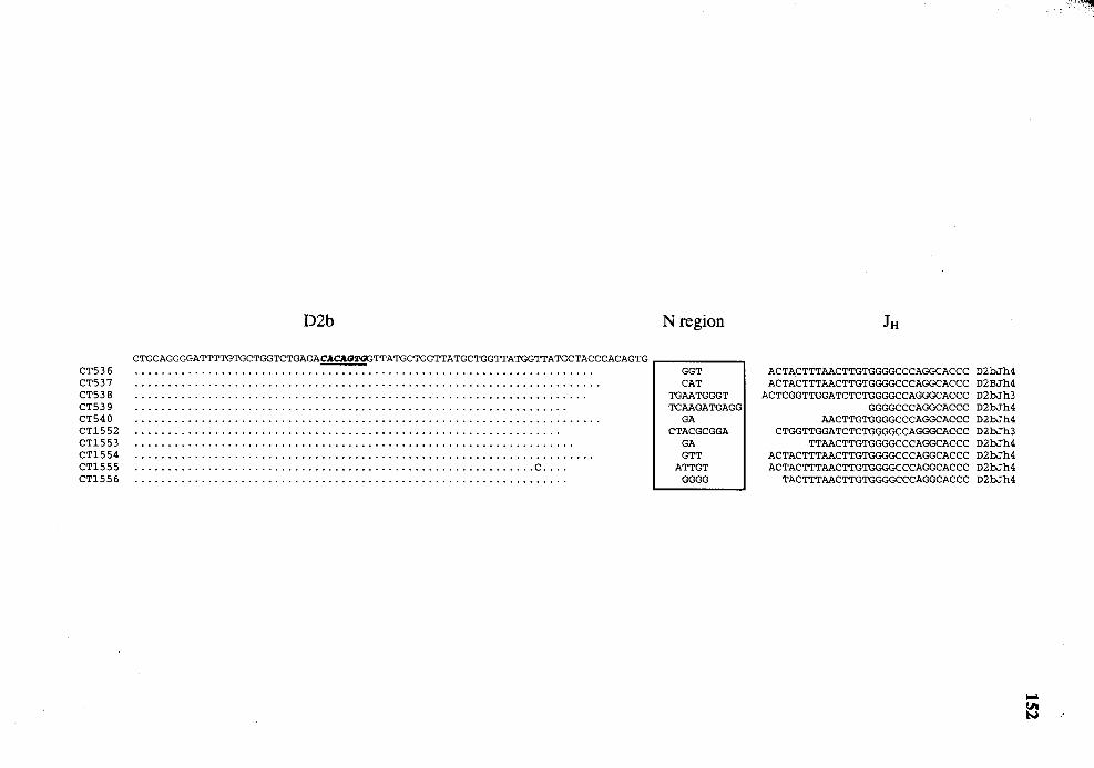
CT536 CT537 CT538 CT539 CT540 CT1552 CT1553 CT1554 CT1555 CT1556
D2b
CTGCAGGGGATTTTGTGCTGGTCTGAGACACAGT<13TTATGCTGGTTATGCTGGTTATGGTTATGCTACCCACAGTG
............................................................ c ....
N region
GGT CAT
TGAATGGGT TCAAGATGAGG
GA CTACGCGGA
GA GTT
ATTGT GGGG
JH
ACTACTTTAACTTGTGGGGCCCAGGCACCC D2bJh4 ACTACTTTAACTTGTGGGGCCCAGGCACCC D2BJh4
ACTCGGTTGGATCTCTGGGGCCAGGGCACCC D2bJh3 GGGGCCCAGGCACCC D2bJh4
AACTTGTGGGGCCCAGGCACCC D2bJh4 CTGGTTGGATCTCTGGGGCCAGGGCACCC D2bJh3
TTAACTTGTGGGGCCCAGGCACCC D2bJh4 ACTACTTTAACTTGTGGGGCCCAGGCACCC D2bJh4 ACTACTTTAACTTGTGGGGCCCAGGCACCC D2bJh4
TACTTTAACTTGTGGGGCCCAGGCACCC D2bJh4
,-:··
Figure 28
153
Comparison of the nucleotide sequences of the VD2b genes isolated from
splenic DNA with those of germline V H 1-a3 and D2b genes. Dots denote
the similarity to the germline genes; the N regions are boxed.

VH1-a3 VD290 VD291 VD292 VD293 VD295 VD296 VD297 VD298 VD299
VD290 VD291 VD292 VD293 VD295 VD296 VD297 VD298 VD299
GCGAAAGGCCGATTCACCATCTCCAAAACCTCGTCGACCACGGTGACTCTGCAAATGACCAGTCTGACAGCCGCGGACACGGCCACCTATTTCTGTGCGAGAGACACAC7.l'G
N-region
GCCCC AGCCTGG
CTGCCCC GCTTCGATACTTA
G G
GAGGATTACTATACTT GTGACTGGTGCTGGTGGC
TTCCTACGCCCCAA
D2b
GTTATGCTGGTTATGCTGGTTATGGTTATGCTACCCACA<n'QCCTCAGGCCTCACATAAAACTCAGCCACAGCC

155
genes are intact suggesting that they are similar to the DJ genes in that they too potentially
can be rearranged to VDJ genes.
Fetal VDJ Genes
It is rather unusual that only 10% of rabbit B cell hybridomas have 2 VDJ genes,
one of which must be productive and the other, presumably, nonproductive. Because of
the junctional diversity and N-nucleotide addition at the VD and DJ junctions, the
generation of nonproductive VDJ genes is certain to occur. In mouse and human, more
than 60% of B cells have rearranged VDJ genes on both IgH alleles due to the high
frequency of nonproductive rearrangements. One possible explanation for the low
frequency of nonproductive rearrangements in rabbit is that rabbit B cells, unlike mouse
and human B cells, do not generate nonproductive VDJ genes. An alternative explanation
would be that only one IgH allele can be rearranged to a complete VDJ gene.
I attempted to determine whether at any time during development, nonproductive
VDJ genes can be found more frequently. In the case that I find many nonproductive VDJ
genes, the benefit is two-fold. First, it will provide the evidence against the possibility
that rabbit B cells do not generate nonproductive VDJ genes. Further, the VH gene usage
in these nonproductive VDJ genes will distinguish whether the preferential utilization of
V H 1 resulted from its preferential rearrangement. Because nonproductive VDJ genes
cannot be selected for or against by antigen selection process, the V H gene usage in the
nonproductive VDJ genes would reflect the VH gene(s) utilized in the rearrangements.
In previous studies for VDJ genes in newborn and adult rabbits, nonproductive
VDJ genes were found infrequently (Tunyaplin C. and Knight K. L. , unpublished data). I
asked whether nonproductive VDJ genes might be found at higher frequency in the fetal
VDJ gene repertoire, especially at the developmental stages preceding the appearance of
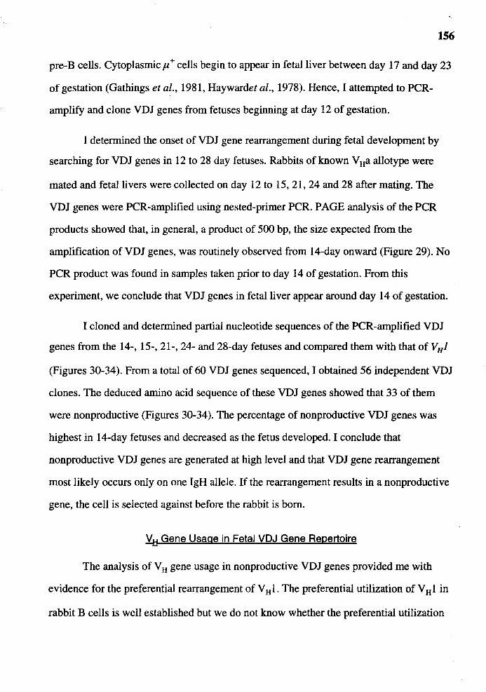
156
pre-B cells. Cytoplasmicµ+ cells begin to appear in fetal liver between day 17 and day 23
of gestation (Gathings et al., 1981, Haywardet al., 1978). Hence, I attempted to PCR
amplify and clone VDJ genes from fetuses beginning at day 12 of gestation.
I determined the onset of VDJ gene rearrangement during fetal development by
searching for VDJ genes in 12 to 28 day fetuses. Rabbits of known V Ha allotype were
mated and fetal livers were collected on day 12 to 15, 21, 24 and 28 after mating. The
VDJ genes were PCR-amplified using nested-primer PCR. PAGE analysis of the PCR
products showed that, in general, a product of 500 bp, the size expected from the
amplification of VDJ genes, was routinely observed from 14-day onward (Figure 29). No
PCR product was found in samples taken prior to day 14 of gestation. From this
experiment, we conclude that VDJ genes in fetal liver appear around day 14 of gestation.
I cloned and determined partial nucleotide sequences of the PCR-amplified VDJ
genes from the 14-, 15-, 21-, 24- and 28-day fetuses and compared them with that of VHJ
(Figures 30-34). From a total of 60 VDJ genes sequenced, I obtained 56 independent VDJ
clones. The deduced amino acid sequence of these VDJ genes showed that 33 of them
were nonproductive (Figures 30-34). The percentage of nonproductive VDJ genes was
highest in 14-day fetuses and decreased as the fetus developed. I conclude that
nonproductive VDJ genes are generated at high level and that VDJ gene rearrangement
most likely occurs only on one lgH allele. If the rearrangement results in a nonproductive
gene, the cell is selected against before the rabbit is born.
V H Gene Usage in Fetal VDJ Gene Repertoire
The analysis of V H gene usage in nonproductive VDJ genes provided me with
evidence for the preferential rearrangement of V H 1. The preferential utilization of V H 1 in
rabbit B cells is well established but we do not know whether the preferential utilization

Figure 29
157
PAGE analysis of nested-primer PCR for fetal VDJ genes. The gestational
stage at which fetal liver was obtained are indicated. The white arrows
indicate the position of the PCR product expected from a VDJ gene.
PCR amplification of fetal VDJ genes <(
-c -c 0 0
I I
>. >. ctS ctS -c -c
I I
M C\J ~ T""" T"""
z 0 E '-
-0 ~ -c - CJ) -o,o
I C: I
>. 0 >. a:s E a:s -c -c I - I
M MctSLO T""" CJ) T"""
-c -c -c 0 0 0
I I I
>. >. >. ctS ctS ctS -c -c -c
I I I
MT""' ~ <X> C\J C\J C\J
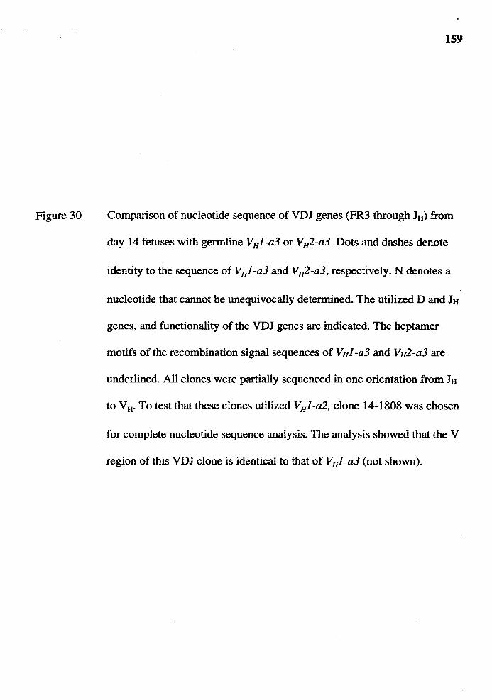
Figure 30
159
Comparison of nucleotide sequence of VDJ genes (FR3 through JH) from
day 14 fetuses with germline VHJ-a3 or VH2-a3. Dots and dashes denote
identity to the sequence of VHJ-a3 and VH2-a3, respectively. N denotes a
nucleotide that cannot be unequivocally determined. The utilized D and JH
genes, and functionality of the VDJ genes are indicated. The heptamer
motifs of the recombination signal sequences of VHJ-a3 and VH2-a3 are
underlined. All clones were partially sequenced in one orientation from JH
to VH. To test that these clones utilized VHJ-a2, clone 14-1808 was chosen
for complete nucleotide sequence analysis. The analysis showed that the V
region of this VDJ clone is identical to that of VHJ-a3 (not shown).

VH1-a3 14-1525F 14-1526F 14-1527F 14-1579F 14-1580F 14-1581F 14-lBOBF
VH2-a3 14-1523F 14-1524F 14-1578F
14-1525F 14-1526F 14-1527F 14-1579F 14-1580F
.14-1581F 14-lBOBF
14-1523F 14-1524F 14-1578F
A K G R F T I S K T S S T T V T L Q M T S L T A A D T A T Y F C A R GCGAAAGGCCGATTCACCATCTCCAAAACCTCGTCGACCACGGTGACTCTGCAAATGACCAGTCTGACAGCCGCGGACACGGCCACCTATTTCTGTGCGAGAGACACAG2'G
•...•••..•••••.. N ••• C ..••••..••••...••.••.•••••..••••••.••••••.••••••••••••..•••••••. ................................................................ c ..................................... .
GAATGGCCGAATCATCATCTCCAGGGACAACGCCACAACACGGTGTCTCGGCAAATGAACAGTCTGACAGCCAGGGATAGGGCCCTGTATTTCTGTGCAGGAGACACAGTG
N region D region N region JH
CAA GTTATGCTGGTAGTAGTTATTATA T TTTAACTTGTGGGGCCCAGGCACCCTGG DS ,JH4' non-productive TCCGA GCTGGTAGTAGTTATTA TAACTTGTGGGGCCCAGGCACCCTGG DS ,JH4' non-productive
GG GTTACTATACTTATGGTTATGCTGGTTATGCTTATGCTA GAGGAA GGGGCCCAGGCACCCTGG D2a,JH4, non-productive TCCCCC TGCTGGTTATGCTGGTTATGGTTATG GGGG CTTGTGGGGCCCAGGCACCCTGG D2b,JH4, productive
GGT GCTACGATGACTATGGTGATTA GG CGGTTGGATCTCTGGGGCCAGGGCACCCTGG Dl ,JH3, non-productive CAA TTACTATACTTATGGTTATGCTGGTTATGCTTATGCTACC TTAACTTGTGGGGCCCAGGCACCCTGG D2a,JH4, non-productive GTA ATAACTACTACCAGCATAAC GTGG TGGTTGGATCTCTGGGGCCAGGGCACCCTGG DS (inverted) , JHS
non-productive AG GCTGGTTATGCTGGTTATGGTTATGC CCT ACTACTTTAACTTGTGGGGCCCAGGCACCCTGG D2b,JH4, non-productive
TATGCTGGTAGTAGTTATTA ACTTGTGGGGCCCAGGCACCCTGG DS ,JH4, non-productive TTACTATACTTATGGTTATGCTGGTTATGCTTATGCTAC AAGG ACTTGTGGGGCCCAGGCACCCTGG 02a,JH4, non-productive
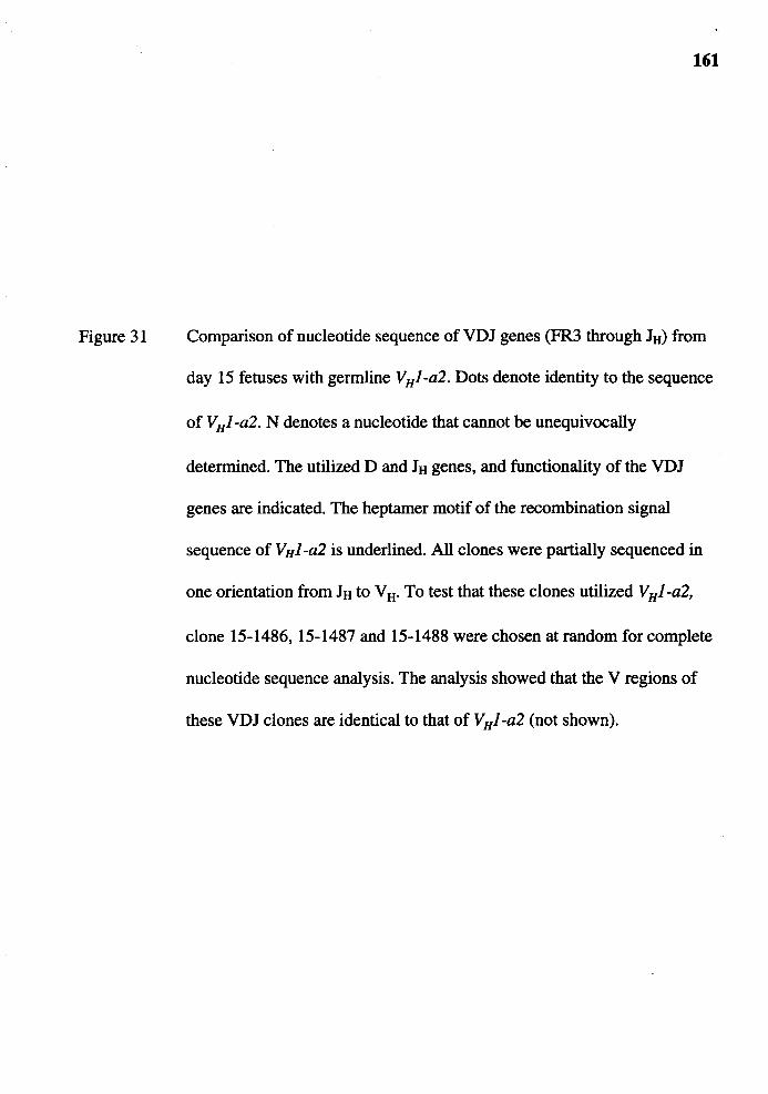
Figure 31
161
Comparison of nucleotide sequence of VDJ genes (FR3 through Itt) from
day 15 fetuses with germline VHJ-a2. Dots denote identity to the sequence
of V HJ -a2. N denotes a nucleotide that cannot be unequivocally
determined. The utilized D and Itt genes, and functionality of the VDJ
genes are indicated. The heptamer motif of the recombination signal
sequence of V HJ -a2 is underlined. All clones were partially sequenced in
one orientation from Itt to V tt· To test that these clones utilized V HJ -a2,
clone 15-1486, 15-1487 and 15-1488 were chosen at random for complete
nucleotide sequence analysis. The analysis showed that the V regions of
these VDJ clones are identical to that of V HJ-a2 (not shown).
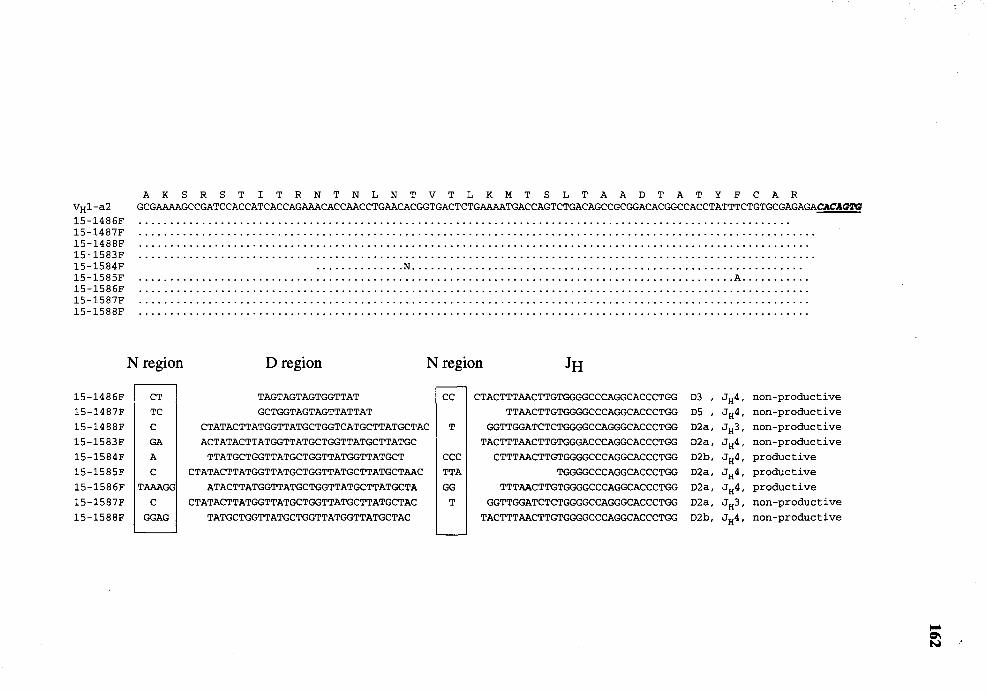
VH1-a2 15-1486F 15-1487F 15-1488F 15-1583F 15-1584F 15-1585F 15-1586F 15-1587F 15-1588F
15-1486F
15-1487F
15-1488F
15-1583F
15-1584F
15-1585F
15-1586F
15-1587F
15-1588F
A K S R S T I T R N T N L N T V T L K M T S L T A A D T A T Y F C A R GCGAAAAGCCGATCCACCATCACCAGAAACACCAACCTGAACACGGTGACTCTGAAAATGACCAGTCTGACAGCCGCGGACACGGCCACCTATTTCTGTGCGAGAGACACAGTG
.............. N ............................................................. . .•••...•••••...••••...••.••...••..•••••..••.•..•.••..•••••..••••.••••.••••.•.•••••.•••••...••• A ..•.•••...•
N region D region N region JH
CT TAGTAGTAGTGGTTAT cc CTACTTTAACTTGTGGGGCCCAGGCACCCTGG D3 ' JH4, non-productive TC GCTGGTAGTAGTTATTAT TTAACTTGTGGGGCCCAGGCACCCTGG D5 , JH4, non-productive c CTATACTTATGGTTATGCTGGTCATGCTTATGCTAC T GGTTGGATCTCTGGGGCCAGGGCACCCTGG D2a, JH3, non-productive GA ACTATACTTATGGTTATGCTGGTTATGCTTATGC TACTTTAACTTGTGGGACCCAGGCACCCTGG D2a, JH4, non-productive A TTATGCTGGTTATGCTGGTTATGGTTATGCT CCC CTTTAACTTGTGGGGCCCAGGCACCCTGG D2b, JH4, productive c CTATACTTATGGTTATGCTGGTTATGCTTATGCTAAC TTA TGGGGCCCAGGCACCCTGG D2a, JH4, productive
TAAAGG ATACTTATGGTTATGCTGGTTATGCTTATGCTA GG TTTAACTTGTGGGGCCCAGGCACCCTGG D2a, JH4, productive c CTATACTTATGGTTATGCTGGTTATGCTTATGCTAC T GGTTGGATCTCTGGGGCCAGGGCACCCTGG D2a, JH3, non-productive
GGAG TATGCTGGTTATGCTGGTTATGGTTATGCTAC TACTTTAACTTGTGGGGCCCAGGCACCCTGG D2b, JH4, non-productive
Figure 32
163
Comparison of nucleotide sequence of VDJ genes (FR3 through JH) from
day 21 fetuses with germline V8 1-a3 or V8 2-a3. Dots and dashes denote
identity to the sequence of V8 1-a3 and V8 2-a3, respectively. The utilized
D and JH genes, and functionality of the VDJ genes are indicated. The
heptamer motifs of the recombination signal sequences of V 8 1 -a3 and
V8 2-a3 are underlined. All clones were partially sequenced in one
orientation from JH to V H· To test that these clones utilized V 8 1-a3, clone
21-1489 was chosen at random for complete nucleotide sequence analysis.
The analysis showed that the V region of this VDJ clone is identical to that
of V8 1-a2 (not shown).
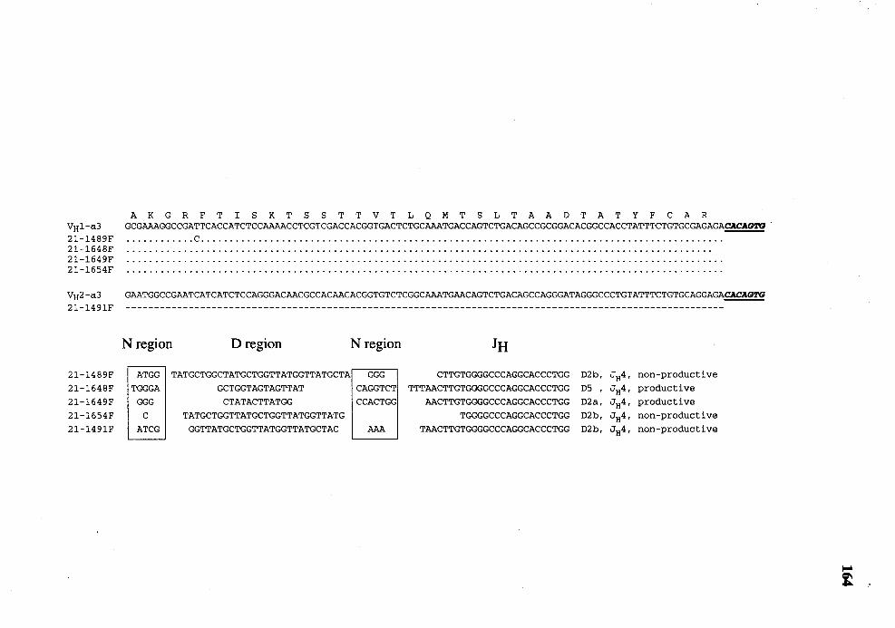
VH1-a3 21-1489F 21-1648F 21-1649F 21-1654F
VH2-a3 21-1491F
21-1489F
21-1648F
21-1649F
21-1654F
21-1491F
A K G R F T I S K T S S T T V T L Q M T S L T A A D T A T Y F C A R GCGAAAGGCCGATTCACCATCTCCAAAACCTCGTCGACCACGGTGACTCTGCAAATGACCAGTCTGACAGCCGCGGACACGGCCACCTATTTCTGTGCGAGAGACACAG2'G ............ c .......................................................................................... .
GAATGGCCGAATCATCATCTCCAGGGACAACGCCACAACACGGTGTCTCGGCAAATGAACAGTCTGACAGCCAGGGATAGGGCCCTGTATTTCTGTGCAGGAGACACAG2'G
N region D region N region JH
ATGG TATGCTGGCTATGCTGGTTATGGTTATGCTA GGG CTTGTGGGGCCCAGGCACCCTGG D2b, JH4, non-productive TGGGA GCTGGTAGTAGTTAT CAGGTCT TTTAACTTGTGGGGCCCAGGCACCCTGG DS ' JH4, productive
GGG CTATACTTATGG CCACTGG AACTTGTGGGGCCCAGGCACCCTGG D2a, JH4, productive c TATGCTGGTTATGCTGGTTATGGTTATG TGGGGCCCAGGCACCCTGG D2b, JH4' non-productive
ATCG GGTTATGCTGGTTATGGTTATGCTAC AAA TAACTTGTGGGGCCCAGGCACCCTGG D2b, JH4' non-productive
Figure 33
165
Comparison of nucleotide sequence of VDJ genes (FR3 through 1tt) from
day 24 fetuses with germline V HJ -al or V HJ-a3. Dots and dashes denote
identity to the sequence of VHJ-al and VHJ-a3, respectively. N denotes a
nucleotide that cannot be unequivocally determined. The utilized D and JH
genes, and functionality of the VDJ genes are indicated. The utilized D
gene of clone 24-1799F is too short to be identified accurately. The
utilized D gene of clone 24-1765F cannot be identified. The heptamer
motifs of the recombination signal sequences of VHJ-al and VHJ-a3 are
underlined. All clones were partially sequenced in one orientation from JH
toVH.
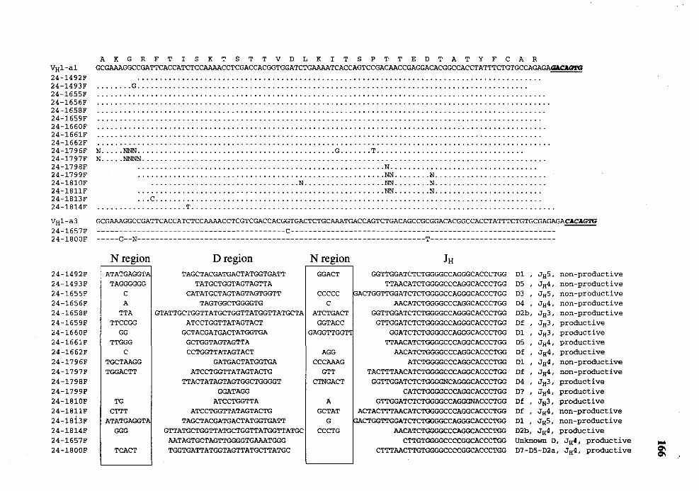
VHl-al 24-1492F 24-1493F 24-1655F 24-1656F 24-1658F 24-1659F 24-1660F 24-1661F 24-1662F 24-1796F 24-1797F 24-1798F 24-1799F 24-lBlOF 24-lBllF 24-1813F 24-1814F
VH1-a3 24-1657F 24-lBOOF
24-1492F 24-1493F 24-1655F 24-1656F 24-1658F 24-1659F 24-1660F 24-1661F 24-1662F 24-1796F 24-1797F 24-1798F 24-1799F 24-lBlOF 24-lBllF 24-1Bi3F 24-1814F 24-1657F 24-lBOOF
A K G R F T I S K T S T T V D L K I T S P T T E D T A T Y F C A R GCGAAAGGCCGATTCACCATCTCCAAAACCTCGACCACGGTGGATCTGAAAATCACCAGTCCGACAACCGAGGACACGGCCACCTATTTCTGTGCCAGAGA~
•••••.•• G ••.•••••••••••••••••.•.••.••••••••••••••.•.••.•••••••••••••••••••••••••••••••••••••••••
N ••••• NNN •.••.••.•.......••••••••••••••.•.••..••••••• G ••••••• T •••••....•.•••.•••••.••..•••••••• N •.••• NNNN ••••••.•••••.•••••.•.•.••.••••••••.•••••••...•.•..•••••••••••••••....•••••••••••.....•••••
....................................................... N .......................•......... •••••••..••••.•.•...••.••••••••.•••••••.•..•••••••••••• NN ••••.... N ....••••••.•.....••••••••
................................. N .................. NN ••••••.• N ................•........ •••••••.•••••..........•••••••••••••••••••..••••••••••• NN •••••... N •••.•.••••.......••••••• ... c ..................................................................................... .
.................... T ................................................................................ .
GCGAAAGGCCGATTCACCATCTCCAAAACCTCGTCGACCACGGTGACTCTGCAAATGACCAGTCTGACAGCCGCGGACACGGCCACCTATTTCTGTGCGAGAGACACAGTG ------------------------------------------c----------------------------------------------------------------G--N----------------------------------------------------------------T----------------------------
N region D region N region JH ATATGAGGTA TAGCTACGATGACTATGGTGATT GGACT GGTTGGATCTCTGGGGCCAGGGCACCCTGG Dl , JHS, non-productive
TAGGGGGG TATGCTGGTAGTAGTTA TTAACATCTGGGGCCCAGGCACCCTGG DS , JH4, non-productive c CATATGCTAGTAGTAGTGGTT ccccc GACTGGTTGGATCTCTGGGGCCAGGGCACCCTGG D3 ' JHS, non-productive A TAGTGGCTGGGGTG c AACATCTGGGGCCCAGGCACCCTGG D4 , JH4, non-productive
TTA GTATTGCTGGTTATGCTGGTTATGGTTATGCTA ATCTGACT GGTTGGATCTCTGGGGCCAGGGCACCCTGG D2b, JH3, non-productive TTCCGG ATCCTGGTTATAGTACT GGTACC GTTGGATCTCTGGGGCCAGGGCACCCTGG Df , JH3, productive
GG GCTACGATGACTATGGTGA GAGGTTGGT" GGATCTCTGGGGCCAGGGCACCCTGG Dl , JH3, productive TTGGG GCTGGTAGTAGTTA TTAACATCTGGGGCCCAGGCACCCTGG DS ' JH4, productive
c CCTGGTTATAGTACT AGG AACATCTGGGGCCCAGGCACCCTGG Df , JH4, productive TGCTAAGG GATGACTATGGTGA CCCAAAG ATCTGGGGCCCAGGCACCCTGG Dl , JH4, non-productive TGGACTT ATCCTGGTTATAGTACTG GTT TACTTTAACATCTGGGGCCCAGGCACCCTGG Df , JH4, non-productive
TTACTATAGTAGTGGCTGGGGT CTNGACT GGTTGGATCTCTGGGGNCAGGGCACCCTGG D4 ' JH3, productive GGATAGG CATCTGGGGCCCAGGCACCCTGG D? ' JH4, productive
TG ATCCTGGTTA A GTTGGATCTCTGGGGCCAGGGNACCCTGG Df , JH3, productive CTTT ATCCTGGTTATAGTACTG GCTAT ACTACTTTAACATCTGGGGCCCAGGCACCCTGG Df , JH4, non-productive
ATATGAGGTA TAGCTACGATGACTATGGTGATT G PACTGGTTGGATCTCTGGGGCCAGGGCACCCTGG Dl ' JHS, non-productive GGG GTTATGCTGGTTATGCTGGTTATGGTTATGC CCCTG AACATCTGGGGCCCAGGCACCCTGG D2b, JH4, productive
AATAGTGCTAGTTGGGGTGAAATGGG CTTGTGGGGCCCCGGCACCCTGG Unknown D, JH4, productive TCACT TGGTGATTATGGTAGTTATGCTTATGC CTTTAACTTGTGGGGCCCCGGCACCCTGG D7-D5-D2a, JH4, productive """ ~
Figure 34
167
Comparison of nucleotide sequence of VDJ genes (FR3 through JH) from
day 28 fetuses with germline VH1-a3 or VH2-a3. Dots and dashes denote
identity to the sequence of VHJ-a3 and VH2-a3, respectively. N denotes a
nucleotide that cannot be unequivocally determined. The utilized D and JH
genes, and functionality of the VDJ genes are indicated. The heptamer
motifs of the recombination signal sequences of V Hl-a3 and V H2-a3 are
underlined. All clones were partially sequenced in one orientation from JH
toVH.
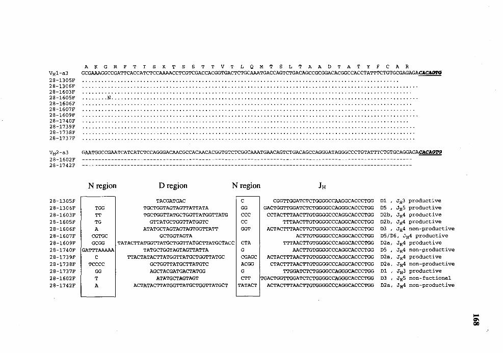
VH1-a3 28-1305F 28-1306F 28-1603F 28-1605F 28-1606F 28-1607F 28-1609F 28-1740F 28-1739F 28-1738F 28-1737F
VH2-a3 28-1602F 28-1742F
28-1305F 28-1306F 28-1603F 28-1605F 28-1606F 28-1607F 28-1609F 28-1740F 28-1739F 28-1738F 28-1737F 28-1602F 28-1742F
A K G R F T I S K T S S T T V T L Q M T S L T A A D T A T Y F C A R GCGAAAGGCCGATTCACCATCTCCAAAACCTCGTCGACCACGGTGACTCTGCAAATGACCAGTCTGACAGCCGCGGACACGGCCACCTATTTCTGTGCGAGAGAc.\CAG!'G
..•..... N ........................•........ ····································•·········•·········•·····
....................................................................................................... · .....
GAATGGCCGAATCATCATCTCCAGGGACAACGCCACAACACGGTGTCTCGGCAAATGAACAGTCTGACAGCCAGGGATAGGGCCCTGTATTTCTGTGCAGGAGACACAG!l'G
N region D region N region JH
TACGATGAC c CGGTTGGATCTCTGGGGCCAAGGCACCCTGG Dl , JH3 productive TGG TGCTGGTAGTAGTTATTATA GG GACTGGTTGGATCTCTGGGGCCAGGGCACCCTGG D5 , JH5 productive TT TGCTGGTTATGCTGGTTATGGTTATG CCC CCTACTTTAACTTGTGGGGCCCAGGCACCCTGG D2b, JH4 productive TG GTTATGCTGGTTATGGTC cc TTTAACTTGTGGGGCCCAGGCACCCTGG D2b, JH4 productive A ATATGCTAGTAGTAGTGGTTATT GGT ACTACTTTAACTTGTGGGGCCCAGGCACCCTGG D3 ' JH4 non-productive
CGTGC GCTGGTAGTA ACTTGTGGGGCCCAGGCACCCTGG D5/D6, JH4 productive GCGG TATACTTATGGTTATGCTGGTTATGCTTATGCTACC CTA TTTAACTTGTGGGGCCCAGGCACCCTGG D2a, JH4 productive
GATTTAAAAA TATGCTGGTAGTAGTTATTA G AACTTGTGGGGCCCAGGCACCCTGG D5 ' JH4 non-productive c TTACTATACTTATGGTTATGCTGGTTATGC CGAGC ACTACTTTAACTTGTGGGGCCCAGGCACCCTGG D2a, JH4 productive
TCCCC GCTGGTTATGCTTATGTC ACGG CTACTTTAACTTGTGGGGCCCAGGCACCCTGG D2a, JH4 non-productive GG AGCTACGATGACTATGG G TTGGATCTCTGGGGCCAGGGCACCCTGG Dl , JH3 productive T ATATGCTAGTAGT CTT TGACTGGTTGGATCTCTGGGGCCAGGGCACCCTGG D3 ' JH5 non-fuctional A ACTATACTTATGGTTATGCTGGTTATGCT TATACT ACTACTTTAACTTGTGGGGCCCAGGCACCCTGG D2a, JH4 non-productive
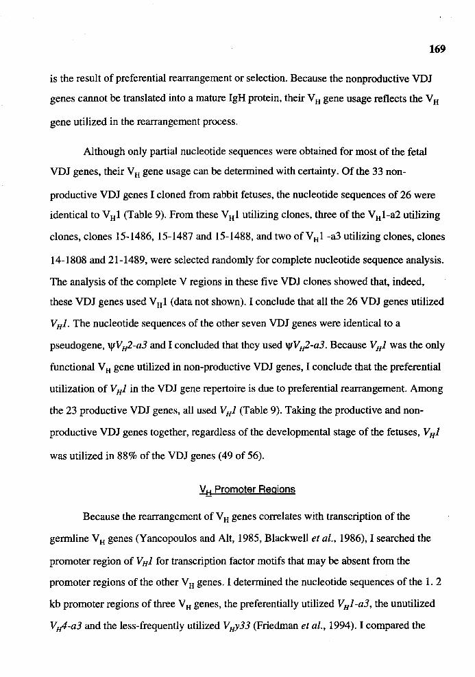
169
is the result of preferential rearrangement or selection. Because the nonproductive VDJ
genes cannot be translated into a mature IgH protein, their V H gene usage reflects the V H
gene utilized in the rearrangement process.
Although only partial nucleotide sequences were obtained for most of the fetal
VDJ genes, their V H gene usage can be determined with certainty. Of the 33 non-
productive VDJ genes I cloned from rabbit fetuses, the nucleotide sequences of 26 were
identical to V H 1 (Table 9). From these V H 1 utilizing clones, three of the V H 1-a2 utilizing
clones, clones 15-1486, 15-1487 and 15-1488, and two of VHl -a3 utilizing clones, clones
14-1808 and 21-1489, were selected randomly for complete nucleotide sequence analysis.
The analysis of the complete V regions in these five VDJ clones showed that, indeed,
these VDJ genes used V H 1 (data not shown). I conclude that all the 26 VDJ genes utilized
V HJ. The nucleotide sequences of the other seven VDJ genes were identical to a
pseudogene, 'lfVH2-a3 and I concluded that they used 'lfVH2-a3. Because VHJ was the only
functional V H gene utilized in non-productive VDJ genes, I conclude that the preferential
utilization of V HJ in the VDJ gene repertoire is due to preferential rearrangement. Among
the 23 productive VDJ genes, all used VHJ (Table 9). Taking the productive and non
productive VDJ genes together, regardless of the developmental stage of the fetuses, VHJ
was utilized in 88% of the VDJ genes (49 of 56).
V:!::i Promoter Regions
Because the rearrangement of V H genes correlates with transcription of the
germline VH genes (Yancopoulos and Alt, 1985, Blackwell et al., 1986), I searched the
promoter region of VHJ for transcription factor motifs that may be absent from the
promoter regions of the other V H genes. I determined the nucleotide sequences of the 1. 2
kb promoter regions of three V H genes, the preferentially utilized V HJ -a3, the unutilized
V Jtl-a3 and the less-frequently utilized VHy33 (Friedman et al., 1994). I compared the
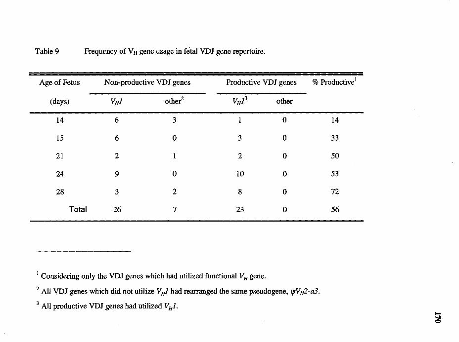
Table 9 Frequency of Vtt gene usage in fetal VDJ gene repertoire.
Age of Fetus Non-productive VDJ genes Productive VDJ genes % Productive1
(days) VHJ other VH13 other
14 6 3 1 0
15 6 0 3 0
21 2 1 2 0
24 9 0 10 0
28 3 2 8 0
Total 26 7 23 0
1 Considering only the VDJ genes which had utilized functional VH gene.
2 All VDJ genes which did not utilize VHJ had rearranged the same pseudogene, l/fVH2-a3.
3 All productive VDJ genes had utilized V HJ.
14
33
50
53
72
56
nucleotide sequence of these promoters to each other and to two other rabbit V H
promoters (Figure 35) (Bernstein et al., 1985).
171
One of the most important motifs for transcription factor is the highly conserved
octamer motif (Eaton and Calame, 1987), which is found among the promoters of VH, VL,
Fµ, Ex: and TCR genes (reviewed in McDougall et al., 1989, Calame and Eaton, 1988).
The octamer motif of all rabbit V H promoters examined, except for the promoter of the
pseudogene \j/CL42. V H25, are identical to each other and to the consensus sequence of
the octamer motif (Figure 35). A potential TAT A box was identified 10 bp upstream of
the octamer motif of all the V H promoters except for that of the pseudogene \j/CIA2.
V H34. Like the octamer motif, the TAT A box of each V H promoter (except for the
promoter of the pseudogene, tpCIA2.V8 34) is identical to each other (Figure 35).
Another conserved motif among the V 8 promoter is the heptamer motif. Mutation
studies showed that mutation of the heptamer reduced the activity of the V H promoter by
30% to 80% suggesting that the heptamer is an important motif for the activity of V H
promoters (reviewed in Staudt and Lenardo, 1991). Two heptamer-like motifs were found
in each of the five rabbit V 8 promoters examined (Figure 35). One of them is located 13
bp downsteam of the octamer and all the V 8 promoters have identical heptamer motif at
this position. Another heptamer-like motif was found 20 bp upstream
of the octamer. The heptamer at this position differs considerably among the five
V 8 promoters. It is possible that these differences in the heptamer-like motif may result in
different levels of promoter activity. However, these heptamer-like motifs themselves
differ from the consensus sequence of the heptamer (CTCATGA) and therefore, the
argument that the differences in the heptamer-like motifs may result in different promoter
activity is not overly strong.
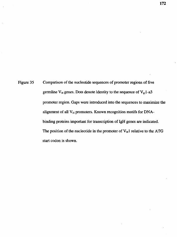
Figure 35
172
Comparison of the nucleotide sequences of promoter regions of five
germline VH genes. Dots denote identity to the sequence of VH1-a3
promoter region. Gaps were introduced into the sequences to maximize the
alignment of all VH promoters. Known recognition motifs for DNA
binding proteins important for transcription of IgH genes are indicated.
The position of the nucleotide in the promoter of VHl relative to the ATG
start codon is shown.
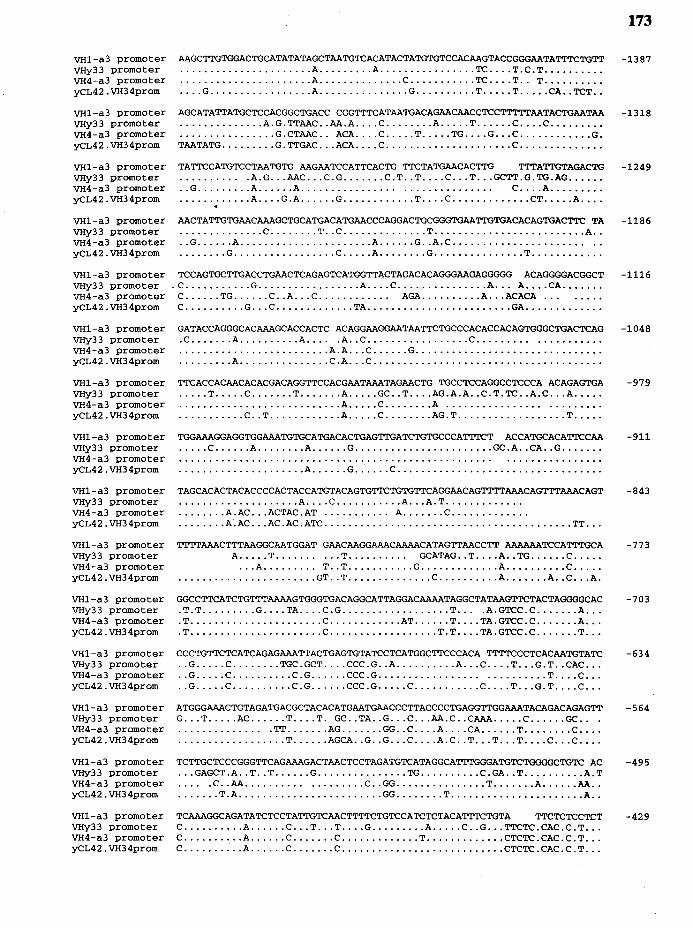
VH1-a3 promoter VHy33 promoter VH4-a3 promoter yCL42.VH34prom
VH1-a3 promoter VHy33 promoter VH4-a3 promoter yCL42.VH34prom
VH1-a3 promoter VHy33 promoter VH4-a3 promoter yCL42.VH34prom
VH1-a3 promoter VHy33 promoter VH4-a3 promoter yCL42.VH34prom
VH1-a3 promoter VHy33 promoter VH4-a3 promoter yCL42.VH34prom
VH1-a3 promoter VHy33 promoter VH4-a3 promoter yCL42.VH34prom
VH1-a3 promoter VHy33 promoter VH4-a3 promoter yCL42.VH34prom
VH1-a3 promoter VHy33 promoter VH4-a3 promoter yCL42.VH34prom
VH1-a3 promoter VHy33 promoter VH4-a3 promoter yCL42.VH34prom
VH1-a3 promoter VHy33 promoter VH4-a3 promoter yCL42.VH34prom
VH1-a3 promoter VHy33 promoter VH4-a3 promoter yCL42.VH34prom
VH1-a3 promoter VHy33 promoter VH4-a3 promoter yCL42.VH34prom
VH1-a3 promoter VHy33 promoter VH4-a3 promoter yCL42.VH34prom
VH1-a3 promoter VHy33 promoter VH4-a3 promoter yCL42.VH34prom
VH1-a3 promoter VHy33 promoter VH4-a3 promoter yCL42.VH34prom
AAGCTTGTGGACTGCATATATAGCTAATGTCACATACTATGTGTCCACAAGTACCGGGAATATTTCTGTT ...................... A ......... A ................ TC .... T.C.T ......... . ...................... A .............. C ........... TC .... T .. T ......... . .... G ................. A ............... G .......... T ..... T ..... CA .. TCT ..
AGCATATTATGCTCCACGGCTGACC CGGTTTCATAATGACAGAACAACCTCCTTTTTAATACTGAATAA .............. A.G. TTAAC .. AA.A .... C ........ A ..... T ...... C .... C ........ . ................ G. CTAAC. . ACA .... C ..... T ..... TG .... G ... C ............ G. TAATATG ......... G. TTGAC ... ACA .... C ..................... C ............. .
TATTCCATGTCCTAATGTG AAGAATCCATTCACTG TTCTATGAACACTTG TTTATTGTAGACTG . . . . . . . . . . .A.G ... AAC ... C. G ....... C. T .. T .... C ... T ... GCTT .G. TG.AG ..... . .. G ......... A ...... A................ . . . . . . . . . . . . . . . C .... A ........ . ............ A .... G .A ...... G ............ T .... C ............. CT ..... A ... . .. AACTATTGTGAACAAAGCTGCATGACATGAACCCAGGACTGCGGGTGAATTGTGACACAGTGACTTC TA ••••••........ C ........ T .. C .............. T .....•...••••••••••••••.• A •• .. G ...... A ...................... A ...... G .. A.C ....................... . •••••••• G ••••••••••••••••• C ••••• A •••••••• G ••••••••••••••• T ••••••••••••
TCCAGTGCTTGACCTGAACTCAGAGTCATGGTTACTAGACACAGGGAAGAGGGGG ACAGGGGACGGCT . C ........... G ................. A .... C ............... A ... A .... CA ...... .
C ...... TG ...... C .. A ... C............ AGA .......... A ... ACACA .. . C .......... G ... C ............. TA ........................ GA ............ .
GATACCAGGGCACAAAGCACCACTC ACAGGAAGGAATAATTCTGCCCACACCACAGTGGGCTGACTCAG .C ....... A .......... A ..... A .. C ................. C ................... . ......................... A.A ... C ...... G .............................. . ......... A ............... C.A ... C ..................................... .
TTCACCACAACACACGACAGGTTCCACGAATAAATAGAACTG TGCCTCCAGGCCTCCCA ACAGAGTGA ..... T ..... C ....... T ....... A ..... GC .. T .... AG.A.A .. C. T. TC .. A.C ... A .... . . . • • . . . . . • • • • • • • • • • . . . . . • • • A ••••• C •.....•. A .••.•••••..•••••••.•••••. ........... C .. T ............ A ..... C ........ AG.T .................. T .... .
TGGAAAGGAGGTGGAAATGTGCATGACACTGAGTTGATCTGTGCCCATTTCT ACCATGCACATTCCAA ..... C ...... A ........ A ...... G ....................... GC .A .. CA .. G ...... .
••••••••••••••••••••• A •••••• G •••••• C ••••••••••••••••••••••••••••••••••
TAGCACACTACACCCCACTACCATGTACAGTGTTCTGTGTTCAGGAACAGTTTTAAACAGTTTAAACAGT .................... A .... C ........... A ... A. T ............ . ........ A.AC ... ACTAC.AT ........... A ....... C ............ . ........ A.AC ... AC .AC .ATC ......................................... TT .. .
TTTTAAACTTTAAGGCAATGGAT GAACAAGGAAACAAAACATAGTTAACCTT AAAAAATCCATTTGCA A ..... T .......... T.......... GCATAG .. T .... A .. TG ...... C .... .
•.. A ••••••.•• T •. T ••••••••••• G ••••••••••••• A •••••••••• C ••••• ....................... GT .. T .............. C .......... A ....... A .. C ... A.
GGCCTTCATCTGTTTAAAAGTGGGTGACAGGCATTAGGACAAAATAGGCTATAAGTTCTACTAGGGGCAC . T. T ......... G .... TA .... C .G .................. T... .A. GTCC. C ....... A .. . . T ...................... C ............ AT ...... T .... TA.GTCC. C ....... A .. . .T ...................... C .................. T.T .... TA.GTCC .C ....... T .. .
CCCTGTTCTCATCAGAGAAATTACTGAGTGTATCCTCATGGCTTCCCACA TTTTCCCTCACAATGTATC .. G ..... C ........ TGC . GCT .... CCC . G .. A .......... A ... C .... T ... G. T .. CAC .. . .. G ..... C .......... C . G ...... CCC . G . . . . . . . . . . . . . . . . . . ......... T .... C .. . .. G ..... C .......... C. G ...... CCC .G ..... C ........... C .... T ... G. T .... C .. .
ATGGGAAACTGTAGATGACGCTACACATGAATGAACCCTTACCCCTGAGGTTGGAAATACAGACAGAGTT G ... T ..... AC ...... T .... T. GC .. TA .. G ... C ... AA.C .. CAAA ..... C ...... GC. . . . . . . . . . . . . . . . . . TT ....... AG ....... GG .. C .... A .... CA ...... T ........ C ... . .................. T ...... AGCA .. G .. G ... C .... A. C .. T ... T ... T .... C ... C ... .
TCTTGCTCCCGGGTTCAGAAAGACTAACTCCTAGATGTCATAGGCATTTGGGATGTCTGGGGCTGTC AC ... GAGCT .A .. T .. T ...... G ............... TG .......... C. GA .. T .......... A. T . . . . .C .. AA ................... C .. GG ............... T ....... A ...... AA .. ....... T.A ........................ GG ........ T ...................... A ..
TCAAAGGCAGATATCTCCTATTGTCAACTTTTCTGTCCATCTCTACATTTCTGTA TTCTCTCCTCT C .......... A ...... C ... T ... T .... G ......... A ..... C .. G ... TTCTC. CAC. C. T .. . C .......... A ...... C ....... C ............. T ............. CTCTC . CAC . C . T .. . C .......... A ...... C ....... C ........................... CTCTC .CAC.C .T .. .
173
-1387
-1318
-1249
-1186
-1116
-1048
-979
-911
-843
-773
-703
-634
-564
-495
-429
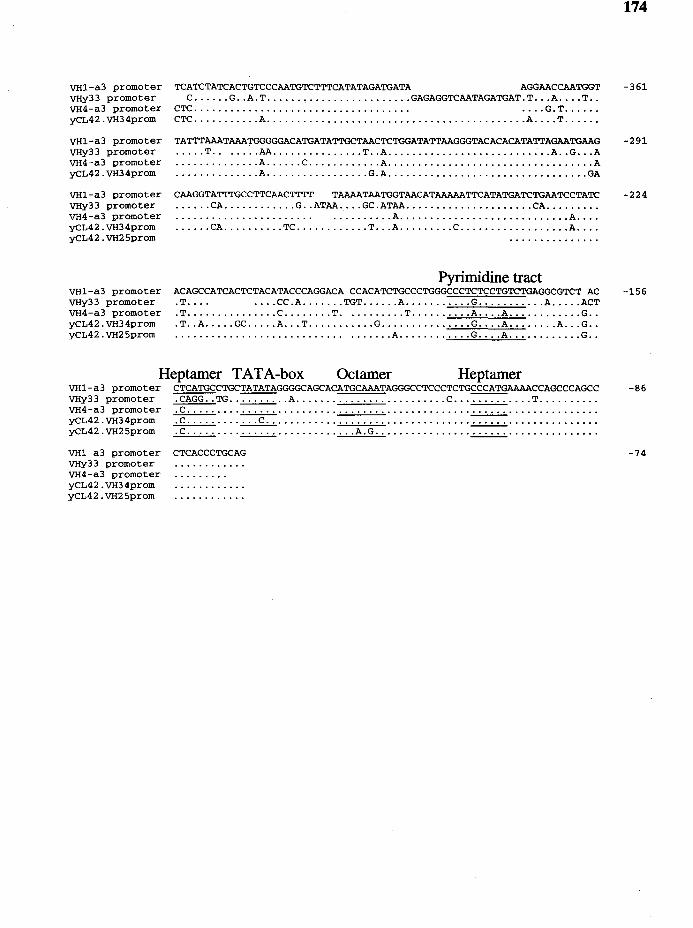
VH1-a3 promoter VHy33 promoter VH4-a3 promoter yCL42.VH34prom
VH1-a3 promoter VHy33 promoter VH4-a3 promoter yCL42.VH34prom
VH1-a3 promoter VHy33 promoter VH4-a3 promoter yCL42.VH34prom yCL42.VH25prom
VH1-a3 promoter VHy33 promoter VH4-a3 promoter yCL42.VH34prom yCL42.VH25prom
TCATCTATCACTGTCCCAATGTCTTTCATATAGATGATA AGGAACCAATGGT C ...... G .. A.T ........................ GAGAGGTCAATAGATGAT.T ... A .... T ..
CTC.................................... . ... G.T ..... . CTC ........... A ........................................... A .... T ..... .
TATTTAAATAAATGGGGGACATGATATTGCTAACTCTGGATATTAAGGGTACACACATATTAGAATGAAG ..... T ....... AA ............... T .. A ........................... A .. G ... A .............. A ...... C ............ A .................................. A .............. A ................. G.A ................................. GA
CAAGGTATTTGCCTTCAACTTTT TAAAATAATGGTAACATAAAAATTCATATGATCTGAATCCTATC ...... CA ............ G .. ATAA .... GC.ATAA ..................... CA ........ . • • • • • • • • • • • • • • • • • • • • . . • • ••..••••• A •••••••••••••••••••••••••••• A •••• ...... CA .......... TC ............ T ... A ......... C .................. A ... .
Pyrimidine tract ACAGCCATCACTCTACATACCCAGGACA CCACATCTGCCCTGGGCCCTCTCCTGTCTGAGGCGTCT AC . T. . . . . ... CC .A ....... TGT ...... A ........... G ........... A ..... ACT .T ••••••.•••••••. C .••••••• T •.•••••••. T •••••••••• A •••• A •••••••••••• G •• . T .. A ..... GC ..... A ... T ........... G ............... G .... A ........ A ... G .. •.••••••.•.••••••••••.••••••••••••• A •••.•••••••• G •••• A •••••••••••• G ••
Heptamer TATA-box Octamer Heptamer
174
-361
-291
-224
-156
VH1-a3 promoter CTCATGCCTGCTATATAGGGGCAGCACATGCAAATAGGGCCTCCCTCTGCCCATGAAAACCAGCCCAGCC -86 VHy33 promoter .CAGG .. TG .. ~ .. A ......................... c ... ~ .... T ......... . VH4-a3 promoter . C ......... ~ ................................ ~ .............. . yCL42.VH34prom .c ............ c .................................. ~ .............. . yCL42.VH25prom .c ........ ·~· ............ A.G ............... ·...:....:....:.· ............. .
VH1-a3 promoter VHy33 promoter VH4-a3 promoter yCL42.VH34prom yCL42.VH25prom
CTCACCCTGCAG -74
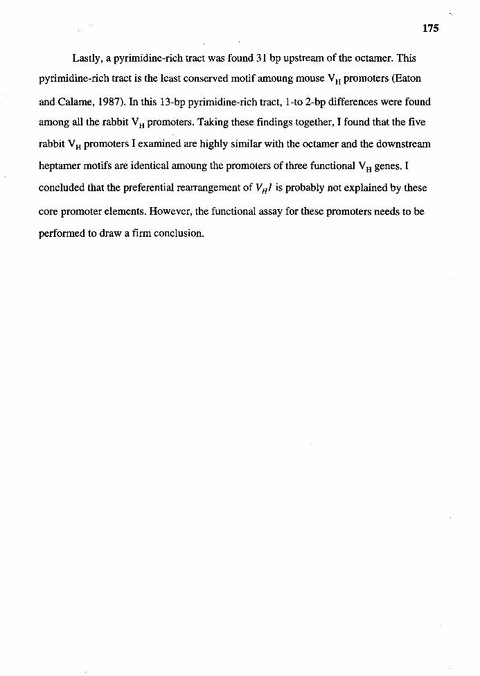
Lastly, a pyrimidine-rich tract was found 31 bp upstream of the octamer. This
pyrimidine-rich tract is the least conserved motif amoung mouse V H promoters (Eaton
175
and Calame, 1987). In this 13-bp pyrimidine-rich tract, 1-to 2-bp differences were found
among all the rabbit V H promoters. Taking these findings together, I found that the five
rabbit V H promoters I examined are highly similar with the octamer and the downstream
heptamer motifs are identical amoung the promoters of three functional V H genes. I
concluded that the preferential rearrangement of V 8 1 is probably not explained by these
core promoter elements. However, the functional assay for these promoters needs to be
performed to draw a firm conclusion.
CHAPTER IV
DISCUSSION
I have shown in this study that the status of the IgH gene on the unexpressed allele
of rabbit B cells is different from that in B cells of other species in two aspects. First,
50% of rabbit B cells do not rearrange their unexpressed IgH alleles (Table 8). The V H
and JH genes on the unexpressed allele of these B cells remain in germline configuration.
This is in contrast to mouse and human in which the unexpressed IgH alleles are
rearrranged to a DJ or a nonproductive VDJ gene (Atkinson et al., 1993, Alt et al., 1984,
Reth, 1986). In chicken, approximately 8% of bursa! cells have JH on the unexpressed
allele in germline configuration (Barth and Humphries, 1988, Benatar et al., 1992,
Reynaud et al., 1989). However, the status of VH in these cells has not been determined.
Second, for those B cells in rabbit which had rearranged the unexpressed IgH allele, most
of them (85%) rearranged DJ (Table 8). Only 15% of them rearranged two VDJ genes,
one of which is presumably nonproductive (Table 8). This is in contrast to mouse and
human B cells in which nonproductive VDJ genes are such a common occurrence that
60% to 70% of B cells have nonproductive VDJ genes on the unexpressed IgH alleles.
But, in this aspect rabbit B cells are similar to those in chicken where the incidence of
nonproductive VDJ genes is low (Ferradini et al., 1994). The possibility that rabbit B
cells generate only productive VDJ genes was ruled out when I found that many VDJ
genes isolated from early fetal development were nonproductive. Rather, it is likely that if
a pre-B cell rearranges a nonproductive VDJ gene, it dies without rearranging a VDJ gene
176
177
on the second IgH allele. Regarding the intermediates of IgH gene rearrangements on the
unexpressed IgH alleles, I found only DJ genes on the unexpressed allele of the
hybridomas. For this reason, I think that the order of IgH gene rearrangement for most of
the VDJ genes is that D rearranges to J and then V rearranges to DJ. Currently, it is no
known what role VD gene rearrangement plays in the IgH gene rearrangements in rabbit.
The other major result of my work is the analysis of V H gene usage in
nonproductive VDJ genes cloned from rabbit fetuses. Our laboratory has shown
previously that rabbit B cells preferentially utilize V H 1 in their VDJ genes (Knight and
Becker, 1990, Raman et al., 1994). In this study, I found that many of the VDJ genes in
fetus are nonproductive and the only functional V H gene utilized is V H 1. Because the V H
gene utilization in these nonproductive VDJ genes cannot be influenced by the selection
process, I conclude that the preferential utilization of V H 1 resulted from its preferential
rearrangement.
Rabbit B Cells do not Rearrange DJ Genes on Both Alleles
before Rearranging VDJ gene
During the development of B cells in mouse, human and chicken, the first
rearrangement of IgH genes is D to JH on both alleles. Therefore, the DJ gene
rearrangement in B-lineage cells is believed the default step of IgH gene rearrangement
without any regulation. In contrast, rabbit B cells do not rearrange DJ genes on both IgH
alleles before VDJ gene rearrangement indicating that DJ gene rearrangement in rabbit is
not a default step as it is in other species.
An alternative explanation for the lack of a DJ gene on the unexpressed IgH allele
is that the DJ gene rearrangement is an inefficient process in rabbit. The IgH gene
rearrangement has been shown to be dependent on the accessibility of the IgH locus to
recombinase enzymes as suggested by the correlation of the rearrangement and the
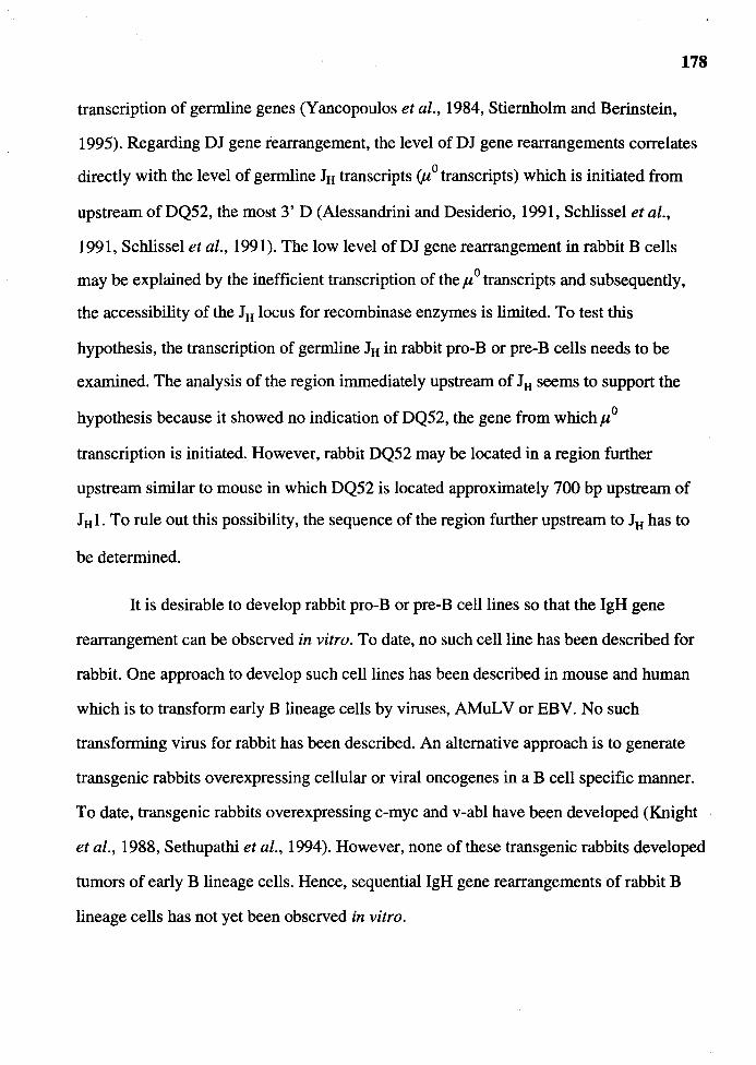
178
transcription of germline genes (Yancopoulos et al., 1984, Stiernholm and Berinstein,
1995). Regarding DJ gene rearrangement, the level of DJ gene rearrangements correlates
directly with the level of germline J8 transcripts (µ 0 transcripts) which is initiated from
upstream of DQ52, the most 3' D (Alessandrini and Desiderio, 1991, Schlissel et al.,
1991, Schlissel et al., 1991). The low level of DJ gene rearrangement in rabbit B cells
may be explained by the inefficient transcription of the µ 0 transcripts and subsequently,
the accessibility of the J8 locus for recombinase enzymes is limited. To test this
hypothesis, the transcription of germline J8 in rabbit pro-B or pre-B cells needs to be
examined. The analysis of the region immediately upstream of J8 seems to support the
hypothesis because it showed no indication of DQ52, the gene from whichµ 0
transcription is initiated. However, rabbit DQ52 may be located in a region further
upstream similar to mouse in which DQ52 is located approximately 700 bp upstream of
J8 1. To rule out this possibility, the sequence of the region further upstream to J8 has to
be determined.
It is desirable to develop rabbit pro-B or pre-B cell lines so that the IgH gene
rearrangement can be observed in vitro. To date, no such cell line has been described for
rabbit. One approach to develop such cell lines has been described in mouse and human
which is to transform early B lineage cells by viruses, AMuLV or EBV. No such
transforming virus for rabbit has been described. An alternative approach is to generate
transgenic rabbits overexpressing cellular or viral oncogenes in a B cell specific manner.
To date, transgenic rabbits overexpressing c-myc and v-abl have been developed (Knight
et al., 1988, Sethupathi et al., 1994). However, none of these transgenic rabbits developed
tumors of early B lineage cells. Hence, sequential lgH gene rearrangements of rabbit B
lineage cells has not yet been observed in vitro.

Nonproductive VDJ Genes Occur Infrequently in Rabbit
and Chicken B Cells
179
I report in this study that nonproductive VDJ genes are found infrequently in B
cells from adult rabbit, which is similar to the finding in chicken. In chicken, DJ
progenitors undergo V to DJ gene rearrangement only on one allele and essentially no
second rearrangement is allowed if the first rearrangement results in a nonproductive VDJ
gene (Ferradini et al., 1994). I have considered the explanation, for the scarcity of
nonproductive VDJ genes in rabbit, that nonproductive rearrangements may occur
infrequently and therefore, the second rearrangement is generally not necessary. However,
because I found many nonproductive VDJ genes in rabbit fetuses, I conclude that IgH
gene rearrangement in rabbit generates nonproductive VDJ genes like in other species.
Therefore,, I propose that the rearrangement in rabbit is similar to that in chicken in that
no second rearrangement is allowed when the first rearrangement fails to generate a
productive VDJ gene.
It appears as if nonproductive VDJ genes are not desirable in these cells. Although
we do not know for certain why this is the case in rabbit and chicken B cells, we can
speculate why chicken B cells do not have nonproductive VDJ genes on their
unexpressed IgH allele. Chicken B cells diversify their VDJ genes by somatic gene
conversion, the process in which the nucleotide sequences of donor genes are duplicated
onto the coding region of the VDJ genes. Gene conversion in chicken occurs both in the
V H and the CDR3 regions. The reading frame of nonproductive VDJ genes can be
restored if the gene conversion occurs in the CDR3 region. Such a gene conversion event
could result in B cells expressing two VDJ genes simultaneously.
The reason why rabbit B cells do not have nonproductive VDJ genes is less clear.
Although rabbit B cells, like chicken, diversify their VDJ genes by somatic gene
conversion-like process, no gene conversion-like event has been documented in the
CDR3 region. Consequently, nonproductively rearranged VDJ genes in rabbit B cells
cannot be converted to productive ones like in chicken.
180
How rabbit confines the generation of VDJ gene to only one allele is not yet
known. Several possibilities can explain the phenomenon and they are discussed below.
lgH Gene Rearrangement may Occur during a Narrow
Window during B Cell Development
This model explains that, during differentiation of B cells, DJ gene
rearrangements on each lgH alleles occur independently of each other (Figure 36). Once
DJ is rearranged on one allele, VDJ gene rearrangement occurs rapidly regardless of the
lgH gene rearrangement on the second allele. Productive VDJ gene on the first allele
would provide the cell with survival and allelic exclusion signals while a nonproductively
rearranged gene would not. These cells then die without rearranging the second allele
because the period during which V(D)J gene rearrangement can occur has passed.
Although I think that this possibility is the most likely explanation, it is not
without a shortcoming. The shortcoming of this model is that it cannot explain the allelic
exclusion in cells which rearrange DJ and, subsequently, V to DJ on both alleles at the
same time. Assuming that the chance of generating a productive VDJ gene is one-third
(the chance that VH is rearranged into the same reading frame of JH), the probability of
two productive VDJ genes to occur in one cell is (113 x 1/3) x 100 = 11 %. If we take into
account that the frequency of B cells with two VDJ genes in rabbit is 10% (Table 8), the
frequency of B cells in the periphery with two productive VDJ genes would be 1.1 %
(10% of 11 % ). This concern of violating allelic exclusion is irrelevant, however, if there
is a selection against B cells with two productive VDJ genes. But the finding of B cells in
lg transgenic mice expressing transgenic and endogenous lg genes simultaneously
discounts the negative

Figure 36 Schematic representation of the "narrow window for rearrangement"
model.
181

pre-B cell
develops into mature B cell
--1 ~J---1 --
dies because the window for rearranging a VDJ gene on the second allele has passed
183
selection of such double producers (Grosschedl et al., 1984, Rusconi and Kohler, 1985,
Weaver et al., 1985, Stall et al., 1988, Vos and Hodes, 1992, Rath et al., 1989, Forni,
1990).
Rabbit B Cells are Inefficient in Repairing
the Double-Strand Breaks
The existence of germline V H• D and JH genes on the unexpressed allele, although
uncommon in mouse, human and chicken B cells, is common for B cells in SCID mice
(Kotloff et al., 1993). This is not unexpected, however, considering that SCID mice are
defective in repairing the lethal double-strand-breaks (<ls-breaks) generated during the
rearrangement process. It is already so rare to find cells with a VDJ gene that the chance
of findings cells with DJ on the unexpressed allele, which requires another successful
DNA repair, is virtually none.
The finding that rabbit B cells have their IgH allele on the unexpressed allele in
germline configuration is similar to the finding of SCID B cells and the above
explanation for SCID mice may apply to rabbit (Figure 37). However, I do not think that
the explanation in SCID mice is applicable to rabbit for three reasons. First, the DNA
repair mechanism is a process highly conserved throughout evolution (reviewed in Jeggo,
1990). There is no apparent reason for rabbit to be an exception. Second, 50% of rabbit B
cells have rearranged their unexpressed lgH allele to DJ or VDJ genes. And finally, rabbit
B cells diversify their VDJ genes by gene conversion which also involves DNA repair
machinery. If the DNA repair machinery in rabbit is not efficient, it is unlikely that the
rabbit would evolve to use gene conversion as a mechanism to diversify their VDJ genes.
The possibility that rabbit B cells may be inefficient in DNA-repair can be tested by
determining the susceptibility of rabbit B cell lines to <ls-breaks generated by X- ·
irradiation, as compared to cell lines from other species.

Figure 37
184
Schematic representation of the "inefficient double-strand breaks repair"
model.
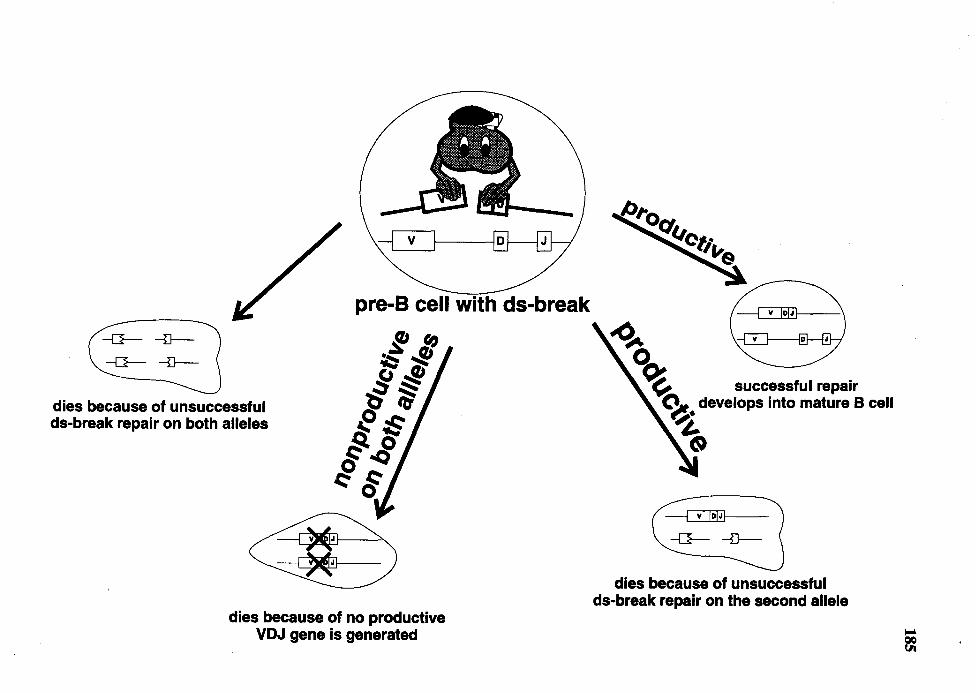
dies because of unsuccessful ds-break repair on both alleles
pre-B cell with ds-break
,f ro ·~ -JP 'J-! Ii ,5§
?) 'te e~
g_ ~ §-o
:: §
dies because of no productive VDJ gene is generated
~ 00! ~ successful repair
(\_.. develops into mature B cell ~· ~
dies because of unsuccessful ds-break repair on the second allele
186
V(D)J Gene Rearrangement Requires a Desuppression
We can also speculate that the V to DJ gene rearrangement is actively suppressed
and that a V to DJ gene rearrangement requires the IgH gene be released (desuppressed)
from the suppression. If the factor(s) required for the desuppression is expressed in such
a low level that only one IgH allele can be desuppressed (Figure 38), as a result,
only one allele can be rearranged to a VDJ gene. The suppression may be mediated by a
silencer similar to the one reported by Luaster et al. (Lauster et al., 1993) who found a
silencer for the rearrangement process between VA. and J.A. genes in chicken. This silencer,
when included in the VA. and J.A. transgenic construct, decreased the level of transgene
rearrangements in transgenic mice. Abutting both 5' and 3' ends of the silencer are anti
silencers which counteract the function of the silencer thus allowing the rearrangement of
the trans gene. The report of a similar silencer-anti-silencer between V H 1 and D in
chicken also supports this model (Ferradini et al., 1994).
The definitive approach to determine whether such silencer-anti-silencer are
present in rabbit IgH locus is to generate several transgenic mouse lines using artificial
rearrangement substrates which contain germline V H• D and JH genes together with
varying amount of the germline region between V H and JH genes and compare the level of
rearrangement of the transgenes. However, this approach is cumbersome and not suitable
as initial approach. Because the silencer in chicken A. gene also functions as a strong
silencer and suppresses the expression of reporter gene in a heterologous promoter system ·
(Lauster et al., 1993), it is possible to locate the silencer in the rabbit IgH locus by
searching for the germline region which possesses silencer activity.
187
Figure 38 Schematic representation of the "suppression and desuppression" model.
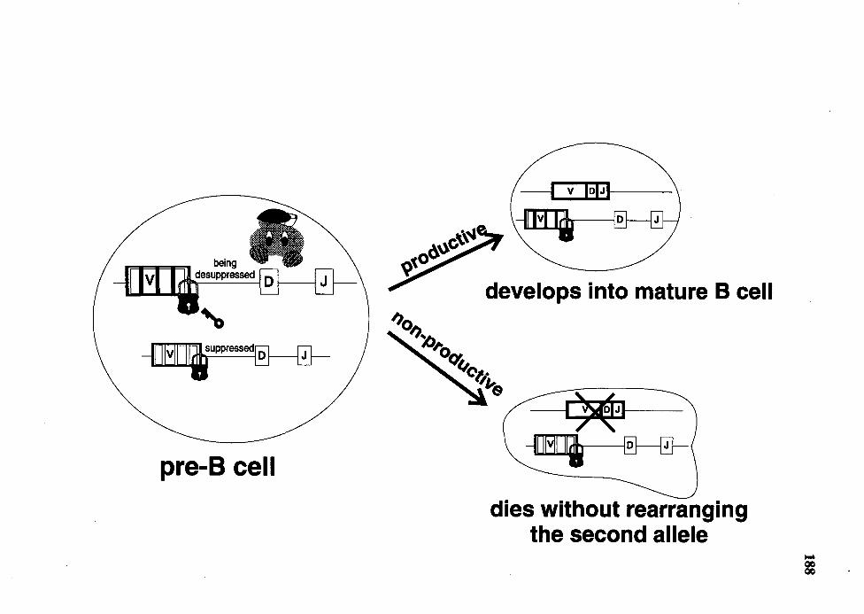
pre-B cell
-I v lolJ-1 --
develops into mature B cell
dies without rearranging the second allele
189
One lgH Allele is Inactivated
An alternative explanation for the low frequency of VDJ genes on the
unexpressed allele is that one lgH allele is inactivated and cannot be rearranged to a VDJ
gene (Figure 39). Although there is no precedence for such inactivation of the IgH gene in
any other species, we can speculate how this might occur in rabbit.
One lgH allele is hypermethylated
Methylation of DNA has been shown to affect several chromosomal activities.
Hypermethylation results in suppression of gene expression (reviewed in Tilghman,
1993), condensation of the chromosome (reviewed in Tilghman, 1993) and reduction of
V(D)J gene rearrangement (Goodhardt et al., 1993, Hsieh and Lieber, 1992, Fernex et al.,
1994, Engler et al., 1991). Therefore, it is not surprising that lgH genes are
hypermethylated in non-B cells (Storb and Arp, 1983) and they become hypomethylated
during the development of B cells (Storb and Arp, 1983). This hypomethylation correlates
with the rearrangement and expression of the lgH genes. In rabbit, it is possible that
during B cell development only one of the two IgH alleles was demethylated so that VDJ
gene rearrangement can occur. If this rearrangement fails to yield a productive VDJ gene,
the rearrangement on the second allele is prohibited because it was not demethylated. To
test this possibility, we need to compare the methylation pattern of IgH genes in purified
B-cell progenitors, pro- and pre-B cells, and B cells. This experiment is not feasible at the
present time due to the lack of antibody to surface markers specific for early B lineage
cells.
One lgH allele is genetically imprinted
Genetic imprinting is an epigenetic phenomenon which results phenotypically in
the expression of a gene on one of the two homologous alleles being inhibited (reviewed
190
in Varmuza and Mann, 1994). Five autosomal genes to date have been shown to be
genetically imprinted (reviewed in Varmuza and Mann, 1994, Chess et al., 1994). Rabbit
may imprint one of its IgH alleles so even if productive VDJ genes are generated on this
allele, B cells cannot express them. However, I doubt that rabbit genetically imprints its
IgH genes because Kitsberg et al. (Kitsberg et al., 1993) ruled out the possibility of
genetic imprinting of IgH genes in mouse. If rabbit IgH genes are imprinted, this would
probably be unique to rabbit. This possibility can be tested by determining whether the
two rabbit lgH alleles replicate asynchronously. An imprinted allele was shown to
replicate later in the cell cycle than the expressed allele (Kitsberg et al., 1993).
Asynchronous replication of rabbit lgH genes would then indicate that one of the lgH
alleles is imprinted. Such asynchronous replication of the IgH gene must be found not
only in non-B cells but also in every cell type because the imprinting process is
independent of the expression of genes (Chess et al., 1994, Kitsberg et al., 1993).
Intermediates of lgH Gene Rearrangements in Rabbit
Rabbit appears to be unique regarding the intermediates of lgH gene
rearrangement in that both VD and DJ genes are found. Rabbit is the first species to be
reported on VD genes which utilized conventional D gene, D2b (Figure 28). (Shin et al.
(1993) reported the cloning of a VD gene previously from a EBV-transformed human B
cell line but that VD gene had used a nonconventional DIR gene). Further, the fact that I
can PCR-amplify many different VD genes from splenic DNA suggests that VD genes
may be the alternative intermediates to DJ genes in VDJ gene rearrangements. However,
the extent to which DJ gene rearrangements occur must be much higher than that of VD
gene rearrangements because most of the rearrangements on the unexpressed alleles of B
cell hybridomas are DJ genes.
191
Figure 39 Schematic representation of the "inactivation of one IgH allele" model.
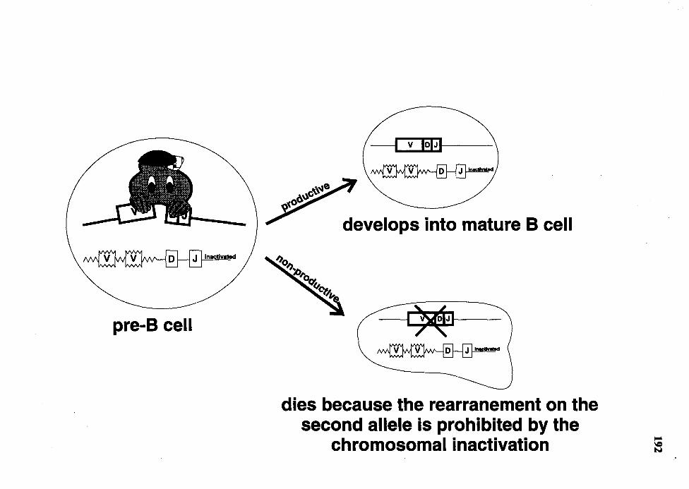
pre-B cell
develops into mature B cell
dies because the rearranement on the second allele is prohibited by the
chromosomal inactivation
193
The VD gene rearrangement is probably not unique to rabbit. I predict that VD
gene rearrangements also occur in other species, such as mouse, human and chicken, but
they have not not detected because Southern analysis used in previous reports to study the
lgH gene rearrangements (Alt et al., 1984, Chen et al., 1993, Nickerson et al., 1989,
Benatar et al., 1992, Reynaud et al., 1989) is not as sensitive as PCR, which I used in this
study. I predict that VD genes are generated at low levels in these species, as well as in
rabbit, and that VD genes could be detected in all species by using PCR.
Currently, we do not know whether the VD or DJ genes can be rearranged to form
a complete VDJ gene. Although one of them may represent a misstep in the
rearrangement process, the intact RSSs of D gene located 3' of VD or 5' of DJ genes
(Figures 23, 27 and 28) argue against this possibility. Instead, they suggest that these
intermediates could be further rearranged to a VDJ gene. This issue can be approached by
examining the rearrangement of artificial rearrangement substrates containing either V
and DJ, or VD and J in transgenic animals or in vitro rearrangement assay. The best
approach is to generate gene-targeted mice using the DJ- or VD-containing substrates
such that the rearrangement of these intermediates can be studied in the context of
endogenous IgH locus.
YH 1 is Preferentially Rearranged
It has been previously shown that 70% to 80% of rabbit B cells preferentially
utilized V Hl. However, previous studies did not discern whether the preferential
utilization of V H 1 resulted from preferential rearrangement or selection for B cells in
which V H 1 was rearranged. In this study, I found evidence to support the preferential
rearrangement of V H 1 when the analysis of V H gene usage in nonproductive VDJ genes
showed that V H 1 was the only functional V H gene utilized (26 of 33 nonproductive VDJ
genes utilized V H 1, Table 9). However, the finding does not rule out the possibility of a
selection process for B cells which rearranged and expressed V H 1. At present, the
molecular basis for the preferential rearrangement of V H 1 is not understood. I discuss
several possible mechanisms for such preferential rearrangement below.
RSS
194
The variation of the RSS has been shown to affect the efficiency of V(D)J gene
rearrangement (Hesse et al., 1989, Akamatsu et al., 1994). One obvious explanation for
the preferential rearrangement of V H 1 is that its RSS is more suitable for the
rearrangement than those of other V H genes. By comparing the RSS of all the functional
V H genes we have cloned, I found that this explanation is unlikely to be the case. As
shown in Figure 40, the RSS of the V H genes are very similar, many of which are
identical to that of V H 1. I conclude that the RSS alone is not sufficient to explain the
preferential rearrangement of V H 1.
Promoters
Because Y ancopoulos et al. (Y ancopoulos and Alt, 1985) reported that the
rearrangement of V H genes correlate with transcription of the germline V H genes, I
attempted to correlate the preferential rearrangement of V H 1 with the activity of its
promoter region by searching for transcription factor motifs which may be present in V H 1
promoter but absent from the promoter regions of the other V H genes. By comparing the
nucleotide sequences of the 1. 2 kb promoter regions of three V H genes, the preferentially
utilized V Hl-a3, the less-frequently utilized V Hy33, the unutilized V Jt4-a3 (Friedman et
al., 1994) and two other rabbit Vtt promoters (Bernstein et al., 1985), I found that the four
motifs important for the activity of mouse V H promoters, the octamer, the heptamer, the
pyrimidine-rich tract and the TATA-box (Eaton and Calame, 1987), were similar_in all of
the V H promoters (Figure 35). I suggest that the preferential rearrangement of V HJ is
probably not explained by these core promoter elements. However, to draw a firm
Figure 40
195
Comparison of the RSSs of all several functional germline V H genes. Dots
denote identity to the RSS of V H 1-al. Slashes indicate gaps which are
introduced to maximize the alignment. The heptamer and nonamer
elements of the RSS are indicated.
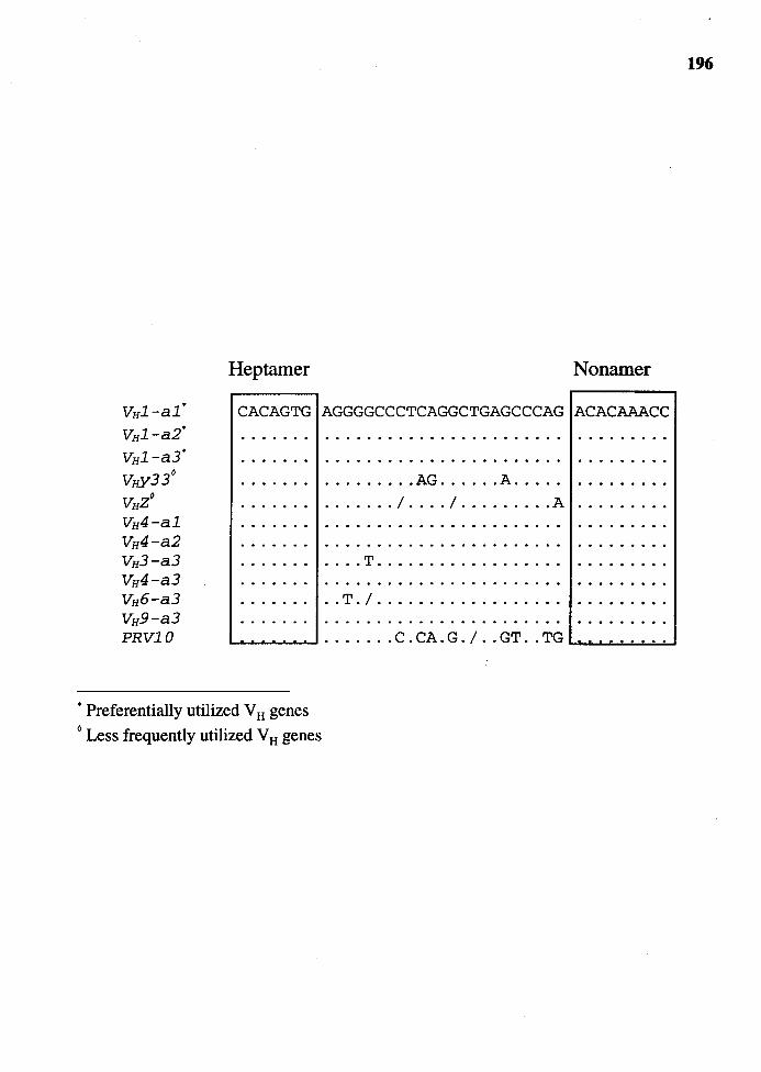
Hep tamer Nonamer
VHl-al'
vH1-a2·
VH1-a3'
Vi,y33°
VHZ0
VH4-al VH4-a2 VH3-a3 VH4-a3 VH6-a3 VH9-a3 PRVlO
CACAGTG AGGGGCCCTCAGGCTGAGCCCAG ACACAAACC
......... AG ...... A .... .
....... I .... I ........ . A
.... T ................. .
.. T. I . ................ .
................... .__ ................ C . CA. G . I .. GT .. TG . . ...
• Preferentially utilized V H genes 0 Less frequently utilized V H genes
196
197
conclusion, we need to compare the ability of these promoters to drive the expression of
reporter genes, such as CAT or Luciferase, in a transient transfection system.
Another experimental approach worth considering is the examination of the
germline VH transcript. If the higher activity of the VHl promoter, compared to that of
other V H genes, is the explanation for preferential rearrangement of V H 1, then we would
expect to find a higher level of germline V H 1 transcripts than of other V H genes in pre-B
cells which are undergoing V(D)J gene rearrangement. But as mentioned earlier, the
caveat of this experiment for the moment is the lack of technology to purify rabbit pre-B
cells.
Proximity of VH 1 to D Region
Yancopoulos et al. (Yancopoulos et al., 1984) found that murine fetal pre-B cell
lines preferentially utilized the most D-proximal VH gene, VH81X, in their VDJ gene
rearrangements. They proposed that the proximity of the V H gene to the D rendered it
more accessible to the rearrangement process. Because rabbit V HJ is also the most D
proximal VH gene (Knight and Becker, 1990), it may be readily accessible to the
recombinase enzymes like the V H81X in mouse. Although we could cite the utilization of
the Vtt gene immediately 5' to VHJ ('JIVH2-a3) to argue in favor ofthis hypothesis, it is
not likely the explanation for the preferential rearrangement of V HJ for two reasons. First,
the Vtt genes other than VHJ which are used most frequently in productive VDJ genes are
not immediately upstream of VHJ. Rather, these genes, VHx, VHY and VHz, are located
more than 50 kb upstream of V HJ, beyond V Hl 1 (Knight and Becker, 1990, Raman et al.,
1994). Furthermore, I found that 'JIVtt2 was used only in the fetal VDJ genes derived
from a3 allotype fetuses and not in a 1 or a2 fetuses.
198
Methylation Status of V H 1
The preferential rearrangement of V HJ could also be explained by the methylation
status of V HJ. Hsieh and Lieber ( 1992) demonstrated that a hypermethylated artificial
rearrangement substrate became inaccessible for V(D)J recombination after it was
transfected into a pre-B cell line. Furthermore, Femex et al. (Femex et al., 1994) found in
transgenic mice that hypermethylation of an lg transgene correlated with decreased
rearrangement. Possibly V HJ in rabbit is undermethylated relative to other V H genes and
is therefore preferentially rearranged.
Chromosome Topology
Another possibility to explain preferential rearrangement of V HJ is the presence of
a matrix-associated region (MAR) in close proximity to V HJ. Chen et al. (1993) showed
that the rearrangement of JH genes was impaired after the MARs located upstream of Eµ
was disrupted by gene targeting technology. It is possible that rabbit VHJ may be located
near a MAR sequence and that this proximity may render V HJ susceptible to preferential
rearrangement. At present, there is no good consensus sequence for MAR and therefore,
the presence of MAR in any region of the chromosome has to be determined empirically.
This involves determining whether the chromosomal DNA from the region of interest can
bind to nuclear-matrix. By this method, four MARs have been identified in the mouse
IgH locus between D genes and the 3'-most CH gene, Ca (Cockerill, 1990). However, the
correlation between V H gene usage and MARs has not yet been analyzed. If the
association of V H genes to MARs influences their usage in the VDJ gene repertoire, I
would expect that rabbit IgH locus may contain as few as four MARs because only four
V H genes appear to generate the entire rabbit VDJ gene repertoire.
The study of V(D)J gene rearrangement is important for our understanding of
humoral immunity. Rabbit presents a unique system to study the preferential VH gene

199
utilization and rearrangement because of its limited V H gene usage in the VDJ gene
repertoire. The knowledge we obtain from studying rabbit V(D)J gene rearrangement will
complement that obtained from other species. Ultimately, we hope to integrate all the
knowledge about humoral immunity from every species so that a single picture of the
humoral immune system with no species barrier may emerge.
REFERENCES
Aguilera, R. J., Akira, S., Okazaki, K. and Sakano, H. (1987). A pre-B cell nuclear protein that specifically interacts with the immunoglobulin V-J recombination sequences. Cell 51:909.
Akamatsu, Y., Tsurushita, N., Nagawa, F., Matsuoka, M., Okazaki, K., Imai, M. and Sakano, H. (1994). Essential residues in V(D)J recombination signals. J. Immonol. 153:4520.
Akira, S., Okazaki, K. and Sakano, H. (1987). Two pairs ofrecombination signals are sufficient to cause immunoglobulin V-(D)-J joining. Science 238: 1134.
Alessandrini, A. and Desiderio, S. V. (1991). Coordination of immunoglobulin DJH transcription and D-to-JH rearrangement by promoter-enhancer approximation. Mol. Cell. Biol. 11:2096.
Allegrucci, M., Young, C. G., Alexander, C. B., Newman, B. A. and Mage, R. G. (1991). Preferrential rearrangement in normal rabbits of the 3' VHa allotype gene that is deleted in Alicia mutants; somatic hypermutation/conversion may play a major role in generating the heterogeneity of rabbit heavy chain variable region sequences. Eur. J. Immonol. 21:411.
Alt, F., Rosenberg, N., Lewis, S., Thomas, E. and Baltimore, D. (1981). Organization and reorganization of immunoglobulin genes in A-MuLV-transformed cells: rearrangement of heavy but not light chain genes. Cell 27:381.
Alt, F. W. (1987). Development of the primary antibody repertoire. Science 238:1079.
Alt, F. W. and Baltimore, D. (1982). Joining of immunoglobulin heavy chain gene segments: implications from a chromosome with evidence of three D-JH fusions. Proc. Natl. Acad. Sci. USA79:4118.
Alt, F. W., Yancopoulos, G.D., Blackwell, T. K., Wood, C., Thomas, E., Boss, M., Coffman, R., Rosenberg, N., Tonegawa, S. and Baltimore, D. (1984). Ordered . rearrangement of immunoglobulin heavy chain variable region segments. Embo J. 3:1209.
200

201
Altenburger, W., Steinmetz, M. and Zachau, H. G. (1980). Functional and non-functional joining in immunoglobulin light chain genes of a mouse myeloma. Nature 287:603.
Anderson, S. J., Abraham, K. M., Nakayama, T., Singer, A. and Perlmutter, R. M. (1992). Inhibition of T-cell receptor beta-chain gene rearrangement by overexpression of the nonreceptor protein tyrosine kinase p56lck. Embo J. 11:4877.
Anderson, S. J., Levin, S. D. and Perlmutter, R. M. (1993). Protein tyrosine kinase p56lck controls allelic exclusion of T-cell receptor beta-chain genes. Nature 365:552.
Atkinson, M. J., Paige, C. J. and Wu, G. E. (1993). Map position and usage of 3' VH family members: usage is not position dependent. Int. Immunol. 5:1577.
Barth, C. F. and Humphries, E. H. (1988). Expression of v-rel induces mature B-cell lines that reflect the diversity of avian immunoglobulin heavy- and light-chain rearrangements. Mol. Cell. Biol. 8:5358.
Beck, E.G., Jack, H. M. and Wabl, M. (1987). Allelic inclusion in a pre-B-cell line that generates immunoglobulin heavy chain genes in vitro. Proc. Natl. Acad. Sci. USA 84:1060.
Becker, R. S. and Knight, K. L. (1990). Somatic diversification of immunoglobulin heavy chain VDJ genes: evidence for somatic gene conversion in rabbits. Cell 63:987.
Becker, R. S., Suter, M. and Knight, K. L. (1990). Restricted utilization of VH and DH genes in leukemic rabbit B cells. Eur. J. Immonol. 20:397.
Becker, R. S., Zhai, S. K., Currier, S. J. and Knight, K. L. (1989). lg VH, DH, and JH germ-line gene segments linked by overlapping cosmid clones of rabbit DNA. J. Immonol. 142: 1351.
Benatar, T., Tkalec, L. and Ratcliffe, M. J. (1992). Stochastic rearrangement of immunoglobulin variable-region genes in chicken B-cell development. Proc. Natl. Acad. Sci. USA 89:7615.
Benger, J.C. and Cox, D. W. (1989). Polymorphisms of the immunoglobulin heavy-chain delta gene and association with other constant-region genes. Am. J. Hum. Gen. 45:606.
Berinstein, N., Levy, S. and Levy, R. (1989). Activation of an excluded immunoglobulin allele in a human B lymphoma cell line. Science 244:337.
Berman, J.E. and Alt, F. W. (1990). Human heavy chain variable region gene diversity, organization, and expression. [Review]. Int. Rev. Immunol. 5:203.
Berman, J. E., Mellis, S. J., Pollock, R., Smith, C. L., Suh, H., Heinke, B., Kowal, C., Surti, U., Chess, L., Cantor, C.R. and et, a. I. (1988). Content and organization of the

202
human lg VH locus: definition of three new VH families and linkage to the lg CH locus. EmboJ. 7:727.
Bernard, 0., Gough, N. M. and Adams, J.M. (1981). Plasmacytomas with more than one immunoglobulin kappa mRNA: implications for allelic exclusion. Proc. Natl. Acad. Sci. USA 78:5812.
Bernard, 0., Hozumi, N. and Tonegawa, S. (1978). Sequences of mouse immunoglobulin light chain genes before and after somatic changes. Cell 15: 1133.
Bernstein, K. E., Alexander, C. B. and Mage, R. G. (1985). Germline VH genes in an a3 rabbit not typical of any one VHa allotype. J. Immonol. 134:3480.
Blackwell, T. K. (1984). Joining of immunoglobulin heavy chain variable region gene segments in vivo and within a recombination substrate. UCLA Symp. Mol. Cell. Biol., New Series 19:537.
Blackwell, T. K., Moore, M. W., Yancopoulos, G.D., Suh, H., Lutzker, S., Selsing, E. and Alt, F. W. (1986). Recombination between immunoglobulin variable region gene segments is enhanced by transcription. Nature 324:585.
Bollum, F. (1974). In "The Enzymes", vol. 10. p. 145. Academic Press, New York.
Borgulya, P., Kishi, H., Uematsu, Y. and von, B. H. (1992). Exclusion and inclusion of alpha and beta T cell receptor alleles. Cell 69:529.
Bos, N. A. and Meeuwsen, C. G. (1989). B cell repertoire in adult antigen-free and conventional neonatal BALB/c mice. I. Preferential utilization of the CH-proximal VH gene family PC7183. Eur. J. Immonol. 19: 1811.
Bosma, M. J. and Carroll, A. M. (1991). The SCID mouse mutant: definition, characterization, and potential uses. [Review]. Ann. Rev. Immunol9:323.
Bottaro, A., Cariota, U., DeMarchi, M. and Carbonara, A. 0. (1991). Pulsed-field electrophoresis screening for immunoglobulin heavy-chain constant-region (IGHC) multigene deletions and duplications. Am. J. Hum. Gen. 48:745.
Brack, C., Hirama, M., Lenhard, S. R. and Tonegawa, S. (1978). A complete immunoglobulin gene is created by somatic recombination. Cell 15:1.
Brodeur, P.H., Osman, G. E., Mackle, J. J. and Lalor, T. M. (1988). The organization of the mouse lgh-V locus. Dispersion, interspersion, and the evolution of VH gene family clusters. J. Exp. Med. 168:2261.
Brodeur, P.H. and Riblet, R. (1984). The immunoglobulin heavy chain variable region (lgh-V) locus in the mouse. I. One hundred Igh-V genes comprise seven families of
203
homologous genes. Eur. J. Immonol. 14:922.
Brodeur, P.H., Thompson, M.A. and Riblet, R. (1984). The content and organization of mouse IGH-V families. UCLA Symp. Mol. and Cell. Biol., new series 18:445.
Brueckner, M., D'Eustachio, P. and Horwich, A. L. (1989). Linkage mapping of a mouse gene, iv, that controls left-right asymmetry of the heart and viscera. Proc. Natl. Acad. Sci. USA 86:5035.
Buluwela, L., Albertson, D. G., Sherrington, P., Rabbitts, P. H., Spurr, N. and Rabbitts, T. H. (1988). The use of chromosomal translocations to study human immunoglobulin gene organization: mapping DH segments within 35 kb of the C mu gene and identification of a new DH locus. Embo J. 7:2003.
Burnett, R. C., Hanly, W. C., Zhai, S. K. and Knight, K. L. (1989). The IgA heavy-chain gene family in rabbit: cloning and sequence analysis of 13 C alpha genes. Embo J. 8:4041.
Calame, K. and Eaton, S. (1988). Transcriptional controlling elements in the immunoglobulin and T cell receptor loci. [Review]. Adv. Immunol. 43:235.
Cancro, M. P., K.ienker, L. J., Ferguson, S. E. and Allman, D. M. (1991). B-cell repertoire development. [Review]. Immunol. Series 55:25.
Capra, J. D. and Kehoe, J.M. (1975). Hypervariable regions, idiotypy, and the antibodycombining site. Adv. Immunol. 20: 1.
Casanova, J. L., Romero, P., Widmann, C., Kourilsky, P. and Maryanski, J. L. (1991). T cell receptor genes in a series of class I major histocompatibility complex-restricted cytotoxic T lymphocyte clones specific for a Plasmodium berghei nonapeptide: implications for T cell allelic exclusion and antigen-specific repertoire. J. Exp. Med. 174:1371.
Cebra, J. J., Colberg, J.E. and Dray, S. (1965). Rabbit lymphoid cells differentiated with respect to a-, y-- andµ- heavy polypeptide chains and to allotypic markers Aaland Aa2. J. Exp. Med. 123:547.
Cebra, J. J., Colberg, J.E. and Dray, S. (1966). Rabbit lymphoid cells differentiated with respect to a-, y--, and u- heavy polypeptide chains and to allotypic markers Aal, Aa2. J. Exp. Med. 123:547.
Chaabani, H., Bech, H. N. and Cox, D. W. (1985). A multigene deletion within the immunoglobulin heavy-chain region. Am. J. Hum. Gen. 37: 1164.
Champagne, E., Sagman, U., Biondi, A., Lewis, W. H., Mak, T. W. and Minden, M. D. ( 1988). Structure and rearrangement of the T cell receptor J alpha locus in T cells and
204
leukemic T cell lines. Eur. J. Immonol. 18: 1033.
Chen, J. and Alt, F. W. (1993). Gene rearrangement and B-cell development. [Review]. Curr. Op. Immunol. 5:194.
Chen, J., Trounstine, M., Alt, F. W., Young, F., Kurahara, C., Loring, J. F. and Huszar, D. (1993). lmmunoglobulin gene rearrangement in B cell deficient mice generated by targeted deletion of the JH locus. Int. Immunol 5:647.
Chen, J., Young, F., Bottaro, A., Stewart, V., Smith, R. K. and Alt, F. W. (1993). Mutations of the intronic IgH enhancer and its flanking sequences differentially affect accessibility of the JH locus. Embo J. 12:4635.
Cherepakhin, V. V., Jackubov, L. Z., Ibraghimov, A. R. and Rokhlin, 0. V. (1987). Allelic exclusion frequency analysis and molecular characteristics of immunoglobulins secreted by the hybridomas expressing both allelic genes. Immunol. Lett. 15:33.
Cherif, D. and Berger, R. (1990). New localizations of VH sequences by in situ hybridization with biotinylated probes. Genes, Chrom. Cancer 2: 103.
Chess, A., Simon, I., Cedar, H. and Axel, R. (1994). Allelic inactivation regulates olfactory receptor gene expression. Cell 78:823.
Chou, H. S., Behlke, M. A., Godambe, S. A., Russell, J. H., Brooks, C. G. and Loh, D. Y. (1986). T cell receptor genes in an alloreactive CTL clone: implications for rearrangement and germline diversity of variable gene segments. Embo J. 5:2149.
Christoph, T. and Krawinkel, U. (1989). Physical linkage of variable, diversity and joining gene segments in the immunoglobulin heavy chain locus of the mouse. Eur. J. Immonol. 19: 1521.
Chukwuocha, R. U. and Feeney, A. J. (1993). Role of homology-directed recombination: predominantly productive rearrangements of Vh81X in newborns but not in adults. Mol. Immunol. 30:1473.
Cockerill, P. N. (1990). Nuclear matrix attachment occurs in several regions of the lgH locus. Nucleic Acids Res. 18:2643.
Cohen, S. and Milstein, C. (1967). Structure and biological properties of immunoglobulins. [Review]. Adv. Immunol. 7:1.
Coleclough, C., Perry, R. P., Karjalainen, K. and Weigert, M. (1981). Aberrant rearrangements contribute significantly to the allelic exclusion of immunoglobulin gene expression. Nature 290:372.
Corcos, D., Iglesias, A., Dunda, 0., Bucchini, D. and Jami, J. (1991). Allelic exclusion in
transgenic mice expressing a heavy chain disease-like human mu protein. Eur. J. Immonol. 21:2711.
205
Cory, S. and Adams, J.M. (1980). Deletions are associated with somatic rearrangement of immunoglobulin heavy chain genes. Cell 19:37.
Costa, T. E., Suh, H. and Nussenzweig, M. C. (1992). Chromosomal position of rearranging gene segments influences allelic exclusion in transgenic mice. Proc. Natl. Acad. Sci. USA 89:2205.
Couez, D., Malissen, M., Buferne, M., Schmitt, V. A. and Malissen, B. (1991). Each of the two productive T cell receptor alpha-gene rearrangements found in both the AlO and BM 3. 3 T cell clones give rise to an alpha chain which can contribute to the constitution of a surface-expressed alpha beta dimer. Int. Immunol 3:119.
Cuisinier, A. M., Fumoux, F., Moinier, D., Boubli, L., Guigou, V., Milili, M., Schiff, C., Fougereau, M. and Tonnelle, C. (1990). Rapid expansion of human immunoglobulin repertoire (VH, V kappa, V lambda) expressed in early fetal bone marrow. New Biol. 2:689.
Cuisinier, A. M., Guigou, V., Boubli, L., Fougereau, M. and Tonnelle, C. (1989). Preferential expression of VHS and VH6 immunoglobulin genes in early human B-cell ontogeny. Scand. J. Immonol. 30:493.
Currier, S. J., Gallarda, J. L. and Knight, K. L. (1988). Partial molecular genetic map of the rabbit VH chromosomal region. J. Immonol. 140:1651.
Dembic, Z., Haas, W., Weiss, S., Mccubrey, J., Kiefer, H., von, B. H. and Steinmetz, M. ( 1986). Transfer of specificity by murine alpha and beta T-cell receptor genes. Nature 320:232.
Desiderio, S. V., Yancopoulos, G.D., Paskind, M., Thomas, E., Boss, M.A., Landau, N., Alt, F. W. and Baltimore, D. (1984). Insertion of N regions into heavy-chain genes is correlated with expression of terminal deoxytransferase in B cells. Nature 311:752.
Dildrop, R. (1984). A new classification of mouse VH sequences. Immunol. Today 5:85.
Dildrop, R. (1985). VH-gene expression in murine lipopolysaccharide blasts distributeds over the nine known VH-gene groups and may be random. Eur. J. Immunol. 15: 1154.
DiPietro, L.A., Sethupathi, P., Kingzette, M., Zhai, S. K., Suter, M. and Knight, K. L. ( 1992). Limited repertoire of utilized VH gene segments in a VHa3-allotype-suppressed rabbit. Int. Immunol 4:555.
DiPietro, L.A., Short, J. A., Zhai, S. K., Kelus, A. S., Meier, D. and Knight, K. L. (1990). Limited number of immunoglobulin VH regions expressed in the mutant rabbit "Alicia".
Eur. J. Jmmonol. 20: 1401.
Dray, S., Young, G. 0. and Gerald, L. (1963). Immunochemical identification and genetics of rabbit y-globulin allotypes. J. Immunol 91:403.
Dreyer, W. J. and Bennett, J.C. (1965). The molecular basis of antibody formation: a paradox. Proc. Natl. Acad. Sci. USA 54:864.
206
Dubiski, S. (1969). Immunochemistry and genetics of a "new" allotypic specificity Ae14 of rabbit y-G immunoglobulins: recombination in somatic cells. J. Immunol. 103: 120.
Early, P., Huang, H., Davis, M., Calame, K. and Hood, L. (1980). An immunoglobulin heavy chain variable region gene is generated from three segments of DNA: VH, D and JH. Cell 19:981.
E~ton, S. and Calame, K. (1987). Multiple DNA sequence elements are necessary for the function of an immunoglobulin heavy chain promoter. Proc. Natl. Acad. Sci. USA 84:7634.
Engler, P., Roth, P., Kim, J. Y. and Storb, U. (1991). Factors affecting the rearrangement efficiency of an lg test gene. J. Immonol. 146:2826.
Erikson, J., Finan, J., Nowell, P. C. and Croce, C. M. (1982). Translocation of immunoglobulin VH genes in Burkitt lymphoma. Proc. Natl. Acad. Sci. USA 79:5611.
Erikson, J., Miller, D. A., Miller, 0. J., Abcarian, P. W., Skurla, R. M., Mushinski, J. F. and Croce, C. M. (1985). The c-myc oncogene is translocated to the involved chromosome 12 in mouse plasmacytoma. Proc. Natl. Acad. Sci. USA 82:4212.
Feddersen, R. M. and Van, N. B. (1985). Double recombination of a single immunoglobulin kappa-chain allele: implications for the mechanism of rearrangement. Proc. Natl. Acad. Sci. USA 82:4793.
Feeney, A. J. (1990). Lack of N regions in fetal and neonatal mouse immunoglobulin VD-J junctional sequences. J. Exp. Med. 172:1377.
Feinstein, A. (1963). Character and allotypy of an immune globulin in rabbit colostrum. Nature 199: 1197.
Feng, S. H. and Stein, K. E. ( 1991 ). VH gene family expression in mice with the xid defect. J. Exp. Med. 174:45.
Fenton, R. G., Marrack, P., Kappler, J. W., Kanagawa, 0. and Seidman, J. G. (1988). Isotypic exclusion of gamma delta T cell receptors in transgenic mice bearing a _ rearranged beta-chain gene. Science 241: 1089.

207
Femex, C., Caillol, D., Capone, M., Krippl, B. and Ferrier, P. (1994). Sequences affecting the V(D)J recombinational activity of the IgH intronic enhancer in a transgenic substrate. Nucleic Acids Res. 22:792.
Ferradini, L., Reynaud, C. A., Lauster, R. and Weill, J.C. (1994). Rearrangement of the chicken lambda light chain locus: a silencer/antisilencer regulation. [Review]. Semin. Immunol. 6:165.
Finger, L. R., Harvey, R. C., Moore, R. C., Showe, L. C. and Croce, C. M. (1986). A common mechanism of chromosomal translocation in T- and B-cell neoplasia. Science 234:982.
Fitts, M. G. and Metzger, D. W. (1990). Identification ofrabbit genomic lg-VH pseudogenes that could serve as donor sequences for latent allotype expression. J. Immonol. 145:2713.
Flanagan, J. G. and Rabbitts, T. H. (1982). Arrangement of human immunoglobulin heavy chain constant region genes implies evolutionary duplication of a segment containing gamma, epsilon and alpha genes. Nature 300:709.
Foon, K. A. and Piro, L. D. (1992). Lymphocytic leukemias: new insights into biology and therapy. [Review]. Leukemia 4:26.
Forni, L. (1990). Extensive splenic B cell activation in IgM-transgenic mice. Eur. J. Immonol. 20:983.
Freitas, A. A., Andrade, L., Lembezat, M. P. and Coutinho, A. (1990). Selection of VH gene repertoires: differentiating B cells of adult bone marrow mimic fetal development. Int. Immunol 2:15.
Freitas, A. A., Lembezat, M. P. and Coutinho, A. (1989). Expression of antibody Vregions is genetically and developmentally controlled and modulated by the B lymphocyte environment. Int. Immunol 1:342.
Friedman, M. L., Tunyaplin, C., Zhai, S. K. and Knight, K. L. (1994). Neonatal VH, D, and JH gene usage in rabbit B lineage cells. J. Immonol. 152:632.
Fujimoto, S. and Yamagishi, H. (1987). Isolation of an excision product of T-cell receptor· alpha-chain gene rearrangements [published erratum appears in Nature 1987 Jun 4-10;327(6121):439]. Nature 327:242.
Furutani, M., Yanagi, Y., Fujisawa, I., Nakayama, T., Kishimoto, H., Kuida, K., Asano, Y. and Tada, T. (1989). Post-transcriptional allelic exclusion of two functionally rearranged T cell receptor alpha genes. Int. Immunol 1:281.
Gallarda, J. L., Gleason, K. S. and Knight, K. L. (1985). Organization of rabbit
208
immunoglobulin genes. I. Structure and multiplicity of germ-line VH genes. J. Immonol. 135:4222.
Gathings, W. E., Lawton, A. R. and Cooper, M. D. (1977). Immunofluorescent studies of the development of pre-B cells, B lymphocytes and immunoglobulin isotype diversity in humans. Eur. J. Immonol. 7:804.
Gathings, W. E., Mage, R. G., Cooper, M. D., Lawton, A. R. and Young, C. G. (1981). Immunofluorescence studies on the expression of VH a allotypes by pre-B and B cells of homozygous and heterozygous rabbits. Eur. J. Immonol. 11:200.
Gathings, W. E., Mage, R. G., Cooper, M. D. and Young, C. G. (1982). A subpopulation of small pre-B cells in rabbit bone marrow expresses kappa light chains and exhibits allelic exclusion of b locus allotypes. Eur. J. Immonol. 12:76.
Gauss, G. H. and Lieber, M. R. (1992). The basis for the mechanistic bias for deletional over inversional V(D)J recombination. Genes Dev. 6: 1553.
Gerondakis, S., Boyd, A., Bernard, 0., Webb, E. and Adams, J.M. (1984). Activation of immunoglobulin mu gene expression involves stepwise demethylation. Embo J. 3:3013.
Gilfillan, S., Dierich, A., Lemeur, M., Benoist, C. and Mathis, D. (1993). Mice lacking TdT: mature animals with an immature lymphocyte repertoire [published erratum appears in Science 1993 Dec 24;262(5142):1957]. Science 261:1175.
Gold, M. R., Chiu, R., Ingham, R. J., Saxton, T. M., van, 0. I., Watts, J. D., Affolter, M. and Aebersold, R. ( 1994). Activation and serine phosphorylation of the p561ck protein tyrosine kinase in response to antigen receptor cross-linking in B lymphocytes. J. Immonol. 153:2369.
Goodhardt, M., Cavelier, P., Doyen, N., Kallenbach, S., Babinet, C. and Rougeon, F. (1993). Methylation status of immunoglobulin kappa gene segments correlates with their recombination potential. Eur. J. Immonol. 23: 1789.
Gough, N. M. and Bernard, 0. (1981). Sequences of the joining region genes for immunoglobulin heavy chains and their role in generation of antibody diversity. Proc. Natl. Acad. Sci. USA 78:509.
Goverman, J., Minard, K., Shastri, N., Hunkapiller, T., Hansburg, D., Sercarz, E. and Hood, L. ( 1985). Rearranged beta T cell receptor genes in a helper T cell clone specific for lysozyme: no correlation between V beta and MHC restriction. Cell 40:859.
Grandien, A. (1994). Positive and Negative selection of antibody repertoires during B-cell differentiation. Immunol. Rev. 137:56.
Grandien, A., Coutinho, A. and Andersson, J. (1990). Selective peripheral expansion and
209
activation of B cells expressing endogenous immunoglobulin in mu-transgenic mice. Eur. J. Immonol. 20:991.
Grosschedl, R., Weaver, D., Baltimore, D. and Costantini, F. (1984). Introduction of a mu immunoglobulin gene into the mouse germ line: specific expression in lymphoid cells and synthesis of functional antibody. Cell 38:647.
Gu, H., Forster, I. and Rajewsky, K. (1990). Sequence homologies, N sequence insertion and JH gene utilization in VHDJH joining: implications for the joining mechanism and the ontogenetic timing of Lyl B cell and B-CLL progenitor generation. Embo J. 9:2133.
Guigou, V., Cuisinier, A. M., Tonnelle, C., Moinier, D., Fougereau, M. and Fumoux, F. (1990). Human immunoglobulin VH and VK repertoire revealed by in situ hybridization. Mol. Immunol. 27:935.
Hagemeijer, A. (1992). Cytogenetics and oncogenes. [Review]. Leukemia 4:16.
Halligan, B. D. and Desiderio, S. V. (1987). Identification of a DNA binding protein that recognizes the nonamer recombinational signal sequence of immunoglobulin genes. Proc. Natl. Acad. Sci. USA 84:7019.
Hamaguchi, Y., Matsunami, N., Yamamoto, Y. and Honjo, T. (1989). Purification and characterization of a protein that binds to the recombination signal sequence of the immunoglobulin J kappa segment. Nucleic Acids Res. 17:9015.
Hardy, R.R. (1994). CD5 B cells, a fetal B cell lineage. Adv. Immunol. 55:297.
Hardy, R.R., Carmack, C. E., Shinton, S. A., Riblet, R. J. and Hayakawa, K. (1989). A single VH gene is utilized predominantly in anti-BrMRBC hybridomas derived from purified Ly-1 B cells. Definition of the VHl 1 family. J. Immonol. 142:3643.
Hayward, A. R., Simons, M.A., Lawton, A. R., Mage, R. G. and Cooper, M. D. (1978). Pre-B and B cells in rabbits. Ontogeny and allelic exclusion of kappa light chain genes. J. Exp. Med. 148:1367.
Heath, W. R. and Miller, J. F. ( 1993). Expression of two alpha chains on the surface of T cells in T cell receptor transgenic mice. J. Exp. Med. 178: 1807.
Hengartner, H., Meo, T. and Muller, E. (1978). Assignment of genes for immunoglobulin kappa and heavy chains to chromosomes 6 and 12 in mouse. Proc. Natl. Acad. Sci. USA 75:4494.
Hesse, J.E., Lieber, M. R., Gellert, M. and Mizuuchi, K. (1987). Extrachromosomal DNA substrates in pre-B cells undergo inversion or deletion at immunoglobulin V-(D)-J joining signals. Cell 49:775.
210
Hesse, J.E., Lieber, M. R., Mizuuchi, K. and Gellert, M. (1989). V(D)J recombination: a functional definition of the joining signals. Genes Dev. 3: 1053.
Hirama, T., Takeshita, S., Yoshida, Y. and Yamagishi, H. (1991). Structure of extrachromosomal circular DNAs generated by immunoglobulin light chain gene rearrangements. Immunol. Lett. 27: 19.
Hochtl, J., Muller, C.R. and Zachau, H. G. (1982). Recombined flanks of the variable and joining segments of immunoglobulin genes. Proc. Natl. Acad. Sci. USA 79: 1383.
Hochtl, J. and Zachau, H. G. (1983). A novel type of aberrant recombination in immunoglobulin genes and its implications for V-J joining mechanism. Nature 302:260.
Holmberg, D. (1987). High connectivity, natural antibodies preferentially use 7183 and QUPC52 VH families. Eur. J. Immunol. 17:399.
Honjo, T. (1983). Immunoglobulin genes. [Review]. Ann. Rev. Immunol1:499.
Hozumi, N. and Tonegawa, S. (1976). Evidence for somatic rearrangement of immunoglobulin genes coding for variable and constant regions. Proc. Natl. Acad. Sci. USA 73:3628.
Hsieh, C. L. and Lieber, M. R. (1992). CpG methylated minichromosomes become inaccessible for V(D)J recombination after undergoing replication. Embo J. 11:315.
Hu, H., Queiro, M. R., Tilanus, M. G., de, W.R. and Schuurman, H.J. (1993). Expression of T-cell receptor alpha and beta variable genes in normal and malignant human T cells. Brit. J. Haemat. 84:39.
Huang, C., Stewart, A. K., Schwartz, R. S. and Stollar, B. D. (1992). lmmunoglobulin heavy chain gene expression in peripheral blood B lymphocytes. J. Clin. Inv. 89: 1331.
Huang, C. and Stollar, B. D. (1993). A majority oflg H chain cDNA of normal human adult blood lymphocytes resembles cDNA for fetal lg and natural autoantibodies. J. Immonol. 151:5290.
Iacomini, J., Yannoutsos, N., Bandyopadhay, S. and Imanishi, K. T. (1991). Endogenous immunoglobulin expression in mu transgenic mice. Int. Immunol 3: 185.
Ichihara, Y., Matsuoka, H. and Kurosawa, Y. (1988). Organization of human immunoglobulin heavy chain diversity gene loci. Embo J. 7:4141.
Iglesias, A., Lamers, M. and Kohler, G. (1987). Expression of immunoglobulin delta chain causes allelic exclusion in transgenic mice. Nature 330:482.
Iliev, A., Spatz, L., Ray, S. and Diamond, B. (1994). Lack of allelic exclusion permits
211
autoreactive B cells to escape deletion. J. Immonol. 153:3551.
Imanishi, K. T., Bandyopadhay, S., Busto, P. and Huang, C. A. (1993). Endogenous lg production in mu transgenic mice. I. Allelic exclusion at the level of expression. J. /mmonol.
Jeggo, P.A. (1990). Studies on mammalian mutants defective in rejoining double-strand breaks in DNA. [Review]. Mut. Res. 239:1.
Jeong, H. D. (1988). Strain-dependent expression of VH gene families. J. Immunol. 140:2436.
Jeong, H. D. and Teale, J.M. (1989). VH gene family repertoire of resting B cells. Preferential use of D-proximal families early in development may be due to distinct B cell subsets. J. Immonol. 143:2752.
Jeong, H. D. and Teale, J.M. (1990). Contribution of the CDS+ B cell to D-proximal VH family expression early in ontogeny. J. Immonol. 145:2725.
Kabat, E. A., Wu, T. T., Perry, H. M., Gottesman, K. S. and Foeller, C. (1991). "Sequences of proteins of immunological interest", 5 Ed. U. S. Department of Health and Human Services.
Kallenbach, S., Brinkmann, T. and Rougeon, F. (1993). Rag-1: a topoisomerase? Int. Immunol 5:231.
Kallenbach, S. and Rougeon, F. (1992). A V(D)J site-specific recombination model involving no compulsory double-stranded break formation at the coding segments. Res. in /mmunol. 143:873.
Kastner, D. L., Mcintyre, T. M., Mallett, C. P., Hartman, A. B. and Steinberg, A. D. ( 1989). Direct quantitative in situ hybridization studies of lg VH utilization. A comparison between unstimulated B cells from autoimmune and normal mice. J. /mmonol. 143:2761.
Kaushik, A., Shulze, D., Bona, C. and Kelsoe, G. (1989). Murine VK gene expression does not follow the VH paradigm. J. Exp. Med. 169:1859.
Kelus, A. S. and Steinberg, C. M. ( 1991 ). Is there a high rate of mitotic recombination between the loci encoding immunoglobulin VH and CH regions in gonial cells?
Immunogenetics 33:255.
Kenny, J. J., Finkelman, F., Macchiarini, F., Kopp, W. C., Storb, U. and Longo, D. L. (1989). Alteration of the B cell surface phenotype, immune response to phosphocholine and the B cell repertoire in M167 mu plus kappa transgenic mice. J. Immonol. 142:4466.
Kim, B. S. and Dray, S. (1973). Expression of the a, x, and y variable region genes of heavy chains among IgG, IgM, and IgA molecules of normal and a locus allotypesuppressed rabbits. J. Immonol. 111:750.
212
Kindt, T. J. and Mandy, W. J. (1972). Recombination of genes coding for constant and variable regions of immunoglobulin heavy chains. J. Immunol. 108: 1110.
Kindt, T. J., Mandy, W. J. and Todd, C. W. (1970). Association of allotypic specificities of group a with allotypic specificities Aaa and A12 in rabbit immunoglobulin. Biochem. 9:2028.
Kirkham, P. M., Mortari, F., Newton, J. A. and Schroeder, H. J. (1992). Immunoglobulin VH clan and family identity predicts variable domain structure and may influence antigen binding. Embo J. 11:603.
Kirsch, I. R., Morton, C. C., Nakahara, K. and Leder, P. (1982). Human immunoglobulin heavy chain genes map to a region of translocations in malignant B lymphocytes. Science 216:301.
Kitamura, D. and Rajewsky, K. (1992). Targeted disruption of mu chain membrane exon causes loss of heavy-chain allelic exclusion [see comments]. Nature 356: 154.
Kitsberg, D., Selig, S., Brandeis, M., Simon, I., Keshet, I., Driscoll, D. J., Nicholls, R. D. and Cedar, H. (1993). Allele-specific replication timing of imprinted gene regions. Nature 364:459.
Kleinfield, R. W. and Weigert, M. G. (1989). Interspersion of the VHQ52 and VH7183 gene families in the NFS/N mouse. J. Immonol. 142:4483.
Knight, K. L. ( 1992). Restricted VH gene usage and generation of antibody diversity in rabbit. [Review]. Ann. Rev. Immunoll0:593.
Knight, K. L. and Becker, R. S. (1987). Isolation of genes encoding bovine IgM, IgG, IgA and IgE chains. Vet. Immunol. & Immunopath. 17:17.
Knight, K. L. and Becker, R. S. (1990). Molecular basis of the allelic inheritance of rabbit immunoglobulin VH allotypes: implications for the generation of antibody diversity. Cell 60:963.
Knight, K. L., Burnett, R. C. and McNicholas, J.M. (1985). Organization and polymorphism of rabbit immunoglobulin heavy chain genes. J. Immonol. 134:1245.
Knight, K. L., Johnson, A., Coligan, J.E. and Kindt, T. J. (1987). Partial amino acid sequence analysis of rabbit MHC class II molecules isolated from two-dimensional polyacrylamide gels. Mol. Immunol. 24:449.
Knight, K. L., Malek, T. R. and Dray, S. (1975). Human immunoglobulins with al allotypic determinants of rabbit immunoglobulin heavy chains. Nature 253:216.
213
Knight, K. L., Spieker, P.H., Kazdin, D.S. and Oi, V. T. (1988). Transgenic rabbits with lymphocytic leukemia induced by the c-myc oncogene fused with the immunoglobulin heavy chain enhancer. Proc. Natl. Acad. Sci. USA 85:3130.
Kodaira, M., Kinashi, T., Umemura, I., Matsuda, F., Noma, T., Ono, Y. and Honjo, T. (1986). Organization and evolution of variable region genes of the human immunoglobulin heavy chain. J. Mol. Biol. 190:529.
Kofler, R. (1988). A new murine lg VH gene family. J. Immonol. 140:4031.
Kohler, G. (1980) Proc. Natl. Acad. Sci USA 68:219.
Komori, T., Okada, A., Stewart, V. and Alt, F. W. (1993). Lack ofN regions in antigen receptor variable region genes of TdT-deficient lymphocytes [published erratum appears in Science 1993 Dec 24;262(5142):1957]. Science 261:1171.
Kotloff, D. B., Bosma, M. J. and Ruetsch, N. R. (1993). V(D)J recombination in peritoneal B cells ofleaky scid mice. J. Exp. Med. 178:1981.
Krimpenfort, P., Ossendorp, F., Borst, J., Melief, C. and Berns, A. (1989). T cell depletion in transgenic mice carrying a mutant gene for TCR-beta. Nature 341:742.
Kroemer, G., Faessler, R., Strohal, R., Wick, G. and Kofler, R. (1987). Evidence for a new murine immunoglobulin heavy chain variable region gene family. Immunol. Inv. 16:107.
Kuida, K., Furutani, S. M., Saito, T., Kishimoto, H., Sano, K. and Tada, T. (1991). Posttranslational attainment of allelic exclusion of the T cell receptor alpha chain in a T cell clone. Int. Immunol 3:75.
Kurosawa, Y. and Tonegawa, S. (1982). Organization, structure, and assembly of immunoglobulin heavy chain diversity DNA segments. J. Exp. Med. 155:201.
Kurosawa, Y., von, B. H., Haas, W., Sakano, H., Trauneker, A. and Tonegawa, S. (1981). · Identification of D segments of immunoglobulin heavy-chain genes and their rearrangement in T lymphocytes. Nature 290:565.
Kwan, S. P., Max, E. E., Seidman, J. G., Leder, P. and Scharff, M. D. (1981). Two kappa immunoglobulin genes are expressed in the myeloma S 107. Cell 26:57.
Lafaille, J. J., DeCloux, A., Bonneville, M., Takagaki, Y. and Tonegawa, S. (1989). Junctional sequences of T cell receptor gamma delta genes: implications for gamma delta
214
T cell lineages and for a novel intermediate of V-(D)-J joining. Cell 59:859.
Lai, E., Wilson, R. K. and Hood, L. E. (1989). Physical maps of the mouse and human immunoglobulin-like loci. [Review]. Adv. Immunol. 46:1.
Lamers, M. C., Vakil, M., Kearney, J. F., Langhorne, J., Paige, C. J., Julius, M. H., Mossmann, H., Carsetti, R. and Kohler, G. (1989). Immune status of a mu, kappa transgenic mouse line. Deficient response to bacterially related antigens. Eur. J. Immonol. 19:459.
Landau, N. R., Schatz, D. G., Rosa, M. and Baltimore, D. (1987). Increased frequency of N-region insertion in a murine pre-B-cell line infected with a terminal deoxynucleotidyl transferase retroviral expression vector. Mol. Cell. Biol. 7:3237.
Landucci-Tosi, S., Mage, R. G. and Dubiski, S. (1970). Distribution of allotypic specifities Al, A2, A14 and A15 among immunoglobulin G molecules. J. Immunol. 104:641.
Lauster, R., Reynaud, C. A., Martensson, I. L., Peter, A., Bucchini, D., Jami, J. and Weill, J.C. (1993). Promoter, enhancer and silencer elements regulate rearrangement of an immunoglobulin transgene. Embo J. 12:4615.
Lawler, A. M., Kearney, J. F., Kuehl, M. and Gearhart, P. J. (1989). Early rearrangements of genes encoding murine immunoglobulin kappa chains, unlike genes encoding heavy chains, use variable gene segments dispersed throughout the locus. Proc. Natl. Acad. Sci. USA 86:6744.
Lawler, A. M., Lin, P. S. and Gearhart, P. J. (1987). Adult B-cell repertoire is biased toward two heavy-chain variable-region genes that rearrange frequently in fetal pre-B cells. Proc. Natl. Acad. Sci. USA 84:2454.
Lefranc, M. P., Lefranc, G. and Rabbitts, T. H. (1982). Inherited deletion of immunoglobulin heavy chain constant region genes in normal human individuals. Nature 300:760.
Leiden, J.M., Dialynas, D. P., Duby, A. D., Murre, C., Seidman, J. and Strominger, J. L. ( 1986). Rearrangement and expression of T-cell antigen receptor genes in human Tlymphocyte tumor lines and normal human T-cell clones: evidence for allelic exclusion of Ti beta gene expression and preferential use of a J beta 2 gene segment. Mol. Cell. Biol. 6:3207.
Lennox, E. S. and Cohn, M. (1967). Immunoglobulins. Annu. Rev. Biochem. 36:365.
Lewis, S. and Gellert, M. (1989). The mechanism of antigen receptor gene assembly. [Review]. Cell 59:585.
Lewis, S., Gifford, A and Baltimore, D. (1984). Joining of V kappa to J kappa gene segments in a retroviral vector introduced into lymphoid cells. Nature 308:425.
21S
Lewis, S., Gifford, A and Baltimore, D. (1985). DNA elements are asymmetrically joined during the site-specific recombination of kappa immunoglobulin genes. Science 228:677.
Lewis, S., Rosenberg, N., Alt, F. and Baltimore, D. (1982). Continuing kappa-gene rearrangement in a cell line transformed by Abelson murine leukemia virus. Cell 30:807.
Lewis, S. M., Hesse, J.E., Mizuuchi, K. and Gellert, M. (1988). Novel strand exchanges in V(D)J recombination. Cell SS: 1099.
Lieber, M. R., Hesse, J.E., Lewis, S., Bosma, G. C., Rosenberg, N., Mizuuchi, K., Bosma, M. J. and Gellert, M. (1988). The defect in murine severe combined immune deficiency: joining of signal sequences but not coding segments in V(D)J recombination. Cell SS:7.
Lieber, M. R., Hesse, J.E., Mizuuchi, K. and Gellert, M. (1988). Lymphoid V(D)J recombination: nucleotide insertion at signal joints as well as coding joints. Proc. Natl. Acad. Sci. USA 8S:8588.
Lin, W. C. and Desiderio, S. (1993). RegulationofV(D)Jrecombinationactivatorprotein RAG-2 by phosphorylation. Science 260:953.
Livant, D., Blatt, C. and Hood, L. (1986). One heavy chain variable region gene segment subfamily in the BALB/c mouse contains 500-1000 or more members. Cell 47:461.
Logtenberg, T., Schutte, M. E., Inghirami, G., Berman, J.E., Gmelig, M. F., Insel, R. A., Knowles, D. M. and Alt, F. W. (1989a). Immunoglobulin Vtt gene expression in human B cell lines and tumors: biased V H gene expression in chronic lymphocytic leukemia. Int. Immunol 1:362.
Logtenberg, T., Young, F. M., van, E. J., Gmelig, M. F., Berman, J.E. and Alt, F. W. (1989b). Frequency of Vwgene utilization in human EBY-transformed B-cell lines: the most Jwproximal Vtt segment encodes autoantibodies. J. Autoimm.
Ma, A., Fisher, P., Dildrop, R., Oltz, E., Rathbun, G., Achacoso, P., Stall, A. and Alt, F. W. (1992). Surface IgM mediated regulation of RAG gene expression in E mu-N-myc B cell lines. Embo J. 11:2727.
Maeda, T., Sugiyama, H., Tani, Y. and Kishimoto, S. (1989). The Dlttcomplex remains active in recombination to VH segments after the loss of mu-chain expression in mupositive pre-B cells. J. Immonol. 142:3652.
Mage, R. G., Dray, S., Gilman-Sachs, A, Hamers-Casterman, C., Hamers, R., Hanly, W.
216
C., Kindt, T. J., Knight, K. L., Mandy, W. J. and Naessens, J. (1982). Rabbit heavy chain haplotypes-allotypic determinants expressed by VH- CH recombinants. Immunogenetics
15:287.
Mage, R. G., Young-Cooper, G. 0. and Alexander, C. B. (1971). Genetic control of variable and constant regions of immunoglobulin heavy chain. Nature New Biol. 230:63.
Malissen, M., McCoy, C., Blanc, D., Trucy, J., Devaux, C., Schmitt, V. A., Fitch, F., Hood, L. and Malissen, B. (1986). Direct evidence for chromosomal inversion during Tcell receptor beta-gene rearrangements. Nature 319:28.
Malissen, M., Trucy, J., Jouvin, M. E., Cazenave, P.A., Scollay, R. and Malissen, B. ( 1992). Regulation of TCR alpha and beta gene allelic exclusion during T-cell development. [Review]. Immunol. Today 13:315.
Malissen, M., Trucy, J., Letourneur, F., Rebai, N., Dunn, D. E., Fitch, F. W., Hood, L. and Malissen, B. (1988). AT cell clone expresses two T cell receptor alpha genes but uses one alpha beta heterodimer for allorecognition and self MHC-restricted antigen recognition. Cell 55:49.
Malynn, B. A. (1987). Expression of the immunoglobulin heavy-chain variable gene repertoire. Curr. Top. Microbial. Immunol. 135:75.
Malynn, B. A., Yancopoulos, G.D., Barth, J.E., Bona, C. A. and Alt, F. W. (1990). Biased expression of JH-proximal VH genes occurs in the newly generated repertoire of neonatal and adult mice. J. Exp. Med. 171:843.
Mandy, W. J. and Todd, C. W. (1970). Rabbit immunoglobulin allotype A12: A new agglutinating specificity. Biochem. Genetics. 4:59.
Manz, J., Denis, K., Witte, 0., Brinster, R. and Storb, U. (1988). Feedback inhibition of immunoglobulin gene rearrangement by membrane mu, but not by secreted mu heavy chains [published erratum appears in J Exp Med 1989 Jun 1;169(6):2269]. J. Exp. Med. 168:1363.
Marche, P. N. and Kindt, T. J. (1986). Two distinct T-cell receptor alpha-chain transcripts. in a rabbit T-cell line: implications for allelic exclusion in T cells. Proc. Natl. Acad. Sci. USA 83:2190.
Matsuda, F., Lee, K. H., Nakai, S., Sato, T., Kodaira, M., Zong, S. Q., Ohno, H., Fukuhara, S. and Honjo, T. (1988). Dispersed localization of D segments in the human immunoglobulin heavy-chain locus. Embo J. 7:1047.
Matsuda, F., Shin, E. K., Hirabayashi, Y., Nagaoka, H., Yoshida, M. C., Zong, S. Q. and Honjo, T. (1990). Organization of variable region segments of the human immunoglobulin heavy chain: duplication of the D5 cluster within the locus and
217
interchromosomal translocation of variable region segments. Embo J. 9:2501.
Matsuda, F., Shin, E. K., Nagaoka, H., Matsumura, R., Haino, M., Fukita, Y., Taka, i. S., Imai, T., Riley, J. H., Anand, R. and et, a. I. (1993). Structure and physical map of 64 variable segments in the 3'0. 8-megabase region of the human immunoglobulin heavychain locus. Nature Genetics. 3:88.
Matsumura, R., Matsuda, F., Nagaoka, H., Fujikura, J., Shin, E. K., Fukita, Y., Haino, M. and Honjo, T. (1994). Structural analysis of the human VH locus using nonrepetitive intergenic probes and repetitive sequence probes. Evidence for recent reshuffling. J. Immonol. 152:660.
Matsunami, N., Hamaguchi, Y., Yamamoto, Y., Kuze, K., Kangawa, K., Matsuo, H., Kawaichi, M. and Honjo, T. (1989). A protein binding to the J kappa recombination sequence of immunoglobulin genes contains a sequence related to the integrase motif. Nature 342:934.
Max, E. E., Seidman, J. G. and Leder, P. (1979). Sequences of five potential recombination sites encoded close to an immunoglobulin kappa constant region gene. Proc. Natl. Acad. Sci. USA 76:3450.
McCormack, W. T., Laster, S. M., Marzluff, W. F. and Roux, K. H. (1985). Dynamic gene interactions in the evolution of rabbit VH genes: a four codon duplication and block homologies provide evidence for intergenic exchange. Nucleic Acids Res. 13:7041.
McDougall, S., Eaton, S., Peterson, C. L. and Calame, K. (1989). Transcriptional regulation of immunoglobulin heavy chain and T-cell receptor beta chain genes. Adv. Exp. Med. & Biol. 254:77.
Medrano, L. and Dutrillaux, B. (1984). Chromosomal location of immunoglobulin genes: partial mapping of these genes in the rabbit and comparison with lg genes carrying chromosomes of man and mouse. [Review]. Adv. Cancer Res. 41:323.
Meek, K., Johansson, B., Schulman, J., Bona, C. and Capra, J. D. (1989). Nucleotide changes in sequential variants of influenza virus hemagglutinin genes and molecular structures of corresponding monoclonal antibodies specific for each variant. Proc. Natl. Acad. Sci. USA 86:4664.
Mirro, J. J. (1992). Pathology and immunology of acute leukemia. [Review]. Leukemia 4:13.
Mombaerts, P., Iacomini, J., Johnson, R. S., Herrup, K., Tonegawa, S. and Papaioannou, V. E. (1992). RAG-I-deficient mice have no mature Band T lymphocytes. Cell 68:869.
Mortari, F., Newton, J. A., Wang, J. Y. and Schroeder, H.J. (1992). The human cord blood antibody repertoire. Frequent usage of the VH7 gene family. Eur. J. Immonol.
218
22:241.
Morzycka, W. E., Lee, F. E. and Desiderio, S. V. (1988). Unusual immunoglobulin gene rearrangement leads to replacement of recombinational signal sequences. Science 242:261.
Muller, W., Ruther, U., Vieira, P., Rombach, J., Reth, M. and Rajewsky, K. (1989). Membrane-bound IgM obstructs B cell development in transgenic mice. Eur. J. Immonol. 19:923.
Nickerson, K. G., Berman, J., Glickman, E., Chess, L. and Alt, F. W. (1989). Early human IgR gene assembly in Epstein-Barr virus-transformed fetal B cell lines. Preferential utilization of the most JR-proximal D segment (DQ52) and two unusual VR-
r related rearrangements. J. Exp. Med. 169:1391.
Nishida, Y., Kataoka, T., Ishida, N., Nakai, S., Kishimoto, T., Bottcher, I. and Ronjo, T. (1981). Cloning of mouse immunoglobulin epsilon gene and its location within the heavy chain gene cluster. Proc. Natl. Acad. Sci. USA 78:1581.
Nisonoff, A. and Woernley, D. L. Effect of hydrolysis by papain on the combining sites of an antibody. Nature 183: 1325.
Nisonoff, A., Wissler, F. C., Lipman, L. N. and Woernley, D. L. Separation of univalent fragments from the bivalent rabbit antibody molecule by reduction of disulfide bonds. Arch. Biochem. & Biophys. 89:230.
Nottenburg, C., St, J. T. and Weissman, I. L. (1987). Unusual immunoglobulin DNA sequences from the nonexpressed chromosome of mouse normal B lymphocytes: implications for allelic exclusion and the DNA rearrangement process. J. Immonol. 139:1718.
Nottenburg, C. and Weissman, I. L. (1981). Cmu gene rearrangement of mouse immunoglobulin genes in normal B cells occurs on both the expressed and nonexpressed chromosomes. Proc. Natl. Acad. Sci. USA 78:484.
Nussenzweig, M. C., Shaw, A. C., Sinn, E., Campos, T. J. and Leder, P. (1988). Allelic exclusion in transgenic mice carrying mutant human IgM genes. J. Exp. Med. 167:1969.
Dettinger, M.A., Schatz, D. G., Gorka, C. and Baltimore, D. (1990). RAG-1 and RAG-2, adjacent genes that synergistically activate V(D)J recombination. Science 248:1517.
Offen, D., Spatz, L., Escowitz, R., Factor, S. and Diamond, B. (1992). Induction of tolerance to an IgG autoantibody. Proc. Natl. Acad. Sci. USA 89:8332.
Okazaki, K., Davis, D. D. and Sakano, R. (1987). T cell receptor beta gene sequences in the circular DNA of thymocyte nuclei: direct evidence for intramolecular DNA deletion
in V-D-J joining. Cell 49:477.
Osman, G. E., Brodeur, P.H., Rosenberg, N. and Wortis, H. H. (1992). The lg VH repertoire of fetal liver-derived pre-B cells is influenced by the expression of a gene linked to X-linked immune deficiency. J. Immonol. 148:1928.
219
Padovan, E., Casorati, G., Dellabona, P., Meyer, S., Brockhaus, M. and Lanzavecchia, A. (1993). Expression of two T cell receptor alpha chains: dual receptor T cells. Science 262:422.
Pascual, V. (1993). Analysis oflg H chain gene segment utilization in human fetal liver: revisiting the "proximal utilization hypothesis". J. Immunol. 151:4164.
Pascual, V. and Capra, J. D. (1991). Human immunoglobulin heavy-chain variable region genes: organization, polymorphism, and expression. [Review]. Adv. Immunol. 49:1.
Pennell, C. A., Sheehan, K. M., Brodeur, P.H. and Clarke, S. H. (1989). Organization and expression of VH gene families preferentially expressed by Ly-1 + (CDS) B cells. Eur. J. Immonol. 19:2115.
Pergola, F., Zdzienicka, M. Z. and Lieber, M. R. (1993). V(D)J recombination in mammalian cell mutants defective in DNA double-strand break repair. Mol. Cell. Biol. 13:3464.
Perlmutter, R. M. (1985). Developmentally controlled expression of immunoglobulin VH genes. Sciences 227:1597.
Perlmutter, R. M. ( 1987). Programmed development of the antibody repertoire. Curr. Top. Microbial. Immunol. 135:95.
Perlmutter, R. M. (1988). Diversification of the human fetal antibody repertoire. UCLA Symp. Mol. Cell. Biol., New Series 85:
Pemis, B., Chiappino, M. B., Kelus, A. S. and Gell, P. G. H. (1965). Cellular localization of immunoglobulins with different allotypic specificities in rabbit lymphoid issues. J. Exp. Med. 122:853.
Perry, R. P., Kelley, D. E., Coleclough, C., Seidman, J. G., Leder, P., Tonegawa, S., Matthyssens, G. and Weigert, M. (1980). Transcription of mouse kappa chain genes: implications for allelic exclusion. Proc. Natl. Acad. Sci. USA 77:1937.
Pircher, H., Ohashi, P., Miescher, G., Lang, R., Zikopoulos, A., Burki, K., Mak, T. W., MacDonald, H. R. and Hengartner, H. (1990). T cell receptor (TcR) beta chain transgenic mice: studies on allelic exclusion and on the TcR+ gamma/delta population. Eur. J. Immonol. 20:417.
Porter, R.R. (1958) Separation and isolation of fractions ofrabbit gamma-globulin containing the antibody and antigenic combining sites. Nature 182:670.
Potter, M. and Lieberman, R. (1967). Genetics of immunoglobulins in the mouse. [Review]. Adv. Immunol. 7:91.
Raman, C., Spieker, P.H., Yam, P. C. and Knight, K. L. (1994). Preferential VH gene usage in rabbit lg-secreting heterohybridomas. J. Immonol. 152:3935.
Ramsden, D. A., Baetz, K. and Wu, G. E. (1994). Conservation of sequence in recombination signal sequence spacers. Nucleic Acids Res. 22: 1785.
Rath, S., Durdik, J., Gerstein, R. M., Selsing, E. and Nisonoff, A. (1989). Quantitative analysis of idiotypic mimicry and allelic exclusion in mice with a mu lg transgene. J. Immonol. 143:2074.
Raulet, D. H., Garman, R. D., Saito, H. and Tonegawa, S. (1985). Developmental regulation of T-cell receptor gene expression. Nature 314: 103.
Ravetch, J. V., Siebenlist, U., Korsmeyer, S., Waldmann, T. and Leder, P. (1981). Structure of the human immunoglobulin mu locus: characterization of embryonic and rearranged J and D genes. Cell 27:583.
220
Ravichandran, K. S., Osborne, B. A. and Goldsby, R. A. (1994). Quantitative analysis of the B cell repertoire by limiting dilution analysis and fluorescent in situ hybridization. Cell. Immunol. 154:309.
Rawlings, D. J. (1994). Bruton's tyrosine kinase is a key regulator in B cells development. Immunol. Rev. 138: 105.
Reininger, L., Kaushik, A., lzui, S. and Jaton, J.C. (1988). A member of a new VH gene family encodes antibromelinized mouse red blood cell autoantibodies. Eur. J. Immonol. 18:1521.
Reininger, L., Ollier, P., Poncet, P., Kaushik, A. and Jaton, J.C. (1987). Novel V genes encode virtually identical variable regions of six murine monoclonal anti-bromelaintreated red blood cell autoantibodies. J. Immonol. 138:316.
Reth, M., Petrac, E., Wiese, P., Lobel, L. and Alt, F. W. (1987). Activation of V kappa gene rearrangement in pre-B cells follows the expression of membrane-bound immunoglobulin heavy chains. Embo J. 6:3299.
Reth, M. G. (1986). VHDJH formation and DJH replacement during pre-B differentiation: non-random usage of gene segments. EMBO J. 5:2131.
Reynaud, C. A., Anquez, V., Grimal, H. and Weill, J.C. (1987). A hyperconversion
221
mechanism generates the chicken light chain preimmune repertoire. Cell 48:379.
Reynaud, C. A., Dahan, A., Anquez, V. and Weill, J.C. (1989). Somatic hyperconversion diversifies the single Vh gene of the chicken with a high incidence in the D region. Cell 59:171.
Riblet, R., Tutter, A. and Brodeur, P. (1986). Polymorphism and evolution of Igh-V gene families. Curr. Top. Microbial. Immunol. 127:168.
Ritchie, K. A., Brinster, R. L. and Storb, U. (1984). Allelic exclusion and control of endogenous immunoglobulin gene rearrangement in kappa transgenic mice. Nature 312:517.
Roeder, W., Maki, R., Traunecker, A. and Tonegawa, S. (1981). Linkage of the four gamma subclass heavy chain genes. Proc. Natl. Acad. Sci. USA 78:474.
Rolink, A., Grawunder, U., Haasner, D., Strasser, A. and Melchers, F. (1993). Immature surface lg+ B cells can continue to rearrange kappa and lambda L chain gene loci. J. Exp. Med. 178: 1263.
Rosenberg, N. and Baltimore, D. (1976). A quantitative assay for transformation of bone marrow cells by Abelson murine leukemia virus. J. Exp. Med. 143: 1453.
Rosenberg, N., Baltimore, D. and Scher, C. D. (1975). In vitro transformation of lymphoid cells by Abelson murine leukemia virus. Proc. Natl. Acad. Sci. USA 72: 1932.
Rosenshein, I. L. and Marchalonis, J. J. (1985). Broad phylogenetic expression of heavychain determinants detected by rabbit antisera to V Ha allotypes. Mol. Immunol. 22: 1177.
Rothe, J., Ryser, S., Mueller, U., Steinmetz, M. and Bluethmann, H. (1993). Functional expression of a human TCR beta gene in transgenic mice. Int. Immunol 5: 11.
Roux, K. H., Dhanarajan, P., Gottschalk, V., McCormick, W. T. and Renshaw, R. W. (1991). Latent al VH germline genes in an a2a2 rabbit. Evidence for gene conversion at
both the germline and somatic levels. J. Immonol. 146:2027.
Rubinstein, D. B., Schwartz, R. S., Guillaume, T., Leblanc, P. and Stewart, A. K. (1994). Germline complexity, restriction fragment length polymorphism, and coding region sequences of the human V H 7 gene family identified with family-specific FR3 segment
oligonucleotides. Mol. Immunol. 31:713.
Rusconi, S. and Kohler, G. (1985). Transmission and expression of a specific pair of rearranged immunoglobulin mu and kappa genes in a transgenic mouse line. Nature 314:330.
Sadofsky, M. J., Hesse, J.E. and Gellert, M. (1994). Definition of a core region of RAG-
2 that is functional in V(D)J recombination. Nucleic Acids Res. 22: 1805.
Sadofsky, M. J., Hesse, J.E., McBlane, J. F. and Gellert, M. (1994). Expression and V(D)J recombination activity of mutated RAG-1 proteins. Nucleic Acids Res. 22:
222
Sakano, H., Huppi, K., Heinrich, G. and Tonegawa, S. (1979). Sequences at the somatic recombination sites of immunoglobulin light-chain genes. Nature 280:288.
Sakano, H., Kurosawa, Y., Weigert, M. and Tonegawa, S. (1981). Identification and nucleotide sequence of a diversity DNA segment (D) of immunoglobulin heavy-chain genes. Nature 290:562.
Sakano, H., Maki, R., Kurosawa, Y., Roeder, W. and Tonegawa, S. (1980). Two types of somatic recombination are necessary for the generation of complete immunoglobulin heavy-chain genes. Nature 286:676.
Sambrook, J., Fritsch E. F. and Maniatis, T. Laboratory Manual for Molecular Biology. New York: Cold Spring Harbor Laboratory Press, 2nd edition, 1989.
Sanz, I. (1991). Multiple mechanisms participate in the generation of diversity of human H chain CDR3 regions. J. Immonol. 147: 1720.
Sanz, I., Wang, S.S., Meneses, G. and Fischbach, M. (1994). Molecular characterization of human lg heavy chain DIR genes. J. Immonol. 152:3958.
Sasso, E. H., Van, D. K. and Milner, E. C. (1990). Prevalence and polymorphism of human VH3 genes. J. Immonol. 145:2751.
Schatz, D. G., Gettinger, M. A. and Baltimore, D. (1989). The V(D)J recombination activating gene, RAG-1. Cell 59: 1035.
Schlissel, M., Voronova, A. and Baltimore, D. (1991). Helix-loop-helix transcription factor E47 activates germ-line immunoglobulin heavy-chain gene transcription and rearrangement in a pre-T-cell line. Genes Dev. 5:1367.
Schlissel, M. S., Corcoran, L. M. and Baltimore, D. (1991). Virus-transformed pre-B cells show ordered activation but not inactivation of immunoglobulin gene rearrangement and transcription. J. Exp. Med. 173:711.
Schroeder, H.J., Hillson, J. L. and Perlmutter, R. M. (1990). Structure and evolution of mammalian VH families. Int. Immunol 2:41.
Schroeder, H.J., Walter, M.A., Hotker, M. H., Ebens, A., Willems, van, Dijk, K, Liao, L. C., Cox, D. W., Milner, E. C. and Perlmutter, R. M. (1988). Physical linkage of a human immunoglobulin heavy chain variable region gene segment to diversity and
223
joining region elements. Proc. Natl. Acad. Sci. USA 85:8196.
Schroeder, H.J. and Wang, J. Y. (1990). Preferential utilization of conserved immunoglobulin heavy chain variable gene segments during human fetal life. Proc. Natl. Acad. Sci. USA 87:6146.
Schroeder, H. W. (1987). Early restriction of the human antibody repertoire. Science 238:791.
Schulze, D., H. (1987). Genotypic analysis of B cell colonies by in situ hybridization Stiochiometric Expression of Three VH families in adult C57BU6 and BALB/c mice. J. Exp. Med. 166:163.
Seidman, J. G., Edgell, M. H. and Leder, P. (1978). Immunoglobulin light-chain structural gene sequences cloned in a bacterial plasmid. Nature 271:582.
Seidman, J. G. and Leder, P. (1978). The arrangement and rearrangement of antibody genes. Nature 276:790.
Selsing, E., Voss, J. and Storb, U. (1984). Immunoglobulin gene 'remnant' DNA-implications for antibody gene recombination. Nucleic Acids Res. 12:4229.
Sethupathi, P., Spieker, P.H., Polet, H., Yam, P. C., Tunyaplin, C. and Knight, K. L. (1994). Lymphoid and non-lymphoid tumors in E kappa-myc transgenic rabbits. Leukemia 8:2144.
Sevag, M. S. (1935) Biochem. Z 273:419.
Sheehan, K. M., Mainville, C. A., Willert, S. and Brodeur, P.H. (1993). The utilization of individual VH exons in the primary repertoire of adult BALB/c mice. J. Immonol. 151:5364.
Sher, I. (1982). CBA/N immune defective mice: evidence of the failure of a B cells subpopulation to be expressed. Immunol. Rev. 64: 119.
Shimizu, A., Takahashi, N., Yamawaki, K. Y., Nishida, Y., Kataoka, T. and Honjo, T. (1981). Ordering of mouse immunoglobulin heavy chain genes by molecular cloning. Nature 289:149.
Shimizu, A., Takahashi, N., Yaoita, Y. and Honjo, T. (1982). Organization of the constant-region gene family of the mouse immunoglobulin heavy chain. Cell 28:499.
Shimizu, T. and Yamagishi, H. (1992). Biased reading frames of pre-existing DH--JH coding joints and preferential nucleotide insertions at VH--DJH signal joints of excision products of immunoglobulin heavy chain gene rearrangements. Embo J. 11:4869.
224
Shin, E. K., Matsuda, F., Fujikura, J., Akamizu, T., Sugawa, H., Mori, T. and Honjo, T. (1993). Cloning of a human immunoglobulin gene fragment containing both VH-D and D-JH rearrangements: implication for VH-D as an intermediate to VH-D-JH formation. Eur. J. Immonol. 23:2365.
Shinkai, Y., Rathbun, G., Lam, K. P., Oltz, E. M., Stewart, V., Mendelsohn, M., Charron, J., Datta, M., Young, F., Stall, A. M. and et, a. I. (1992). RAG-2-deficient mice lack mature lymphocytes owing to inability to initiate V(D)J rearrangement. Cell 68:855.
Shirakata, M., Huppi, K., Usuda, S., Okazaki, K., Yoshida, K. and Sakano, H. (1991). HMGl-related DNA-binding protein isolated with V-(D)-J recombination signal probes. Mol. Cell. Biol. 11:4528.
Short, J. A., Sethupathi, P., Zhai, S. K. and Knight, K. L. (1991). VDJ genes in VHa2 allotype-suppressed rabbits. Limited germline VH gene usage and accumulation of somatic mutations in D regions. J. Immonol. 147:4014.
Siden, E., Alt, F. W., Shinefeld, L., Sato, V. and Baltimore, D. (1981). Synthesis of immunoglobulin mu chain gene products precedes synthesis of light chains during Blymphocyte development. Proc. Natl. Acad. Sci. USA 78: 1823.
Silver, D. P., Spanopoulou, E., Mulligan, R. C. and Baltimore, D. (1993). Dispensable sequence motifs in the RAG-1 and RAG-2 genes for plasmid V(D)J recombination. Proc. Natl. Acad. Sci. USA 90:6100.
Snodgrass, H. R., Dembic, Z., Steinmetz, M. and von, B. H. (1985). Expression of T-cell antigen receptor genes during fetal development in the thymus. Nature 315:232.
Snodgrass, H. R., Kisielow, P., Kiefer, M., Steinmetz, M. and von, B. H. (1985). Ontogeny of the T-cell antigen receptor within the thymus. Nature 313:592.
Stall, A. M., Kroese, F. G., Gadus, F. T., Sieckmann, D. G., Herzenberg, L. A. and Herzenberg, L. A. ( 1988). Rearrangement and expression of endogenous immunoglobulin genes occur in many murine B cells expressing transgenic membrane lgM. Proc. Natl. Acad. Sci. USA 85:3546.
Staudt, L. M. and Lenardo, M. J. (1991). Immunoglobulin gene transcription. [Review]. Ann. Rev. Immunol 9:313.
Stiernholm, N. B. and Berinstein, N. L. (1995). A mutated promoter of a human lg V lambda gene segment is associated with reduced germ-line transcription and a low frequency of rearrangement. J. Immonol. 154: 1748.
Storb, U. and Arp, B. (1983). Methylation patterns of immunoglobulin genes in lymphoid cells: correlation of expression and differentiation with undermethylation. Proc. Natl.
Acad. Sci. USA 80:6642.
Storb, U., Wilson, R., Selsing, E. and Walfield, A. (1981). Rearranged and germline immunoglobulin kappa genes: different states of DNase I sensitivity of constant kappa genes in immunocompetent and nonimmune cells. Biochem. 20:990.
225
Taccioli, G. E., Cheng, H. L., Varghese, A. J., Whitmore, G. and Alt, F. W. (1994). A DNA repair defect in Chinese hamster ovary cells affects V(D)J recombination similarly to the murine scid mutation. J. Biol. Chem. 269:7439.
Taccioli, G. E., Cheng, H. L., Varghese, A. J., Whitmore, G. and Alt, F. W. (1994). A DNA repair defect in Chinese hamster ovary cells affects V(D)J recombination similarly to the murine scid mutation. J. Biol. Chem. 269:7439.
Taccioli, G. E., Gottlieb, T. M., Blunt, T., Priestley, A., Demengeot, J., Mizuta, R., Lehmann, A. R., Alt, F. W., Jackson, S. P. and Jeggo, P.A. (1994). Ku80: product of the XRCC5 gene and its role in DNA repair and V(D)J recombination. Science 265: 1442.
Taccioli, G. E., Rathbun, G., Oltz, E., Stamato, T., Jeggo, P.A. and Alt, F. W. (1993). Impairment of V(D)J recombination in double-strand break repair mutants. Science 260:207.
Tilghman, S. M. (1993). DNA methylation: a phoenix rises [comment]. Proc. Natl. Acad. Sci. USA 90:8761.
Timmers, E., Kenter, M., Thompson, A., Kraakman, M. E., Berman, J.E., Alt, F. W. and Schuurman, R. K. (1991). Diversity of immunoglobulin heavy chain gene segment rearrangement in B lymphoblastoid cell lines from X-linked agammaglobulinemia patients. Eur. J. Immonol. 21:2355.
Todd, C. W. (1963). Allotypy in rabbit 19S protein. Biochem. Biophys. Res. Commun. 11:170.
Tonegawa, S. (1983). Somatic generation of antibody diversity. [Review]. Nature 302:575.
Tonegawa, S., Brack, C., Hozumi, N. and Schuller, R. (1977). Cloning of an immunoglobulin variable region gene from mouse embryo. Proc. Natl. Acad. Sci. USA 74:3518.
Tonegawa, S., Maxam, A. M., Tizard, R., Bernard, 0. and Gilbert, W. (1978). Sequence of a mouse germ-line gene for a variable region of an immunoglobulin light chain. Proc. Natl. Acad. Sci. USA 75:1485.
Tsao, B. P., Ohnishi, K., Cheroutre, H., Mitchell, B., Teitell, M., Mixter, P., Kronenberg, M. and Hahn, B. H. (1992). Failed self-tolerance and autoimmunity in IgG anti-DNA

226
transgenic mice. J. Immonol. 149:350.
Tsujimoto, Y., Cossman, J., Jaffe, E. and Croce, C. M. (1985). Involvement of the bcl-2 gene in human follicular lymphoma. Science 228: 1440.
Tsujimoto, Y., Jaffe, E., Cossman, J., Gorham, J., Nowell, P. C. and Croce, C. M. (1985). Clustering of breakpoints on chromosome 11 in human B-cell neoplasms with the t(l 1;14) chromosome translocation. Nature 315:340.
Tsukamoto, A., Weissman, I. L. and Hunt, S. V. (1984). Allelic exclusion in rat kappa immunoglobulin chains: extent of Jk rearrangement in normal B lymphocytes. Embo J. 3:975.
Tucker, P. W., Liu, C. P., Mushinski, J. F. and Blattner, F. R. (1980). Mouse immunoglobulin D: messenger RNA and genomic DNA sequences. Science 209: 1353.
Turka, L. A., Schatz, D. G., Oettinger, M. A., Chun, J. J., Gorka, C., Lee, K., McCormack, W. T. and Thompson, C. B. (1991). Thymocyte expression ofRAG-1 and RAG-2: termination by T cell receptor cross-linking. Science 253:778.
Tutter, A. and Riblet, R. (1988). Duplications and deletions of Vh genes in inbred strains of mice. Immunogenetics 28: 125.
Tutter, A. and Riblet, R. (1988). Selective and neutral evolution in the murine Igh-V locus. Curr. Top. Microbial. Immunol. 137:107.
Tutter, A. and Riblet, R. (1989). Conservation of an immunoglobulin variable-region gene family indicates a specific, noncoding function. Proc. Natl. Acad. Sci. USA 86:7460.
Tutter, A. and Riblet, R. (1989). Evolution of the immunoglobulin heavy chain variable region (Igh-V) locus in the genus Mus. Immunogenetics 30:315.
Uematsu, Y., Ryser, S., Dembic, Z., Borgulya, P., Krimpenfort, P., Berns, A., von, B. H. and Steinmetz, M. (1988). In transgenic mice the introduced functional T cell receptor beta gene prevents expression of endogenous beta genes. Cell 52:831.
van Meerwijk, J., Iglesias, A., Hansen, H. T., Bluethmann, H. and Steinmetz, M. (1991). Allelic exclusion of a T cell receptor-beta minilocus. J. Immonol. 147:3224.
Van, N. B., Coleclough, C., Perry, R. P. and Weigert, M. (1982). DNA between variable and joining gene segments of immunoglobulin kappa light chain is frequently retained in cells that rearrange the kappa locus. Proc. Natl. Acad. Sci. USA 79:262.
Varmuza, S. and Mann, M. (1994). Genetic Imprintings. Trends in Genetics 10:118.
Viale, A. -C., Coutinho, A. and Freitas, A. A. (1992). Differential expression of VH gene
families in peripheral B cell repertoires of newborn or adult immunoglobulin H chain congenic mice. J. Exp. Med. 175:1449.
227
Vos, Q. and Hodes, R. J. (1992). Immunoglobulin (lg) mu, kappa transgenic mice express transgenic idiotype on endogenously rearranged IgM and IgA molecules by secretion of chimeric molecules. J. Exp. Med. 176:951.
Wabl, M and Steinberg, C (1982). A theory of allelic and isotypic exclusion for immunoglobulin genes. Proc. Natl. Acad. Sci USA 79:6976.
Wagner, S. D., Williams, G. T., Larson, T., Neuberger, M. S., Kitamura, D., Rajewsky, K., Xian, J. and Bruggemann, M. (1994). Antibodies generated from human immunoglobulin miniloci in transgenic mice. Nucleic Acids Res. 22: 1389.
Walter, M.A., Surti, U., Hofker, M. H. and Cox, D. W. (1990). The physical organization of the human immunoglobulin heavy chain gene complex. Embo J. 9:3303.
Weaver, D., Costantini, F., Imanishi, K. T. and Baltimore, D. (1985). A transgenic immunoglobulin mu gene prevents rearrangement of endogenous genes. Cell 42: 117.
Weiler, E. (1965). Differential activity of allelic gamma-globulin genes in antibodyproducing cells. Proc. Natl. Acad. Sci. USA 54:1765.
Winter, E., Radbruch, A. and Krawinkel, U. (1985). Members of novel VH gene families are found in VDJ regions of polyclonally activated B-lymphocytes. Embo J. 4:2861.
Wood, C. and Tonegawa, S. (1983). Diversity and joining segments of mouse immunoglobulin heavy chain genes are closely linked and in the same orientation: implications for the joining mechanism. Proc. Natl. Acad. Sci. USA 80:3030.
Wu, G., E. (1988). VH gene family utilization is regulated by a locus outside of the VH region. J. Exp. Med 167: 1499.
Wu, G. E., Atkinson, M. J., Ramsden, D. A. and Paige, C. J. (1990). VH gene repertoire. [Review]. Semin. Immunol. 2:207.
Yamagishi, H., Tsuda, T., Fujimoto, S., Toda, M., Kato, K., Maekawa, Y., Umeno, M. and Anai, M. (1983). Purification of small polydisperse circular DNA of eukaryotic cells by use of ATP-dependent deoxyribonuclease. Gene 26:317.
Yancopoulos, G.D. (1988). Developmentally regulated and strain-specific expression of murine VH gene families. J. Exp. Med. 168:417.
Yancopoulos, G.D. and Alt, F. W. (1985). Developmentally controlled and tissuespecific expression of unrearranged VH gene segments. Cell 40:271.
228
Y ancopoulos, G. D. and Alt, F. W. ( 1986). Regulation of the assembly and expression of variable-region genes. [Review]. Ann. Rev. Immuno/4:339.
Yancopoulos, G.D., Desiderio, S. V., Paskind, M., Kearney, J. F., Baltimore, D. and Alt, F. W. (1984). Preferential utilization of the most JR-proximal VH gene segments in preB cell lines. Nature 311:727.
Yoshida, N. (1987). lg gene rearrangement and expression in the progeny of B-cells progenitors in the course of clonal expansion in bone marrow culture. EMBO J. 6:2735.
Zouali, M. and Theze, J. (1991). Probing VH gene-family utilization in human peripheral B cells by in situ hybridization. J. Immonol. 146:2855.
VITA
The author, Chainarong Tunyaplin, was born in Bangkon, Thailand on September
5, 1963 to Sopoan and Suwanna Tunyaplin. After the completion of his high school
education, he entered Chiang Mai University, Thailand, where he earned his MD degree
in 1987.
After receiving his MD, he joined Department of Microbiology, Faculty of
Medicine, Chiang Mai University as an instructor. He taught medical immunology course
for medical, dental, nursing and pharmacy students. After two years as the member of the
department, he joined the graduate program of Department of Microbiology and
Immunology, Loyola University of Chicago as a graduate student. In 1991, he joined the
laboratory of Dr. K. L. Knight where he studied the IgH gene rearrangements in rabbit B
cells.
After the completion of his Ph.D. degree, he will return to Thailand where he will
continue to teach immunology.
Publication:
Tunyaplin, C. and Knight, K. L. Fetal VDJ gene repertoire in rabbit: Evidence for preferential rearrangement of V HJ. Submitted.
Tunyaplin, C. and Knight, K. L. Gene rearrangement on the unexpressed IgH allele in rabbit B cells. In preparation.
229

230
Knight, K. L. and Tunyaplin, C. Rabbit IgH genes in Immunoglobulin Genes. In print.
Friedman, M. L., Tunyaplin, C., Zhai, S. K. and Knight, K. L. (1994). Neonatal VH o ' ' and JH gene usage in rabbit B lineage cells. J. Immonol. 152:632.
Sethupathi, P., Spieker, P.H., Polet, H., Yam, P. C., Tunyaplin, C. and Knight, K. L. (1994). Lymphoid and non-lymphoid tumors in E kappa-myc transgenic rabbits. Leukemia 8:2144.
Approval Sheet
The dissertation submitted by Chainarong Tunyaplin has been read and approved by the following committee:
Katherine L. Knight, Ph.D. Professor and Chair Department of Microbiology and Immunology Loyola University Medical Center
Charles F. Lange, Ph.D. Professor Department of Microbiology and Immunology Loyola University Medical Center
Susan C. Balcer, Ph.D. Assistant Professor Department of Microbiology and Immunology Loyola University Medical Center
Hans-Martin Jack, Ph.D. Assistant Professor Department of Microbiology and Immunology Loyola University Medical Center
Pamela L. Witte, Ph.D. Associate Professor Department of Cell Biology, Neurobiology and Anatomy Loyola University Medical Center
Phong T. Le, Ph.D. Assistant Professor Department of Cell Biology, Neurobiology and Anatomy Loyola University Medical Center
The final copies have been examined by the director of the dissertation and the signature which appears below verifies the fact that any necessary changes have been incorporated and that the dissertation is now given final approval by the committee with reference to content and form.
The dissertation is therefore accepted in partial fulfillment of the requirements for the degree of DOCTOR OF PHILOSOPHY.
~ 21:/rrr! Date Director's Signature