pengaruh gaya kepemimpinan situasional, locus - Repository ...
Upload
independentCategory
view
0download
0
RESEARCH ARTICLE
Complex Genomic Rearrangements at thePLP1 Locus Include Triplication andQuadruplicationChristine R. Beck1, Claudia M. B. Carvalho1,2, Linda Banser3, Tomasz Gambin1,Danielle Stubbolo3, Bo Yuan1, Karen Sperle3, Suzanne M. McCahan3,4, Marco Henneke5,Pavel Seeman6, James Y. Garbern7†, Grace M. Hobson3,4,8*, James R. Lupski1,9,10*
1 Department of Molecular and Human Genetics, Baylor College of Medicine, Houston, Texas, United Statesof America, 2 Centro de Pesquisas Rene Rachou- FIOCRUZ, Belo Horizonte, Minas Gerais, Brazil,3 Nemours Biomedical Research, Alfred I. duPont Hospital for Children, Wilmington, Delaware, UnitedStates of America, 4 Jefferson Medical College, Thomas Jefferson University, Philadelphia, Pennsylvania,United States of America, 5 Department of Pediatrics and Adolescent Medicine, Division of PediatricNeurology, University Medical Center Göttingen, Georg August University, Göttingen, Germany,6 Department of Pediatric Neurology, DNA Laboratory, 2nd Faculty of Medicine, Charles University andMotol University Hospital, Prague, Czech Republic, 7 University of Rochester Medical Center, Rochester,New York, United States of America, 8 University of Delaware, Department of Biological Sciences, Newark,Delaware, United States of America, 9 Department of Pediatrics and Human Genome Sequencing Center,Baylor College of Medicine, Houston, Texas, United States of America, 10 Texas Children’s Hospital,Houston, Texas, United States of America
†Deceased.* [email protected] (GMH); [email protected] (JRL)
AbstractInverted repeats (IRs) can facilitate structural variation as crucibles of genomic rearrange-
ment. Complex duplication—inverted triplication—duplication (DUP-TRP/INV-DUP) rear-
rangements that contain breakpoint junctions within IRs have been recently associated with
bothMECP2 duplication syndrome (MIM#300260) and Pelizaeus-Merzbacher disease
(PMD, MIM#312080). We investigated 17 unrelated PMD subjects with copy number gains
at the PLP1 locus including triplication and quadruplication of specific genomic intervals—
16/17 were found to have a DUP-TRP/INV-DUP rearrangement product. An IR distal to
PLP1 facilitates DUP-TRP/INV-DUP formation as well as an inversion structural variation
found frequently amongst normal individuals. We show that a homology—or homeology—
driven replicative mechanism of DNA repair can apparently mediate template switches with-
in stretches of microhomology. Moreover, we provide evidence that quadruplication and po-
tentially higher order amplification of a genomic interval can occur in a manner consistent
with rolling circle amplification as predicted by the microhomology-mediated break induced
replication (MMBIR) model.
PLOS Genetics | DOI:10.1371/journal.pgen.1005050 March 6, 2015 1 / 26
OPEN ACCESS
Citation: Beck CR, Carvalho CMB, Banser L,Gambin T, Stubbolo D, Yuan B, et al. (2015) ComplexGenomic Rearrangements at the PLP1 Locus IncludeTriplication and Quadruplication. PLoS Genet 11(3):e1005050. doi:10.1371/journal.pgen.1005050
Editor: Nancy B. Spinner, University ofPennsylvania, UNITED STATES
Received: September 9, 2014
Accepted: February 2, 2015
Published: March 6, 2015
Copyright: © 2015 Beck et al. This is an openaccess article distributed under the terms of theCreative Commons Attribution License, which permitsunrestricted use, distribution, and reproduction in anymedium, provided the original author and source arecredited.
Data Availability Statement: All relevant data arewithin the paper and its Supporting Information filesexcept the aCGH data, which were deposited inGEO. The accession numbers are GSE63594 (http://www.ncbi.nlm.nih.gov/geo/query/acc.cgi?acc=GSE63594) and GSE64122 (http://www.ncbi.nlm.nih.gov/geo/query/acc.cgi?acc=GSE64122).
Funding: This work was supported in part by grantsfrom the US National Institute of NeurologicalDisorders and Stroke (R01NS058529 to JRL;R01NS058978 to GMH), the National HumanGenome Research Institute and National Heart,Lung, and Blood Institute (U54HD006542), and the

Author Summary
Genomic architecture, such as direct or inverted repeats, can facilitate structural variation(SV) of the human genome. SV can consist of deletion, duplication, or inversion of a geno-mic segment, or combinations thereof, the latter referred to as complex genomic rear-rangements (CGR). CGR are defined as requiring two or more novel DNA breakpointjunctions. We described a CGR product at theMECP2 locus with an unusual pattern con-sisting of an inverted triplicated segment flanked by duplicated segments of the genome.This complex CGR is facilitated by inverted repeats in a process that mechanistically couldoccur by two template switches mediated by replicative DNA repair. We now investigatethe PLP1 locus and demonstrate that 16/17 CGR independent events present with duplica-tion—inverted triplication—duplication pattern facilitated by two inverted repeats, similarto events involvingMECP2. We show that the same inverted repeats facilitating CGR for-mation are also responsible for an inversion polymorphism observed frequently in thenormal population. Intriguingly, one CGR was found to have a quadruplication resultingin the presence of four copies of a genomic segment. Breakpoint studies suggest thisquadruplication occurred in a manner consistent with rolling circle amplification as pre-dicted by previously postulated models.
IntroductionInverted repeats (IRs) are a common architectural feature within the human genome and canpredispose loci to rearrangement [1–3]. An IR-mediated inversion that disrupts the Factor VIIIgene causes ~45% of severe hemophilia A cases [4]. The importance of IRs to human genomicrearrangements and resultant genomic disorders and the expanded scope by which IRs can fa-cilitate genomic change are now apparent [2,3,5–7]. The abundance of inverted low copy re-peats (LCRs) or segmental duplications genome-wide suggests that ~12% of the genome maybe susceptible to inversion mediated by IRs [2]. Fosmid paired-end sequencing of 8 human ge-nomes from diverse populations shows that ~50–100 large genomic inversions not representedin the human genome reference sequence are present in the personal genome of each individu-al. In total, 224 non-redundant inversions were identified in 8 genomes; these events are pri-marily mediated by larger blocks of homology [8]. Earlier work provided experimentalevidence for genome-wide inversions and suggested these can occur somatically and withaging [9]. Moreover, inverted repetitive regions that are smaller than conventional LCRs, desig-nated self-chains, are also associated with genomic instability furthering the impact of IRs onboth structural human differences and phenotypes [3].
Recently, IRs were shown to mediate complex duplication—inverted triplication—duplica-tion (DUP-TRP/INV-DUP) rearrangements, leading toMECP2 duplication syndrome(MIM#300260), Duchenne Muscular Dystrophy (MIM#310200), VIPR2 triplication, CHRNA7triplication, and Pelizaeus-Merzbacher disease (PMD, MIM#312080) [1,10–13]. The mecha-nisms for such complex genomic rearrangements (CGRs) have only begun to be elucidated.
Genomic rearrangements leading to the duplication of the X-linked proteolipid protein 1(PLP1) gene are the major mutational cause for PMD and explain ~80% of patients; point mu-tations in PLP1 occur less frequently, and higher copy number gains (e.g. triplications) and dele-tions are rare [14–17]. CGR can cause PMD by duplicating PLP1 via a mechanism that resultsin a DUP-TRP/INV-DUP structure [1]. Consistent with a gene dosage hypothesis, and as estab-lished for both homozygous duplication [18] and heterozygous triplication [19] at the CMT1Alocus, triplication of PLP1 can lead to a more severe form of PMD than duplication [13,14].
Inverted Repeats Mediate Complex Genomic Rearrangements
PLOS Genetics | DOI:10.1371/journal.pgen.1005050 March 6, 2015 2 / 26
National Institute of Medical Sciences(P20GM103464 to GMH). CRB is an HHMI fellow ofthe Damon Runyon Cancer Research Foundation(DRG 2155-13), and was also supported by the BrainDisorders and Development training grant at BaylorCollege of Medicine (T32NS043124-11). PS issupported by a Ministry of Health of the CzechRepublic grant, NT-14348 and DRO UniversityHospital Motol, 0064203. The funders had no role instudy design, data collection and analysis, decision topublish, or preparation of the manuscript.
Competing Interests: JRL has stock ownership in23andMe, Ion Torrent Systems, and Lasergen, Inc., isa paid consultant for Regeneron Pharmaceuticalsand is a co-inventor on multiple United States andEuropean patents related to molecular diagnostics forinherited neuropathies, eye diseases and bacterialgenomic fingerprinting. The Department of Molecularand Human Genetics at Baylor College of Medicinederives revenue from the chromosomal microarrayanalysis (CMA) and clinical exome sequencingoffered in the Medical Genetics Laboratory (MGL;http://www.bcm.edu/geneticlabs/). GMH receives payand the Nemours Biomedical Research departmentderives revenue from the Molecular DiagnosticsLaboratory, Alfred I. duPont Hospital for Children fordiagnostic testing. Other authors have no disclosuresrelevant to the manuscript.

Using high-density array comparative genomic hybridization (aCGH), DUP-TRP/INV-DUP rearrangements primarily contain one variable breakpoint at the proximal (centromeric)end [1]; however, distal breakpoints for the triplication to duplication and duplication to nor-mal copy number transitions cluster at inverted LCRs distal toMECP2 [1]. The proposedmechanism for these CGR involved a two-step process: i) break-induced replication (BIR)within homologous regions of the inverted LCRs forming a breakpoint junction (Jct1) and ii)microhomology-mediated BIR (MMBIR) or non-homologous end-joining forming a secondjunction (Jct2). Mutational signatures observed at the latter junction include microhomology,templated insertions, and increased point mutation frequency [1,20]. However, in bothMECP2 and PLP1 DUP-TRP/INV-DUP rearrangements, delineation of unique breakpointjunctions within the IR has been hampered by the complexity of large blocks of homologous se-quences creating challenges to mapping Jct1 at base pair resolution.
To further investigate mechanisms for CGR formation we analyzed a cohort of 17 unrelatedPMD patients with copy number gains at the PLP1 locus, including duplications, triplicationsand quadruplication. Analysis of phenotypically normal individuals elucidated a common in-version polymorphism associated with the IRs distal to PLP1. Southern blotting experimentsestablished an estimated frequency for the inversion. We postulated and confirmed that theLCR substrates responsible for the inversion are also responsible for one breakpoint junction(Jct 1) in each PMD associated CGR. Additionally, we document a DUP-TRP/INV-DUP rear-rangement product structure at the PLP1 locus in the personal genomes of 16 subjects withPMD and provide evidence that such CGR can occur by replicative mechanisms [21]. Finally,we investigated the quadruplication of a genomic segment proximal to PLP1 and found the po-tential mechanism of formation to be consistent with rolling-circle replication leading to am-plification—a mechanism predicted by the MMBIR model [22].
Results
Inversion Polymorphism Discovery, Frequency and RecurrenceThe 186 kb genomic interval (ChrX: 103,172,000–103,358,000 in hg19) located ~150 kb distal toPLP1 contains a complex genomic architecture in the haploid reference genome. This regionconsists of an array of IRs, with the ~40 kb outer C and D repeats having ~93% identity, the mid-dle A1a and A1b repeats ~20 kb in size and ~99% identical, and the innermost ~10 kb A2 andA3 repeats showing ~87% identity both with each other and with A1a and A1b (Fig. 1A)[16,23,24]. The IR architecture predicts the potential for inversion mediated by non-allelic ho-mologous recombination (NAHR), resulting in at least two structural haplotypes, analogous tothe H1 and H2 structural variant (SV) alleles at theMECP2 locus [1]. Indeed, in silico analysis ofthe human genome SV track from the UCSC Genome Browser (www.genome.ucsc.edu) suggeststhe existence of such an SV allele [25]. The browser track indicates fosmids consistent with inver-sions spanning both of the A1a and A1b LCRs in 5 of 9 individuals (S1 Fig) [8,26]. These data in-dicate that there was an inversion between A1a and A1b LCRs and that the inversion haplotypeexists at a relatively high allele frequency as a non-pathogenic rearrangement in HapMap indi-viduals (Fig. 1B). Further investigation mapped the apparent ectopic crossover for the NAHR-mediated inversion in a fosmid from the G248 library to nucleotide-level resolution (S2 Fig).
To directly examine the inversion SV polymorphism between A1a and A1b, we designed aSouthern blotting assay and genotyped multiple individuals from different populations of ori-gin for reference (arbitrarily designated H1) or inversion (H2) structural haplotypes. Thescheme of the assay is depicted in Fig. 1C, wherein Southern analysis leads to predicted visiblefragments of 25 kb for H1 and/or 29 kb for H2. As the rearrangement is on the X chromosome,males should have only one allele, and females, two. Genotyping 17 individuals (including 3
Inverted Repeats Mediate Complex Genomic Rearrangements
PLOS Genetics | DOI:10.1371/journal.pgen.1005050 March 6, 2015 3 / 26

males) with this assay discerned 31 haplotypes of the X chromosome (Figs. 1C, S3, and S1Table). The frequencies of structural haplotypes were 13/31 H2 (~42%) and 18/31 H1 (~58%),with 4 individuals hemizygous or homozygous for H2 and 7 for H1. The remaining 6 femaleswere heterozygous for both H1 and H2. The 17 individuals were of Japanese, CEPH NorthernEuropean, Han Chinese, Yoruban and unknown populations of origin, and all populationscontained both H1 and H2 structural haplotypes (S2 Table).
We hypothesized that the similarity between LCRs A1a and A1b and their relatively largelength and proximity (~20 kb repeats of ~99% identity separated by ~50 kb) could predispose
Fig 1. Inversion discovery and frequency between A1a and A1b. A) A depiction of the PLP1 genomic region on chromosome Xq22.2. PLP1 is proximal tothe LCR structures and the black horizontal arrow indicates direction of transcription. There are three IRs present at chrX:103,171,387–103,359,682: the outerC and D repeats, middle A1a and A1b repeats, and inner A2 and A3 repeats [23,24]. The gene and the IR structures are separated by ~150 kb.B) A commoninversion, discovered in the HGSV resource, is depicted between the A1a and A1b repeats. This inversion was present in at least 5/9 of individuals in the fosmidresource, and is possible in three additional people (S1 Fig).C) A Southern blotting scheme to distinguish between reference and inversion alleles is depicted,with A1a, A1b, A2 and A3 represented as above. Female genomes have two alleles, and phenotypically normal males have one allele (blot quantitated in S1Table). Digestion with BssSI (depicted by black vertical lines) and detection with a probe proximal to LCR A1a (indicated by red star) should distinguishreference (25 kb, red) from inverted (29 kb, purple) alleles. Nine individuals from the HapMap population were studied for inversion via Southern blotting.Gender of the individual is indicated by circles (female) or squares (male) above the blot. DNA identifiers (NA numbers) are consistent with Coriell names (http://ccr.coriell.org/), and fosmid libraries (ABC library identifiers) are as in Kidd et al. [49]. The population of origin for each individual and the genotypes are indicatedat the bottom of the blot. H = Han Chinese, J = Japanese, Y = Yoruban, and U = unknown. Genotyping for 8 additional individuals is depicted in S3 Fig.
doi:10.1371/journal.pgen.1005050.g001
Inverted Repeats Mediate Complex Genomic Rearrangements
PLOS Genetics | DOI:10.1371/journal.pgen.1005050 March 6, 2015 4 / 26

to recurrent events [27,28]. We analyzed the genomic region encompassing the two LCRs andidentified multiple adjacent single nucleotide polymorphisms (SNPs) spanning the region inlinkage disequilibrium and delineating a haplotype block extending for ~0.5 Mb with a recom-bination rate of 0.3 centimorgans per Mb. The two SNP haplotype blocks were evenly distribut-ed between the 14 different populations from the 1,000 genomes project [29]. SuperimposingSouthern blotting results for individuals homozygous or hemizygous for SV haplotypes on topof the SNP haplotypes enabled phasing; 6/7 inversion (H2) alleles were on one SNP haplotypeand 1/7 was on the other (belonging to individual NA18947, S4 Fig), whereas homozygous H1alleles occurred on either SNP haplotype. Heterozygous calls are uninformative, as the struc-tural haplotype information cannot be phased to the SNP data. These data suggest that the in-version is likely recurrent in the population and makes population estimation of the structuralvariant using SNP genotyping unlikely to reflect the true population frequency.
Breakpoint Mapping of PLP1 RearrangementsSixteen patients with PMD and one diagnosed with spastic paraplegia type two (SPG2;MIM#312920) were examined by aCGH for copy number variation (CNV) in PLP1 and thesurrounding genomic region. A schematic of CNV observed in the personal genomes from 17patients is depicted in Fig. 2A. PLP1 duplications were detected in 10 patients (BAB1290,BAB2389, P250, P298, P500, P558, P842, P1389, P1407 and P113), whereas triplications weredetected in 6 patients (BAB3698, P518, P642, P674, P820, and P1150). The one SPG2 patient,BAB1612/P374 has been described previously, and the phenotype of this individual is ascribedto a potential position effect [30]. The distal breakpoints in all subjects appear to cluster in ap-proximately the same genomic location; however, there are few probes on the arrays that canspecify unique loci within the C/D, A1a/A1b, and A2/A3 LCRs due to the repeat nature of theregion. Thus, determining the precise LCRs involved in the breakpoints required alternatemapping approaches.
Array and semi-quantitative PCR data, summarized in Fig. 2A (see also S5 Fig and Table 1),indicate that the region of rearrangement spans from 145 kb (BAB1612/P374) to ~4,000 kb(BAB2389). Triplicated genomic segments range in size from 254 bp (P298/P255) to 575 kb(P642). Proximal triplication and duplication copy number transitions differed in each individualand were not located within LCRs. The distal copy number transitions group within a 100 kb re-gion of uncertainty as described above. The triplication present in P298/P255 was too small to bedetected by aCGH; however, a 254 bp triplicated genomic segment was detected both by amplifi-cation and sequence analysis with unique flanking primers and by quantitative PCR (S6 Fig).
FISH was performed on nuclei prepared from peripheral blood lymphocytes from P642,P1150, and P113. This independently corroborated interpretation of array and semi-quantita-tive PCR data using an orthogonal experimental approach. Moreover, FISH determined wheth-er extra copies of the genomic segments were located in or near the PLP1 locus as opposed toelsewhere in the genome; arrays determine neither the orientation nor the position of a copynumber segment, but only specify the genomic segment that underwent a gain in copy number(Fig. 2B-D). Interphase nuclei of patients P642 and P1150 showed, as expected, one green con-trol probe signal and revealed three closely-spaced red PLP1 probe signals indicating triplica-tion at the PLP1 locus; P113 had two red PLP1 signals indicating duplication at that locus, butfour proximal probe signals confirmed the additional quadruplication (Fig. 2D). Metaphasespreads of all 3 patients gave one green control probe signal, one red presumably merged PLP1probe signal on the X chromosome, and no signals on other chromosomes, also indicating thatthe triplications and duplications were at the PLP1 locus rather than being located elsewhere inthe genome and that the triplications were too small to resolve on metaphase chromosomes.
Inverted Repeats Mediate Complex Genomic Rearrangements
PLOS Genetics | DOI:10.1371/journal.pgen.1005050 March 6, 2015 5 / 26

Marker Genotypes Suggest Intra-Chromosomal EventsWe investigated haplotypes using genetic markers, 2 short tandem repeats (STRs) and 9 SNPs,mapping over a 258 kb region of the duplications with 4 markers mapping within PLP1 andthe remainder distal to it. We observed that 12 of 13 patients tested (P250, P255/298,BAB1612/P374, P500, P518, P558, P642, P674, P820, P842, P1150, P1389, and P1407), weremonomorphic displaying only one form for each marker genotype (S3 Table). The DNA fromBAB1612/P374 was only interrogated at the 7 sites distal to PLP1, since this is where his tripli-cated/duplicated region lies. In this subject, only one form was detected for all markers exceptthe STR furthest distal to PLP1 where two were detected. The finding of an absence of bi-allelic
Fig 2. Complex rearrangements involving PLP1 have clustered breakpoints in IRs. A) 17 individuals are depicted with schematics of their array results.Duplications are indicated in red, triplications are indicated in blue, and the quadruplication is indicated in gold. Coordinates and LCR blocks are indicated inthe cartoon above the array results (A2 and A3 are unlabeled). P1150 and P113 contain copy number neutral segments within the rearrangement, leading toa copy number of 1 indicated by white space in the array schematic (see also S5 and S6 Figs).B) FISH results for P642, depicting a large triplication of thePLP1 region, including the gene. All three copies of the four probes are on Xq22.2. Array data are at the top and qPCR data are below, with LCRs indicated inyellow and pink. Colors for copy number in array data are as in panel A. Probe locations for FISH denoted by vertical lines. C) FISH results for P1150, similarto those in B. The figure depicts duplication of the two proximal probes 1 and 2 and triplication of the distal 3 and 4 probes, as proposed by the array dataabove.D) FISH results for patient P113, depicting a quadruplication/duplication rearrangement of the region. High-resolution aCGH showed greatercomplexities, including a small triplication and a normal copy number segment within the duplication (See Figs. 5 and S5). In all FISH studies, nuclei arestained with DAPI, and a marker for the short arm of chromosome X, AL353698, is shown in green. Vertical arrows with numbers in the array data aboveindicate positions of FISH probes throughout the PLP1 region.
doi:10.1371/journal.pgen.1005050.g002
Inverted Repeats Mediate Complex Genomic Rearrangements
PLOS Genetics | DOI:10.1371/journal.pgen.1005050 March 6, 2015 6 / 26

loci in these multi-copy regions of X is most parsimoniously explained by the occurrence ofintra-chromosomal rearrangement events, as has also been observed for DUP-TRP/INV-DUPrearrangements at theMECP2 locus [20].
Junction Analyses of Proximal BreakpointsIn P1150, we obtained a breakpoint junction between the proximal (centromeric) endpoint ofthe rearrangement and the proximal end of the triplicated region via inverse PCR. We then hy-pothesized that our other patients with duplication-triplication-duplication copy numberchanges could potentially have the same CGR product structure and explored this hypothesisby long-range PCR on each individual personal genome. We were able to amplify and sequenceacross the proximal breakpoint junction in all 16 patients (Table 1). The 16 junctions each indi-cate that the triplicated region is inverted with respect to the proximal duplication region (S6Fig); in 6 cases the triplication encompasses PLP1. This is a potentially analogous rearrange-ment structure to that previously described forMECP2 CGRs; therefore, we denote the non-recurrent junctions in Table 1 as Jct2 [1].
We had previously mapped and sequenced across the duplication breakpoint junction of pa-tient P255, who had a 254 bp inverted duplication [24]. We no longer had DNA from P255 tointerrogate the copy number of the region; therefore, we tested an affected family member,P298, by qPCR and found, as anticipated, that the region is triplicated.
The Jct2 sequences in the 16 patients are shown in Table 1. Fourteen patients contain one ormore breakpoint junctions displaying microhomology. Patients P558 and P842 have blunt junc-tions. In 13 of the patients, endpoints at Jct2 are in repetitive element sequences, and in P1389,one end was in an LCR (Table 1). Patient BAB1612/P374 contained a LINE2-mediated event (L2/L2, both within the same LCR) that did not result in a chimeric element. Patients P518 andBAB3698 contain chimeric AluS elements formed in the generation of this junction. In BAB3698,there are 47 bp of identity between the two AluSx elements at the transition from triplication toduplication. In P518, the rearrangement occurs through the formation of twoAluS chimeric junc-tions, the first (from proximal to middle segment) in the same orientation in 14 bp of identity,and the second (frommiddle segment to triplication) in 34 bp of identity (see S6 Fig). This com-plex breakpoint junction contains a segment of 488 bp that consists of an AluSx3, an L1 sequence(L1ME), and an AluSq2. Interestingly, the distal (triplication) to middle junction occurs betweenthese two Alu elements that are only separated by 310 bp, suggesting a potential U-turn caused byinverted Aluswithin close proximity [31,32], similar to the situation in Jct1 but mediated by shortAlu sequences instead of LCRs (S6 Fig). The breakpoint junction mutational signatures are con-sistent with replicative mechanisms such as MMBIR or a homeologous (near homologous) re-combination event between similar Alu elements at each instance of Jct2 [22,33,34].
Complexities that included an additional template switch were observed in Jct2 from indi-viduals P500, P518, BAB1290 and BAB2389 (Table 1 and S6 Fig). Such events have been postu-lated to reflect reduced processivity of the replisome mediating MMBIR during initial templateswitching [22]. We also amplified across a breakpoint junction present in P1150, indicating a27 kb deletion on one of the duplicated copies (Figs. 2 and S5). At that junction, there is a bp ofmicrohomology (S6 Fig). The overall findings for Jct2 are consistent with both long distancetemplate switching and a microhomology-mediated mechanistic process such as FoSTeS/MMBIR [21,22].
DUP-TRP/INV-DUP Distal BreakpointsAfter Jct2 was determined for the 16 patients, we hypothesized that a likely genomic arrange-ment consistent with this junction was one in which one copy of the triplicated region was
Inverted Repeats Mediate Complex Genomic Rearrangements
PLOS Genetics | DOI:10.1371/journal.pgen.1005050 March 6, 2015 7 / 26

Tab
le1.
UniqueJu
nctions(Jct2)
inDUP-TRP/IN
V-D
UPrearrangem
ents.
Patient
Array
Triplic
ation
startpoint
Triplic
ation
size
(bp)
Proximal
dup.s
tart
point
Duplic
ation
size
(bp)
Microhomologya
Inse
rted
Seq
uen
ce()b
Blunten
djunctions
Chim
eric
Elemen
t(s)
Junctionin
repetitiveelem
ent
orLCRc
1P25
0Nim
bleG
en10
3145
195
7847
610
2866
025
4583
10-gag
TTGActg-
-g(a)
gTTGActg-
N/A
N/A
MER10
1//N/A
2P29
8Affy
metrix
1032
2341
725
410
2943
388
3809
47-aac
AAtgc-
N/A
N/A
N/A
N/A
3BAB
1612
/P37
4
Affy
metrix
1031
9823
525
436
1031
7914
014
5195
-aag
AGGTtcc-
N/A
N/A
N/A
L2c(LCR)//L2
c(sam
eLC
R)
4P50
0Nim
bleG
en/
Agilent
1030
7331
415
0357
1027
2360
760
0728
-ctcTAGctt...
...aggtcatttat-
93bpof
1027
3936
6(+)...ggt(c)attt
(a)t-
N/A
N/A
L1ME4a
//HAL1
b//L2
5P51
8Nim
bleG
en10
3004
306
2193
6510
2324
702
9996
33-ggcC
T...ACca
t...
...atTA...AAtta-
488b
pof
1030
0394
0(+)
N/A
AluS(34b
p)/A
luS
(14b
p)
AluSq//
AluSx...AluSq//
AluSg
6P55
8Nim
bleG
en10
3149
848
7382
310
2804
340
5199
95N/A
N/A
-ttgg/tctg-
N/A
L1PA2//MIR
7P64
2Nim
bleG
en10
2648
490
5751
8110
2627
018
6973
17-ggtA
Gcc
t-N/A
N/A
N/A
L1M4c
//N/A
8P67
4Nim
bleG
en10
2963
070
2606
0110
2793
421
5309
14-actAggc-
N/A
N/A
N/A
N/A
9P82
0Nim
bleG
en10
2847
223
3764
4810
2770
530
5538
05-tag
GAGaA
Aggt-
N/A
N/A
N/A
L1PA7//MIRb
10P84
2Nim
bleG
en10
3196
894
2677
710
2642
482
6818
53N/A
N/A
-gaa
a/ttct-
N/A
L1ME3B
//AluSc
11P11
50Nim
bleG
en10
3019
045
2046
2610
2803
592
5207
43-ttcAGgaa
-N/A
N/A
N/A
HERVH//L1
MA9
12P13
89Affy
metrix
1031
8081
742
854
1028
8505
143
9284
-ggaG
cag-
N/A
N/A
N/A
LCR//N/A
13P14
07Affy
metrix
/Agilent
1030
8838
413
5287
1027
4480
857
9527
-agca
TGctca
-N/A
N/A
N/A
L2a//N/A
14BAB12
90Agilent
1031
2800
895
663
1025
2871
379
5622
-atgATTTag
g...
...ttttatagc-
43bpof
1025
5010
8(+),
...ttt(tat)agc-
N/A
N/A
L1PA3//L1
MB4//
L1M5
15BAB23
89Agilent
1030
8114
514
2528
9938
8802
3935
533
-aaa
Ctgg...
...cttTATac
t-57
bpof
1030
9362
2(-)
N/A
N/A
N/A
//N/A
//L1
MEc
16BAB36
98Agilent
1029
9568
022
7991
1029
4478
737
9548
-tag
gGATGG...
...G
ACCTca
gg-
N/A
N/A
AluSx(47
bpiden
tity)
AluSx//AluSx
AllDUP-TRP/IN
V-D
UPrearrang
emen
tsat
Xq2
2.2ha
vean
approx
imatetriplicationen
dpoint
ofChrX:103
2236
71an
ddistal
duplicationen
dpoint
ofChrX:103
3243
35,a
ndall
coordina
tesarewith
resp
ecttohg
19/G
RCh3
7.The
brea
kpoint
junc
tions
arede
tailedin
S6Fig.N
/Astan
dsforno
tapp
licab
le(the
reisno
microho
molog
y,inse
rted
sequ
ence
,blunt
junc
tion,
chim
ericelem
ent,no
rrepe
titiveelem
entw
iththisbrea
kpoint
orat
thesp
ecified
side
ofthejunc
tion)
aMicroho
molog
yat
brea
kpoint
isindica
tedwith
capitalletters.D
ashe
sindica
tese
quen
cessu
rrou
ndingthejunc
tions
,and
...indica
teseither
inse
rted
sequ
ence
slead
ingto
two
junc
tions
orex
tend
edho
molog
ydu
eto
ach
imericelem
ent.
bInse
rted
sequ
ence
/add
ition
altemplatesw
itche
sat
thebrea
kpoint
junc
tions
.Smallins
ertio
nsareindica
tedin
brac
kets
(),w
hereas
larger
inse
rtions
areindica
tedwith
the
coordina
tesof
theinse
rted
sequ
ence
san
dthestrand
(+or-).
c Rep
etitive
elem
ents
orlowco
pyrepe
ats(LCRs)
pres
enta
tthe
brea
kpoint
junc
tions
areindica
tedwith
therepe
atna
me.
//indica
testhejunc
tion.
OnlyP51
8an
dBAB36
98co
ntain
junc
tions
resu
lting
inch
imericelem
ents.
doi:10.1371/journal.pgen.1005050.t001
Inverted Repeats Mediate Complex Genomic Rearrangements
PLOS Genetics | DOI:10.1371/journal.pgen.1005050 March 6, 2015 8 / 26

situated in an inverse orientation between the two copies of the duplicated region and that theother two copies of the triplication were embedded within the duplicated regions, i.e. a DUP-TRP/INV-DUP structure [1].
Patients with presumed DUP-TRP/INV-DUP rearrangements with sufficient DNA availablewere subjected to Southern blotting (10/16 total) to examine whether the same repeats involvedin the common inversion polymorphism are also involved in the CGR and to investigate onwhich structural haplotype the rearrangement occurred. The Southern scheme in Fig. 1C wasused to analyze patient DNAs; however, in a male with PMD caused by DUP-TRP/INV-DUPinvolving the A1a and A1b repeats, the Southern blot does not reflect the normal copy numberof one allele of the X chromosome (either H1 or H2) (Fig. 3A, S4 Table). Instead, the rearrange-ment gives rise to two copies of the original haplotype plus an additional “flipped” haplotype inan affected individual with DUP-TRP/INV-DUP leading to PMD, similar to the observationdescribed for theMECP2 locus [1]. This assay can presumably distinguish the SV haplotype onwhich the genomic rearrangement occurred. A representative gel and labeled blot are shown inFig. 3B, with the dosage of the bands indicating that subjects BAB1290 and BAB1612/P374both carried the inversion H2 structural haplotype prior to the rearrangement.
Interestingly, the 10 individuals examined by this assay appeared to use the A1a and A1bLCRs as the substrates for their rearrangements, in spite of two other IRs being located in closeproximity (BIR between C/D would lead to duplication of H1 or H2 and A2/A3 would lead totriplication) (Figs. 1A, 3C and D). Individuals BAB1290, BAB1612/P374, BAB2389, BAB3698,P500, P518, and P642 all contained a rearrangement that had occurred on the inverted H2 al-lele, while P250, P298, and P558 had a Southern blot result indicating the rearrangement oc-curred on an H1 haplotype (S4 Table).
A three-generation family was studied in which the two maternal grandparents were unaf-fected, and subsequent Southern blotting and aCGH data indicated that the grandmother(BAB4179) was not a carrier and that she had two copies of the inverted H2 locus (Figs. 3C andS5). The grandfather was unavailable, but did not have PMD; therefore, the de novo rearrange-ment can be inferred to have occurred in between the grandparent and the maternal genera-tion. The mother (BAB3700) was a carrier of the rearrangement and had equal dosage of H1and H2 on a Southern Blot. The affected son (BAB3698) had Southern results consistent withrearrangement on H2, and his carrier sister (BAB3699) had similar results to the mother.These findings are consistent with the de novo DUP-TRP/INV-DUP occurring in associationwith “flipping” the H2 haplotype to an H1 haplotype, a mechanism similar to that observed forCGRs at theMECP2 locus (Fig. 3A) [1]. The assay results in this family are most parsimoniouswith the rearrangement occurring on one of the grandmother’s inversion-containing alleles(H2), and having balanced copy number in BAB3700 and BAB3699 due to the additional allelebeing a reference (H1) 25 kb band. This would result in a 2:2 dosage of 29 kb:25 kb bands onthe Southern blot, which we observe in both BAB3700 and 3699 (Fig. 3C and S4 Table).
Breakpoint Junctions or Crossovers Within LCRsAs Jct1 occurs within the LCR region distal to PLP1, the junctional products are not readilyamplified and sequenced by long PCR with primers anchored to unique flanking sequence. Weadopted an alternative strategy to complement the Southern blotting assay above. Using asemi-quantitative PCR approach, we first confirmed that each of the patients has duplicationof A1a and A1b LCRs (black primer pair in Fig. 3E) and triplication of a region proximal toA1a (red primer pair and black/red primer pair in Figs. 3E, S7). This PCR approach indepen-dently verified the Southern Blot results and suggested a crossover breakpoint within the A1aor A1a/A1b chimera present on H2 (Fig. 3E). We attempted to more narrowly define the
Inverted Repeats Mediate Complex Genomic Rearrangements
PLOS Genetics | DOI:10.1371/journal.pgen.1005050 March 6, 2015 9 / 26
Fig 3. CGRs at the PLP1 locus use A1a and A1b repeats. A) Southern scheme from Fig. 1 applied to a DUP-TRP/INV-DUP rearrangement results ineither two copies of a 25 kb band and one of a 29 kb band if the rearrangement occurred on H1 (shown at the top), or the reciprocal copy dosage if therearrangement occurred on H2 (bottom). Colors for LCRs are as in Fig. 1. B) Digested DNA from 2 control individuals (NA15510 and NA10851) and twoPMD/SPG2 patients (BAB1290 and BAB1612/P374, respectively). Southern below depicts control individuals have expected, gender appropriate 2 and 1copies, and affected males have three copies, with dosage of 2:1 H2:H1, indicating the rearrangement likely occurred on H2 (indicated within the blacksquare for each patient- data for this blot and panels C and D are quantitated in S4 Table). C) BAB2389 and BAB3698 may also have rearrangements on theinverted allele (H2, indicated at the top of the image). BAB3698 is depicted with his carrier mother, sister (BAB3700 and BAB3699, indicated by a dot in acircle), and non-carrier grandmother (BAB4179). The grandfather was unaffected and unavailable for study.D) P250, P298, and P558 all likely containrearrangements on H1 (~1:2 ratio of H2:H1) and P500, P518, and P642 contain rearrangements on H2. All rearrangement progenitor haplotypes areindicated for the patients above the Southern blot. E) The reference genomic structure of H1 is shown (inner A2 and A3 repeats are unlabeled). The qPCRprimer pairs amplify a unique region outside of the A1a LCR (in red), inside of both A1a and A1b LCRs (in black) or from the A1a LCR to a unique regionoutside (red/black pair below). These will give rise to one copy (red pair and red/black pair) or two copies (black pair) in a non-rearranged X chromosome in amale individual. DUP-TRP/INV-DUP (on right) in an H1 haplotype will give rise to four copies amplified by the black pair (2x normal control) and three copiesby the red pair and red/black pair (3X normal control) (S7 Fig). F) Analysis of Jct1 has successfully cloned one breakpoint in BAB1612/P374. The structure ofthe LCR-mediated rearrangement on H2 is depicted at the top (A1a and A1b are simplified to “A” and “B” and inner A2 and A3 repeats are unlabeled).Overlapping clones for each region of the two LCRs were generated (numbered 1–4, S8 Fig), and results for BAB1612/P374 were obtained for section 1clones that both contain and lack the breakpoint. Multiple clones from this region are depicted along with the reference sequences for LCRs A1a and A1bbelow. The breakpoint from individual BAB1612/P374 occurred in stretch of 24bp of microhomology (bracket-denoted region).
doi:10.1371/journal.pgen.1005050.g003
Inverted Repeats Mediate Complex Genomic Rearrangements
PLOS Genetics | DOI:10.1371/journal.pgen.1005050 March 6, 2015 10 / 26

crossover region in our patients by using sequence differences between the LCRs (paralogoussequence variants or PSVs), but patients appeared to lack apparent PSVs between A1a and A1bthat were at the corresponding genomic locations in the hg19 reference sequence [35].
To determine sequences across Jct1, we designed a PCR-cloning assay that allowed us toamplify large (~12–16 kb), overlapping portions of both A1a and A1b LCRs that are implicatedin the rearrangements [35] (Figs. 3F, S8). Three individuals were subjected to this analysis(BAB1612/P374, BAB2389, and BAB1290), however BAB2389 and BAB1290 appear to haveJct1 within a large region of identity (>8 kb) in the center of the LCR that lacks PSVs betweencloned segments; therefore, further refinement of the breakpoint junction was intractable usingthis method. Additionally, in P255/298, a PCR approach using one primer at the proximal du-plication junction and one within the LCR corroborated that the breakpoint indeed occurredwithin this>8kb stretch of identity.
In contrast to the three other individuals for whom we sought to find Jct1 at base pair reso-lution, in BAB1612/P374 we were able to detect an LCR-mediated breakpoint within 24 bp ofmicrohomology flanked by A1a and A1b sequences (Fig. 3F). The point of crossover withinthis sequence was confirmed by direct PCR amplification and sequence analysis from genomicDNA followed by comparison to the PSVs present on cloned A1a and A1b sequences from thesame individual; its identification elucidates Jct1 within an LCR, a heretofore un-investigatedjunction at the nucleotide level of resolution.
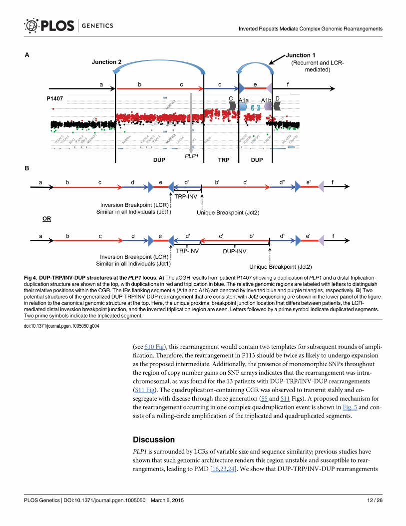
DUP-TRP/INV-DUP Rearrangement StructureThe DUP-TRP/INV-DUP structure hypothesized for these 16 individuals postulates that al-though there are 4 copy number transitions in these patients, there are only two breakpoint junc-tions (Fig. 4A). We have sequenced Jct2 in all 16 patients; Southern blotting and quantitativePCR were used to determine Jct1, and direct junction sequencing was successful for BAB1612/P374 (Figs. 3, 4 and S6). Additionally, due to the small size (~ 254 bp) of the triplication in P255/298, a PCR approach using one primer at the proximal duplication junction and one within theLCR validated the overall structure of this rearrangement as DUP-TRP/INV-DUP.
Quadruplication by Rolling-Circle AmplificationWe have discerned two junctions from patient P113 with proximal quadruplication and dupli-cation of PLP1 using long-range PCR (Figs. 5A, S6). Junction 1 consists of one fork stalling andtemplate switching (FoSTeS) event—FoSTeS 1 (Fig. 5A). The second junction, between theproximal end of the triplication and the distal end of the quadruplication, consists of FoSTeSevents 2 and 3 (S6 Fig for sequences of all junctions). We determined that the rearrangementwas on the inverted H2 allele using PCR genotyping of the haplotype present in P113 (S9 Fig).Additionally, digital PCR (dPCR) data indicate that the FoSTeS 1 occurs in one copy, andFoSTeS 2/3 occurs in 2 copies (S5 Table).
This quadruplication rearrangement is also associated with a de novo point mutation (G in-sertion) ~50 bp away from the junction that appeared to occur concurrent with the rearrange-ment, as observed for other CGR mediated by a replicative process (S6 Fig) [20]. Themechanism by which copy number increased from 3 to 4 copies and generated the quadrupli-cation is suggestive of a rolling circle amplification, wherein one breakpoint is repeated twicein the process of replicating ~280 kb (S5 Table) [22,36–38]. The FISH data for this individualshows the rearrangement to be contained on the X chromosome, and family data including theproband P113, his mother P154, his affected uncle P117, and grandmother P088 suggest thatthe structure is stable in 4 individuals from 3 generations, diminishing the likelihood of recom-bination-based amplification (Figs. 2D and S5). If the amplification were mediated by NAHR
Inverted Repeats Mediate Complex Genomic Rearrangements
PLOS Genetics | DOI:10.1371/journal.pgen.1005050 March 6, 2015 11 / 26
(see S10 Fig), this rearrangement would contain two templates for subsequent rounds of ampli-fication. Therefore, the rearrangement in P113 should be twice as likely to undergo expansionas the proposed intermediate. Additionally, the presence of monomorphic SNPs throughoutthe region of copy number gains on SNP arrays indicates that the rearrangement was intra-chromosomal, as was found for the 13 patients with DUP-TRP/INV-DUP rearrangements(S11 Fig). The quadruplication-containing CGR was observed to transmit stably and co-segregate with disease through three generation (S5 and S11 Figs). A proposed mechanism forthe rearrangement occurring in one complex quadruplication event is shown in Fig. 5 and con-sists of a rolling-circle amplification of the triplicated and quadruplicated segments.
DiscussionPLP1 is surrounded by LCRs of variable size and sequence similarity; previous studies haveshown that such genomic architecture renders this region unstable and susceptible to rear-rangements, leading to PMD [16,23,24]. We show that DUP-TRP/INV-DUP rearrangements
Fig 4. DUP-TRP/INV-DUP structures at the PLP1 locus. A) The aCGH results from patient P1407 showing a duplication of PLP1 and a distal triplication-duplication structure are shown at the top, with duplications in red and triplication in blue. The relative genomic regions are labeled with letters to distinguishtheir relative positions within the CGR. The IRs flanking segment e (A1a and A1b) are denoted by inverted blue and purple triangles, respectively. B) Twopotential structures of the generalized DUP-TRP/INV-DUP rearrangement that are consistent with Jct2 sequencing are shown in the lower panel of the figurein relation to the canonical genomic structure at the top. Here, the unique proximal breakpoint junction location that differs between patients, the LCR-mediated distal inversion breakpoint junction, and the inverted triplication region are seen. Letters followed by a prime symbol indicate duplicated segments.Two prime symbols indicate the triplicated segment.
doi:10.1371/journal.pgen.1005050.g004
Inverted Repeats Mediate Complex Genomic Rearrangements
PLOS Genetics | DOI:10.1371/journal.pgen.1005050 March 6, 2015 12 / 26

are a frequent CGR product at the PLP1 locus and that they are facilitated by a complex IR butspecifically mediated via the ~20 kb A1a and A1b 99% identical repeats. These particular IRsare not only driving CGR observed in patients but additionally mediate a common SV poly-morphism—copy-number neutral inversions at Xq22.2. The latter can complicate interpreta-tion of CGRs in the region; a proposed breakpoint can also appear in an unaffected individualin the guise of an inverted allele [13]. Additionally, the recurrence of this inversion might con-found the correlation of diagnostic SNPs with structural information, leading to the underesti-mation of the frequency in a population [39]. These data suggest that IRs with a high degree ofidentity that are involved in non-pathogenic inversions can also drive seemingly recurrentbreakpoints in non-recurrent rearrangements associated with disease and that this occurs atmultiple genomic loci [1,12,13]. Indeed, a previous determination of genes potentially subjectto CNV via DUP-TRP/INV-DUP due to proximity of homologous IRs predicted the PLP1gene might be affected [2].
The proximal junctions, or Jct2, in the DUP-TRP/INV-DUP rearrangements at the PLP1locus are depicted in S6 Fig. Jct2 is non-recurrent in the 16 individuals, with different genomic
Fig 5. A quadruplication and potential rolling-circle mechanism of rearrangement. A) P113 aCGH result is shown. At top, colored arrows representsegments of triplicated (blue), quadruplicated (gold), and duplicated (red) sequence in this individual. A1a and A1b are represented by inverted blue andpurple triangles, respectively. Copy number (C#) of the various segments is enumerated below the aCGH result, as is the rearrangement structure on the H2haplotype (see S9 Fig). Breakpoints are depicted by blue dashed arrows in the schematic of the rearrangement and are labeled with the template switchingevents associated with the junction. Breakpoint sequences are depicted in S6 Fig, and dPCR data indicating duplication of FoSTeS events 2 & 3 is in S5Table. B) The proposed rolling circle mechanism leading to the triplication and quadruplication is depicted, with the FoSTeS events 2 & 3 leading to invasionof the fork into the already replicated strand. This establishes the rolling circle, depicted on the right. The event ends with a double strand break or a forkdisassociation event, and the breakpoint involving FoSTeS event 1 (S6 Fig). The structure of the overall rearrangement is depicted at the bottom, with colorsrelating to panel A for orientation.
doi:10.1371/journal.pgen.1005050.g005
Inverted Repeats Mediate Complex Genomic Rearrangements
PLOS Genetics | DOI:10.1371/journal.pgen.1005050 March 6, 2015 13 / 26

coordinates for each breakpoint. Interestingly, investigation revealed that 14 of 16 Jct2 se-quences contained microhomology of 1–4 bp at one or more of the FoSTeS events in the junc-tion. Two of these Jct2 sequences involved larger stretches of microhomology; one containedan Alu-Alu chimeric event with 47 bp of perfect identity at the junction and the second CGRcontained two Alu-Alu chimeras, one containing 14 bp and the other with 34 bp of perfectidentity at the junction (Table 1). These data suggest that a replicative mechanism is involvedin the formation of Jct2 in a majority of cases. Previously, we proposed that MMBIR or NHEJcould be responsible for Jct2 [1]. Here, we expand this ‘”two-step hypothesis” to include home-ologous recombination within divergent repeats or similar sequences [33,40]. This is especiallyrelevant to Alu-Alumediated junctions, where the region of perfect identity may not be exten-sive enough to employ homology-driven repair, but extensive base-pairing outside the regionof identity could aid in driving a recombination coupled replication driven rearrangement pro-cess at these loci [34]. In the 16 patients with DUP-TRP/INV-DUP rearrangements presented,2 contain a Jct2 breakpoint resulting in the formation of a chimeric Alu element.
We hypothesize that the PMD-associated CGR are caused by BIR or MMBIR; these replica-tive processes have been shown to be error-prone, perhaps because they utilize a polymerase/replisome with reduced fidelity (induced point mutations) as well as reduced processivity (tem-plate switching) relative to intergenerational DNA polymerases [20,41]. Evidence now indi-cates that BIR/MMBIR-associated mutation results from conservative replication coupled witha migrating bubble [42,43]. Thus, DUP-TRP/INV-DUP CGRs involving PLP1 have the poten-tial to additionally impact patient health through point mutations on the X chromosome.These hypotheses need further investigation through large-scale genomic sequencing. Never-theless, although few in number, de novo point mutations apparently acquired concomitantlywith the DUP-TRP/INV-DUP rearrangement in P250 (insertion of an A) and the quadruplica-tion rearrangement in P113 (insertion of a G) were not seen in the corresponding, contiguous(non-breakpoint containing) section of the X chromosome for these intrachromosomal events,a finding consistent with observations made at theMECP2 locus and de novomutation withCGR formation [20]. Given that on average, ~600 bp were sequenced at each junction, this sug-gests a rate of 2 mutations in ~15 kb of sequencing, consistent with the elevated point mutationrate observed in association with replication-based mechanisms of repair [20,41].
Junction 1 is present at seemingly identical loci, occurring within a complex inverted repeatstructure in the 16 DUP-TRP/INV-DUP rearrangements studied. Further analysis has shownthat at least one of these breakpoint junctions is in a region of 24 bp of microhomology and threeoccur within a>8 kb region of identity within A1a and A1b. The proposed mechanism for Jct1is BIR within a region of ectopic, inverted homology [1]. Our data reveal that the template switchcan occur within smaller regions of identity within A1a and A1b, suggesting that either MMBIRor homeologous recombination, rather than an homologous recombination within IRs may bean alternative mechanism for the formation of these seemingly recurrent junctions [22,31].
Previously, a study of 36 PMD patients identified 3 cases with duplicated copies of PLP1 in-serted outside of Xq22 [24]. Conversely, in this study all 16 subjects with junctions in IRs con-tain the extra copy or copies of the gene on Xq22, therefore suggesting that the mechanism ofCNV results in a contiguous rearrangement (triplicated or duplicated regions in tandem,Fig. 2, Table 1). Additionally, all 16 individuals queried by Southern blotting and/or qPCRmethodologies indicate that the A1a and A1b inverted LCRs mediate PLP1 DUP-TRP/INV-DUP rearrangements. Although two other IRs in the region, albeit with less sequence identity(the 93% identical outer C/D and 87% identical innermost A2/A3 repeats), could presumablymediate the junction between distal duplication and distal triplication breakpoints, these 16cases use the A1a and A1b specific repeats. A1a and A1b are ~20 kb in length (versus ~30 kbfor C/D and ~10 kb for A2/A3) and are separated by ~50 kb (versus ~140 kb for C/D and ~30
Inverted Repeats Mediate Complex Genomic Rearrangements
PLOS Genetics | DOI:10.1371/journal.pgen.1005050 March 6, 2015 14 / 26

kb for A2/A3). Therefore, the higher level of sequence identity between the A1a and A1b re-peats (~99%), added to the shorter inter-repeat distance and the length of the LCR may bothincrease the likelihood of NAHR leading to the inversion [28,44] and potentiate these repeatsas substrates for replication pausing, fork invasion, and reversal through BIR [31]. This is thesecond locus for which DUP-TRP/INV-DUP cases with recurrent Jct1 mediated by IRs hasbeen described. InMECP2 DUP-TRP/INV-DUP, the K1 and K2 LCRs participate in both non-pathogenic inversions and the rearrangements present in patients [1,27]. Such empirical stud-ies may enable refinement of current predictions for IRs that can predispose regions of the ge-nome to DUP-TRP/INV-DUP [2].
Our data further implicate a “two-step process” of BIR paired with MMBIR to generateCGRs resulting in duplication of copy number sensitive genes proximal to IRs [1]. The rear-rangements in the 16 patients with DUP-TRP/INV-DUP contain just two junctions that resultin four copy number transition states. This complex pattern on array CGH is due to just twotemplate switches, Jct1 occurring within the LCRs A1a and A1b distal to the PLP1 gene and re-sulting in an inversion, and Jct2 occurring at varying locations proximal to junction 1 and re-suming the pattern of normal replication, resulting in a rescue from the potential formation ofa dicentric chromosome (Fig. 4) [45].
The observations at the quadruplication-containing CGR in P113 are consistent with roll-ing-circle amplification (Fig. 5). The rarity of quadruplication at PLP1 could be due to selectivepressures from the increased severity of PMD with additional copies of PLP1 (4 versus 3); it isnotable that the quadruplication observed herein does not include the dosage sensitive PLP1gene [14]. One junction in this CGR (between IRs A1a and A1b) occurs at a similar location asin the DUP-TRP/INV-DUP structures, and PCR genotyping suggests that the interpretation ofthe rearrangement is complicated by the inversion structural variation, resulting in H2 (S9Fig). At the proximal junction, the fork template switches twice, invading upstream and leadingto a rolling-circle [22,36–38]. After almost two complete copies of the circle (35 kb short of theoverall 280 kb), the next junction is a template switch from the proximal end of the quadrupli-cated region to the distal end of the duplicated region within the LCR region. Our observationsare most parsimoniously explained by a rolling-circle amplification event, as predicted forhigher-order genomic segment amplification in the MMBIR model [22]. Due to the observa-tions of: i) triplicated and quadruplicated segments, ii) the accompanying point mutation asso-ciated with CGR formation, and iii) the prevalence of intrachromosomal rearrangements atthis locus, a replicative model for CGR formation is likely [20,42,43]. The quadruplication-containing CGR provides evidence for an important next step in the MMBIR model, allowingfor higher-order amplification to occur, as is often observed in cancer [22,46,47].
In summary, our studies confirmed a unique rearrangement product consisting of a DUP-TRP/INV-DUP structure in 16 individuals, with 6 containing triplication of PLP1 [1]. We alsoelucidated a common, recurrent inversion polymorphism between two IRs distal to this gene.Jct1 occurs between the same repeats that mediate the non-pathogenic inversion, and sequenc-ing of a DUP-TRP/INV-DUP breakpoint within the LCRs showed that these junctions canoccur within short stretches of identity within a larger repeat of ~20 kb. This study of break-point junctions involved in both DUP-TRP/INV-DUP and higher-order amplification leadingto quadruplication implicate replicative mechanisms in the generation of these CGRs. Addi-tionally, we provide experimental evidence supporting the contentions that: i) IRs contributeto genome instability, ii) LCRs can mediate replication-based mechanisms, and iii) short repet-itive sequences, such as Alu, can provide microhomology to facilitate template switching. Theprevalence of DUP-TRP/INV-DUP events involving PLP1 brings attention to the importanceof this mechanism and the potentially broader impact of this rearrangement structure in geneand genome evolution.
Inverted Repeats Mediate Complex Genomic Rearrangements
PLOS Genetics | DOI:10.1371/journal.pgen.1005050 March 6, 2015 15 / 26

Materials and Methods
Inversion AscertainmentTo determine whether there is a polymorphic inversion in the LCRs distal to PLP1, we exam-ined the genomic information for 9 individuals contained in the human genome structural var-iation (HGSV) track of the UCSC Genome Browser [8,25,26]. The HGSV track (hg18)contains data on discordant fosmid end sequences from libraries of 9 individuals from diversegeographical regions. Discordant end sequence orientations of fosmids spanning LCRs A1a orA1b [23] and having both ends present in unique sequence (not LCRs) indicate potential inver-sions [26]. Individuals with at least one clone independently spanning each of the LCRs suggestthat there is an inversion between the two repeats (S1 Fig).
Inversion Haplotypes and AnalysisPhased data from the 1000 genomes project [29] was used to create plots of two haplotypes inthe region spanning from LCRs A1a to A1b (Hg19 coordinates, ChrX:103223669–103324337)[48]. One thousand genomes data was cross-correlated with homozygous genotypes deter-mined from Southern Blots to elucidate phased haplotypes that contain inversion alleles. Re-sults were plotted using custom (in-house) scripts implemented in the R programminglanguage (S4 Fig).
Personal Genomes from Subjects and Patients InvestigatedFamilies with PMD or rearrangements of Xq22.2 including PLP1 were obtained by physicianreferral or self-referral. Patients were enrolled through informed consent in research protocolsapproved by the Institutional Review Boards at Baylor College of Medicine (BCM) and the Ne-mours Alfred I. duPont Hospital for Children. The rearrangements present in patientsBAB1290, BAB1612/P374, BAB2389, P250, P255, P500, P518, and P558 were published previ-ously [1,24,30]. Two of the patients with PLP1 triplication (P518 and P674) were described ashaving more severe disease than patients with duplication [14]. Control DNAs from HapMapindividuals [48] were obtained from the Coriell Institute for Medical Research cell repositories.
DNA Digestion and Fragment Separation for Southern Blotting AnalysisApproximately 10 μg of genomic DNA from each patient was digested using BssSI. The DNAwas diluted to 60 μl and digested for 4 hours at 37°C with 16U, heat inactivated at 80°C for 20minutes, and the digest was then repeated with 12U for 3 hours and subsequent heat inactiva-tion (leading to a 10-fold overdigestion). The digested DNAs were then precipitated and con-centrated using standard sodium acetate precipitation, and were reconstituted in 25 μl of waterwith gentle mixing overnight. Concentrations were determined using a NanoDrop spectropho-tometer, and samples were then loaded along with an 8–48 kb ladder on a 0.6% Tris- BoricAcid-EDTA (TBE) gel and run in 1X TBE buffer for ~3 days at 50–60 volts. DNA restrictiondigestion products, i.e. bands on gels, were then visualized with ethidium bromide staining.
Probe Design for Southern Blot AnalysesProbe DNA was prepared using primers A1a proximal probe For- 50-AATGCAGCTCAAAG-GAAAGC-30 and A1a proximal probe Rev- 50-AGCCACTGACCAGTGATTTTC-30 and am-plifying a 514 bp fragment from BAC clone RP11–462K21 (https://bacpac.chori.org) DNAprepared using a QIAprep spin miniprep kit. The resultant PCR bands were resolved on 1%agarose and Tris-Acetate-EDTA gels and purified using a Zymoclean Gel DNA Recovery Kit
Inverted Repeats Mediate Complex Genomic Rearrangements
PLOS Genetics | DOI:10.1371/journal.pgen.1005050 March 6, 2015 16 / 26

(Zymo Research). Probe DNA was validated by Sanger sequencing, using both forward and re-verse primers and was frozen at-20°C in 90 ng aliquots.
Southern BlottingSouthern Blotting was carried out as previously described [23]. Briefly, DNAs were subjectedto electrophoresis for sufficient duration to distinguish 25 and 29 kb fragment sizes and werethen transferred to a Sure Blot positively charged nylon membrane by standard ‘sandwich’methodology for 2–3 days. Approximately 80 ng of DNA was labeled with 32P-dCTP by ran-dom priming for 2–4 hours at 37°C using the Random Primed DNA labeling kit (Roche).Membranes were pre-hybridized for 4 hours in 10% dextran sulfate/1M NaCl/1%SDS (hybrid-ization solution) with 4mg of sheared salmon sperm DNA at 65°C. Probe was pre-associated inhybridization solution with ~1mg sheared placental DNA at 65°C for ~2 hours, then added tothe pre-hybridized membrane. Hybridization was carried out at 65°C overnight (~18 hours).The following day, the blot was washed and analyzed using autoradiography for bands corre-sponding to PLP1 A1a structural haplotype information (~25 and 29 kb).
Array CGH AnalysesTo determine the size, genomic content, and extent of PLP1 rearrangements, a high-densityoligonucleotide array from Agilent was custom-designed to examine PMD patients. The 4 x 44K microarray was designed using the Agilent eArray website (https://earray.chem.agilent.com/earray/) and was used to visualize the rearrangements of three patients in this study (BAB1290,BAB1612/P374, and BAB2389), the family containing individuals BAB3698, BAB3699,BAB3700, and BAB4179, and to complement existing array data for P500, P1407, and P113.The family of P113 was explored using Agilent arrays, including patients P113 and P117, aswell as the mother of P113, P154, and grandmother, P088, who are both carriers. Probe labelingand hybridization were conducted as previously described, with NA15510 and NA10851 usedas reference DNAs for female and male individuals, respectively (Accession GSE63594) [1].Purified DNA samples from P113, P250, P500, P518, P558, P642, P674, P820, P842 and P1150were submitted to NimbleGen for array service with normal male control NM002 as a refer-ence. The NimbleGen X chromosome CGH fine-tiling array with oligonucleotide probes of 45to 85 bases in length with median spacing of 106 bp throughout the whole X chromosome wasused. Patient DNA samples P255/P298, BAB1612/P374, P1389 and P1407 were submitted tothe Biomolecular Core Lab at duPont Hospital for Children for hybridization to AffymetrixCytogenetics 2.7M Array. DNA sample P1407 was submitted to Coriell’s Genotyping and Mi-croarray Center for hybridization on Affymetrix GenomeWide Human SNP Array 6.0. Alldata from Affymetrix arrays were analyzed with GeneChip Command Console SoftwareAGCC. NimbleGen and Affymetrix Cytogenetics array data were aligned with qPCR data andplotted using the R programming language (Affymetrix and NimbleGen data are under Acces-sion GSE64122) (Figs. 2B, C, D and S5).
Semi-Quantitative Multiplex PCR (qPCR) for Detection of Copy NumberSemi-quantitative multiplex PCR was performed using a QIAGENMultiplex PCR kit accordingto the manufacturer’s protocol to analyze regions on the X chromosome in and surroundingPLP1 to determine copy number. Primer pairs were selected using the NCBI primer design tool(primers available upon request). In each experiment, five control DNAs were used, two knownto have duplications in the region of interest without CGRs and three normal controls known tobe single copy in the region of interest. A primer pair that amplifies a region of the human dystro-phin (DMD) gene on the p-arm of the X chromosome was included in each multiplex reaction
Inverted Repeats Mediate Complex Genomic Rearrangements
PLOS Genetics | DOI:10.1371/journal.pgen.1005050 March 6, 2015 17 / 26

for amplification of a single-copy region. Products were separated by electrophoresis on a 4%NuSieve 3:1 agarose gel (Lonza, Walkersville MD) and stained with ethidium bromide. Net in-tensity of each band was determined using a Molecular Imaging system with Kodak Gel LogicImaging software or AlphaImager HP. Copy number was determined by calculating the ratio ofthe net intensities of bands in the test region to dystrophin single-copy region for each DNA sam-ple and then normalized by dividing by the average of the ratios of test region to dystrophin ofthe three normal controls. Theoretical ratios were: one, single-copy; two, duplication; three, trip-lication. Alternatively, quantitative PCR was performed as above except that one primer of eachpair was labeled with 6-FAM and samples were submitted to the Biomolecular Core Lab atduPont Hospital for Children for capillary electrophoresis on an ABI PRISM 3130XL DNA Ana-lyzer. Analysis of copy number was determined as above, by using area under the peak as deter-mined by Peak Scanner software rather than net intensity. Triplicated, quadruplicated andduplicated regions were mapped to within several kb of their endpoints using these methods.
Fluorescent in situ Hybridization (FISH) to Confirm Copy NumberInterphase nuclei and metaphase chromosomes were prepared from 700 μl of whole bloodstored in sodium heparin Vacutainer tubes as follows. Blood was placed in α-MEM, 20%FBS,1% L-glutamine, 50 μg/ml gentamycin and treated with 150 μl Phytohemagluttenin (Invitro-gen, Carlsbad CA). The cultures were incubated at 37°C for 72 hours in upright position afterwhich they were treated with 100 μl colcemid by trituration followed by incubation at 37°C for30 min. Cultures were then subjected to centrifugation at 350 x g for 6 minutes. The superna-tant liquid was discarded and the pellet was suspended in 10 ml 75mM KCl pre-warmed to37°C and incubated at 37°C for 15 minutes. Then 1 ml of fixative (3:1 mixture of methanol:ace-tic acid) was added slowly. The preparation was washed 3 times in 10 ml of fixative with pellet-ing by centrifugation at 350xg for 6 minutes after washes. The resulting pellet of interphase andmetaphase chromosomes was then stored in fixative at -20°C. Chromosomes and nuclei weredropped onto pre-cleaned Fisherbrand slides in a CDS-5 Glovebox environmental Chamber,(Thermotron, Holland Michigan) set at 25°C and 50% humidity. Slides were stored at -20°C ina vacuum under dessication until use.
FISH was performed using cosmid clone U125A1 and BAC clone RP13–188A5 obtainedfrom the BACPAC resource center. Cosmid and BAC DNAs were isolated using the QIAGENPlasmid purification and QIAGEN Large-Construct kits, respectively. One μg of U125A1 DNAwas labeled with Biotin-16-dUTP and 1μg RP13–188A5 DNA was labeled with digoxigeninusing the DIG-Nick Translation Mix. Labeled probes were purified using Nuctrap probe purifi-cation columns according to the manufacturer’s protocol. After hybridization to chromosomesand nuclei according to standard protocol, biotinylated U125A1 was bound to Cy3-labeledstreptavidin, further amplified with biotinylated antiavidin (Vector Laboratories, BurlingameCA) and detected with a second layer of Cy3-labeled streptavidin. Simultaneously, RP13–188A5 labeled with digoxigenin was coupled with mouse antidigoxigenin, detected with rabbitanti-mouse FITC (Jackson ImmunoResearch Laboratories) and further amplified with goatanti-rabbit FITC antibody. Nuclei and chromosome spreads were counterstained with 4’,6-dia-midino-2-phenylindole (DAPI) and cover slips were mounted using Vectashield antifade solu-tion. Images were captured using a Leica DM RXA2 fluorescence microscope and Openlabimaging software (Perkin Elmer, WalthamMA).
STR and SNP Analyses to Determine Origin of Extra Genomic CopiesTo examine whether an intra- or inter-chromosomal origin occurred for the extra genomicsegments in each patient’s genome, we analyzed 2 STRs and 9 single SNPs within the
Inverted Repeats Mediate Complex Genomic Rearrangements
PLOS Genetics | DOI:10.1371/journal.pgen.1005050 March 6, 2015 18 / 26

duplicated/triplicated region common to most patients. Sites were chosen based on marker ge-notypes displaying a high degree of heterozygosity in HapMap samples. S6 Table depicts thedbSNP identifiers, locations with respect to Chromosome X sequence NT_011651.17, andprimers used to amplify the SNP or STR. Regions of interest were amplified from DNA withHotStar Taq DNA polymerase (Qiagen) for products<1kb or Expand High Fidelity PCR sys-tem (Roche) for products>1kb. Patients P250, P255/298, BAB1612/P374, P500, P518, P558,P642, P674, P820, P842, P1150, P1389, and P1407 were analyzed. Products containing the STRwere amplified with one primer within the pair fluorescently-labeled so product sizes could beevaluated by capillary electrophoresis on ABI’s PRISM 3130 XL DNA Analyzer. Products con-taining the SNP were purified using QIAquick PCR or Gel purification kits, then sequencedwith the Big Dye Terminator kit v. 3.1 (Life Technologies), according to the manufacturer’s in-structions. Patients P113, P117 and carriers P154 and P088 were subjected to genotyping usingthe Illumina OmniExpress SNP array analyses at the human genome sequencing center ofBCM. Data from the analyses was visualized by plotting the B allele frequencies versus the Xchromosome coordinates encompassing the quadruplication genomic rearrangement, as wellas the Log ratio of SNP intensity (S11 Fig).
Jct2 AnalysesInverse PCR was used to obtain the first junction of patient P1150. Briefly, DNA was digestedwith NheI and ligated to form circles. PCR primers were designed to amplify in opposite direc-tions around the circle by long-range PCR using the Expand High Fidelity PCR system (S6Table). When the PCR products were analyzed on an agarose gel, a product was found that wasunique to the patient. The product was subjected to DNA sequencing according to the manu-facturer’s instructions, then purified with the Filtration Cartridge (Edge Biosystems, Inc., Gai-thersburg MD) and separated using an ABI PRISM 3130 xl Genetic Analyzer. DNA sequencewas analyzed using Vector NTI sequence analysis software.
Proximal junctions were obtained for the personal genomes from the remaining triplicationpatients by long-range PCR using appropriately positioned primers at the endpoints of copynumber changes (Table 1 and S6 Table) in 25 μl reactions with 50–100 ng of patient DNAusing TaKaRa LA Taq or using the Expand High Fidelity PCR dNTPack kit according to themanufacturers’ instructions. PCR products were prepared for sequencing by using the standardExoSAP-IT protocol (Affymetrix, Santa Clara CA) or by using the Qiagen PCR purification kitand DNA sequencing reactions were performed as indicated above using primers used in am-plification or internal primers as indicated in S6 Table. Sequences were aligned to the humangenome reference sequence, and breakpoints are depicted in S6 Fig. We had previously re-ported the sequence across the junction in P255 [24].
Jct1 AnalysesPCR was conducted across Jct1 from DNAs prepared from patients with DUP-TRP/INV-DUPCGRs using a QIAGENMultiplex PCR. Two control DNAs duplicated through this region andthree control DNAs with a single copy at this locus were amplified in parallel. Along with a dys-trophin primer pair (Hdys 23F-6FAM and Hdys 23 R) for a single copy region of the humandystrophin gene, we used primers pairs V362H12-F19–6FAM and V362H12-R19 (red arrows),and V362H12-F24–6FAM and V362H12-R24 (black arrows), and V362H12-F19–6FAM andV362H12-R24 (one red, one black arrow) (Figs. 3E, S8, S6 Table). Fluorescently labeled PCRproducts were diluted 1:100 in sterile HPLC water and subjected to capillary electrophoresisusing an ABI PRISM 3130 XL DNA Analyzer. Copy number analysis was performed as previ-ously described using the Peak Scanner software [24].
Inverted Repeats Mediate Complex Genomic Rearrangements
PLOS Genetics | DOI:10.1371/journal.pgen.1005050 March 6, 2015 19 / 26

To subclone breakpoints in PMD DUP-TRP/INV-DUP patients, we amplified patientDNAs containing rearrangements (from BAB1612/P374, BAB2389, and BAB1290) with PCRprimers that anneal within the A1a and A1b LCRs and uniquely flanking primers. This yieldedfour overlapping segments of the two LCRs (S6 Fig). These PCR products were then subjectedto electrophoresis in crystal violet 0.8% agarose gels, purified using the SNAP purification kitfrom Invitrogen, and cloned into TOPO XL cloning vectors. Resultant clones for each of thefour segments were screened by digestion and sequenced in their entirety. At least two clonesfor each region, obtained from independent PCR reactions, were screened for the breakpointand the corresponding A1a or A1b region. Sequence analysis was conducted using the Laser-gene 9 DNA analysis software suite.
Copy Number Analysis of Junctions in the Quadruplication by dPCRCopy number of junctions in the quadruplication patients and a carrier were determined bydPCR using QuantStudioTM 3D Digital PCR System (Life Technologies), according to themanufacturer’s instructions. Concentration of DNA was determined by QubitR dsDNA BRassay (Life Technologies) using the Qubit 2.0 fluorometer (Life Technologies). Sample DNAwas digested with SphI (NEBiolabs) to separate multiple copies of interest that may be locatedon the same molecule without disrupting the region of amplification. Digests were performedusing 400 ng of DNA in a 10 μl reaction containing 10U of SphI and incubating at 37°C for1.5 hr, followed by heat-inactivation of the enzyme at 65°C for 20 min. The digest was dilutedto 40 μl with RNase-free water to yield a concentration of 10 ng/μl DNA. Primers and probesused in the dPCR assays are in S6 Table. Reactions for dPCR included 1x QuantStudioTM 3DDigital PCR Master Mix, 1x TaqMan Copy Number Reference Assay for human RNaseP (LifeTechnologies, Cat. # 4403328, VIC label), 1–1.5x PrimeTime qPCR 5’ nuclease assay (IDT,FAM label) for jct1 or jct2/3 and 40–60ng DNA in a 16 μl volume. Fifteen μl of this mix wasused to load the Digital PCR 20K Chip (Life Technologies); chips were processed according tothe manufacturer’s instructions.
Supporting InformationS1 Fig. Inversions in the HGSV. The Human Genome Structural Variation track (NCBI36/hg18) prediction of inversion from the UCSC genome browser. The locations of A1a and A1bare noted. Green horizontal lines indicate inversion fosmids (discordant end orientations) withrespect to the reference sequence. Five individuals have strong support (both LCRs spanned)for an inversion—G248, ABC10, ABC11, ABC12, and ABC13, and three more have weak sup-port—ABC7, ABC8, and ABC14; ABC9 appears to be homozygous for thereference orientation.(PDF)
S2 Fig. Breakpoint of the inversion in G248. One inversion fosmid spanning A1a from indi-vidual NA15510 (library G248) was previously sequenced in its entirety (GI:121495926)(Kiddet al. 2010). The other breakpoint of this inversion was not investigated, as the fosmid sequencewas unavailable. Analysis of this clone with respect to the reference sequence for LCRs A1a (inblue) and A1b (in purple) revealed an apparent switch from paralogous sequence variants(PSVs) belonging to one LCR to those of the other. This occurred after 18525 bp of A1a in ablock of 334 base pairs of identical sequence and prior to 1492 bp of A1b (Lindsay et al. 2006).Seven LCR A1a-specific PSVs in the 1000 bp proximal and 10 A1b-specific PSVs in 200 bp dis-tal to the 334 bp region of perfect identity signified a historical recombination event betweenA1a and A1b. These data indicate a putative NAHR-mediated switch ~1500 bp from the end of
Inverted Repeats Mediate Complex Genomic Rearrangements
PLOS Genetics | DOI:10.1371/journal.pgen.1005050 March 6, 2015 20 / 26

LCR A1a, mediating the inversion. The breakpoint sequence occurred within 334bp of 100%identity (ChrX:103242193–103242526 in A1a) between the two reference LCRs at this location,and two putative PRDM9 binding sites/homologous recombination hotspot motifs are presentat the distal end of the region of homology (sequence depicted in red)(Myers et al. 2008; Myerset al. 2010). These data implicate NAHR as the likely mechanism for the inversion.(PDF)
S3 Fig. Additional genotyping of inversion allele in phenotypically normal individuals. Tenindividuals from the HapMap population studied for inversion. Gender of the individual is indi-cated by circles (female) or squares (male) above the figure. DNA identifiers (NA numbers) areconsistent with Coriell names (http://ccr.coriell.org/), and fosmid libraries (ABC library identifi-ers) are as in Kidd et al. (Kidd et al. 2010). The population of origin for each individual and thegenotypes are indicated at the bottom of the blot. H = Han Chinese, Y = Yoruban, C = CEPH,and U = unknown. Samples repeated between this blot and the one in Fig. 1 are indicated withan asterisk (�). Lanes 2 and 3 and lanes 7 and 8 were separated by samples that did not producereadily visible bands and were therefore removed from the figure. These lanes are indicated withwhite space in the image of the Southern blot. Quantitation of the blot is presented in S1 Table.(PDF)
S4 Fig. Inversions between A1a and A1b LCRs appear to be recurrent. Reference and inver-sion (H1 and H2) structural haplotypes inferred from Southern blotting were plotted over thesequence haplotypes from the 1000 genomes project. Individuals are denoted on the left—eachfemale has two alleles (A1 and A2) whereas males only have one. Homozygous H2 alleles arehighlighted in yellow. Each colored dot represents a non-reference sequence of a SNP at thatgiven location. Red denotes a cytosine, blue an adenine, green is guanine, and gold is thymine,whereas a reference call is shown in white. The orange bars denote location of A1a and A1b re-peats with inverted arrows indicating orientation. NA18947 is homozygous for H2, and A1 liesin the upper haplotype. The 6 other alleles from homozygous or hemizygous H2 individualsare on the bottom haplotype.(PDF)
S5 Fig. Array and semi-quantitative PCR results. A) Semi-quantitative PCR results are dis-played below NimbleGen and Affymetrix array data. Location of PLP1 denoted by vertical greyarrow, and array results are depicted as in Fig. 2, with duplications in red and triplications inblue. The position of the LCR region is shown in yellow (C and D repeats) and purple (A1a,A1b, A2 and A3 repeats). B) Agilent aCGH data is depicted, with red probes indicating amplifi-cation and green deletion. Shading in BAB3698 pedigree denotes location of aberration. Pedi-grees for families of probands BAB3698 and P113 are shown. Details regarding arrays and PCRare discussed in the Materials and Methods section.(PDF)
S6 Fig. Jct2 and other breakpoint junctions from patients in the study. For DUP-TRP/INV-DUP cases, Jct2 is detailed with Dist. (-) representing the distal (minus strand) and the triplica-tion start sequence; Prox. is the proximal region (plus strand) and the duplication start se-quence. Mid. (where applicable) represents the middle, inserted sequence. BPT is the sequenceof Jct2 itself. For other breakpoints junctions (P113, both junctions; P1150 ‘deletion’), proximaland distal are centromeric and telomeric, respectively. The vertical lines in the alignments indi-cate sequences that align perfectly. Red sequences indicate microhomology, and green se-quence for BAB1290 indicates potential sequence involved in the second template switch. Jct2of P518 involves two Alumediated template switches 488 bp apart. The schematic below theP518 junction sequence depicts the rearrangement. P113 involves a complex rearrangement
Inverted Repeats Mediate Complex Genomic Rearrangements
PLOS Genetics | DOI:10.1371/journal.pgen.1005050 March 6, 2015 21 / 26
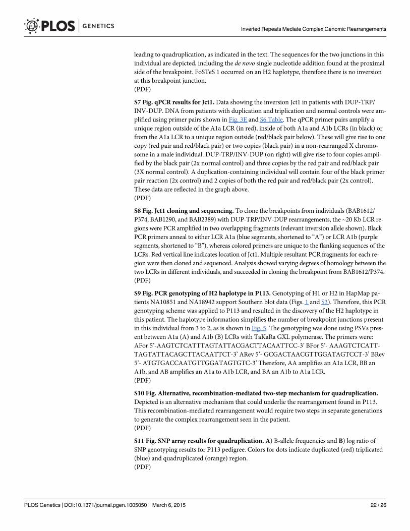
leading to quadruplication, as indicated in the text. The sequences for the two junctions in thisindividual are depicted, including the de novo single nucleotide addition found at the proximalside of the breakpoint. FoSTeS 1 occurred on an H2 haplotype, therefore there is no inversionat this breakpoint junction.(PDF)
S7 Fig. qPCR results for Jct1. Data showing the inversion Jct1 in patients with DUP-TRP/INV-DUP. DNA from patients with duplication and triplication and normal controls were am-plified using primer pairs shown in Fig. 3E and S6 Table. The qPCR primer pairs amplify aunique region outside of the A1a LCR (in red), inside of both A1a and A1b LCRs (in black) orfrom the A1a LCR to a unique region outside (red/black pair below). These will give rise to onecopy (red pair and red/black pair) or two copies (black pair) in a non-rearranged X chromo-some in a male individual. DUP-TRP/INV-DUP (on right) will give rise to four copies ampli-fied by the black pair (2x normal control) and three copies by the red pair and red/black pair(3X normal control). A duplication-containing individual will contain four of the black primerpair reaction (2x control) and 2 copies of both the red pair and red/black pair (2x control).These data are reflected in the graph above.(PDF)
S8 Fig. Jct1 cloning and sequencing. To clone the breakpoints from individuals (BAB1612/P374, BAB1290, and BAB2389) with DUP-TRP/INV-DUP rearrangements, the ~20 Kb LCR re-gions were PCR amplified in two overlapping fragments (relevant inversion allele shown). BlackPCR primers anneal to either LCR A1a (blue segments, shortened to “A”) or LCR A1b (purplesegments, shortened to “B”), whereas colored primers are unique to the flanking sequences of theLCRs. Red vertical line indicates location of Jct1. Multiple resultant PCR fragments for each re-gion were then cloned and sequenced. Analysis showed varying degrees of homology between thetwo LCRs in different individuals, and succeeded in cloning the breakpoint from BAB1612/P374.(PDF)
S9 Fig. PCR genotyping of H2 haplotype in P113. Genotyping of H1 or H2 in HapMap pa-tients NA10851 and NA18942 support Southern blot data (Figs. 1 and S3). Therefore, this PCRgenotyping scheme was applied to P113 and resulted in the discovery of the H2 haplotype inthis patient. The haplotype information simplifies the number of breakpoint junctions presentin this individual from 3 to 2, as is shown in Fig. 5. The genotyping was done using PSVs pres-ent between A1a (A) and A1b (B) LCRs with TaKaRa GXL polymerase. The primers were:AFor 5’-AAGTCTCATTTAGTATTACGACTTACAATTCC-3’ BFor 5’- AAAGTCTCATT-TAGTATTACAGCTTACAATTCT-3’ ARev 5’- GCGACTAACGTTGGATAGTCCT-3’ BRev5’- ATGTGACCAATGTTGGATAGTGTC-3’ Therefore, AA amplifies an A1a LCR, BB anA1b, and AB amplifies an A1a to A1b LCR, and BA an A1b to A1a LCR.(PDF)
S10 Fig. Alternative, recombination-mediated two-step mechanism for quadruplication.Depicted is an alternative mechanism that could underlie the rearrangement found in P113.This recombination-mediated rearrangement would require two steps in separate generationsto generate the complex rearrangement seen in the patient.(PDF)
S11 Fig. SNP array results for quadruplication. A) B-allele frequencies and B) log ratio ofSNP genotyping results for P113 pedigree. Colors for dots indicate duplicated (red) triplicated(blue) and quadruplicated (orange) region.(PDF)
Inverted Repeats Mediate Complex Genomic Rearrangements
PLOS Genetics | DOI:10.1371/journal.pgen.1005050 March 6, 2015 22 / 26

S1 Table. Semi-quantitative analysis of HapMap Southern blots. Southern blot genotypingof the inversion in 17 HapMap individuals was quantitated using GelAnalyzer 2010 software.Briefly, lanes and bands were designated according to the histograms, and normalization wasdetermined automatically by the valley-to-valley normalization function, Resultant band inten-sities (Raw Volume) were used to generate ratios of the bands if more than one was present.This ratio is represented in the final column of the table, and heterozygous HapMap femaleshave an expected ratio of 1. Asterisks indicate DNAs that were on both blots.(PDF)
S2 Table. Population distribution of H1 and H2. Southern blot genotyping of the inversionin 4 HapMap populations, as well as one individual of unknown population of origin.(PDF)
S3 Table. Results of SNP and STR genotyping.(PDF)
S4 Table. Semi-quantitative analysis of patient Southern blots. Southern blot genotyping ofDUP-TRP/INV-DUP patients was quantitated using GelAnalyzer 2010 software, as in S1Table. This ratio is represented in the final column of the table. Ratios of ~2 in the patients in-dicate H2 rearrangements, while less than one indicate H1 (expected ratio of ~0.5). Carrier fe-males from BAB3698 family and females without rearrangements have expected ratios of ~1.(PDF)
S5 Table. dPCR results for FoSTeS Jct 1 and 2/3 from qadruplication. The table depicts digi-tal PCR results from Jct/FoSTeS 1 and Jct/FoSTeS 2/3 in patient P113, the affected uncle of theproband P117, and the carrier mother of P113, P154. In each experiment, FoSTeS 1 has a copynumber of 1, and FoSTeS 2/3 has a copy number of 2. Pooled normal control individuals(6NLs) have neither junction. �Copies/genome is calculated for each junction as (copies/micro-liter jct)/([copies/microliter RNaseP]/2). Note that RNase P is autosomal and so has two copiesper male or female genome (one on each allele). The junctions are located on the X chromo-some; there is only one X allele in males, and it is affected in the patients. Female carriers havejunctions on one X allele but not the other.(PDF)
S6 Table. Primers and probes used in the manuscript. SNP primers amplify de novo nucleo-tide changes, and Inv. PCR primers were used in inverse PCR of sample P1150. Additionalprimers are available upon request.(PDF)
AcknowledgmentsWe are thankful to the families who participated in this study. Thanks to Dr. Klára Brožová forclinical assistance and Professor Feng Zhang for critical review of the manuscript. We acknowl-edge the Nemours Biomolecular Core Laboratory. The content is solely the responsibility ofthe authors and does not necessarily represent the official views of the granting agenciesand foundations.
Author ContributionsConceived and designed the experiments: CRB CMBC LB GMH JRL. Performed the experi-ments: CRB CMBC LB DS BY KS. Analyzed the data: CRB CMBC LB TG DS SMM.
Inverted Repeats Mediate Complex Genomic Rearrangements
PLOS Genetics | DOI:10.1371/journal.pgen.1005050 March 6, 2015 23 / 26

Contributed reagents/materials/analysis tools: MH PS JYG SMM. Wrote the paper: CRBCMBC LB GMH JRL.
References1. Carvalho CM, Ramocki MB, Pehlivan D, Franco LM, Gonzaga-Jauregui C, et al. (2011) Inverted geno-
mic segments and complex triplication rearrangements are mediated by inverted repeats in the humangenome. Nat Genet 43: 1074–1081. doi: 10.1038/ng.944 PMID: 21964572
2. Dittwald P, Gambin T, Gonzaga-Jauregui C, Carvalho CM, Lupski JR, et al. (2013) Inverted low-copyrepeats and genome instability—a genome-wide analysis. HumMutat 34: 210–220. doi: 10.1002/humu.22217 PMID: 22965494
3. ZhouW, Zhang F, Chen X, Shen Y, Lupski JR, et al. (2013) Increased genome instability in humanDNA segments with self-chains: homology-induced structural variations via replicative mechanisms.HumMol Genet 22: 2642–2651. doi: 10.1093/hmg/ddt113 PMID: 23474816
4. Lakich D, Kazazian HH Jr., Antonarakis SE, Gitschier J (1993) Inversions disrupting the factor VIIIgene are a common cause of severe haemophilia A. Nat Genet 5: 236–241. PMID: 8275087
5. Hermetz KE, Newman S, Conneely KN, Martin CL, Ballif BC, et al. (2014) Large inverted duplications inthe human genome form via a fold-back mechanism. PLoS Genet 10: e1004139. doi: 10.1371/journal.pgen.1004139 PMID: 24497845
6. Giorda R, Ciccone R, Gimelli G, Pramparo T, Beri S, et al. (2007) Two classes of low-copy repeatscomediate a new recurrent rearrangement consisting of duplication at 8p23.1 and triplication at 8p23.2.HumMutat 28: 459–468. PMID: 17262805
7. Lange J, Skaletsky H, van Daalen SK, Embry SL, Korver CM, et al. (2009) Isodicentric Y chromosomesand sex disorders as byproducts of homologous recombination that maintains palindromes. Cell 138:855–869. doi: 10.1016/j.cell.2009.07.042 PMID: 19737515
8. Kidd JM, Cooper GM, DonahueWF, Hayden HS, Sampas N, et al. (2008) Mapping and sequencing ofstructural variation from eight human genomes. Nature 453: 56–64. doi: 10.1038/nature06862 PMID:18451855
9. Flores M, Morales L, Gonzaga-Jauregui C, Dominguez-Vidana R, Zepeda C, et al. (2007) RecurrentDNA inversion rearrangements in the human genome. Proc Natl Acad Sci U S A 104: 6099–6106.PMID: 17389356
10. Soler-Alfonso C, Carvalho CM, Ge J, Roney EK, Bader PI, et al. (2014) CHRNA7 triplication associated withcognitive impairment and neuropsychiatric phenotypes in a three-generation pedigree. Eur J HumGenet.
11. Beri S, Bonaglia MC, Giorda R (2013) Low-copy repeats at the human VIPR2 gene predispose to recur-rent and nonrecurrent rearrangements. Eur J HumGenet 21: 757–761. doi: 10.1038/ejhg.2012.235PMID: 23073313
12. Ishmukhametova A, Chen JM, Bernard R, de Massy B, Baudat F, et al. (2013) Dissecting the structureand mechanism of a complex duplication-triplication rearrangement in the DMD gene. HumMutat 34:1080–1084. doi: 10.1002/humu.22353 PMID: 23649991
13. Shimojima K, Mano T, Kashiwagi M, Tanabe T, Sugawara M, et al. (2012) Pelizaeus-Merzbacher dis-ease caused by a duplication-inverted triplication-duplication in chromosomal segments including thePLP1 region. Eur J Med Genet 55: 400–403. doi: 10.1016/j.ejmg.2012.02.013 PMID: 22490426
14. Wolf NI, Sistermans EA, Cundall M, Hobson GM, Davis-Williams AP, et al. (2005) Three or more copiesof the proteolipid protein gene PLP1 cause severe Pelizaeus-Merzbacher disease. Brain 128: 743–751. PMID: 15689360
15. Garbern J, Hobson G (2002) Prenatal diagnosis of Pelizaeus-Merzbacher disease. Prenat Diagn 22:1033–1035. PMID: 12424770
16. Inoue K, Osaka H, Thurston VC, Clarke JT, Yoneyama A, et al. (2002) Genomic rearrangements result-ing in PLP1 deletion occur by nonhomologous end joining and cause different dysmyelinating pheno-types in males and females. Am J HumGenet 71: 838–853. PMID: 12297985
17. Inoue K, Osaka H, Imaizumi K, Nezu A, Takanashi J, et al. (1999) Proteolipid protein gene duplicationscausing Pelizaeus-Merzbacher disease: molecular mechanism and phenotypic manifestations. AnnNeurol 45: 624–632. PMID: 10319885
18. Lupski JR, de Oca-Luna RM, Slaugenhaupt S, Pentao L, Guzzetta V, et al. (1991) DNA duplication as-sociated with Charcot-Marie-Tooth disease type 1A. Cell 66: 219–232. PMID: 1677316
19. Liu P, Gelowani V, Zhang F, Drory VE, Ben-Shachar S, et al. (2014) Mechanism, Prevalence, and MoreSevere Neuropathy Phenotype of the Charcot-Marie-Tooth Type 1A Triplication. Am J HumGenet 94:462–469. doi: 10.1016/j.ajhg.2014.01.017 PMID: 24530202
Inverted Repeats Mediate Complex Genomic Rearrangements
PLOS Genetics | DOI:10.1371/journal.pgen.1005050 March 6, 2015 24 / 26

20. Carvalho CM, Pehlivan D, Ramocki MB, Fang P, Alleva B, et al. (2013) Replicative mechanisms forCNV formation are error prone. Nat Genet 45: 1319–1326. doi: 10.1038/ng.2768 PMID: 24056715
21. Lee JA, Carvalho CM, Lupski JR (2007) A DNA replication mechanism for generating nonrecurrent re-arrangements associated with genomic disorders. Cell 131: 1235–1247. PMID: 18160035
22. Hastings PJ, Ira G, Lupski JR (2009) A microhomology-mediated break-induced replication model forthe origin of human copy number variation. PLoS Genet 5: e1000327. doi: 10.1371/journal.pgen.1000327 PMID: 19180184
23. Lee JA, Inoue K, Cheung SW, Shaw CA, Stankiewicz P, et al. (2006) Role of genomic architecture inPLP1 duplication causing Pelizaeus-Merzbacher disease. HumMol Genet 15: 2250–2265. PMID:16774974
24. Woodward KJ, Cundall M, Sperle K, Sistermans EA, Ross M, et al. (2005) Heterogeneous duplicationsin patients with Pelizaeus-Merzbacher disease suggest a mechanism of coupled homologous and non-homologous recombination. Am J HumGenet 77: 966–987. PMID: 16380909
25. Kent WJ, Sugnet CW, Furey TS, Roskin KM, Pringle TH, et al. (2002) The human genome browser atUCSC. Genome Res 12: 996–1006. PMID: 12045153
26. Tuzun E, Sharp AJ, Bailey JA, Kaul R, Morrison VA, et al. (2005) Fine-scale structural variation of thehuman genome. Nat Genet 37: 727–732. PMID: 15895083
27. Small K, Iber J, Warren ST (1997) Emerin deletion reveals a common X-chromosome inversion mediat-ed by inverted repeats. Nat Genet 16: 96–99. PMID: 9140403
28. Liu P, Lacaria M, Zhang F, Withers M, Hastings PJ, et al. (2011) Frequency of nonallelic homologousrecombination is correlated with length of homology: evidence that ectopic synapsis precedes ectopiccrossing-over. Am J HumGenet 89: 580–588. doi: 10.1016/j.ajhg.2011.09.009 PMID: 21981782
29. The 1000 Genomes Project Consortium, Abecasis GR, Altshuler D, Auton A, Brooks LD, et al. (2010)A map of human genome variation from population-scale sequencing. Nature 467: 1061–1073. doi: 10.1038/nature09534 PMID: 20981092
30. Lee JA, Madrid RE, Sperle K, Ritterson CM, Hobson GM, et al. (2006) Spastic paraplegia type 2 associ-ated with axonal neuropathy and apparent PLP1 position effect. Ann Neurol 59: 398–403. PMID:16374829
31. Mizuno K, Miyabe I, Schalbetter SA, Carr AM, Murray JM (2013) Recombination-restarted replicationmakes inverted chromosome fusions at inverted repeats. Nature 493: 246–249. doi: 10.1038/nature11676 PMID: 23178809
32. Zhang Y, Saini N, Sheng Z, Lobachev KS (2013) Genome-wide screen reveals replication pathway forquasi-palindrome fragility dependent on homologous recombination. PLoS Genet 9: e1003979. doi:10.1371/journal.pgen.1003979 PMID: 24339793
33. deWind N, Dekker M, Claij N, Jansen L, van Klink Y, et al. (1999) HNPCC-like cancer predisposition inmice through simultaneous loss of Msh3 and Msh6 mismatch-repair protein functions. Nat Genet 23:359–362. PMID: 10545954
34. Boone PM, Yuan B, Campbell IM, Scull JC, Withers MA, et al. (2014) The Alu-Rich Genomic Architec-ture of SPAST Predisposes to Diverse and Functionally Distinct Disease-Associated CNV Alleles. AmJ HumGenet 95: 143–161. doi: 10.1016/j.ajhg.2014.06.014 PMID: 25065914
35. Lindsay SJ, Khajavi M, Lupski JR, Hurles ME (2006) A chromosomal rearrangement hotspot can beidentified from population genetic variation and is coincident with a hotspot for allelic recombination.Am J HumGenet 79: 890–902. PMID: 17033965
36. Yu C, Bonaduce MJ, Klar AJ (2012) Remarkably high rate of DNA amplification promoted by the mat-ing-type switching mechanism in Schizosaccharomyces pombe. Genetics 191: 285–289. doi: 10.1534/genetics.112.138727 PMID: 22377633
37. McEachern MJ, Haber JE (2006) Break-induced replication and recombinational telomere elongation inyeast. Annu Rev Biochem 75: 111–135. PMID: 16756487
38. Andersson DI, Hughes D (2009) Gene amplification and adaptive evolution in bacteria. Annu RevGenet 43: 167–195. doi: 10.1146/annurev-genet-102108-134805 PMID: 19686082
39. Sudmant PH, Kitzman JO, Antonacci F, Alkan C, Malig M, et al. (2010) Diversity of human copy numbervariation andmulticopy genes. Science 330: 641–646. doi: 10.1126/science.1197005 PMID: 21030649
40. Rayssiguier C, Thaler DS, Radman M (1989) The barrier to recombination between Escherichia coliand Salmonella typhimurium is disrupted in mismatch-repair mutants. Nature 342: 396–401. PMID:2555716
41. Deem A, Keszthelyi A, Blackgrove T, Vayl A, Coffey B, et al. (2011) Break-induced replication is highlyinaccurate. PLoS Biol 9: e1000594. doi: 10.1371/journal.pbio.1000594 PMID: 21347245
Inverted Repeats Mediate Complex Genomic Rearrangements
PLOS Genetics | DOI:10.1371/journal.pgen.1005050 March 6, 2015 25 / 26

42. Saini N, Ramakrishnan S, Elango R, Ayyar S, Zhang Y, et al. (2013) Migrating bubble during break-in-duced replication drives conservative DNA synthesis. Nature 502: 389–392. doi: 10.1038/nature12584PMID: 24025772
43. Wilson MA, Kwon Y, Xu Y, ChungWH, Chi P, et al. (2013) Pif1 helicase and Poldelta promote recombi-nation-coupled DNA synthesis via bubble migration. Nature 502: 393–396. doi: 10.1038/nature12585PMID: 24025768
44. Dittwald P, Gambin T, Szafranski P, Li J, Amato S, et al. (2013) NAHR-mediated copy-number variantsin a clinical population: mechanistic insights into both genomic disorders and Mendelizing traits. Ge-nome Res 23: 1395–1409. doi: 10.1101/gr.152454.112 PMID: 23657883
45. Mizuno K, Lambert S, Baldacci G, Murray JM, Carr AM (2009) Nearby inverted repeats fuse to generateacentric and dicentric palindromic chromosomes by a replication template exchange mechanism.Genes Dev 23: 2876–2886. doi: 10.1101/gad.1863009 PMID: 20008937
46. Watanabe T, Tanabe H, Horiuchi T (2011) Gene amplification system based on double rolling-circlereplication as a model for oncogene-type amplification. Nucleic Acids Res 39: e106. doi: 10.1093/nar/gkr442 PMID: 21653557
47. Windle BE, Wahl GM (1992) Molecular dissection of mammalian gene amplification: newmechanisticinsights revealed by analyses of very early events. Mutat Res 276: 199–224. PMID: 1374515
48. The International HapMap Consortium (2005) A haplotype map of the human genome. Nature 437:1299–1320. PMID: 16255080
49. Kidd JM, Graves T, Newman TL, Fulton R, Hayden HS, et al. (2010) A human genome structural varia-tion sequencing resource reveals insights into mutational mechanisms. Cell 143: 837–847. doi: 10.1016/j.cell.2010.10.027 PMID: 21111241
Inverted Repeats Mediate Complex Genomic Rearrangements
PLOS Genetics | DOI:10.1371/journal.pgen.1005050 March 6, 2015 26 / 26
Copyright © 2022 FDOKUMEN