A Thiol Peroxidase Is an H2O2 Receptor and Redox-Transducer in Gene Activation
Direct Covalent Attachment of DNA Microarrays by Rapid Thiol−Ene“Click” ChemistryJorge Escorihuela,† María-Jose Banuls,† Santiago Grijalvo,‡ Ramon Eritja,‡ Rosa Puchades,†
and Angel Maquieira*,†
†Centro de Reconocimiento Molecular y Desarrollo Tecnologico, Departamento de Química, Universitat Politecnica de Valencia,Camino de Vera s/n, 46022 Valencia, Spain‡Networking Center on Bioengineering, Biomaterials and Biomedicine (CIBER-BBN) and Institute for Advanced Chemistry ofCatalonia (IQAC−CSIC), Chemical and Biomolecular Nanotechnology Department, Jordi Girona 18-26, 08034 Barcelona, Spain
*S Supporting Information
ABSTRACT: A rapid strategy for the covalent immobilizationof DNA onto silicon-based materials using the UV-initiatedradical thiol−ene reaction is presented in this study. Followingthis approach, thiol- and alkene-modified oligonucleotideprobes were covalently attached in microarray format, reachingimmobilization densities around 6 pmol·cm−2. The developedmethodology presents the advantages of spatially controlledprobe anchoring (using a photomask), direct attachmentwithout using cross-linkers (one-pot fashion), and shortirradiation times (20 min). Using the described strategy,hybridization efficiencies up to 65% with full complementary strands were reached. The approach was evaluated by scoringsingle-base pair mismatches with discrimination ratios around 15. Moreover, the efficacy of the proposed DNA detection schemeis further demonstrated through the assay on a genomic target of bacterial Escherichia coli.
■ INTRODUCTION
Array technology monitors the interaction of a set of molecules,such as nucleic acids and proteins, with a predetermined libraryof molecular probes.1 In the development of a useful andreliable chemistry for producing DNA arrays, the accessibilityand functionality of the surface-bound probe, the density ofimmobilization, the reproducibility and stability of the attach-ment chemistry, and the fidelity of the anchored sequences areall critical parameters to be considered. In this regard, thedevelopment of efficient surface chemistries to modify thephysical and chemical properties of supports has becomeessential to develop DNA chip technology.2 Thus, differentattachment methodologies have been implemented to anchorprobes to solid substrates.3 Typical methods involve thegeneration of active functional groups on the surface thatreact with an appropriately modified DNA probe. In this regard,organosilane compounds have been widely used in the surfacemodification of many materials and devices.4−9
The site-specific immobilization of biological ligands at well-defined locations on substrates is essentially required for certainbiological assays,10,11 for combinatorial screening, and for thefabrication of biosensors.12 It is therefore of great interest todevelop strategies that perform bioassays through robust andefficient chemical methodologies for the realization of DNAchip technology. As a result of its orthogonality and the abilityto selectively pattern surfaces simply by simple exposure to UVlight, thiol−ene chemistry has recently received increasinginterest as a promising surface modification reaction.13,14 The
thiol−ene reaction, which takes place with UV radiation andforms a thioether bond, is compatible with aqueous mediachemistry, which is crucial for its bioutility.15,16 Thiol−enereactions proceed under mild conditions in the presence ofoxygen, are regioselective, tolerate many functional groups, canbe performed on neat or in benign solvents such as water, andprovide quantitative or near-quantitative yields. Although manyof the initial uses of thiol−ene chemistry focused onphotocurable polymers and resins for its use in protectivecoatings and films,17,18 this novel click reaction has proven tobe an elegant and facile method to anchor biomolecules tosilicon-based surfaces.19−28 The reported methods generallyemploy silane coupling agents and need a further reaction withdifferent secondary polymers or cross-linking reagents toprovide covalent anchoring of probes.In a recent work, we reported on the use of thiol−ene “click”
chemistry for the covalent immobilization of a biotin-derivativeto a silicon based surface, and then, we immobilized biotin-functionalized oligonucleotides through a streptavidin bridge.26
This approach constituted a selective strategy to prepare DNAmicroarrays and allowed the discrimination of geneticvariations. However, covalent attachment of DNA probes isnormally preferred because of its robustness and reproduci-bility. In this regard, the search for new methodologies to
Received: January 24, 2014Revised: February 20, 2014Published: February 21, 2014
Article
pubs.acs.org/bc
© 2014 American Chemical Society 618 dx.doi.org/10.1021/bc500033d | Bioconjugate Chem. 2014, 25, 618−627
covalently attach oligonucleotides to solid supports hasemerged as a hot topic in chemistry and materials science.Herein, we report for the first time a simple and rapid
strategy to covalently bind DNA oligonucleotides on silicon-based substrates using the thiol−ene reaction, without the useof intermediate reagents or cross-linkers. We demonstrate thepotential application of this approach by preparing locally DNAfunctionalized surfaces and developing hybridization assayswhich allow effective discrimination of base mismatch. Also, forthe first time, a DNA microarray created using the thiol−enereaction is used for real samples and bacterial Escherichia coli isspecifically detected.
■ RESULTS AND DISCUSSION
Immobilization Assays. To adopt the thiol−ene reactionfor the immobilization of oligonucleotides, surfaces werefunctionalized with allyltrimethoxysilane (strategy A) and 3-mercaptopropyl trimethoxysilane (strategy B). For that, silicon-oxide-coated silicon slides were chosen as starting material,cleaned with piranha solution, and subsequently functionalizedwith the appropriate organosilane compound (2% in toluene).When investigating the silanization time by means of watercontact angle (WCA) measurements, a rapid increase in thehydrophobicity was initially observed during the first 10 min,followed by a slight variation in the first 2 h, which remained
essentially constant after 4 h. According to this, the silanizationtime was set at 2 h. The silanization reaction was alsoinvestigated in different solvents, but no significant differenceswere observed when evaluating the surface wettability.For the performance of the proposed strategies (Scheme 1),
commercially available thiol-modified oligonucleotides wereused. With strategy B, a set of alkene 5′-terminatedoligonucleotide probes were synthesized (probes H−L). Inthese cases, a spacer was introduced in order to have a suitableseparation between the silicon surface and the oligonucleotideprobes. For that, a hydrophilic spacer, monoallyl-tetraethyleneglycol, was derivatized to its corresponding phosphoramidite(see Supporting Information) and was covalently linked intoseveral oligonucleotide probes with excellent coupling yields(>90%) at their 5′-termini (5′-alkene-modified probes).Next, the covalent anchoring of Cy5-labeled oligonucleotides
through thiol−ene chemistry was investigated. Probe A wasused for the photoimmobilization on alkene-functionalizedsurfaces (Strategy A), whereas the synthesized alkene-probe Hwas anchored onto the thiol-modified surfaces (Strategy B). Forthat purpose, slides were functionalized with allyltrimethox-ysilane or 3-mercaptopropyl trimethoxysilane under thedescribed conditions to obtain the alkene- or thiol-function-alized surfaces, respectively. Then, probes A and H were dropcast onto the functionalized slides and exposed to UV-light(365 nm) to induce the photoimmobilization. After exposure
Scheme 1. Thiol−ene Reaction for the Immobilization of Oligonucleotidesa
aSurfaces were functionalized with allyltrimethoxysilane (Strategy A) and 3-mercaptopropyl trimethoxysilane (Strategy B).
Figure 1. (a) Fluorescence image of an array of probes immobilized at different concentrations and (b) oligonucleotide immobilization density forprobes A (Strategy A) and H (Strategy B).
Bioconjugate Chemistry Article
dx.doi.org/10.1021/bc500033d | Bioconjugate Chem. 2014, 25, 618−627619
and washing, fluorescence was measured with a surfacefluorescence reader (see Supporting Information).Initially, the influence of several parameters, such as time and
pH, was studied. In this regard, no significant differences on theimmobilization were observed for the pH range under study(buffers with different pH from 4 to 12). Additionally, thegrafting of probes was investigated at different irradiation times.To this end, probe A (2 μM) was spotted and incubated atdifferent times. After washing the slides, low signal-to-noiseratios (S/N) were obtained for short times. Better immobiliza-tion efficiencies and S/N ratios were achieved for longer times,with maximum values accomplished after 20 min (seeSupporting Information). Negligible responses (S/N < 3)were obtained from a noncovalent probe anchoring onfunctionalized surfaces when fluorescent labeled oligonucleo-tides without thiol (Strategy A) or alkene group (Strategy B)were used, following strategies A or B, respectively.Furthermore, when working with either unmodified silicon orsurfaces modified with triethoxymethylsilane, no fluorescenceof the Cy5-labeled probe was detected after exposure to UV-light and washings, suggesting a poor nonspecific interaction ofprobe DNA.A quantitative analysis of the immobilization efficiency was
performed using a standard calibration curve, plottingfluorescence intensity and amount of spotted probe (seeSupporting Information). For that, probes A and H wereserially diluted to a range of concentrations (0.05 to 3 μM) andspotted onto the functionalized slides, and after irradiation andwashing, the fluorescence of the spots was analyzed (Figure 1a).As shown in Figure 1b, a surface coverage around 3 and 6pmol·cm−2 was reached for strategies A and B, respectively. Theobtained densities were similar to those reported by otherauthors who work on distinct substrates.29,30 The differences inthe immobilization density can be rationalized by means of themechanism for DNA probe immobilization by the thiol−enereaction. In this regard, for strategy A, thiol radicals weregenerated by UV irradiation in the solution media; hence,thiolated probes may react among them to form disulfidebonds,31 which inhibit the covalent attachment to the surface,subsequently lowering the surface coverage. This radicalquenching was not favored in strategy B, where thiol radicalswere covalently attached to the surface. To study the effect of
the 15 T spacer arm on the immobilization yield, immobiliza-tion studies were done with probe M (containing the samesequence as probe A but lacking the poly T tail). The resultsrevealed a similar immobilization density to that observed forprobe A (2.7 pmol·cm−2), indicating poor spacer influence inthe anchoring step.It is noteworthy that high immobilization densities do not
necessarily imply an improvement in the hybridizationefficiency, so these immobilization results make the strategysuitable to detect DNA with high sensitivities.32 In both cases,the immobilization densities increased with the dispensedoligonucleotide concentration, reaching a plateau at concen-trations around 2 μM. Interestingly, when interchip reprodu-cibility was studied, a standard deviation below 12% wasobtained for measurements on different days, which shows therobustness of the described immobilization approach.Photolitography, which is commonly used in microfabrica-
tion processes, has been recently adapted to constructmicroarrays of proteins,33,34 polymers,35 and oligonucleotides.36
The use of this approach to construct patterned surfaces isessentially accomplished by using contact photomasks. Asshown in Figure 2, we patterned the distribution of probe Aonto an alkene-modified surface by selective irradiation at 365nm through a photomask for 20 min. Subsequent washingsremoved unbound molecules and led to the patterning ofcovalently attached oligonucleotides. These results indicate thatthe covalent attachment of oligonucleotides to the surfaceoccurs specifically through the proposed thiol−ene reaction.The homogeneity and well-defined distribution of thefluorescent DNA motifs displays the efficiency of the couplingreaction and, consequently, the high density of the Cy5-labeledDNA anchored on the organosilane monolayer and its potentialuse in material sciences.
Surface Characterization. The chemical functionalizationof the surface was investigated by employing different surfacecharacterization techniques. After cleaning with piranhasolution, the contact angle was below 10°, indicating that thehydroxylated SiO2 surface was very hydrophilic. Upon reactionwith allytrimethoxysilane, the WCA increased to 84° (seeSupporting Information), which is in accordance with thepresence of a more hydrophobic layer on the surface. Finally,after the immobilization of DNA probe by UV irradiation, the
Figure 2. (A) Schematic illustration of the surface patterning. Surface modification: (a) silanization; (b) drop casting of Cy5-labeled DNA; (c)irradiation through a photomask; (d) removal of the photomask and washings. (B) Fluorescence image obtained after irradiation through aphotomask.
Bioconjugate Chemistry Article
dx.doi.org/10.1021/bc500033d | Bioconjugate Chem. 2014, 25, 618−627620
WCA decreased to 51°, as observed for similar systems (50° forDNA immobilization on other Si surfaces).37 For the thiolatedsurface used in strategy B, a WCA of 58° was measured afterorganosilanization, whereas after DNA probe attachmentdropped to 53°, which was similar to the value obtained forstrategy A.Evidence for the viability of the thiol−ene chemistry
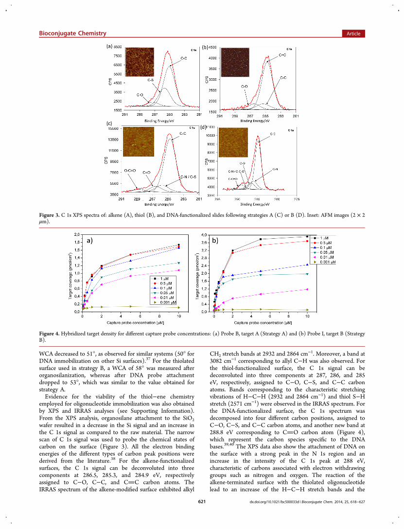
employed for oligonucleotide immobilization was also obtainedby XPS and IRRAS analyses (see Supporting Information).From the XPS analysis, organosilane attachment to the SiO2wafer resulted in a decrease in the Si signal and an increase inthe C 1s signal as compared to the raw material. The narrowscan of C 1s signal was used to probe the chemical states ofcarbon on the surface (Figure 3). All the electron bindingenergies of the different types of carbon peak positions werederived from the literature.38 For the alkene-functionalizedsurfaces, the C 1s signal can be deconvoluted into threecomponents at 286.5, 285.3, and 284.9 eV, respectivelyassigned to C−O, C−C, and CC carbon atoms. TheIRRAS spectrum of the alkene-modified surface exhibited alkyl
CH2 stretch bands at 2932 and 2864 cm−1. Moreover, a band at
3082 cm−1 corresponding to allyl C−H was also observed. Forthe thiol-functionalized surface, the C 1s signal can bedeconvoluted into three components at 287, 286, and 285eV, respectively, assigned to C−O, C−S, and C−C carbonatoms. Bands corresponding to the characteristic stretchingvibrations of H−C−H (2932 and 2864 cm−1) and thiol S−Hstretch (2571 cm−1) were observed in the IRRAS spectrum. Forthe DNA-functionalized surface, the C 1s spectrum wasdecomposed into four different carbon positions, assigned toC−O, C−S, and C−C carbon atoms, and another new band at288.8 eV corresponding to CO carbon atom (Figure 4),which represent the carbon species specific to the DNAbases.39,40 The XPS data also show the attachment of DNA onthe surface with a strong peak in the N 1s region and anincrease in the intensity of the C 1s peak at 288 eV,characteristic of carbons associated with electron withdrawinggroups such as nitrogen and oxygen. The reaction of thealkene-terminated surface with the thiolated oligonucleotidelead to an increase of the H−C−H stretch bands and the
Figure 3. C 1s XPS spectra of: alkene (A), thiol (B), and DNA-functionalized slides following strategies A (C) or B (D). Inset: AFM images (2 × 2μm).
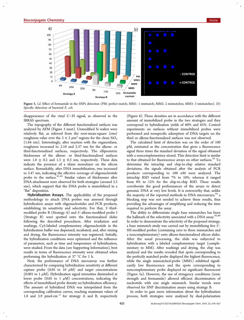
Figure 4. Hybridized target density for different capture probe concentrations: (a) Probe B, target A (Strategy A) and (b) Probe I, target B (StrategyB).
Bioconjugate Chemistry Article
dx.doi.org/10.1021/bc500033d | Bioconjugate Chem. 2014, 25, 618−627621
disappearance of the vinyl C−H signal, as observed in theIRRAS spectrum.The topography of the different functionalized surfaces was
analyzed by AFM (Figure 3 inset). Unmodified Si wafers wererelatively flat, as inferred from the root-mean-square (rms)roughness value over the 2 × 2 μm2 regions for the clean SiO2(1.64 nm). Interestingly, after reaction with the organosilane,roughness increased to 2.10 and 2.37 nm for the alkene- orthiol-functionalized surfaces, respectively. The ellipsomtricthicknesses of the alkene- or thiol-functionalized surfaceswere 1.0 ± 0.2 and 1.3 ± 0.3 nm, respectively. These dataindicate the presence of a silane monolayer on the siliconsurface. Remarkably, after DNA immobilization, rms increasedto 3.47 nm, indicating the effective coverage of oligonucleotideprobe to the surface.41,42 Similar values of thicknesses afterDNA attachment were observed for both strategies (around 3.3nm), which support that the DNA probe is immobilized in a“flat” disposition.Hybridization Assays. The applicability of the proposed
methodology to attach DNA probes was assessed throughhybridization assays with oligonucleotides and PCR products,establishing its sensitivity and selectivity. For that, 5′-thiol-modified probe B (Strategy A) and 5′-alkene-modified probe I(Strategy B) were spotted onto the functionalized slidesfollowing the described procedures. After irradiation andwashings, Cy5-labeled complementary oligonucleotide in thehybridization buffer was dispensed, incubated, and, after rinsingand drying, the fluorescence intensity was registered. Initially,the hybridization conditions were optimized and the influenceof parameters, such as time and temperature of hybridization,were studied. From the data (see Supporting Information), bestresults in terms of fluorescence intensity were obtained whenperforming the hybridization at 37 °C for 1 h.Next, the performance of DNA microarray was further
characterized by comparing hybridization sensitivity at differentcapture probe (0.05 to 10 μM) and target concentrations(0.001 to 1 μM). Hybridization signal intensities diminished atlower probe (0.05 to 1 μM) concentrations, indicating theeffects of immobilized probe density on hybridization efficiency.The amount of hybridized DNA was interpolated from thecorresponding calibration curves, reaching maximum values of1.8 and 3.9 pmol·cm−2 for strategy A and B, respectively
(Figure 4). These densities are in accordance with the differentamount of immobilized probe in the two strategies and theycorrespond to hybridization yields of 60% and 65%. Controlexperiments on surfaces without immobilized probes wereperformed and nonspecific adsorption of DNA targets on thethiol or alkene-functionalized surfaces was not observed.The calculated limit of detection was on the order of 100
pM, estimated as the concentration that gives a fluorescencesignal three times the standard deviation of the signal obtainedwith a noncomplementary strand. This detection limit is similarto that obtained for fluorescence arrays on other surfaces.43 Todetermine the intrachip and chip-to-chip relative standarddeviations, the signals obtained after the analysis of PCRproducts corresponding to 100 nM were analyzed. Theintrachip RSD varied from 7% to 10%, whereas it rangedfrom 8% to 12% for the chip-to-chip RSD. These resultscorroborate the good performances of the arrays to detectgenomic DNA at very low levels. It is noteworthy that, unlikethe majority of the reported methods in DNA microarrays,44 ablocking step was not needed to achieve these results, thusproviding the advantages of simplifying and reducing the timerequired to perform the assay.The ability to differentiate single base mismatches has been
the hallmark of the selectivity associated with a DNA assay.45,46
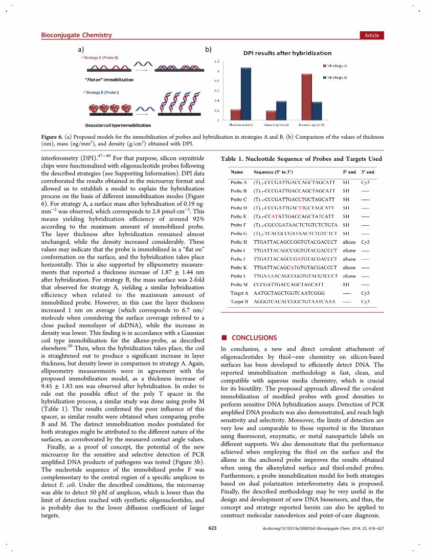
In order to demonstrate the selectivity of the proposed strategy,a base mismatch study was carried out by immobilizing five 5′-SH-modified probes (containing zero to three mismatches anda noncomplementary) onto alkene-functionalized silicon slides.After the usual processing, the slide was subjected tohybridization with a labeled complementary target (comple-mentary to MM). After washings and drying, the chip wasanalyzed and the results revealed that spots corresponding tothe perfectly matched probe displayed the highest fluorescence,while the single mismatched-probe (MM1) exhibited signifi-cantly low fluorescence and the spots corresponding tononcomplementary probe displayed no significant fluorescent(Figure 5a). However, the use of stringency conditions (ionicstrength and formamide) allowed efficient discrimination ofnucleotide with one single mismatch. Similar trends wereobserved for SNP discrimination assays using strategy B.In order to gain more information about the hybridization
process, both strategies were analyzed by dual-polarization
Figure 5. (a) Effect of formamide in the SNPs detection (PM: perfect match; MM1: 1 mismatch; MM2: 2 mismatches; MM3: 3 mismatches). (b)Specific detection of bacterial E. coli.
Bioconjugate Chemistry Article
dx.doi.org/10.1021/bc500033d | Bioconjugate Chem. 2014, 25, 618−627622
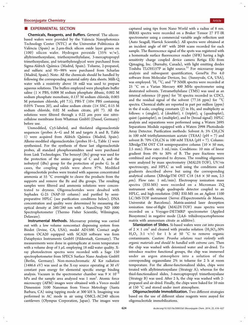
interferometry (DPI).47−49 For that purpose, silicon oxynitridechips were functionalized with oligonucleotide probes followingthe described strategies (see Supporting Information). DPI datacorroborated the results obtained in the microarray format andallowed us to establish a model to explain the hybridizationprocess on the basis of different immobilization modes (Figure6). For strategy A, a surface mass after hybridization of 0.19 ng·mm−2 was observed, which corresponds to 2.8 pmol·cm−2. Thismeans yielding hybridization efficiency of around 92%according to the maximum amount of immobilized probe.The layer thickness after hybridization remained almostunchanged, while the density increased considerably. Thesevalues may indicate that the probe is immobilized in a “flat on”conformation on the surface, and the hybridization takes placehorizontally. This is also supported by ellipsometry measure-ments that reported a thickness increase of 1.87 ± 1.44 nmafter hybridization. For strategy B, the mass surface was 2-foldthat observed for strategy A, yielding a similar hybridizationefficiency when related to the maximum amount ofimmobilized probe. However, in this case the layer thicknessincreased 1 nm on average (which corresponds to 6.7 nm/molecule when considering the surface coverage referred to aclose packed monolayer of dsDNA), while the increase indensity was lower. This finding is in accordance with a Gaussiancoil type immobilization for the alkene-probe, as describedelsewhere.50 Then, when the hybridization takes place, the coilis straightened out to produce a significant increase in layerthickness, but density lower in comparison to strategy A. Again,ellipsometry measurements were in agreement with theproposed immobilization model, as a thickness increase of9.45 ± 1.83 nm was observed after hybridization. In order torule out the possible effect of the poly T spacer in thehybridization process, a similar study was done using probe M(Table 1). The results confirmed the poor influence of thisspacer, as similar results were obtained when comparing probeB and M. The distinct immobilization modes postulated forboth strategies might be attributed to the different nature of thesurfaces, as corroborated by the measured contact angle values.Finally, as a proof of concept, the potential of the new
microarray for the sensitive and selective detection of PCRamplified DNA products of pathogens was tested (Figure 5b).The nucleotide sequence of the immobilized probe F wascomplementary to the central region of a specific amplicon todetect E. coli. Under the described conditions, the microarraywas able to detect 50 pM of amplicon, which is lower than thelimit of detection reached with synthetic oligonucleotides, andis probably due to the lower diffusion coefficient of largertargets.
■ CONCLUSIONS
In conclusion, a new and direct covalent attachment ofoligonucleotides by thiol−ene chemistry on silicon-basedsurfaces has been developed to efficiently detect DNA. Thereported immobilization methodology is fast, clean, andcompatible with aqueous media chemistry, which is crucialfor its bioutility. The proposed approach allowed the covalentimmobilization of modified probes with good densities toperform sensitive DNA hybridization assays. Detection of PCRamplified DNA products was also demonstrated, and reach highsensitivity and selectivity. Moreover, the limits of detection arevery low and comparable to those reported in the literatureusing fluorescent, enzymatic, or metal nanoparticle labels ondifferent supports. We also demonstrate that the performanceachieved when employing the thiol on the surface and thealkene in the anchored probe improves the results obtainedwhen using the alkenylated surface and thiol-ended probes.Furthermore, a probe immobilization model for both strategiesbased on dual polarization interferometry data is proposed.Finally, the described methodology may be very useful in thedesign and development of new DNA biosensors, and thus, theconcept and strategy reported herein can also be applied toconstruct molecular nanodevices and point-of-care diagnosis.
Figure 6. (a) Proposed models for the immobilization of probes and hybridization in strategies A and B. (b) Comparison of the values of thickness(nm), mass (ng/mm2), and density (g/cm3) obtained with DPI.
Table 1. Nucleotide Sequence of Probes and Targets Used
Bioconjugate Chemistry Article
dx.doi.org/10.1021/bc500033d | Bioconjugate Chem. 2014, 25, 618−627623
■ EXPERIMENTAL SECTION
Chemicals, Reagents, and Buffers. General. The silicon-based wafers were provided by the Valencia NanophotonicsTechnology Center (NTC) at the Universitat Politecnica deValencia (Spain) as 2-μm-thick silicon oxide layer grown on(100) silicon wafer. Hydrogen peroxide (35% w/w),allyltrimethoxysilane, triethoxymethylsilane, 3-mercaptopropyltrimethoxysilane, and tetraethyleneglycol were purchased fromSigma-Aldrich Quimica (Madrid, Spain). Toluene, 2-propanol,and sulfuric acid 95−98% were purchased from Scharlau(Madrid, Spain). Note: All the chemicals should be handled byfollowing the corresponding material safety data sheets. Milli-Qwater with a resistivity above 18 mΩ was used to prepareaqueous solutions. The buffers employed were phosphate buffersaline (1 × PBS, 0.008 M sodium phosphate dibasic, 0.002 Msodium phosphate monobasic, 0.137 M sodium chloride, 0.003M potassium chloride, pH 7.5), PBS-T (10× PBS containing0.05% Tween 20), and saline sodium citrate (1× SSC, 0.15 Msodium chloride, 0.02 M sodium citrate, pH 7). All thesolutions were filtered through a 0.22 μm pore size nitro-cellulose membrane from Whatman GmbH (Dassel, Germany)before use.Unmodified, Cy5-labeled, and thiolated oligonucleotide
sequences (probes A−G and M and targets A and B, Table1) were acquired from Aldrich Quimica (Madrid, Spain).Alkene-modified oligonucleotides (probes H−L, Table 1) weresynthesized. For the synthesis of these last oligonucleotideprobes, all standard phosphoramidites used were purchasedfrom Link Technologies. The benzoyl (Bz) group was used forthe protection of the amino group of C and A, and theisobutyryl (iBu) group for the protection of probe G. In allcases, the coupling yields were above 97%. Modifiedoligonucleotide probes were treated with aqueous concentratedammonia at 55 °C overnight to cleave the products from thesupports and remove the Bz and iBu groups. The resultingsamples were filtered and ammonia solutions were concen-trated to dryness. Oligonucleotides were desalted withSephadex G-25 (NAP-10 column) and purified by semi-preparative HPLC (see purification conditions below). DNAconcentration and quality were determined by measuring theoptical density at 260/280 nm with a NanoDrop ND 1000Spectrophotometer (Thermo Fisher Scientific, Wilmington,Delaware).Instrumental Methods. Microarray printing was carried
out with a low volume noncontact dispensing system fromBiodot (Irvine, CA, USA), model AD1500. Contact anglesystem OCA20 equipped with SCA20 software was fromDataphysics Instruments GmbH (Filderstadt, Germany). Themeasurements were done in quintuplicate at room temperaturewith a volume drop of 5 μL employing 18 mΩ water quality. X-ray photoelectron spectra were recorded with a Sage 150spectrophotometer from SPECS Surface Nano Analysis GmbH(Berlin, Germany). Non-monochromatic Al Kα radiation(1486.6 eV) was used as the X-ray source operating at 30 eVconstant pass energy for elemental specific energy bindinganalysis. Vacuum in the spectrometer chamber was 9 × 10⁻⁹
hPa and the sample area analyzed was 1 mm2. Atomic forcemicroscopy (AFM) images were obtained with a Veeco modelDimension 3100 Nanoman from Veeco Metrology (SantaBarbara, CA) using tapping mode at 300 kH h. Imagining wasperformed in AC mode in air using OMCL-AC240 siliconcantilevers (Olympus Corporation, Japan). The images were
captured using tips from Nano World with a radius of 8 nm.IRRAS spectra were recorded on a Bruker Tensor 27 FT-IRspectrometer using a commercial variable angle reflection unit(Auto Seagull, Harrick Scientific). All spectra were obtained atan incident angle of 68° with 2048 scans recorded for eachsample. The fluorescence signal of the spots was registered witha homemade surface fluorescence reader (SFR) having a highsensitivity charge coupled device camera Retiga EXi fromQimaging, Inc. (Burnaby, Canada), with light emitting diodesToshiba TLOH157P as light source.51 For microarray imageanalysis and subsequent quantification, GenePix Pro 4.0software from Molecular Devices, Inc. (Sunnyvale, CA, USA),was employed. 1H, 13C, and 31P NMR spectra were recorded at25 °C on a Varian Mercury 400 MHz spectrometer usingdeuterated solvents. Tetramethylsilane (TMS) was used as aninternal reference (0 ppm) for 1H spectra recorded in CDCl3and the residual signal of the solvent (77.16 ppm) for 13Cspectra. Chemical shifts are reported in part per million (ppm)in the d scale, coupling constants (J) in Hz, and multiplicity asfollows: s (singlet), d (doublet), t (triplet), q (quadruplet),quint (quintuplet), m (multiplet), and br (broad signal). HPLCanalysis and separations were performed using a Waters 2695Separations Module equipped with a Waters 2998 PhotodiodeArray Detector. Purification methods: Solvent A: 5% CH3CNin 100 mM triethylammonium acetate (TEAA) (pH = 7) andsolvent B: 70% CH3CN in 100 mM TEAA (pH = 7). Column:XBridgeTM OST C18 semipreparative column (10 × 50 mm,2.5 mm). Flow rate: 3 mL/min. Conditions: 10 min of lineargradient from 0% to 30% of B. The pure fractions werecombined and evaporated to dryness. The resulting oligomerswere analyzed by mass spectrometry (MALDI-TOF), UV/visspectroscopy, and HPLC as well using the conditions andgradients described above but using the correspondinganalytical column (XBridgeTM OST C18 (4.6 × 50 mm, 2.5μm). Flow rate: 1 mL/min). Electrospray ionization massspectra (ESI-MS) were recorded on a Micromass ZQinstrument with single quadrupole detector coupled to anHPLC, and high-resolution (HR) ESI-MS on an Agilent 1100LC/MS-TOF instrument (Servei d’Espectrometria de Masses,Universitat de Barcelona). Matrix-assisted laser desorptionionization time-of-flight (MALDI-TOF) mass spectra wererecorded on a Voyager-DETMRP spectrometer (AppliedBiosystems) in negative mode (2,4,6- trihidroxyacetophenonematrix with ammonium citrate as additive).
Silanization of Slides. Si-based wafers were cut into piecesof 2 × 1 cm2 and cleaned with piranha solution (H2SO4:30%H2O2 3:1 v/v) for 1 h at 50 °C to remove organiccontaminants. Caution: Piranha solutions react violently withorganic materials and should be handled with extreme care. Thenthe chip was washed with deionized water and air-dried. Tointroduce reactive functional groups, the chip was immersedunder an argon atmosphere into a solution of thecorresponding organosilane 2% in toluene for 2 h at roomtemperature. For the alkene-functionalized slides, chips weretreated with allyltrimetoxysilane (Strategy A); whereas for thethiol-functionalized slides, 3-mercaptopropyl trimethoxysilane(Strategy B) was used. After 2 h, the chip was washed with 2-propanol and air-dried. Finally, the chips were baked for 10 minat 150 °C and stored under inert atmosphere.
Oligonucleotide Immobilization. Two different strategiesbased on the use of different silane reagents were asayed foroligonucleotide immobilizations.
Bioconjugate Chemistry Article
dx.doi.org/10.1021/bc500033d | Bioconjugate Chem. 2014, 25, 618−627624

Strategy A. Silicon-oxide slides were treated following theabove-described procedure to obtain the corresponding alkene-functionalized surfaces. To perform this study, oligonucleotideprobe A (Table 1), consisting of 5′ SH-, 3′ Cy5 oligomer ofsequence (T)15-(CCC GAT TGA CCA GCT AGC ATT) wasused to evaluate the efficiency of the platform towardoligonucleotide immobilization. For that, different probe Asolutions were prepared and spotted (40 nL) onto the alkene-functionalized surface and exposed to UV-light at 365 nm witha mercury capillary lamp (6 mW/cm2, Jelight Irvine, CA, USA)placed at a fixed distance (∼0.5 cm) from the slide for 20 minto induce the immobilization. Finally, slides were thoroughlyrinsed with PBS-T and water and air-dried. Immobilizationresults were obtained from the fluorescence signals using theSFR. Measurements were made by accumulation of emittedlight by the samples during 15 s with a device gain of 5.Strategy B. Silicon-oxide slides were treated following the
above-described procedure to obtain the corresponding thiol-functionalized slides. Thus, oligonucleotide probe H (Table 1),consisting of 5′-alkene-, 3′ Cy5 oligomer of sequence TTGATT ACA GCC GGT GTA CGA CCC T, was used toevaluate the efficiency of the platform toward oligonucleotideimmobilization. For that, different probe H solutions wereprepared and spotted (40 nL) onto the thiol-functionalizedsurface and exposed to UV-light at 365 nm for 20 min toinduce the immobilization. Finally, slides were thoroughlyrinsed with PBS-T and water and air-dried. Immobilizationresults were obtained from the fluorescence intensity of thespots registered with the SFR.Hybridization Assays. To study the hybridization
efficiency on the developed platform, silicon-based slides werefunctionalized with the appropriate organosilane reactive asdescribed above. Afterward, solutions containing oligonucleo-tide 5′ SH- or alkene-labeled (probes B or I) were spotted (40nL) onto the functionalized slides creating the microarrays.Then slides were exposed to UV-light at 365 nm (6 mW/cm2)for 20 min, washed with water and air-dried. After washing, 50μL of the complementary strand was spread out with acoverslip. After incubation in a slim box for 45 min at 37 °C,the coverslip was gently removed and the chip was washed withPBS-T and deionized water. The fluorescence intensity of thespots was registered with the SFR.Detection of Mismatches. Strategy A. To investigate the
sensitivity of the system, four oligonucleotides sequences,namely, SH-(T)15-(CCC GAT TGA CCA GCT AGC ATT)(probe B, PM), SH-(T)15-(CCC GAT TGA CCT GCT AGCATT) (probe C, MM1), SH-(T)15-(CCC GAT TGA TTAGCT AGC ATT) (probe D, MM2), and SH-(T)15-CCA TATTGA CCA GCT ATC ATT) (probe E, MM3) having zero,one, two, and three base mismatches, respectively, were spotted(40 nL/spot, 0.5 μM) with a noncontact dispenser onto analkene-functionalized silicon oxide chip. After immobilizationand blocking as described above, the microarray was subjectedto hybridization with a labeled oligomer (target A), Cy5-(AATGCT AGC TGG TCA ATC GGG) in SSC under differentconditions for 1 h at 37 °C. After washing and drying, thefluorescence was detected in the SFR.Strategy B. In this case, four alkene-modified oligonucleo-
tides sequences previously synthesized, namely, alkene-(TTGATT ACA GCC GGT GTA CGA CCC T) (probe I, PM),alkene-(TTG ATT ACA GCC GAT GTA CGA CCC T)(probe J, MM1), alkene-(TTG ATT ACA GCA TGT GTACGA CCC T) (probe K, MM2), and alkene-(TTG AAA ACA
GCC GGT GTA CGT CCC T) (probe L, MM3) having zero,one, two, and three base mismatches, respectively, were spotted(40 nL/spot, 0.5 μM) with a noncontact dispenser onto anthiol-functionalized silicon oxide chip. After immobilization andblocking as described above, the microarray was subjected tohybridization with a labeled oligomer (target B), Cy5-(AGGGTC ACA CCG GCT GTA ATC AAA) in SSC underdifferent conditions for 1 h at 37 °C. After washing and drying,the fluorescence was detected in the SFR.
Detection of Escherichia coli. Silicon-based slides werefunctionalized with allyltrimethoxysilane as described above.Then, solutions containing SH-labeled probe F (E. coli specificprobe) and probe G (negative control probe) were spottedonto the functionalized slides creating the microarrays. Positiveand negative controls were included to check the quality of theassay. Afterward, slides were exposed to UV-light at 365 nm for20 min and subsequently washed and air-dried. Then, Cy5-labeled PCR product solutions (50 μL) in hybridization buffer(1× SSC) were distributed on the chip. PCR duplexes werefirst melted by 5 min incubation at 95 °C followed by fastcooling for 1 min on ice. After incubating 1 h at 37 °C, theslides were washed with PBS-T, rinsed with deionized water,and air-dried.
Synthesis of Monoallyl-tetraethylene glycol Phos-phoramidite 3-[2-[2-[2-(allyloxyethoxy)ethoxy]-ethoxy]-ethoxy-(diisopropylamino)phosphanyl]-oxapropan-eni-trile. Monoallyl-tetraethylene glycol (150 mg; 0.640 mmol)was previously dissolved in anhydrous acetonitrile (3 × 10 mL).The solvent was evaporated under reduced pressure and theresulting alcohol was dried in vacuum for one hour. The alcoholwas then dissolved in anhydrous dichloromethane (2 mL) andDIEA (2.0 equiv) was added dropwise at room temperature.The solution was cooled at 0 °C and phosphine reagent (1.1equiv) was carefully added dropwise. The reaction was stirred 5min at 0 °C and 1 h at room temperature and monitored byTLC. Dichloromethane was added (10 mL) and organic layerwas washed with a NaHCO3 0.5 M solution (1 × 5 mL) andbrine (1 × 5 mL). Finally, solvent was evaporated and crudewas purified by flash chromatography (Hex/AcOEt 1% to 20.1H NMR (400 MHz, CDCl3) δ 5.87 (m, 1H), 5.20 (m, 2H),4.00 (t, J = 7.1 Hz; 2H), 3.97 (t, J = 7.0 Hz; 2H), 3.81 (m, 2H),3.62 (m, 14H), 3.58 (m, 4H), 2.64 (m, 2H), 1.16 (d, J = 7.02Hz; 6H), 1.14 (d, J = 7.01 Hz; 6H). 13C NMR (100 MHz,CDCl3) δ 134.7, 117.0, 72.2, 71.1, 70.5, 69.3, 62.6, 58.4, 42.9,24.6, 20.3. 31P NMR (162 MHz, CDCl3) δ 149.9. HRMS (ESI+): m/z calcd for C20H39N2O6P ([M+H]+) 435.2618; found435.2617.
■ ASSOCIATED CONTENT*S Supporting InformationHPLC analysis and mass spectra of designed oligonucleotideprobes, water contact angle measurements, surface character-ization (XPS, AFM and IRRAS figures), oligonucleotidecalibration curves, DPI measurements. This material is availablefree of charge via the Internet at http://pubs.acs.org.
■ AUTHOR INFORMATIONCorresponding Author*Phone: 34-963873415; fax: 34-963879349; E-mail:[email protected].
NotesThe authors declare no competing financial interest.
Bioconjugate Chemistry Article
dx.doi.org/10.1021/bc500033d | Bioconjugate Chem. 2014, 25, 618−627625
■ ACKNOWLEDGMENTSFinancial support from Ministerio de Ciencia e Innovacion(CTQ2010-15943) and Generalitat Valenciana (PROMETEO/2010/008 and ACOMP/2012/158) is acknowledged. Theauthors are grateful to the Servei d’Espectrometria de Masses ofthe Universitat de Barcelona for the spectroscopic facilities.
■ REFERENCES(1) Baldi, P., and Hatfield, G. W. (2011) DNA microarrays and geneexpression: from experiments to data analysis and modeling, pp 7−15,Cambridge University Press, Cambridge.(2) Sassolas, A., Leca-Bouvier, B. D., and Blum, L. J. (2008) DNAbiosensors and microarrays. Chem. Rev. 108, 109−139.(3) Samanta, D., and Sarkar, A. (2011) Immobilization of bio-molecules on self-asembled monolayers: methods and sensorapplications. Chem. Soc. Rev. 40, 2567−2592.(4) Crick, C. R., and Parkin, I. P. (2010) Preparation andcharacterisation of super-hydrophobic surfaces. Chem.Eur. J. 16,3568−3588.(5) Shylesh, S., Schunemann, V., and Thiel, W. R. (2010)Magnetically separable nanocatalysts: bridges between homogeneousand heterogeneous catalysis. Angew. Chem., Int. Ed. 49, 3428−3459.(6) Valentini, L., Cardinali, M., and Kenny, J. M. (2010) Selectivedeposition of semiconducting single-walled carbon nanotubes ontoamino-silane modified indium tin-oxide surface for the development ofpoly(3-hexylthiophene)/carbon-nanotube photovoltaic heterojunc-tions. Carbon 48, 861−867.(7) LeMieux, M. C., Roberts, M., Barman, S., Jin, Y. W., Kim, J. M.,and Bao, Z. N. (2008) Self-sorted, aligned nanotube networks for thin-film transistors. Science 321, 101−104.(8) Melde, B. J., Johnson, B. J., and Charles, P. T. (2008)Mesoporous silicate materials in sensing. Sensors 8, 5202−5228.(9) Dugas, V., Demesmay, C., Chevolot, Y., and Souteyrand, E.(2010) Use of organosilanes in biosensors, Nova Science Publishing Inc.,New York.(10) Mrksich, M., and Whitesides, G. M. (1995) Patterning self-assembled monolayers using microcontact printing - a new technologyfor biosensors. Trends Biotechnol. 13, 228−235.(11) Singhvi, R., Kumar, A., Lopez, G. P., Stephanopoulos, G. N.,Wang, D. I., Whitesides, G. M., and Ingber, D. E. (1994) Engineeringcell shape and function. Science 264, 696−698.(12) Place, E. S., Evans, N. D., and Stevens, M. M. (2009)Complexity in biomaterials for tissue engineering. Nat. Mater. 8, 457−470.(13) Schulz, C., Nowak, S., Frohlich, R., and Ravoo, B. J. (2012)Covalent layer-by-layer assembly of redox active molecular multilayerson silicon (100) by photochemical thiol-ene chemistry. Small 8, 569−577.(14) Chen, Y. X., Triola, G., and Waldmann, H. (2011)Bioorthogonal Chemistry for site-specific labeling of surfaceimmobilization of proteins. Acc. Chem. Res. 44, 762−773.(15) Dondoni, A. (2008) The emergence of thiol−ene coupling as aclick process for materials and bioorganic chemistry. Angew. Chem., Int.Ed. 47, 8995−8997.(16) Hoyle, C. E., and Bowman, C. N. (2010) Thiol-ene clickchemistry. Angew. Chem., Int. Ed. 49, 1540−1573.(17) Kempe, K., Krieg, A., Becer, C. R., and Schubert, U. S. (2012)“Clicking” on/with polymers: a rapidly expanding field for thestraightforward preparation of novel macromolecular architectures.Chem. Soc. Rev. 41, 176−191.(18) Lowe, A. (2010) Thiol-ene “click” reactions and recentapplications in polymer and materials synthesis. Polym. Chem. 1,17−36.(19) Jonkheijm, P., Weinrich, D., Kohn, M., Engelkamp, H.,Christianen, P. C. M., Kuhlmann, J., Maan, J. C., Nusse, D.,Schroeder, H., Wacker, R., Breinbauer, R., Niemeyer, C. M., andWaldmann, H. (2008) Photochemical surface patterning by the thiol-ene reaction. Angew. Chem., Int. Ed. 47, 4421−4424.
(20) Bertin, A., and Schlaad, H. (2009) Mild and versatile (bio-)functionalization of glass surfaces via thiol−ene photochemistry.Chem. Mater. 21, 5698−5700.(21) Gupta, N., Lin, B. F., Campos, L. M., Dimitriou, M. D., Hikita,T. S., Treat, N. D., Tirrell, M. V., Clegg, D. O., Kramer, E. J., andHawker, C. J. (2010) A versatile approach to high-throughputmicroarrays using thiol-ene chemistry. Nat. Chem. 2, 138−145.(22) Weinrich, D., Lin, P.-C., Jonkheijm, P., Nguyen, U. T. T.,Schroder, H., Niemeyer, C M., Alexandrov, K., Goody, R., andWaldmann, H. (2010) Oriented immobilization of farnesylatedproteins by the thiol-ene reaction. Angew. Chem., Int. Ed. 49, 1252−1257.(23) Madan, N., Terry, A., Harb, J., Davis, R. C., Schlaad, H., andLinford, M. R. (2011) Thiol−ene−thiol photofunctionalization ofthiolated monolayers with polybutadiene and functional thiols,including thiolated DNA. J. Phys. Chem. C 115, 229318−22938.(24) Wendeln, C., Rinnen, S., Schulz, C., Kaufmann, T., Arlinghaus,H. F., and Ravoo, B. J. (2012) Rapid preparation of multifunctionalsurfaces for orthogonal ligation by microcontact chemistry. Chem.Eur. J. 18, 5880−5888.(25) Wasserberg, D., Steentjes, T., Stopel, M. H. W., Huskens, J.,Blum, C., Subramaniam, V., and Jonkheijm, P. (2012) Patterningperylenes on surfaces using thiol-ene chemistry. J. Mater. Chem. 22,16606−16610.(26) Escorihuela, J., Banuls, M. J., Puchades, R., and Maquieira, A.(2012) DNA microarrays on silicon surfaces through thiol-enechemistry. Chem. Commun. 48, 2116−2118.(27) Lafleur, J. P., Kwapiszewski, R., Jensen, T. G., and Kutter, J. P.(2013) Rapid photochemical surface patterning of proteins in thiol-ene based microfluidic devices. Analyst 138, 845−849.(28) Oberleitner, B., Dellinger, A., Deforet, M., Galtayries, A.,Castanet, A.-S., and Semetey, V. (2013) A facile and versatile approachto design self-assembled monolayers on glass using thiol−enechemistry. Chem. Commun. 49, 1615−1617.(29) Gong, P., Harbers, G. M., and Grainger, D. W. (2006) Multi-technique comparison of immobilized and hybridized oligonucleotidesurface density on commercial amine-reactive microarray slides. Anal.Chem. 78, 2342−2351.(30) Li, Y., Wang, Z., Ou, L. M. L., and Yu, H.-Z. (2007) DNAdetection on plastic: surface activation protocol to convertpolycarbonate substrates to biochip platforms. Anal. Chem. 79, 426−433.(31) Kim, H. J., Yoon, J. H., and Yoon, S. (2010) Photooxidativecoupling of thiophenol derivatives to disulfides. J. Phys. Chem. A 114,12010−12015.(32) Tamarit-Lopez, J., Morais, S., Puchades, R., and Maquieira, A.(2011) Oxygen plasma treated interactive polycarbonate DNAmicroarraying platform. Bioconjugate Chem. 22, 2573−2580.(33) Li, Y., and Zuilhof, H. (2012) Photochemical grafting andpatterning of organic monolayers on indium tin oxide substrates.Langmuir 28, 5350−5359.(34) Wasserberg, D., Nicosia, C., Tromp, E. E., Subramaniam, V.,Huskens, J., and Jonkheijm, P. (2013) Oriented protein immobiliza-tion using covalent and noncovalent chemistry on a thiol-reactive self-reporting surface. J. Am. Chem. Soc. 135, 3104−3111.(35) Weitekamp, R. A., Atwater, H. A., and Grubbs, R. H. (2013)Photolithographic olefin metathesis polymerization. J. Am. Chem. Soc.135, 16817−16820.(36) Escorihuela, J., Banuls, M. J., Puchades, R., and Maquieira, A.(2012) Development of oligonucleotide microarrays onto si-basedsurfaces via thioether linkage mediated by UV irradiation. BioconjugateChem. 23, 2121−2128.(37) Ham, H. O., Liu, Z., Lau, K. H. A., and Messersmith, P. B.(2011) Facile DNA immobilization on surfaces through a catechol-amine polymer. Angew. Chem., Int. Ed. 50, 732−736.(38) Caipa Campos, M. A., Paulusse, J. M. J., and Zuilhof, H. (2010)Functional monolayers on oxide-free silicon surfaces via thiol-ene clickchemistry. Chem. Commun. 46, 5512−5214.
Bioconjugate Chemistry Article
dx.doi.org/10.1021/bc500033d | Bioconjugate Chem. 2014, 25, 618−627626
(39) Graf, N., Gross, T., Wirth, T., Weigel, W., and Unger, W. (2009)Application of XPS and ToF-SIMS for surface chemical analysis ofDNA microarrays and their substrates. Anal. Bioanal. Chem. 393,1907−1912.(40) Lee, C.-Y., Gong, P., Harbers, G. M., Grainger, D. W., Castner,D. G., and Gamble, L. J. (2006) Surface coverage and structure ofmixed DNA/alkylthiol monolayers on gold: characterization by XPS,NEXAFS, and fluorescence intensity measurements. Anal. Chem. 78,3316−3325.(41) Rouillat, M. H., Dugas, V., Martin, J. R., and Phaner-Goutorbe,M. (2005) Characterization of DNA chips on the molecular scalebefore and after hybridization with an atomic force microscope. Appl.Surf. Sci. 252, 1765−1771.(42) Phaner-Goutorbe, M., Dugas, V., Chevolot, Y., and Souteyrand,E. (2011) Silanization of silica and glass slides for DNA microarrays byimpregnation and gas phase protocols: A comparative study.Mater. Sci.Eng., C 31, 384−390.(43) Wang, C., Jia, X.-M., Jiang, C., Zhuang, G.-N., Yan, Q., and Xiao,S.-J. (2012) DNA microarray fabricated on poly(acrylic acid) brushes-coated porous silicon by in situ rolling circle amplification. Analyst 137,4539−4545.(44) Bammler, T., Beyer, R. P., Bhattacharya, S., Boorman, G. A.,Boyles, A., Bradford, B. U., Bumgarner, R. E., Bushel, P. R., Chaturvedi,K., Choi, D., Cunningham, M. L., Dengs, S., Dressman, H. K., Fannin,R. D., Farun, F. M., Freedman, J. H., Fry, R. C., Harper, A., Humble,M. C., Hurban, P., Kavanagh, T. J., Kaufmann, W. K., Kerr, K. F., Jing,L., Lapidus, J. A., Lasarev, M. R., Li, J., Li, Y. J., Lobenhofer, E. K., Lu,X., Malek, R. L., Milton, S., Nagalla, S. R., O’Malley, J. P., Palmer, V. S.,Pattee, P., Paules, R. S., Perou, C. M., Phillips, K., Qin, L. X., Qiu, Y.,Quigley, S. D., Rodland, M., Rusyn, I., Samson, L. D., Schwartz, D. A.,Shi, Y., Shin, J. L., Sieber, S. O., Slifer, S., Speer, M. C., Spencer, P. S.,Sproles, D. I., Swenberg, J. A., Suk, W. A., Sullivan, R. C., Tian, R.,Tennant, R. W., Todd, S. A., Tucker, C. J., Van Houten, B., Weis, B.K., Xuan, S., and Zarbl, H. (2005) Standardizing global geneexpression analysis between laboratories and across platforms. Nat.Methods 2, 351−356.(45) Banoub, J. H., Newton, R. P., Esmans, E., Ewing, D. F., andMackenzie, G. (2005) Recent developments in mass spectrometry forthe characterization of nucleosides, nucleotides, oligonucleotides, andnucleic acids. Chem. Rev. 105, 1869−1916.(46) Wang, K., Tang, Z., Yang, C. J., Kim, Y., Fang, X., Li, W., Wu, Y.,Medley, C. D., Cao, Z., Li, J., Colon, P., Lin, H., and Tan, W. (2009)Molecular engineering of DNA: molecular beacons. Angew. Chem., Int.Ed. 48, 856−870.(47) Escorihuela, J., Banuls, M. J., García-Castello, J., Toccafondo, V.,García-Ruperez, J., Puchades, R., and Maquieira, A. (2012) Chemicalsilicon surface modification and bioreceptor attachment to developcompetitive integrated photonic biosensors. Anal. Bioanal. Chem. 404,2831−2840.(48) Xu, P., Huang, F., and Liang, H. (2013) Real-time study of aDNA strand displacement reaction using dual polarization interfer-ometry. Biosens. Bioelectron. 41, 505−510.(49) Lopez-Paz, J. L., Gonzalez-Martínez, M. A., Escorihuela, J.,Banuls, M. J., Puchades, R., and Maquieira, A. (2014) Direct and label-free monitoring oligonucleotide immobilization, non-specific bindingand DNA biorecognition. Sens. Actuator, B 192, 221−228.(50) Elhadj, S., Singh, G., and Saraf, R. F. (2004) Optical propertiesof an immobilized DNA monolayer from 255 to 700 nm. Langmuir 20,5539−5543.(51) Mira, D., Llorente, R., Morais, S., Puchades, R., Maquieira, A.,and Marti, J. (2004) High throughput screening of surface-enhancedfluorescence on industrial standard digital recording media. Proc. SPIE5617, 364−373.
Bioconjugate Chemistry Article
dx.doi.org/10.1021/bc500033d | Bioconjugate Chem. 2014, 25, 618−627627
Copyright © 2022 FDOKUMEN


![Photochemistry of the azoalkanes 2,3-diazabicyclo[2.2.1]hept-2-ene and spiro[cyclopropane-7,1'-[2,3]-diazabicyclo[2.2.1]hept-2-ene]: on the questions of one-bond vs. two-bond cleavage](https://static.fdokumen.com/doc/165x107/631c10f0a906b217b906c563/photochemistry-of-the-azoalkanes-23-diazabicyclo221hept-2-ene-and-spirocyclopropane-71-23-diazabicyclo221hept-2-ene.jpg)
