Thiol redox transitions in cell signaling: a lesson from N-acetylcysteine
Full Paper
Selective Cell Adhesion and BiosensingApplications of Bio-Active Block CopolymersPrepared by CuAAC/Thiol-ene Double ClickReactions
Gizem Oyman Eyrilmez, Sean Doran, Eljesa Murtezi, Bilal Demir,Dilek Odaci Demirkol, Hakan Coskunol, Suna Timur,* Yusuf Yagci*
N-Acetyl-L-cysteine (NAC)-capped poly(methyl me
thacrylate)-b-polycaprolactone blockcopolymer (PMMA-b-PCL-NAC) was prepared using the previously described one-pot photo-induced sequential CuAAC/thiol-ene double click procedure. PMMA-b-PCL-NAC had previouslyshown good applicability as a matrix for cell adhesion of cells from the Vero cell line(African green monkey kidney epithelial). Here, in this work, PMMA-b-PCL-NAC served as an excellent immobilization matrix for biomoleculeconjugation. Covalent binding of RGD (R: arginine,G: glycine, and D: aspartic acid) peptide sequenceonto the PMMA-b-PCL-NAC-coated surface was per-formed via EDC chemistry. RGD-modified PMMA-b-PCL-NAC (PMMA-b-PCL-NAC-RGD) as a non-toxic cellproliferation platform was used for selective ‘‘integ-rin avb3-mediated cell adhesion and biosensingstudies. Both optical and electrochemical techniqueswere used to monitor the adhesion differencesbetween ‘‘integrin avb3’’ receptor positive andnegative cell lines on to the designed biofunctionalsurfaces.G. Oyman EyrilmezDepartment of Biotechnology, Graduate School of Natural andApplied Sciences, Ege University, 35100 Izmir, TurkeyS. Doran, E. Murtezi, Y. YagciFaculty of Science and Letters, Department of Chemistry, IstanbulTechnical University, Maslak 34469 Istanbul, TurkeyS. Timur, B. Demir, D. Odaci DemirkolFaculty of Science, Department of Biochemistry, Ege University,35100 Izmir, Turkey
D. OdaciInstituteEge UnivE-mail: sH. CoskuFaculty oIzmir, TuY. YagciFaculty ofor AdvaUniversitE-mail: y
aSupporting Information is available online from the Wiley OnlineLibrary or from the author.
� 2015 WILEY-VCH Verlag GmbH & Co. KGaA, Weinheim wileyonlinelibrary.com
Early View Publication; these are NOT the fin
Demirkol, S. Timuron Drug Abuse, Toxicology and Pharmaceutical Science,ersity, 35100 Izmir, [email protected], Psychiatry Department, Ege University, 35100rkey
f Science, Chemistry Department, Center of Excellencenced Materials Research (CEAMR), King Abdulazizy, PO Box 80203, Jeddah 21589 Saudi [email protected]
Macromol. Biosci. 2015, DOI: 10.1002/mabi.201500099 1
al page numbers, use DOI for citation !! R
www.mbs-journal.de
G. Oyman Eyrilmez et al.
2
REa
1. Introduction
Cell-on-a-chip systems have a great importance because of
theirnumerousadvantageoussuchas increasedsensitivity,
continuous flow, and real time analysis.[1] Besides, these
systems can be used to monitor cellular viability, cell
adhesion and proliferation, drug toxicity, and electro-
chemical sensing.[2,3] For cell-on-a-chip, the improvement
of the biocompatibility of cell culture materials is very
important. Therefore, an enormous amount of research has
been conducted toward the design of functional surfaces
with increased sensitivity and biocompatibility.[4–7]
Especially, biodegradable synthetic polymers such as
polycaprolactone (PCL), poly(lactic acid) (PLA), and poly-
(lactideco-glycotide) (PLGA) have been employed as scaf-
folds for organ tissue growth but they generally show
poor cytocompatibility. The surface modification of these
synthetic biocompatible polymers with specific functional
groups and biomolecules has been shown to change their
surface properties and improve cytocompatibility.[8] The
use of so-called click chemistry is a very effective route for
modification of polymeric and macromolecular structures
and has become widespread. These transformations are
very high yielding and highly modular giving rise to little
or no side products without the need for column
chromatography, among other favorable attributes.[9]
Among the archetypal click reactions are included the
copper catalyzed azide–alkyne cycloaddition (CuAAC)[10]
and the thiol-ene reaction.[11] Both reactions have been
successfully employed extensively in macromolecular
modification[12] related to bioapplications.
Recently, our group took advantage of their favorable
properties to employ them sequentially in a one-pot
procedure under photochemical conditions to obtain
N-acetyl-L-cysteine (NAC)-capped poly(methyl methacry-
late)-b-polycaprolactone block copolymer (PMMA-b-PCL-NAC). [13] PMMA-b-PCL-NAC was employed as a matrix
for cell adhesion of cells from the Vero cell line (African
green monkey kidney epithelial) and showed cell prolifer-
ation comparable to commercially available cell culture
plates. Recent years, there has been lots of studies about
improving cell adhesion, proliferation, and viability on
polymer-modified surfaces for cell-on-a chip systems.[14–16]
Especially, extracellular matrix (ECM) proteins, such as
fibronectin, collagen, and laminin-coated surfaces, have
widespread usage due to the their role in themodulation of
integrin-dependent cell adhesion.[17–19] Integrins are cell
surface transmembrane proteins that interact with ECM
molecules. They are formed from 19 a- and 8 b-subunits
that are expressed in 25 different a/b heterodimeric
combinations on the cell surface.[20,21] Also, integrin probes
can be used for sensing of selectively adhered and
proliferated cells. Particularly, integrin alpha V beta 3
(avb3) is an important receptor for ECM proteins via their
Macromol. Biosci. 2015, DOI: 1
� 2015 WILEY-VCH Verlag Gmb
rly View Publication; these are NOT the final pag
RGD (R: arginine, G: glycine, and D: aspartic acid) peptide
sequence. Although integrin avb3 is poorly expressed in
most healthy cells, it is highly expressed in many
tumors.[22–28] Therefore, integrin avb3 is an interesting
biological target for cancer treatment, imaging, and also
sensing.[29]
Researchers have developed numerous surfaces to
investigate integrin-mediated cell adhesion and prolifer-
ation.[30–33] Recently, researchers have worked toward
improving new electrochemical cell-sensing surfaces for
cell-on-a-chip systems. With this goal in mind, several cell
detection based electrochemical methods have appeared
mostly using mammalian cells, especially cancerous cell
lines.[34] Normally, self-assembled modification of elec-
trode surfaces have been used for bio-recognition of the
relevant cell line. On the other hand, novel nanocomposite
and/or polymeric adhesivematerials could be attributed to
this kind of approach. In our previous work, folic acid and
poly(caprolactone)-modified clay nanocomposite were
used in the selective cell attachment of folic acid overex-
pressed HeLa (ovarian cancer cell line) cells against A549
(lung cancer cell line). [5] Here, PMMA-b-PCL-NAC was
synthesized according to the procedure published previ-
ously.[13] PMMA-b-PCL-NAC served as an excellent immo-
bilization matrix for RGD modification. Introduction of
RGD onto the PMMA-b-PCL-NAC-coated surface was
performed through covalent binding using the well-
established two-step carbodiimide coupling method.[35]
Briefly, RGD-modified PMMA-b-PCL-NAC (PMMA-b-PCL-NAC-RGD) was successfully used as a targeted surface for
cell culture applications to discriminate the proliferation
behaviors of integrin avb3 receptor positive (U87-MG) and
negative (HaCaT) cell lines.
2. Experimental Section
2.1. Materials
RGD peptide, EDC (1-ethyl-3-(3-dimethylaminopropyl) carbodii-
mide), Triton X-100, formaldehyde (37.0%), 4, 6-diamino-2-phenyl-
indol (DAPI) were purchased from Sigma. THF (tetrahydrofuran),
phosphate-buffered saline (pH 7.4, PBS) which was prepared using
8.0 g � L-1 NaCl, 0.2 g � L-1 KCl, 1.4 g � L-1 Na2HPO4.2H20, and 0.2 g
KH2PO4were obtained fromMerck. NAC end-capped PMMA-b-PCL-NAC was prepared as previously described.[13]
Dulbecco’s modified Eagle medium (DMEM), Eagle’s minimum
essential medium (EMEM), fetal bovine serum (FBS), penicillin/
streptomycin (P/S) (10 000/10 000 units), and 200mM L-glutamine
were purchased from Lonza. CytoPainter Phalloidin-iFluor 555
reagent, Anti-Integrin alpha V beta 3 (avb3) antibody (ab78289),
and Goat Anti-Mouse IgG H&L (Alexa Fluor 488) (ab150117) were
purchased from Abcam.
FT-IR spectrawere recordedonPerkin–Elmer FT-IR spectrumone
spectrometerwith an ATR Accessory (ZnSe, PikeMiracle Accessory)
and cadmium telluride (MCT) detector. Resolution was 4 cm�1 and
0.1002/mabi.201500099
H & Co. KGaA, Weinheim www.MaterialsViews.com
e numbers, use DOI for citation !!
Selective Cell Adhesion and Biosensing Applications . . .
www.mbs-journal.de
24 scans with 0.2 cm � s�1 scan speed. A Philips XL-30S FEG model
SEMwasused. SEManalyseswere carriedout at5.0 kVand50000�magnifications.
2.2. Cell Culture Studies
U87-MG (ATCC) and HaCaT (CLS) cell lines were maintained in
EMEM and DMEM, respectively. Both of them supplemented with
10.0%FBS, and1.0%P/Sat37 8C inahumidified incubatorwith5.0%
CO2 in air. All cells were sub-cultured at 80% confluency by
trypsinization every 2 or 3 d.
The expression of integrin avb3 in U87-MG cells and the lack
of expression in HaCaT as a control cell line were confirmed by
flow cytometry analyses. For flow cytometry studies, cells were
harvested and washed once in cold PBS and in incubation buffer
consisting of PBS supplementedwith2.0% FBS.After suspending in
incubation buffer, 1� 106 cells were collected and centrifuged. For
cell staining, 2.0mg Anti-Integrin avb3 antibody was added and
incubated at room temperature for 1h. Negative control staining
wasperformedusingmatchedmouseIgG1, isotypecontrolantibody
(BD Biosciences, Heidelberg). After then, secondary staining was
performed by using Goat Anti-Mouse IgG H&L (Alexa Fluor 488)
antibody (1:2 000). Unbound antibodies were removed bywashing
three times in 1.0mL incubation buffer. Immediately before flow
cytometryanalysiscellsweresuspendedin1.0mLincubationbuffer
and thenanalyzed inaBDFACSflowcytometer. 10000gatedevents
were observed in total and the living cellswere gated in a dot plot of
forward versus side scatter signals. For drawing dot plots FlowJo
software (Tree Star, San Carlos, CA) was used.
PMMA-b-PCL-NAC and PMMA-b-PCL-NAC-RGD were compared
to the commercial PS cell culture plates to observe their properties
as cell culturematerials. Thus, 0.5mg �mL�1 PMMA-b-PCL-NAC (in
5.0%THF)was added into polystyrene-coated 96-well plates. Plates
were dried for 72h at room temperatures. To activate carboxyl
groups of PMMA-b-PCL-NAC, 0.2M EDC (in pH 7.4 PBS) were added
into plates and incubated for 15min. Then, PMMA-b-PCL-NAC-
coated plates were incubated with 0.05mg �mL�1 RGD peptide
overnight and rinsed with PBS three times to remove unbound
molecules. Afterward, the resulting PMMA-b-PCL-NAC-RGD-coated plates were sterilized under UV radiation for 15min and
used for the cell culture experiments. All coating experimentswere
performed at ambient temperature.
To observe time-dependent cellular viability, cells were
incubated for 4, 24, 48, and 72h on PMMA-b-PCL-NAC, PMMA-b-PCL-NAC-RGD, and also unmodified polystyrene-coated plates (PS).
At the end of each cultivation time, cells were treated with
0.5mg �mL�1 MTT (3-(4, 5-dimethylthiazol-2-yl)-2,5-diphenylte-
trazolium bromide) for 4 h. Then, 10.0% of SDS (in HCl) was added
per well and incubated overnight. Then, UV�vis absorption was
measured at 570nm with 630nm as reference wavelength using
a microplate reader (Bio-Tek Instruments, Inc., Winooski, VT). The
conventional PS surface was used as control for each experiment.
HaCaT and U87 MG cells were cultured during 72h to compare
their proliferation behaviors on surfaces. CytoPainter Phalloidin-
iFluor 555 Reagent and anti-integrin avb3 antibody were used to
stain F-actin filaments and integrin transmembrane proteins of
cells, respectively. For immunofluorescence staining, cells were
fixed with 4.0% formaldehyde in PBS for 30min at 37 8C after 72h
Macromol. Biosci. 2015, DOI:
� 2015 WILEY-VCH Verlag Gmwww.MaterialsViews.com
Early View Publication; these are NO
incubation times on surfaces. Permeabilization of cells was
facilitated by treatment with 0.1% Triton X-100 for 4min. Then,
cells were stained for actin and integrin avb3 using CytoPainter
Phalloidin-iFluor 555 Reagent (1:1 000) and anti-integrin avb3
antibody (1:500), followed by Alexa 488 fluorophore-conjugated
secondaryantibody (1:500).Also,DAPI (1.0mg �mL�1) stainingwas
performed at ambient temperature during 5min. After extensive
washingwith PBS, the cellswere imaged using anOlympus CKX71
fluorescencemicroscopewitha20�objective. The cell numberwas
quantified for each surface by using Image J (NIH) software.
2.3. Construction of Electrochemical Cell-Sensing
Platform
Theglassycarbonelectrodes (GCEs)werefirstpolishedusingapiece
of cloth with various sized alumina slurry, rinsed with distilled
water, and then sonicated for 5min in ethanol: distilled water to
remove any substances on the electrode surface. During all the
electrochemical measurements, a three electrode cell with GCE as
the working electrode, Ag/AgCl electrode with 3.0M KCl as a
reference electrode and platinum (Pt) as the counter electrodewere
used equipped with PalmSens Potentiostat (Palm Instruments,
Houten, the Netherlands). In order to construct the biosensor
surface for the electrochemical cell sensing, initially 0.5mgPMMA-
b-PCL-NACwas dissolved in 100mL THF and 900mL PBS pH 7.4, and
then 15 mL of this mixture was dropped onto the mirror-like GCE
surface. After drying, the surface was treated with 0.2M EDC
(prepared in PBS, pH 7.4) for 15min in order to activate –COOH
groups. Subsequently, the electrodewas rinsedwith distilledwater,
and then 50mL of RGD tripeptide (0.05mg �mL�1 in PBS pH7.4)was
dropped to the modified electrode. Afterward, the electrode was
allowedtostandfor1htogenerateacovalentbondbetween–COOH
groupsofPMMA-b-PCL-NACand–NH2 functionalityofRGDpeptide.
At the final step, RGD-modified GCE surfaces were treated with
different concentrations of cells for 2h at ambient conditions (both
glioblastoma (U87-MG as integrin avb3 receptor overexpressed cell
line) and healthy keratinocyte (HaCaT- as negative control) were
tested for the selective binding). Monitoring of the cell-binding
capacity of bioactive surface was determined by using differential
pulse voltammetry (DPV) and cyclic voltammetry (CV) techniques.
CV (between–0.4 and –0.8V) andDPV (between–0.2 and –0.6V)
techniques were performed after each modification using [Fe-
(CN)6]3–/4– as a water soluble redox probe (10mM). Cell binding to
the surface caused a decrease inDPV signalswhichwere correlated
with the cell loaded onto the surface. Differences between the
current signals were calculated as follows:
10.1002
bH & Co
T the
DI ¼ Io� Ic
where Io is the mean current at zero cell concentration and Ic is
the mean current at any concentration after cell binding onto
the PMMA-b-PCL-NAC-RGD-covered surfaces.
2.4. Statistical Analysis
GraphPad Prism version 5.03 software (GraphPad Software, San
Diego, CA) was used to obtain graphs and for statistical analyses.
The non-parametric Mann–Whitney U-test was used to compare
/mabi.201500099
. KGaA, Weinheim 3
final page numbers, use DOI for citation !! R
www.mbs-journal.de
G. Oyman Eyrilmez et al.
4
REa
relative cell numbers per surface area among different surfaces.
Statistical significancewas denotedwith *, **, and *** for p�0.05, p� 0.01, and p � 0.001, respectively.
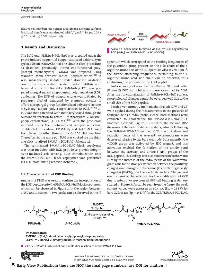
Scheme 2. Amide bond formation via EDC cross-linking betweenRGD (–NH2) and PMMA-b-PCL-NAC (–COOH).
3. Results and Discussion
The NAC end- PMMA-b-PCL-NAC was prepared using the
photo-induced sequential copper-catalyzed azide–alkyne
cycloaddition (CuAAC)/thiol-ene double-click procedure,
as described previously. Bromo end-functional poly-
(methyl methacrylate) (PMMA) was prepared using
standard atom transfer radical polymerization.[36] It
was subsequently azidated under standard azidation
conditions using sodium azide to afford PMMA with
terminal azide functionality (PMMA-N3). PCL was pre-
pared along standard ring-opening polymerization (ROP)
guidelines. The ROP of e-caprolactone was initiated by
propargyl alcohol, catalyzed by stannous octoate to
afford a propargyl group functionalized polycaprolactone,
a-hydroxyl–alkyne–poly(e-caprolactone) (A-PCL).[37,38] A-PCL was then esterified with methacrylic acid through the
Mitsonobu reaction to afford a-methacrylate–v-alkyne–
poly(e-caprolactone) (A-PCL-MA).[38] With the precursors
in hand, using the photo-induced one-pot sequential
double-click procedure, PMMA-N3 and A-PCL-MA were
first clicked together through the CuAAC click reaction.
Thereafter, in the same pot, NAC was clicked via the thiol-
ene click to afford PMMA-b-PCL-NAC (Scheme 1).
The synthesized PMMA-b-PCL-NAC block copolymer
was then modified with RGD peptide to provide integrin
avb3-mediated cell sensing. RGD immobilization onto
the PMMA-b-PCL-NAC block copolymer was performed
via EDC cross-linking reaction (Scheme 2).
3.1. Characterization of RGD Binding
Analysis of FT-IR was used to confirm the incorporation of
theRGDpeptideonto the PMMA-PCL-NACblock copolymer,
which can be observed in Figure 1. In the region between
1 550 and 1 650 cm�1, two peaks can be observed in the IR
Scheme 1. Photo CuAAC/thiol-ene double click reaction to afford PM
Macromol. Biosci. 2015, DOI: 1
� 2015 WILEY-VCH Verlag Gmb
rly View Publication; these are NOT the final pag
spectrum which correspond to the bending frequencies of
the guanidine group present on the side chain of the l-
arginine amino acid of the RGD peptide. Also at 2 916 cm�1,
the alkane stretching frequencies pertaining to the l-
arginine amino acid side chain can be observed, thus
confirming the presence of the RGD peptide.
Surface morphologies before (Figure S1) and after
(Figure 2) RGD immobilization were examined by SEM.
After the functionalization of PMMA-b-PCL-NAC surface,
morphological changes cannot be observed well due to the
small size of the RGD peptide.
Besides, voltammetry methods that include DPV and CV
were applied during the measurements in the presence of
ferricyanide as a redox probe. Hence, both methods were
conducted to characterize the PMMA-b-PCL-NAC-RGD-
modified electrode. Figure 3 illustrates the CV and DPV
diagramsof the eachmodification step gradually. Following
the PMMA-b-PCL-NAC-modified GCE, the oxidation and
reduction peaks of the relevant voltammogram were
decreased relative to the bare electrode. Subsequently, the
–COOH group was activated by EDC reagent, and this
activation enabled the formation of the amide bond
between the carboxyl and amine (–NH2) groups of the
RGDpeptide. This linkagewasalsoevidenced inbothCVand
DPV by the increase of the redox peaks of the voltammo-
grams due to the stronger attraction between the positively
chargedguanidinogroupof arginine (R) and thenegativeSslycharged d [Fe(CN)6] on the electrode surface. The general
electrochemical characteristic for the modification of GCE
due to integrin overexpressed U87 cell binding is demon-
strated in Figure 3. As can be seen from the figure, the peak
current values were assessed as 60.4 mA (Epc¼ 0.33V) for
bareGCE,46mA(Epc¼ 0.37V) for theGCE/PMMA-b-PCL-NAC,
MA-PCL-NAC.
0.1002/mabi.201500099
H & Co. KGaA, Weinheim www.MaterialsViews.com
e numbers, use DOI for citation !!
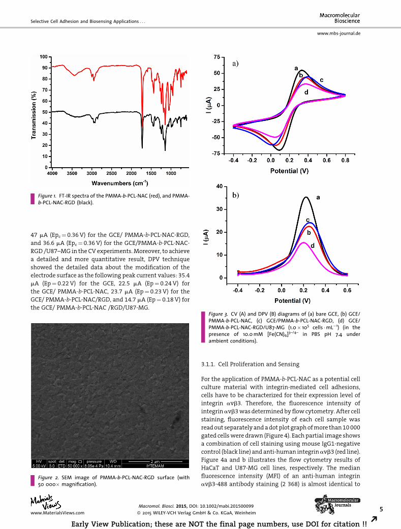
Figure 1. FT-IR spectra of the PMMA-b-PCL-NAC (red), and PMMA-b-PCL-NAC-RGD (black).
Selective Cell Adhesion and Biosensing Applications . . .
www.mbs-journal.de
47 mA (Epc¼ 0.36V) for the GCE/ PMMA-b-PCL-NAC-RGD,
and 36.6 mA (Epc¼ 0.36V) for the GCE/PMMA-b-PCL-NAC-
RGD /U87–MG in the CV experiments.Moreover, to achieve
a detailed and more quantitative result, DPV technique
showed the detailed data about the modification of the
electrode surface as the following peak current values: 35.4
mA (Ep¼ 0.22V) for the GCE, 22.5 mA (Ep¼ 0.24V) for
the GCE/ PMMA-b-PCL-NAC, 23.7 mA (Ep¼ 0.23V) for the
GCE/ PMMA-b-PCL-NAC/RGD, and 14.7 mA (Ep¼ 0.18V) for
the GCE/ PMMA-b-PCL-NAC /RGD/U87-MG.
Figure 2. SEM image of PMMA-b-PCL-NAC-RGD surface (with50 000� magnification).
Figure 3. CV (A) and DPV (B) diagrams of (a) bare GCE, (b) GCE/PMMA-b-PCL-NAC, (c) GCE/PMMA-b-PCL-NAC-RGD, (d) GCE/PMMA-b-PCL-NAC-RGD/U87-MG (1.0� 105 cells �mL�1) (in thepresence of 10.0mM [Fe(CN)6]3–/4– in PBS pH 7.4 underambient conditions).
Macromol. Biosci. 2015, DOI:
� 2015 WILEY-VCH Verlag Gmwww.MaterialsViews.com
Early View Publication; these are NO
3.1.1. Cell Proliferation and Sensing
For the application of PMMA-b-PCL-NAC as a potential cell
culture material with integrin-mediated cell adhesions,
cells have to be characterized for their expression level of
integrin avb3. Therefore, the fluorescence intensity of
integrinavb3wasdeterminedbyflowcytometry. After cell
staining, fluorescence intensity of each cell sample was
readout separatelyandadotplotgraphofmore than10000
gated cellswere drawn (Figure 4). Each partial image shows
a combination of cell staining using mouse IgG1-negative
control (black line) andanti-human integrinavb3 (red line).
Figure 4a and b illustrates the flow cytometry results of
HaCaT and U87-MG cell lines, respectively. The median
fluorescence intensity (MFI) of an anti-human integrin
avb3-488 antibody staining (2 368) is almost identical to
10.1002/mabi.201500099
bH & Co. KGaA, Weinheim 5
T the final page numbers, use DOI for citation !! R
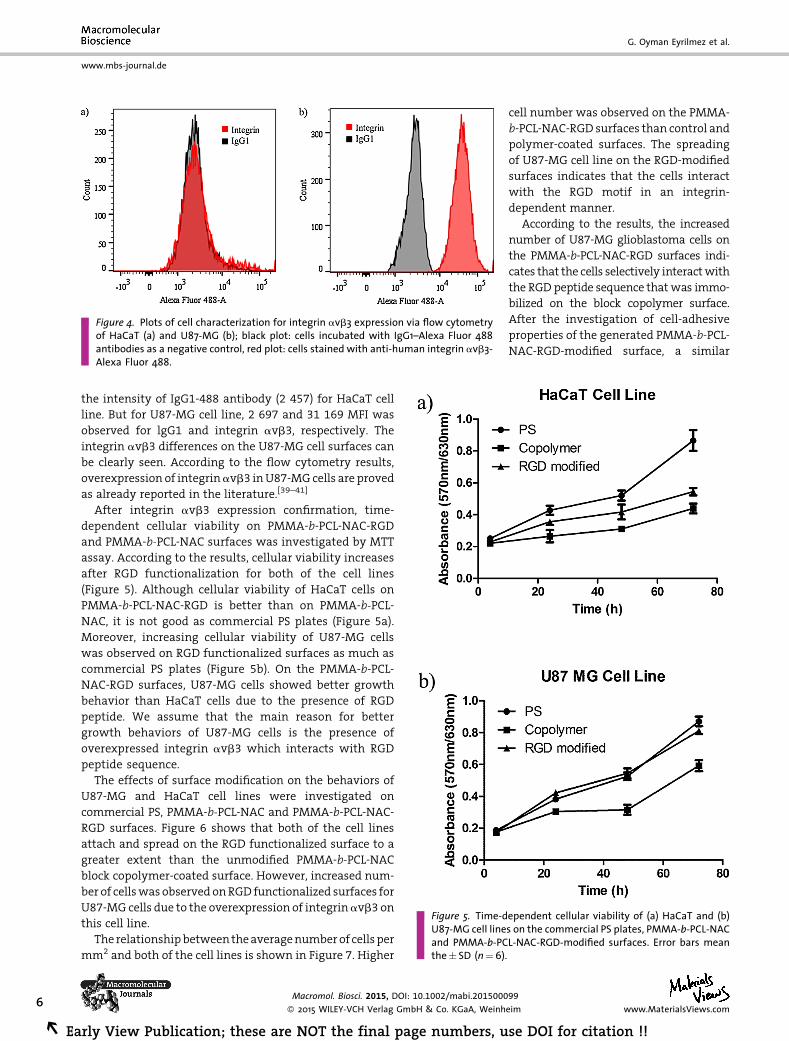
Figure 4. Plots of cell characterization for integrin avb3 expression via flow cytometryof HaCaT (a) and U87-MG (b); black plot: cells incubated with IgG1–Alexa Fluor 488antibodies as a negative control, red plot: cells stained with anti-human integrin avb3-Alexa Fluor 488.
Figure 5. Time-dependent cellular viability of (a) HaCaT and (b)U87-MG cell lines on the commercial PS plates, PMMA-b-PCL-NACand PMMA-b-PCL-NAC-RGD-modified surfaces. Error bars meanthe� SD (n¼6).
www.mbs-journal.de
G. Oyman Eyrilmez et al.
6
REa
the intensity of IgG1-488 antibody (2 457) for HaCaT cell
line. But for U87-MG cell line, 2 697 and 31 169 MFI was
observed for lgG1 and integrin avb3, respectively. The
integrin avb3 differences on the U87-MG cell surfaces can
be clearly seen. According to the flow cytometry results,
overexpression of integrinavb3 inU87-MG cells are proved
as already reported in the literature.[39–41]
After integrin avb3 expression confirmation, time-
dependent cellular viability on PMMA-b-PCL-NAC-RGD
and PMMA-b-PCL-NAC surfaces was investigated by MTT
assay. According to the results, cellular viability increases
after RGD functionalization for both of the cell lines
(Figure 5). Although cellular viability of HaCaT cells on
PMMA-b-PCL-NAC-RGD is better than on PMMA-b-PCL-NAC, it is not good as commercial PS plates (Figure 5a).
Moreover, increasing cellular viability of U87-MG cells
was observed on RGD functionalized surfaces as much as
commercial PS plates (Figure 5b). On the PMMA-b-PCL-NAC-RGD surfaces, U87-MG cells showed better growth
behavior than HaCaT cells due to the presence of RGD
peptide. We assume that the main reason for better
growth behaviors of U87-MG cells is the presence of
overexpressed integrin avb3 which interacts with RGD
peptide sequence.
The effects of surface modification on the behaviors of
U87-MG and HaCaT cell lines were investigated on
commercial PS, PMMA-b-PCL-NAC and PMMA-b-PCL-NAC-
RGD surfaces. Figure 6 shows that both of the cell lines
attach and spread on the RGD functionalized surface to a
greater extent than the unmodified PMMA-b-PCL-NAC
block copolymer-coated surface. However, increased num-
ber of cellswasobservedonRGD functionalized surfaces for
U87-MG cells due to the overexpression of integrinavb3 on
this cell line.
The relationshipbetweentheaveragenumberof cellsper
mm2 and both of the cell lines is shown in Figure 7. Higher
Macromol. Biosci. 2015, DOI: 10.1002/mabi.2015000
� 2015 WILEY-VCH Verlag GmbH & Co. KGaA, Weinh
rly View Publication; these are NOT the final page numbers, u
cell number was observed on the PMMA-
b-PCL-NAC-RGD surfaces than control and
polymer-coated surfaces. The spreading
of U87-MG cell line on the RGD-modified
surfaces indicates that the cells interact
with the RGD motif in an integrin-
dependent manner.
According to the results, the increased
number of U87-MG glioblastoma cells on
the PMMA-b-PCL-NAC-RGD surfaces indi-
cates that the cells selectively interactwith
the RGD peptide sequence thatwas immo-
bilized on the block copolymer surface.
After the investigation of cell-adhesive
properties of the generated PMMA-b-PCL-NAC-RGD-modified surface, a similar
99
eim www.MaterialsViews.com
se DOI for citation !!
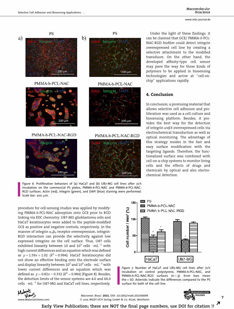
Figure 6. Proliferation behaviors of (a) HaCaT and (b) U87-MG cell lines after 72 hincubation on the commercial PS plates, PMMA-b-PCL-NAC and PMMA-b-PCL-NAC-RGD surfaces. Actin (red), integrin (green), and DAPI (blue) staining were performed.Scale bar: 200 mm.
Figure 7. Number of HaCaT and U87-MG cell lines after 72 hincubation on control polystyrene, PMMA-b-PCL-NAC, andPMMA-b-PCL-NAC-RGD surfaces (n¼ 3). Error bars meanthe� SD. Asterisks indicate the differences compared to the PSsurface for both of the cell line.
Selective Cell Adhesion and Biosensing Applications . . .
www.mbs-journal.de
procedure for cell-sensing studies was applied by modify-
ing PMMA-b-PCL-NAC adsorption onto GCE prior to RGD
linking via EDC chemistry. U87-MG glioblastoma cells and
HaCaT keratinocytes were added to the peptide-modified
GCE as positive and negative controls, respectively. In the
manner of integrin avb3 receptor overexpression, integrin-
RGD interaction can provide the selectivity against low
expressed integrins on the cell surface. Thus, U87 cells
exhibited linearity between 10 and 105 cells �mL�1 with
high currentdifferences andanequationwhichwasdefined
as y¼ 1.59xþ 1.02 (R2¼ 0.994). HaCaT keratinocytes did
not show an effective binding onto the electrode surface
and display linearity between 102 and 105 cells �mL�1 with
lower current differences and an equation which was
defined as y¼ 0.82x – 0.332 (R2¼ 0.984) (Figure 8). Besides,
the detection limits of the sensor systems are 4.0 and 66.0
cells �mL�1 for U87-MG and HaCaT cell lines, respectively.
Macromol. Biosci. 2015, DOI: 10.1002/mabi.20150009
� 2015 WILEY-VCH Verlag GmbH & Co. KGaA, Weinhewww.MaterialsViews.com
Early View Publication; these are NOT the final pag
Under the light of these findings, it
can be claimed that GCE/ PMMA-b-PCL-NAC-RGD biofilm could detect integrin
overexpressed cell line by creating a
selective attachment to the modified
transducer. On the other hand, the
developed affinity-type cell sensor
may pave the way for those kinds of
polymers to be applied in biosensing
technologies and arrive at ‘‘cell-on-
chip’’ applications rapidly.
4. Conclusion
In conclusion, a promisingmaterial that
allows selective cell adhesion and pro-
liferation was used as a cell culture and
biosensing platform. Besides, it pro-
vides the best way for the detection
of integrin avb3 overexpressed cells via
electrochemical transduction as well as
optical monitoring. The advantage of
this strategy resides in the fast and
easy surface modification with the
targeting ligands. Therefore, the func-
tionalized surface was combined with
cell-on-a-chip systems to monitor living
cells and the effects of drugs and
chemicals by optical and also electro-
chemical detection.
9
im 7
e numbers, use DOI for citation !! R
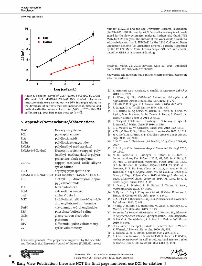
Figure 8. Linearity curves of GCE/ PMMA-b-PCL-NAC-RGD/U87-MG and GCE /PMMA-b-PCL-NAC-RGD /HaCaT electrodes[measurements were carried out via DPV technique related tothe difference of currents that was mentioned in material andmethod and in the presence of 10.0mM, [Fe(CN)6]�3/–4 within PBSbuffer, pH 7.4. Error bars mean the� SD (n¼ 3)].
www.mbs-journal.de
G. Oyman Eyrilmez et al.
8
REa
5. Appendix/Nomenclature/Abbreviations
NAC
rly View Publicatio
N-acetyl-L-cysteine
PCL
polycaprolactonePLA
poly(lactic acid)PLGA
poly(lactideco-glycotide)PMMA
poly(methyl methacrylate)PMMA-b-PCL-NAC
N-acetyl-L-cysteine-capped poly-(methyl methacrylate)-b-polyca-prolactone block copolymer
CuAAC
copper catalyzed azide–alkynecycloaddition
RGD
arginylglysylaspartic acidPMMA-b-PCL-NAC-RGD
RGD-modified PMMA-b-PCL-NACEDC
1-ethyl-3-(3 dimethylaminopro-pyl) carbodiimide
THF
tetrahydrofuranECM
extracellular matrixavb
alpha V beta 3MTT
3-(4,5-dimethylthiazol-2-yl)-2,5-diphenyltetrazolium bromide
DAPI
40,6-diamidino-2 phenylindolePBS
phosphate-buffered salineGCEs
glassy carbon electrodesPt
platinumDPV
differential pulse voltammetryCV
cyclic voltammetryAcknowledgements: This project was supported by the Scientificand Technological Research Council of Turkey (TUBITAK, project
Macromol. Biosci. 2015, DOI: 1
� 2015 WILEY-VCH Verlag Gmb
n; these are NOT the final pag
number 113Z918) and the Ege University Research Foundation(14-FEN-023). EGE University, AREL Central Laboratory is acknowl-edged for the flow cytometry analyses. Authors also thank IYTEMAM for SEM analyses. The authors of this workwould also like toacknowledge and thank T€UB_ITAK for the 2236 Co-Funded BrainCirculation Scheme (Co-Circulation scheme), partially supportedby the EC-FP7 Marie Curie Actions-People-COFUND and coordi-nated by B_IDEB as a source of funding.
Received: March 21, 2015; Revised: April 21, 2015; Publishedonline:DOI: 10.1002/mabi.201500099
Keywords: cell adhesion; cell sensing; electrochemical biosensor;selective surfaces
[1] E. Primiceri, M. S. Chiriac�o, R. Rinaldi, G. Maruccio, Lab Chip2013, 13, 3789.
[2] P. Wang, Q. Liu, Cell-Based Biosensors: Principles andApplications, Artech House, MA, USA 2009, p. 271.
[3] J. El-Ali, P. K. Sorger, K. F. Jensen, Nature 2006, 442, 403.[4] R. Langer, D. A. Tirrell, Nature 2004, 428, 487.[5] F. B. Barlas, D. Ag Seleci, M. Ozkan, B. Demir, M. Seleci, M.
Aydin, M.A. Tasdelen, H. M. Zareie, S. Timur, S. Ozcelik, Y.Yagci, J. Mater. Chem. B 2014, 2, 6412.
[6] P. Neirynck, J. Schimer, P. Jonkheijm, L-G. Milroy, P. Cigler, L.Brunsveld, J. Mater. Chem. B 2014, 3, 539.
[7] S. R. Meyers, M. W. Grinstaff, Chem. Rev. 2012, 112, 1615.[8] Y. Zhu, C. Gao, X. Liu, J. Shen, Biomacromolecules 2002, 3, 1312.[9] H. C. Kolb, M. G. Finn, K. B. Sharpless, Angew. Chem. Int. Ed.
Engl. 2001, 40, 2004.[10] C.W. Torn�e, C. Christensen, M. Meldal, J. Org. Chem. 2002, 67,
3057.[11] C. E. Hoyle, C. N. Bowman, Angew. Chem. Int. Ed. Engl. 2010,
49, 1540.[12] a) D. Bacinello, E. Garanger, D. Taton, K. C. Tam, S.
Lecommandoux, Eur. Polym. J 2015, 62, 363. b) K. Bury, F.Du Prez, D. Neugebauer, Macromol. Biosci. 2013, 13, 1520.c) S. M. Brosnan, H. Schlaad, Polymer 2014, 55, 5509. d) B.Dervaux, F. E. Du Prez, Chem. Sci. 2012, 3, 959. e) M. A.Tasdelen, Y. Yagci, Angew. Chem. Int. Ed. 2013, 52, 5930. f) S.Doran, Y. Yagci, Polym. Chem. 2015, 6, 946. g) E. Murtezi, Y.Yagci, Macromol. Rapid Commun. 2014, 35, 1782. h) A. B.Lowe, Polym. Chem. 2010, 1, 17.
[13] S. Doran, E. Murtezi, F. B. Barlas, S. Timur, Y. Yagci,Macromolecules 2014, 47, 3608.
[14] G. Oyman, C. Geyik, R. Ayranci, M. Ak, D. Odaci Demirkol, S.Timur, H. Coskunol, RSC Adv. 2014, 4, 53411.
[15] R. A. D’Sa, P. J. Dickinson, J. Raj, B. K. Pierscionek, B. J. Meenan,Soft Matter 2011, 7, 608.
[16] J. Yang, D. H. Kim, J. L. Hendricks, M. Leach, R. Northey, D. C.Martin, Acta Biomater. 2005, 1, 125.
[17] Polymers for Regenerative Medicine, C. Werner, Ed., Advancesin Polymer Science, Vol. 203, Springer, Berlin, Heidelberg 2006.
[18] D. Liu, C. A. Che Abdullah, R. P. Sear, J. L. Keddie, Soft Matter2010, 6, 5408.
[19] U. Geissler, U. Hempel, C. Wolf, D. Scharnweber, H. Worch,K. Wenzel, J. Biomed. Mater. Res. 2000, 51, 752.
[20] Y. Takada, X. Ye, S. Simon, Genome Biol. 2007, 8, 215.[21] B. Alberts, A. Johnson, J. Lewis, M. Raff, K. Roberts, P. Walter,
Molecular Biology of the Cell, 5st ed., Garland Science, Taylor& Francis Group, LLC, NewYork, USA 2008, p. 1170.
0.1002/mabi.201500099
H & Co. KGaA, Weinheim www.MaterialsViews.com
e numbers, use DOI for citation !!
Selective Cell Adhesion and Biosensing Applications . . .
www.mbs-journal.de
[22] A. R. Hsu, A. Veeravagu, W. Cai, L. C. Hou, V. Tse, X. Chen,Recent Pat. Anticancer Drug Discov. 2007, 2, 143.
[23] Z. Liu, F. Wang, X. Chen, Drug Dev. Res. 2008, 69, 329.[24] B. Felding-Habermann, T. E. O’Toole, J. W. Smith, E. Fransvea,
Z. M. Ruggeri, M. H. Ginsberg, P. E. Hughes, N. Pampori, S. J.Shattil, A. Saven, B. M.Mueller, Proc. Natl. Acad. Sci. USA 2001,98, 1853.
[25] C. R. Cooper, C. H. Chay, K. J. Pienta, Neoplasia 2002, 4, 191.[26] R. Soldi, S. Mitola, M. Strasly, P. Defilippi, G. Tarone, F.
Bussolino, EMBO J. 1999, 18, 882.[27] S.M.Albelda, S. A.Mette,D. E. Elder, R. Stewart, L. Damjanovich,
M. Herlyn, C. A. Buck, Cancer Res. 1990, 50, 6757.[28] S. Zitzmann, V. Ehemann, M. Schwab, Cancer Res. 2002, 62,
5139.[29] F. Danhier, A. Le Breton, V. Pr�eat, Mol. Pharm. 2012, 9, 2961.[30] S. Regis, S. Youssefian, M. Jassal, M. Phaneuf, N. Rahbar, S.
Bhowmick, Polym. Eng. Sci. 2014, 54, 2587.[31] T. A. Petrie, J. E. Raynor, C. D. Reyes, K. L. Burns, D. M. Collard,
A. J. Garcı́a, Biomaterials 2008, 29, 2849.
Macromol. Biosci. 2015, DOI:
� 2015 WILEY-VCH Verlag Gmwww.MaterialsViews.com
Early View Publication; these are NO
[32] M. S. Tjin, A. W. C. Chua, D. R. Ma, S. T. Lee, E. Fong,Macromol.Biosci. 2014, 14, 1125.
[33] T. A. Petrie, J. E. Raynor, D. W. Dumbauld, T. T. Lee, S. Jagtap,K. L. Templeman, D. M. Collard, A. J. Garcia, Sci. Transl. Med.2010, 2, 45.
[34] L. Qu, J. Xu, X. Tan, Z. Liu, L. Xu, R. Peng, ACS Appl. Mater.Interfaces 2014, 6, 7309.
[35] G. T. Hermanson, Bioconjugate Techniques, 3rd edition,Academic Press, London, UK 2013, p. 127.
[36] K. Matyjaszewski, Macromolecules 2012, 45, 4015.[37] T. Cai, M. Li, B. Zhang, K-G. Neoh, E-T. Kang, J. Mater. Chem. B
2014, 2, 814.[38] I. Javakhishvili, S. Hvilsted, Biomacromolecules 2009, 10, 74.[39] H. Wu, H. Chen, Y. Sun, Y. Wan, F. Wang, B. Jia, X. Su, Cancer
Lett. 2013, 335, 75.[40] Y. Wu, X. Zhang, Z. Xiong, Z. Cheng, D. R. Fisher, S. Liu, S. S.
Gambhir, X. Chen, J. Nucl. Med. 2005, 46, 1707.[41] H. Wang, K. Chen, W. Cai, Z. Li, L. He, A. Kashefi, X. Chen,Mol.
Cancer Ther. 2008, 7, 1044.
10.1002/mabi.201500099
bH & Co. KGaA, Weinheim 9
T the final page numbers, use DOI for citation !! R
Copyright © 2022 FDOKUMEN


![Photochemistry of the azoalkanes 2,3-diazabicyclo[2.2.1]hept-2-ene and spiro[cyclopropane-7,1'-[2,3]-diazabicyclo[2.2.1]hept-2-ene]: on the questions of one-bond vs. two-bond cleavage](https://static.fdokumen.com/doc/165x107/631c10f0a906b217b906c563/photochemistry-of-the-azoalkanes-23-diazabicyclo221hept-2-ene-and-spirocyclopropane-71-23-diazabicyclo221hept-2-ene.jpg)