Targeted Delivery and Enhanced Cytotoxicity of Cetuximab−Saporin by Photochemical Internalization...
13
Subscriber access provided by UNIV OF OSLO Molecular Pharmaceutics is published by the American Chemical Society. 1155 Sixteenth Street N.W., Washington, DC 20036 Article Targeted Delivery and Enhanced Cytotoxicity of Cetuximab−Saporin by Photochemical Internalization in EGFR-Positive Cancer Cells Wai Lam Yip, Anette Weyergang, Kristian Berg, Hanne H. Tnnesen, and Pl K. Selbo Mol. Pharmaceutics, 2007, 4 (2), 241-251 • DOI: 10.1021/mp060105u Downloaded from http://pubs.acs.org on November 25, 2008 More About This Article Additional resources and features associated with this article are available within the HTML version: • Supporting Information • Links to the 3 articles that cite this article, as of the time of this article download • Access to high resolution figures • Links to articles and content related to this article • Copyright permission to reproduce figures and/or text from this article
-
Upload
rr-research -
Category
Documents
-
view
18 -
download
0
Transcript of Targeted Delivery and Enhanced Cytotoxicity of Cetuximab−Saporin by Photochemical Internalization...
Subscriber access provided by UNIV OF OSLO
Molecular Pharmaceutics is published by the American Chemical Society. 1155Sixteenth Street N.W., Washington, DC 20036
Article
Targeted Delivery and Enhanced Cytotoxicity of Cetuximab−Saporinby Photochemical Internalization in EGFR-Positive Cancer Cells
Wai Lam Yip, Anette Weyergang, Kristian Berg, Hanne H. Tnnesen, and Pl K. SelboMol. Pharmaceutics, 2007, 4 (2), 241-251 • DOI: 10.1021/mp060105u
Downloaded from http://pubs.acs.org on November 25, 2008
More About This Article
Additional resources and features associated with this article are available within the HTML version:
• Supporting Information• Links to the 3 articles that cite this article, as of the time of this article download• Access to high resolution figures• Links to articles and content related to this article• Copyright permission to reproduce figures and/or text from this article

Subscriber access provided by UNIV OF OSLO
Molecular Pharmaceutics is published by the American Chemical Society. 1155Sixteenth Street N.W., Washington, DC 20036
Targeted Delivery and Enhanced Cytotoxicity ofCetuximab -Saporin by Photochemical Internalization in
EGFR-Positive Cancer Cells
Wai Lam Yip, † Anette Weyergang,† Kristian Berg, † Hanne H. Tønnesen,‡ andPål K. Selbo*,†
Department of Radiation Biology, Institute for Cancer Research, The Norwegian RadiumHospital, Oslo, Norway, and Department of Pharmaceutics, School of Pharmacy,
UniVersity of Oslo, Oslo, Norway
Received October 5, 2006; Revised Manuscript Received December 12, 2006; Accepted December 19, 2006
Abstract: Photochemical internalization (PCI) is a novel technology of macromolecular delivery.By PCI, endocytosed membrane-impermeable therapeutic drugs are photochemically releasedfrom entrapment in endo-lysosomal compartments to the cytosol of target cells. In the presentreport, we describe the in vitro proof-of-concept for PCI of cetuximab-saporin, an immunotoxintargeting EGFR-expressing cells. This immunotoxin consists of the chimeric murine-human IgG1
monoclonal antibody cetuximab (C225 or Erbitux) bound to the type I ribosome-inactivatingprotein toxin saporin by a biotin-streptavidin linkage. The photochemical treatment enhancedthe cytotoxicity of the immunotoxin in a synergistic manner in three different EGFR-expressingcarcinoma cell lines derived from different tumor tissues (colorectal, HCT-116; prostate, DU-145; and epidermis, A-431). Both cytotoxicity of cetuximab-saporin and epifluorescence ofAlexa488-cetuximab were evaluated by competition with cetuximab demonstrating specificbinding and uptake of cetuximab-saporin in EGFR positive cells. In the EGFR-negative uterinesarcoma cell line MES-SA, neither binding nor preferential accumulation of Alexa488-cetuximabwas detected. In addition, PCI enhanced the cytotoxicity of cetuximab-saporin to the sameextent as streptavidin-saporin in the MES-SA cells. In conclusion, PCI enhances selectivity ofthe cytotoxicity of the immunotoxin cetuximab-saporin in EGFR-expressing cells.
Keywords: Photochemical internalization; photodynamic therapy; drug delivery; immunotoxin; EGFR;saporin; drug targeting; cetuximab
IntroductionThe utilization of macromolecules in the therapy of cancer
and other diseases is becoming increasingly important.Recent advances in molecular biology and biotechnologyhave made it possible to improve targeting and design ofcytotoxic agents, DNA complexes, and other macromolecules
for clinical applications. In many cases the targets ofmacromolecular therapeutics are intracellular. However,degradation of macromolecules in endocytic vesicles afteruptake by endocytosis is a major intracellular barrier for thetherapeutic application of macromolecules having intracel-lular targets of action.
Photochemical internalization (PCI) is a novel technologyfor the release of endocytosed macromolecules into thecytosol. The technology is based on the activation by lightof photosensitizers (PSs) located in endocytic vesicles toinduce the release of macromolecules from the endocyticvesicles.1-3 Thereby, endocytosed molecules can be releasedto reach their target of action before being degraded in
* To whom correspondence should be addressed. Mailing ad-dress: Department of Radiation Biology, Institute for CancerResearch, The Norwegian Radium Hospital, Montebel-lo, N-0310 Oslo, Norway. E-mail: [email protected]: +47-22934261. Fax:+47-22934270.
† The Norwegian Radium Hospital.‡ University of Oslo.
articles
10.1021/mp060105u CCC: $37.00 © 2007 American Chemical Society VOL. 4, NO. 2, 241-251 MOLECULAR PHARMACEUTICS 241Published on Web 01/31/2007

lysosomes. PCI has been shown to stimulate intracellulardelivery of a large variety of macromolecules and othermolecules that do not readily penetrate the plasma membrane,including type I ribosome-inactivating proteins (RIPs), DNAdelivered as gene-encoding plasmids or by means of aden-ovirus or adeno-associated virus, peptide nucleic acids(PNAs) and chemotherapeutic agents such as bleomycin andin some cases doxorubicin. The efficacy and specificity ofPCI of macromolecular therapeutics has been improved bycombining the macromolecules with targeting moieties, suchas the epidermal growth factor.4 In general, PCI can induceefficient light-directed delivery of macromolecules into thecytosol, indicating that it may have a variety of usefulapplications for site-specific drug delivery as for examplein gene therapy, vaccination, and cancer treatment. Recently,we demonstrated that PCI of the affinity toxin EGF-saporin,using mouse EGF, resulted in enhanced cytotoxicity in twoEGFR-positive cancer cell lines.5 EGF-saporin was ef-ficiently endocytosed, delivery was specific, and the com-bination between the affinity toxin and photochemicalactivation of the PS (PCI of the affinity toxin) inducedsynergistic antiproliferative effects.
In the present study, we wanted to further exploit andimprove the concept of PCI-induced drug delivery using animmunotoxin targeting EGFR. The therapeutically approvedcetuximab (also known as C225 or Erbitux), which is achimeric humanized murine mAb recognizing the epidermalgrowth factor receptor (EGFR),6 was linked to the type Iribosome-inactivating protein saporin.7 PCI of cetuximab-saporin was established as a proof-of-concept in threedifferent EGFR-positive human cancer cell lines. In addition,an EGFR-negative cancer cell line was utilized as control.The results reported here indicate that cetuximab-saporin
binds specifically to EGFR-positive cells and is subsequentlytaken up by endocytosis. PCI significantly enhances thecytosolic delivery and the cytotoxicity of cetuximab-saporin.PCI of cetuximab-saporin is a unique combination strategyencompassing three different ways of cell inactivation: (1)receptor blockage and internalization by cetuximab; (2)cytotoxic effect of the photochemical treatment; (3) the RIPactivity of saporin.
Experimental SectionCell Lines and Culture Conditions. All cell lines were
purchased from ATCC (Manassas, VA). Three human cancercell lines expressing EGFR were used: HCT-116 colorectalcarcinoma (ATCC No. CCL-247), DU-145 prostate carci-noma (ATCC No. HTB-81), and A-431 epidermoid carci-noma of the skin (ATCC No. CRL-1555). Additionally, theEGFR negative human uterus sarcoma MES-SA cell line(ATCC No. CRL-1976) was selected as control cells. HCT-116 and DU-145 cells were maintained in RPMI-1640medium (Sigma-Aldrich, St. Louis, MO). MES-SA cells werecultured in McCoy’s medium (Sigma-Aldrich) while A-431cells were cultured in DMEM (Bio Whittaker Europe,Velviers, Belgium). All media were supplemented with 10%bovine calf serum, 100 units/mL penicillin, 100µg/mLstreptomycin (Sigma-Aldrich), and 2 mM glutamine (Sigma-Aldrich). All cell lines were incubated at 37°C in ahumidified atmosphere containing 5% CO2 and subculturedthree times a week.
Drugs and Chemicals.The PS LumiTrans (TPPS2a, meso-tetraphenylporphine with two sulfonate groups on adjacentphenyl rings) was a generous gift from PCI Biotech AS(Oslo, Norway). A stock solution of 0.35 mg/mL was keptat -20 °C. All work with TPPS2a was carried out undersubdued light. Streptavidin-saporin was purchased fromAdvanced Targeting Systems (San Diego, CA). The strepta-vidin-saporin conjugate contains on average two saporinmolecules per streptavidin molecule. Biotinylation kit wasobtained from Pierce (Rockford, IL). Cetuximab (C225) waspurchased from E. Merck AB (Stockholm, Sweden), and thesolution of 2 mg/mL was kept at 4°C. Tyrphostin AG-1478(Sigma-Aldrich) was dissolved in dimethyl sulfoxide (DMSO;Sigma-Aldrich) to yield a stock solution of 50 mM.
Light Source. Illuminations of cells were performed byusing LumiSource (PCI Biotech, Oslo, Norway). This lampconsists of four standard light tubes (18 W/tube, Osram L18/67), which emit blue light with a main peak at ap-proximately 435 nm. The irradiance varies less than 10%across the whole illumination area (765 cm2) with an outputof 11 mW/cm2. The light box is air-cooled during lightexposure, preventing cells from being exposed to hyperther-mia, and keeps the irradiance stable.
Preparation of Cetuximab-Saporin. Cetuximab wasbiotinylated according to the manufacturer’s instructions(Pierce Biotechnology Inc, Rockford, IL). Briefly, a solutionof 2 mL of 2 mg/mL cetuximab was mixed with 100µL of1 M NaHCO3 to adjust the mAb solution to pH∼8.Cetuximab was then mixed with 54µL of 10 mM NHS-
(1) Berg, K.; Selbo, P. K.; Prasmickaite, L.; Tjelle, T. E.; Sandvig,K.; Moan, J.; Gaudernack, G.; Fodstad, O.; Kjølsrud, S.; Anholt,H.; Rodal, G. H.; Rodal, S. K.; Høgset, A. Photochemicalinternalization: a novel technology for delivery of macromoleculesinto cytosol.Cancer Res.1999, 59, 1180-1183.
(2) Selbo, P. K.; Sandvig, K.; Kirveliene, V.; Berg, K. Release ofgelonin from endosomes and lysosomes to cytosol by photo-chemical internalization.Biochim. Biophys. Acta2000, 1475,307-313.
(3) Selbo, P. K.; Sivam, G.; Fodstad, O.; Sandvig, K.; Berg, K.InViVo Documentation of Photochemical Internalization, a NovelApproach to Site Specific Cancer Therapy.Int. J. Cancer2001,92, 761-766.
(4) Berg, K.; Hogset, A.; Prasmickaite, L.; Weyergang, A.; Bonsted,A.; Dietze, A.; Lou, P. J.; Bown, S. G.; Norum, O. J.; Mollergard,H. M. T.; Selbo, P. K. Photochemical internalization (PCI): Anovel technology for activation of endocytosed therapeutic agents.Med. Laser Appl.2006, 21, 239-250.
(5) Weyergang, A.; Selbo, P. K.; Berg, K. Photochemically stimulateddrug delivery increases the cytotoxicity and specificity of EGF-saporin.J. Controlled Release2006, 111, 165-173.
(6) Graham, J.; Muhsin, M.; Kirkpatrick, P. Cetuximab.Nat. ReV.Drug DiscoVery 2004, 3, 549-550.
(7) Barbieri, L.; Battelli, M. G.; Stirpe, F. Ribosome-inactivatingproteins from plants.Biochim. Biophys. Acta1993, 1154, 237-282.
articles Yip et al.
242 MOLECULAR PHARMACEUTICS VOL. 4, NO. 2
biotin that was made in 99.9% dimethyl sulfoxide (DMSOfrom Aldrich, Stenheim, Germany) immediately before, andthen mixed at room temperature for 30 min. Sephadex G-25medium desalting columns (Amersham Biosciences, Amer-shamplace, U.K.) were used to remove free NHS-biotin fromthe biotin-cetuximab solution. The concentration of cetux-imab-biotin was measured using the DC protein assay kit2 (BioRad, Laboratories, Philadelphia, PA), and verifiedspectrophotometrically at 280 nm. It was assumed that allcetuximab was biotinylated. Then, a solution of 10µMbiotinylated cetuximab was mixed with a stock solution ofstreptavidin-saporin at a molar ratio cetuximab-biotin:streptavidin-saporin of 4:1 for 15 min at room temperature.Due to the very high affinity constant of the binding ofstreptavidin to biotin (1015 M-1),8 the linking reaction wasassumed complete. The immunotoxin solution was thendiluted with sterile PBS (pH 7.0) to a concentration of 300nM and aliquoted and stored at-80 °C.
Photochemical Treatments.Cells were harvested with500 µg/L trypsin and 200 mg/mL EDTA and seeded out at3000 cells per well in 96-well plates (Nunclon, Nunc,Roskilde, Denmark) for the MTT assay (as described below)or 1000 cells/well in 6-well plates (Nunclon) for colony-forming ability experiments. Cells were allowed to attachto the bottom of the wells for 24 h prior to the start ofexperiments. HCT-116 and DU-145 cells were then eitherincubated with 0.2µg/mL TPPS2a for PDT only or, for thePCI experiments, coincubated with increasing concentrationsof cetuximab-saporin, streptavidin-saporin, or cetuximabfor 18 h, unless otherwise stated. For studies with A-431cells, 0.1µg/mL TPPS2a was used, since the cell line hashigher sensitivity to TPPS2a-PDT than the other cell linesused in the present study. The monolayers were then washedtwice with drug-free culture medium before it was replacedwith fresh drug-free medium and chased for 4 h. Cells weresubsequently exposed to increasing doses of LumiSourcelight. This PCI protocol is referred to as “light after” PCI.Alternatively, “light first” PCI strategy was performed. Inthese experiments, the cells were first subjected to PDT asdescribed above, and immediately after light exposure cellswere incubated with the immunotoxin for 18 h before thecells were washed twice and, subsequently, incubatedovernight followed by the MTT viability assay, which wasperformed 48 h post light exposure (as described below).
Cytotoxicity Assays.The cell viability was measured bythe MTT assay 48 h after light exposure, or alternatively,the colony-forming ability of the cells was assessed 7-10days after light exposure as recently described.9
Analysis of EGFR and Phosphorylated EGFR.HCT-116 cells were seeded out in Falcon 3003 dishes (10× 103
cells/cm2) overnight. Cells were then incubated with 100 nMcetuximab for 18 h (day 2) or 2 h (day 3) prior to treatmentwith 100 ng/mL EGF for 2 min before the monolayers werewashed once with ice-cold PBS and kept on ice. PBS wasaspirated, and 100µL of lysis buffer containing 62.5 mMTris-HCl pH 6.8, 2% w/v SDS, 10% glycerol, 50 mM DTT,0.01% bromphenol blue, 10µL/mL Protease InhibitorCocktail (Sigma-Aldrich), 10µL/mL Phosphatase InhibitorCocktail 1 (Sigma-Aldrich), 10µL/mL Phosphatase InhibitorCocktail 2 (Sigma-Aldrich), 1 mM PMSF, 1 mM NaVO4, 1mM NaF, and 20 mMâ-glycerolphosphate was added tothe cells. The cell lysates were collected and transferred to1.8 mL plastic tubes (Eppendorf, Hamburg, Germany).Proteins were then subjected to SDS-PAGE and Westernblot followed by antibody-based analysis of total andphosphorylated (p) EGFR as described previously.5 Primarymouse Ab (Cat. No. 2236) against human p-EGFR and rabbittotal EGFR Ab (Cat. No. 2232) was obtained from CellSignaling Technology (Danvers, MA) and diluted 1:1000before incubation of secondary HRP conjugated donkey-anti-rabbit Ab (Cat. No. W401B) that was purchased fromPromega (Madison, WI) or HRP conjugated sheep-antimouse (Cat. No. Na 931) from Amersham Biosciences, bothdiluted 1:5000 prior to incubation of the membrane. En-hanced chemiluminescence (ECL+; Amersham) was usedfor signal detection.
Preparation and Validation of Alexa488-Cetuximab.Cetuximab was labeled with Alexa Fluor 488 (∼0.9 kDa)according to the instructions of the manufacturer (MolecularProbes, Eugene, OR). The Ab-dye conjugate is herebynamed Alexa488-cetuximab. Ten micrograms of Alexa488-cetuximab or unconjugated cetuximab was subjected to4-15% linear gradient SDS-PAGE. Fluorescence intensityof the dye conjugate was detected using the Storm 860scanner (Amersham/GE Health, London, U.K.). Thereafter,the gel was stained with Coomassie blue for comparing theprotein band of Alexa488-cetuximab with cetuximab. Al-exa488 labeled goat-anti-mouse mAb (Molecular Probes)were used as a negative control not targeting EGFR.
Fluorescence Microscopy.For cetuximab uptake studies,cells were seeded out in Falcon 3001 dishes (50× 103 cellsper dish,∼5 × 10³ cells/cm2) overnight. The cells wereincubated with 100 nM Alexa488-cetuximab, without orin the presence of 2µM cetuximab, for 18 h. Then the cellswere washed and incubated in drug-free medium for 4 h.The cells were examined by a Zeiss Axioplan epi-fluorescence and phase contrast microscope equipped witha 63× oil-immersion objective (Zeiss, Oberkochen, Ger-many). The images were acquired by utilizing a cooledcharge-coupled device (CCD) camera (Astromed 3200,Astromed, Cambridge, U.K.). A 450-490 nm band-passexcitation filter, a beam splitter at 510 nm, and a 510-550nm emission filter were used for the recording of Alexa 488fluorescence (micrographs were all set with the samesensitivity range). Fluorescence of TPPS2awas recorded usinga 395-440 nm band-pass excitation filter, a 460 nm dichroicbeam splitter, and a 610 nm long-pass filter. The AnalySISD
(8) Diamandis, E. P.; Christopoulos, T. K. The biotin-(strept)avidinsystem: principles and applications in biotechnology.Clin. Chem.1991, 37, 625-636.
(9) Selbo, P. K.; Weyergang, A.; Bonsted, A.; Bown, S. G.; Berg,K. Photochemical internalization of therapeutic macromolecularagents: a novel strategy to kill multidrug resistant cancer cells.J. Pharmacol. Exp. Ther.2006, 319, 604-612.
DeliVery of Cetuximab-Saporin in Cancer Cells by PCI articles
VOL. 4, NO. 2 MOLECULAR PHARMACEUTICS 243
Soft Imaging System (Olympus, Hamburg, Germany) wasutilized to process and analyze the digital images.
Results and DiscussionOverexpression of EGFR is a hallmark of numerous
cancers and has become a major target for drug delivery andtherapy. Despite the anticancer effects of the monoclonalantibody cetuximab and the small molecule tyrosine kinaseinhibitors (TKI) gefitinib and erlotinib in clinical trials, theydo not appear to be curative as monotherapies. However,improved antitumor effects are obtained when such EGFR-targeting drugs are combined with conventional anticancerdrugs10 or radiotherapy.11 Therefore, development of morepotent treatment strategies is highly needed.
In the present work we aimed to develop and investigatethe concept of targeting EGFR-positive cancer cells with themodel immunotoxin cetuximab-saporin using PCI as drugdelivery method. PCI is based on the principles of photo-dynamic therapy (PDT), which is approved worldwide fordifferent cancer indications.12,13 PDT is dependent on threecomponents, all of which are crucial to accomplish successfultherapeutic outcome: (1)Light of wavelengths that cor-respond to the absorption properties of a (2)photosensitizer(PS). After light activation, the PS utilizes the absorbedenergy in (3)oxygendependent reactions to form reactiveoxygen species (ROS) of which singlet oxygen (1O2) hasbeen characterized as the most important.14 For PCI it isnecessary to use a PS which localizes in endo-lysosomalcompartments.1,2
Selection of EGFR-Positive and EGFR-Negative Con-trol Cells. To demonstrate a general applicability of PCI ofEGFR-targeting immunotoxins we used three different hu-man carcinoma cell lines derived from different tumortissues: (1) HCT-116, colon carcinoma cells; (2) DU-145,prostate carcinoma cells; and (3) A431, epithelial carcinomacells, all previously shown to be EGFR positive.5,15-17 Thesarcoma cell line MES-SA was included as an EGFR-negative control cell line.5 As can be seen by Western blots
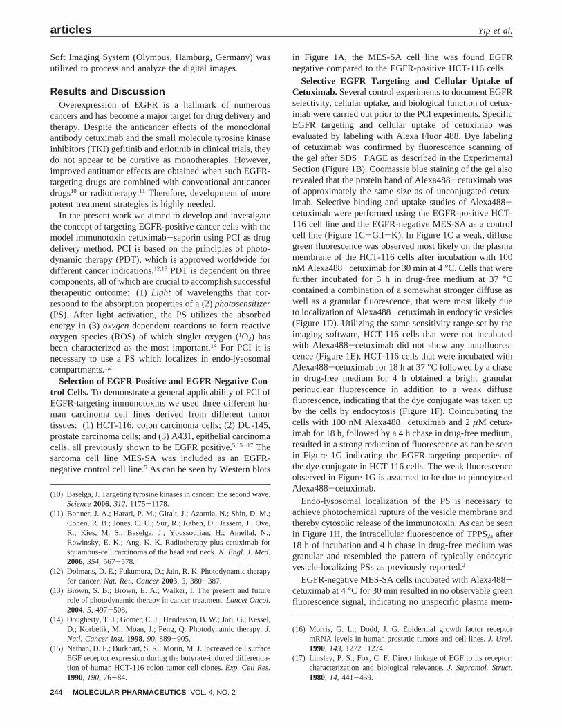
in Figure 1A, the MES-SA cell line was found EGFRnegative compared to the EGFR-positive HCT-116 cells.
Selective EGFR Targeting and Cellular Uptake ofCetuximab. Several control experiments to document EGFRselectivity, cellular uptake, and biological function of cetux-imab were carried out prior to the PCI experiments. SpecificEGFR targeting and cellular uptake of cetuximab wasevaluated by labeling with Alexa Fluor 488. Dye labelingof cetuximab was confirmed by fluorescence scanning ofthe gel after SDS-PAGE as described in the ExperimentalSection (Figure 1B). Coomassie blue staining of the gel alsorevealed that the protein band of Alexa488-cetuximab wasof approximately the same size as of unconjugated cetux-imab. Selective binding and uptake studies of Alexa488-cetuximab were performed using the EGFR-positive HCT-116 cell line and the EGFR-negative MES-SA as a controlcell line (Figure 1C-G,I-K). In Figure 1C a weak, diffusegreen fluorescence was observed most likely on the plasmamembrane of the HCT-116 cells after incubation with 100nM Alexa488-cetuximab for 30 min at 4°C. Cells that werefurther incubated for 3 h in drug-free medium at 37°Ccontained a combination of a somewhat stronger diffuse aswell as a granular fluorescence, that were most likely dueto localization of Alexa488-cetuximab in endocytic vesicles(Figure 1D). Utilizing the same sensitivity range set by theimaging software, HCT-116 cells that were not incubatedwith Alexa488-cetuximab did not show any autofluores-cence (Figure 1E). HCT-116 cells that were incubated withAlexa488-cetuximab for 18 h at 37°C followed by a chasein drug-free medium for 4 h obtained a bright granularperinuclear fluorescence in addition to a weak diffusefluorescence, indicating that the dye conjugate was taken upby the cells by endocytosis (Figure 1F). Coincubating thecells with 100 nM Alexa488-cetuximab and 2µM cetux-imab for 18 h, followed by a 4 hchase in drug-free medium,resulted in a strong reduction of fluorescence as can be seenin Figure 1G indicating the EGFR-targeting properties ofthe dye conjugate in HCT 116 cells. The weak fluorescenceobserved in Figure 1G is assumed to be due to pinocytosedAlexa488-cetuximab.
Endo-lysosomal localization of the PS is necessary toachieve photochemical rupture of the vesicle membrane andthereby cytosolic release of the immunotoxin. As can be seenin Figure 1H, the intracellular fluorescence of TPPS2a after18 h of incubation and 4 h chase in drug-free medium wasgranular and resembled the pattern of typically endocyticvesicle-localizing PSs as previously reported.2
EGFR-negative MES-SA cells incubated with Alexa488-cetuximab at 4°C for 30 min resulted in no observable greenfluorescence signal, indicating no unspecific plasma mem-
(10) Baselga, J. Targeting tyrosine kinases in cancer: the second wave.Science2006, 312, 1175-1178.
(11) Bonner, J. A.; Harari, P. M.; Giralt, J.; Azarnia, N.; Shin, D. M.;Cohen, R. B.; Jones, C. U.; Sur, R.; Raben, D.; Jassem, J.; Ove,R.; Kies, M. S.; Baselga, J.; Youssoufian, H.; Amellal, N.;Rowinsky, E. K.; Ang, K. K. Radiotherapy plus cetuximab forsquamous-cell carcinoma of the head and neck.N. Engl. J. Med.2006, 354, 567-578.
(12) Dolmans, D. E.; Fukumura, D.; Jain, R. K. Photodynamic therapyfor cancer.Nat. ReV. Cancer2003, 3, 380-387.
(13) Brown, S. B.; Brown, E. A.; Walker, I. The present and futurerole of photodynamic therapy in cancer treatment.Lancet Oncol.2004, 5, 497-508.
(14) Dougherty, T. J.; Gomer, C. J.; Henderson, B. W.; Jori, G.; Kessel,D.; Korbelik, M.; Moan, J.; Peng, Q. Photodynamic therapy.J.Natl. Cancer Inst.1998, 90, 889-905.
(15) Nathan, D. F.; Burkhart, S. R.; Morin, M. J. Increased cell surfaceEGF receptor expression during the butyrate-induced differentia-tion of human HCT-116 colon tumor cell clones.Exp. Cell Res.1990, 190, 76-84.
(16) Morris, G. L.; Dodd, J. G. Epidermal growth factor receptormRNA levels in human prostatic tumors and cell lines.J. Urol.1990, 143, 1272-1274.
(17) Linsley, P. S.; Fox, C. F. Direct linkage of EGF to its receptor:characterization and biological relevance.J. Supramol. Struct.1980, 14, 441-459.
articles Yip et al.
244 MOLECULAR PHARMACEUTICS VOL. 4, NO. 2
brane binding of cetuximab (Figure 1I). MES-SA cellsincubated with Alexa488-cetuximab for 18 h, followed by4 h chase in drug-free medium, showed only a weak granularfluorescence (Figure 1J). The same fluorescence pattern andintensity were observed in MES-SA cells coincubated withAlexa488-cetuximab and 20-fold excess of cetuximab (2µM) (Figure 1K), indicating that the weak granular fluores-cence signal is due to non-receptor mediated endocytosis.In addition, an Alexa488 labeled goat-anti-mouse mAb wasincluded as a non EGFR-targeting control mAb in the HCT-116 cells. There were no differences in the fluorescenceintensity or fluorescence pattern between HCT-116 cellsincubated with Alexa488 labeled goat-anti-mouse mAb and
Alexa488-cetuximab and 20-fold excess of cetuximab (datanot shown). The fluorescence pattern of Alexa488 labeledgoat-anti-mouse mAb in HCT-116 cells was weak andgranular resembling the fluorescence of Alexa488-cetux-imab in MES-SA cells, also indicating that mAbs withouttargeting capabilities are taken up by unspecific endocytosis.
The observations described above demonstrate that cetux-imab selectively binds to EGFR and is taken up by receptor-mediated endocytosis in HCT 116 cells. Additionally, theresults indicate that cetuximab to a lower extent is taken upby unspecific endocytosis. Unspecific uptake of cetuximab-saporin in normal cells could therefore be a potentiallimitation causing unspecific toxicity. However, it should be
Figure 1. Characterization of cellular binding, uptake, and effect of cetuximab. (A) EGFR expression of HCT-116 and MES-SAcells measured by antibody staining of Western blots. Ponceu staining of membranes confirmed even protein loading (not shown).(B) SDS-PAGE of Alexa488-cetuximab. Quality of labeling was confirmed by using both fluorescence scanning and Coomassieblue staining. (C-H) Fluorescence microscopy of HCT-116 incubated with 100 nM Alexa488-cetuximab. The cells were analyzedafter 30 min on ice (C); 30 min on ice, then 3 h chase in drug-free medium for at 37 °C (D); no treatment (E); 18 h incubationat 37 °C, then 4 h chase in drug-free medium at 37 °C (F) or as in F but Alexa488-cetuximab coincubated with 2 µM cetuximab(G). In panel H, HCT-116 cells were incubated with 1 µg/mL TPPS2a for 18 h, then 4 h chase in drug-free medium at 37 °C.(I-L) MES-SA cells were incubated with 100 nM Alexa488-cetuximab after 30 min on ice (I), with Alexa488-cetuximab for 18h followed by a 4 h chase at 37 °C (J), or as in panel J, but coincubated with 2 µM cetuximab (K). (L) The cells were incubatedwith 1 µg/mL TPPS2a for 18 h followed by a 4 h chase in drug-free medium at 37 °C. (M) Influence of cetuximab and TKI on EGFinduced Tyr1068 phosphorylation of EGFR in HCT-116 cells. One hundred ng/mL EGF was used to activate EGFR when indicatedin the figure. The cells were incubated with 100 nM cetuximab for the indicated time and subsequently isolated for analysis orchased in drug-free medium for 4 h before analysis ((18h)*). As a control, 1 h incubation of 20 µM tyrphostin AG1478 was usedto block EGF-induced activation of EGFR (lane 6).
DeliVery of Cetuximab-Saporin in Cancer Cells by PCI articles
VOL. 4, NO. 2 MOLECULAR PHARMACEUTICS 245
noted that PCI causes additional specificity by the preferentialaccumulation of PS in the neoplastic lesions14 as well as thesite-specific light exposure which is required to stimulatePS-mediated IT activation. PCI may therefore reduce thedosage of cancer therapeutic drugs needed.
Inhibition of EGFR Phosphorylation by Cetuximab.Before construction of the immunotoxin we evaluated theability of cetuximab to antagonize EGF-stimulated activationof EGFR in the HCT-116 cells. As can be seen in Figure1M, the phosphorylation of Tyr1068 of EGFR by 100 ng/mL (16.7 nM) exogenous EGF is completely inhibited by100 nM cetuximab administered 2 h or 18 hprior to theEGF treatment. After removal of cetuximab followed by 4h incubation in drug-free medium, a weak reactivation ofEGFR by exogenous EGF was observed. As a control weused tyrphostine AG1478, a selective EGFR TKI, to inhibitphosphorylation of EGFR (p-EGFR). It was found that a 1h incubation with 20µM tyrphostine completely blockedEGF-mediated phosphorylation of EGFR. The Western blotdata confirmed that cetuximab exerts EGFR-targeting prop-erties in the HCT-116 cells.
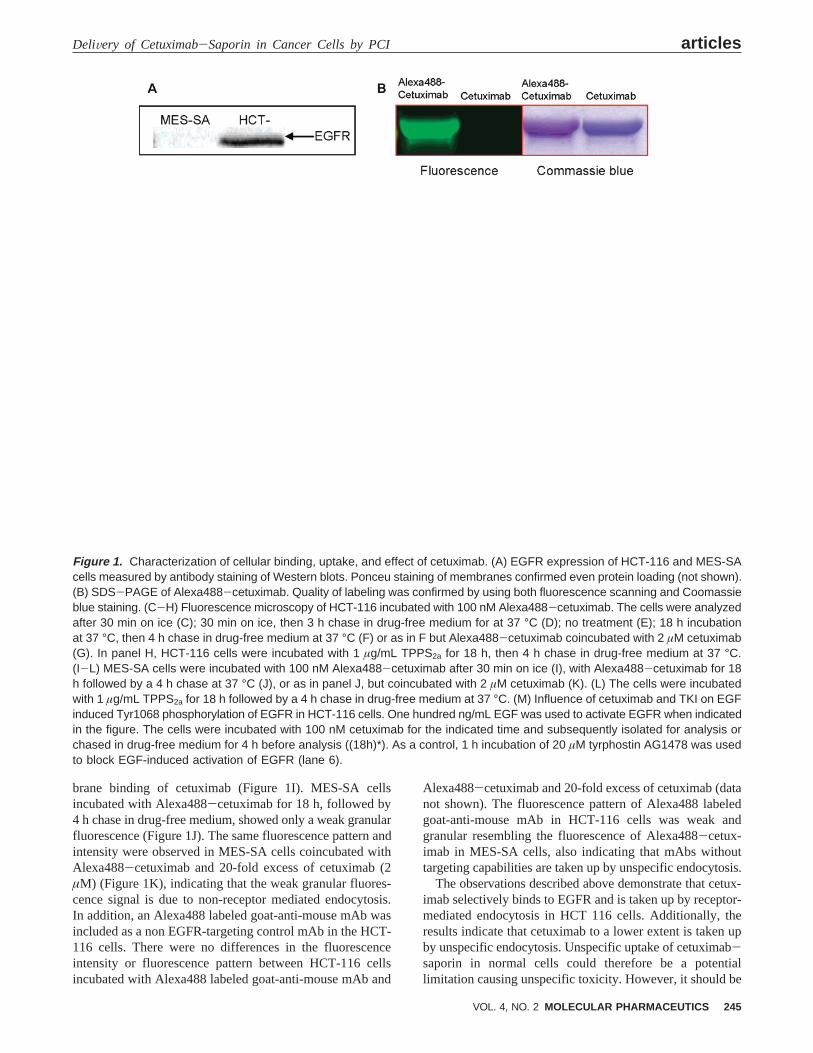
PDT in Combination with Cetuximab. The cytotoxicityinduced by PDT (PS+ light) in HCT-116 cells is shown inFigure 2A. LD50 was achieved with∼100 s of light exposureafter 18 h of TPPS2a incubation and 4 h of chase in drug-free medium, and was selected as the light dose for treatmentof HCT-116 cells if not otherwise described. The cellularcytotoxicity of nonconjugated cetuximab incubated for 72 hwas evaluated, either as monotherapy or in combination withPDT. As can be seen in Figure 2B, only subtoxic responseswere achieved when treating HCT-116 cells for 72 h withup to 100 nM of cetuximab alone, and only∼10% decreasein viability was observed at the highest concentration ofcetuximab. This is in accordance with recent findingsreporting that cetuximab as monotherapy of HCT-116 cellsresults in poor growth inhibition controlin Vitro.18 Inaddition, cetuximab alone failed to show any antitumoractivity in 7 out of 11 xenografts with EGFR-positive tumorcells, including HCT-116 xenografts.19
TPPS2a-PDT was combined with cetuximab alone to mimica PCI protocol. Thus, after 18 h coincubation of TPPS2a andcetuximab, the cells were washed and chased for 4 h indrug-free medium before exposure to light. In these experimentsno enhanced cytotoxicity was observed with increasing dosesof cetuximab (Figure 2B). It should be noted, as shown in
Figure 1M, that EGF activates EGFR of HCT-116 cells 4 hafter cetuximab is removed from the medium. Thus, for localcontrol of tumor cells, it is important to inactivate all cellsafter a single treatment with PDT combined with cetuximabto prevent reactivation of EGFR and regrowth of malignantcells; otherwise repeated treatments are necessary.
Interestingly, del Carmen and co-workers have demon-strated the rational use of a combination of PDT and repeated
(18) Balin-Gauthier, D.; Delord, J. P.; Rochaix, P.; Mallard, V.;Thomas, F.; Hennebelle, I.; Bugat, R.; Canal, P.; Allal, C. In vivoand in vitro antitumor activity of oxaliplatin in combination withcetuximab in human colorectal tumor cell lines expressingdifferent level of EGFR.Cancer Chemother. Pharmacol.2006,57, 709-718.
(19) Wild, R.; Fager, K.; Flefleh, C.; Kan, D.; Inigo, I.; Castaneda,S.; Luo, F. R.; Camuso, A.; McGlinchey, K.; Rose, W. C.Cetuximab preclinical antitumor activity (monotherapy and com-bination based) is not predicted by relative total or activatedepidermal growth factor receptor tumor expression levels.Mol.Cancer Ther.2006, 5, 104-113.
Figure 2. Cytotoxic effects of PDT and PCI of saporin.Relative viability of HCT-116 cells treated with (A) PDT(TPPS2a + increasing light doses), (B) PDT with a fixed lightdose (100 s) and increasing concentrations of cetuximab orcetuximab alone, and (C) PCI of saporin. The cells weretreated with PDT with a fixed light dose (100 s) and increasingconcentrations of saporin or saporin without light exposure.The cells were treated with 0.2 µg/mL TPPS2a and asotherwise described in the Experimental Section. Results arethe average of three samples from at least two independentexperiments. Cell viability was assessed by the MTT assay48 h post light exposure, and the values were normalizedrelative to untreated cells. Bars are standard error (SE).
articles Yip et al.
246 MOLECULAR PHARMACEUTICS VOL. 4, NO. 2
injections of cetuximab, using the PS benzoporphyrin deriva-tive monoacid A (BPD-MA), which induced a synergisticgrowth inhibition of advanced and recurrent epithelial ovariancancer cellsin ViVo.20 However, in that study cetuximab wasdelivered 4 times with several days between the injections.Recently, it was demonstrated in a multinational, randomizedstudy that radiotherapy combined with cetuximab enhancedlocoregional control and reduced mortality of patients withadvanced head and neck cancer,11 demonstrating the strengthof combination treatments.
The effect of saporin alone and PCI of saporin wasevaluated in the HCT-116 cells. It has previously been shownthat PCI of saporin induces synergistic cytotoxicityin Vitro.1
Accordingly, it was found in the HCT 116 cells that acombination of a photochemical treatment and saporin (30kDa), both inducing a 50% reduction in viability when givenseparately, caused 95% reduction of cell viability when giventogether (Figure 2C).
PCI of Cetuximab-Saporin: Cytotoxic Effects inEGFR-Positive Cells.In view of the fact that saporin alone
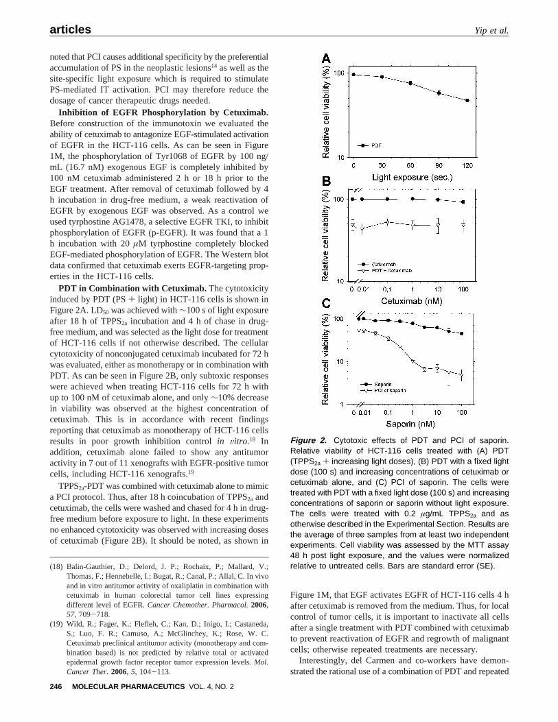
also has a low cytotoxic potential toward whole cells andanimals,5,7 cetuximab was linked to saporin to improve thecellular uptake and specificity. In addition, we used PCI ofcetuximab-saporin to increase the cytosolic delivery of thetoxin. The IT was formed on the basis of a streptavidin-biotin linkage as described in the Experimental Section. Todemonstrate a general applicability of the present IT-basedEGFR-targeting strategy, PCI of cetuximab-saporin wasperformed in three different EGFR-positive cancer cell linesof different tissue origin. The toxicity of streptavidin-saporinand cetuximab-saporin in combination with TPPS2a insubdued light was evaluated in the HCT-116 cells (Figure3A). The cytotoxicity of cetuximab-saporin (300 pM) inHCT-116 cells was not influenced by the presence of TPPS2a
in the absence of light exposure (P > 0.05). The cytotoxiceffect of the IT cetuximab-saporin was reduced by a 20-fold excess of cetuximab, demonstrating the specificity ofthe IT. Only a subtoxic response was achieved afterincubating the HCT-116 cells with 300 pM streptavidin-saporin. The same pattern of cytotoxicity from these com-pounds as seen in the HCT-116 cells was also observed inthe DU-145 and A431 cell lines (data not shown). Theenhanced cytotoxicity of cetuximab-saporin as comparedto streptavidin-saporin indicates increased uptake and re-localization of saporin to the cytosol of the cells.
(20) del Carmen, M. G.; Rizvi, I.; Chang, Y.; Moor, A. C.; Oliva, E.;Sherwood, M.; Pogue, B.; Hasan, T. Synergism of epidermalgrowth factor receptor-targeted immunotherapy with photody-namic treatment of ovarian cancer in vivo.J. Natl. Cancer Inst.2005, 97, 1516-1524.
Figure 3. Cytotoxic effects of PCI of cetuximab-saporin in HCT-116 cells. (A) Toxin conjugates incubated with or withoutTPPS2a or cetuximab in the absence of light exposure as indicated in the figure. (B) PCI of toxin conjugates at differentconcentrations and a PDT dose demonstrated to kill ∼45% of the cells. (C) Light dose-dependent PDT, PCI of 3 pM cetuximab-saporin, 3 pM cetuximab-saporin only, or PDT combined with 3 pM cetuximab. (D) Relative clonal cell survival of HCT-116cells treated with PDT at different light doses, PCI of 3 pM streptavidin-saporin, PCI of 3 pM cetuximab-saporin, or PCI of 3pM cetuximab-saporin combination in the presence of 6 nM cetuximab. Results are the average of three samples from at leasttwo independent experiments. Cytotoxicity as assessed by the MTT assay 48 h post light exposure or by the clonal cell survivalassay assessed 10-14 days post light exposure. Values were normalized to nontreated cells; bars, SE.
DeliVery of Cetuximab-Saporin in Cancer Cells by PCI articles
VOL. 4, NO. 2 MOLECULAR PHARMACEUTICS 247
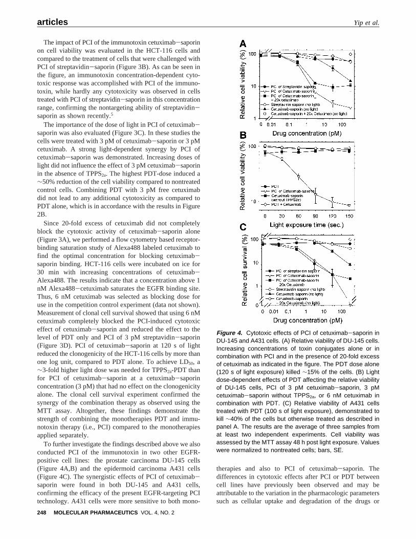
The impact of PCI of the immunotoxin cetuximab-saporinon cell viability was evaluated in the HCT-116 cells andcompared to the treatment of cells that were challenged withPCI of streptavidin-saporin (Figure 3B). As can be seen inthe figure, an immunotoxin concentration-dependent cyto-toxic response was accomplished with PCI of the immuno-toxin, while hardly any cytotoxicity was observed in cellstreated with PCI of streptavidin-saporin in this concentrationrange, confirming the nontargeting ability of streptavidin-saporin as shown recently.5
The importance of the dose of light in PCI of cetuximab-saporin was also evaluated (Figure 3C). In these studies thecells were treated with 3 pM of cetuximab-saporin or 3 pMcetuximab. A strong light-dependent synergy by PCI ofcetuximab-saporin was demonstrated. Increasing doses oflight did not influence the effect of 3 pM cetuximab-saporinin the absence of TPPS2a. The highest PDT-dose induced a∼50% reduction of the cell viability compared to nontreatedcontrol cells. Combining PDT with 3 pM free cetuximabdid not lead to any additional cytotoxicity as compared toPDT alone, which is in accordance with the results in Figure2B.
Since 20-fold excess of cetuximab did not completelyblock the cytotoxic activity of cetuximab-saporin alone(Figure 3A), we performed a flow cytometry based receptor-binding saturation study of Alexa488 labeled cetuximab tofind the optimal concentration for blocking cetuximab-saporin binding. HCT-116 cells were incubated on ice for30 min with increasing concentrations of cetuximab-Alexa488. The results indicate that a concentration above 1nM Alexa488-cetuximab saturates the EGFR binding site.Thus, 6 nM cetuximab was selected as blocking dose foruse in the competition control experiment (data not shown).Measurement of clonal cell survival showed that using 6 nMcetuximab completely blocked the PCI-induced cytotoxiceffect of cetuximab-saporin and reduced the effect to thelevel of PDT only and PCI of 3 pM streptavidin-saporin(Figure 3D). PCI of cetuximab-saporin at 120 s of lightreduced the clonogenicity of the HCT-116 cells by more thanone log unit, compared to PDT alone. To achieve LD50, a∼3-fold higher light dose was needed for TPPS2a-PDT thanfor PCI of cetuximab-saporin at a cetuximab-saporinconcentration (3 pM) that had no effect on the clonogenicityalone. The clonal cell survival experiment confirmed thesynergy of the combination therapy as observed using theMTT assay. Altogether, these findings demonstrate thestrength of combining the monotherapies PDT and immu-notoxin therapy (i.e., PCI) compared to the monotherapiesapplied separately.
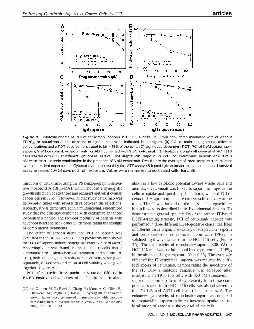
To further investigate the findings described above we alsoconducted PCI of the immunotoxin in two other EGFR-positive cell lines: the prostate carcinoma DU-145 cells(Figure 4A,B) and the epidermoid carcinoma A431 cells(Figure 4C). The synergistic effects of PCI of cetuximab-saporin were found in both DU-145 and A431 cells,confirming the efficacy of the present EGFR-targeting PCItechnology. A431 cells were more sensitive to both mono-
therapies and also to PCI of cetuximab-saporin. Thedifferences in cytotoxic effects after PCI or PDT betweencell lines have previously been observed and may beattributable to the variation in the pharmacologic parameterssuch as cellular uptake and degradation of the drugs or
Figure 4. Cytotoxic effects of PCI of cetuximab-saporin inDU-145 and A431 cells. (A) Relative viability of DU-145 cells.Increasing concentrations of toxin conjugates alone or incombination with PCI and in the presence of 20-fold excessof cetuximab as indicated in the figure. The PDT dose alone(120 s of light exposure) killed ∼15% of the cells. (B) Lightdose-dependent effects of PDT affecting the relative viabilityof DU-145 cells, PCI of 3 pM cetuximab-saporin, 3 pMcetuximab-saporin without TPPS2a, or 6 nM cetuximab incombination with PDT. (C) Relative viability of A431 cellstreated with PDT (100 s of light exposure), demonstrated tokill ∼40% of the cells but otherwise treated as described inpanel A. The results are the average of three samples fromat least two independent experiments. Cell viability wasassessed by the MTT assay 48 h post light exposure. Valueswere normalized to nontreated cells; bars, SE.
articles Yip et al.
248 MOLECULAR PHARMACEUTICS VOL. 4, NO. 2
differences in the cell survival and cell death signaling.Altogether, these results confirm the targeting concept of PCIthat has previously been shown.5,21,22
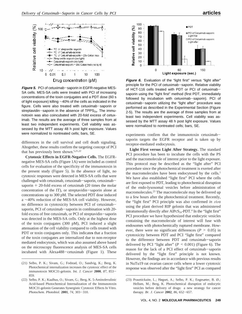
Cytotoxic Effects in EGFR-Negative Cells.The EGFR-negative MES-SA cells (Figure 1A) were included as controlcells for evaluation of the specificity of the immunotoxin inthe present study (Figure 5). In the absence of light, nocytotoxic responses were detected in MES-SA cells that werechallenged with cetuximab, cetuximab-saporin, cetuximab-saporin+ 20-fold excess of cetuximab (20 times the molarconcentration of the IT), or streptavidin-saporin alone atconcentrations up to 300 pM. The selected PDT dose induceda ∼40% reduction of the MES-SA cell viability. However,no difference in cytotoxicity between PCI of cetuximab-saporin, PCI of cetuximab-saporin in combination with 20-fold excess of free cetuximab, or PCI of streptavidin-saporinwas detected in the MES-SA cells. Only at the highest doseof the toxin conjugates (300 pM), PCI induced a slightattenuation of the cell viability compared to cells treated withPDT or toxin conjugates only. This indicates that a fractionof the toxin conjugates are internalized due to non-receptormediated endocytosis, which was also assumed above basedon the microscopy fluorescence analysis of MES-SA cellsincubated with Alexa488-cetuximab (Figure 1). These
experiments confirm that the immunotoxin cetuximab-saporin targets the EGFR receptor and is taken up byreceptor-mediated endocytosis.
Light First versus Light After Strategy. The standardPCI procedure has been to incubate the cells with the PSand the macromolecule of interest prior to the light exposure.This protocol may be described as the “light after” PCIprocedure since the photochemical treatment is exerted afterthe macromolecules have been endocytosed by the cells.1
We have also established “light first” PCI where the cellsare first exposed to PDT, leading to photochemical disruptionof the endo-lysosomal vesicles before administration ofmacromolecules.23 The macromolecule may be delivered upto a few hours after the photochemical treatment. Recently,the “light first” PCI principle was also confirmedin ViVousing the plant derived RIP gelonin that was administeredintratumorally directly after AlPcS2a-PDT.4 In the “light first”PCI procedure we have hypothesized that endocytic vesiclescontaining the macromolecule of interest will fuse withendosomes with photochemically ruptured membrane. How-ever, there were no significant differences (P > 0.05) incytotoxicity between PDT and PCI “light first” comparedto the difference between PDT and cetuximab-saporindelivered by PCI “light after” (P < 0.001) (Figure 6). Thereason for the lack of a PCI effect of cetuximab-saporindelivered by the “light first” principle is not known.However, the findings are in accordance with previous resultsin NuTu19 rat ovarian cancer cells where a lower cytotoxicresponse was observed after the “light first” PCI as compared
(21) Selbo, P. K.; Sivam, G.; Fodstad, O.; Sandvig, K.; Berg, K.Photochemical internalization increases the cytotoxic effect of theimmunotoxin MOC31-gelonin.Int. J. Cancer2000, 87, 853-859.
(22) Selbo, P. K.; Kaalhus, O.; Sivam, G.; Berg, K. 5-AminolevulinicAcid-based Photochemical Internalization of the ImmunotoxinMOC31-gelonin Generates Synergistic Cytotoxic Effects In Vitro.Photochem. Photobiol.2001, 74, 303-310.
(23) Prasmickaite, L.; Høgset, A.; Selbo, P. K.; Engesæter, B. Ø.;Hellum, M.; Berg, K. Photochemical disruption of endocyticvesicles before delivery of drugs: a new strategy for cancertherapy.Br. J. Cancer2002, 86, 652-657.
Figure 5. PCI of cetuximab-saporin in EGFR-negative MES-SA cells. MES-SA cells were treated with PCI of increasingconcentrations of the toxin conjugates and a PDT dose (60 sof light exposure) killing ∼40% of the cells as indicated in thefigure. Cells were also treated with cetuximab-saporin orstreptavidin-saporin in the absence of TPPS2a. The immu-notoxin was also coincubated with 20-fold excess of cetux-imab. The results are the average of three samples from atleast two independent experiments. Cell viability was as-sessed by the MTT assay 48 h post light exposure. Valueswere normalized to nontreated cells; bars, SE.
Figure 6. Evaluation of the “light first” versus “light after”principle for the PCI of cetuximab-saporin. Relative viabilityof HCT-116 cells treated with PDT or PCI of cetuximab-saporin using the “light first” method (first PDT, immediatelyfollowed by incubation with cetuximab-saporin). PCI ofcetuximab-saporin utilizing the “light after” procedure wasperformed as described in the Experimental Section (Figure2-5). The results are the average of three samples from atleast two independent experiments. Cell viability was as-sessed by the MTT assay 48 h post light exposure. Valueswere normalized to nontreated cells; bars, SE.
DeliVery of Cetuximab-Saporin in Cancer Cells by PCI articles
VOL. 4, NO. 2 MOLECULAR PHARMACEUTICS 249
to “light after” PCI for the delivery of EGF-saporin.5 Studiesin progress in our laboratory accordingly show reduced EGF-mediated EGFR phosphorylation after TPPS2a-PDT in Nu-Tu19 cells (data not published). The present results maytherefore be explained by a PDT-induced damage of the EGFreceptor, which may result in a reduced EGFR-mediatedendocytosis. Photochemical damage of EGFR utilizinganother PS has previously been describedin Vitro and inViVo.24
Potential in ViWo Use of PCI of Cetuximab-Saporin.The streptavidin-biotin linkage used in the present reporthas been shown to be a simple design for evaluation of newtargeting moieties for the treatment of cancer. The results inthe present study and in a recent report utilizing EGF-saporin5 document that the specificity is maintained. How-ever, the immunoconjugate cetuximab-saporin is relativelylarge (>270 kDa) and, due to diffusion limitations in thetissue, may be regarded as unsuitable for treatment of solidtumors. Therefore, further work should focus on synthesizingimmunotoxins with smaller size, e.g., using a disulfide bridgebetween mAb and toxin or making a recombinant constructwhere the Fv fragment of a growth factor-targeting mAb isfused with a protein toxin. The use of EGF as a targetingligand may be beneficial due to its small size and relativelylow affinity to EGFR, both of which may stimulate tissuepenetration. In addition, EGF stimulates endocytosis of theligand-toxin complex EGF-saporin. On the other hand, therelatively low affinity of EGF for EGFR indicates a require-ment for higher doses of the ligand-toxin complex thanwhen using a high affinity ligand. In addition, EGF is well-known for its stimulatory activity on cell growth, survival,metastasis, and angiogenesis.25 In comparison, cetuximabbinds strongly to EGFR and acts both by inhibiting intrac-ellular signaling and through antibody-dependent cell cyto-toxicity. In addition, it is an approved and much used mAbin cancer therapy.
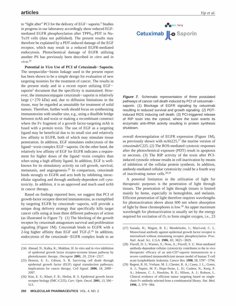
Based on findings reported here, we suggest that PCI ofgrowth-factor receptor directed immunotoxins, as exemplifiedby targeting EGFR by cetuximab-saporin, will provide aunique drug delivery strategy that specifically kills targetcancer cells using at least three different pathways of action(as illustrated in Figure 7): (1) The blocking of the growthreceptor by cetuximab antagonizes survival and proliferationsignaling (Figure 1M). Cetuximab binds to EGFR with a2-log higher affinity than EGF and TGF-â.26 In addition,endocytosis of the cetuximab-EGFR complex leads to an
overall downregulation of EGFR expression (Figure 1M),as previously shown with mAb225,27 the murine version ofcetuximab/C225. (2) The ROS-mediated cytotoxic responsesafter the photochemical exposure (PDT) result in apoptosisor necrosis. (3) The RIP activity of the toxin after PCI-induced cytosolic release results in cell inactivation by meansof inhibition of the cellular protein synthesis. In addition,antibody-mediated cellular cytotoxicity could be a fourth wayof inactivating tumor cells.28,29
A potential limitation in the utilization of light fortherapeutic purposes is the penetration of light throughtissues. The penetration of light through tissues is limitedmainly by heme, especially in hemoglobin, and melanin.Efficient penetration of light therefore requires wavelengthsfor photoactivation above about 600 nm where absorptionof light by these chromophores is low.30 An upper maximumwavelength for photoactivation is usually set by the energyrequired for excitation of O2 to form singlet oxygen, i.e., 23
(24) Ahmad, N.; Kalka, K.; Mukhtar, H. In vitro and in vivo inhibitionof epidermal growth factor receptor-tyrosine kinase pathway byphotodynamic therapy.Oncogene2001, 20, 2314-2317.
(25) Henson, E. S.; Gibson, S. B. Surviving cell death throughepidermal growth factor (EGF) signal transduction pathways:Implications for cancer therapy.Cell Signal.2006, 18, 2089-2097.
(26) Kim, E. S.; Khuri, F. R.; Herbst, R. S. Epidermal growth factorreceptor biology (IMC-C225).Curr. Opin. Oncol.2001, 13, 506-513.
(27) Sunada, H.; Magun, B. E.; Mendelsohn, J.; MacLeod, C. L.Monoclonal antibody against epidermal growth factor receptor isinternalized without stimulating receptor phosphorylation.Proc.Natl. Acad. Sci. U.S.A.1986, 83, 3825-3829.
(28) Flavell, D. J.; Warnes, S.; Noss, A.; Flavell, S. U. Host-mediatedantibody-dependent cellular cytotoxicity contributes to the in vivotherapeutic efficacy of an anti-CD7-saporin immunotoxin in asevere combined immunodeficient mouse model of human T-cellacute lymphoblastic leukemia.Cancer Res.1998, 58, 5787-5794.
(29) Begent, R. H.; Verhaar, M. J.; Chester, K. A.; Casey, J. L.; Green,A. J.; Napier, M. P.; Hope-Stone, L. D.; Cushen, N.; Keep, P.A.; Johnson, C. J.; Hawkins, R. E.; Hilson, A. J.; Robson, L.Clinical evidence of efficient tumor targeting based on single-chain Fv antibody selected from a combinatorial library.Nat. Med.1996, 2, 979-984.
Figure 7. Schematic representation of three postulatedpathways of cancer cell death induced by PCI of cetuximab-saporin. (1) Blockage of EGFR signaling by cetuximabresulting in reduced survival and growth signaling. (2) PDT-induced ROS inducing cell death. (3) PCI-triggered releaseof RIP toxin into the cytosol, where the toxin exerts itsenzymatic anti-rRNA activity resulting in protein synthesisshutdown.
articles Yip et al.
250 MOLECULAR PHARMACEUTICS VOL. 4, NO. 2
kcal/mol, which is equivalent to the excess energy of thelong-lived form of singlet oxygen. This requires that thephotosensitizer must be excited at wavelengths below about850 nm. The therapeutic wavelength window is thereforeusually defined as 600-800 nm, but shorter wavelengths maybe used when thin lesions are to be treated. The treatmentof internal organs is generally of no limitations any moredue to the development of light sources and light applicatorsin PDT. Necrosis has been reported up to 3 cm into thetreated tissue.31
ConclusionsThe main goal of the present report was to demonstrate a
proof-of-principle concept using PCI of an EGFR-targeting
immunotoxin. We have demonstrated in three differentEGFR-positive cell lines, and in EGFR-negative control cells,derived from different cancer tissues that PCI of theimmunotoxin cetuximab-saporin is a potent and specificanticancer cell therapy. Based on the present study wepropose that our PCI concept introduces a unique triplecytotoxicity by (1) cetuximab, (2) PDT, and (3) the photo-chemically internalized saporin. The efficacy and multipleselectivity of the PCI drug delivery concept documented inthis report warrant further preclinical investigations.
Acknowledgment. We thank the Norwegian CancerSociety (Grant C 96138/007) and the Norwegian RadiumHospital Research Foundation for financial support.
Abbreviations UsedEGFR, epidermal growth factor receptor; IT, immuno-
toxin; MTT, 3-(4,5-dimethylthiazol-2-yl)-2,5-diphenyltetra-zolium bromide; PCI, photochemical internalization; PDT,photodynamic therapy; PS, photosensitizer; RIP, ribosome-inactivating protein; ROS, reactive oxygen species; TKI,tyrosine kinase inhibitor; TPPS2a, meso-tetraphenylporphinewith two sulfonate groups on adjacent phenyl rings.
MP060105U
(30) Berg, K.; Selbo, P. K.; Weyergang, A.; Dietze, A.; Prasmickaite,L.; Bonsted, A.; Engesaeter, B. O.; Angell-Petersen, E.; Warloe,T.; Frandsen, N.; Hogset, A. Porphyrin-related photosensitizersfor cancer imaging and therapeutic applications.J. Microsc.2005,218, 133-147.
(31) Chen, Q.; Huang, Z.; Luck, D.; Beckers, J.; Brun, P. H.; Wilson,B. C.; Scherz, A.; Salomon, Y.; Hetzel, F. W. Preclinical studiesin normal canine prostate of a novel palladium-bacteriopheophor-bide (WST09) photosensitizer for photodynamic therapy ofprostate cancers.Photochem. Photobiol.2002, 76, 438-445.
DeliVery of Cetuximab-Saporin in Cancer Cells by PCI articles
VOL. 4, NO. 2 MOLECULAR PHARMACEUTICS 251