Nanobiopolymer for Direct Targeting and Inhibition of EGFR Expression in Triple Negative Breast...
9
Nanobiopolymer for Direct Targeting and Inhibition of EGFR Expression in Triple Negative Breast Cancer Satoshi Inoue 1 , Rameshwar Patil 1 , Jose Portilla-Arias 1 , Hui Ding 1 , Bindu Konda 1 , Andres Espinoza 1 , Dmitriy Mongayt 2 , Janet L. Markman 1 , Adam Elramsisy 1 , H. Westley Phillips 1 , Keith L. Black 1 , Eggehard Holler 1 , Julia Y. Ljubimova 1 * 1 Department of Neurosurgery, Cedars-Sinai Medical Center, Los Angeles, California, United States of America, 2 Center for Pharmaceutical Biotechnology and Nanomedicine, Northeastern University, Boston, Massachusetts, United States of America Abstract Treatment options for triple negative breast cancer (TNBC) are generally limited to cytotoxic chemotherapy. Recently, anti- epidermal growth factor receptor (EGFR) therapy has been introduced for TNBC patients. We engineered a novel nanobioconjugate based on a poly(b-L-malic acid) (PMLA) nanoplatform for TNBC treatment. The nanobioconjugate carries anti-tumor nucleosome-specific monoclonal antibody (mAb) 2C5 to target breast cancer cells, anti-mouse transferrin receptor (TfR) antibody for drug delivery through the host endothelial system, and Morpholino antisense oligonucleotide (AON) to inhibit EGFR synthesis. The nanobioconjugates variants were: (1) P (BioPolymer) with AON, 2C5 and anti-TfR for tumor endothelial and cancer cell targeting, and EGFR suppression (P/AON/2C5/TfR), and (2) P with AON and 2C5 (P/AON/ 2C5). Controls included (3) P with 2C5 but without AON (P/2C5), (4) PBS, and (5) P with PEG and leucine ester (LOEt) for endosomal escape (P/mPEG/LOEt). Drugs were injected intravenously to MDA-MB-468 TNBC bearing mice. Tissue accumulation of injected nanobioconjugates labeled with Alexa Fluor 680 was examined by Xenogen IVIS 200 (live imaging) and confocal microscopy of tissue sections. Levels of EGFR, phosphorylated and total Akt in tumor samples were detected by western blotting. In vitro western blot showed that the leading nanobioconjugate P/AON/2C5/TfR inhibited EGFR synthesis significantly better than naked AON. In vivo imaging revealed that 2C5 increased drug-tumor accumulation. Significant tumor growth inhibition was observed in mice treated with the lead nanobioconjugate (1) [P = 0.03 vs. controls; P,0.05 vs. nanobioconjugate variant (2)]. Lead nanobioconjugate (1) also showed stronger inhibition of EGFR expression and Akt phosphorylation than other treatments. Treatment of TNBC with the new nanobioconjugate results in tumor growth arrest by inhibiting EGFR and its downstream signaling intermediate, phosphorylated Akt. The nanobioconjugate represents a new generation of nanodrugs for treatment of TNBC. Citation: Inoue S, Patil R, Portilla-Arias J, Ding H, Konda B, et al. (2012) Nanobiopolymer for Direct Targeting and Inhibition of EGFR Expression in Triple Negative Breast Cancer. PLoS ONE 7(2): e31070. doi:10.1371/journal.pone.0031070 Editor: Maria G. Castro, University of Michigan School of Medicine, United States of America Received July 28, 2011; Accepted January 2, 2012; Published February 15, 2012 Copyright: ß 2012 Inoue et al. This is an open-access article distributed under the terms of the Creative Commons Attribution License, which permits unrestricted use, distribution, and reproduction in any medium, provided the original author and source are credited. Funding: This work was supported by grants from National Institutes of Health (R01 CA123495 and R01 CA CA136841) to JYL and the Winnick Family Foundation(M01 RR00425) to JYL. The funders had no role in study design, data collection and analysis, decision to publish, or preparation of the manuscript. Competing Interests: SI, EH, KLB and JYL are inventors on a relevant patent application PCT/US10/62515 ‘‘Polymalic Acid-Based Nanobiopolymer Compositions and Methods for Treating Cancer’’ filed by Cedars-Sinai Medical Center and Arrogene Nanotechnology Inc. in December 2010. * E-mail: [email protected] Introduction Triple negative breast cancer (TNBC) is an aggressive breast cancer phenotype characterized by lack of expression of estrogen receptor (ER) and progesterone receptor (PR), as well as the absence of overexpression of human epidermal growth factor receptor-2 (HER-2) [1]. TNBC often presents as an advanced-stage disease and is treated mostly by systemic administration of conventional chemotherapy due to the lack of specific molecular markers expression [2]. Immunohistochemical analysis showed that TNBC is associated with a high expression of proliferation marker Ki-67 as well as several other markers favoring cancer cell growth, including mutated p53, cyclin E, epidermal growth factor receptor-1 (HER- 1, EGFR), vimentin, P-cadherin, and mutated BRCA1. Anti-EGFR therapy has been increasingly recognized as an important treatment for breast cancer patients [3]. EGFR is a member of the EGFR/ErbB/HER family of type I transmem- brane tyrosine kinase receptors including ErbB1/HER-1 (EGFR), ErbB2/HER-2/neu, ErbB3/HER-3, and ErbB4/HER-4 [4,5]. High expression of EGFR induces erroneous development and unrestricted proliferation in a number of human malignancies, including breast cancer [4]. Tumors overexpressing EGFR represent clinically aggressive cases [6]. They tend to have more rapid cell cycle progression, greater chemoresistance, inhibition of apoptosis, increased angiogenesis, cell motility, and higher rates of metastasis [7]. The clinical data indicated that EGFR expression had a significant prognostic value in TNBC patients [8], with high EGFR levels correlating with poor prognosis. Therefore, EGFR is a potential therapeutic target for the successful treatment of TNBC. A variety of modalities for blocking EGFR expression and/or function in cancer cells including anti-EGFR monoclonal antibodies (mAbs) and EGFR tyrosine kinase inhibitors (TKI) have been proven effective, particularly when used in combination [4,5,7]. However, all of the conventional small molecule drugs are PLoS ONE | www.plosone.org 1 February 2012 | Volume 7 | Issue 2 | e31070
-
Upload
independent -
Category
Documents
-
view
1 -
download
0
Transcript of Nanobiopolymer for Direct Targeting and Inhibition of EGFR Expression in Triple Negative Breast...
Nanobiopolymer for Direct Targeting and Inhibition ofEGFR Expression in Triple Negative Breast CancerSatoshi Inoue1, Rameshwar Patil1, Jose Portilla-Arias1, Hui Ding1, Bindu Konda1, Andres Espinoza1,
Dmitriy Mongayt2, Janet L. Markman1, Adam Elramsisy1, H. Westley Phillips1, Keith L. Black1, Eggehard
Holler1, Julia Y. Ljubimova1*
1 Department of Neurosurgery, Cedars-Sinai Medical Center, Los Angeles, California, United States of America, 2 Center for Pharmaceutical Biotechnology and
Nanomedicine, Northeastern University, Boston, Massachusetts, United States of America
Abstract
Treatment options for triple negative breast cancer (TNBC) are generally limited to cytotoxic chemotherapy. Recently, anti-epidermal growth factor receptor (EGFR) therapy has been introduced for TNBC patients. We engineered a novelnanobioconjugate based on a poly(b-L-malic acid) (PMLA) nanoplatform for TNBC treatment. The nanobioconjugate carriesanti-tumor nucleosome-specific monoclonal antibody (mAb) 2C5 to target breast cancer cells, anti-mouse transferrinreceptor (TfR) antibody for drug delivery through the host endothelial system, and Morpholino antisense oligonucleotide(AON) to inhibit EGFR synthesis. The nanobioconjugates variants were: (1) P (BioPolymer) with AON, 2C5 and anti-TfR fortumor endothelial and cancer cell targeting, and EGFR suppression (P/AON/2C5/TfR), and (2) P with AON and 2C5 (P/AON/2C5). Controls included (3) P with 2C5 but without AON (P/2C5), (4) PBS, and (5) P with PEG and leucine ester (LOEt) forendosomal escape (P/mPEG/LOEt). Drugs were injected intravenously to MDA-MB-468 TNBC bearing mice. Tissueaccumulation of injected nanobioconjugates labeled with Alexa Fluor 680 was examined by Xenogen IVIS 200 (live imaging)and confocal microscopy of tissue sections. Levels of EGFR, phosphorylated and total Akt in tumor samples were detectedby western blotting. In vitro western blot showed that the leading nanobioconjugate P/AON/2C5/TfR inhibited EGFRsynthesis significantly better than naked AON. In vivo imaging revealed that 2C5 increased drug-tumor accumulation.Significant tumor growth inhibition was observed in mice treated with the lead nanobioconjugate (1) [P = 0.03 vs. controls;P,0.05 vs. nanobioconjugate variant (2)]. Lead nanobioconjugate (1) also showed stronger inhibition of EGFR expressionand Akt phosphorylation than other treatments. Treatment of TNBC with the new nanobioconjugate results in tumorgrowth arrest by inhibiting EGFR and its downstream signaling intermediate, phosphorylated Akt. The nanobioconjugaterepresents a new generation of nanodrugs for treatment of TNBC.
Citation: Inoue S, Patil R, Portilla-Arias J, Ding H, Konda B, et al. (2012) Nanobiopolymer for Direct Targeting and Inhibition of EGFR Expression in Triple NegativeBreast Cancer. PLoS ONE 7(2): e31070. doi:10.1371/journal.pone.0031070
Editor: Maria G. Castro, University of Michigan School of Medicine, United States of America
Received July 28, 2011; Accepted January 2, 2012; Published February 15, 2012
Copyright: � 2012 Inoue et al. This is an open-access article distributed under the terms of the Creative Commons Attribution License, which permitsunrestricted use, distribution, and reproduction in any medium, provided the original author and source are credited.
Funding: This work was supported by grants from National Institutes of Health (R01 CA123495 and R01 CA CA136841) to JYL and the Winnick FamilyFoundation(M01 RR00425) to JYL. The funders had no role in study design, data collection and analysis, decision to publish, or preparation of the manuscript.
Competing Interests: SI, EH, KLB and JYL are inventors on a relevant patent application PCT/US10/62515 ‘‘Polymalic Acid-Based Nanobiopolymer Compositionsand Methods for Treating Cancer’’ filed by Cedars-Sinai Medical Center and Arrogene Nanotechnology Inc. in December 2010.
* E-mail: [email protected]
Introduction
Triple negative breast cancer (TNBC) is an aggressive breast
cancer phenotype characterized by lack of expression of estrogen
receptor (ER) and progesterone receptor (PR), as well as the
absence of overexpression of human epidermal growth factor
receptor-2 (HER-2) [1].
TNBC often presents as an advanced-stage disease and is treated
mostly by systemic administration of conventional chemotherapy
due to the lack of specific molecular markers expression [2].
Immunohistochemical analysis showed that TNBC is associated
with a high expression of proliferation marker Ki-67 as well as
several other markers favoring cancer cell growth, including
mutated p53, cyclin E, epidermal growth factor receptor-1 (HER-
1, EGFR), vimentin, P-cadherin, and mutated BRCA1.
Anti-EGFR therapy has been increasingly recognized as an
important treatment for breast cancer patients [3]. EGFR is a
member of the EGFR/ErbB/HER family of type I transmem-
brane tyrosine kinase receptors including ErbB1/HER-1 (EGFR),
ErbB2/HER-2/neu, ErbB3/HER-3, and ErbB4/HER-4 [4,5].
High expression of EGFR induces erroneous development and
unrestricted proliferation in a number of human malignancies,
including breast cancer [4]. Tumors overexpressing EGFR
represent clinically aggressive cases [6]. They tend to have more
rapid cell cycle progression, greater chemoresistance, inhibition of
apoptosis, increased angiogenesis, cell motility, and higher rates of
metastasis [7]. The clinical data indicated that EGFR expression
had a significant prognostic value in TNBC patients [8], with high
EGFR levels correlating with poor prognosis. Therefore, EGFR is
a potential therapeutic target for the successful treatment of
TNBC.
A variety of modalities for blocking EGFR expression and/or
function in cancer cells including anti-EGFR monoclonal
antibodies (mAbs) and EGFR tyrosine kinase inhibitors (TKI)
have been proven effective, particularly when used in combination
[4,5,7]. However, all of the conventional small molecule drugs are
PLoS ONE | www.plosone.org 1 February 2012 | Volume 7 | Issue 2 | e31070
quickly metabolized and cleared through the kidneys, thus
requiring high therapeutic concentrations, causing cardio- or
other toxicities as side effects. They are also characterized by lack
of tumor specificity. Increasingly, nano-based therapeutics have
been catching a great deal of attention for cancer treatment. For
instance, hyperthermia induced by gold nanoshells sensitized
radioresistant TNBC to radiation treatment [9].
The multifunctional polymeric delivery system demonstrated
significantly higher antitumor activity in primary and metastatic
cancers when compared with drug alone and a pegylated anti-
cancer agent [10].
Our aim is to develop an efficient drug delivery system to reach
the tumor site specifically, with the ability to carry multiple anti-
tumor therapeutic components simultaneously without harmful
effects on normal organs. A new nanobioconjugate was designed
and synthesized, which specifically delivered anti-EGFR Morpho-
lino antisense oligonucleotides (AON) into breast cancer cells and
efficiently inhibited tumor growth in vivo. The novel drug is based
on poly(b-L-malic acid) (PMLA) platform, which is non-toxic, non-
immunogenic, biodegradable, and is a tumor specific drug delivery
system with covalently conjugated anti-transferrin receptor (TfR)
mAb for transcytosis across the endothelial system [11,12]. The
drugs based on PMLA have been successfully used to treat HER-2
positive breast cancer and gliomas [13,14,15,16]. The nanobio-
conjugate can target specific cell and receptor types based on the
incorporation of mAbs into its structure [17]. PMLA can be
covalently conjugated to potential anticancer components such as
AONs while allowing them to retain their therapeutic activity
[14,18,19,20]. The targeting effect has been enhanced by tandem
conjugation of mAbs and anticancer components, making it a
novel platform with more efficiently targeted drug delivery
[16,21]. Multiple biomolecules can be attached to the platform
allowing the nanobioconjugate to block several molecular markers
at the same time to reduce tumor growth, angiogenesis, invasion,
and metastasis.
Morpholino AONs that specifically bind to mRNA and inhibit
protein synthesis are well established as one of the most powerful
specific tools for gene/protein inhibition. Preclinical studies of
AONs for cancer treatment showed promising results, and
Morpholinos are very stable in plasma making them good
candidates for systemic treatment [22,23,24]. AONs have been
used in vitro and in vivo and have showed significant inhibition of
various genes [24,25].
The nucleosome-specific mAb 2C5 has been of interest to
targeted drug therapy because of its binding specificity toward
tumor cells. This mAb is able to recognize and bind to a wide
variety of surface bound nucleosomes that are expressed on the
tumor cells of multiple cancer cell lines of multiple origins [26,27].
2C5 successfully enhanced the distribution of doxorubicin-loaded,
long-circulating, polyethylene glycol-coated liposomes (Doxil,
ALZA Corp.) in tumors in vivo [28].
The present nanobioconjugates were designed to target breast
cancer with 2C5 anti-tumor mAb and to inhibit the protein
synthesis of EGFR by specific AON. Additionally, a mAb to TfR
was attached to allow the nanoplatform to target the tumor
vasculature and pass through it. This unique combination of
features resulted in a highly specific drug, which accumulated in
the tumor tissue and inside tumor cells.
Results
1. Synthesis of Polymalic acid nanobioconjugatesThe nanobioconjugates were synthesized for TNBC treatment
using the hierarchic organization as described previously, with
modifications [13]. Of the two sequences of AON specific for
EGFR (see materials), version 2 knocked down EGFR expression
better than version 1 (data not shown). Thus, we selected version 2
to be conjugated to the polymer platform. The absolute molecular
weight of the leading version of nanobioconjugate shown in Fig. 1
was 1,300 kDa determined by light scattering and close to the
calculated value based on design. Hydrodynamic diameters (nano
sizes) and f potentials of the nanobioconjugates in Fig. 1 are
summarized in Table 1. It has been reported that f potentials in
the range of 24.1 to 25.7 mV should be ideal to allow the
nanoparticles to attach to the cell membrane and for nanoparticle
internalization [29,30].
2. Various tumors express 2C5 antigen and transferrinreceptor
To investigate the expression level of 2C5 nucleosomal antigen
and TfR on different tumors, Western blot analysis was performed
to detect 2C5 antigen and TfR on breast cancer cell lines, MDA-
MB-468, MCF-7, and BT-474. High TfR expression was seen in
all cell lines tested. Although there was some fluctuation in the
expression level of 2C5 antigen, it was detected in all cell lines
(Fig. 2a). The highest 2C5 antigen expression was seen in MDA-
MB-468.
3. Nanobioconjugate carrying both tumor targeting 2C5mAb and EGFR AON (P AON/2C5) significantly inhibitsEGFR expression on EGFR positive breast cancer cells invitro as compared to naked EGFR AON
To evaluate the suppressive effect of nanobioconjugate on the
expression of EGFR in high (MDA-MB-468, EGFR+++) and
moderate (SKBR-3, EGFR++) EGFR-positive breast cancer cells,
western blot analysis was performed. Both EGFR-positive cell lines
were treated with PBS (control), AON transduction reagent
Endoporter (5 mM), two different concentrations of EGFR AON
(5 mM or 10 mM) with Endoporter, or three different concentra-
tions of nanobioconjugate (P /2C5 /AON) (1.25 mM, 2.5 mM or
5 mM by AON). In both MDA-MB-468 and SKBR-3, the EGFR
expression was inhibited by high concentration of AON in
comparison with the Endoporter control.
P/AON/2C5 also efficiently inhibited EGFR expression in
both breast cancer cell lines. Interestingly, low concentrations
(1.25 mM for SKBR-3 and 2.5 mM for MDA-MB-468) of
nanobioconjugate were sufficient enough to markedly inhibit
EGFR expression. Furthermore, 5 mM of nanobioconjugate was
more effective than 10 mM of naked AON (Fig. 2b).
4. P/2C5 specifically accumulates in the tumor areaThe mAb 2C5 has nucleosome-restricted specificity and is able
to recognize human tumor cells. 2C5 also works as a specific
ligand for liposome targeting to cells in vitro and in vivo [31]. To
confirm its tumor targeting property and successful conjugation of
the drug, imaging analysis was performed using the MDA-MB-468
subcutaneous tumor model. The fluorescent signal intensities in
the tumor and normal organs were measured and tumor/
reference intensity ratios were calculated as described previously
[15,21]. The imaging studies showed that the nanobioconjugate
with anti-tumor 2C5 attached to PMLA showed specific drug
delivery into implanted human breast tumors. Twenty-four hours
after the intravenous injection of drug, it accumulated mostly in
the tumor and draining organs, kidney and liver (Fig. 3a). Little
drug accumulation was seen in any organs other than the draining
organs (Fig. 3b).
Nanodrug to Treat Triple Negative Breast Cancer
PLoS ONE | www.plosone.org 2 February 2012 | Volume 7 | Issue 2 | e31070
Table 1. Nanobioconjugate versions, their sizes, and f potentials.
Nanobioconjugates Size in nm (SD) Polydispersity index (SD) Zeta potential in mV
PMLA 6.660.1 0.160.01 222.9
P/PEG(5%)/LOEt(40%)/MEA(5%) 8.160.1 0.7960.01 28.45
Nanobioconjugate 1P/PEG(5%)/LOEt(40%)/2C5(0.12%)
12.760.2 0.6960.02 24.13
Nanobioconjugate 2P/PEG(5%)/LOEt(40%)/EGFR(2.0%)/2C5(0.12%)
13.060.2 0.7160.02 23.28
Nanobioconjugate 3 P/PEG(5%)/LOEt(40%)/EGFR(2.0%)/2C5(0.12%)/MsTfR(0.12%)
14.860.2 0.7360.02 23.92
doi:10.1371/journal.pone.0031070.t001
Figure 1. Nanobioconjugate schematic. The new version of nanobioconjugate was designed to inhibit EGFR expression by Morpholino AON invitro and in vivo. The components are Morpholino AON to EGFR (1) conjugated to the scaffold by disulfide bonds that are cleaved by glutathione inthe cytoplasm to release the free AONs; targeting antibodies 2C5 and TfR either alone or as a combination of mAbs to tumor cells (2a), mouse TfR (2b)for tumor endothelial and cancer cell targeting, and receptor-mediated endocytosis; PEG for drug protection (3); L-leucine ethyl ester together withpolymer-COOH (4) for endosomal escape of the drug, and optional fluorescent reporter dye (fluorescein or Alexa Fluor 680) for imaging (5).doi:10.1371/journal.pone.0031070.g001
Nanodrug to Treat Triple Negative Breast Cancer
PLoS ONE | www.plosone.org 3 February 2012 | Volume 7 | Issue 2 | e31070
5. 2C5 antibody shows active tumor targeting effect onMDA-MB-468 breast cancer animal model
To confirm the tumor specificity of 2C5 mAb covalently
attached to the nanoplatform, the Alexa Fluor 680-labeled
nanobioconjugates (control P/IgG, P/2C5) or P/2C5/TfR were
injected through the tail vein of mice and images were taken
24 hours after the drug injection. Control polymer with IgG
showed only weak signal in the tumor (Fig. 4a). In contrast, P/2C5
showed higher accumulation in the tumor area in comparison
with P/IgG. Finally, P/2C5/TfR showed significantly enhanced
accumulation in the tumor in comparison to the other two groups
as determined in three independent experiments (Fig. 4a).
Subsequent confocal microscopy was performed on sections of
tumors removed 24 hours after intravenous injection of Alexa
Fluor 680-labeled drugs. Again, higher drug accumulation was
observed inside cancer cells in the case of P/2C5 as compared with
control. The highest drug accumulation was seen in the P/2C5/
TfR injected group (Fig. 4b).
6. The lead drug significantly suppresses EGFR-positivetriple negative breast tumor growth in vivo
We investigated the therapeutic effect of the nanobioconjugate
upon intravenous treatment using subcutaneous mouse models of
human breast tumor xenografts. We used the MDA-MB-468 cell
line that overexpresses EGFR for in vivo treatment experiments.
MDA-MB-468 tumor-bearing mice were treated with either PBS
(control), P (polymer alone, control), P/2C5, P/AON/2C5, or P/
AON/2C5/TfR. No body weight loss, morbidity, or death was
observed, indicating that all treatments were well tolerated (data
not shown).
There was no anti-tumor effect in the PBS-, P-, or P/2C5-
treated mice. Mice treated with P/AON/2C5 showed significant
Figure 2. Expression levels of 2C5 and EGFR (after treatment). (2a) Expression level of 2C5 antigen and transferrin receptor in breast cancercells. 2C5 antigen and TfR expression was studied by western blot analysis. All cell lines expressed TfR and 2C5 antigen. Consistent expression of TfRwas seen in all cell lines. 2C5 antigen was also expressed by all cell lines, with the highest expression in MDA-MB-231 (not shown). b-Actin was aninternal control to normalize gel loading. (2b) Inhibition of EGFR expression in vitro by P/AON/2C5. EGFR overexpressing breast cancer cells MDA-MB-468 (TNBC) and SKBR-3 were treated with either PBS (control), Endoporter (5 mM), two different concentrations of EGFR AON (5 mM or 10 mM) withEndoporter, or three different concentrations of nanobioconjugate (P/AON/2C5) (1.25 mM, 2.5 mM or 5 mM). Two different sequences of EGFR AONswere used for this study (shown in Materials and Methods, 1. Reagents section). Since EGFR AON version 2 inhibited EGFR expression better thanversion 1 in vitro, version 2 (shown here) was chosen for the entire study. 72 hour after treatment, total cell protein was harvested and subjected toWestern blot analysis as described in Materials and Methods. Decreased EGFR expression was observed in AON, or P/AON/2C5-treated tumor cells butnot in PBS or Endoporter-treated cells. In both cell lines, 10 mM of AON was required to inhibit EGFR. On the other hand, low concentrations (1.25 mMfor SKBR-3 and 2.5 mM for MDA-MB-468) of the nanobioconjugate significantly inhibited EGFR expression. GAPDH was an internal control tonormalize gel loading.doi:10.1371/journal.pone.0031070.g002
Nanodrug to Treat Triple Negative Breast Cancer
PLoS ONE | www.plosone.org 4 February 2012 | Volume 7 | Issue 2 | e31070

Figure 3. Significant accumulation of P/2C5 in MDA-MB-468 breast tumor in vivo. (3a), An MDA-MB-468 subcutaneous tumor-bearingmouse was administered with P/2C5 intravenously. 24 hours later, the animal showed drug distribution mostly in the tumor, as well as in kidney andliver (drug clearing organs). (3b), Other than in kidney and liver, the drug was seen exclusively in the tumor.doi:10.1371/journal.pone.0031070.g003
Figure 4. Tumor-specific active targeting effect of the 2C5 antibody in vivo. MDA-MB-468 subcutaneous tumor-bearing mice were injectedwith Alexa Fluor 680-labeled nanobioconjugates (P/IgG, control; P/2C5 or P/2C5/TfR) through their tail vein. The images were taken 24 hours after thedrug injection using Xenogen IVIS 200 system. Although the P/IgG barely accumulated in the tumor by EPR effect, the P/2C5 accumulated in thetumor area significantly more than control (Fig. 4a and 4b, **p = 0.001). The highest drug accumulation was seen in P/2C5/TfR treated group wheretwo different tumor targeting antibodies were combined (Fig. 4a and 4b: p = 0.002 vs P/2C5; ***p = 0.0001 vs P/IgG). Confocal microscopy confirmeddrug delivery efficiency of the nanopolymer with dual targeting antibodies, P/2C5/TfR (Fig. 4c).doi:10.1371/journal.pone.0031070.g004
Nanodrug to Treat Triple Negative Breast Cancer
PLoS ONE | www.plosone.org 5 February 2012 | Volume 7 | Issue 2 | e31070
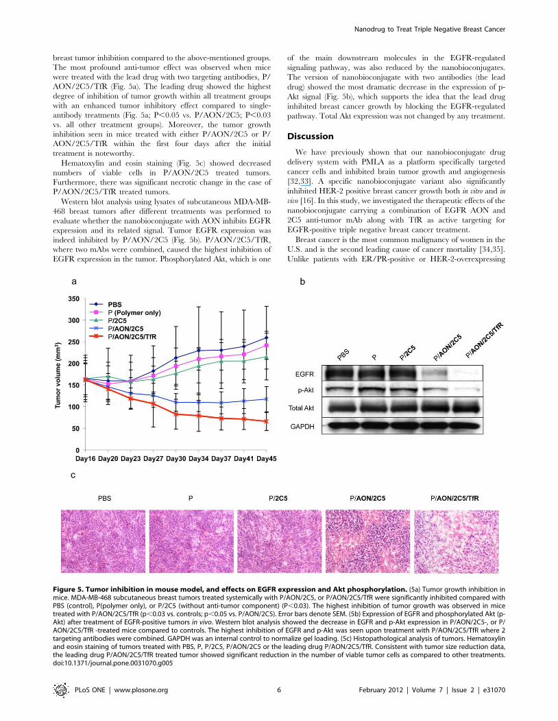
breast tumor inhibition compared to the above-mentioned groups.
The most profound anti-tumor effect was observed when mice
were treated with the lead drug with two targeting antibodies, P/
AON/2C5/TfR (Fig. 5a). The leading drug showed the highest
degree of inhibition of tumor growth within all treatment groups
with an enhanced tumor inhibitory effect compared to single-
antibody treatments (Fig. 5a; P,0.05 vs. P/AON/2C5; P,0.03
vs. all other treatment groups). Moreover, the tumor growth
inhibition seen in mice treated with either P/AON/2C5 or P/
AON/2C5/TfR within the first four days after the initial
treatment is noteworthy.
Hematoxylin and eosin staining (Fig. 5c) showed decreased
numbers of viable cells in P/AON/2C5 treated tumors.
Furthermore, there was significant necrotic change in the case of
P/AON/2C5/TfR treated tumors.
Western blot analysis using lysates of subcutaneous MDA-MB-
468 breast tumors after different treatments was performed to
evaluate whether the nanobioconjugate with AON inhibits EGFR
expression and its related signal. Tumor EGFR expression was
indeed inhibited by P/AON/2C5 (Fig. 5b). P/AON/2C5/TfR,
where two mAbs were combined, caused the highest inhibition of
EGFR expression in the tumor. Phosphorylated Akt, which is one
of the main downstream molecules in the EGFR-regulated
signaling pathway, was also reduced by the nanobioconjugates.
The version of nanobioconjugate with two antibodies (the lead
drug) showed the most dramatic decrease in the expression of p-
Akt signal (Fig. 5b), which supports the idea that the lead drug
inhibited breast cancer growth by blocking the EGFR-regulated
pathway. Total Akt expression was not changed by any treatment.
Discussion
We have previously shown that our nanobioconjugate drug
delivery system with PMLA as a platform specifically targeted
cancer cells and inhibited brain tumor growth and angiogenesis
[32,33]. A specific nanobioconjugate variant also significantly
inhibited HER-2 positive breast cancer growth both in vitro and in
vivo [16]. In this study, we investigated the therapeutic effects of the
nanobioconjugate carrying a combination of EGFR AON and
2C5 anti-tumor mAb along with TfR as active targeting for
EGFR-positive triple negative breast cancer treatment.
Breast cancer is the most common malignancy of women in the
U.S. and is the second leading cause of cancer mortality [34,35].
Unlike patients with ER/PR-positive or HER-2-overexpressing
Figure 5. Tumor inhibition in mouse model, and effects on EGFR expression and Akt phosphorylation. (5a) Tumor growth inhibition inmice. MDA-MB-468 subcutaneous breast tumors treated systemically with P/AON/2C5, or P/AON/2C5/TfR were significantly inhibited compared withPBS (control), P(polymer only), or P/2C5 (without anti-tumor component) (P,0.03). The highest inhibition of tumor growth was observed in micetreated with P/AON/2C5/TfR (p,0.03 vs. controls; p,0.05 vs. P/AON/2C5). Error bars denote SEM. (5b) Expression of EGFR and phosphorylated Akt (p-Akt) after treatment of EGFR-positive tumors in vivo. Western blot analysis showed the decrease in EGFR and p-Akt expression in P/AON/2C5-, or P/AON/2C5/TfR -treated mice compared to controls. The highest inhibition of EGFR and p-Akt was seen upon treatment with P/AON/2C5/TfR where 2targeting antibodies were combined. GAPDH was an internal control to normalize gel loading. (5c) Histopathological analysis of tumors. Hematoxylinand eosin staining of tumors treated with PBS, P, P/2C5, P/AON/2C5 or the leading drug P/AON/2C5/TfR. Consistent with tumor size reduction data,the leading drug P/AON/2C5/TfR treated tumor showed significant reduction in the number of viable tumor cells as compared to other treatments.doi:10.1371/journal.pone.0031070.g005
Nanodrug to Treat Triple Negative Breast Cancer
PLoS ONE | www.plosone.org 6 February 2012 | Volume 7 | Issue 2 | e31070
breast cancer, systemic treatment options for patients with TNBC
are limited due to the lack of a molecular target [1]. Recently, a
number of targeted agents have been considered for TNBC
including poly(ADP-ribose) polymerase (PARP) inhibitor, mTOR
(mammalian target of rapamycin) inhibitor, anti-VEGF mAb, and
anti-EGFR mAb [3].
EGFR activates downstream signaling pathways, such as the
phosphoinositide-3 kinase (PI3K)/Akt pathway, which mediates
cell proliferation, survival, and migration [36,37]. Anti-EGFR
therapy has been increasingly recognized as a potential treatment
for breast cancer patients, and recently, advances in this direction
have been made [5,38]. Furthermore, EGFR is an independent
prognostic factor for patients with TNBC [8]. Therefore, EGFR-
targeted therapy is one of the promising strategies for TNBC
treatment and can potentially improve therapeutic outcome in
TNBC patients.
Several novel EGFR inhibitors are currently in pre-clinical and
clinical development including mAbs and small-molecule tyrosine
kinase inhibitors. Gefitinib (IressaTM, AstraZeneca) is a small-
molecule tyrosine kinase inhibitor of EGFR that binds reversibly
to the ATP-binding site of EGFR. However, phase II trials of
Gefitinib in refractory metastatic breast cancer yielded disap-
pointing responses [39]. Cetuximab (ErbituxTM, ImClone
Systems Incorporated) is a mAb that binds with high affinity to
the extracellular domain of EGFR, competes for ligand binding
and blocks activation of the receptor by EGF or TGF-a. It also
induces antibody-mediated receptor dimerization resulting in
receptor downregulation [38,40]. However, the phase I trial of
Cetuximab in combination with Paclitaxel against advanced
breast cancer was not considered promising [41]. Recently, the
dual kinase inhibitor, Lapatinib, which possesses tyrosine kinase
receptor inhibitory activity against both EGFR and HER-2,
appeared to improve the current model of tyrosine kinase
inhibition and became a novel anti-EGFR therapy [42].
However, despite this progress in anti-EGFR based cancer
treatments, they lack of tumor-specific delivery, which should
result in inefficient anti-tumor activity. For therapeutic effect,
high doses are needed, which lead to serious toxicity. The
nanobioconjugates are ideal drug delivery tools with enhanced
specificity to the tumor and are capable of carrying multiple drug
combinations on a single platform. The drug delivery efficiency is
significantly improved when the nanopolymer is combined with
multiple components such as targeting antibodies against a tumor
marker by exploiting both passive targeting through EPR effect
and active targeting by mAbs [43,44,45].
The synthesis of the complete nanobioconjugate on polymalic
acid nanoplatform with covalently attached EGFR AON in
combination with 2C5 and TfR mAbs has been successfully
achieved (Fig. 1). AON is a stable and promising option for cancer
treatment [24,25]. AON blocked EGFR on glioma cells as good as
siRNA resulting in significant tumor growth inhibition in vivo and
in vitro [46]. As Table 1 shows, the size (smaller than 30 nm) of
these conjugates and their slightly negative f potential make them
ideal for the interaction with the cell membrane and intracellular
internalization. In addition, the homogeneities of each product
were reflected by their polydispersity index.
Western blot analysis showed that P/AON/2C5 significantly
inhibited the expression of EGFR in vitro (Fig. 2b). The low dose of
nanobioconjugate was more effective than the high dose of naked
AON. This result suggests that the nanobioconjugate is a very
efficient delivery vehicle in terms of drug uptake and efficacy.
More importantly, unlike commercially available transduction
agents, PMLA is non-toxic and non-immunogenic, which is ideal
for systemic treatment.
It has been shown that 2C5 specifically targets tumor cells, and
thus it increases anti-cancer effect and reduces side effects of the
treatment [27,28,31]. In accordance with previously published
data, the nanobioconjugate carrying 2C5 mAb specifically
recognized the tumor, which suggests that the activity of the
tumor targeting 2C5 antibody was not changed during chemical
synthesis. A new version of nanobioconjugate carrying tumor
targeting 2C5 mAb and EGFR AON, P/AON/2C5, significantly
inhibited breast tumor growth in vivo (Fig. 5). Breast tumor growth
was inhibited even more when the animals were treated with P/
AON/2C5/TfR where two targeting antibodies were combined
on one nanobiopolymer. Previously, we have proven that tandem
configuration of specific antibodies enhanced tumor targeting
[16,21]. Therefore, targeting tumor vascular endothelium by
mouse anti-TfR mAb in combination with anti-tumor 2C5 mAb
significantly enhanced tumor targeting effect of the nanobiocon-
jugate.
Western blot analysis using tumor samples proved that the
nanobioconjugate efficiently delivered the AONs via attached
targeting antibodies into the tumor cells and this explained the
significant treatment effect observed with novel nanobioconjugate
to inhibit the growth of TNBC. A plausible mechanism of action
of this nanobioconjugate appears to be the inhibition through
EGFR block of Akt phosphorylation, which is a downstream target
of EGFR. A similar blockage of Akt phosphorylation in breast
cancer was also observed after successful treatment of HER-2-
positive breast cancer-bearing mice using another nanobioconju-
gate that inhibits HER-2 synthesis and activity [12].
The results of this study suggest that tumor-targeted nanobio-
conjugate carrying EGFR AON with significant anti-tumor
activity against EGFR-positive TNBC may represent a new
generation of cancer therapeutics with a potential for efficacy
against triple negative breast cancers.
Materials and Methods
1. ReagentsTwo versions of MorpholinoTM-39-NH2 antisense oligonucleo-
tides to EGFR were custom made by Gene Tools (Philomath,
OR):
Version 1: 59- GGTCGCATCGCTGCTCCCCGAAGAG-39,
Version 2: 59- TCGCTCCGGCTCTCCCGATCAATAC-39
Highly purified, endotoxin-free poly(b-L-malic acid), Mw
(weight-averaged) = 100 kDa, polydispersity = 1.1, was obtained
from the culture broth of Physarum polycephalum. Rat anti-mouse
TfR mAb R17217 (mTfR) was purchased from Southern Biotech
(Birmingham, AL). Cysteamine (2-mercaptoethyl-1-amine hydro-
chloride), N-hydroxysuccinimide, other reagents and solvents were
of highest available purity and purchased from Sigma-Aldrich (St.
Louis, MO).
Mouse autoimmune mAb 2C5 recognizing tumor cell surface-
bound nucleosomes released from neighboring apoptotic tumor
cells was a gift from Prof. V.P. Torchilin (Northeastern University,
Boston, MA).
2. Synthesis of polymalic acid nanobioconjugatesThe nanobioconjugates contain five to six key components
(Fig. 1): PMLA as the backbone of delivery carrier, Morpholino
AON for the inhibition of EGFR protein synthesis, tumor
vasculature targeting anti-mouse TfR mAb, targeting anti-tumor
2C5 mAb, 40% leucine ethyl ester as an endosome escape unit to
achieve cytoplasmic AON delivery, and 5% PEG5000 to increase
stability in the bloodstream. The preconjugate containing 40%
leucine ethyl ester, 5% PEG5000 and 10% of cysteamine (%
Nanodrug to Treat Triple Negative Breast Cancer
PLoS ONE | www.plosone.org 7 February 2012 | Volume 7 | Issue 2 | e31070

referring to the total amount of carboxyl groups in PMLA) was
synthesized by the methods described previously [13]. The
antibodies attached to the preconjugate were qualitatively and
quantitatively assayed by size exclusion HPLC. ELISA with purified
TfR and 2C5 antigen was used to verify functional reactivity of
attached antibodies as described. Conjugates for imaging were
fluorescently labeled with Alexa FluorH 680 C2-maleimide
(Invitrogen, Carlsbad, CA) by forming thioether with sulfhydryl
groups. Conjugate without AONs and mAb was used as a control.
3. The nanobioconjugate characterizationChemical and physical characterization of nanobioconjugate
was performed by various methods including L-malate dehydro-
genase assay, PEG colorimetric determination and protein
quantification, size and f potential, HPLC, and ELISA. HPLC
was performed on a Hitachi analytical Elite LaChrom HPLC-UV
system and size exclusion column BioSep-SEC-S 3000 column.
The nanobioconjugate variants were characterized with respect to
their size (hydrodynamic diameter) on the basis of noninvasive
back-scattering (NIBS) and f potential from electrophoretic
mobility based on the Helmholtz-Smoluchowski formula, using
electrophoresis M3-PALS [19]. Both measurements were per-
formed in a Zetasizer Nano System ZS90 (Malvern Instruments,
Malvern, UK). Data on molecular size and f potential from three
independent measurements represent mean 6 standard deviation.
4. Cell lines and culture conditionsHuman breast cancer cell lines MDA-MB-468 (TNBC, EGFR
positive), SKBR-3 (EGFR-positive), BT-474 (EGFR-positive), and
MCF-7 (EGFR-negative) were obtained from American Type
Culture Collection (Manassas, VA). SKBR-3 was cultured in
McCoy’s 5A medium with 10% fetal bovine serum and antibiotics.
All other cell lines were cultured in DMEM with the same
supplements.
5. NomenclatureThe term ‘‘nanobioconjugate’’ denotes the drug delivery system
with PMLA as the nanoplatform and various functional groups
covalently attached to it, specifically the rat anti-mouse TfR mAbs, and
leucine ethylester (LOEt) as the endosomal escape unit. The newly
synthesized versions of nanobioconjugate to treat EGFR-positive
breast cancer contained a single anti-cancer drug (EGFR AON) with
either one mAb (2C5) or two mAbs (2C5 and TfR). All versions of
nanobioconjugates used in this study are shown in Fig. 1 and Table 1.
6. Western blottingThe protein samples were harvested from a variety of breast
cancer cells (shown in Fig. 2.) and the expression level of 2C5
antigen and TfR were detected by western blotting as described
previously [47]. MDA-MB-468 EGFR-positive TNBC cells and
EGFR-positive SKBR-3 breast cancer cells were treated with one
of the following: PBS (control), Endoporter (control, transduction
reagent), AON to EGFR with Endoporter, and P/AON/2C5.
Cell lysates were collected 72 hours after treatment. EGFR and
glyceraldehyde 3-phosphate dehydrogenase (GAPDH, to normal-
ize gel load) expression was analyzed by western blotting.
MDA-MB-468 TNBC subcutaneous breast tumor bearing mice
were treated with controls (PBS); P(Polymer control), P/2C5, P/
AON/2C5, and P/AON/2C5/TfR. Cell lysates were prepared
from excised breast tumor samples and analyzed by western
blotting to detect EGFR, total Akt, phosphorylated Akt (p-Akt),
and GAPDH (for equal gel loading). All of the antibodies used
were purchased from Cell Signaling Technology (Beverly, MA).
7. Tumor xenografts in nude miceAll experiments with animals were performed in accordance
with the protocols approved by the Cedars-Sinai Medical Center
Institutional Animal Care and Use Committee (IACUC). Athymic
mice were purchased from Taconic (Hudson, NY). A total of
16107 MDA-MB-468 cells suspended in 150 ml of Matrigel (BD
Biosciences, Bedford, MA) were injected into the right flanks of 35
mice (5 mice per group), and treatment was commenced when
tumors reached an average size of .120 mm3 (21 days after
injection). Mice were divided into 5 treatment groups and
administered either sterile PBS (control), P (Polymer control), P/
2C5, P/AON/2C5, or P/AON/2C5/TfR through the tail vein
twice a week. Treatments were performed 8 times (for 4 weeks).
Tumor sizes were measured with calipers twice a week, and tumor
volumes were determined using the formula: (length6width2)6(p/6).
Four days after the last treatment, the animals were anesthetized
with 3% isoflurane-air mixture and sacrificed by cervical
dislocation. Tumor samples were stained with hematoxylin and
eosin for morphological observation.
The same subcutaneous tumor model was used for the imaging
study.
8. In vivo imaging studyTo assess the organ localization of nanobioconjugates, 16107
MDA-MB-468 human breast cancer cells suspended in 150 ml of
Matrigel were implanted into the right thigh of athymic mice
(CrTac:NCr-Foxn1nu Homozygous, Taconic) as described above.
When tumors grew up to 120 mm3, 160 ml of Alexa Fluor 680-
labeled Polycefin variants were injected intravenously at the
concentration of 4 mM. PMLA with IgG, P/IgG, was used as a
control. For the assessment of drug distribution and localization in
nude mice, animals were studied in a Xenogen IVIS 200 imager
under isoflurane anesthesia at different time points (before drug
administration, 1 h, 3 h, 6 h, and 24 h after the injection of the
drug). Twenty-four hours after drug administration, mice were
euthanized. Intra-arterial PBS perfusion was done in order to wash
out the circulating drugs in blood vessels. The tumor and major
organs were harvested to detect the fluorescent signal. The
fluorescent signal intensities in the tumor and different organs
were analyzed by Xenogen Living ImageH software, Version 2.50
(WaveMetrix, USA).
9. Confocal microscopyTo assess the tissue localization of nanobioconjugates, tumor-
bearing mice were injected through the tail vein with either Alexa
Fluor 680-labeled P/IgG (control), Alexa Fluor 680-labeled P/
2C5, or Alexa Fluor 680-labeled P/2C5/TfR, as above. Twenty-
four hours after drug administration, mice were euthanized,
tumors were harvested, snap-frozen in liquid nitrogen, and
embedded in OCT compound for the fluorescent signal detection
on sections by confocal microscopy (TCS SP5 X microscope;
Leica Microsystems, Mannheim, Germany).
10. Statistical analysisStudent’s t-test (for two groups) and analysis of variance
(ANOVA, for three or more groups) were used to calculate
statistical significance of the experimental results. GraphPad
Prism4 program (GraphPad Software, San Diego, CA) was
utilized for all calculations. Data are presented as mean 6
standard error of mean (SEM). The significance level was set at
P,0.05.
Nanodrug to Treat Triple Negative Breast Cancer
PLoS ONE | www.plosone.org 8 February 2012 | Volume 7 | Issue 2 | e31070
Author Contributions
Conceived and designed the experiments: SI JYL EH KB. Performed the
experiments: SI BK JM RP JP HD EH A. Espinoza A. Elramsisy HWP
DM. Analyzed the data: SI JYL EH KB HD RP. Contributed reagents/
materials/analysis tools: DM. Wrote the paper: SI JYL EH HD RP JP.
Read and approved the final manuscript: SI JYL EH KB BK JM RP JP A.
Espinoza A. Elramsisy DM HD HWP.
References
1. Dent R, Trudeau M, Pritchard KI, Hanna WM, Kahn HK, et al. (2007) Triple-
negative breast cancer: clinical features and patterns of recurrence. Clin Cancer
Res 13: 4429–4434.
2. Bevers TB, Anderson BO, Bonaccio E, Buys S, Daly MB, et al. (2009) NCCN
clinical practice guidelines in oncology: breast cancer screening and diagnosis.
J Natl Compr Canc Netw 7: 1060–1096.
3. Pal SK, Childs BH, Pegram M (2011) Triple negative breast cancer: unmet
medical needs. Breast Cancer Res Treat 125: 627–636.
4. Agrawal A, Gutteridge E, Gee JM, Nicholson RI, Robertson JF (2005) Overview
of tyrosine kinase inhibitors in clinical breast cancer. Endocr Relat Cancer 12
Suppl 1: S135–144.
5. Flynn JF, Wong C, Wu JM (2009) Anti-EGFR Therapy: Mechanism
andAdvances in Clinical Efficacy in Breast Cancer. J Oncol 2009: 526963.
6. Baselga J (2002) Why the epidermal growth factor receptor? The rationale for
cancer therapy. Oncologist 7 Suppl 4: 2–8.
7. Huang S, Armstrong EA, Benavente S, Chinnaiyan P, Harari PM (2004) Dual-
agent molecular targeting of the epidermal growth factor receptor (EGFR):
combining anti-EGFR antibody with tyrosine kinase inhibitor. Cancer Res 64:
5355–5362.
8. Liu D, He J, Yuan Z, Wang S, Peng R, et al. (2010) EGFR expression correlates
with decreased disease-free survival in triple-negative breast cancer: a
retrospective analysis based on a tissue microarray. Med Oncol.
9. Atkinson RL, Zhang M, Diagaradjane P, Peddibhotla S, Contreras A, et al.
(2010) Thermal enhancement with optically activated gold nanoshells sensitizes
breast cancer stem cells to radiation therapy. Sci Transl Med 2: 55ra79.
10. Chandna P, Khandare JJ, Ber E, Rodriguez-Rodriguez L, Minko T (2010)
Multifunctional tumor-targeted polymer-peptide-drug delivery system for
treatment of primary and metastatic cancers. Pharm Res 27: 2296–2306.
11. van Renswoude J, Bridges KR, Harford JB, Klausner RD (1982) Receptor-
mediated endocytosis of transferrin and the uptake of fe in K562 cells:
identification of a nonlysosomal acidic compartment. Proc Natl Acad Sci U S A
79: 6186–6190.
12. Xia CQ, Shen WC (2001) Tyrphostin-8 enhances transferrin receptor-mediated
transcytosis in Caco-2- cells and inreases hypoglycemic effect of orally
administered insulin-transferrin conjugate in diabetic rats. Pharm Res 18:
191–195.
13. Lee BS, Fujita M, Khazenzon NM, Wawrowsky KA, Wachsmann-Hogiu S, et
al. (2006) Polycefin, a new prototype of a multifunctional nanoconjugate based
on poly(beta-L-malic acid) for drug delivery. Bioconjug Chem 17: 317–326.
14. Lee B-S, Vert M, Holler E (2002) Water-soluble aliphatic polyesters: poly(malic
acid)s. In: Doi YSA, ed. Biopolymers. Weinheim: Wiley–VCH. pp 75–103.
15. Ding H, Inoue S, Ljubimov AV, Patil R, Portilla-Arias J, et al. (2010) Inhibition
of brain tumor growth by intravenous poly (beta-L-malic acid) nanobioconjugate
with pH-dependent drug release [corrected]. Proc Natl Acad Sci U S A 107:
18143–18148.
16. Inoue S, Ding H, Portilla-Arias J, Hu J, Konda B, et al. (2011) Polymalic Acid-
Based Nanobiopolymer Provides Efficient Systemic Breast Cancer Treatment by
Inhibiting both HER2/neu Receptor Synthesis and Activity. Cancer Res 71:
1454–1464.
17. Ljubimova JY, Fujita M, Ljubimov AV, Torchilin VP, Black KL, et al. (2008)
Poly(malic acid) nanoconjugates containing various antibodies and oligonucle-
otides for multitargeting drug delivery. Nanomedicine (Lond) 3: 247–265.
18. Braud C, Vert M (1992) Degradation of poly(p-malic acid) - monitoring of
oligomers formation by aqueous SEC and HPCE. Polym Bull 29: 177–183.
19. Gasslmaier B, Holler E (1997) Specificity and direction of depolymerization of
beta-poly(L-malate) catalysed by polymalatase from Physarum polycephalum–
fluorescence labeling at the carboxy-terminus of beta-poly(L-malate).
Eur J Biochem 250: 308–314.
20. Gasslmaier B, Krell CM, Seebach D, Holler E (2000) Synthetic substrates and
inhibitors of beta-poly(L-malate)-hydrolase (polymalatase). Eur J Biochem 267:
5101–5105.
21. Fujita M, Lee BS, Khazenzon NM, Penichet ML, Wawrowsky KA, et al. (2007)
Brain tumor tandem targeting using a combination of monoclonal antibodies
attached to biopoly(beta-L-malic acid). J Control Release 122: 356–363.
22. Busch RK, Perlaky L, Valdez BC, Henning D, Busch H (1994) Apoptosis in
human tumor cells following treatment with p120 antisense oligodeoxynucleo-
tide ISIS 3466. Cancer Lett 86: 151–157.
23. Sekhon HS, London CA, Sekhon M, Iversen PL, Devi GR (2008) c-MYC
antisense phosphosphorodiamidate morpholino oligomer inhibits lung metastasis
in a murine tumor model. Lung Cancer 60: 347–354.
24. Wu B, Lu P, Benrashid E, Malik S, Ashar J, et al. (2010) Dose-dependentrestoration of dystrophin expression in cardiac muscle of dystrophic mice by
systemically delivered morpholino. Gene Ther 17: 132–140.
25. Kinali M, Arechavala-Gomeza V, Feng L, Cirak S, Hunt D, et al. (2009) Localrestoration of dystrophin expression with the morpholino oligomer AVI-4658 in
Duchenne muscular dystrophy: a single-blind, placebo-controlled, dose-escala-tion, proof-of-concept study. Lancet Neurol 8: 918–928.
26. Elbayoumi TA, Pabba S, Roby A, Torchilin VP (2007) Antinucleosome
antibody-modified liposomes and lipid-core micelles for tumor-targeted deliveryof therapeutic and diagnostic agents. J Liposome Res 17: 1–14.
27. Elbayoumi TA, Torchilin VP (2008) Tumor-specific antibody-mediated targeteddelivery of Doxil reduces the manifestation of auricular erythema side effect in
mice. Int J Pharm 357: 272–279.28. ElBayoumi TA, Torchilin VP (2009) Tumor-targeted nanomedicines: enhanced
antitumor efficacy in vivo of doxorubicin-loaded, long-circulating liposomes
modified with cancer-specific monoclonal antibody. Clin Cancer Res 15:1973–1980.
29. Lorenz MR, Holzapfel V, Musyanovych A, Nothelfer K, Walther P, et al. (2006)Uptake of functionalized, fluorescent-labeled polymeric particles in different cell
lines and stem cells. Biomaterials 27: 2820–2828.
30. Wilhelm C, Billotey C, Roger J, Pons JN, Bacri JC, et al. (2003) Intracellularuptake of anionic superparamagnetic nanoparticles as a function of their surface
coating. Biomaterials 24: 1001–1011.31. Gupta B, Levchenko TS, Mongayt DA, Torchilin VP (2005) Monoclonal
antibody 2C5-mediated binding of liposomes to brain tumor cells in vitro and in
subcutaneous tumor model in vivo. J Drug Target 13: 337–343.32. Fujita M, Khazenzon NM, Ljubimov AV, Lee BS, Virtanen I, et al. (2006)
Inhibition of laminin-8 in vivo using a novel poly(malic acid)-based carrierreduces glioma angiogenesis. Angiogenesis 9: 183–191.
33. Ljubimova JY, Fujita M, Khazenzon NM, Lee BS, Wachsmann-Hogiu S, et al.(2008) Nanoconjugate based on polymalic acid for tumor targeting. Chem Biol
Interact 171: 195–203.
34. Herbst RS, Bajorin DF, Bleiberg H, Blum D, Hao D, et al. (2006) ClinicalCancer Advances 2005: major research advances in cancer treatment,
prevention, and screening–a report from the American Society of ClinicalOncology. J Clin Oncol 24: 190–205.
35. Jemal A, Siegel R, Ward E, Hao Y, Xu J, et al. (2009) Cancer statistics, 2009.
CA Cancer J Clin 59: 225–249.36. Kallergi G, Agelaki S, Kalykaki A, Stournaras C, Mavroudis D, et al. (2008)
Phosphorylated EGFR and PI3K/Akt signaling kinases are expressed incirculating tumor cells of breast cancer patients. Breast Cancer Res 10: R80.
37. Sibilia M, Fleischmann A, Behrens A, Stingl L, Carroll J, et al. (2000) The EGFreceptor provides an essential survival signal for SOS-dependent skin tumor
development. Cell 102: 211–220.
38. Lurje G, Lenz HJ (2009) EGFR signaling and drug discovery. Oncology 77:400–410.
39. Baselga J, Albanell J, Ruiz A, Lluch A, Gascon P, et al. (2005) Phase II andtumor pharmacodynamic study of gefitinib in patients with advanced breast
cancer. J Clin Oncol 23: 5323–5333.
40. Adams GP, Weiner LM (2005) Monoclonal antibody therapy of cancer. NatBiotechnol 23: 1147–1157.
41. Modi S, D’Andrea G, Norton L, Yao TJ, Caravelli J, et al. (2006) A phase Istudy of cetuximab/paclitaxel in patients with advanced-stage breast cancer.
Clin Breast Cancer 7: 270–277.42. Nahta R, Yuan LX, Du Y, Esteva FJ (2007) Lapatinib induces apoptosis in
trastuzumab-resistant breast cancer cells: effects on insulin-like growth factor I
signaling. Mol Cancer Ther 6: 667–674.43. Liu X, Wang Y, Nakamura K, Kubo A, Hnatowich DJ (2008) Cell studies of a
three-component antisense MORF/tat/Herceptin nanoparticle designed forimproved tumor delivery. Cancer Gene Ther 15: 126–132.
44. Maeda H, Bharate GY, Daruwalla J (2009) Polymeric drugs for efficient tumor-
targeted drug delivery based on EPR-effect. Eur J Pharm Biopharm 71:409–419.
45. Maeda H, Fang J, Inutsuka T, Kitamoto Y (2003) Vascular permeabilityenhancement in solid tumor: various factors, mechanisms involved and its
implications. Int Immunopharmacol 3: 319–328.
46. Kang CS, Zhang ZY, Jia ZF, Wang GX, Qiu MZ, et al. (2006) Suppression ofEGFR expression by antisense or small interference RNA inhibits U251 glioma
cell growth in vitro and in vivo. Cancer Gene Ther 13: 530–538.47. Inoue S, Branch CD, Gallick GE, Chada S, Ramesh R (2005) Inhibition of Src
kinase activity by Ad-mda7 suppresses vascular endothelial growth factorexpression in prostate carcinoma cells. Mol Ther 12: 707–715.
Nanodrug to Treat Triple Negative Breast Cancer
PLoS ONE | www.plosone.org 9 February 2012 | Volume 7 | Issue 2 | e31070