Hair cell regeneration in vestibular epithelia - DiVA Portal
Ocular surface epithelia contain ABCG2-dependent sidepopulation cells exhibiting features associated with stem cells
Murat T. Budak1, Onder S. Alpdogan2, Mingyuan Zhou3, Robert M. Lavker3,*, M.A. MuratAkinci1, and J. Mario Wolosin1,*
1 Department of Ophthalmology, Mount Sinai School of Medicine, One Gustave L. Levy Place, New York,NY 10029, USA
2 Department of Medicine and Pediatrics, Memorial Sloan-Kettering Cancer Center, 1275 York Avenue, NewYork, NY 10021, USA
3 Department of Dermatology, The Feinberg School of Medicine at Northwestern University, 303 EastChicago Avenue, Chicago, IL 60611, USA
AbstractSummary—When cell populations are incubated with the DNA-binding dye Hoechst 33342 andsubjected to flow cytometry analysis for Hoechst 33342 emissions, active efflux of the dye by theABCG2/BCRP1 transporter causes certain cells to appear as a segregated cohort, known as a sidepopulation (SP). Stem cells from several tissues have been shown to possess the SP phenotype. Asthe lack of specific surface markers has hindered the isolation and subsequent biochemicalcharacterization of epithelial stem cells this study sought to determine the existence of SP cells andexpression of ABCG2 in the epithelia of the ocular surface and evaluate whether such SP cells hadfeatures associated with epithelial stem cells. Human and rabbit limbal-corneal and conjunctivalepithelial cells were incubated with Hoechst 33342, and analyzed and sorted by flow cytometry.Sorted cells were subjected to several tests to determine whether the isolated SP cells displayedfeatures consistent with the stem cell phenotype. Side populations amounting to <1% of total cells,which were sensitive to the ABCG2-inhibitor fumitremorgin C, were found in the conjunctival andlimbal epithelia, but were absent from the stem cell-free corneal epithelium. Immunohistochemistrywas used to establish the spatial expression pattern of ABCG2. The antigen was detected in clustersof conjunctival and limbal epithelia basal cells but was not present in the corneal epithelium. SP cellswere characterized by extremely low light side scattering and contained a high percentage of cellsthat: showed slow-cycling prior to tissue collection; exhibited an initial delay in proliferation afterculturing; and displayed clonogenic capacity and resistance to phorbol-induced differentiation; allfeatures that are consistent with a stem cell phenotype.
KeywordsStem cells; Side population; Epithelia; Cornea; Limbus; Conjunctiva
IntroductionThe vertebrate ocular surface is lined with two stratified, constantly renewing tissues, thelimbal/corneal and conjunctival epithelia. These two closely related but distinct cell lineages(Kruse et al., 1990; Wei et al., 1993; Wei et al., 1996; Moyer et al., 1996), arise simultaneously
* Authors for correspondence (e-mail: [email protected]; [email protected]).Supported by EY 014878, EY 015132 and EY 01867 (J. M.W.) EY 13711, EY 06769 (R. M. L.) and by an unrestricted grant to theDepartment of Ophthalmology from Research to Prevent Blindness. J. M. W. is the recipient of a RPB Senior Scientific Investigatoraward.
NIH Public AccessAuthor ManuscriptJ Cell Sci. Author manuscript; available in PMC 2005 September 29.
Published in final edited form as:J Cell Sci. 2005 April 15; 118(Pt 8): 1715–1724.
NIH
-PA Author Manuscript
NIH
-PA Author Manuscript
NIH
-PA Author Manuscript
from a few PAX6-positive ectodermal cells that remain in the embryonic ectodermal surfacefollowing formation of the lens vesicle by the bulk of the PAX6+ cells (Davis and Reed,1996; Koroma et al., 1997; Wolosin et al., 2002).
The self-renewing nature of the limbal/corneal and conjunctival epithelia makes their long-term survival ultimately dependent on small populations of stem cells. In the limbal/cornealepithelia, SCs are exclusively situated in the basal cell layer of the limbus, the outer vascularrim at the junction between the cornea and the conjunctiva (Schemer et al., 1986; Cotsarelis etal., 1989). The epithelium covering the transparent central cornea contains no stem cells(Schermer et al., 1986; Cotsarelis et al., 1989; Wolosin et al., 2000). Several lines of evidencesuggest that conjunctival epithelial stem cells are concentrated in the fornix (Wei et al.,1995; Pellegrini et. al., 1999), though stem cells may also be present in other areas (Pelligriniet al., 1999; Wirtschafter et al., 1999; Chen et al., 2003; Nagasaki and Zhao, 2005).
The ability to isolate viable somatic stems cells will provide new, invaluable means tocharacterize these cells and use them in gene therapy protocols (Bradfute and Goodell, 2002).The identification of a number of surface markers has facilitated the routine preparation ofhematopoietic stem cells (Civin, 1992; Silvestri, 1992). Identification of comparable markersin epithelial systems has remained a challenging task. An alternative method for the purificationof stem cells exploits their special spectral emission pattern following staining with the DNAdye Hoechst 33342 (Hoechst). In the free state the dye exhibits a fluorescence emission peakat 465 nm (Hoechst blue). In the DNA-bound state in live cells, electronic interactions betweenthe densely packed Hoechst molecules cause a bathochromic shift leading to a secondaryemission at 675 nm (Hoechst red). Hoechst-stained bone marrow cells subjected to flowcytometry reveal a population of cells displaying very low fluorescent emission intensities anda reduced bathochromic shift. The cohort, or side population (SP), reflects a reduction of DNA-bound dye in these cells due to their ability to actively efflux Hoechst. The bone marrow SPcells are enriched in hematopoietic cells with multiple stem cell characteristics, including thecapacity for long-term lineage reconstitution (Goodell et al., 1996; Uchida et al., 2003).
Recently, it was demonstrated that Hoechst efflux in the SP cells is mediated by the G2 subtypemember of the ATP Binding Cassette (ABC) transporter, and the authors proposed that the SPphenotype may be a feature displayed by multiple adult stem cell types (Zhou et al., 2001).Several studies have provided support for this proposal (Scharenberg et al., 2002; Lechner etal., 2002; Kim and Morshead, 2003; Alison, 2003; Lassalle et al., 2003; Bhattacharya et al.,2003; Summer et al., 2003). Preliminary reports on the presence of SP cells in the limbal zonehave been published (Watanabe et al., 2004; Wolosin et al., 2004) We describe now theexistence of ABCG2-dependent SP cells in the human and rabbit ocular surface epithelia anddemonstrate that these cells display many features that are consistent with a stem cellphenotype.
Materials and MethodsTissue procurement and processing
Rabbit tissues were dissected from eyes obtained from local abattoirs. Human corneas andconjunctivas unsuitable for human transplant, all from unidentifiable donors, were obtainedfrom the National Disease Research Interchange (Philadelphia, PA) within 48 hours ofcollection. For the cell kinetics studies (see below), 3-month-old albino New Zealand rabbitswere housed in an American Association of Laboratory Animal Care-accredited facility withfree access to food and water. All animal procedures complied with the USPHS policy onhumane care and use of laboratory animals. For limbal dissection, corneas were quartered, andafter careful trimming of conjunctival remnants, placed on a black cutting board. Tangentialillumination was used to visualize and surgically dissect the rabbit limbus as a discrete 0.5–
Budak et al. Page 2
J Cell Sci. Author manuscript; available in PMC 2005 September 29.
NIH
-PA Author Manuscript
NIH
-PA Author Manuscript
NIH
-PA Author Manuscript

0.8 mm-wide whitish outer strip and the human limbus as a discrete ~1-mm-wide zonecontaining the Palisades of Vogt. Tissues were incubated for 16–20 hours at 4°C in 5 mg/mlDispase (Roche, Indianapolis, IN) dissolved in 4-(2-hydroxyethyl)-l-piperazineethanesulfonicacid-buffered 1:1 mix of Dulbecco’s modified Eagle’s medium and Ham-F12 (hD/F-12) withmoderate shaking. This results in the spontaneous release of whole epithelial sheets.
Flow cytometryPure epithelial sheets were incubated for 20–30 minutes at 37°C in trypsin. When >90% of thepopulation consisted of single cells, the suspension was complemented with one volume ofhD/F-12 containing 20% fetal calf serum (FCS), cells were dispersed using a fire-polishedPasteur pipette, sequentially filtered through 40 μm and 10 μm nylon meshes, suspended at106 cells/ml in hD/F-12 containing 2% FCS, and incubated with 2–5 μg Hoechst (Sigma, StLouis, MO) for 45–90 minutes at 37°C under slow swirling motion. To inhibit ABCtransporters, 10 μM reserpine (Sigma), 10–100 μM verapamil (Sigma) or 10 μM fumitremorginC (a generous gift from Susan Bates, NCI, Bethesda, MD) were added 5 minutes before theaddition of Hoechst. Cells were spun down, resuspended at 106 cells/ml in HBSS with 4% PCSand 2 μg/ml propidium iodide (PI; Roche, Indiana, IN), analyzed for fluorescent emissions andside and forward light scattering (SSC and FSC, respectively) and sorted in a MoFlo(Cytomation, Fort Collins, CO) cytometer. Instrument gains were adjusted to set the main cellcohort, which comprises most of the cells containing one copy of DNA (G0 and G1, or G0-G1, cells; see Fig. 1), at the center of the plots. Data for viable cells (PI staining intensity below1% of the maximal staining) were analyzed for parametric correlations and annotated usingFCSExpress (DeNovo Software, Toronto, Canada).
For direct microscopic observation, human limbal cells were spun down and gentlyresuspended in a minimal volume of HBSS with 0.1% Try pan Blue and examined in an invertedmicroscope using phase-contrast and transmitted light illuminations.
BrdU labeling in vivoThree 2ML2 osmotic Alzet mini pumps (Durect, Cupertino, CA) calibrated to produce aconstant delivery of ~24 mg bromodeoxyuridine (BrdU)/day for 14–16 days, were implantedsubcutaneously into 3 kg (3–4 months old) rabbits. Most animals were sacrificed 11 days later,when continuous steady state label pumping had proceeded for 10 days. In one case the pumpswere removed after 17 days, and the animal was left undisturbed for 38 days prior to sacrificeand tissue collection. Small segments of the limbal/corneal and conjunctival tissues were fixedovernight at 4°C in 50 mM glycine, 70% ethanol, pH 3.0 and embedded in paraffin. Themajority of the tissue was used to isolate the limbal and conjunctival epithelium, sort singlecell suspensions by flow cytometry and to determine the BrdU content of the sorted fractions.
BrdU detection in tissue sectionsParaffin-embedded tissues were cut into 4-μm-thick sections, deparaffinized, rehydrated inPBS, immersed in 2N HC1 for 30 minutes and digested with trypsin for 15 minutes. BrdUincorporation was revealed by indirect immunofluorescence. Sections were blocked in 5% goatserum in PBS and incubated for 2 hours at 37°C with 10 μg/ml anti-BrdU mouse monoclonalantibody (Roche) made in 0.5% Tween 20, 1% goat serum in PBS (T-PBS). After washings,sections were stained for 1 hour with 1 μg/ml Alexa 488-conjugated goat anti-mouse IgG(Molecular Probes, Eugene, OR), counterstained with 0.01% PI-PBS, mounted with anti-fading medium and photographed in an Olympus epifluorescence microscope equipped forgreen (for Alexa 488), red (for PI) and dual fluorescence image capture.
Budak et al. Page 3
J Cell Sci. Author manuscript; available in PMC 2005 September 29.
NIH
-PA Author Manuscript
NIH
-PA Author Manuscript
NIH
-PA Author Manuscript

BrdU detection in sorted cellsLimbal and conjunctival epithelial cells were alternatively sorted according to their Hoechst33342 staining, or light-scattering properties. Sorted cells were either cytospun onto poly-L-lysine-coated glass slides using a Shandon Cytospin3 centrifuge (Shandon, Wyman, MA) orconcentrated and plated overnight on the slides for selection of adherent (i.e. basal) cells. Slideswere immersed in 70% ethanol for 30 minutes, briefly air-dried, immersed in 0.07 N NaOHfor 2 minutes, overlaid with FITC-conjugated monoclonal anti-BrdU antibody (Pharmingen,San Diego, CA) prepared in T-PBS for 30 minutes, counterstained with 0.01% PI-PBS andmounted and photographed as described above. BrdU-free cells were processed in parallel toserve as controls.
ABCG2 detection in tissue sectionsFreshly dissected tissue segments were embedded in Cryomatrix™ (Shandon, Pittsburgh, PA),frozen in liquid nitrogen, cut into 8- to 10-μm-thick sections in a cryotome, collected on gelatin-coated glass slides. The sections were fixed in cold methanol, briefly air-dried, rehydrated inPBS and blocked with 5% goat serum. ABCG2 was detected by indirect immunofluorescenceusing one of two anti-ABCG2 monoclonal antibodies, BXP21 (Chemicon, Temecula, CA) andBXP34 (Alexis, San Diego, CA) as the primary reagent and Alexa 488 goat anti-mouse IgGas the secondary antibody. Both primary antibodies recognize internal epitopes within the C-terminus of human ABCG2 (Maliepaard et al., 2001). Stained samples were mounted andphotographed as described above.
Measurements of initial proliferative ratesLimbal and conjunctival cells (106 cells/ml) were incubated for 30 minutes with 0.5 μMcarboxy-fluorescein diacetate, succinymidyl ester (CFDA-SE; Molecular Probes). Thisfluorescein derivative reacts covalently with amino groups resulting in its long-term retentionwithin cells. Thus, decreases in cellular fluorescence reflect primarily cell division (Oostendorpet al., 2000). 100,000 CFDA-SE-loaded epithelial cells were plated on 2.5 cm permeable Falcontissue culture inserts in which 3T3 cells were plated at 4000 cells/cm2 on the underside of thepermeable synthetic membrane. In one experiment cells were harvested after either 16 or 64hours, incubated with Hoechst and analyzed in the MoFlo cytometer. In a second experiment,cells were collected after 24 to 96 hours, lightly fixed with 0.1% formaldehyde for 30 minutesand stored at 4°C in 1% BSA When the last batch of cells was collected, all batches wereanalyzed by flow cytometry using a FACSCaliburTM Excalibur (BDBiosciences, San Jose,CA) analytical cytometer.
Clonogenic capacity assaySP and G0-G1 cells were plated at low density (10–50 cells/cm2) in 20% FCS-hD/F12 onto100 mm Petri dishes containing a layer of Swiss 3T3 fibroblasts which were incubated with 8μg/ml mitomycin C (Sigma) for 3 hours to elicit permanent cell proliferation arrest, and thenplated at a rate of 2000/cm2 48 hours prior to epithelial cell addition. Where indicated, phorbolmyristate acetate (PMA; Sigma) was added 4 hours later and removed with the first mediumrefreshment at 72 hours post-plating. For colony visualization, dishes were fixed in coldmethanol and incubated for 10 minutes in 50% methanol, 10% acetic acid, 0.01% CoomassieBrilliant Blue R250.
RNA preparation and real-time quantitative PCROne μg RNA aliquots, isolated using Tri-Reagent (MRC, Cincinnati, HO) and incorporatingmaterial from four distinct specimens, were converted into cDNA using reverse transcriptase(Omniscript™; Qiagen, Valencia, CA). No attempt was made to remove residual genomicDNA. An identical mock reaction omitting the enzyme was carried out in parallel. The products
Budak et al. Page 4
J Cell Sci. Author manuscript; available in PMC 2005 September 29.
NIH
-PA Author Manuscript
NIH
-PA Author Manuscript
NIH
-PA Author Manuscript

of these reactions were subjected to quantitative real-time PCR in an AbiPrism 7900H sequencedetection instrument (annealing: 20 seconds at 58°C; DNA melting: 10 seconds at 95°C) usingtriplicates samples containing QuantiTect™ SYBR® Green PCR mix (Qiagen), reverse-transcribed product derived from 100 ng of initial RNA and primer pairs (150 μM) designedto amplify either ABCG2 (ABCG2fwd: TGCAAC-ATGTACTGGCGAAGA; ABCG2rev:TCTTCCACAAGCCCCA-GG), or β-actin (β-actinfwd: AGTACTCCGTGTGGATCGGC;β-actinrev: GCTGATCCACATCTGCTGGA) (Abbott et al., 2002). The generation of singleamplified DNA sequences with the expected size (74 bp for ABCG2 and 68 bp for β-actin),was determined by electrophoresis of the reaction products in 4% agarose gels containing0.05% Ethidium Bromide. Cycle thresholds (Ct) for the different samples were used todetermine relative amounts of ABCG2 message according to the delta-delta Ct method.
ResultsLimbal and conjunctival epithelia contain ABCG2-dependent side populations
Initial studies with rabbit cells established the best conditions for the generation ofdistinguishable SP cells. From these studies we concluded that the optimal Hoechstconcentration for the conjunctival epithelium, when performing 90-minute incubations, occursat 3.0–3.5 μM. For the limbal epithelium better results were obtained by reducing the dyeconcentration to 2.0–2.5 μM and the incubation time to 60 minutes.
SP cells were present in rabbit and human limbal and conjunctival epithelia (Fig. 1A-D andTable 1). Inclusion of the ABCG2-specific inhibitor fumitremorgin C (Rabindran et al.,2000; Robey et al., 2001; Nakanishi et al., 2003) resulted in a 80–90% reduction in the size ofthe SPs in both ocular surface epithelia (Fig. 1E,F), confirming that the SP phenotype primarilyreflects the activity of the ABCG2 transporter (Zhou et al., 2001). Reserpine, a powerful pan-ABC transporter inhibitor (Zhou et al., 2001; Murayama et al., 2002), completely abolishedSP cells (Fig. 1D,F inserts). In contrast, verapamil caused only a 20–30% decrease in rabbitconjunctival SP cells, consistent with the demonstrated lack of effectiveness of this P-glycoprotein inhibitor on ABCG2 activity (Zhou et al., 2001; Ozvegy et al., 2001).
ABCG2 is expressed in the ocular surface epithelia in discreet clustersThe expression of the fumitremorgin C-sensitive ABCG2 transporter was investigated at themRNA and protein levels. Real-time quantitative PCR combined with product sizeconfirmation by agarose chromatography (a single 71 bp band; not shown) demonstrated theexpression of the message for this protein throughout the ocular surface. Overall message levelswere extremely low: in the limbal epithelium ABCG2 they amounted to 1/4800 of the β-actinvalue. Our measurements indicated very similar message content for the conjunctival andlimbal epithelia (ratio=0.96). In contrast, in the stem and SP cell-free corneal epithelium themessage amounted to only 1/26.3 of the level measured in the limbal counterpart.
Immunofluorescence detected ABGC2 protein in the periphery of both limbal and conjunctivalepithelia cells. Both monoclonal antibodies used generated similar patterns but only results forthe clone displaying lower background, BXP21, are shown (Fig. 2). In the limbal/cornealepithelium, ABCG2 antigenicity was observed exclusively in the limbal zone. The stainoccurred in cell clusters comprising basal and proximal suprabasal cells. Stain intensity washigher in the cells at the center of each cluster (Fig. 2A-C). The greatest accumulation of suchclusters occurred within the Palisades of Vogt (Townsend, 1991), the area generally believedto contain the highest concentration of stem cells within the human limbus. In some of thepalisades proximal to the conjunctiva, all basal and suprabasal epithelial cells appeared to beABCG2-positive. There was no ABCG2 stain in the corneal zone, i.e., the small amount ofABCG2 message expressed in this area did not translate into any visible protein expression
Budak et al. Page 5
J Cell Sci. Author manuscript; available in PMC 2005 September 29.
NIH
-PA Author Manuscript
NIH
-PA Author Manuscript
NIH
-PA Author Manuscript

(Fig. 2D). In the conjunctival epithelium, ABCG2 stain was detected at low levels in a largefraction of the basal cells throughout the tissue, though it appears to be more intense in theGoblet cell-rich areas (Fig. 2E-G).
Ocular epithelial SP cells display physical and proliferative features consistent with a stemcell phenotype
Analysis of the correlation between light scattering and Hoechst emission features yieldedsignificant insight into physical features of the ocular surface epithelia SP cells (Fig. 3). FSCis generated by particles that are relatively large with respect to the wavelength of the incidentlight, and thus, this parameter is considered to be proportional to the size of the cell. SSC isgenerated by particles in the range of, or smaller than, the length of the analyzing beam.Therefore, the magnitude of this signal reflects cell cytosolic and/or cell membrane complexity.The SP cells displayed low FSC, but more significantly, extremely low SSC (LSSC; Fig. 3A).Furthermore, analysis of the correlations between light scattering/Hoechst emission patternsin the opposite direction (Fig. 3B) revealed that only a fraction (~50% in most experiments)of the LSSC cells belong to the SP. To further characterize the SP cells, we compared thesecells with cells collected from the core of the main cell cohort (G0-G1, see Fig. 1). WhereasG0-G1 cells ranged in size from between 12–18 μm, the SP contained a high proportion ofcells with diameters smaller than 10 μm. These cells displayed remarkably low contrast levels(Fig. 4). In addition, examination under regular incident light showed that >95% of the G0-G1and SP cells excluded Trypan Blue, indicating that the sorting process did not cause any overtcellular damage. Finally, when rabbit or human corneal epithelial cells were subjected tosimilar analyses, neither SP nor LSSC were detected (Fig. 5).
Several experiments were performed to examine the presumptive stem cell status of the SPcells. Slow or infrequent cycling, is one of the most unique features of adult stem cells (Leblond,1964; Bickenbach, 1981; Cotsarelis et al., 1989; Pavlovitch et al., 1991; Morris and Potten,1994; Wei et al., 1995; Fortunel et al., 1998; Lehrer et al., 1998; Braun et al., 2003; Tumbaret al., 2004). Accordingly, we performed experiments aimed at identifying slow- and fast-cycling cells in the conjunctival and limbal epithelia and in their respective flow cytometrycohorts. After 10 days of continuous BrdU infusion, histology revealed that a great proportionof the basal conjunctival (Fig. 6A) and limbal/corneal (Fig. 6D) epithelial cells incorporatedBrdU. A substantial fraction of the suprabasal cells were also stained, reflecting the continuousstratifying activity of these tissues. BrdU+ cells were identified within the sorted populations(Fig. 7E-H). Percentiles of BrdU+ derived from these images are gathered in Table 2. In oneexperiment (Table 2, experiment number 1) using four rabbits simultaneously, half of the cellsfor each epithelium were sorted according to Hoechst staining to collect SP and non-SP (takenfrom the center of G0-G1 cohorts) cells. The other half was sorted according to light scatteringproperties, to collect LSSC and non-LSSC cells (taken from the center of the main cell group;see Fig. 3). The non-SP, non-LSSC samples, representing the majority of the cells in thepopulation, contained BrdU cells in proportions similar to those observed in the tissue sections.The SP and LSSC fractions displayed much lower percentiles of BrdU+ cells (Fig. 7A,B). Inanother experiment SP and non-SP (G0-G1) cells were collected from the epithelia of a singlerabbit and plated overnight to isolate spontaneously adherent (i.e. basal) cells. For theconjunctiva, 87% of the adherent G0-G1 cells were BrdU+ (Fig. 7C; Table 2, experimentnumber 2). In contrast, only 9% of the adherent SP cells contained BrdU (Fig. 7D). The resultsfor the limbal cells were qualitatively similar, however, the very small number of cellsrecovered for the SP hindered a quantitative assessment for this tissue. Overall, these resultsare consistent with the notion that SP cells and, in fact, the overlapping LSSC cohorts consistmostly of cells that are not rapidly cycling.
Budak et al. Page 6
J Cell Sci. Author manuscript; available in PMC 2005 September 29.
NIH
-PA Author Manuscript
NIH
-PA Author Manuscript
NIH
-PA Author Manuscript

To complement these studies, we performed an experiment (Table 2, experiment number 3)conforming to the classical protocol used to identify label-retaining cells (LRCs). BrdU isinfused for a prolonged period of time. This causes the labeling of all rapidly dividing cellsand of the fraction of the slow-cycling cells that happen to undergo division during the labelinfusion interval. After a subsequent chase period, only the slow-cycling cells, which remainedquiescent or underwent only 1–2 divisions, could retain a detectable concentration of label(Leblond, 1964). Thus, based on the results of the BrdU uptake experiments, we extended theBrdU infusion to the full pump life (14–16 days), to maximize the initial labeling index andchased for a period of 38 days. As expected, after this chase period BrdU+ basal cells werescarce (Fig. 6). In the conjunctival epithelium, prominently stained cells were present in theGoblet cell-rich palpebral and fornical zones (Fig. 6B,C). Fewer BrdU-retaining basal cellswere observed in the Goblet cell-free palpebral conjunctiva (Fig. 6B), or in the bulbar area(Fig. 6E). In the limbal epithelium, BrdU label was present in a fraction of the basal cells (Fig.6E). Cells displaying various levels of staining were observed within the suprabasal strata inboth epithelial lineages, in particular near the epithelial surface, indicating that labeldisappearance during the 6-week chase interval reflects not its only dilution through multiplecell divisions but also loss through cell exfoliation. Consistent with the tissue immunostainingpatterns, cytometer-sorted conjunctival epithelial G0-G1 fraction, which represents themajority of the cell population, contained very few BrdU+ cells (Fig. 7E). In contrast, 22% ofthe cells in the conjunctival SP sample were BrdU-rich (Fig. 7F; Table 2, experiment number3). With respect to the study of the limbal cells, we needed to consider that when dealing witha small number of cells (such as in the case of the limbus) the multiple steps involved in Hoechstanalysis leads to substantial cell attrition. Thus, given the high degree of overlap between SPand LSSC cohorts and the similarity of BrdU uptake rates observed for both populations (Table2), we chose to sort limbal cells according to light scattering into LSSC and non-LSSC cohorts,thereby circumventing the Hoechst loading step. Results were similar to those obtained for theSP/non-SP-based conjunctival cell comparison (Fig. 7G,H; Table 2). Only the LSSCpopulation contained a substantial percent of BrdU+ cells.
Further tests of cycling rate were based on the proliferative behavior of the LSSC and SP cellsimmediately after transference to the in vitro environment. We reasoned that cells that exhibitslow or infrequent cycling in vivo might require a longer time to initiate proliferation in culturethan cells that are rapidly cycling. Indeed, rabbit LSSC cells (about half of which are SP cells;Fig. 3), contained a high proportion of the cells that remained quiescent during the first 72hours after seeding (Fig. 8). In contrast, the great majority of the cells in the much larger generalpopulation began to proliferate within the first 48 hours (or, alternatively, may have detachedfrom the plate during this period). Similar results were obtained using conjunctival epithelialcells (data not shown). The cultured CFDA-SE-loaded cells were also sorted according to theirHoechst emission features and analyzed for CFDA fluorescence retention (Table 3). Sixteenhours after seeding, when few cells have completed a round of cell division, the CFDA-SEfluorescence of the average SP cell amounted to about 80% and 70% of that for the G0-G1 orthe S-G2/M cells respectively. This initial intensity ratio is consistent with the smaller massof the SP cell. However, after another 48 hours of culture, the fluorescence of the non-SP cellsunderwent a fourfold reduction, whereas the fluorescence of the SP cells had decreased by onlyone half. As loss of fluorescence primarily reflects cell division, these studies indicate the slow-cycling nature of the SP cells during the early post-seeding period.
In addition to the studies of cycling rate, we assessed the colony-forming efficiency (CFE) ofSP and non-SP cell fractions. Freshly trypsinized limbal and conjunctival rabbit epithelial cellsdisplayed CFEs of between 6–9% after 14 days of culture (based on at least ten experimentsin each case). Incubation with Hoechst did not change CFEs. Cell processing through the flowcytometer has only a moderate effect (CFE decreased from 5.3 to 4.2% for the limbal sample;n =2). The CFE for the selected limbal cell G0-G1 fraction, 5.1% (n=3), was similar to that of
Budak et al. Page 7
J Cell Sci. Author manuscript; available in PMC 2005 September 29.
NIH
-PA Author Manuscript
NIH
-PA Author Manuscript
NIH
-PA Author Manuscript

the whole population (Fig. 9A). Considering such results, the CFE for rabbit SP cells grownin FCS-hD/F12 was poor, amounting to 2.9±1.2% (n=3) and 3.6±2.0% (n=4), for the limbal(Fig. 9D) and conjunctival epithelia, respectively. Nevertheless, cells within almost all coloniesderived from the SP fraction remained small and highly compact for the first 14 days of culture,features associated with undifferentiated cells. In contrast, many of the G0-G1-derived coloniesceased to grow after 8–10 days in culture and developed large irregularly shaped cells indicativeof a more differentiated phenotype. To obtain some insight on the proliferative capacity of theSP-derived colonies, three large clones from the rabbit limbal SP culture were seriallycultivated using permeable inserts containing a layer of feeder 3T3 cells (Wolosin et al.,2000). We terminated this serial cultivation when a cumulative number of about 109 cells werereached. As the three initial colonies represent three founding cells, on average the progeny ofeach of these cells must have undergone ~35 population doublings, indicating the largeproliferative capacity of the founding SP cells.
Finally, we examined the effects of PMA on the SP cells. Addition of PMA to culturedepidermal keratinocytes or corneal epithelial cells induces the majority of cells to ceaseproliferating and commence differentiation. However, a small subset of cells can selectivelywithstand this ‘differentiative effect’ of phorbol esters. Multiple observations suggest that thisPMA-resistant cell population consists of stem cells (Hawley-Nelson et al., 1982; Parkinsonet al., 1983; Yuspa, 1983; Kruse and Tseng, 1991). Therefore, we tested the effects of PMAon the sorted ocular epithelial cell cohorts. When we added PMA to the G0-G1 populationduring the 4–72 hours post-plating interval, cells ceased to proliferate and clone productionwas minimal or nil (Fig. 9B). In contrast, many of the SP cells withstood the effect of PMAand went on to form large colonies (Fig. 9E). Furthermore, during continuous microscopicmonitoring of the PMA-treated SP dishes, we noticed the appearance of clusters of four to eightsmall cells around day 9. By day 14, these same dishes contained multiple visible microcolonies(triple arrow in Fig. 9E). Control experiments where the epithelial cells were seeded at thecenter of the dish using a plastic ring that was removed on day 3, demonstrated that suchmicrocolonies are unlikely to represent putative satellites originating from cells that havesomehow migrated from the original colonies. To explore the long-term behavior of thesemicrocolonies, the large colonies were wiped off (fewer cells were plated in this experimentto reduce the number of PMA-resilient colonies by day 14) and culture was continued foranother two weeks. During this time the microcolonies grew to form tightly packed coloniesof small cells. The number of these late colonies was far greater than the number of the PMA-resilient fast growing colonies (Fig. 9F). The PMA-treated G0-G1 dishes failed to generateany such slow-developing colonies (Fig. 9C).
DiscussionCells displaying an SP phenotype were initially identified in bone marrow as a small cohortof blood progenitors displaying stem cell features (Goodell et al., 1996). We demonstrate inthis report that (1) cells with the SP phenotype are present in the conjunctival and limbal/cornealepithelial lineages; (2) within the limbal/corneal epithelium, SP cells are present in the cellsfrom the limbal zone, where the stem cells reside (Schermer et al., 1986; Cotsarelis et al.,1989; Lehrer et al., 1998), but absent from the stem cell-free corneal epithelial domain; (3) atleast 50% of the SP cells display a light-scattering phenotype characterized by an extremelylow SSC; (4) the ocular surface SP cells display features consistent with adult stem cells; and(5) the LSSC cells display the same stem cell-associated properties as the SP cells. Thesefindings represent a substantial step towards the isolation and characterization of the stem cellspresent within the ocular anterior surface epithelium.
Our conclusion on the stem cell nature of the isolated ocular surface epithelial SP and LSSCcells are based on a multifaceted examination of the properties of these SP cells. The strongest
Budak et al. Page 8
J Cell Sci. Author manuscript; available in PMC 2005 September 29.
NIH
-PA Author Manuscript
NIH
-PA Author Manuscript
NIH
-PA Author Manuscript

and most significant evidence is provided by the observation that the SP and LSSC cohortscontain a high proportion of slow-cycling cells, the most recognized feature of stem cells inself-renewing adult tissues. The results of label uptake during the 10-day infusion and labelretention after a 14–16-day infusion followed by an extended label chase are consistent. Theyshow that in these populations slightly more than 1% of the cells undergo cell division eachday (e.g. in the conjunctival SP, 15% of the cells incorporate label during the 10-day infusionand 22% of the cells incorporated and retained label following the 14–16-day infusion). Thus,we can conclude that in these nearly mature rabbits the average SP and LSSC cell cycles onceevery 2–3 months.
The manner in which the SP and LSSC cells behaved when placed in culture provided furtherevidence that these cells are slow-cycling. Relatively quiescent stem cells frequently requirea longer time to initiate proliferation upon transference to a cell culture environment whencompared with cells that are rapidly dividing (Oostendorp et al., 2000; Ladd et al., 1997; Punzelet al., 2002). These cells remained quiescent for at least the first 72 hours after seeding, whereasthe non-SP and non-LSSC cells began to divide within 24–48 hours. Taken together, theseresults establish the slow-cycling nature of the limbal and conjunctival epithelial SP cells aswell as the LSSC cell populations. The extremely low light side-scattering phenotype isindicative of minimal intracellular complexity or ‘primitiveness’, which is a property consistentwith the highly undifferentiated nature of stem cells.
Other major properties ascribed to stem cells from stratified epithelia are their ability to initiateclonal growth and yield colonies consisting of small cells that have a long survival time, knownas holoclones (Barrandon and Green, 1987; Wei et al., 1993; Rochat et al., 1994; Pelligrini etal., 1999; Pelligrini et al., 2001; Papini et al., 2003), and the capacity of these cells to surviveand clonally proliferate following exposure to PMA. (Hawley-Nelson et al., 1982, Parkinsonet al., 1983, Yuspa, 1983, Kruse and Tseng, 1993) Consistent with these features we obtainedclones exclusively composed of small cells that displayed a substantial degree of generationalcapacity and capacity to survive the effect of PMA These functional features notwithstanding,our SP cells displayed a low (~3%) CFE. A literature search, though, revealed that stem cell-rich fractions exhibiting substantially lower CFEs than the source whole populations have beenobserved in adult rat epidermis (Pavlovitch et al., 1991) and in neonatal human foreskin (Li etal., 1998). This is remarkable considering that whole populations include a large proportion ofsuprabasal, differentiated cells. Thus, it is possible that in our CFE assays, the cell progenypresent in the non-SP G0-G1 fraction display much better clone forming abilities than the SPcells. Relevant to this hypothesis, hematopoietic stem cells with long-term bone marrowrepopulating capacity have been shown to have minimal to no clonal capacity (Sutherland etal., 1990: Haylock et al., 1992) Yet, when cultured under standard conditions the same cellsgive rise to a progeny of clonogenic cells, presumably as a result of initial differentiation events.Additionally, as suggested others (Li et al., 1998) short-term cultures may be inappropriate forassessing the full proliferative capacity of epithelial fractions enriched in stem cells. In thePMA-exposed SP cell dishes slow-forming colonies were substantially more numerous thanthe large colonies formed within the first two weeks (see Fig. 9E,F). Thus, CFE calculationbased exclusively on fast-forming colonies may not be valid as an assessment tool for stemcell enrichment, at least until optimal conditions for the in vitro activation of these cells areidentified (Ladd et al., 1997).
The majority of the SP phenotype in the ocular surface cells is caused by ABCG2 activity, asevidenced by the marked reduction in SP cells by fumitremorgin C. However, the ABCG2expression patterns seen with immunohistochemistry (Fig. 1) suggest the existence ofsignificantly higher numbers of ABCG2+ cells than SP cells Multiple factors could accountfor this apparent discrepancy. The SP is a function of the ratio between the passive influx andactive efflux of Hoechst. Thus, part of the SP phenomenon may reflect low Hoechst passive
Budak et al. Page 9
J Cell Sci. Author manuscript; available in PMC 2005 September 29.
NIH
-PA Author Manuscript
NIH
-PA Author Manuscript
NIH
-PA Author Manuscript

cell membrane permeability, due to a unique lipid composition, relative paucity of ‘leak’channels, or a very limited surface membrane. In addition, certain cells displaying anABCG2+ phenotype by immunohistochemistry may not have enough efflux activity to cancelthe passive influx rate. Given the cluster organization of the ABCG2 antigen in limbal epithelialcells (Fig. 2), it is tempting to speculate that the SP cells are restricted to those ABCG2+ cellslocated at the center of each cluster, in a manner analogous to the hierarchical epidermalproliferative unit (Potten, 1974,Potten, 1991).
In summary, we have found that the SP phenotype is useful for isolating fractions of limbaland conjunctival epithelial basal cells that are enriched in stem cells. Furthermore, our studiesindicate that a light-scatter phenotype characterized by extremely low side scattering mayconstitute an alternative means for the isolation of stem cells from the ocular surface epithelia.The ability of either of these cells to reconstitute epithelia in vivo remains to be evaluated.
ReferencesAbbott BL, Colapietro AM, Barnes Y, Marini E, Andreeff M, Sorrentino BP. Low levels of ABCG2
expression in adult AML blast samples. Blood 2002;100:4594–4601. [PubMed: 12393637]Alison MR. Tissue based stem cells: ABC transporter proteins take centre stage. J Pathol 2003;200:547–
550. [PubMed: 12898588]Barrandon Y, Green H. Three clonal types of keratinocyte with different capacities for multiplication.
Proc Natl Acad Sci USA 1987;84:2302–2306. [PubMed: 2436229]Bhattacharya S, Jackson JD, Das AV, Thoreson WB, Kuszynski C, James J, Joshi S, Ahmad I. Direct
Identification and Enrichment of Retinal Stem Cells/Progenitors by Hoechst Dye Efflux Assay. InvestOphthalmol Vis Sci 2003;44:2764–2773. [PubMed: 12766085]
Bickenbach JR. Identification and behavior of label-retaining cells in oral mucosa and skin. J Dent Res1981;60:1611–1620. [PubMed: 6943171]
Bradfute SB, Goodell MA. Adenoviral transduction of mouse hematopoietic stem cells. Mol Ther2003;7:334–340. [PubMed: 12668129]
Braun KM, Niemann C, Jensen UB, Sundberg JP, Silva-Vargas V, Watt FM. Manipulation of stem cellproliferation and lineage commitment: visualisation of label retaining cells in holemounts of mouseepidermis. Development 2003;130:5241–5255. [PubMed: 12954714]
Chen W, Ishikawa M, Yamaki K, Sakuragi S. Wistar rat palpebral conjunctiva contains more slow-cyclingstem cells that have larger proliferative capacity implication for conjunctival epithelial homeostasis.Jpn J Ophthalmol 2003;47:119–128. [PubMed: 12738543]
Civin CI. Identification and positive selection of human progenitor/stem cells for bone marrowtransplantation. Prog Clin Biol Res 1992;377:461–472. [PubMed: 1279715]
Cotsarelis G, Cheng SZ, Dong G, Sun TT, Lavker RM. Existence of slow-cycling limbal epithelial basalcells that can be preferentially stimulated to proliferate: implications on epithelial stem cells. Cell1989;57:201–209. [PubMed: 2702690]
Davis JA, Reed RR. Role of O1f-1 and Pax-6 transcription factors in neurodevelopment. J Neurosci1996;16:5082–5094. [PubMed: 8756438]
Fortunel N, Batard P, Hatzfeld A, Monier MN, Panterne B, Lebkowski J, Hatzfeld J. High proliferativepotential-quiescent cells a working model to study primitive quiescent hematopoietic cells. J CellSci 1998;111:1867–1875. [PubMed: 9625749]
Goodell MA, Brose K, Paradis G, Conner AS, Mulligan RC. Isolation and functional properties of murinehematopoietic stem cells that are replicating in vivo. J Exp Med 1996;183:1797–1806. [PubMed:8666936]
Hawley-Nelson P, Stanley JR, Schmidt J, Gullino M, Yuspa SH. The tumor promoter 12-O-tetradecanoylphorbol-13-acetate accelerates keratinocyte differentiation and stimulates growth of anunidentified cell type in cultured human epidermis. Exp Cell Res 1982;137:155–167. [PubMed:6173242]
Haylock DN, To LB, Dowse TL, Juttner CA, Simmons PJ. Ex vivo expansion and maturation of peripheralblood CD34+ cells into the myeloid lineage. Blood 1992;80:1405–1412. [PubMed: 1381625]
Budak et al. Page 10
J Cell Sci. Author manuscript; available in PMC 2005 September 29.
NIH
-PA Author Manuscript
NIH
-PA Author Manuscript
NIH
-PA Author Manuscript

Kim M, Morshead C. Morshead Distinct Populations of Forebrain Neural Stem and Progenitor Cells CanBe Isolated Using Side-Population Analysis. J Neurosci 2003;23:10703–10709. [PubMed:14627655]
Koroma BM, Yang JM, Sundin OH. The Pax-6 homeobox gene is expressed throughout the corneal andconjunctival epithelia. Invest Ophthalmol Vis Sci 1997;38:108–120. [PubMed: 9008636]
Kruse FE, Chen JJ, Tsai RJ, Tseng SC. Conjunctival transdifferentiation is due to the incomplete removalof limbal basal epithelium. Invest Ophthalmol Vis Sci 1990;31:1903–1913. [PubMed: 2211036]
Kruse FE, Tseng SC. A tumor promoter-resistant subpopulation of progenitor cells is larger in limbalepithelium than in corneal epithelium. Invest Ophthalmol Vis Sci 1993;34:2501–2511. [PubMed:7686894]
Ladd AC, Pyatt R, Gothot A, Rice S, McMahel J, Traycoff CM, Srour EF. Orderly process of sequentialcytokine stimulation is required for activation and maximal proliferation of primitive human bonemarrow CD34+ hematopoietic progenitor cells residing in G0. Blood 1997;90:658–668. [PubMed:9226166]
Lassalle B, Bastos H, Louis JP, Riou L, Testart J, Dutrillaux B, Fouchet P, Allemand I. Side Populationcells in adult mouse testis express Bcrp1gene and are enriched in spermatogonia and germinal stemcells. Development 2003;131:479–487. [PubMed: 14681185]
Leblond CP. Classification of cell populations on the basis of their proliferative behavior. Natl CancerInst Monogr 1964;14:119–50. [PubMed: 14147128]
Lechner A, Leech CA, Abraham EJ, Nolan AL, Habener JF. Nestin-positive progenitor cells derivedfrom adult human pancreatic islets of Langerhans contain side population (SP) cells defined byexpression of the ABCG2 (BCRP1) ATP-binding cassette transporter. Biochem Biophys ResCommun 2002;293:670–674. [PubMed: 12054520]
Lehrer MS, Sun TT, Lavker RM. Strategies of epithelial repair: modulation of stem cell and transitamplifying cell proliferation. J Cell Sci 1998;111:2867–2875. [PubMed: 9730979]
Li A, Simmons PJ, Kaur P. Identification and isolation of candidate human keratinocyte stem cells basedon cell surface phenotype. Proc Natl Acad Sci USA 1998;31:3902–3907. [PubMed: 9520465]
Nagasaki T, Zhao J. Uniform distribution of epithelial stem cells in the bulbar conjunctiva. InvestOphthalmol Vis Sci 2005;46:126–132. [PubMed: 15623764]
Nakanishi T, Doyle LA, Hassel B, Wei Y, Bauer KS, Wu S, Pumplin DW, Fang HB, Ross DD. Functionalcharacterization of human breast cancer resistance protein (BCRP, ABCG2) expressed in the oocytesof Xenopus laevis. Mol Pharmacol 2003;641:452–462.
Morris RJ, Potten CS. Slowly cycling (label-retaining) epidermal cells behave like clonogenic stem cellsin vitro. Cell Prolif 1994;27:279–289. [PubMed: 10465012]
Maliepaard M, Scheffer GL, Faneyte IF, van Gastelen MA, Pijnenborg AC, Schinkel AH, van de VijverMJ, Scheper RJ, Schellens JH. Subcellular localization and distribution of the breast cancer resistanceprotein transporter in normal human tissues. Cancer Res 2001;61:3458–3464. [PubMed: 11309308]
Moyer PD, Kaufman AH, Zhang Z, Kao CW, Spaulding AG, Kao WW. Conjunctival epithelial cells canresurface denuded cornea, but do not transdifferentiate to express cornea-specific keratin 12following removal of limbal epithelium in mouse. Differentiation 1996;60:31–38. [PubMed:8935926]
Murayama A, Matsuzaki Y, Kawaguchi A, Shimazaki T, Okano H. Flow cytometric analysis of neuralstem cells in the developing and adult mouse brain. J Neurosci Res 2002;69:837–847. [PubMed:12205677]
Oostendorp RA, Audet J, Eaves CJ. High-resolution tracking of cell division suggests similar cell cyclekinetics of hematopoietic stem cells stimulated in vitro and in vivo. Blood 2000;95:855–862.[PubMed: 10648396]
Ozvegy C, Litman T, Szakads G, Nagy Z, Bates S. Functional Characterization of the Human MultidrugTransporter, ABCG2, Expressed in Insect Cells. Biochem Biophys Res Commun 2001;285:111–117.[PubMed: 11437380]
Papini S, Cecchetti D, Campani D, Fitzgerald W, Grivel JC, Chen S, Margolis L, Revoltella RP. Isolationand clonal analysis of human epidermal keratinocyte stem cells in long-term culture. Stem Cells2003;21:481–494. [PubMed: 12832701]
Budak et al. Page 11
J Cell Sci. Author manuscript; available in PMC 2005 September 29.
NIH
-PA Author Manuscript
NIH
-PA Author Manuscript
NIH
-PA Author Manuscript

Parkinson EK, Grabham P, Emmerson A. A subpopulation of cultured human keratinocytes which isresistant to the induction of terminal differentiation-related changes by phorbol, 12-myristate, 13-acetate: evidence for an increase in the resistant population following transformation. Carcinogenesis1983;4:857–861. [PubMed: 6191883]
Pavlovitch JH, Rizk-Rabin M, Jaffray P, Hoehn H, Poot M. Characteristics of homogeneously smallkeratinocytes from newborn rat skin: possible epidermal stem cells. Am J Physiol 1991;261:C964–C972. [PubMed: 1767823]
Pellegrini G, Golisano O, Paterna P, Lambiase A, Bonini S, Rama P, de Luca M. Location and clonalanalysis of stem cells and their differentiated progeny in the human ocular surface. J Cell Biol1999;145:769–782. [PubMed: 10330405]
Pellegrini G, Dellambra E, Golisano O, Martinelli E, Fantozzi I, Bondanza S, Ponzin D, McKeon F, deLuca M. p63 identifies keratinocyte stem cells. Proc Natl Acad Sci USA 2001;98:3156–3161.[PubMed: 11248048]
Potten CS. Regeneration in epithelial proliferative units as exemplified by small intestinal crypts. CIBAFound Symp 1991;160:54–71. [PubMed: 1752171]
Potten CS. The epidermal proliferative unit: the possible role of the central basal cell. Cell Tissue Kinet1974;7:77–88. [PubMed: 4129708]
Punzel M, Zhang T, Liu D, Eckstein V, Ho AD. Functional analysis of initial cell divisions defines thesubsequent fate of individual human CD34(+)CD38(−) cells. Exp Hematol 2002;30:464–472.[PubMed: 12031653]
Rabindran SK, Ross DD, Doyle LA, Yang W, Greenberger LM. Fumitremorgin C reverses multidrugresistance in cells transfected with the breast cancer resistance protein. Cancer Res 2000;60:47–50.[PubMed: 10646850]
Robey RW, Honjo Y, van de Laar A, Miyake K, Regis JT, Litman T, Bates SE. A functional assay fordetection of the mitoxantrone resistance protein, MXR (ABCG2). Biochim Biophys Acta2001;1512:171–182. [PubMed: 11406094]
Rochat A, Kobayashi K, Barrandon Y. Location of stem cells of human hair follicles by clonal analysis.Cell 1994;76:1063–1073. [PubMed: 8137423]
Scharenberg CW, Harkey MA, Torok-Storb B. The ABCG2 transporter is an efficient Hoechst 33342efflux pump and is preferentially expressed by immature human hematopoietic progenitors. Blood2002;99:507–512. [PubMed: 11781231]
Schermer A, Galvin S, Sun TT. Differentiation-related expression of a major 64 K corneal keratin in vivoand in culture suggests limbal location of corneal epithelial stem cells. J Cell Biol 1986;103:49–62.[PubMed: 2424919]
Silvestri F, Banavali S, Baccarani M, Preisler HD. The CD34 hemopoietic progenitor cell associatedantigen: biology and clinical applications. Haematologica 1992;77:265–273. [PubMed: 1385274]
Summer R, Kotton DN, Sun X, Ma B, Fitzsimmons K, Fine A. SP (Side Population) Cells and Bcrp1Expression in Lung. Am J Physiol Lung Cell Mol Physiol 2003;285:L97–L104. [PubMed: 12626330]
Sutherland HJ, Lansdorp PM, Henkelman DH, Eaves AC, Eaves CJ. Functional characterization ofindividual human hematopoietic stem cells cultured at limiting dilution on supportive marrow stromallayers. Proc Natl Acad Sci USA 1990;87:3584–3588. [PubMed: 2333304]
Townsend WM. The limbal palisades of Vogt. Trans Am Ophthalmol Soc 1991;89:721–756. [PubMed:1808821]
Tumbar T, Guasch G, Greco V, Blanpain C, Lowry WE, Rendl M, Fuchs E. Defining the epithelial stemcell niche in skin. Science 2004;303:359–363. [PubMed: 14671312]
Uchida N, Dykstra B, Lyons KJ, Leung FY, Eaves CJ. Different in vivo repopulating activities of purifiedhematopoietic stem cells before and after being stimulated to divide in vitro with the same kinetics.Exp Hematol 2003;31:1338–1347. [PubMed: 14662343]
Watanabe H, Okano T, Tano Y, Watanabe K, Nishida K, Yamato M, Umemoto T, Sumide T, YamamotoK, Maeda N. Human limbal epithelium contains side population cells expressing the ATP-bindingcassette transporter ABCG2. FEBS Lett 2004;565:6–10. [PubMed: 15135043]
Wei ZG, Wu RL, Lavker RM, Sun TT. In vitro growth and differentiation of rabbit bulbar, fornix, andpalpebral conjunctival epithelia Implications on conjunctival epithelial transdifferentiation and stemcells. Invest Ophthalmol Vis Sci 1993;34:1814–1828. [PubMed: 8473120]
Budak et al. Page 12
J Cell Sci. Author manuscript; available in PMC 2005 September 29.
NIH
-PA Author Manuscript
NIH
-PA Author Manuscript
NIH
-PA Author Manuscript
Wei ZG, Cotsarelis G, Sun TT, Lavker RM. Label-retaining cells are preferentially located in fornicalepithelium: implications on conjunctival epithelial homeostasis. Invest Ophthalmol Vis Sci1995;36:236–246. [PubMed: 7822151]
Wei ZG, Sun TT, Lavker RM. Rabbit conjunctival and corneal epithelial cells belong to two separatelineages. Invest Ophthalmol Vis Sci 1996;37:523–533. [PubMed: 8595952]
Wirtschafter JD, Ketcham JM, Weinstock RJ, Tabesh T, McLoon LK. Mucocutaneous junction as themajor source of replacement palpebral conjunctival epithelial cells. Invest Ophthalmol Vis Sci1999;40:3138–3146. [PubMed: 10586935]
Wolosin JM, Xiong X, Schutte M, Stegman Z, Tieng A. Stem cells and differentiation stages in the limbo-corneal epithelium. Prog Retin Eye Res 2000;19:223–255. [PubMed: 10674709]
Wolosin JM, Schutte M, Zieske JD, Budak MT. Changes in connexin43 in early ocular surfacedevelopment. Curr Eye Res 2002;24:430–438. [PubMed: 12525970]
Wolosin JM, Budak MT, Akinci MAM. Ocular surface epithelial and stem cell development. Intl J DevBiol 2004;48:981–991.
Yuspa SH. Alterations in epidermal functions resulting from exposure to initiators and promoters ofcarcinogenesis. Curr Probl Dermatol 1983;11:227–241. [PubMed: 6653156]
Zhou S, Schuetz JD, Bunting KD, Colapietro AM, Sampath J, Morris JJ, Lagutina L, Grosveld GC, OsawaM, Nakauchi H, Sorrentino BP. The ABC transporter Bcrpl/ABCG2 is expressed in a wide varietyof stem cells and is a molecular determinant of the side-population phenotype. Nat Med 2001;7:1028–1034. [PubMed: 11533706]
Budak et al. Page 13
J Cell Sci. Author manuscript; available in PMC 2005 September 29.
NIH
-PA Author Manuscript
NIH
-PA Author Manuscript
NIH
-PA Author Manuscript

Fig. 1.Hoechst plots of human and rabbit ocular surface epithelial cells. Human (A,B) and rabbit (C-F) limbal cell plots obtained using 2.0 μM dye (left-hand graphs) and conjunctival cell plotsusing 3.0 μM dye (right-hand graphs). (C,D) Controls. (E,F) Identical experiments to C andD, respectively, but with 10 μM fumitremorgin C added 5 minutes prior to the introduction ofHoechst. Inserts in D and F are a different experiment with rabbit conjunctiva in controlcondition or with 20 μM reserpine. The three main zones present in these plots, the SP, themain cohort of cells containing one copy of DNA (cells in G0-G1) and the cohort of most cellscontaining DNA in excess of one copy by virtue of being in the S, G2 or M phase of the cellcycle (S-G2/M) are indicated.
Budak et al. Page 14
J Cell Sci. Author manuscript; available in PMC 2005 September 29.
NIH
-PA Author Manuscript
NIH
-PA Author Manuscript
NIH
-PA Author Manuscript
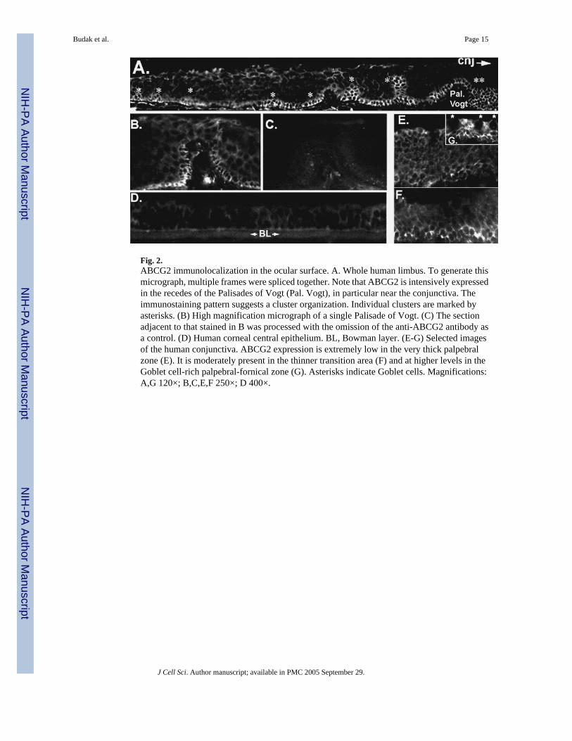
Fig. 2.ABCG2 immunolocalization in the ocular surface. A. Whole human limbus. To generate thismicrograph, multiple frames were spliced together. Note that ABCG2 is intensively expressedin the recedes of the Palisades of Vogt (Pal. Vogt), in particular near the conjunctiva. Theimmunostaining pattern suggests a cluster organization. Individual clusters are marked byasterisks. (B) High magnification micrograph of a single Palisade of Vogt. (C) The sectionadjacent to that stained in B was processed with the omission of the anti-ABCG2 antibody asa control. (D) Human corneal central epithelium. BL, Bowman layer. (E-G) Selected imagesof the human conjunctiva. ABCG2 expression is extremely low in the very thick palpebralzone (E). It is moderately present in the thinner transition area (F) and at higher levels in theGoblet cell-rich palpebral-fornical zone (G). Asterisks indicate Goblet cells. Magnifications:A,G 120×; B,C,E,F 250×; D 400×.
Budak et al. Page 15
J Cell Sci. Author manuscript; available in PMC 2005 September 29.
NIH
-PA Author Manuscript
NIH
-PA Author Manuscript
NIH
-PA Author Manuscript

Fig. 3.Reciprocal correlations between Hoechst emission and light scattering features in ocularsurface cells. (A) Human limbal epithelium. The scatter plot for all the cells (All cells) showsa main cohort and a small group of cells (LSSC) displaying very low SSC values. Note that~50% of the SP cells are LCCS cells and that some of the LSSC cells are not SP cells. (B)Representative experiment of the distribution of LSSC cells within the Hoechst plot in therabbit conjunctiva. Note that the LSSC cells (zone highlighted with a gray mask) distributeequally between the SP and G0-G1 zone. Axis denominations are provided in capital italicswithin the frames.
Budak et al. Page 16
J Cell Sci. Author manuscript; available in PMC 2005 September 29.
NIH
-PA Author Manuscript
NIH
-PA Author Manuscript
NIH
-PA Author Manuscript

Fig. 4.Phase-contrast micrographs of human limbal epithelial G0-G1 and SP cells. The non-SP cellswere collected from the center of the G0-G1 spot. Bar, 30 μm.
Budak et al. Page 17
J Cell Sci. Author manuscript; available in PMC 2005 September 29.
NIH
-PA Author Manuscript
NIH
-PA Author Manuscript
NIH
-PA Author Manuscript

Fig. 5.Hoechst and scatter plots of human corneal cells. Note the complete absence of SP and thepaucity of LSSC cells.
Budak et al. Page 18
J Cell Sci. Author manuscript; available in PMC 2005 September 29.
NIH
-PA Author Manuscript
NIH
-PA Author Manuscript
NIH
-PA Author Manuscript

Fig. 6.Immunofluorescence micrographs of BrdU in rabbit ocular surface epithelia. (A,B) Palpebralconjunctiva. The location of the mucocutaneous junction (mcj) is indicated. Arrowheads pointto some of the Goblet cells. Frame A shows a representative section from the rabbit subjectedto 10 days of BrdU treatment. About 50% of the cells incorporate label (yellow-greenfluorescence). Labeling extends to the suprabasal compartment indicating the rapid nature ofcell turnover and stratification in this epithelium. Frame B shows an equivalent section fromthe animal subjected to a 38-day BrdU chase. Label is scant. Within the proliferative basallayer BrdU is present in cells in the Goblet cell-rich zone but it is essentially absent within thebasal cells of the highly layered and Goblet-cell free zone proximal to the mucocutaneousjunction. (C) Fornical conjunctiva of the same specimen shown in B. In this area the epitheliumpresents a lobular-like organization and displays substantial BrdU retention in basal cells. (D,E)Sections incorporating bulbar conjunctiva and the limbal (Li) and central corneal (Co) zones.There is substantial BrdU incorporation in all three domains (D). Following the 38-day chase,strong BrdU retention is seen only in the cells of the limbus (E). Bar, 100 μm.
Budak et al. Page 19
J Cell Sci. Author manuscript; available in PMC 2005 September 29.
NIH
-PA Author Manuscript
NIH
-PA Author Manuscript
NIH
-PA Author Manuscript

Fig. 7.Distribution of BrdU in rabbit ocular surface epithelial cells sorted by flow cytometry. (A-D)Representative images of the experiments with limbal and conjunctival cells sorted followinga 10-day label infusion. Frames A and B are limbal SP and LSSC cells. Note that in bothpopulations the percentile of BrdU+ cells (yellow-green fluorescence) is low. Images werecaptured using dual red-green emission filtering. Unlabeled cells are shown as inserts forreference purposes. Frames C and D belong to the adherent cell fractions (Methods) fromconjunctival G0-G1 and SP cells, respectively. For this experiment, green and red emissionswere captured independently and superimposed, as graphically depicted on the left-hand sideof C. Note that in the G0-G1 fraction the majority of cells incorporate BrdU whereas only afew SP cells are BrdU-positive. (E-H) Images from an experiment where the rabbit was infusedfor the full pump duration (14–16 days) followed by a 38-day label chase. Frames F and Ecorrespond to conjunctival G0-G1 and SP respectively. Frames G and H are from the non-LSSC and LSSC limbal cells. Small inserts are images of BrdU-free nuclei.
Budak et al. Page 20
J Cell Sci. Author manuscript; available in PMC 2005 September 29.
NIH
-PA Author Manuscript
NIH
-PA Author Manuscript
NIH
-PA Author Manuscript

Fig. 8.CFDA-SE retention of freshly plated limbal epithelial cells as a function of their SSC. Thetime elapsed since plating is indicated within each plot. The decrease in CFDA-SE fluorescenceprimarily reflects cell division. The general population of adherent cells exhibits largedecreases in fluorescent intensity in the 24–48, 48–72 and 72–96 hour intervals whereas thegreat majority of the LSSC cells seem to retain most of the initial fluorescence for the first 72hours. The experiment shown is representative of two experiments with limbal and twoexperiments with conjunctival epithelial cells.
Budak et al. Page 21
J Cell Sci. Author manuscript; available in PMC 2005 September 29.
NIH
-PA Author Manuscript
NIH
-PA Author Manuscript
NIH
-PA Author Manuscript
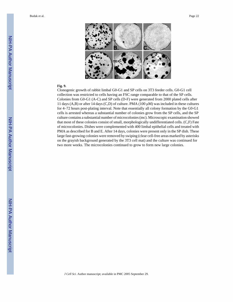
Fig. 9.Clonogenic growth of rabbit limbal G0-G1 and SP cells on 3T3 feeder cells. G0-G1 cellcollection was restricted to cells having an FSC range comparable to that of the SP cells.Colonies from G0-G1 (A-C) and SP cells (D-F) were generated from 2000 plated cells after11 days (A,B) or after 14 days (C,D) of culture. PMA (100 μM) was included in these culturesfor 4–72 hours post-plating interval. Note that essentially all colony formation by the G0-G1cells is arrested whereas a substantial number of colonies grow from the SP cells, and the SPculture contains a substantial number of microcolonies (mc). Microscopic examination showedthat most of these colonies consist of small, morphologically undifferentiated cells. (C,F) Fateof microcolonies. Dishes were complemented with 400 limbal epithelial cells and treated withPMA as described for B and E. After 14 days, colonies were present only in the SP dish. Theselarge fast-growing colonies were removed by swiping (clear cell-free areas marked by asteriskson the grayish background generated by the 3T3 cell mat) and the culture was continued fortwo more weeks. The microcolonies continued to grow to form new large colonies.
Budak et al. Page 22
J Cell Sci. Author manuscript; available in PMC 2005 September 29.
NIH
-PA Author Manuscript
NIH
-PA Author Manuscript
NIH
-PA Author Manuscript

NIH
-PA Author Manuscript
NIH
-PA Author Manuscript
NIH
-PA Author Manuscript
Budak et al. Page 23
Table 1SP cell yields for human and rabbit limbal and conjunctival tissues
N Total cells/tissue SP (% viablecells)
SP (% of total) SP/tissue
Human limbus 6 5,5 × 10⁵ 0.64 0.49 2695Human conjunctiva 4 48 × 105 0.92 0.58 27,840Rabbit limbus 8 3,3 × 10⁵ 1.21 0.88 2904Rabbit conjunctiva 16 38 × 105 1.43 0.96 36,840
Percentiles and total cell yields were calculated for each experiment and used to obtain the mean values shown in the table. N is the number of replicateexperiments.
J Cell Sci. Author manuscript; available in PMC 2005 September 29.

NIH
-PA Author Manuscript
NIH
-PA Author Manuscript
NIH
-PA Author Manuscript
Budak et al. Page 24
Table 2Percentage of BrdU-positive cells in various flow cytometry fractions obtained from Hoechst-loaded rabbit limbaland conjunctival epithelia
Experiment Conditions Tissue SP G0/G1 LSSC non-LSSC
1 10-day infusion Limbal-cs 13 57 11 51Conjunctival-cs 15 47 14 41
2 10-day infusion Conjunctival-pl 9 873 > 14-day infusion Limbal-cs 18 <1
38-day chase Conjunctival-cs 22 <1
Cs and pi indicate cytospun and plated cells, respectively All percentiles are based on counting at least 240 cells
J Cell Sci. Author manuscript; available in PMC 2005 September 29.

NIH
-PA Author Manuscript
NIH
-PA Author Manuscript
NIH
-PA Author Manuscript
Budak et al. Page 25
Table 3Yields and CFDA fluorescence in the various cohorts of cultured rabbit limbal cells
Time in culture SP G1 S-G2/M SP/G1 ×100
16 hours % viable cells 3.4 59.3 12.6 5.7CFDA fluorescence* 312 389 463 80
64 hours % viable cells 0.93 41.9 24.8 2.3CFDA fluorescence* 222 110 132 201
*Arbitrary relative units.
J Cell Sci. Author manuscript; available in PMC 2005 September 29.
Copyright © 2022 FDOKUMEN