Functional Differences between Keratins of Stratified and Simple Epithelia
13
The Rockefeller University Press, 0021-9525/98/10/487/13 $2.00 The Journal of Cell Biology, Volume 143, Number 2, October 19, 1998 487–499 http://www.jcb.org 487 Functional Differences between Keratins of Stratified and Simple Epithelia Elizabeth Hutton,* Rudolph D. Paladini, ‡ Qian-Chun Yu,* Mei Yen,* Pierre A. Coulombe, ‡ and Elaine Fuchs* *Howard Hughes Medical Institute, Department of Molecular Genetics and Cell Biology, The University of Chicago, Chicago, IL 60637; and ‡ Department of Biological Chemistry, Johns Hopkins University School of Medicine, Baltimore, Maryland 21205 Abstract. Dividing populations of stratified and simple epithelial tissues express keratins 5 and 14, and keratins 8 and 18, respectively. It has been suggested that these keratins form a mechanical framework important to cellular integrity, since their absence gives rise to a blis- tering skin disorder in neonatal epidermis, and hemor- rhaging within the embryonic liver. An unresolved fun- damental issue is whether different keratins perform unique functions in epithelia. We now address this question using transgenic technology to express a K16- 14 hybrid epidermal keratin transgene and a K18 sim- ple epithelial keratin transgene in the epidermis of mice null for K14. Under conditions where the hybrid epi- dermal keratin restored a wild-type phenotype to new- born epidermis, K18 partially but not fully rescued. The explanation does not appear to reside in an inability of K18 to form 10-nm filaments with K5, which it does in vitro and in vivo. Rather, it appears that the keratin network formed between K5 and K18 is deficient in withstanding mechanical stress, leading to perturba- tions in the keratin network in regions of the skin that are subjected either to natural or to mechanically in- duced trauma. Taken together, these findings suggest that the loss of a type I epidermal keratin cannot be fully compensated by its counterpart of simple epithe- lial cells, and that in vivo, all keratins are not equiva- lent. Key words: keratins • intermediate filaments • func- tional redundancy • epidermis • epithelia K eratins belong to the superfamily of intermediate filament (IF) 1 proteins, which have the remark- able capacity to assemble into 10-nm filaments in vitro, in the absence of auxiliary proteins or factors (for re- view see Fuchs and Weber, 1994). They are composed of two sequence types, both of which share a common, largely a-helical secondary structure, capable of forming coiled-coil heterodimers, which then further assemble to form obligatory heteropolymers. Type I keratins include K9-K20 and type II keratins encompass K1-K8 (Moll et al., 1982). While most combinations of type I and type II keratins can copolymerize in vitro (Franke et al., 1983), keratins are often coexpressed as specific pairs in vivo, where they are largely restricted to epithelial tissues (Sun et al., 1984). The innermost, mitotically active (basal) layer of many stratified epithelial tissues, including the epidermis, cor- nea, and tongue, express the type II keratin K5 and the type I keratin K14 (Fuchs and Green, 1980; Moll et al., 1982; Nelson and Sun, 1983; Byrne et al., 1994). A second natural partner for K5 is K15, which is more abundantly expressed in internal stratified squamous epithelia, such as esophagus (Moll et al., 1982; Leube et al., 1988; Lloyd et al., 1995). As keratinocytes of stratified tissues withdraw from the cell cycle and commit to differentiate, they often downregulate transcription of K5/K14 (K5/K15); concomi- tantly, as the differentiating cells move into the upper lay- ers, they induce one of five to six different pairs of kera- tin genes depending upon the particular stratified tissue (Fuchs and Green, 1980; Moll et al., 1982). In contrast to these elaborate patterns in stratified tissues, simple epithe- lia express K8 and K18, and only under certain circum- stances do they express additional keratins K7 and K19 (Moll et al., 1982; Wu et al., 1982). One role that has been ascribed to the various keratin filament networks of stratified squamous epithelia is to impart mechanical integrity to cells, without which, the cells become fragile and prone to rupturing. This function was initially proposed on the basis of transgenic mice engi- neered to express a dominant-negative K14 mutant (Vas- Address all correspondence to Elaine Fuchs, Howard Hughes Medical In- stitute, Department of Molecular Genetics and Cell Biology, The Univer- sity of Chicago, 5841 S. Maryland Avenue, Room N314, MC1028, Chi- cago, IL 60637. Tel.: (773) 702-1347. Fax: (773) 702-0141. 1. Abbreviations used in this paper: BS3, bis(sulfosuccinimidyl)suberate; EBS, epidermis bullosa simplex; IF, intermediate filament.
Transcript of Functional Differences between Keratins of Stratified and Simple Epithelia
The Rockefeller University Press, 0021-9525/98/10/487/13 $2.00The Journal of Cell Biology, Volume 143, Number 2, October 19, 1998 487–499http://www.jcb.org 487
Functional Differences between Keratins of Stratified and Simple Epithelia
Elizabeth Hutton,* Rudolph D. Paladini,
‡
Qian-Chun Yu,* Mei Yen,* Pierre A. Coulombe,
‡
and Elaine Fuchs*
*Howard Hughes Medical Institute, Department of Molecular Genetics and Cell Biology, The University of Chicago, Chicago,
IL 60637; and
‡
Department of Biological Chemistry, Johns Hopkins University School of Medicine, Baltimore, Maryland 21205
Abstract.
Dividing populations of stratified and simple epithelial tissues express keratins 5 and 14, and keratins 8 and 18, respectively. It has been suggested that these keratins form a mechanical framework important to cellular integrity, since their absence gives rise to a blis-tering skin disorder in neonatal epidermis, and hemor-rhaging within the embryonic liver. An unresolved fun-damental issue is whether different keratins perform unique functions in epithelia. We now address this question using transgenic technology to express a K16-14 hybrid epidermal keratin transgene and a K18 sim-ple epithelial keratin transgene in the epidermis of mice null for K14. Under conditions where the hybrid epi-dermal keratin restored a wild-type phenotype to new-born epidermis, K18 partially but not fully rescued. The
explanation does not appear to reside in an inability of K18 to form 10-nm filaments with K5, which it does in
vitro
and in vivo. Rather, it appears that the keratin network formed between K5 and K18 is deficient in withstanding mechanical stress, leading to perturba-tions in the keratin network in regions of the skin that are subjected either to natural or to mechanically in-duced trauma. Taken together, these findings suggest that the loss of a type I epidermal keratin cannot be fully compensated by its counterpart of simple epithe-lial cells, and that in vivo
,
all keratins are not equiva-lent.
Key words: keratins • intermediate filaments • func-
tional redundancy • epidermis • epithelia
K
eratins
belong to the superfamily of intermediatefilament (IF)
1
proteins, which have the remark-able capacity to assemble into 10-nm filaments in
vitro, in the absence of auxiliary proteins or factors (for re-view see Fuchs and Weber, 1994). They are composed oftwo sequence types, both of which share a common,largely
a
-helical secondary structure, capable of formingcoiled-coil heterodimers, which then further assemble toform obligatory heteropolymers. Type I keratins includeK9-K20 and type II keratins encompass K1-K8 (Moll etal., 1982). While most combinations of type I and type IIkeratins can copolymerize in vitro (Franke et al., 1983),keratins are often coexpressed as specific pairs in vivo,where they are largely restricted to epithelial tissues (Sunet al., 1984).
The innermost, mitotically active (basal) layer of many
stratified epithelial tissues, including the epidermis, cor-nea, and tongue, express the type II keratin K5 and thetype I keratin K14 (Fuchs and Green, 1980; Moll et al.,1982; Nelson and Sun, 1983; Byrne et al., 1994). A secondnatural partner for K5 is K15, which is more abundantlyexpressed in internal stratified squamous epithelia, such asesophagus (Moll et al., 1982; Leube et al., 1988; Lloyd etal., 1995). As keratinocytes of stratified tissues withdrawfrom the cell cycle and commit to differentiate, they oftendownregulate transcription of K5/K14 (K5/K15); concomi-tantly, as the differentiating cells move into the upper lay-ers, they induce one of five to six different pairs of kera-tin genes depending upon the particular stratified tissue(Fuchs and Green, 1980; Moll et al., 1982). In contrast tothese elaborate patterns in stratified tissues, simple epithe-lia express K8 and K18, and only under certain circum-stances do they express additional keratins K7 and K19(Moll et al., 1982; Wu et al., 1982).
One role that has been ascribed to the various keratinfilament networks of stratified squamous epithelia is toimpart mechanical integrity to cells, without which, thecells become fragile and prone to rupturing. This functionwas initially proposed on the basis of transgenic mice engi-neered to express a dominant-negative K14 mutant (Vas-
Address all correspondence to Elaine Fuchs, Howard Hughes Medical In-stitute, Department of Molecular Genetics and Cell Biology, The Univer-sity of Chicago, 5841 S. Maryland Avenue, Room N314, MC1028, Chi-cago, IL 60637. Tel.: (773) 702-1347. Fax: (773) 702-0141.
1.
Abbreviations used in this paper
: BS3, bis(sulfosuccinimidyl)suberate;EBS, epidermis bullosa simplex; IF, intermediate filament.
The Journal of Cell Biology, Volume 143, 1998 488
sar et al., 1991), and it has been documented by K14 nullmutations identified in both mice (Lloyd et al., 1995) andhumans (Chan et al., 1994; Rugg et al., 1994). In all ofthese cases, the skin displays features of epidermolysisbullosa simplex (EBS), a blistering disorder typified bybasal epidermal cell rupturing upon mild mechanical stress(Fitzpatrick et al., 1993; Fuchs and Cleveland, 1998). Simi-larly, mice expressing a mutant version of the K10 gene,normally expressed in differentiating epidermal cells, ex-hibit features of epidermolytic hyperkeratosis (EH), a dis-order analogous to EBS but involving suprabasal ratherthan basal cell degeneration (Fuchs et al., 1992; Porter etal., 1996). In humans, EBS and EH are generally autoso-mal dominant skin disorders, and correlate with the ap-pearance of clumps or aggregates of keratin filaments inthe cell cytoplasm (Anton-Lamprecht, 1994).
It is now well established that these mechanically in-duced blistering disorders of the epidermis involve muta-tions in keratins (Bonifas et al., 1991; Coulombe et al.,1991; Cheng et al., 1992; Chipev et al., 1992; Lane et al.,1992; Rothnagel et al., 1992). Recently, a number of otherkeratin disorders have been identified genetically, andthey involve additional stratified epithelial tissues, includ-ing cornea, esophagus, and hair; they are characterized bycell degeneration and mechanical fragility (for review seeFuchs and Cleveland, 1998). Where tested, the severity ofthese diseases seems to correlate with the degree to whichthe keratin mutants perturb IF assembly in vitro (Letai etal., 1993).
Human disorders involving simple epithelial keratinshave not yet been established, although a K18 mutationwas reported recently in a patient with cryptogenic cirrho-sis, a liver disorder (Ku and Omary, 1994; Ku et al., 1997),and transgenic mice expressing a mutant K18 developchronic hepatitis (Ku et al., 1995, 1996). Simple epithelialkeratins may also function to maintain hepatocyte integ-rity as evidenced by (
a
) the death of some K8 null mouseembryos resulting from hemorrhaging of the liver at thetime of vascularization (Baribault et al., 1993); (
b
) the livernecrosis in adult K8 mice when subjected to hepatectomy(Loranger et al., 1997); and (
c
) the signs of hepatitis seenin adult K18 null mice, which also display K8 aggregates intheir hepatocytes (Magin et al., 1998). Taken together,these findings suggest a special importance of simple epi-thelial keratins in the liver. A role for K8/K18 in intestinalepithelia has also surfaced in that one strain of K8 nullmice displays marked colorectal hyperplasia and inflam-mation (Baribault et al., 1994). Whether simple epithelialkeratins function to impart mechanical integrity, however,is considerably less clear than it is for stratified epithelialkeratins.
If keratins have comparable functions in different tis-sues, then what is the functional significance of the multi-plicity of keratin sequences? Is it that keratin genes havesimply duplicated and diverged over evolution, but func-tionally the gene products are redundant? Or, did differ-ent keratin genes evolve to suit the particular functionalneeds of the tissues in which they are expressed? To beginto explore the answer to this fundamental issue of keratinbiology, we have used transgenic technology to replaceK14 with K18 gene expression in K14 null mice. We showthat while K18 can coassemble with K5 into a keratin fila-
ment network in vivo and in vitro, the keratin network isunable to withstand mechanical trauma, giving rise to nat-ural skin blistering in the paws and to mechanically in-duced blistering in the back. Our findings suggest that ker-atins within a single family type have specially tailoredproperties and are not functionally redundant. They fur-ther support the notion that keratins have evolved to suitthe particular structural and functional needs of differentepithelial tissues.
Materials and Methods
Generation of Transgenes
A K14 transgene was generated using 2,100 bp of the human K14 pro-moter to drive expression of a hybrid human K16-14 cDNA. The hybridK16-14 cDNA was assembled from human K14 cDNA and human K16cDNA. A conserved SphI site was used to engineer the hybrid cDNA, re-sulting in an encoded 49-kD protein containing 368 amino acid (a.a.) resi-dues of human K16 and 105 a.a. of human K14 (Wawersik et al., 1997). Ahuman K18 transgene was generated using 2,100 bp of human K14 pro-moter to drive expression of a full-length human K18 cDNA, provided byDr. R. Oshima (Burnham Institute, La Jolla, CA) (Oshima et al., 1986).The 1,425-bp K18 cDNA was placed in a mammalian epidermal expres-sion vector (Vassar et al., 1989), modified to contain a globin intron se-quence in the 5
9
untranslated region.
Engineering the K14 Null Mice Replaced by Either the K14 or K18 Transgene
Genetically engineered transgenes were microinjected into fertilized, sin-gle cell embryos (CD-1 for K18; B6C3 F2 for K16-14), and after culturingthe embryos to the two cell stage, they were transferred to the oviducts ofpseudopregnant female mice (Vassar et al., 1991; Paladini and Coulombe,1998). 3 wk after birth, ear or tail samples were taken for DNA analysis.Presence of the transgene was determined by PCR or Southern blotting ofthese DNAs. Mice expressing the transgenes were then bred to mice thatwere null for the K14 gene (Lloyd et al., 1995). Genotyping was deter-mined by Southern blot analysis and/or PCR using the conditions andprobes described (Lloyd et al., 1995; Paladini and Coulombe, 1998).
Immunoblot Analyses
Intermediate filaments were isolated from skin samples as described (Wuet al., 1982), and keratins were solubilized in a solution of 8 M urea and10%
b
-mercaptoethanol. Water-soluble proteins were concentrated at4
8
C with a Microcon 10 concentrator (Amicon W.R. Grace & Co., Bev-erly, MA). After electrophoretic transfer of SDS-PAGE–resolved IF pro-teins to nitrocellulose, blots were pre-incubated overnight in 1% gelatin(Sigma Chemical Co., St. Louis, MO), 20 mM Tris HCl, pH 7.6, 150 mMNaCl, and 0.1% Tween-20, followed by incubation with primary antibodyfor 1 h and washing three times for 10 min each in PBS, 0.5% Tween-20.After this procedure, blots were then developed using either a HRP-con-jugated secondary antibody and chemiluminescence ECL (AmershamCorp., Arlington Heights, IL) or an alkaline phosphatase–conjugated sec-ondary antibody and color development (Bio-Rad Laboratories, Her-cules, CA). Primary antibodies included: (
a
) mouse CY-90 anti-K18 mAb(Sigma Chemical Co.), (
b
) rabbit polyclonal anti-K14 COOH-terminalpeptide antibody (Stoler et al., 1988), or (
c
) guinea pig polyclonal anti-K5COOH-terminal peptide antibody (Lersch et al., 1989). For successive in-cubations after ECL development, blots were stripped at 70
8
C in 62.5 mMTris HCl, pH 7.6, 2% SDS, 0.7%
b
-mercaptoethanol. To quantitate sig-nals, blots were scanned into a densitometer (Molecular Dynamics,Sunnyvale, CA), and protein levels were determined with ImageQuantsoftware (Molecular Dynamics).
Immunohistochemistry
Animals were killed at 1, 2, or 5 d of age, or as adults, and skin was frozenin isopentane, sectioned (8
m
m), and then processed for immunofluores-cence immunohistochemistry. Antisera were used at the following dilu-tions: anti-K14, 1:200 (Stoler et al., 1988); anti-K18, 1:200 (Sigma Chemi-
Hutton et al.
K14/K18 Replacement in Knockout Mice
489
cal Co.); anti-K6, 1:200 (gift of D. Roop, Baylor College of Medicine,Houston, TX); anti-K5, 1:200 (Lersch et al., 1989). All secondary antibod-ies, conjugated to either FITC or Texas red were obtained from JacksonImmunoResearch Laboratories (West Grove, PA).
Ultrastructural Analyses
Tissues were obtained from the skin of duplicate K14 null, K14 wild-type,K14 null/K16-14 transgenic, K14 null/K18 transgenic, and K14 wild-type/K18 transgenic animals, killed at 2–3 d of age. For conventional transmis-sion electron microscopy, tissues were immediately fixed in 2.5% glutaral-dehyde and 4% formaldehyde, pH 7.3, for 24 h, followed by postfixationwith 1% osmium tetroxide for 1 h. After en bloc staining and dehydrationwith ethanol, the samples were embedded with LX-112 medium (LaddResearch Industries, Burlington, Vermont). All tissues had been properlyoriented to ensure that the entire thickness of epidermis or mucous mem-brane would be sectioned perpendicular to the normal tissue plane. Semi-thin sections were stained with toluidine blue. Ultrathin sections of 80 nmwere cut with a diamond knife, mounted on uncoated grids, stained withuranyl acetate and lead citrate, and then observed with a JEOL-CXIIelectron microscope (JEOL USA, Inc., Peabody, MA) operated at 60 kv.
For immunoelectron microscopy, the methods of Warhol et al. (1985)and Coulombe et al. (1989) were used with some modifications. The tis-sues were (
a
) fixed with 2% PFA, 0.1% glutaraldehyde in PBS for 6 h, (
b
)dehydrated with ethanol and embedded with Lowicryl K4M medium(Ladd Research Industries) at
2
20
8
C, and then (
c
) polymerized with UVlight for 7 d at
2
20
8
C. Ultrathin sections were mounted on Formvar/car-bon-coated nickel grids. All sections were first incubated overnight withpolyclonal rabbit antisera monospecific for either K14 (1:200), K5 (1:100),or K18 (1:100), followed by a 45-min incubation with a secondary anti-body, 20-nm gold–conjugated goat anti–rabbit IgG. All grids were thenbriefly stained with uranyl acetate and lead citrate, and then examinedwith a JEOL-CXII electron microscope at 60 kV.
Filament Assembly Studies
The assembly properties of K5-K18 and K5-K14 were compared using as-says previously described (Coulombe and Fuchs, 1990; Wawersik et al.,1997). Plasmids pET-K5 and pET-K14 (Coulombe and Fuchs, 1990) andpET-K18 (Ku et al., 1997) were transformed into
Escherichia coli
strainBL21 (DE3) to generate mg amounts of the corresponding recombinanthuman keratins. Purified type I and type II keratins were mixed in a
z
45:55 molar ratio at a final concentration of 500
m
g/ml in 6 M urea buffer, in-cubated for 1 h at room temperature, and then fractionated by anion ex-change chromatography on a Pharmacia Mono Q column (PharmaciaBiotech, Inc., Piscataway, NJ). Collected fractions were analyzed by SDS-PAGE and those containing type I–type II heterotypic complexes werepooled.
To assay for tetramer formation, the K5-K18 and K5-K14 samples weredialyzed against 25 mM sodium phosphate and 10 mM
b
-ME, containing6 M or 8 M urea at pH 7.4, to remove Tris ions. Protein concentration wasadjusted to 200
m
g/ml, and chemical cross-linking was performed by add-ing BS3 (Bis-[sulfosuccinimidyl] suberate; Pierce Chemical Co., Rockford,IL) to a final concentration of 5 mM for 1 h at room temperature. Cross-linked products (6
m
g proteins) were resolved on a 4–16% gradient SDS-PAGE, and stained with Coomassie blue.
To assay for polymerization into filaments, the K5-K18 and K5-K14samples (200
m
g/ml) were serially dialyzed against the following buffers atroom temperature: (
a
) 9 M urea, 25 mM Tris-HCl, 10 mM
b
-ME, pH 8.1;(
b
) 4 M urea, 25 mM Tris-HCl, 5 mM
b
-ME, pH 7.4; and (
c
) 5 mM Tris-HCl, 5 mM
b
-ME, pH 7.4. Assemblies were examined by negative staining(1% aqueous uranyl acetate) using a Zeiss electron microscope (CarlZeiss Inc., Thornwood, NY). Polymerization efficiency was determined bysubjecting final assemblies (40
m
l aliquots,
z
8
m
g proteins) to centrifuga-tion at 100,000
g
for 60 min at 4
8
C (Airfuge; Beckman Coulter, Inc., Ful-lerton, CA). Supernatant and pellet fractions were analyzed by SDS-PAGE and Coomassie blue staining.
Results
A K16-14 Hybrid Transgene Rescues Blistering in K14 Null Mice
The hallmark of K14 null mice is that they display marked
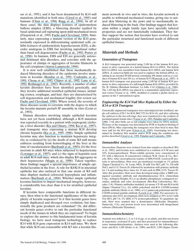
blistering over their paws shortly after birth (Lloyd et al.,1995). Our first aim was to determine if we could rescuethis phenotype by expressing a K14-like transgene using2,100 bp of the human K14 promoter (Vassar et al., 1989).Fig. 1 shows the rescue vector, which contains a significantportion of human K16 coding sequence to facilitate its dis-tinction by size from endogenous K14, and to facilitatequantitation of transgene levels relative to endogenousK14 levels. The hybrid retains the COOH-terminal epi-tope of K14, to which a peptide-specific antibody exists(Stoler et al., 1988). Transgenic mice were generated bystandard procedures, and confirmed by Southern blotanalysis (Paladini and Coulombe, 1998). Mice from twoK16-14–expressing lines were generated. Expression ofthe hybrid K16-14 at
#
75% relative to endogenous K14levels does not cause any detectable alteration in the skinof transgenic mice (Paladini and Coulombe, 1998).
To quantitate the levels of transgene expression for thepresent study, we isolated the IF proteins from tail skinsand conducted SDS-PAGE, followed by immunoblotanalysis to detect the transgene product and the endoge-nous K14, both of which share identical COOH-terminalsequences (Fig. 1). The antibody detected an additional49-kD band in the transgene skin extract (Fig. 1
A
) thatwas not present in the wild-type skin extract (Fig. 1
B
).The 52-kD band corresponding to endogenous K14 wasdetected in both transgenic and wild-type skin extracts.Densitometry scanning revealed that in mice heterozygousfor the K16-K14 transgene locus, the transgene productwas present at 17%
6
3% the level of K14 in the lower ex-pressing line (shown) and at
z
50% the level of K14 in thehigher expressing line (data not shown). These mice werethen bred to the K14 null animals to generate mice with abasal epidermal layer null for K14 and positive for theK16-14 transgene. Animals were genotyped by Southernblot analysis.
Figure 1. Expression of K16-K14 in transgenic mice. (A) A sche-matic of the replacement vector, showing 2,100 bp of the humanK14 promoter (hK14), followed by the 59 intron and 59 untrans-lated sequence from the rabbit b-globin gene (b int), followed bya K16-14 hybrid transgene followed by the K14 39 untranslatedsequence (see Allen et al., 1996 for expression vector). The dot-ted vertical lines demarcate the relative portions of K16 versusK14 coding sequences used (see Paladini and Coulombe, 1998).(B) IF proteins were isolated from the skins of K16-K14 trans-genic (lane 1) and control (lane 2) neonatal mice. Proteins werethen subjected to SDS-PAGE. Gels were subjected to immuno-blot analysis using an antibody against the COOH terminus ofK14, also present in the 49-kD transgene product. Relativeamounts of transgenic versus wild-type K14 were determined bydensitometry scanning. Sample shown is from the low expressingK16-14 transgenic mouse.

The Journal of Cell Biology, Volume 143, 1998 490
Mice from both K16-K14 transgenic/K14 null lines ap-peared normal at birth and exhibited no blistering overtheir body or paws. Animals remained blister-free as theybecame adults. A more detailed analysis of these mice willbe described elsewhere. Histological analysis revealed nosigns of epidermal abnormalities, even in the palmar re-gion of the paws, subjected naturally to substantial me-chanical stress (Fig. 2
A
). In palmar areas and elsewherethroughout the K16-14 replacement skin, an ordered pro-gram of differentiation was observed, with mitotically ac-tive basal cells in the inner layer, spinous cells, granularlayer cells, and stratum corneum (Fig. 2,
A
and
B
). Thiswas in marked contrast to K14 null skin, which displayedgross blistering within 2 d of birth (Lloyd et al., 1995). Blis-tering was very bad in the palmar regions of K14 null skinwhere rete ridges are typically seen (not shown), and wasprominent even in areas with hair follicles (Fig. 2
C
). Atthe histological level, clear cytoplasm and intraepidermalrupturing was seen within the basal layer (Fig. 2
C
; Lloydet al., 1995). These findings demonstrate that in the pres-ence of K16-14, the skin blistering phenotype typical ofneonatal K14 null mice was rescued.
To investigate whether there might be more subtle aber-rations in the basal cell cytoarchitecture of K16-14 trans-genic/K14 null replacement epidermis, we examined theskin by electron microscopy. As shown in Fig. 3, replace-ment paw skin basal cells showed abundant keratin fila-ments (
A
), not seen in the K14 null cells (
B
). These filaments
associated with desmosomes and with hemidesmosomes,as expected for wild-type keratin networks. Furthermore,no signs of microscopic blistering were visible in thesesamples. Overall, the macroscopic and microscopic ap-pearance of the K16-14 skin was similar to that of K14
6
or wild-type skin (Fig. 3
C
).
Generating K18 Transgenic Mice and Quantitating the Level of Transgene Expression
To test the functional significance of the multiplicity ofkeratin sequences, we next focused our attention on re-placing the major epidermal type I keratin K14 with themajor simple epithelial type I keratin K18. These keratinsshare only 48% sequence identity at the amino acid level,and are the most distantly related among the type I ker-atins. Fig. 4 illustrates the K14 promoter–K18 transgenethat was injected into fertilized mouse eggs to engineertransgenic mice. In this case, we did not use any epitopetags or manipulations of K18, which might complicate in-terpretation of the rescue. Mice that test positive bySouthern blot analysis for the K14 promoter–driven K18transgene were then bred to the F1 generation.
We first demonstrated that the
a
K18 mAb used for ourstudies did not cross-react with endogenous skin keratins.As judged by indirect immunofluorescence of frozen tis-sue from normal mice, no
a
K18 staining was observed(Fig. 4,
A
and
B
). In contrast to control skin, tissue from
Figure 2. The cytolysis in the basal layer of K14null epidermis is missing when these cells expressthe K16-14 transgene. Semithin (0.75 mm) sec-tions of neonatal paw skin biopsies from (A andB) K14 null/K16-14 transgenic and (C) K14 nullmice. Skins were embedded in Epon and stainedwith Toluidine blue. Section in A is from centralpaw, showing that even in areas rich in reteridges, the K16-14–expressing, K14 null paw skinappears indistinguishable from wild type. Basal
cells from the equivalent region of K14 null skin were fully cytolyzed, leading to complete separation of the upper epidermis (notshown). Areas of paw skin from areas where hair follicles were still sparse, but where rete ridges were no longer present were still nor-mal in the replacement skin (B), but partially blistered in the K14 null skin (C). Bar, 30 mm.
Figure 3. K14 null mice expressingthe K16-14 transgene have basalepidermal cells that are rich in kera-tin filament bundles and similar towild-type basal cells. Paw skin sam-ples were taken from neonatal age-matched animals that were eitherK16-14 transgenic/K14 null, K14null, or wild type. Skins were pro-cessed for electron microscopy. (A)Basal cell of K16-14 paw skin inarea of rete ridge formation. Basalcells show bundles of keratin fila-ments (Kf) in cytoplasm. No signsof microblistering or basal cell cy-tolysis were evident here, or else-where throughout the skin. (B)
Basal layer of K14 null mouse skin. Note that keratin filament bundles are largely absent. Note also the presence of basal cell cytol-ysis (asterisks). (C) Basal layer from wild-type mouse skin. Note presence of bundles of keratin filaments (Kf). Nu, nucleus; BL,basal lamina; Mi, mitochondria; De, desmosome; Hd, hemidesmosome. Bar, 0.5 mm.
Hutton et al.
K14/K18 Replacement in Knockout Mice
491
several K18 transgenic mouse lines displayed strong stain-ing with
a
K18, and this paralleled the staining pattern ob-served with
a
K14 (example shown in Fig. 4,
C
and
D
, re-spectively). Both
a
K18 and
a
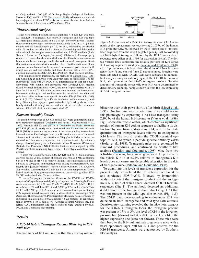
K14 staining often extendedbeyond the basal and into the suprabasal layers, reflectiveof the ability of epidermal keratins to persist long aftertheir synthesis (Fuchs and Green, 1980; Roop et al., 1987).Overall, these data demonstrated that K18 was faithfullyexpressed in a pattern identical to that of K14.
To examine the behavior of K18 protein in basal epi-dermal keratinocytes, we conducted two-dimensional gelelectrophoresis on keratins extracted from K18 transgenicmouse skin. Coomassie blue staining revealed two to threespots, not present in control skin samples, that migrated at
the molecular size (
z
44 kD) and pKi (5.5) expected forK18 (Fig. 5
A
). The identity of these spots was confirmedby
aK18 immunoblot analysis (Fig. 5 B). The size and pKiof K18 in transgenic mouse epidermis was comparable tothat observed in simple epithelia (Moll et al., 1982), sug-gesting that its expression in epidermis did not result inany substantial processing or modifications of the protein.
Heterozygous K18 positive mice were mated with K14heterozygous mice, and eventually bred further to produceK18 positive/K14 null animals. As shown in Fig. 5 C, thekeratin extracts from the skins of these animals lacked theK14 spots, but still contained K18 spots of approximatelythe same isoelectric points, size, and levels as those seen inthe transgenic skin extracts.
To assess the levels of K18 transgene expression relativeto endogenous K14, we had to modify the approach wehad used for the K16-14 protein (see Fig. 1 B), since K18and K14 did not share a common antibody epitope. In thefirst approach, we subjected the Coomassie-stained, two-dimensional gels of transgenic samples to densitometryscanning, and compared directly the relative density of theK18 versus K14 spots. Using this method, transgene ex-pression ranged from 5% to 45% of the wild-type K14 lev-els depending upon the mouse. The example shown in Fig.5 A is from the transgenic line used for this and subse-quent studies, which on a background of two K14 alleles is
Figure 4. K18 replacement vector and transgene expression inmouse skin. (Top) Schematic depicting the K18 replacementvector. The backbone of the vector is as described in Fig. 1.The full-length human K18 cDNA (Oshima et al., 1986) wasintroduced into the BamHI site of the vector. Frozen, methanol-fixed sections of back skins (10 mm) of neonatal K18 transgenicmice (tg) on a wild-type K14 background were subjected todouble-immunofluorescence microscopy using antibodies againstK18 (FITC-conjugated secondary) and K14 (Texas red–conju-gated secondary), respectively (see Materials and Methods). Aand B, wild-type skin; C and D, K18 transgenic skin. Bar, 40 mm.
Figure 5. Polyacrylamide gel analysis of IF proteins. IF proteinswere extracted from the skins of neonatal transgenic mice ex-pressing the K18 transgene on a wild-type (A and B) or K14 null(C) background. A–C, duplicate gels were either stained withCoomassie blue (A and C) or transferred to nitrocellulose paperfor immunoblot analysis with K18 (B) or K14 (not shown) anti-bodies. Shown are the type I keratins. K10 is far more abundantthan K14 (spot below K10 in A). Note that the K14 spot, con-firmed by immunoblot analysis, is missing in C. (Note: we do notknow the identity of the very acidic spot in C; it does not migrateat a pKi or size known to keratins, and it was not consistently ob-tained in our 0.6 M KCl–insoluble extracts.) (D and E) Duplicatesamples of IF proteins from wild-type (wt) and K18 transgenic(Tg) mouse skins and dilutions of purified recombinant humanK18 or K14 were subjected to one-dimensional gel electrophore-sis and immunoblot analysis using aK18 or aK14 antibodies.Loadings were: wt, 4 mg extract; Tg, 2 mg and 4 mg extract, respec-tively. Recombinant protein loadings are as indicated in nano-grams.

The Journal of Cell Biology, Volume 143, 1998 492
35% 6 5%. In the second approach, we used one dimen-sional PAGE of IF extracts from this line, followed by im-munoblot analysis with antibodies against K18 and K14,respectively. In this case, we used varying dilutions of puri-fied recombinant human K18 and K14 as standards, sothat we could determine the approximate number of nano-grams of K18 and K14 in the transgenic skin extracts (Fig.5, D and E). Two different anti-K18 antibodies, multiplegels, and multiple exposures were used in the quantita-tion, which again provided an estimate that the samplecontained K18 at levels that were 35% 6 5% the levelsof K14.
To further explore the K18 protein on a K14 null back-ground, we conducted immunoblot analysis, this time us-ing one-dimensional gel electrophoresis that enabled us toexamine and compare more samples. As shown in Fig. 6,the immunoblot data verified the presence of K18 and theabsence of K14 in the skin of some offspring from het-erozygous matings. Moreover, the K18 isolated from theseskins was found in the insoluble cytoskeletal fraction,along with the other keratins, consistent with the notion
that it existed as part of the keratin network. The levels ofK18 were at least comparable to, if not higher than, thosein the K18 transgenic mice.
Immunofluorescence of skin sections from the K18transgenic/K14 null mice revealed a pattern of aK18 anti-body staining that was similar to what we saw in the trans-genic mouse skin; in this case, however, K14 was absent(Fig. 7, A and B, respectively). This K18 was strictly due totransgene expression and not to induction of endogenous
Figure 6. Immunoblot analyses of skin IF proteins from wild-type, K14 null, K18 transgenic, and K18 rescue mice. Triton X-100–insoluble and –soluble protein extracts were prepared from backskins of 1–2-d-old mice, resolved by electrophoresis through 10%SDS–polyacrylamide gels (Wu et al., 1982) and transferred to ni-trocellulose paper. The blot was then sequentially hybridizedwith antibodies against K18, K14, and K5. After each hybridiza-tion, bound antibody was visualized by chemiluminescence (Am-ersham Corporation), and the blot was then stripped to removethe bound antibody before proceeding with the next antibody.Extracts are as indicated: WT, wild-type; KO, K14 null; K18 tg,K18 transgenic; and K18 res, two different lines of K18 rescue.Note that both transgene protein and endogenous keratins residein the insoluble fraction. Molecular mass standards at right in kD.
Figure 7. K18 expression is still maintained and does not induceK6 when K18 transgenic mice are bred onto a K14 null back-ground. Neonatal back skins and paw skins of control (notshown) and K181/K14 null mice (K18 res) were frozen andmethanol-fixed, sectioned (10 mm), and then stained with anti-bodies against the keratins indicated. (Note: depending upon thefixation/processing conditions, antibody staining of K18 some-times extended into the suprabasal layers. This happened incon-sistently in control as well as transgenic skin, and in all cases, wasalways paralleled by an identical staining pattern with aK5.) Notethe wild-type staining pattern of aK6 in the outer root sheath ofhair follicles; suprabasal epidermal induction of K6, a typicalmarker of hyperproliferative disorders did not occur in K18 res-cue skin. Bar, 40 mm.

Hutton et al. K14/K18 Replacement in Knockout Mice 493
K18, since no aK18 staining was detected in K14 null skin(not shown). The pattern of expression of K18 paralleledthat of K5, the normal partner of K14 (Fig. 7, C and D).The exchange that we genetically engineered between K18and K14 did not result in the induction within the basallayer of K6 and K16, the keratin pair known to be ex-pressed normally in the outer root sheath of the hair folli-cle (see Fig. 7 E) and induced upon wound healing and avariety of hyperproliferative disorders in the skin (Sun etal., 1984; Mansbridge and Knapp, 1987; Paladini et al.,1996). Thus, the genetic exchange appeared to occur in theabsence of other alterations in keratin expression.
Expression of the K18 Transgene on a K14 Null Background Restores Back Skin Morphology, but Not Paw Blistering
When bred on either a K14 wild-type background or a K141/2 heterozygous background, our K18 transgenic miceappeared phenotypically and histologically indistinguish-able from the wild type. These animals lived to adulthood,showed no loss in viability, and developed no signs of ab-errations in their skin or hair coat. This was important,since there are a number of cases where ectopic expressionof a keratin can induce phenotypic changes (Powell andRogers, 1990; Blessing et al., 1993; Paladini and Cou-lombe, 1998).
Even when bred to produce K181/K14 null mice, cer-tain features of the animals were typical of wild-type mice.Most notably, the back skin morphology of K181/K14 nullmice was similar to that of wild-type back skin, exhibitingno signs of basal cell cytolysis or of alterations in terminaldifferentiation (Fig. 8 A). Within 2–3 d after birth, how-ever, the paws of these mice began to display signs of EBS,including signs of basal cell degeneration within the cyto-plasm of basal cells (Fig. 8 B). In most regions of the ven-tral paw, basal epidermal cells showed gross signs of cytol-ysis (Fig. 8 C), and often the basal layer was completelydegenerated, leading to detachment of the epidermis, andonly remnants of basal cells attached to the blister floor(Fig. 8 D). Typical of EBS, suprabasal layers remained in-tact, reflective of expression of K1 and K10, concomitantwith downregulation of K5 and K14 in these layers (Fuchsand Green, 1980; Roop et al., 1987). However, in both se-verely cytolyzed and in detached regions, the overall mor-phology of the epidermis was distorted, suggesting thatsignificant perturbations had occurred in the biochemistryof these cells, presumably beginning before the time atwhich they exited the basal layer.
The phenotype of these mice was sufficiently severe thatthey died within several days after birth unless given spe-cial care. While we did not analyze internal complicationsin detail, the mice also suffered from degeneration of thetongue and oral epithelia, places where the K14 promoteris known to be active. Taken together, the expression ofK18 seemed to restore some, but clearly not all, of the de-fects caused by removal of K14 expression in mice.
Restoration of the 10-nm Filament Networkin Basal Cells of K181/K14 Null Back Skin, but Not Paw Skin
To evaluate the effects of K18 expression on cytoskeletal
architecture, we first examined the basal keratinocytes inthe paw and back skin of K18 transgenic mice on a wild-type background. At the ultrastructural level, filamentbundles within K18 basal epidermal cells appeared indis-tinguishable from those in wild-type basal epidermal cells,and no signs of keratin clumps or other perturbations weredetected (Fig. 9 A). Desmosomes and hemidesmosomesappeared normal, and both were surrounded by denselystaining keratin, as expected for the comparable wild-typestructures. Basal keratinocytes cultured from the skin ofK18 transgenic mice also displayed a seemingly normalkeratin network, which colabeled with antibodies againstK18, K5, and K14 (data not shown). Taken together, thesefindings were in agreement with similar in vitro findings byLu and Lane (1990), who demonstrated that K18 is able tointegrate into an epidermal keratin network without per-turbation. The result was also consistent with the normalappearance of the K18 transgenic mice.
We next examined the cytoskeletal architecture of theskin of K18 positive/K14 null mice. A number of the backskin basal cells of these mice displayed seemingly normalcytoskeletal networks, with bundles of cytoplasmic kera-tin filaments connecting to hemidesmosomes and desmo-somes (Fig. 9 B). This was in contrast to the back skin ofK14 null mice, but it was similar to that seen in the K16-14rescue mice (Fig. 3). In these regions of the skin, no signsof microblisters were detected, and the basal cells ap-peared generally healthy.
Whereas the majority of back skin basal cells from K18transgenic/K14 null mice appeared ultrastructurally nor-mal, an occasional cell exhibited signs of degeneration
Figure 8. K18 expression does not rescue the blistered paw phe-notype of K14 null mice. Neonatal back skins and paw skins fromK181/K141, K181/K142, and wild-type littermates were em-bedded in Epon, sectioned (0.75 mm), and then stained withToluidine blue. Sections shown are from K18 transgenic skin on aK14 null background. (A) back skin, showing no obvious abnor-malities; (B) paw skin, depicting early signs of basal cell degener-ation (arrowheads); (C) paw skin showing clear signs of basal cellcytolysis (arrows); (D) paw skin showing blister resulting fromcompletely degenerated basal epidermal layer. Double arrow,blister; arrowheads in D, fragments of basal cells left on the blis-ter floor, indicative of basal cell rupturing. Bars: (A and B) 40mm; (C and D) 20 mm.

The Journal of Cell Biology, Volume 143, 1998 494
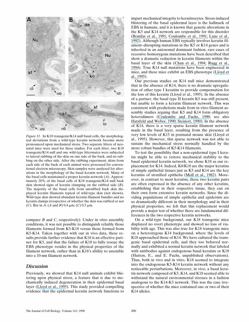
(Fig. 9 C, asterisk). Some vacuolization was also seen, afeature also frequently found in Weber-Cockayne EBS,the mildest form of human EBS (for review see Anton-Lamprecht, 1994; Fuchs and Cleveland, 1998). A few bun-dles of keratin filaments were still detected in these cells,but regions of the cytoplasm were devoid of such filaments(Fig. 9 C, large arrowhead). In a few regions of the cyto-plasm of these cells, a mixture of filaments and aggregatesof keratin-like material were detected (Fig. 9 C9). Clumpsof keratin were not seen in the K14 null skin (Lloyd et al.,1995), but are characteristic of dominant-negative actingkeratin mutants in mice and in humans (Vassar et al., 1991;Anton-Lamprecht, 1994).
Abnormalities in keratin networks were significantlymore prominent in the basal cells of the paw skin of K18
positive/K14 null mouse skin, i.e., in regions where overtskin blistering was also readily detected (Fig. 9 D). Inmany of these cells, large areas of cytoplasm were devoidof keratin, while other areas were densely packed withclumps or aggregates of keratin-like material. Theseclumps often associated with desmosomes (Fig. 9 D, inset),and at higher magnification, they appeared as aggregatesof short filamentous-like structures (D9). In Fig. 9 D0, de-picting the junction between a basal cell (BC) and aspinous cell (SP), note that the spinous cells contained anarray of keratin filaments; this is again consistent with theswitch to expression of K1 and K10 in terminally differen-tiating layers.
Immunoelectron microscopy confirmed that the clumpsof material in the basal cells were keratin, and that they la-
Figure 9. Ultrastructure of basalcells from the back skins andpaw skins of K18 transgenicmice bred on either a wild-typeor a K14 null background. Skinsof 1–2-d-old K18 transgenicmice on either a wild-type orK14 null background were pro-cessed for electron microscopyas described in the Materialsand Methods. (A) basal cellsfrom K18 transgenic/K14 wild-type back skin, depicting normalkeratin filament bundles (kf)and desmosomes (de). Paw skinshowed similar morphology. (B–D) basal cells from K18 trans-genic/K14 null back skin (B andC) or paw skin (D). The major-ity of basal cells in back skin dis-played normal morphology andkeratin filament bundles, similarto that seen in B. An occasionalbasal cell from back skin exhib-ited signs of cytolysis (asterisksand small arrowheads), withsome regions of the cytoplasmdevoid of keratin filaments(large arrowhead in C) andother regions showing somesmall aggregates or clumps ofkeratin (C9, kc). Many cells frompaw skin displayed prominentclumping of keratin both in thecytoplasm and associated withthe desmosomes (D and inset toD, respectively). D9 and D0show higher magnification to vi-sualize these clumps of keratinin more detail. Note that spinouscells (SP) contained a largelynormal keratin network, reflec-tive of the induction of K1 andK10 in these layers. BL, basallamina; mi, mitochondria; hd,hemidesmosome; Nu, nucleus; BC,basal cell. Bar in A: (A) 0.4 mm;(B, C9, D9, D0) 0.3 mm; (C) 0.9 mm;(D) 0.8 mm; (inset to D) 0.1 mm.

Hutton et al. K14/K18 Replacement in Knockout Mice 495
beled not only with antibodies against K18, but also withantibodies against K5 (Fig. 10). Since these clumps werenot detected in K14 null mice, we conclude that the clumpsrepresent a new structure produced from a combination ofK18 and K5, or possibly other cytoplasmic proteins.
The Keratin Network in K18 Replacement Skin Cannot Withstand Mechanical Trauma
A priori, the detection of keratin clumps in the K18 re-placement skin could imply that the assembly process isdefective, or that there is an excess of K5 relative to K18.Either case might result in the accumulation of partially
polymerized keratin material. Alternatively, it could be anindication that the resulting keratin network formed byK18/K5 filaments is unstable, and collapses partially uponmechanical stress. To distinguish between these possibili-ties, we conducted an experiment to see how the basal epi-dermal layer would perform under mechanical stress.
Four 2-d-old K14 null/K181 replacement mice and twocontrol littermates were subjected to mild rubbing of theback skin, which was then processed for ultrastructuralanalysis immediately thereafer (within 10 min). In eachcase, only the left side of the middle lower back wasrubbed (12 times with a cotton-tipped applicator); theright side of each mouse was not rubbed, and was used as acontrol for each mouse.
Some basal epidermal cells of rubbed, K18 transgenic/K14 null skin survived the rubbing, as judged by the pres-ence of a seemingly normal keratin filament network andgenerally healthy cytoplasm (Fig. 11 A). However, a num-ber of cells displayed clumps or aggregates of keratin ma-terial (Fig. 11 B). The aberrancies in keratin networkswere similar, but more extensive than those seen in occa-sional back skin cells of unrubbed K18 replacement skinand in many paw skin cells. As expected, we did not ob-serve signs of cell cytolysis, a delayed response which inhuman EBS occurs subsequent to alterations in cytoskele-tal architecture (for review see Anton-Lamprecht, 1994).
In contrast to the basal cells from rubbed back skin ofthe K181/K14 null mice, cells from similarly rubbed backskin of control mice appeared uniformly healthy, with lit-tle or no perturbations in overall morphology (Fig. 11 C).Taken together, our findings suggest that the perturba-tions detected in basal cells of K18 replacement skin areaccentuated by mechanical stress.
When Combined in the Presence of 6 MUrea, K5 and K18 Associate in a 1:1 Ratio to Form a Complex That Then Assembles into Filaments Efficiently In Vitro
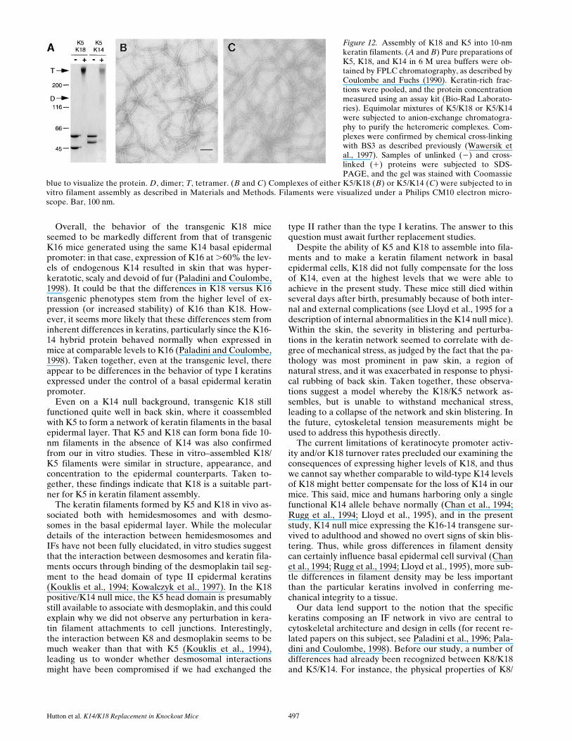
Our in vivo studies suggested that K5 and K18 can assem-ble into keratin filaments. To test this hypothesis in vitroand analyze this interaction in more detail, we first demon-strated that K5 and K18 can form stable heterotypic com-plexes under 6 M urea buffer conditions. We showed thisby anion exchange chromatography (not shown) and bychemical cross-linking (Fig. 12 A). Cross-linking using BS3showed that the major K5-K18 and K5-K14 speciesformed under these conditions was a 240-kD product, withsmall amounts of an z130-kD product (Fig. 12 A). Asshown previously (Coulombe and Fuchs, 1990), these spe-cies correspond to covalently cross-linked heterotetramersand heterodimers of keratins, respectively. When the ureaconcentration was raised to 8 M under otherwise identicalconditions, the K5-K18 tetramers were destabilized to agreater extent than K5-K14 ones (data not shown). Takentogether, these data show that K18 can readily form het-erotetramers in combination with K5, although these areslightly less stable than those formed from K5-K14.
When dialyzed under standard epidermal keratin as-sembly buffer, K5-K18 heterotypic complexes polymer-ized efficiently (90%) into 10-nm filaments that are similarto those formed by K5-K14 (.95% efficiency) (Fig. 12,
Figure 10. Keratin clumps in K18 transgenic/K14 null basal cellscontain a mixture of K5 and K18. Paw skin from K18 transgenic/K14 null animals was embedded in Lowicryl and subjected to 30-nm gold labeling with antibodies against either K18 (A) or K5(B) as described previously (Coulombe et al., 1989). de, desmo-some; hd, hemidesmosome; kc, keratin clumps. Bar, 0.2 mm.
The Journal of Cell Biology, Volume 143, 1998 496
compare B and C, respectively). Under in vitro assemblyconditions, it was not possible to distinguish reliably thosefilaments formed from K5-K18 versus those formed fromK5-K14. Taken together with our in vivo data, these re-sults provide further evidence that K18 is an effective part-ner for K5, and that the failure of K18 to fully rescue theEBS phenotype resides in the physical properties of thefilament network, rather than in K18’s ability to assembleinto a 10-nm filament network.
DiscussionPreviously, we showed that K14 null animals exhibit blis-tering upon physical stress, a feature that is due to me-chanically induced degeneration in their epidermal basallayer (Lloyd et al., 1995). This study provided compellingevidence that the epidermal keratin network functions to
impart mechanical integrity to keratinocytes. Stress-inducedblistering of the basal epidermal layer is the hallmark ofEBS in humans, and it is known that genetic alterations inthe K5 and K14 network are responsible for this disorder(Bonifas et al., 1991; Coulombe et al., 1991; Lane et al.,1992). Although human EBS typically involves keratin fil-ament–disrupting mutations in the K5 or K14 genes and isinherited in an autosomal dominant fashion, rare cases ofrecessive homozygous mutations have been described thatshow a dramatic reduction in keratin filaments within thebasal layer of the skin (Chan et al., 1994; Rugg et al.,1994). True K14 null mutations have been engineered inmice, and these mice exhibit an EBS phenotype (Lloyd etal., 1995).
Our previous studies on K14 null mice demonstratedthat in the absence of K14, there is no dramatic upregula-tion of other type I keratins to provide compensation forthe loss of this keratin (Lloyd et al., 1995). In the absenceof a partner, the basal type II keratin K5 was still present,but unable to form a keratin filament network. This wasconsistent with predictions made from in vitro filament as-sembly studies arguing that K5 and K14 form obligatoryheterodimers (Coulombe and Fuchs, 1990; see alsoHatzfeld and Weber, 1990; Steinert, 1990). In the absenceof K14, there is a very sparse keratin filament networkmade in the basal layer, resulting from the presence ofvery low levels of K15 in postnatal mouse skin (Lloyd etal., 1995). However, this sparse network was not able tosustain the mechanical stress normally handled by themore robust bundles of K5-K14 filaments.
To test the possibility that a non-epidermal type I kera-tin might be able to restore mechanical stability to thebasal epidermal keratin network, we chose K18 as our re-placement for K14. Indeed, K8/K18 are the major keratinsof simple epithelial tissues just as K5 and K14 are the keykeratins of stratified epithelia (Moll et al., 1982). More-over, in contrast to most keratins, these two keratin pairsare often expressed in the absence of any other keratins,establishing that in their respective tissue, they can ontheir own form extensive keratin networks. Since the di-viding populations of simple epithelia and epidermis areso dramatically different in their morphology and in theirphysical properties, we felt that this replacement wouldprovide a major test of whether there are fundamental dif-ferences in the two respective keratin networks.
On a wild-type background, our K18 transgenic micedisplayed no overt phenotype and showed no loss of via-bility with age. This was also true for K18 transgenic miceon a heterozygous K14 background, where the levels ofK18 approached those of K14. We have cultured the trans-genic basal epidermal cells, and they too behaved nor-mally and exhibited a normal keratin network that labeledwith antibodies against endogenous basal keratins or K18(Hutton, E., and E. Fuchs, unpublished observations).Thus, both in vivo and in vitro, K18 seemed to integrateinto the endogenous K5-K14 keratin network without anynoticeable perturbations. Moreover, in vivo, a basal kera-tin network composed of K5, K14, and K18 seemed able towithstand the natural environmental stresses in a fashionanalogous to the K14-K5 network. This was the case irre-spective of whether the mice contained one or two of theirK14 alleles.
Figure 11. In K18 transgenic/K14 null basal cells, the morpholog-ical deviations from a wild-type keratin network become morepronounced upon mechanical stress. Two separate litters of neo-natal mice were used for these studies. For each litter, two K18transgenic/K14 null and one wild-type littermates were subjectedto lateral rubbing of the skin on one side of the back, and no rub-bing on the other side. After the rubbing experiment, skins fromeach side of the back of each animal were processed for conven-tional electron microscopy. Skin samples were analyzed for alter-ations in the morphology of the basal keratin network. Many ofthe basal cells maintained a proper keratin network (A). Approx-imately 20% of the basal cells of K18 transgenic/K14 null backskin showed signs of keratin clumping on the rubbed side (B).The majority of the basal cells from unrubbed back skin dis-played keratin filaments typical of wild-type skin (not shown).Wild-type skin showed abundant keratin filament bundles and nokeratin clumps irrespective of whether the skin was rubbed or not(C). Bar in A: (A and B) 0.4 mm; (C) 0.3 mm.
Hutton et al. K14/K18 Replacement in Knockout Mice 497
Overall, the behavior of the transgenic K18 miceseemed to be markedly different from that of transgenicK16 mice generated using the same K14 basal epidermalpromoter: in that case, expression of K16 at .60% the lev-els of endogenous K14 resulted in skin that was hyper-keratotic, scaly and devoid of fur (Paladini and Coulombe,1998). It could be that the differences in K18 versus K16transgenic phenotypes stem from the higher level of ex-pression (or increased stability) of K16 than K18. How-ever, it seems more likely that these differences stem frominherent differences in keratins, particularly since the K16-14 hybrid protein behaved normally when expressed inmice at comparable levels to K16 (Paladini and Coulombe,1998). Taken together, even at the transgenic level, thereappear to be differences in the behavior of type I keratinsexpressed under the control of a basal epidermal keratinpromoter.
Even on a K14 null background, transgenic K18 stillfunctioned quite well in back skin, where it coassembledwith K5 to form a network of keratin filaments in the basalepidermal layer. That K5 and K18 can form bona fide 10-nm filaments in the absence of K14 was also confirmedfrom our in vitro studies. These in vitro–assembled K18/K5 filaments were similar in structure, appearance, andconcentration to the epidermal counterparts. Taken to-gether, these findings indicate that K18 is a suitable part-ner for K5 in keratin filament assembly.
The keratin filaments formed by K5 and K18 in vivo as-sociated both with hemidesmosomes and with desmo-somes in the basal epidermal layer. While the moleculardetails of the interaction between hemidesmosomes andIFs have not been fully elucidated, in vitro studies suggestthat the interaction between desmosomes and keratin fila-ments occurs through binding of the desmoplakin tail seg-ment to the head domain of type II epidermal keratins(Kouklis et al., 1994; Kowalczyk et al., 1997). In the K18positive/K14 null mice, the K5 head domain is presumablystill available to associate with desmoplakin, and this couldexplain why we did not observe any perturbation in kera-tin filament attachments to cell junctions. Interestingly,the interaction between K8 and desmoplakin seems to bemuch weaker than that with K5 (Kouklis et al., 1994),leading us to wonder whether desmosomal interactionsmight have been compromised if we had exchanged the
type II rather than the type I keratins. The answer to thisquestion must await further replacement studies.
Despite the ability of K5 and K18 to assemble into fila-ments and to make a keratin filament network in basalepidermal cells, K18 did not fully compensate for the lossof K14, even at the highest levels that we were able toachieve in the present study. These mice still died withinseveral days after birth, presumably because of both inter-nal and external complications (see Lloyd et al., 1995 for adescription of internal abnormalities in the K14 null mice).Within the skin, the severity in blistering and perturba-tions in the keratin network seemed to correlate with de-gree of mechanical stress, as judged by the fact that the pa-thology was most prominent in paw skin, a region ofnatural stress, and it was exacerbated in response to physi-cal rubbing of back skin. Taken together, these observa-tions suggest a model whereby the K18/K5 network as-sembles, but is unable to withstand mechanical stress,leading to a collapse of the network and skin blistering. Inthe future, cytoskeletal tension measurements might beused to address this hypothesis directly.
The current limitations of keratinocyte promoter activ-ity and/or K18 turnover rates precluded our examining theconsequences of expressing higher levels of K18, and thuswe cannot say whether comparable to wild-type K14 levelsof K18 might better compensate for the loss of K14 in ourmice. This said, mice and humans harboring only a singlefunctional K14 allele behave normally (Chan et al., 1994;Rugg et al., 1994; Lloyd et al., 1995), and in the presentstudy, K14 null mice expressing the K16-14 transgene sur-vived to adulthood and showed no overt signs of skin blis-tering. Thus, while gross differences in filament densitycan certainly influence basal epidermal cell survival (Chanet al., 1994; Rugg et al., 1994; Lloyd et al., 1995), more sub-tle differences in filament density may be less importantthan the particular keratins involved in conferring me-chanical integrity to a tissue.
Our data lend support to the notion that the specifickeratins composing an IF network in vivo are central tocytoskeletal architecture and design in cells (for recent re-lated papers on this subject, see Paladini et al., 1996; Pala-dini and Coulombe, 1998). Before our study, a number ofdifferences had already been recognized between K8/K18and K5/K14. For instance, the physical properties of K8/
Figure 12. Assembly of K18 and K5 into 10-nmkeratin filaments. (A and B) Pure preparations ofK5, K18, and K14 in 6 M urea buffers were ob-tained by FPLC chromatography, as described byCoulombe and Fuchs (1990). Keratin-rich frac-tions were pooled, and the protein concentrationmeasured using an assay kit (Bio-Rad Laborato-ries). Equimolar mixtures of K5/K18 or K5/K14were subjected to anion-exchange chromatogra-phy to purify the heteromeric complexes. Com-plexes were confirmed by chemical cross-linkingwith BS3 as described previously (Wawersik etal., 1997). Samples of unlinked (2) and cross-linked (1) proteins were subjected to SDS-PAGE, and the gel was stained with Coomassie
blue to visualize the protein. D, dimer; T, tetramer. (B and C) Complexes of either K5/K18 (B) or K5/K14 (C) were subjected to invitro filament assembly as described in Materials and Methods. Filaments were visualized under a Philips CM10 electron micro-scope. Bar, 100 nm.
The Journal of Cell Biology, Volume 143, 1998 498
K18 heterodimers and filaments assembled from them dif-fer markedly from K5/K14 heterodimers and their corre-sponding filaments (Franke et al., 1983; Hatzfeld andFranke, 1985; Coulombe and Fuchs, 1990; Hatzfeld andWeber, 1990; Hoffmann and Franke, 1997). Moreover, invivo, K8/K18 networks can reorganize during mitosis insome cells, whereas K5/K14 networks do not, suggestingthe possibility that K8/K18 networks may be more dy-namic than K5/K14 networks (Franke et al., 1983). Dra-matic sequence and size differences in the head and tailsegments and in the posttranslational modifications of ker-atins are likely to account for at least a part of these dra-matic variations in keratin networks (Lu and Lane, 1990;Liao et al., 1995 and references therein; for review seeFuchs and Weber, 1994). It is tempting to speculate thatK18 cannot fully compensate for K14 because it creates amore dynamic keratin network that is not entirely compat-ible with the structural requirements imposed upon a tis-sue like the epidermis.
The seemingly less stable aspects of the K5/K18 networkcould also have arisen from genetically forcing an undesir-able partner upon K5. That keratins have distinct prefer-ences for their partners has been recently inferred fromK18 knockout studies, where when left without its normalpartner, K8 can successfully compete for K19, normallythe partner of K7 (Magin et al., 1998). To assess whetherK8/K18 can replace K5/K14 must await further replace-ment studies.
In summary, our study suggests that the multiplicity ofkeratin sequences is not simply an evolutionary quirk, butthat these proteins have diverged to perform specific func-tions in higher eukaryotic epithelia. Whereas keratin net-works are a universal feature of epithelia, their density,composition, and organization vary dramatically and seemto be tailored to the varied shapes and structural require-ments of individual epithelial cells.
A special thank you goes to L. Degenstein for her help in transgenic miceaspects of this work, and in conducting the mouse skin rubbing experi-ments. We thank D. Dugger for technical assistance in transgenic mouseengineering; G. Strasser and D. Lourim for technical assistance in somephases of the cell and molecular biology; Dr. C. Bauer for his expert assis-tance in electron microscopy and in assisting with some of the photogra-phy and figure preparations; E. Smith and C. Wellek for their help withthe computer-assisted art work; J. Fradette (University Laval, Quebec,Canada) for providing the chemical cross-linking data; Dr. M.B. Omary(Stanford University, Palo Alto, CA) for providing the pET-K18 bacterialexpression clone; and Dr. D. Roop (Baylor University School of Medi-cine) for his gift of anti-K6 antiserum.
This work was supported by grants from the National Institutes ofHealth (AR27883, to E. Fuchs; AR44232 to P. Coulombe). E. Fuchs is aHoward Hughes Medical Institute investigator.
Received for publication 9 July 1998 and in revised form 2 September1998.
References
Allen, E., Q.-C. Yu, and E. Fuchs. 1996. Abnormalities in desmosomes, prolif-eration and differentiation in the epidermis of mice expressing a mutant des-mosomal cadherin. J. Cell Biol. 133:1367–1382.
Anton-Lamprecht, I. 1994. Ultrastructural identification of basic abnormalitiesas clues to genetic disorders of the epidermis. J. Invest. Dermatol. 103:65–125.
Baribault, H., J. Price, K. Miyai, and R.G. Oshima. 1993. Mid-gestational le-thality in mice lacking keratin 8. Genes Dev. 7:1191–1201.
Baribault, H., J. Penner, R.V. Iozzo, and M. Wilson-Heiner. 1994. Colorectal
hyperplasia and inflammation in keratin 8-deficient FVB/N mice. GenesDev. 8:2964–2973.
Blessing, M., U. Ruther, and W. Franke. 1993. Ectopic synthesis of epidermalcytokeratins in pancreatic islet cells of transgenic mice interferes with cy-toskeletal order and insulin production. J. Cell Biol. 120:743–755.
Bonifas, J.M., A.L. Rothman, and E.H. Epstein. 1991. Epidermolysis bullosasimplex: evidence in two families for keratin gene abnormalities. Science.254:1202–1205.
Byrne, C., M. Tainsky, and E. Fuchs. 1994. Programming gene expression in de-veloping epidermis. Development (Camb.). 120:2369–2383.
Chan, Y.-M., I. Anton-Lamprecht, Q.-C. Yu, A. Jackel, B. Zabel, J.-P. Ernst,and E. Fuchs. 1994. A human keratin 14 “knockout”: the absence of K14leads to severe epidermolysis bullosa simplex and a function for an interme-diate filament protein. Genes Dev. 8:2574–2587.
Cheng, J., A.J. Syder, Q.-C. Yu, A. Letai, A.S. Paller, and E. Fuchs. 1992. Thegenetic basis of epidermolytic hyperkeratosis: a disorder of differentiation-specific epidermal keratin genes. Cell. 70:811–819.
Chipev, C.C., B.P. Korge, N. Markova, S.J. Bale, J.J. DiGiovanna, J.G. Comp-ton, and P.M. Steinert. 1992. A leucine→proline mutation in the H1 subdo-main of keratin 1 causes epidermolytic hyperkeratosis. Cell. 70:821–828.
Coulombe, P., and E. Fuchs. 1990. Elucidating the early stages of keratin fila-ment assembly. J. Cell Biol. 111:153–169.
Coulombe, P.A., R. Kopan, and E. Fuchs. 1989. Expression of keratin K14 inthe epidermis and hair follicle: Insights into complex programs of differenti-ation. J. Cell Biol. 109:2295–2312.
Coulombe, P.A., M.E. Hutton, A. Letai, A. Hebert, A.S. Paller, and E. Fuchs.1991. Point mutations in human keratin 14 genes of epidermolysis bullosasimplex patients: genetic and functional analyses. Cell. 66:1301–1311.
Fitzpatrick, T.B., A.Z. Eisen, K. Wolff, I.M. Freedberg, and K.F. Austen. 1993.Dermatology in general medicine. Vol. I and II. McGraw-Hill, Inc., NewYork. 2979 pp.
Franke, W.W., D.L. Schiller, M. Hatzfeld, and S. Winter. 1983. Protein com-plexes of intermediate-sized filaments: melting of cytokeratin complexes inurea reveals different polypeptide separation characteristics. Proc. Natl.Acad. Sci. USA. 80:7113–7117.
Fuchs, E., and D. Cleveland. 1998. A structural scaffolding of intermediate fila-ments in health and disease. Science. 279:514–519.
Fuchs, E., and H. Green. 1980. Changes in keratin gene expression during ter-minal differentiation of the keratinocyte. Cell. 19:1033–1042.
Fuchs, E., and K. Weber. 1994. Intermediate filaments: structure, dynamics,function, and disease. Ann. Rev. Biochem. 63:345–382.
Fuchs, E., R.A. Esteves, and P.A. Coulombe. 1992. Transgenic mice expressinga mutant keratin 10 gene reveal the likely genetic basis for epidermolytic hy-perkeratosis. Proc. Natl. Acad. Sci. USA. 89:6906–6910.
Hatzfeld, M., and W.W. Franke. 1985. Pair formation and promiscuity of cyto-keratins: formation in vitro of heterotypic complexes and intermediate-sizedfilaments by homologous and heterologous recombinations of purifiedpolypeptides. J. Cell Biol. 101:1826–1841.
Hatzfeld, M., and K. Weber. 1990. The coiled coil of in vitro assembled keratinfilaments is a heterodimer of type I and II keratins: Use of site-specific mu-tagenesis and recombinant protein expression. J. Cell Biol. 110:1199–1210.
Hofmann, I., and W.W. Franke. 1997. Heterotypic interactions and filament as-sembly of type I and type II cytokeratins in vitro: viscometry and determina-tions of relative affinities. Eur. J. Cell Biol. 72:122–132.
Kouklis, P., E. Hutton, and E. Fuchs. 1994. Making the connection: Keratin in-termediate filaments and desmosomes proteins. J. Cell Biol. 127:1049–1060.
Kowalczyk, A.P., E.A. Bornslaeger, J.E. Borgwardt, H.L. Palka, A.S. Dhaliwal,C.M. Corcoran, M.F. Denning, and K.J. Green. 1997. The amino-terminaldomain of desmoplakin binds to plakoglobin and clusters desmosomal cad-herin–plakoglobin complexes. J. Cell Biol. 139:773–784.
Ku, N., and M.B. Omary. 1994. Identification of the major physiologic phos-phorylation site of human keratin 18: Potential kinases and a role in filamentreorganization. J. Cell Biol. 127:161–171.
Ku, N., S. Michie, R.G. Oshima, and M.B. Omary. 1995. Chronic Hepatitis,hepatocyte fragility, and increased soluble phosphoglycokeratins in trans-genic mice expressing a keratin 18 conserved arginine mutant. J. Cell Biol.131:1303–1314.
Ku, N., S.A. Michie, R.M. Soetikno, E.Z. Resurreccion, R.L. Broome, R.G.Oshima, and M.B. Omary. 1996. Susceptibility to hepatotoxicity in trans-genic mice that express a dominant-negative human keratin 18 mutant. J.Clin. Invest. 98:1034–1046.
Ku, N.O., T.L. Wright, N.A. Terrault, R. Gish, and M.B. Omary. 1997. Muta-tion of human keratin 18 in association with cryptogenic cirrhosis. J. Clin. In-vest. 99:19–23.
Lane, E.B., E.L. Rugg, H. Navsaria, I.M. Leigh, A.H.M. Heagerty, A. Ishida-Yamamoto, and R.A.J. Eady. 1992. A mutation in the conserved helix termi-nation peptide of keratin 5 in hereditary skin blistering. Nature. 356:244–246.
Lersch, R., V. Stellmach, C. Stocks, G. Giudice, and E. Fuchs. 1989. Isolation,sequence, and expression of a human keratin K5 gene: transcriptional regu-lation of keratins and insights into pairwise control. Mol. Cell. Biol. 9:3685–3697.
Letai, A., P.A. Coulombe, M.B. McCormick, Q.-C. Yu, E. Hutton, and E.Fuchs. 1993. Disease severity correlates with position of keratin point muta-tions in patients with epidermolysis bullosa simplex. Proc. Natl. Acad. Sci.USA. 90:3197–3201.
Hutton et al. K14/K18 Replacement in Knockout Mice 499
Leube, R.E., B.L. Bader, F.X. Bosch, R. Zimbelmann, T. Achtstaetter, andW.W. Franke. 1988. Molecular characterization and expression of the strati-fication-related cytokeratin 4 and 15. J. Cell Biol. 106:1249–1261.
Liao, J., L.A. Lowthert, N. Ku, R. Fernandez, and M.B. Omary. 1995. Dynam-ics of human keratin 18 phosphorylation: Polarized distribution of phosphor-ylated keratins in simple epithelial tissues. J. Cell Biol. 131:1291–1301.
Lloyd, C., Q.-C. Yu, J. Cheng, K. Turksen, L. Degenstein, E. Hutton, and E.Fuchs. 1995. The basal keratin network of stratified squamous epithelia: De-fining K15 function in the absence of K14. J. Cell Biol. 129:1329–1344.
Loranger, A., S. Duclos, A. Grenier, J. Price, M. Wilson-Heiner, H. Baribault,and N. Marceau. 1997. Simple epithelium keratins are required for mainte-nance of hepatocyte integrity. Am. J. Pathol. 151:1673–1683.
Lu, X., and E.B. Lane. 1990. Retrovirus-mediated transgenic keratin expressionin cultured fibroblasts: specific domain functions in keratin stabilization andfilament formation. Cell. 62:681–696.
Magin, T.M., R. Schroder, S. Leitgeb, F. Wanninger, K. Zatloukal, C. Grund,and D.W. Melton. 1998. Lessons from keratin 18 knockout mice: Formationof novel keratin filaments, secondary loss of keratin 7 and accumulation ofliver-specific keratin 8 positive aggregates. J. Cell Biol. 140:1441–1451.
Mansbridge, J.N., and A.M. Knapp. 1987. Changes in keratinocyte maturationduring wound healing. J. Invest. Dermatol. 89:253–262.
Moll, R., W.W. Franke, D.L. Schiller, B. Geiger, and R. Krepler. 1982. The cat-alog of human cytokeratins: patterns of expression in normal epithelia, tu-mors, and cultured cells. Cell. 31:11–24.
Nelson, W., and T.-T. Sun. 1983. The 50- and 58-kdalton keratin classes as mo-lecular markers for stratified squamous epithelia: Cell culture studies. J. CellBiol. 97:244–251.
Oshima, R.G., J.L. Millan, and G. Cecena. 1986. Comparison of mouse and hu-man keratin 18: a component of intermediate filaments expressed prior toimplantation. Differentiation. 33:61–68.
Paladini, R.D., and P.A. Coulombe. 1998. Directed expression of keratin 16 tothe progenitor basal cells of transgenic mouse skin delays skin maturation. J.Cell Biol. 142:1035–1051.
Paladini, R.D., K. Takahashi, N.S. Bravo, and P.A. Coulombe. 1996. Onset ofreepithelialization after skin injury correlates with a reorganization of kera-tin filaments in wound edge keratinocytes: Defining a potential role for ker-atin 16. J. Cell Biol. 132:381–397.
Porter, R.M., S. Leitgeb, D.W. Melton, O. Swensson, R.A. Eady, and T.M. Ma-gin. 1996. Gene targeting at the mouse cytokeratin 10 locus: Severe skin fra-gility and changes of cytokeratin expression in the epidermis. J. Cell Biol.
132:925–936.Powell, B.C., and G.E. Rogers. 1990. Cyclic hairloss and regrowth in transgenic
mice overexpressing an intermediate filament gene. EMBO (Eur. Mol. Biol.Organ.) J. 9:1485–1493.
Roop, D.R., H. Huitfeldt, A. Kilkenny, and S.H. Yuspa. 1987. Regulated ex-pression of differentiation-associated keratins in cultured epidermal cells de-tected by monospecific antibodies to unique peptides of mouse epidermalkeratins. Differentiation. 35:143–150.
Rothnagel, J.A., A.M. Dominey, L.D. Dempsey, M.A. Longley, D.A. Green-halgh, T.A. Gagne, M. Huber, E. Frenk, D. Hohl, and D.R. Roop. 1992. Mu-tations in the rod domains of keratins 1 and 10 in epidermolytic hyperkerato-sis. Science. 257:1128–1130.
Rugg, E.L., W.H.I. McLean, E.B. Lane, R. Pitera, J.R. McMillan, P.J.C. Dop-ping-Hepenstal, H.A. Navsaria, I.M. Leigh, and R.A.J. Eady. 1994. A func-tional “knockout” of human keratin 14. Genes Dev. 8:2563–2573.
Steinert, P.M. 1990. The two-chain coiled-coil molecular of native epidermalkeratin intermediate filaments is a type I-type II heterodimer. J. Biol. Chem.265:8766–8774.
Stoler, A., R. Kopan, M. Duvic, and E. Fuchs. 1988. The use of monospecificantibodies and cRNA probes reveals abnormal pathways of terminal differ-entiation in human epidermal diseases. J. Cell Biol. 107:427–446.
Sun, T.-T., R. Eichner, A. Schermer, D. Cooper, W.G. Nelson, and R.A. Weiss.1984. The transformed phenotype. In The Cancer Cell. Vol. 1. A. Levine, W.Topp, G. van de Woude, and J.D. Watson, editors. Cold Spring Harbor Lab-oratory, Cold Spring Harbor, New York. 169–176.
Vassar, R., M. Rosenberg, S. Ross, A. Tyner, and E. Fuchs. 1989. Tissue-spe-cific and differentiation-specific expression of a human K14 keratin gene intransgenic mice. Proc. Natl. Acad. Sci. USA. 86:1563–1567.
Vassar, R., P.A. Coulombe, L. Degenstein, K. Albers, and E. Fuchs. 1991. Mu-tant keratin expression in transgenic mice causes marked abnormalities re-sembling a human genetic skin disease. Cell. 64:365–380.
Warhol, M.J., J.M. Lucocq, E. Carlemalm, and J. Roth. 1985. Ultrastructural lo-calization of keratin proteins in human skin using low-temperature embed-ding and the protein A-gold technique. J. Invest. Dermatol. 84:69–72.
Wawersik, M., R.D. Paladini, E. Noensie, and P.A. Coulombe. 1997. A prolineresidue in the alpha-helical rod domain of type I keratin 16 destabilizes kera-tin heterotetramers. J. Biol. Chem. 272:32557–32565.
Wu, Y.-J., L.M. Parker, N.E. Binder, M.A. Beckett, J.H. Sinard, C.T. Griffiths,and J.G. Rheinwald. 1982. The mesothelial keratins: a new family of cyto-skeletal proteins identified in cultured mesothelial cells. Cell. 31:693–703.