
Histopathological analysis of renal cystic epithelia in the Pkd2WS25/- mouse model of ADPKD
Upload
independentCategory
view
1download
0
J Physiol 587.12 (2009) pp 2887–2901 2887
Functional interactions between P2X4 and P2X7 receptorsfrom mouse salivary epithelia
Griselda Casas-Pruneda1, Juan Pablo Reyes2, Gabriela Perez-Flores1, Patricia Perez-Cornejo1
and Jorge Arreola2
1School of Medicine and 2Institute of Physics, Universidad Autonoma de San Luis Potosı, San Luis Potosı, SLP, Mexico
Mouse parotid acinar cells express P2X4 and P2X7 receptors (mP2X4R and mP2X7R) whosephysiological function remains undetermined. Here we show that mP2X4R expressed inHEK-293 cells do not allow the passage of tetraethylammonium (TEA+) and promote little,if any, ethidium bromide (EtBr) uptake when stimulated with ATP or BzATP. In contrast,mP2X7R generates slowly decaying TEA+ current, sustained Na+ current and promotesrobust EtBr uptake. However, ATP-activated TEA+ current from acinar cells was unlike thatgenerated by mP2X7R or mP2X4R. Functional interactions between mP2X4R and mP2X7Rwere investigated in HEK cells co-transfected with different mP2X4 : mP2X7 cDNA ratios andusing solutions containing either TEA+ or Na+ ions. Co-expressed channels generated a TEA+
current that displayed faster decay during ATP stimulation than mP2X7R alone. Moreover,cells transfected with a 2 : 1 cDNA ratio displayed decaying kinetics similar to those observedin acinar cells. Concentration–response curves in Na+-containing solutions were constructedfor heterologously expressed mP2X4R, mP2X7R and mP2X4R:mP2X7R co-expressions as wellas acinar cells. The EC50 values determined were 11, 220, 434 and 442 μm, respectively. Na+
currents generated by expressing mP2X4R or mP2X7R alone were potentiated by ivermectin(IVM). In contrast, IVM potentiation in acinar cells and HEK cells co-expressing P2X4 andP2X7 (1 : 1 or 2 : 1 cDNA ratios) was seen only when the ATP concentration was lowered from 5to 0.03 mm. Taken together our observations indicate a functional interaction between murineP2X7 and P2X4 receptors. Such interaction might occur in acinar cells to shape the response toextracellular ATP in salivary epithelia.
(Received 6 December 2008; accepted after revision 22 April 2009; first published online 29 April 2009)Corresponding author J. Arreola: Institute of Physics, Dr Manuel Nava no. 6, Zona Universitaria, San Luis Potosı, SLP,78290, Mexico. Email: [email protected]
The physiological responses to extracellular adenosine5′-triphosphate (ATP) are transduced by eithernon-selective P2X (P2X1–P2X7) ion channels or byG-protein coupled metabotropic P2Y (P2Y1–P2Y11)receptors (North, 2002; Burnstock, 2007). Secretoryepithelial cells from salivary glands express P2X4 and P2X7
ionotropic and P2Y1 and P2Y2 metabotropic receptorsbut their physiological role remains unclear (Tennetiet al. 1998; Turner et al. 1998, 1999). Recently, ithas been proposed that purinergic stimulation inducessaliva secretion in mouse submandibular glands due toactivation of P2X7 receptors (Nakamoto et al. 2009). P2X4
and P2X7 receptors are also co-expressed in immune cellssuch as microglia, monocytes and macrophages (Xiang& Burnstock, 2005; Bours et al. 2006) where they play arole in inflammation and pain. For example, maturationand release of pro-inflammatory interleukin-1β as well asdevelopment of neuropathic pain result from activation
of P2X7R (Perreaux & Gabel, 1994; Solle et al. 2001;Chessell et al. 2005; Dinarello, 2005). In contrast, the roleof P2X4R in immune cells is not yet well understood;nevertheless, it is believed that in microglia P2X4R makesa significant contribution to inflammatory responses andtactile allodynia after nerve injury (Tsuda et al. 2003; Raoufet al. 2007).
It has been shown that functionally active P2X receptorsare assembled as homo- or heterotrimers (Torres et al.1999). Biochemical and functional results indicate thatP2X homomeric channels are produced by P2X1, P2X2,P2X3, P2X4, P2X5, or P2X7 subunits (North, 2002).Potential combinations of subunits to form heteromericchannels include: P2X1–P2X2 (Brown et al. 2002),P2X1–P2X4 (Nicke et al. 2005), P2X1–P2X5 (Torreset al. 1998), P2X2–P2X3 (Lewis et al. 1995), P2X2–P2X6
(King et al. 2000) or P2X4–P2X6 (Le et al. 1998).Depending on subunit composition these channels display
C© 2009 The Authors. Journal compilation C© 2009 The Physiological Society DOI: 10.1113/jphysiol.2008.167395

2888 G. Casas-Pruneda and others J Physiol 587.12
a variety of pharmacological and functional propertiesupon activation by external ATP (Lewis et al. 1995; Kinget al. 2000).
Until recently, there was no evidence that P2X7Rcould interact either functionally or physically with anyother P2X receptor (Torres et al. 1999) suggesting thatP2X7 functioned only as a homotrimer. However, newbiochemical and functional data suggest that P2X7Rsubunits interact with P2X4R subunits and possibly formheteromeric channels (Guo et al. 2007). Moreover, suchinteraction between P2X7R and P2X4R seems to be ofphysiological relevance as suggested by previous reportsfrom ciliated airway epithelia which endogenously expressP2X4R and P2X7R and show ATP-activated currents withnovel characteristics (Ma et al. 2006). Similarly, in salivaryepithelia we have shown that the ATP-activated currentis carried by both cations and anions (Arreola & Melvin,2003; Reyes et al. 2008). This current is absent in knock-outmice lacking P2X7R (Li et al. 2005; Reyes et al. 2008)indicating that P2X7R is essential for generation of anon-selective pore. However, this finding does not ruleout the possible participation of P2X4R. Indeed, we couldnot fully reproduce the characteristics of the whole cellcurrent activated by ATP in acinar cells by heterologousexpression of P2X7R alone in HEK 293, indicating thatthe native current might result from interaction betweenP2X4R and P2X7R.
In this work, we show evidence which indicates thesetwo P2X receptors functionally interact in a heterologoussystem as well as in native parotid acinar cells. Our newfindings indicate that in secretory epithelia P2X4 andP2X7 receptors work together to produce an ATP-activatedcurrent with distinctive functional and pharmacologicalcharacteristics. Our results have been partially publishedin abstract form (Casas-Pruneda et al. 2007, 2008).
Methods
Single parotid acinar cell dissociation
Animal handling procedures were performed inaccordance with regulations of the Animal Care and UseInternal Committee (Comite Interno para el Ciudadoy Uso de Animales de Laboratorio; CICUAL) of theUniversidad Autonoma de San Luis Potosi. Single acinarcells were dissociated from C57Black mice parotid glandsusing a previously published protocol (Arreola et al. 1995).Briefly, mice were placed under CO2 anaesthesia for 30 sand then exsanguinated before dissecting both parotidglands. Glands were minced in Ca2+-free minimumessential medium (SMEM; Gibco BRL, Gaithersburg, MD,USA) supplemented with 1% bovine serum albumin (BSA;Fraction V; Sigma Chemical Co., St Louis, MO, USA). Thetissue was treated for 20 min (37◦C) in SMEM containing0.02% trypsin + 1 mM EDTA (ethylenediaminetetraacetic
acid) + 2 mM glutamine + 1% BSA. Digestion wasstop with 2 mg ml−1 of soybean trypsin inhibitor (SigmaChemical Co.). The tissue was further dispersed by twosequential treatments of 30 and 10 min with liberase(0.02–0.04 mg ml−1, Roche Applied Science, Indianapolis,IN, USA) in SMEM + 2 mM glutamine + 1% BSA.Dispersed cells were centrifuged and washed with basalmedium Eagle (BME; Gibco BRL). The final pellet wasresuspended in Dulbecco’s modified Eagle’s medium(DMEM; Gibco BRL) + 0.1 mg ml−1 gentamicin +0.11 mg ml−1 sodium pyruvate + 10% fetal bovine serum(FBS) and cells plated onto glass coverslips for electro-physiological recordings.
Culture and transient transfection of HEK-293 cells
HEK-293 cells (Invitrogen, Carlsbad, CA, USA) weremaintained in DMEM (Gibco, BRL) at 37◦C in a95% O2–5% CO2 atmosphere. Cells used for transienttransfection experiments were grown on 30 mm Petridishes to 60–70% confluence. Both P2X4 and P2X7
receptors used in this work were originally clonedfrom mouse parotid glands (here referred to asmP2X4R and mP2X7R, respectively) and inserted ina bicistronic vector for expression in mammalian celllines (see Reyes et al. 2008, for cloning description).HEK cells were transfected with vectors (1 μg μl−1)pIRES2-EGFP-P2X7R or pIRES2-EGFP-P2X4R using thePolyfect transfection Reagent (Qiagen, Valencia, CA,USA), according to the manufacturer’s instructions.For experiments with cells co-expressing mP2X4R andmP2X7R, cells were transfected with 2 μg cDNA indifferent ratios. The ratios indicate the proportionalamount of each cDNA used in transfection. After 24 hof transfection, cells were detached using trypsin, replatedonto 5 mm glass coverslips and allowed to attach for at least3–5 h before use. Alternatively, stably transfected HEK cellswith mP2X7R were used. Briefly, HEK cells were grown on30 mm Petri dishes to 80% confluence and transfectedwith pIRES2-EGFP-P2X7R (1 μg μl−1) using the Poly-fect transfection Reagent as usual (Qiagen). Selection wasaccomplished with 0.45 mg ml−1 of geneticin (Gibco BRL)added 24 h after transfection. Cell growth was monitoredevery 2 days and medium containing geneticin waschanged every week. After 2–4 weeks, single fluorescentcolonies containing stably transfected cells began toappear. These colonies were collected and maintained inDMEM + 10% FBS + geneticin (0.45 mg ml−1).
Electrophysiological recordings
Isolated mouse parotid acinar cells or transfected HEKcells were placed in a recording chamber mounted onthe stage of an inverted microscope (Eclipse TE2000-SNikon, Melville, NY, USA) equipped with UV illumination
C© 2009 The Authors. Journal compilation C© 2009 The Physiological Society
J Physiol 587.12 Interaction between P2X7R and P2X4R 2889
(X-Cite120, EXFO, Mississauga, ON, Canada). Enhancedgreen fluorescent protein (EGFP) fluorescence observedunder blue light (∼488 nm) illumination was used toidentify successfully transfected HEK-293 cells. Whole cellcurrents were recorded at room temperature (20–22◦C)using the patch clamp technique (Hamill et al. 1981)with an Axopatch 200B amplifier (Molecular Devices,Sunnyvale, CA, USA). Currents were sampled andfiltered at 1 kHz using the pCLAMP software v8(Molecular Devices). Patch pipettes (Corning 8161,Warner Instruments, LLC, Hamden, CT, USA) hadresistances of 3.5–5 M� when filled with the standardpipette (internal) solution containing (in mM): TEAClor NaCl 140, EGTA 20 and Hepes 20 (pH 7.3; tonicity∼335 mosmol kg−1). Cells were bathed in a standardexternal solution containing (in mM): TEACl or NaCl 140,CaCl2 0.5, D-mannitol 100 and Hepes 20 (pH 7.3; tonicity∼380 mosmol kg−1). ATP (di-Tris salt) was added to theexternal solution at the desired concentrations and the pHwas readjusted to 7.3 with TEAOH or NaOH. Solutionswere gravity-perfused into the recording chamber at a flowrate of ∼4 ml min−1. A 3 M KCl agar bridge was used toground the recording chamber. Liquid junction potentialswere not determined but were assumed to be small due tothe use of external and internal solutions with symmetricalTEACl or NaCl concentrations, and therefore membranevoltages were not corrected.
To determine the kinetics of the ATP-activated currentresulting from expression of different constructs, cells wereheld at 0 mV for 4 s and then stepped to −80 mV for 1 minand then back to 0 mV. ATP was applied for 30 s whenthe cell was at −80 mV. Ivermectin (IVM) effects weredetermined from a time course of the current amplituderecorded by pulsing the membrane to −80 mV for 150 msevery 2.5 s. Receptor channels were activated by different[ATP] applied after a 1.5 min pre-incubation period with3 μM IVM. IVM exposure was flanked by applications ofATP alone. Internal and external solutions used in theseexperiments contained 140 mM NaCl.
Concentration–response curves for mP2X7R wereconstructed by applying increasing [ATP]. For cellsexpressing mP2X4R or mP2X7R–mP2X4R as well as forfresh isolated acinar cells, the concentration–responsecurves were constructed by applying a low [ATP] followedby a 5 min wash period and a final application of 5 mM
ATP. In both cases, the fractional response was calculatedrelative to the response elicited by 5 mM ATP. In either case,the current was monitored by pulsing the membrane to−80 mV for 150 ms every 2.5 s or by continuously holdingthe cells at −80 mV.
Ethidium bromide uptake assay
Ethidium bromide uptake was elicited by stimulationwith 150 μM Bz-ATP. The uptake was determined by
measuring whole cell fluorescence (in arbitrary units offluorescence, auf) using a Hamamatsu camera attachedto an inverted epifluorescence microscope (NikonTE2000, Melville, NY, USA) with a 40× fluorescenceobjective (Reyes et al., 2009). Fluorescence was measuredat 544/610 nm excitation/emission using the ImagingWorkbench software (INDEC Biosystems, Santa Clara,CA, USA). HEK-293 cells transfected with mP2X7R,mP2X4R and mP2X4R:mP2X7R (2 : 1 ratio) cDNA werebathed in a physiological solution (pH 7.3) containing(in mM): NaCl 150, Hepes 10, glucose 13, KCl 2 and0.00127 ethidium bromide (a cell impermeant dye).Images were collected during 15 min every 5 s, before(2 min) and after (13 min) stimulation with Bz-ATP.Background fluorescence was subtracted to obtain theBz-ATP (or ATP)-induced fluorescence as a function oftime. Time courses for dye uptake were fitted with amono-exponential function (eqn (1)) and a time constant(τ) for the uptake process was obtained for each of thedifferent groups.
Data analysis
Whole cell currents measured at −80 mV during longlasting ATP applications were normalized to the peakcurrent value. For normalization purposes, the timeneeded to reach peak amplitude was redefined as t = 0 s.We refer to the kinetics of the current during ATPapplication as decay of ATP-activated current (note thatthis decay is different from channel deactivation whichhappens upon ATP wash). Current decay was analysedusing either a mono- or a bi-exponential equation (eqns(1) and (2)):
y = y0 + A 1e− tτ1 (1)
y = y0 + A 1e− tτ1 + A 2e− t
τ2 (2)
where y0 is the maximum decay at steady state (fractionof residual current) and τ1 and τ2 are time constants ofdecay. Activation time constants (τact) were determinedby fitting a mono-exponential function to the onset phaseof the ATP-activated current. The statistical significanceof data differences was tested using one-way ANOVA(P < 0.05) and Dunnett’s post hoc test between groupstaking the mP2X7R current kinetics alone as a control.Current enhancement induced by IVM was computed asthe current in the presence of IVM divided by controlcurrent both activated by the same ATP concentration(I IVM+ATP/I ATP). ATP-induced Na+ currents before andafter IVM application were also compared using Student’st-test for paired data, and P < 0.05 was taken to indicatesignificance. Data are presented as means ± S.E.M. and nindicates the number of cells tested.
C© 2009 The Authors. Journal compilation C© 2009 The Physiological Society

2890 G. Casas-Pruneda and others J Physiol 587.12
Materials
ATP (di-Tris salt), Bz-ATP, IVM and all other reagentswere purchase from Sigma Chemical Co. Fresh ATPsolutions were prepared daily in TEACl or NaCl containingsolutions. IVM solutions were prepared fresh before everyexperiment. IVM was dissolved in DMSO and the stockswere kept at −4◦C for 2 weeks and then discarded.
Results
In previous work we demonstrated that in the absenceof external Na+, fresh isolated acinar cells from mousesalivary glands stimulated with 5 mM ATP show novel ionpermeability and that such permeability can be attributedmainly to activation of mP2X7R (Arreola & Melvin, 2003;Reyes et al. 2008). However, in acinar cells P2X4Rs arealso present but their contribution to the ATP-generatedcurrent is not clear. Here we investigated whether or notboth receptors shape the acinar cell response to ATP bycomparing the response to external ATP in isolated acinarcells vs. HEK-293 cells co-expressing these receptor types.
Time course of ATP-activated current in acinar cellsversus HEK-293 cells
The ATP-activated current was recorded from freshisolated mouse acinar cells bathed and dialysed witheither 140 mM Na+ or 140 mM TEA+ and stimulated with5 mM ATP. Figure 1A shows an example (n = 8) of thecurrent recorded from a cell in Na+-containing solutions.The ATP-activated current showed a biphasic onset butremained fully sustained during the ATP application.Time to reach the first and second phase was 1.5 ± 0.1and 9 ± 0.6 s, respectively. In contrast, in TEA+ the
Figure 1. Currents induced by external ATP in acinar cellsA, representative whole-cell current carried by Na+ (n = 8) and recorded at −80 mV from a fresh isolated acinarcell. Channels were activated by perfusing 5 mM ATP during the indicated time (black bar). Current was normalizedto maximum peak value. B, representative current recorded from a cell dialysed and bathed in TEA+-containingsolutions and stimulated with 5 mM ATP. C, normalized peak current was shifted to t = 0, averaged and then fittedwith a bi-exponential function (continuous white line) to calculate decay time constants (τ 1 and τ 2) and maximumcurrent decay at steady-state (y0). Black and dark grey lines are mean and S.E.M. values (n = 6), respectively. Cellswere dialysed and bathed with TEA+-containing solutions.
ATP-activated current (Fig. 1B) quickly reached a peak andthen decayed (even in the presence of ATP) to a plateau. Akinetic analysis of the current generated by the receptorswas performed by quantifying the rate of current decayin the presence of ATP. This decay phase of the TEA+
current was fitted with a bi-exponential function (eqn (2)).Figure 1C shows the average of the normalized current andits fit (white line). Two time constants τ1 = 2.1 ± 0.2 s andτ2 = 8.1 ± 1.4 s were obtained. The fraction of the TEA+
current that remained at steady state (y0) was estimated tobe −0.4 ± 0.03 (n = 6).
The current induced by long lasting ATP applicationsto heterologously expressed rat P2X7 and P2X4 receptorsdisplay sustained and transient time courses, respectively(North, 2002). Our data show that in the presenceof Na+ the ATP-activated current recorded in acinarcells is sustained during the entire ATP application,suggesting that most of the current was generated bymP2X7R alone. This also suggests that the contributionof mP2X4R to the total current is either small ornon-existent. If this was the case then the responseto external ATP in the presence of TEA+ had to bedominated by mP2X7R. In order to determine thecontribution of each receptor to the ATP response inacinar cells, we recorded the ATP-activated current fromHEK cells expressing each receptor separately. We startedby recording the ATP-activated current elicited with 5 mM
ATP in cells expressing mP2X7R alone bathed and dialysedin Na+-containing solutions. Figure 2A shows that this wasa sustained current with slow onset kinetics. In contrast,when cells were bathed and dialysed in TEA+-containingsolutions, the current activated by 5 mM ATP reached apeak in less than 0.2 s and slowly decayed during theperiod of ATP application (Fig. 2B). The kinetic analysisof the mono-exponential decay phase in the mP2X7R
C© 2009 The Authors. Journal compilation C© 2009 The Physiological Society

J Physiol 587.12 Interaction between P2X7R and P2X4R 2891
current gave τ1 = 18 ± 4 s and y0 = −0.7 ± 0.03 (n = 15)(Fig. 2C). Conversely, when mP2X4R was expressed aloneand 5 mM ATP was used to activate the current incells bathed and dialysed with solutions containingNa+, a rapidly activating and deactivating current wasrecorded (Fig. 2D). To our surprise, when 5 mM ATPand TEA+-containing solutions were used, no currentsthrough mP2X4R could be recorded at −80 mV (Fig. 2E)or at any other voltage (data not shown). To confirmthat the lack of currents was not due to an ultra-rapiddesensitization produced by the high [ATP] used or to adeficient surface expression of mP2X4R, we first exposedthe cells to a TEA+-containing external solution and thensuperfused them with a solution containing Na+. Thisentire manoeuvre was done in the presence of 5 mM
ATP. When TEA+ was replaced with Na+ a small residualcurrent could be seen (data not shown). On average andafter 20 s of ATP application we recorded –70 ± 10 pA(n = 9) of residual current. Thus, the mP2X4R channelsreached the plasma membrane and were indeed functionalbut not permeable to TEA+.
A kinetic analysis of the current generated in thepresence of Na+ by mP2X4R was performed by quanti-fying the rate of current decay in the presence of ATP.Figure 2F shows the normalized mP2X4R current and the
Figure 2. Whole cell current generated by mP2X7R (upper row) and mP2X4R (lower row) expressed inHEK-293 cellsA and B, current recorded from two different cells expressing mP2X7 receptors dialysed and bathed in solutionscontaining Na+ (A) or TEA+ (B). Note the absence of a decay phase for the Na+ current (n = 7). C, average decayphase of TEA+ currents (n = 15) was fitted with a mono-exponential function. Black and grey lines are mean andS.E.M. values, respectively. D and E, current recorded from two different cells expressing mP2X4R alone dialysedand bathed with solutions containing Na+ (D; representative of n = 8) or TEA (E, representative of n = 13). F,average decay phase of Na+ current through P2X4R (n = 8). Continuous line (shown in white) is the fit with amono-exponential function to estimate the time constant (τ 1) and maximum decay (y0). Black bars above thetraces indicate the duration of 5 mM ATP application. Currents were recorded at −80 mV.
fit to the decay phase with a mono-exponential equation(eqn (1)) that gave a time constant τ1 = 7.7 ± 0.6 s andy0 = −0.2 ± 0.05 (n = 8). In Table 1 activation timeconstants (τact) obtained for P2X receptors expressed aloneor in combination in HEK cells are compared to τact valuesobtained using freshly dissociated acinar cells. All cellswere bathed and dialysed with solutions containing eitherNa+ or TEA+ and τact values for the ATP-activated currentsat −80 mV are listed.
Time constant values obtained for control experimentsare included to show that differences in current kinetics arenot due to the presence of Ca2+ or pannexin-1 activation.Pannexin-1 (panx-1) is an endogenous protein present inHEK cells, which has been reported to couple to P2X7Rin order to open large pores that allow passage of dyemolecules of up to 900 Da (Pelegrin & Surprenant, 2006;Reyes et al. 2009). We know panx-1 is activated under ourexperimental conditions (Fig. 4) so we question whetheractivation of panx-1 could change the kinetics of theATP-activated current. To rule out this possibility weused 1 mM carbenoxolone, a panx-1 inhibitor (Pelegrin& Surprenant, 2006). Results show that the current decayparameters of the ATP-activated current were not alteredby carbenoxolone application in cells expressing mP2X7R(y0 = −0.7 ± 0.03 and τ1 = 13.3 ± 1 s).
C© 2009 The Authors. Journal compilation C© 2009 The Physiological Society

2892 G. Casas-Pruneda and others J Physiol 587.12
Table 1. Amplitude and activation time constants of currents generated by mP2Xreceptors
Receptor combination Itotal (nA) τact (s) n
P2X4 (Na+ and Ca2+) −1.9 ± 0.6 0.3 ± 0.03 8P2X7 (Na+ and Ca2+) −7.3 ± 0.5 ND 5P2X7 (Na+ and Ca2+)∗∗ −7.5 ± 1.2 0.7 ± 0.1 7P2X7 (TEA+ and Ca2+) −1.7 ± 0.2 0.1 ± 0.01 15P2X4/P2X7 2 : 1 (Na+ and Ca2+) −2.8 ± 0.5 0.3 ± 0.1 8P2X4/P2X7 2 : 1 (TEA+ and Ca2+) −1 ± 0.1 0.1 ± 0.02 10P2X4/P2X7 1 : 1 (TEA+ and Ca2+) −1.1 ± 0.3 0.2 ± 0.02 10P2X4/P2X7 1 : 2 (TEA+ and Ca2+) −2 ± 0.3 0.1 ± 0.02 8Acinar cells (TEA+ and Ca2+) −0.5 ± 0.04 0.2 ± 0.03 6Acinar cells (Na+ and Ca2+) Phase 1 = −1.4 ± 0.2
Phase 2 = −2.8 ± 0.4 — 8P2X7 (TEA+ and 0 Ca2+)∗∗ −4.6 ± 0.5 0.1 ± 0.01 6P2X7 (TEA+, Ca2+ and 1 mM CBX)∗∗ −5.1 ± 0.5 0.3 ± 0.04 6
HEK-293 and acinar cells were dialysed with standard intracellular solution and bathedwith extracellular solutions containing either Na+ or TEA+. External solutions hadeither 0.5 mM or 0 mM CaCl2. Most of the experiments were performed using HEK-293cells transiently transfected (∗∗ indicates stable transfection). Activation time constantswere obtained from fitting the onset phase of the current that developed uponexternal ATP application. V m = −80 mV. Main cations in the external solution areindicated in parenthesis. ND = not determined.
mP2X7R current kinetics are changed in the presenceof mP2X4R
Since heterologous expression of either mP2X4 or mP2X7
receptors did not reproduce the current recorded inacinar cells we then co-expressed both receptors using0 : 1, 1 : 1, 1 : 2 and 2 : 1 (P2X4 : P2X7) cDNA ratios toexamine changes in current kinetics which may indicatepossible functional interactions between these receptors.In these experiments, we focused our analysis on therate of current decay seen in TEA+-containing solutionswhen ATP was present. If mP2X7R homotrimers areassembled we expect to see only mP2X7R-like currents (seeFig. 2B) because under these ionic conditions activationof mP2X4R generates no current. However, in cellstransfected with a 1 : 1 cDNA ratio the current decayedeven more (Fig. 3A) than with mP2X7R alone (Fig. 2B).In cells transfected with a 1 : 1 ratio, y0 was −0.5 ± 0.03(n = 10). In addition, the time course of the current decaywas no longer mono-exponential, it was bi-exponential(Fig. 3D) with τ1 = 2.2 ± 0.4 s and τ2 = 20 ± 3 s (n = 10).The presence of a faster component in the current decayindicated to us that mP2X4R somehow accelerate the decayof the TEA+ current generated by mP2X7R. A difficultywith co-expression experiments is the unknown amountof receptor protein that reaches the plasma membrane,and thus we questioned the significance of this finding.To corroborate our results we then co-expressed bothreceptors at three additional cDNA ratios. Figure 3Bshows the normalized current obtained from a celltransfected with a 2 : 1 cDNA (P2X4 : P2X7) ratio. In
this case the current quickly activated and showed aneven faster decay than the currents obtained from cellsexpressing 1 : 1 mP2X4R : mP2X7R or mP2X7R alone.The time course of the current decay (Fig. 3D) wasfitted with a bi-exponential equation with time constantsτ1 = 2 ± 0.4 s and τ2 = 14.2 ± 3.5 s and y0 = −0.5 ± 0.04(n = 10). ATP-activated current from cells transfectedwith 1 : 2 ratio bear a closer resemblance to currentsfrom cells expressing mP2X7R alone (τ1 = 20.6 ± 4.9 s,n = 8), with y0 = −0.6 ± 0.04 (n = 8) (Fig. 3C, D). Finally,estimates of the current remaining at the steady state (y0)for each co-expression (1 : 1, 2 : 1 and 1 : 2 ratio) weresignificantly different from that of mP2X7R (Fig. 3D).These comparisons were made using one-way ANOVA(P < 0.05) and Dunnett’s post hoc test. Note that theonset values of ATP-activated current were not statisticallydifferent (one-way ANOVA followed by Tukey’s test,P < 0.05) in cells transfected with 0 : 1, 1 : 1, 1 : 2 and2 : 1 cDNA ratios; however, total current was larger incells transfected with 1 : 2 cDNA ratio compared to 2 : 1or 1 : 1 (see Table 1). These differences were statisticallysignificant.
mP2X4Rs modulate the ethidium bromide uptakeinduced by mP2X7R activation
Results presented in Figs 1, 2 and 3 demonstrate thatnon-conducting mP2X4R (in TEA+-containing solutions)modify mP2X7R TEA+ current kinetics, suggesting thatthese receptors are able to communicate. As mentionedabove, a reported feature of P2X7R is their ability to
C© 2009 The Authors. Journal compilation C© 2009 The Physiological Society

J Physiol 587.12 Interaction between P2X7R and P2X4R 2893
Figure 3. Co-expression of mP2X4R with mP2X7R accelerates the decay phase of the ATP-activated TEA+currentA–C, representative whole-cell currents recorded from cells transfected with mP2X4R : mP2X7R in 1 : 1 ratio (A),2 : 1 ratio (B) and 1 : 2 ratio (C) (n = 10, 10 and 8, respectively). Currents were recorded at −80 mV from cellsdialysed and bathed with TEA+-containing solutions, exposed to 5 mM ATP during the indicated time (black bar).D, the average decay phase at each condition was plotted and compared to mP2X7R currents (Fig. 2C). Notethat current kinetics become faster by increasing the ratio of mP2X4R : mP2X7R cDNA from 1 : 1 to 2 : 1 andthe latter looks more similar to the current recorded from acinar cells (Fig. 1B). Kinetic parameters at each ratiowere compared to parameters for mP2X7R. Statistically significant differences were obtained at all cDNA ratios(P < 0.05).
couple to other proteins such as panx-1, a proteinthat forms a pore permeable to large molecules suchas ethidium bromide (Kim et al. 2001; Pelegrin &Surprenant, 2006; Reyes et al. 2009). We speculated that theinteraction between mP2X4R and mP2X7R might alsochange coupling between P2X7R and panx-1. Thus, we
Figure 4. Ethidium bromide uptake in HEK-293cells expressing P2X receptorsHEK cells were transfected with mP2X4R, mP2X7R ormP2X4R:mP2X7R (2 : 1 ratio). Ethidium bromide(1.27 μM) was added to the Na+-containing bathsolution and was present throughout the experiment.Cells were stimulated with 150 μM Bz-ATP during theindicated time. Fluorescence images were taken every5 s. Time course of ethidium uptake (reported inarbitrary units) in cells expressing mP2X7R (n = 30),mP2X4R (n = 34) or mP2X4R:mP2X7R (n = 14). Timecourse was fitted with a mono-exponential function(white line) and time constants were calculated. Notethat the presence of mP2X4R slowed down the uptakeobserved with mP2X7R alone. In addition to Bz-ATP,5 mM ATP was used on mP2X4R, but no ethidiumbromide uptake was induced in either case (n = 22).Black lines are data points and dark grey bars are S.E.M.values. Inset table shows time constant values obtainedfrom each dye uptake time course.
next measured the rate of ethidium bromide uptake asan index of panx-1 activation in HEK cells expressingmP2X4R (transient transfection), mP2X7R (stabletransfection) and mP2X4R : mP2X7R (transient trans-fection with 2 : 1 ratio). Figure 4 shows the time courseof ethidium bromide uptake induced by stimulation
C© 2009 The Authors. Journal compilation C© 2009 The Physiological Society

2894 G. Casas-Pruneda and others J Physiol 587.12
Figure 5. ATP concentration–response curves of salivary glandP2X receptorsConcentration–response curves for increasing external [ATP] wereobtained for mP2X4R alone (triangles), mP2X7R alone (circles) or 2 : 1mP2X4R : mP2X7R co-expression (squares) as well as for fresh isolatedacinar cells (diamonds). Experiments were carried out in cells bathedand dialysed with TEA+ (A) or Na+ (B and C) containing solutions.Open symbols: data obtained by pulsing the cells to −80 mV during150 ms every 2.5 s. Filled symbols: data obtained by continuouslyholding the cells at −80 mV. Continuous lines are fits with Hillequation to data obtained using the pulsing method (A and B) or by
with 150 μM Bz-ATP. We opted for Bz-ATP since it is arelatively more selective agonist for P2X7R and at thisconcentration is less effective in activating mP2X4R (Guoet al. 2007). On cells expressing mP2X4R (n = 34), noethidium bromide uptake was induced by addition ofeither 150 μM Bz-ATP or 5 mM ATP (Fig. 4, bottomtime course) even though ATP at this concentrationfully activates the mP2X4 receptor (see Figs 2D and5B). In contrast, when cells expressing mP2X7R alone(stable transfection) were stimulated with Bz-ATP, a rapidand robust ethidium uptake was induced (approximately∼2250 auf). Similarly, cells co-expressing both receptorsshowed dye uptake; however, the extent of this uptake wassmaller (uptake decreased by a factor of 4) and slowerthan that of cells expressing only mP2X7R. Time coursesof ethidium uptake were fitted with a mono-exponentialfunction. The time constants determined for mP2X7Ralone (n = 30) and 2 : 1 mP2X4R : mP2X7R (n = 14)were 108 ± 12 s and 197 ± 14 s, respectively. Differencesbetween these time constants were statistically significantwhen evaluated by a non-paired t-test. Similarly, inanother experiment the amplitude of ethidium bromideuptake was decreased by a factor of ∼6, in cells transientlytransfected with mP2X7R (1377 ± 173 auf, n = 12) or 2 : 1mP2X4R : mP2X7R (227 ± 62 auf, n = 14) and stimulatedwith 5 mM ATP (data not shown). The magnitudeof the Na+ current was decreased 2.6-fold in cellstransiently expressing mP2X4R : mP2X7R compared tocells either stably or transiently expressing mP2X7R alone(Table 1). Thus, the dye uptake seen with mP2X7R alonewas both decreased and slowed down in the presence ofmP2X4R.
Co-expression of mP2X4 and mP2X7 receptors doesnot change ion permeability but modifiesconcentration–response curves to ATP
Communication between mP2X4 and mP2X7 receptorscould take place either by physical interaction amonghomotrimeric channels or by way of heterotrimericchannel assembly or by a combination of these twomechanisms. As shown above, mP2X4Rs are permeable
holding at −80 mV (C). Each data point in C was constructed fromdata collected from 3–7 different acinar cells. HEK cells expressingmP2X4R or acinar cells were exposed only to two concentrations ofATP (low [ATP] first and then to a saturating concentration of 5 mM) ata time. While HEK cells expressing only mP2X7R were exposed to allATP concentrations in an increasing fashion. Inset in panel B, examplesof current (digitized at 2 kHz) obtained from a representative cell heldat −80 mV that was first exposed to 10 nM followed by 5 mM ATP.Continuous lines through data symbols represent fits with the Hillequation to obtain the EC50 values and Hill coefficients shown inTable 2.
C© 2009 The Authors. Journal compilation C© 2009 The Physiological Society

J Physiol 587.12 Interaction between P2X7R and P2X4R 2895
to Na+ but not to TEA+ whilst mP2X7R are permeableto both Na+ and TEA+. Also, the ATP sensitivity ofboth receptors is different. For rat P2X4R the reportedEC50 value for ATP is ∼10 μM while for rat P2X7R it is∼120 μM (Silberberg et al. 2005; Young et al. 2007). Thus, ifcombinations of (P2X4)2–(P2X7)1 and (P2X4)1–(P2X7)2
subunits assemble into heterotrimeric channels then theresulting channels would have a different cation selectivityand/or ATP sensitivity.
To test for the presence of channels with alteredpore properties we determined the permeability ratioTEA+/Na+ (an index of how easily TEA+ access thepore compared to Na+) in cells transfected withmP2X4 : mP2X7 in a 2 : 1 ratio. Whole-cell recordingswere performed using an external solution that contained70 mM NaCl and 70 mM TEACl, while the internalsolution contained 140 mM NaCl. The current wasactivated by 5 mM ATP and the reversal potential wasmeasured. Reversal potential values were −10.5 ± 0.6 mVfor mP2X7R (n = 4) and −11.3 ± 1.1 mV for a 2 : 1mP2X4R:mP2X7R ratio (n = 4). These values are notsignificantly different, which indicates no changes inion permeability in cells expressing either mP2X7R ormP2X4:mP2x7.
We then looked for changes in ATP sensitivity in cellsexpressing different combinations of P2X receptors.To assay changes in ATP sensitivity, concentration–response curves were constructed for heterologouslyexpressed mP2X4R, mP2X7R and mP2X4R:mP2X7Rco-expressions. In Fig. 5A, concentration–responsecurves obtained using TEA+-containing solutions formP2X7 channels alone as well as for mP2X4:mP2x7
channels (transfection with 2 : 1 cDNA ratio) areshown. From their fit with the Hill equation weestimated EC50 values of 37 μM for mP2X7R and33 μM for mP2X4R:mP2X7R, showing that in TEA+
there was no change in ATP sensitivity when thechannels are expressed separately or in combination.Figure 5B illustrates concentration–response curves formP2X4R alone (triangles), mP2X7R alone (circles)and mP2X4R:mP2X7R (squares; transfection ratio 2 : 1)obtained using Na+-containing solutions. Completeconcentration–response curves were obtained for allconstructs by pulsing the cells at −80 mV every 2.5 s (opensymbols). The estimated EC50 and Hill coefficient valueswere 11 μM and 0.8 for mP2X4R and 221 μM and 2.8for mP2X7R when expressed separately. Filled trianglesrepresent data obtained from cells expressing mP2X4R thatwere continuously held at −80 mV and exposed to 11 μM
ATP. The response was nearly identical to that obtainedpulsing the cells to −80 mV. The concentration–responsecurve obtained using co-transfected cells was rathercomplex (Fig. 5B; open squares). First, an increase inATP-activated current was observed in the nanomolarrange which plateaued between 0.01 and 0.1 μM ATP.
Table 2. ATP sensitivity of currents
Receptor combination EC50 (μM) nHill n
P2X7 (TEA+ and Ca2+) 36.6 4.3 6P2X4/P2X7 2 : 1 (TEA+ and Ca2+) 32.9 3.7 6P2X4 (Na+ and Ca2+) 11.5 0.8 4–6P2X7 (Na+ and Ca2+) 220.6 2.8 4–7P2X4/P2X7 2 : 1 (Na+ and Ca2+) 0.003 2.7 3–9
434.5 3.4Acinar cells (Na+ and Ca2+) 442.4 4.5 3–7
Different constructs were expressed in HEK cells and acinarcells were freshly isolated. Experiments were carried out usingexternal and internal solutions containing either TEA+ or Na+.EC50 values and Hill coefficients were estimated from fits to dataas shown in Fig. 5. All responses were normalized to the maximalresponse obtained with 5 mM ATP.
Then a second increase in current was observed in therange of 10–10 000 μM ATP. Data fitting (continuous line)gave a first EC50 of ∼3 nM with a Hill coefficient of 2.7and a second EC50 of 434 μM with a Hill coefficient of3.4. Similar results were obtained by continuously holdingthe cells at −80 mV and then applying the desired [ATP](filled squares). An example of the currents generated afteraddition of 10 nM or 5 mM ATP is shown in the inset ofFig. 5B.
The concentration–response curve for co-transfectedcells cannot be explained by the contribution of twoindependent receptor populations such as mP2X4R andmP2X7R (EC50 = 11 and 221 μM, respectively). Channelsthat display high ATP sensitivity may represent assemblyof heteromeric channels in HEK-293 cells. To find out ifa similar population of receptors was present in acinarcells, a concentration–response curve to ATP was alsoconstructed. Fresh dissociated acinar cells were dialysedand bathed in solutions containing 140 mM Na+. Underthis condition the ATP response displayed a slow onset (notshown), and consequently ATP was superfused for morethan 180 s (low dose) to ensure steady-state conditions.After the first ATP dose a second 5 mM ATP applicationfollowed and this was considered 100% of the response.Figure 5C shows the resulting concentration–responsecurve obtained at −80 mV. The estimated EC50 andHill coefficient values were 442 μM and 4.5, respectively.However, unlike the expression system, we did notobserve a channel population with high ATP sensitivityin freshly isolated acinar cells. Table 2 summarizes theATP sensitivity and Hill coefficient obtained for HEKand acinar cells under TEA+ or Na+ conditions. Notethat the ATP EC50 value in acinar cells was similar tothat obtained in HEK cells co-transfected with P2X4 andP2X7 receptors but twice as big as that obtained in HEKcells expressing only mP2X7R. Best-fit EC50 values werestatistically different using a two-tailed t-test (P < 0.05).
C© 2009 The Authors. Journal compilation C© 2009 The Physiological Society

2896 G. Casas-Pruneda and others J Physiol 587.12
Potentiation of ATP-activated Na+ currentsby ivermectin
Our results from kinetic analysis, ethidium bromideuptake and ATP sensitivity indicated a possibleinteraction between mP2X4 and mP2X7 receptors. Tofurther explore this interaction, the response of P2Xreceptors to ivermectin (IVM) was analysed. IVM worksas an allosteric modulator of P2X4R but not of P2X7R(Khakh et al. 1999). It increases the open probability at all[ATP], which results in an increased maximum response(Silberberg et al. 2007).
Figure 6 shows a representative (n = 7) time course ofcurrent recorded at −80 mV in the absence and presenceof IVM. Data are from a representative HEK cell dialysedand bathed in TEA+-containing solutions. ATP at 5 mM
was applied to activate the TEA+ current and then theexternal solution was changed to a solution containingNa+. This was followed by a 20 s wash period andincubation with 3 μM IVM (1.5 min) prior to a second ATPapplication. In the absence and presence of IVM the TEA+
current was nearly the same (I IVM+ATP/I ATP = 0.9 ± 0.05),but to our surprise the Na+ current was larger in thepresence of IVM (I IVM+ATP/I ATP = 1.2 ± 0.1). Thus, ourdata show that mP2X7Rs are also slightly potentiated byIVM although their potentiation was strongly dependenton the permeant cation.
Moreover, Fig. 7A and D shows that in Na+ containingsolutions 3 μM IVM potentiated the mP2X4R currents(I IVM+ATP/I ATP = 3 ± 0.4) as expected. Comparatively, the
Figure 6. IVM potentiates Na+ currents induced by ATP inmP2X7RTime course was constructed using currents measured at −80 mVbefore, during IVM (3 μM) application (hatched bars) and after IVMwash. Cells were dialysed with solutions containing TEA+. mP2X7
receptors were activated by applying 5 mM ATP (black bars) in TEA+(white bar) or Na+ (grey bar) solutions. In the presence of TEA+ thecurrent in the absence and presence of IVM was nearly the same butwhen Na+ was used the current in the presence of IVM was larger(n = 7).
potentiation observed for the mP2X7R current was smallbut statistically significant (I IVM+ATP/I ATP = 1.4 ± 0.1,Fig. 7B and E).
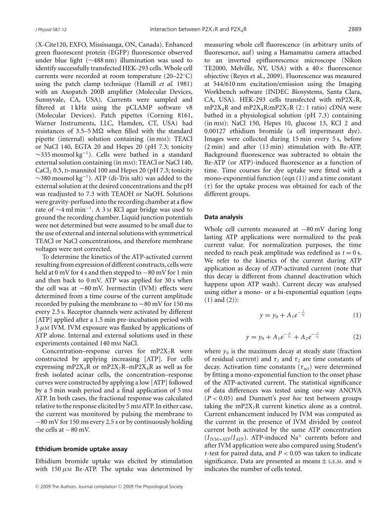
A striking finding was the lack of noticeable potentiationon the current activated by 5 mM ATP (Fig. 7C andF) in cells co-expressing mP2X4R : mP2X7R in a 2 : 1ratio (the same was observed in a 1 : 1 ratio; data notshown) even though IVM potentiated the response ofmP2X4 and mP2X7 receptors separately. Current ratios(I IVM+ATP/I ATP) were 1.1 ± 0.05 (Fig. 7F) and 1.2 ± 0.1in cells transfected with 2 : 1 and 1 : 1 cDNA ratios,respectively. Because the ATP sensitivity of mP2X4R ishigher than that of mP2X7R and certainly 5 mM ATPinduces full activation and deactivation of mP2X4R (seeFigs 2D and 5) we were concerned that the lack ofpotentiation was due to mP2X4R desensitization. Thus, torule out these effects we tested IVM on currents activatedwith 0.03 mM [ATP], a concentration that activates ∼70%of mP2X4R and none of the mP2X7R. At this [ATP]IVM potentiated ∼3.2 times the current generated bymP2X4R (Fig. 7D) but did not induce current in cellsexpressing mP2X7R alone (Fig. 7E). In contrast, in cellstransfected with a 2 : 1 cDNA ratio, IVM potentiated ∼7times the current activated by 0.03 mM ATP (Fig. 7F).Albeit this tendency suggests that the sensitivity toIVM is higher in cells co-expressing both receptors,no statistical significance between the mP2X4R and themP2X4R:mP2X7R was found.
Since IVM did not potentiate the Na+ currents activatedby saturating [ATP] in HEK cells co-expressing bothreceptors, we reasoned that in acinar cells (which naturallyexpress mP2X4R and mP2X7R) the same would occur.Consistent with this idea, IVM failed to enhance the Na+
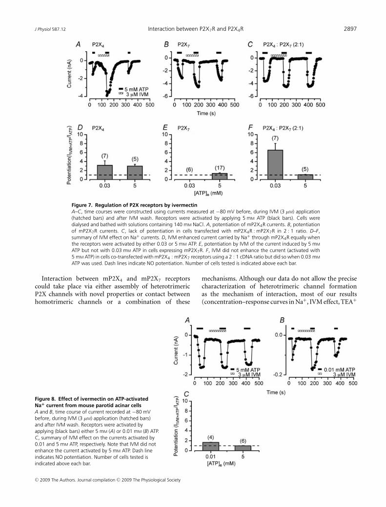
current activated by 5 mM ATP in fresh isolated parotidacinar cells (1 ± 0.05, Fig. 8A and C). Decreasing the ATPconcentration to 0.01 mM resulted in slight but statisticallysignificant potentiation (1.7 ± 0.03, Fig. 8B and C), just aswe saw in HEK cells co-expressing both receptors.
Discussion
In this work we show that heterologous expression ofeither mP2X4 or mP2X7 receptors do not reproduce all thefeatures of the ATP-activated current recorded in acinarcells unless both receptors are co-expressed. Co-expressionof mP2X7R with mP2X4R results in: (1) generation ofwhole cell currents that resemble the ATP-activated TEA+
current in native mouse parotid acinar cells; (2) slowerrate of ethidium bromide uptake than that observed withmP2X7R alone; (3) a channel fraction with lower ATPsensitivity than that of mP2X4R or mP2X7R alone, and(4) lack of potentiation by IVM of Na+ current activatedby 5 mM ATP. The latter is also seen in acinar cells. Thisevidence substantiates a functional interaction betweenmP2X4 and mP2X7 receptors in parotid acinar cells.
C© 2009 The Authors. Journal compilation C© 2009 The Physiological Society
J Physiol 587.12 Interaction between P2X7R and P2X4R 2897
Figure 7. Regulation of P2X receptors by ivermectinA–C, time courses were constructed using currents measured at −80 mV before, during IVM (3 μM) application(hatched bars) and after IVM wash. Receptors were activated by applying 5 mM ATP (black bars). Cells weredialysed and bathed with solutions containing 140 mM NaCl. A, potentiation of mP2X4R currents. B, potentiationof mP2X7R currents. C, lack of potentiation in cells transfected with mP2X4R : mP2X7R in 2 : 1 ratio. D–F,summary of IVM effect on Na+ currents. D, IVM enhanced current carried by Na+ through mP2X4R equally whenthe receptors were activated by either 0.03 or 5 mM ATP. E, potentiation by IVM of the current induced by 5 mM
ATP but not with 0.03 mM ATP in cells expressing mP2X7R. F, IVM did not enhance the current (activated with5 mM ATP) in cells co-transfected with mP2X4 : mP2X7 receptors using a 2 : 1 cDNA ratio but did so when 0.03 mM
ATP was used. Dash lines indicate NO potentiation. Number of cells tested is indicated above each bar.
Interaction between mP2X4 and mP2X7 receptorscould take place via either assembly of heterotrimericP2X channels with novel properties or contact betweenhomotrimeric channels or a combination of these
Figure 8. Effect of ivermectin on ATP-activatedNa+ current from mouse parotid acinar cellsA and B, time course of current recorded at −80 mVbefore, during IVM (3 μM) application (hatched bars)and after IVM wash. Receptors were activated byapplying (black bars) either 5 mM (A) or 0.01 mM (B) ATP.C, summary of IVM effect on the currents activated by0.01 and 5 mM ATP, respectively. Note that IVM did notenhance the current activated by 5 mM ATP. Dash lineindicates NO potentiation. Number of cells tested isindicated above each bar.
mechanisms. Although our data do not allow the precisecharacterization of heterotrimeric channel formationas the mechanism of interaction, most of our results(concentration–response curves in Na+, IVM effect, TEA+
C© 2009 The Authors. Journal compilation C© 2009 The Physiological Society

2898 G. Casas-Pruneda and others J Physiol 587.12
current kinetics and ethidium bromide uptake) suggestthat in native acinar cells and HEK cells mP2X4R andmP2X7R could assemble into heterotrimeric channelswith novel functional and pharmacological properties.Assembly of heterotrimeric P2X4/P2X7 channels is alsosuggested by co-immunoprecipitation of P2X7R withP2X4R in bone marrow derived macrophages (Guoet al. 2007) and by the interaction of P2X7R witha variety of cell membrane anchored proteins (Kimet al. 2001). Nevertheless, physically close homotrimericchannels interacting through protein–protein contactscould also immunoprecipitate together (Murrell-Lagnado& Qureshi, 2008). This has been shown for P2X2 andρ1/GABA, two completely different receptors that interactwith each other (Boue-Grabot et al. 2004).
Our experimental data show that after co-expressionthe resulting mP2X4–mP2X7 channels display novelpharmacological responses such as a lower ATP sensitivityand altered IVM potentiation. In cells expressing eithermP2X7R alone or mP2X4R and mP2X7R, the TEA+
current was activated by ATP with an EC50 of ∼33 μM.In cells expressing mP2X4R or mP2X7R alone theNa+ current was activated with an EC50 of 11 and221 μM, respectively, values similar to those reportedin other tissues (Silberberg et al. 2005; Young et al.2007). In contrast, in cells co-expressing both channelsa quite complex concentration–response curve wasobtained in Na+. This curve is not explained bythe presence of independent mP2X4R (EC50 = 11 μM)and mP2X7R (EC50 = 221 μM) populations. Instead ouranalysis suggests the presence of at least two receptorpopulations whose EC50 values (∼3 nM and 434 μM,respectively) differed significantly from those of theindividual receptors. This observation suggests that inthe heterologous expression system, functional channelsmay include both homo- and hetero-trimers. Assemblyof hetero-trimers, either (mP2X4R)2–(mP2X7R)1 or(mP2X4R)1–(mP2X7R)2, could have altered ATPsensitivity. This could be either extremely high ATP affinity(<10 nM) or low ATP affinity (>400 μM). Althoughthe concentration–response curve in the heterologousexpression system suggests assembly of hetero-trimericchannels, in native cells we did not observed channelswith such high ATP sensitivity. Instead we only observeda single, low ATP sensitivity channel population. Thischannel population was twice as less sensitive to ATPthan mP2X7R channels alone. In addition, we observedthat the ATP-activated current in acinar cells had aslower time course than that of mP2X7R or mP2X4Rchannels. This slower time course of channel activationhas been reported by others working in epithelial cells(Li et al. 2005; Ma et al. 2006). A caveat in ourexperiments is the lack of information about the levelof channel expression in the heterologous expression andthe acinar cells. Clearly transiently or permanently trans-
fected HEK cells expressed larger currents than acinarcells, indicating that higher protein levels are present inthe plasma membrane of HEK cells. If this assumptionis correct then higher level of protein expression wouldresult in different levels of homo- and hetero-trimers,which will directly influence the dose–response relations.Thus, if channels with high ATP sensitivity are presentin low abundance in the acinar cells, their functionalresponse recorded using macroscopic currents may becompromised. Nevertheless, the fact that the kinetics of theATP-activated current in acinar cells are slower than thatof each P2X receptor and that the EC50 is bigger than thatof mP2X7R suggests that in the native cells, P2X receptorsgenerate channels with new pharmacological responses.Another novel pharmacological response observed wasIVM potentiation. IVM potentiates homotrimeric P2X4Rchannels by binding between transmembrane helices nearthe extracellular side (Silberberg et al. 2007). Our datashow that IVM potentiates the Na+ current throughmP2X4R as well as through mP2X7R when activatedby 5 mM ATP. Remarkably, IVM failed to enhance theNa+ current activated by 5 mM ATP in HEK cellsco-transfected with both receptors as well as in acinarcells. Furthermore, the potentiation by IVM of the Na+
current activated by 0.03 mM ATP was larger in cellco-expressing P2X4R and P2X7R channels than in cellexpressing only mP2X4R probably due to activationof the high ATP sensitivity channel population seenduring co-expression experiments (Fig. 5B). A similarbehaviour is observed in acinar cells (Figs 7F and 8C),although the potentiation is not as large. Altogether, thesepharmacological observations are incompatible with theexpression of two homotrimeric channel populations.
Different functional responses seen in cells expressingmP2X4–mP2X7 channels include changes in currentkinetics and ethidium bromide uptake. When mP2X7Ris expressed alone the time course of the Na+ currentswas sustained as shown by others (Hibell et al. 2000)but in TEA+ the currents displayed a slight decay. Thisdecay was accentuated by co-expressing both receptorsdespite the fact that mP2X4Rs do not allow passage ofTEA+. This indicates that mP2X4Rs modify the currentkinetics by interacting with mP2X7Rs to yield channelswhose functional response is changed. In acinar cellsthe TEA+ current displays similar behaviour albeit theEC50 value is larger (Arreola & Melvin, 2003) thanthe one we report here. The rate of current decayrecorded in mP2X4:mP2x7 channels was not altered bycomplete removal of external Ca2+ or addition of 1 mM
carbenoxolone (Pelegrin & Surprenant, 2006). The lackof effect caused by Ca2+ removal was expected sincewith 0.5 mM CaCl2 in the bath solution, Ca2+ wouldcarry less than 2% of the total current in P2X7 channels(Riedel et al. 2007). Thus, the effects of co-expressing bothreceptors are not due to altered Ca2+ influx or activation
C© 2009 The Authors. Journal compilation C© 2009 The Physiological Society

J Physiol 587.12 Interaction between P2X7R and P2X4R 2899
of pannexin-1 hemichannels, but may indeed reflectsubunit interaction. Also, the ethidium bromide uptakeresult hints to a similar conclusion. The dye uptakeobserved in the mP2X4–mP2X7 co-expression wasslower and smaller than the uptake seen with mP2X7
receptors alone. Since ethidium bromide uptake is due topannexin-1 activation probably by the carboxy-terminusof mP2X7R (Becker et al. 2008), our result suggeststhat a physical interaction between P2X receptorsdelays pannexin-1 activation. Alternatively, assembly ofheterotrimeric receptors could produce channels that donot activate pannexin-1 as efficiently as mP2X7R alone.Also, we observed that the magnitude of the Na+ current(which is proportional to receptor density) was smallerin cells co-expressing both receptors. Thus, if the rateof ethidium bromide is proportional to receptor densitythen a decreased receptor density would explain the slowerethidium bromide uptake.
An additional observation to be considered in thedata interpretation is the modulatory effect of mono-valent cations on the function of mP2X7R. Since singlechannel recording of rat P2X7R shows that passage of smalland large cations occurs through the same pore withoutchanging its diameter (Riedel et al. 2007), we expectedto see similar pharmacological and functional responsesindependently of the ion carrying the current. This isnot the case. In this receptor the function of domainssuch as extracellular binding sites for ATP and blockers,the pore and the intracellular domains that couple thereceptor to pannexin-1 are regulated by external Na+
(Reyes et al. 2008; Ma et al. 1999; present study). In theabsence of external Na+, several changes are observed:(1) the pore of mP2X7R becomes permeable to Cl−
and has a higher SCN− conductivity; (2) cibacron bluepotentiates the TEA+ current but blocks the Na+ currentthrough mP2X7R; (3) IVM does not potentiate the TEA+
current but potentiates the Na+ current; (4) the TEA+
current decays in the presence of ATP but the Na+ currentdoes not; (5) the ATP sensitivity of mP2X7R is higherin the presence of TEA+; (6) P2X7Rs couple better topannexin-1 in the presence of TEA+ (Reyes et al. 2008;Virginio et al. 1999; present study); and (7) P2X7R displayslow open probability and fast deactivation when Na+ ispresent (Riedel et al. 2007). In summary, the functionof P2X7R is strongly dependent on external Na+ andsuch Na+ dependence could be passed on to channelsformed by co-expressing both receptors. Although theprecise mechanism by which Na+ regulates these myriadof P2X7R functions remains to be determined, wehypothesized in a previous report that the mP2X7R couldundergo conformational changes when Na+ is removedfrom the external medium thereby resulting in channelswith a different structure but still activated by externalATP (Reyes et al. 2008). If the external Na+ is essentialto maintain the structure of the receptor then its ability
to interact and respond to activators and regulators maybecome altered and/or compromised in the absence ofNa+. This hypothesis may in part explain our data, butmore biochemical and structural data are needed to testthis idea.
In conclusion, our data indicate that a functionalinteraction between mP2X7 and mP2X4 receptors takesplace in epithelial cells from salivary glands. Thisfunctional interaction could be important in generatingthe ATP-activated current and shaping subsequent physio-logical responses to ATP signalling. Such functionalinteraction may also be in place in other cells that naturallyco-express these receptors such as airway ciliated cells,macrophages and hepatocytes (Ma et al. 1999, 2006;Gonzales et al. 2007; Guo et al. 2007; Raouf et al.2007).
References
Arreola J, Melvin JE & Begenisich T (1995). Volume-activatedchloride channels in rat parotid acinar cells. J Physiol 484,677–687.
Arreola J & Melvin JE (2003). A novel chloride conductanceactivated by extracellular ATP in mouse parotid acinar cells.J Physiol 547, 197–208.
Becker D, Woltersdorf R, Boldt W, Schmitz S, Braam U,Schmalzing G & Markwardt F (2008). The P2X7 carboxyl tailis a regulatory module of P2X7 receptor channel activity.J Biol Chem 283, 25725–25734
Boue-Grabot E, Toulme E, Emerit MB & Garret M (2004).Subunit-specific coupling between γ-aminobutyric acid typeA and P2X2 receptor channels. J Biol Chem 279,52517–52525.
Bours MJ, Swennen EL, Di Virgilio F, Cronstein BN & DagneliePC (2006). Adenosine 5′-triphosphate and adenosine asendogenous signaling molecules in immunity andinflammation. Pharmacol Ther 112, 358–404.
Brown SG, Townsend-Nicholson A, Jacobson KA, Burnstock G& King BF (2002). Heteromultimeric P2X(1/2) receptorsshow a novel sensitivity to extracellular pH. J Pharmacol ExpTher 300, 673–680.
Burnstock G (2007). Purine and pyrimidine receptors. Cell MolLife Sci 64, 1471–1483.
Casas-Pruneda G, Reyes JP, Perez-Cornejo P & Arreola J(2007). Functional cross-regulation of murine salivary glandP2X4 and P2X7 receptors. Biophys J 2145-Pos.
Casas-Pruneda G, Reyes JP, Perez-Cornejo P & Arreola J(2008). Interaction between P2X4 and P2X7 receptorsmodulates ivermectin effect. Biophys J 1313-Pos.
Chessell IP, Hatcher JP, Bountra C, Michel AD, Hughes JP,Green P, Egerton J, Murfin M, Richardson J, Peck WL,Grahames CB, Casula MA, Yiangou Y, Birch R, Anand P &Buell GN (2005). Disruption of the P2X7 purinoceptor geneabolishes chronic inflammatory and neuropathic pain. Pain114, 386–396.
Dinarello CA (2005). Blocking IL-1 in systemic inflammation.J Exp Med 201, 1355–1359.
C© 2009 The Authors. Journal compilation C© 2009 The Physiological Society

2900 G. Casas-Pruneda and others J Physiol 587.12
Gonzales E, Prigent S, Abou-Lovergne A, Boucherie S,Tordjmann T, Jacquemin E & Combettes L (2007). Rathepatocytes express functional P2X receptors. FEBS Lett 581,3260–3266.
Guo C, Masin M, Qureshi OS & Murrell-Lagnado RD (2007).Evidence for functional P2X4/P2X7 heteromeric receptors.Mol Pharmacol 72, 1447–1456.
Hamill OP, Marty A, Neher E, Sakmann B & Sigworth FJ(1981). Improved patch-clamp techniques for high-resolution current recording from cells and cell-freemembrane patches. Pflugers Arch 391, 85–100.
Hibell AD, Kidd EJ, Chessell IP, Humphrey PPA & Michel AD(2000). Apparent species differences in the kinetic propertiesof P2X7 receptors. Brit J Pharmacol 130, 167–173.
Khakh BS, Proctor WR, Dunwiddie TV, Labarca C & Lester HA(1999). Allosteric control of gating and kinetics at P2X4
receptor channels. J Neurosci 19, 289–299.Kim M, Jiang LH, Wilson HL, North RA & Surprenant A
(2001). Proteomic and functional evidence for a P2X7
receptor signalling complex. EMBO J 20,6347–6358.
King BF, Townsend-Nicholson A, Wildman SS, Thomas T,Spyer KM & Burnstock G (2000). Coexpression of rat P2X2and P2X6 subunits in Xenopus oocytes. J Neurosci 20,4871–4877.
Le KT, Babinski K & Seguela P (1998). Central P2X4 and P2X6
channel subunits coassemble into a novel heteromeric ATPreceptor. J Neurosci 18, 7152–7159.
Lewis C, Neidhart S, Holy C, North RA, Buell G & SurprenantA (1995). Coexpression of P2X2 and P2X3 receptor subunitscan account for ATP-gated currents in sensory neurons.Nature 377, 432–435.
Li Q, Luo X & Muallem S (2005). Regulation of the P2X7
receptor permeability to large molecules by extracellular Cl−and Na+. J Biol Chem 280, 26922–26927.
Ma W, Korngreen A, Uzlaner N, Priel Z & Silberberg SD(1999). Extracellular sodium regulates airway ciliarymotility by inhibiting a P2X receptor. Nature 400,894–897.
Ma W, Korngreen A, Weil S, Cohen EB, Priel A, Kuzin L &Silberberg SD (2006). Pore properties and pharmacologicalfeatures of the P2X receptor channel in airway ciliated cells.J Physiol 571, 503–517.
Murrell-Lagnado RD & Qureshi OS (2008). Assembly andtrafficking of P2X purinergic receptors (Review). Mol MembrBiol 25, 321–331.
Nakamoto T, Brown D, Catalan M, Gonzalez-Begne M,Romanenko V & Melvin JE (2009). Purinergic P2X7
receptors mediate ATP-induced saliva secretion by themouse submandibular gland. J Biol Chem 284,4815–4822.
Nicke A, Kerschensteiner D & Soto F (2005). Biochemical andfunctional evidence for heteromeric assembly of P2X1 andP2X4 subunits. J Neurochem 92, 925–933.
North RA (2002). Molecular physiology of P2X receptors.Physiol Rev 82, 1013–1067.
Pelegrin P & Surprenant A (2006). Pannexin-1 mediates largepore formation and interleukin-1β release by the ATP-gatedP2X7 receptor. EMBO J 25, 5071–5082.
Perregaux D & Gabel CA (1994). Interleukin-1β maturationand release in response to ATP and nigericin. Evidence thatpotassium depletion mediated by these agents is a necessaryand common feature of their activity. J Biol Chem 269,15195–15203.
Raouf R, Chabot-Dore AJ, Ase AR, Blais D & Seguela P (2007).Differential regulation of microglial P2X4 and P2X7 ATPreceptors following LPS-induced activation.Neuropharmacology 53, 496–504.
Reyes JP, Hernandez-Carballo CY, Perez-Flores G, Perez-Cornejo P & Arreola J (2009). Lack of coupling betweenmembrane stretching and pannexin-1 hemichannels.Biochem Biophys Res Commun 380, 50–53.
Reyes JP, Perez-Cornejo P, Hernandez-Carballo CY, SrivastavaA, Romanenko VG, Gonzalez-Begne M, Melvin JE & ArreolaJ (2008). Na+ modulates anion permeation and block ofP2X7 receptors from mouse parotid glands. J Membr Biol223, 73–85.
Riedel T, Schmalzing G & Markwardt F (2007). Influence ofextracellular monovalent cations on pore and gatingproperties of P2X7 receptor-operated single-channelcurrents. Biophys J 93, 846–858.
Silberberg SD, Chang TH & Swartz KJ (2005). Secondarystructure and gating rearrangements of transmembranesegments in rat P2X4 receptor channels. J Gen Physiol 125,347–359.
Silberberg SD, Li M & Swartz KJ (2007). Ivermectin Interactionwith transmembrane helices reveals widespreadrearrangements during opening of P2X receptor channels.Neuron 54, 263–274.
Solle M, Labasi J, Perregaux DG, Stam E, Petrushova N, KollerBH, Griffiths RJ & Gabel CA (2001). Altered cytokineproduction in mice lacking P2X7 receptors. J Biol Chem 276,125–132.
Tenneti L, Gibbons SJ & Talamo BR (1998). Expression andtrans-synaptic regulation of P2X4 and P2z receptors forextracellular ATP in parotid acinar cells. Effects ofparasympathetic denervation. J Biol Chem 273,26799–26808.
Torres GE, Egan TM & Voigt MM (1999). Hetero-oligomericassembly of P2X receptor subunits. Specificities exist withregard to possible partners. J Biol Chem 274, 6653–6659.
Torres GE, Haines WR, Egan TM & Voigt MM (1998). Co-expression of P2X1 and P2X5 receptor subunits reveals anovel ATP-gated ion channel. Mol Pharmacol 54, 989–993.
Tsuda M, Shigemoto-Mogami Y, Koizumi S, Mizokoshi A,Kohsaka S, Salter MW & Inoue K (2003). P2X4 receptorsinduced in spinal microglia gate tactile allodynia after nerveinjury. Nature 424, 778–783.
Turner JT, Landon LA, Gibbons SJ & Talamo BR (1999).Salivary gland P2 nucleotide receptors. Crit Rev Oral BiolMed 10, 210–224.
Turner JT, Weisman GA, Landon LA, Park M & Camden JM(1998). Salivary gland nucleotide receptors: evidence forfunctional expression of both P2X and P2Y subtypes. Eur JMorphol 36, 170–175.
Virginio C, MacKenzie A, North RA & Surprenant A (1999).Kinetics of cell lysis, dye uptake and permeability changes incells expressing the rat P2X7 receptor. J Physiol 519, 335–346.
C© 2009 The Authors. Journal compilation C© 2009 The Physiological Society

J Physiol 587.12 Interaction between P2X7R and P2X4R 2901
Xiang Z & Burnstock G (2005). Expression of P2X receptors onrat microglial cells during early development. Glia 52,119–126.
Young MT, Pelegrin P & Surprenant A (2007). Amino acidresidues in the P2X7 receptor that mediate differentialsensitivity to ATP and BzATP. Mol Pharmacol 71,92–100.
Author contributions
JA and PPC contributed to conception and design. Allauthors contributed to analisis and data interpretation, wroteand critically revised the manuscript and approval of thefinal version. GCP performed most of the experiments.
JPR performed ATP concentration–response curves in TEAsolutions. GPF carried out the ethidium bromide assay.Experiments were conducted at the School of Medicine andInstitute of Physics of UASLP.
Acknowledgements
We want to thank Carmen Y. Hernandez-Carballo for excellenttechnical assistance. This research was supported by grants fromCONACyT (59889 and 79897 to J.A. and 45895 to P.P.C.) andNIH (PO1-HL18208 to R.E.W. and J.A.). G.C.-P. and G.P.-F.were supported by fellowships from CONACYT (181285 and201794).
C© 2009 The Authors. Journal compilation C© 2009 The Physiological Society
Copyright © 2022 FDOKUMEN


![A-740003 [N-(1-{[(Cyanoimino)(5-quinolinylamino) methyl]amino}-2,2-dimethylpropyl)-2-(3,4-dimethoxyphenyl)acetamide], a Novel and Selective P2X7 Receptor Antagonist, Dose-Dependently](https://static.fdokumen.com/doc/165x107/63441f69596bdb97a9085093/a-740003-n-1-cyanoimino5-quinolinylamino-methylamino-22-dimethylpropyl-2-34-dimethoxyphenylacetamide.jpg)