The P2X7 Receptor Channel Pore Dilates under Physiological Ion Conditions
11
The Journal of General Physiology ARTICLE The Rockefeller University Press $30.00 J. Gen. Physiol. Vol. 132 No. 5 563–573 www.jgp.org/cgi/doi/10.1085/jgp.200810059 563 INTRODUCTION The purinergic P2X receptors (P2XRs) are a family of ligand-gated ion channels composed of seven subunits, termed P2X 1–7 (Egan et al., 2004), and these subunits assemble as homo- or heterotrimers to make functional receptors (Nicke et al., 1998, 2005). The P2X 7 R subunit, initially cloned from a rat brain cDNA library, shares an overall membrane topology with the other members of this family of receptors; it contains two putative pore- forming transmembrane segments, a large cysteine-rich ligand-binding extracellular domain, and intracellularly located N and C termini (Surprenant et al., 1996). Structurally, the receptor is distinguished from other members of P2XRs by its long intracellular C-terminal tail containing multiple protein and lipid interaction motifs, and a cysteine-rich 18–amino acid segment. The P2X 7 R also requires at least a 100-fold higher ATP con- centration for activation than is required for other P2XRs, and removal of divalent cations increases ago- nist potency (Ferrari et al., 2006). Extracellular cations and chloride have complex effects on the channel gat- ing (Virginio et al., 1997; Gudipaty et al., 2001; Li et al., 2003, 2005; Riedel et al., 2007b). Furthermore, during sustained activation, P2X 7 R can elicit a wide range of in- tracellular signaling responses that are usually associated with the G protein–coupled receptors (Dubyak, 2007). Correspondence to Stanko S. Stojilkovic: [email protected] Abbreviations used in this paper: [Ca 2+ ] i , intracellular calcium concen- tration; CBX, carbenoxoline; GT1 cells, gonadotropin-releasing hormone- secreting cells; HEK, human embryonic kidney; KR, Krebs-Ringer; NMEA, 2-(methylamino)ethanol; P2XR, P2X receptor; WT, wild-type. Finally, P2X 7 R operates as a nonselective cationic channel during initial agonist application, but with prolonged application, the receptor also provides a permeation pathway to molecules with a molecular weight of up to 800 D, including the fluorescent dye YO-PRO-1, a pro- cess termed cell “permeabilization.” Initially, it was suggested that the bi-functional perme- ation properties of P2X 7 R reflect a dilation of the inte- gral pore of the channel. Thus, the cation-permeable recombinant channel is activated rapidly (within milli- seconds) after application of agonists, followed by a gradual (within seconds to minutes) increase in perme- ability to NMDG + and fluorescent dyes, both occurring at comparable rates (Surprenant et al., 1996; Chessell et al., 1997; Michel et al., 1999; Gudipaty et al., 2001). It was further suggested that a progressive dilation of the ion-conducting pathway during prolonged agonist ap- plication is not a unique feature of the P2X 7 R, but also occurs in cells expressing P2X 2 R and P2X 4 R (Khakh et al., 1999; Virginio et al., 1999). However, the develop- ment of NMDG + permeability and the uptake of fluo- rescent dyes were not always detected in cells responding to application of agonist with inward currents (Petrou et al., 1997; Klapperstuck et al., 2000). Furthermore, the permeability to NMDG + was reduced in low Na + -con- taining medium and was completely abolished in cells bathed in normal extracellular Na + , questioning the pore dilation hypothesis (Jiang et al., 2005). In addi- tion, the uptake of fluorescent dyes was not affected in cells expressing P2X 7 R in which the receptor-specific The P2X 7 Receptor Channel Pore Dilates under Physiological Ion Conditions Zonghe Yan, Shuo Li, Zhaodong Liang, Melanija Tomic ´, and Stanko S. Stojilkovic Section on Cellular Signaling, Program in Developmental Neuroscience, National Institute of Child Health and Human Development, National Institutes of Health, Bethesda, MD 20892 Activation of the purinergic P2X 7 receptor leads to the rapid opening of an integral ion channel that is permeable to small cations. This is followed by a gradual increase in permeability to fluorescent dyes by integrating the actions of the pannexin-1 channel. Here, we show that during the prolonged agonist application a rapid current that peaked within 200 ms was accompanied with a slower current that required tens of seconds to reach its peak. The secondary rise in current was observed under different ionic conditions and temporally coincided with the devel- opment of conductivity to larger organic cations. The biphasic response was also observed in cells with blocked pannexin channels and in cells not expressing these channels endogenously. The biphasic current was preserved in N-terminal T15A, T15S, and T15V mutants that have low or no permeability to organic cations, reflecting en- hanced permeability to inorganic cations. In contrast, the T15E, T15K, and T15W mutants, and the 18 mutant with deleted P2X 7 receptor–specific 18–amino acid C-terminal segment, were instantaneously permeable to or- ganic cations and generated high amplitude monophasic currents. These results indicate that the P2X 7 receptor channel dilates under physiological ion conditions, leading to generation of biphasic current, and that this process is controlled by residues near the intracellular side of the channel pore.
Transcript of The P2X7 Receptor Channel Pore Dilates under Physiological Ion Conditions
The
Jour
nal o
f G
ener
al P
hysi
olo
gy
A RT I C L E
The Rockefeller University Press $30.00J. Gen. Physiol. Vol. 132 No. 5 563–573www.jgp.org/cgi/doi/10.1085/jgp.200810059 563
I N T R O D U C T I O N
The purinergic P2X receptors (P2XRs) are a family of
ligand-gated ion channels composed of seven subunits,
termed P2X 1 – 7 ( Egan et al., 2004 ), and these subunits
assemble as homo- or heterotrimers to make functional
receptors ( Nicke et al., 1998, 2005 ). The P2X 7 R subunit,
initially cloned from a rat brain cDNA library, shares an
overall membrane topology with the other members of
this family of receptors; it contains two putative pore-
forming transmembrane segments, a large cysteine-rich
ligand-binding extracellular domain, and intracellularly
located N and C termini ( Surprenant et al., 1996 ).
Structurally, the receptor is distinguished from other
members of P2XRs by its long intracellular C-terminal
tail containing multiple protein and lipid interaction
motifs, and a cysteine-rich 18 – amino acid segment. The
P2X 7 R also requires at least a 100-fold higher ATP con-
centration for activation than is required for other
P2XRs, and removal of divalent cations increases ago-
nist potency ( Ferrari et al., 2006 ). Extracellular cations
and chloride have complex effects on the channel gat-
ing ( Virginio et al., 1997 ; Gudipaty et al., 2001 ; Li et al.,
2003, 2005 ; Riedel et al., 2007b ). Furthermore, during
sustained activation, P2X 7 R can elicit a wide range of in-
tracellular signaling responses that are usually associated
with the G protein – coupled receptors ( Dubyak, 2007 ).
Correspondence to Stanko S. Stojilkovic: s t a n k o s @ h e l i x . n i h . g o v
Abbreviations used in this paper: [Ca 2+ ] i , intracellular calcium concen-
tration; CBX, carbenoxoline; GT1 cells, gonadotropin-releasing hormone-
secreting cells; HEK, human embryonic kidney; KR, Krebs-Ringer; NMEA,
2-(methylamino)ethanol; P2XR, P2X receptor; WT, wild-type.
Finally, P2X 7 R operates as a nonselective cationic channel
during initial agonist application, but with prolonged
application, the receptor also provides a permeation
pathway to molecules with a molecular weight of up to
� 800 D, including the fl uorescent dye YO-PRO-1, a pro-
cess termed cell “ permeabilization. ”
Initially, it was suggested that the bi-functional perme-
ation properties of P2X 7 R refl ect a dilation of the inte-
gral pore of the channel. Thus, the cation-permeable
recombinant channel is activated rapidly (within milli-
seconds) after application of agonists, followed by a
gradual (within seconds to minutes) increase in perme-
ability to NMDG + and fl uorescent dyes, both occurring
at comparable rates ( Surprenant et al., 1996 ; Chessell
et al., 1997 ; Michel et al., 1999 ; Gudipaty et al., 2001 ). It
was further suggested that a progressive dilation of the
ion-conducting pathway during prolonged agonist ap-
plication is not a unique feature of the P2X 7 R, but also
occurs in cells expressing P2X 2 R and P2X 4 R ( Khakh
et al., 1999 ; Virginio et al., 1999 ). However, the develop-
ment of NMDG + permeability and the uptake of fl uo-
rescent dyes were not always detected in cells responding
to application of agonist with inward currents ( Petrou
et al., 1997 ; Klapperstuck et al., 2000 ). Furthermore,
the permeability to NMDG + was reduced in low Na + -con-
taining medium and was completely abolished in cells
bathed in normal extracellular Na + , questioning the
pore dilation hypothesis ( Jiang et al., 2005 ). In addi-
tion, the uptake of fl uorescent dyes was not affected in
cells expressing P2X 7 R in which the receptor-specifi c
The P2X 7 Receptor Channel Pore Dilates under Physiological Ion Conditions
Zonghe Yan , Shuo Li , Zhaodong Liang , Melanija Tomic , and Stanko S. Stojilkovic
Section on Cellular Signaling, Program in Developmental Neuroscience, National Institute of Child Health and Human Development, National Institutes of Health, Bethesda, MD 20892
Activation of the purinergic P2X 7 receptor leads to the rapid opening of an integral ion channel that is permeable to small cations. This is followed by a gradual increase in permeability to fl uorescent dyes by integrating the actions of the pannexin-1 channel. Here, we show that during the prolonged agonist application a rapid current that peaked within 200 ms was accompanied with a slower current that required tens of seconds to reach its peak. The secondary rise in current was observed under different ionic conditions and temporally coincided with the devel-opment of conductivity to larger organic cations. The biphasic response was also observed in cells with blocked pannexin channels and in cells not expressing these channels endogenously. The biphasic current was preserved in N-terminal T15A, T15S, and T15V mutants that have low or no permeability to organic cations, refl ecting en-hanced permeability to inorganic cations. In contrast, the T15E, T15K, and T15W mutants, and the � 18 mutant with deleted P2X 7 receptor – specifi c 18 – amino acid C-terminal segment, were instantaneously permeable to or-ganic cations and generated high amplitude monophasic currents. These results indicate that the P2X 7 receptor channel dilates under physiological ion conditions, leading to generation of biphasic current, and that this process is controlled by residues near the intracellular side of the channel pore.

564 P2X 7 R Pore Dilation
Lipofectamine 2000 reagent (Invitrogen) in 2 ml of serum-free Opti-MEM. After 4.5 h of incubation, the transfection mixture was replaced with normal culture medium, and cells were cultured for an additional 24 – 48 h. For electrophysiological measurements, transfected cells were mechanically dispersed and re-cultured on 35-mm dishes for 2 – 10 h.
Current Measurements Whole cell patch clamp recordings were performed using single cells at room temperature using an Axopatch 200B amplifi er (MDS Analytical Technologies), as previously described ( Yan et al., 2005 ). Patch electrodes, fabricated from borosilicate glass (type 1B150F-3; World Precision Instruments, Inc.) using a Flaming Brown horizontal puller (P-97; Sutter Instrument Co.), were heat polished to a fi nal tip resistance of 3.5 – 5.5 MOhm. All current records were programmed, captured, and stored using the pClamp 8.0 software packages in conjunction with the Digidata 1322A A/D converter (MDS Analytical Technologies). Experiments were performed on single cells with an average ca-pacitance of 10 pF. Unless otherwise stated, membrane potential was held at � 60 mV. Current voltage relations were used to esti-mate changes in reversal potential during agonist application and were obtained by voltage ramps from � 80 to +80 mV twice per second during 50 s. Patch electrodes were fi lled with solu-tion containing 145 mM NaCl, 10 mM EGTA, and 10 mM HEPES; the pH was adjusted with 10 M NaOH to 7.35. The os-molarity of this solution was 305 mOsM. The regular Krebs-Ringer (KR)-like bath buffer contained 147 mM NaCl, 3 mM KCl, 1 mM MgCl 2 , 2 mM CaCl 2 , 10 mM glucose, and 10 mM HEPES; the pH was adjusted to 7.35 with 10 M NaOH. For some experiments, 90 or 100% Na + was replaced with mM NMDG + or 2-(methylamino)ethanol (NMEA + ), and the solution pH was adjusted to 7.35 with HCl (Na + /NMDG + -KR buffer and NMDG + -KR buffer, respectively). We also used a buffer containing only 155 mM NMDG, 10 mM glucose, and 10 mM HEPES (NMDG + buffer). The osmolarity of these solutions were 295 – 305 mOsM. ATP and Bz-ATP solution was daily prepared in bath buffer with pH properly readjusted, applied using either an Ultrafast Solu-tion-Switching System (LSS-3200; EXFO Burleigh Products Group Inc.) that was simultaneously program controlled by pClamp 8.0 software through a PZ-150M Amplifi er ( Yan et al., 2006 ) or a RSC-200 Rapid Solution Changer (Biological). Cells with EGFP fl uores-cence were identifi ed before immersing the electrode in bath solution for gigaohm seal.
Fluorescence Imaging Transfected GT1 cells plated on 25-mm coverslips were bathed in KR medium containing 2.5 μ M of either Fura-2 AM or Fura-FF AM (Invitrogen) for 1 h at room temperature. After washing the coverslips with the dye-free KR media, they were mounted on the stage of an Axiovert 135 microscope (Carl Zeiss, Inc.) at-tached to an Attofl uor Digital Fluorescence Microscopy System (Atto Instruments). Cells were examined under a 40 × oil immer-sion objective during exposure to alternating 340- and 380-nm excitation beams, and the intensity of light emission at 520 nm (F 340 and F 380 ) was followed in several single cells simultaneously at the rate of one point per second. For YO-PRO-1 uptake, cells were loaded with Fura-2 and subsequently exposed to 5 μ M YO-PRO-1 (Invitrogen) in KR medium. The same concentration of YO-PRO-1 was present throughout the experiment, and the ex-perimental setup was identical except for the dichroic mirror and the excitation fi lters. Fluorescence was observed upon exci-tation with alternating 380- (Fura-2) and 488-nm (YO-PRO-1) beams. Only cells with low basal F 488 and cells that showed de-creased F 380 in response to agonist, corresponding to an in-crease in intracellular calcium concentrations ([Ca 2+ ] i ), were further analyzed for changes in F 488 , corresponding to YO-PRO-1
C-terminal 18 – amino acid sequence was deleted, but
mutant was not permeable to NMDG + , suggesting that
YO-PRO-1 entry occurs through a separate pathway than
NMDG + ( Jiang et al., 2005 ). Furthermore, cells express-
ing mutant P2X 7 R that was truncated at residue 581 had
negligible ethidium uptake and normal current re-
sponses ( Smart et al., 2003 ). Finally, the coupling of
P2X 7 R to pannexin-1 channels was suggested to account
for cellular entry of fl uorescent dyes ( Pelegrin and
Surprenant, 2006 ).
Here, we addressed the hypothesis that P2X 7 R pore
dilates independently of the expression of pannexin-1
channels. The rat P2X 7 R was expressed in human em-
bryonic kidney (HEK) 293 cells and gonadotropin-re-
leasing hormone-secreting (GT1) cells, which express
pannexin-1 channels endogenously, as well as in C6
glioma cells, which do not express pannexins. In all
cell types, P2X 7 Rs responded to initial agonist applica-
tion with a rapid growth of inward current that peaked
within milliseconds. This response was accompanied
by an additional rise that required tens of seconds to
be completed. Our results also suggest that a transi-
tion from a closed to an opened state of the channel
pore accounts for the rapid rise in current, whereas a
transition from an opened to a dilated state accounts
for the secondary growth of current. The pore dila-
tion occurs in cells bathed in physiological solution,
requires higher agonist concentrations, and is con-
trolled by residues near the intracellular side of the
channel pore.
M AT E R I A L S A N D M E T H O D S
Site-directed Mutagenesis, Cell Culture, and Transfection The pIRES2-EGFP P2X 7 construct ( He et al., 2003 ) was used as a template for production of plasmids containing specifi c amino acid residue point mutations of P2X 7 cDNA using the Quik-Change II XL site-directed mutagenesis kit (Stratagene). The mutagenic oligonucleotide primers were synthesized and PAGE purifi ed by Integrated DNA Technology. Production of the cor-rect mutations and absence of coding errors in these constructs were verifi ed by dye terminator – cycle sequencing (PerkinElmer; performed by Veritas, Inc.). Large-scale plasmid DNAs were pre-pared using a QIAfi lter Plasmid Maxi kit (QIAGEN). HEK293, C6, and GT1 cells were used for the expression of wild-type (WT) and mutant P2X 7 receptors, as described previously ( He et al., 2003 ; Yan et al., 2006 ). HEK293 and C6 cells (American Type Cul-ture Collection) were routinely maintained in Dulbecco ’ s modi-fi ed Eagle ’ s medium containing 10% (vol/vol) fetal bovine serum (Invitrogen) and 1% (vol/vol) penicillin-streptomycin liquid (Invitrogen) in a tissue culture incubator. GT1 cells (provided by R.I. Weiner, University of California, San Francisco, San Francisco, CA) were cultured in Dulbecco ’ s modifi ed Eagle ’ s medium/Ham ’ s F-12 medium (1:1) containing 10% (vol/vol) fetal bovine serum and 100 μ g/ml gentamicin (Invitrogen). For electrophysi-ological measurements, cells were grown on 35-mm dishes at a density of 0.5 × 10 6 cells per dish, whereas for imaging studies GT1 cells were grown on 25-mm coverslips placed in 35-mm dishes at a density of 0.1 × 10 6 cells per dish. Transfection was conducted 24 h after plating the cells using 2 μ g DNA and 5 μ l
Yan et al. 565
were performed in GT1 cells because they do not express
calcium-mobilizing P2Y receptors.
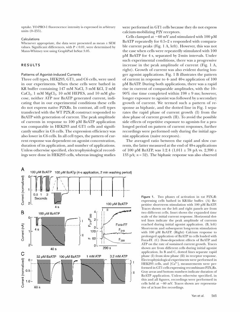
Cells clamped at � 60 mV and stimulated with 100 μ M
BzATP repeatedly for 0.5 – 2 s responded with compara-
ble current peaks ( Fig. 1 A , left). However, this was not
the case when cells were repeatedly stimulated with 100
μ M BzATP for 4 s, separated by 2-min intervals. Under
such experimental conditions, there was a progressive
increase in the peak amplitude of current ( Fig. 1 A ,
right). Growth of current was also evident during lon-
ger agonist applications. Fig. 1 B illustrates the pattern
of current in response to 4- and 40-s application of 100
μ M BzATP. During both applications, there was a rapid
rise in current of comparable amplitudes, with the 10 –
90% rise time completed within 198 ± 9 ms; however,
longer exposure to agonist was followed by a secondary
growth of current. We termed such a pattern of re-
sponse as biphasic, and the dotted line in Fig. 1 sepa-
rates the rapid phase of current growth (I) from the
slow phase of current growth (II). To avoid the possible
side effects of repetitive exposure to agonists for a pro-
longed period on pattern of current responses, further
recordings were performed only during the initial ago-
nist application (naive receptors).
The averaged ratio between the rapid and slow cur-
rents, the latter measured at the end of 40-s applications
of 100 μ M BzATP, was 1:2.4 (1,011 ± 78 pA vs. 2,390 ±
133 pA; n = 52). The biphasic response was also observed
uptake. YO-PRO-1 fl uorescence intensity is expressed in arbitrary units (0 – 255).
Calculations Whenever appropriate, the data were presented as mean ± SEM values. Signifi cant differences, with P < 0.01, were determined by Mann-Whitney test using GraphPad InStat 3.05.
R E S U LT S
Patterns of Agonist-induced Currents Three cell types, HEK293, GT1, and C6 cells, were used
in our experiments. When these cells were bathed in
KR buffer containing 147 mM NaCl, 3 mM KCl, 2 mM
CaCl 2 , 1 mM MgCl 2 , 10 mM HEPES, and 10 mM glu-
cose, neither ATP nor BzATP generated current, indi-
cating that in our experimental conditions these cells
do not express native P2XRs. In contrast, all cell types
transfected with the WT P2X 7 R construct responded to
BzATP with generation of current. The peak amplitude
of currents in response to 100 μ M BzATP application
was comparable in HEK293 and GT1 cells and signifi -
cantly smaller in C6 cells. The expression effi ciency was
also lower in C6 cells. In all cell types, the pattern of cur-
rent response was dependent on agonist concentration,
duration of its application, and number of applications.
Unless otherwise specifi ed, electrophysiological record-
ings were done in HEK293 cells, whereas imaging studies
Figure 1. Two phases of activation in rat P2X 7 R-expressing cells bathed in KR-like buffer. (A) Re-petitive short-term stimulation with 100 μ M BzATP. Traces shown on the left and right panels are from two different cells. Inset shows the expanded time scale of the initial current response. Horizontal dot-ted lines indicate the peak amplitude of currents reached during initial agonist application. (B, left) Short-term and subsequent long-term stimulation with 100 μ M BzATP. (Right) Calcium response to prolonged application of BzATP in cells loaded with Fura-FF. (C) Dose-dependent effects of BzATP and ATP on the rate of sustained current growth. Traces shown are from different cells during initial agonist application. In B and C, dotted lines separate rapid phase (I) from slow phase (II) in receptor response. Electrophysiological experiments were performed in HEK293 cells, and [Ca 2+ ] i measurements were per-formed in GT1 cells expressing recombinant P2X 7 Rs. Gray areas and bottom numbers indicate duration of BzATP application. Unless otherwise specifi ed, in this and all fi gures, recordings were performed in cells held at � 60 mV. Traces shown are representa-tive of at least fi ve recordings.
566 P2X 7 R Pore Dilation
voltage curves constructed from ramp voltage com-
mands revealed a shift in reversal potential during the
40-s agonist application from � 28.2 ± 0.5 mV to a steady-
state of � 17.3 ± 1.3 mV ( Fig. 2 A , right panel, from left
to right). Such a shift was observed in all cells studied
( n = 9). We next repeated these experiments in cells
bathed in KR medium with NMDG + replacing all the ex-
tracellular Na + (NMDG + -KR buffer). Under these ionic
conditions, the rate of the secondary growth in current
was comparable to that observed in cells perfused with
Na + /NMDG + -KR solution, and the 100 μ M BzATP –
evoked current reversal potential changed from � 36.5 ±
0.3 mV to � 24.6 ± 0.8 mV ( Fig. 2 B ). However, in cells
clamped at � 60 mV and exposed to BzATP for 40 s, the
peak amplitude of current differed in two ionic condi-
tions: (1) Na + /NMDG + -KR = 1.3 ± 0.2 nA ( n = 6), 54% of
that observed in cells bathed in KR buffer; (2) NMDG + -
KR = 0.59 ± 0.08 nA ( n = 4), 24% of that observed in
cells bathed in KR buffer. These results suggest that
NMDG + only partially substitutes for Na + as the con-
ducted ion.
The time course of 100 μ M BzATP – induced current
at � 60 mV was substantially different in cells perfused
with medium containing 155 mM NMDG + , 10 mM
HEPES, and 10 mM glucose (NMDG + buffer) and similar
to that reported by others ( Jiang et al., 2005 ). Because
the holding potential of � 60 mV was more positive than
the reversal potential in NMDG + ( � 67 ± 0.2 mV), an
initial outward current developed rapidly after agonist
application, refl ecting an outward movement of intra-
cellular Na + through the channel pore. The peak amp-
litude of this current was 309 ± 53 pA ( n = 8). This was
followed by a decline in current and a shift from out-
ward to inward current within 7 s. A steady-state current
was reached within 20 s ( Fig. 2 C , left), with the mean
value of � 241 ± 60 pA ( n = 8), 10% of that observed in
cells bathed in KR buffer, confi rming lower permeabil-
ity of channel to NMDG + than to small cations. Further-
more, the reversal potential progressively shifted by
+22.5 mV ( n = 15; Fig. 2 C , right), compared with the
+10.9- and +11.7-mV shifts observed in cells bathed in
Na + /NMDG + -KR and NMDG + -KR solutions, respectively.
Finally, the washout of BzATP was not accompanied by
receptor deactivation ( Fig. 2 C , left), but closed when
Na + was reintroduced in medium, which is consistent
with observations by others ( Jiang et al., 2005 ). In con-
trast, deactivation of the channel occurs in cells bathed
in Na + -defi cient/divalent cation-containing medium
( Fig. 2 B ), indicating that this process is not specifi cally
dependent on extracellular Na + , but does require the
presence of small cations.
We also examined the permeability of channels to
NMDG + during the rapid and sustained phase of current
growth. To do this, cells bathed in KR buffer were stimu-
lated with 100 μ M BzATP for 4 and 40 s, followed by
replacement of bath medium with NMDG + medium
in amphotericin-perforated cells, suggesting that the
second phase in current growth does not refl ect dialysis
of cells during the whole cell current recording (not
depicted). To rule out other possible artifacts, we used
intact cells and single cell calcium measurements.
Previous experiments with Fura-2 revealed inability
to report high [Ca 2+ ] i in cells with activated P2X 7 Rs
( Koshimizu et al., 2000 ), prompting us to use the less
sensitive Fura-FF dye. Because HEK293 cells endoge-
nously express Ca 2+ -mobilizing P2Y receptors ( He et al.,
2003 ), GT1 cells were used as an expression system.
In these cells, 100 μ M BzATP also induced biphasic
[Ca 2+ ] i response ( Fig. 1 B , right), with the averaged ratio
between the rapid and slow phase of 1:2.8 comparable
to that observed in current measurements (F 340 /F 380 : 0.83 ±
0.04 vs. 2.32 ± 0.14; n = 23; basal [Ca 2+ ] i subtracted).
However, the leak of Fura-FF (see Fig. 3 ) could infl u-
ence estimates of the F 340 /F 380 ratio during sustained
agonist application.
When 3 – 10 μ M BzATP was applied for 2 min, only
low amplitude monophasic currents were observed,
with the amplitude of current determined by agonist
concentration (3.2 μ M = 19 ± 7.2 pA [ n = 4]; 10 μ M = 34 ±
5.4 pA [ n = 6]). In contrast, cells always responded
with the biphasic current when stimulated with 32 and
100 μ M BzATP ( Fig. 1 C ). The secondary growth of cur-
rent was completed within 80 – 120 s in cells exposed
to 32 μ M BzATP, and within 30 – 70 s in cells exposed
to 100 μ M BzATP, suggesting that the rate of current
growth was determined by agonist concentration. Simi-
lar patterns of responses were observed when cells were
stimulated with ATP, but with a rightward shift in po-
tency. In low micromolar concentrations, ATP always
induced low-amplitude monophasic responses during a
2-min exposure to agonist, with the peak amplitude of
current determined by agonist concentration (100 μ M =
9.3 ± 2.1 pA [ n = 4]; 320 μ M ATP = 35.1 ± 3.4 pA [ n = 7]).
In high millimolar concentrations, ATP induced bipha-
sic responses in all cells, and the rates of secondary cur-
rent growth were dependent on agonist concentration
( Fig. 1 C ). In cells stimulated with 3.2 mM ATP, the ra-
tio between the fi rst and second peak values was 1:3.6
(716 ± 132 pA vs. 2622 ± 319 pA; n = 6), comparable to
that observed in response to 100 μ M BzATP application
(see above).
Biphasic Current and Permeability to NMDG + When 90% of Na + was replaced with NMDG + (Na + /
NMDG + -KR buffer), 100 μ M BzATP still induced bipha-
sic current in naive cells held at � 60 mV, with kinetics
that resembled growth of current observed in cells
bathed in Na + -containing KR medium ( Fig. 2 A , left vs.
Fig. 1 ). Under repetitive 485-ms voltage ramp pulses
from � 80 to +80 mV, delivered twice per second, there
was also a progressive increase in the peak amplitude of
inward current at � 80 mV ( Fig. 2 A , middle). Current

Yan et al. 567
two experimental conditions ( Fig. 2 D , right), and
practically no difference in steady-state currents. These
results indicate that the channel pore is permeable to
NMDG + during sustained but not during initial phase
of current growth.
Biphasic Current and Pannexin-1 Channels P2X 7 Rs operate as bi-functional molecules, and integra-
tion of pannexin-1 channels was suggested to account
for cellular permeability to fl uorescent dyes ( Pelegrin
and Surprenant, 2006 ). In general, the sustained rise in
current could also refl ect gradual integration of pan-
nexin-1 channels into P2X 7 R signaling. To test this
hypothesis, we performed several experiments. Our re-
verse transcriptase – PCR analysis revealed that mRNA
transcripts for pannexin-1 channels are expressed in
GT1 and HEK293 cells (not depicted). In GT1 cells ex-
pressing P2X 7 Rs, we also observed the uptake of YO-
PRO-1. However, it required > 200 s to approach saturation
( Fig. 3 A ), in contrast to biphasic current that leveled
off within 30 – 70 s of stimulation with 100 μ M BzATP
in the same cell type ( Fig. 3 C ). Furthermore, the YO-
PRO-1 uptake was observed in response to 100 μ M ATP
application ( Fig. 3 A ), a concentration that was unable
containing 100 μ M BzATP. All recordings were per-
formed at holding potential of � 60 mV. After 4-s expo-
sures to agonist, replacement of KR with NMDG + buffer
induced a shift from inward to outward current in < 1 s
( Fig. 2 D , left). Once the peak was reached, the current
again turned inward, with the time course highly compa-
rable to that observed in cells bathed in NMDG + medium
only ( Fig. 2 C , left) and with similar amplitude of current
(outward = 270 ± 19 pA; inward = � 168 ± 31; n = 5). This
is in accordance with published data ( Jiang et al., 2005 ).
After a 40-s exposure to agonist, replacement of KR
buffer with NMDG + buffer was also accompanied with
a rapid decrease in the amplitude of inward current,
but without reversing direction of current to outward
( Fig. 2 D , middle); the amplitude of the peak current
was � 188 ± 28 pA and of the steady-state current was
� 394 ± 67 pA ( n = 10). Furthermore, the steady-state
current did not refl ect nonreceptor-mediated perme-
abilization of cells because the washout of agonist with
KR buffer induced a transient rise in current, followed
by complete deactivation of channels ( Fig. 2 D , mid-
dle). Thus, there was a signifi cant difference in the
peak current responses after replacement of Na + -con-
taining KR buffer with NMDG + -containing buffer in
Figure 2. Permeability of P2X 7 R to NMDG + depends on duration of agonist application. (A) Pat-tern of 100 μ M BzATP-induced current and reversal potential in cells bathed in 10% Na + and 90% NMDG + -containing KR buffer during the initial agonist application. (A, left) Current recording during a 40-s agonist application at � 60 mV holding potential. (Middle) Time course of agonist-induced currents in cells under the ramp protocol. (Right) Positive shift in reversal potential observed during the initial 40-s application of BzATP; 0.485-s voltage ramps were deliv-ered twice per second. (B) Pat-terns of BzATP-induced currents (left and middle) and shift in the reversal potential (right) in cells bathed in KR buffer, in which Na + was completely substituted with NMDG + . (C) Time course of BzATP-induced P2X 7 R current in cells bathed in NMDG + buffer (left and middle) and the accom-panied changes in the reversal
potential (right). In this and all fi gures, only 15 out of 100 traces for the current voltage relationship with the equal time intervals are shown. Notice the lack of receptor deactivation in C compared with records shown in A (left) and B (left). (D) Increase in NMDG + per-meability occurs in cells bathed in physiological solution for agonist application of > � 10 s. Application of agonist was initially performed in cells bathed in KR buffer for 4 (left) and 40 s (middle) and was continued in NMDG + medium (gray areas). Notice a difference in re-sponses when replacement of KR buffer with NMDG + buffer was performed during the phase I (left) and II (middle) of current growth. Right panel illustrates the mean ± SEM values of the peak currents reached after replacement of KR buffer with NMDG + buffer in two time points. All experiments were performed in HEK293 cells.

568 P2X 7 R Pore Dilation
not found in other P2XRs. Deletion of this sequence
( � 18 mutant) was reported to affect conductivity for
NMDG + ( Jiang et al., 2005 ). In our hands, the mutant
was unable to generate the biphasic current when cells
were bathed in KR solution. The mutant initially re-
sponded to 100 μ M BzATP with high amplitude of cur-
rent that was reached rapidly, followed by a partial
decline in the current amplitude during prolonged ag-
onist application, and delayed deactivation after wash-
out of agonist ( Fig. 4 A , left trace). A similar pattern,
but not amplitude of current, was observed in cells
to trigger the biphasic current. We also examined the
leak of Ca 2+ dyes Fura-FF and Fura-2 from cells. To elim-
inate changes in fl uorescence induced by Ca 2+ infl ux,
cells were bathed in Na + -containing/Ca 2+ -defi cient KR
buffer. Under such ionic conditions, there was a pro-
gressive leak of Fura-FF and Fura-2 ( Fig. 3 B ) in response
to 100 μ M BzATP, as indicated by parallel changes in
F 380 and F 340 signals, with leak rates comparable to the
rate of YO-PRO-1 uptake.
The possible contribution of pannexin-1 channels in
sustained current response was also evaluated in HEK293
cells expressing WT P2X 7 Rs by applying 30 μ M carben-
oxolone (CBX), an inhibitor of pannexin-1 channels,
for 15 min. This treatment did not affect the pattern of
biphasic response ( Fig. 3 E ); the ratio between the fi rst
and second phases of current, the latter measured at
the end of the 40-s agonist application, was 1:2.38 for
controls (1,036 ± 129 pA vs. 2,521 ± 208 pA; n = 7) and
1:2.38 for CBX-treated cells (874 ± 197 vs. 2,092 ± 215
pA; n = 7). To further test this hypothesis, we used C6
glioma cells as an expression system because these cells
do not express pannexins endogenously ( Lai et al.,
2007 ). When transfected with WT P2X 7 Rs, these cells
responded with biphasic current during prolonged ap-
plication of 100 μ M BzATP ( Fig. 3 D ). These results in-
dicate that endogenous expression of pannexins is not
essential for sustained current growth. A delay in up-
take and leak of fl uorescent dyes could indicate a role
of pannexin-1 channels in this process or that permea-
bility of P2X 7 R channels for dyes refl ects cell death
and downstream effects.
A Role of the Y358-E375 C-terminal Sequence in Sustained Current Growth Immediately after the second transmembrane domain,
P2X 7 R contains a stretch of 18 amino acids, Y358 – E375,
Figure 3. Agonist-induced biphasic current re-sponse is independent of pannexin-1 channels. (A) Agonist-induced diffusion of extracellular YO-PRO-1 into GT1 cells. Fluorescence inten-sities expressed in arbitrary units ( � ex = 488 nm) are shown in response to application of BzATP only (left trace) and ATP followed by BzATP (right trace). (B) Normalized fluorescence in-tensities (gray, � ex = 380 nm; black, � ex = 340 nm) in GT1 cells loaded with Fura-FF (top) or Fura-2 (bottom) and stimulated with 100 μ M BzATP. (C and D) BzATP-induced biphasic response in GT1 (C) and C6 single cells (D). (E) The lack of effects of CBX on pattern of current response in HEK293 cells; control (left) and 30 μ M CBX-treated cells (right). In experiments shown in A, C, D, and E, cells were bathed in KR buffer. In experiments shown in B, cells were bathed in Ca 2+ -defi cient KR buffer.
Figure 4. Effects of deletion of the P2X 7 R-specifi c Y358-E375 C-terminal sequence ( � 18) on the pattern of current and perme-ability to NMDG + . (A) Patterns of currents from cells bathed in normal (left) and NMDG + -containing KR buffer (right). (B) Changes in the reversal potential during the 40-s application of 100 μ M BzATP from cells perfused with NMDG + -KR buffer.
Yan et al. 569
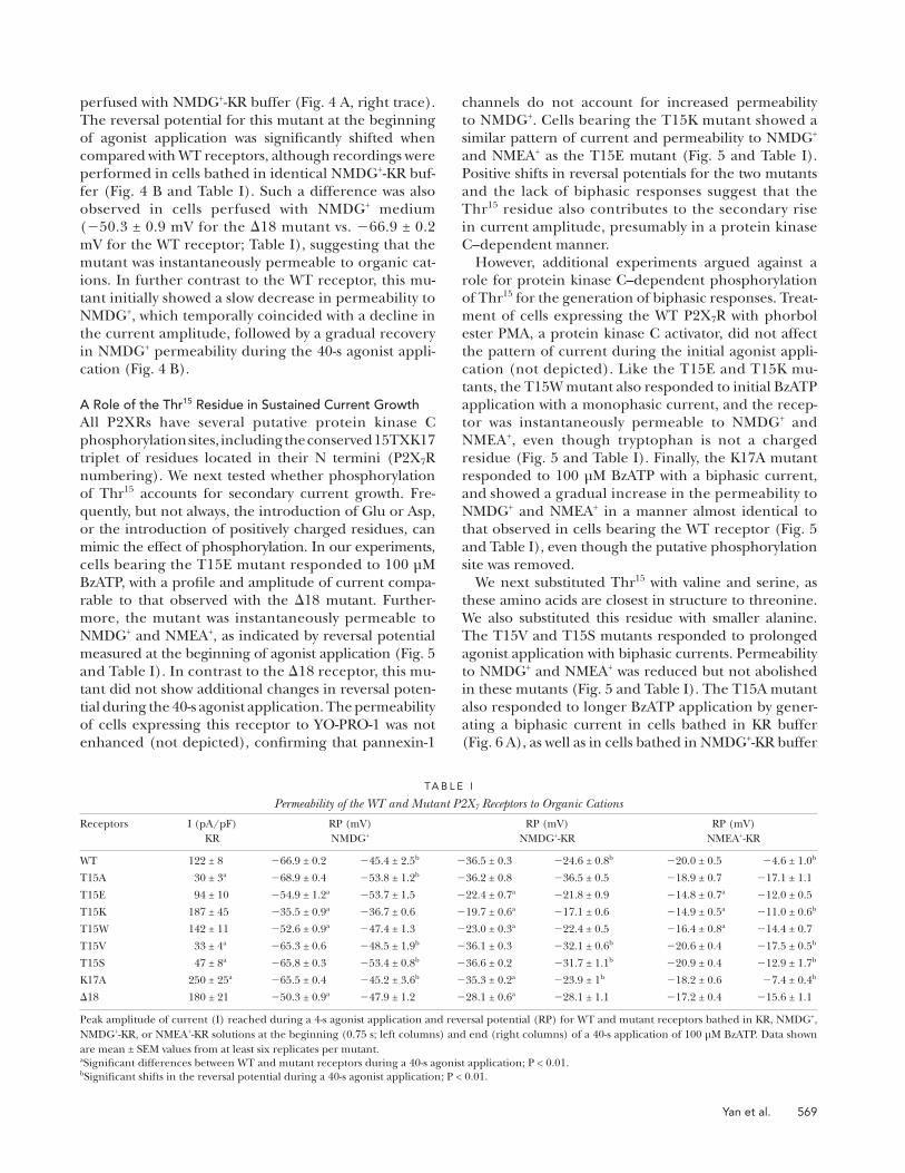
channels do not account for increased permeability
to NMDG + . Cells bearing the T15K mutant showed a
similar pattern of current and permeability to NMDG +
and NMEA + as the T15E mutant ( Fig. 5 and Table I ).
Positive shifts in reversal potentials for the two mutants
and the lack of biphasic responses suggest that the
Thr 15 residue also contributes to the secondary rise
in current amplitude, presumably in a protein kinase
C – dependent manner.
However, additional experiments argued against a
role for protein kinase C – dependent phosphorylation
of Thr 15 for the generation of biphasic responses. Treat-
ment of cells expressing the WT P2X 7 R with phorbol
ester PMA, a protein kinase C activator, did not affect
the pattern of current during the initial agonist appli-
cation (not depicted). Like the T15E and T15K mu-
tants, the T15W mutant also responded to initial BzATP
application with a monophasic current, and the recep-
tor was instantaneously permeable to NMDG + and
NMEA + , even though tryptophan is not a charged
residue ( Fig. 5 and Table I ). Finally, the K17A mutant
responded to 100 μ M BzATP with a biphasic current,
and showed a gradual increase in the permeability to
NMDG + and NMEA + in a manner almost identical to
that observed in cells bearing the WT receptor ( Fig. 5
and Table I ), even though the putative phosphorylation
site was removed.
We next substituted Thr 15 with valine and serine, as
these amino acids are closest in structure to threonine.
We also substituted this residue with smaller alanine.
The T15V and T15S mutants responded to prolonged
agonist application with biphasic currents. Permeability
to NMDG + and NMEA + was reduced but not abolished
in these mutants ( Fig. 5 and Table I ). The T15A mutant
also responded to longer BzATP application by gener-
ating a biphasic current in cells bathed in KR buffer
( Fig. 6 A ), as well as in cells bathed in NMDG + -KR buffer
perfused with NMDG + -KR buffer ( Fig. 4 A , right trace).
The reversal potential for this mutant at the beginning
of agonist application was signifi cantly shifted when
compared with WT receptors, although recordings were
performed in cells bathed in identical NMDG + -KR buf-
fer ( Fig. 4 B and Table I ). Such a difference was also
observed in cells perfused with NMDG + medium
( � 50.3 ± 0.9 mV for the � 18 mutant vs. � 66.9 ± 0.2
mV for the WT receptor; Table I ), suggesting that the
mutant was instantaneously permeable to organic cat-
ions. In further contrast to the WT receptor, this mu-
tant initially showed a slow decrease in permeability to
NMDG + , which temporally coincided with a decline in
the current amplitude, followed by a gradual recovery
in NMDG + permeability during the 40-s agonist appli-
cation ( Fig. 4 B ).
A Role of the Thr 15 Residue in Sustained Current Growth All P2XRs have several putative protein kinase C
phosphorylation sites, including the conserved 15TXK17
triplet of residues located in their N termini (P2X 7 R
numbering). We next tested whether phosphorylation
of Thr 15 accounts for secondary current growth. Fre-
quently, but not always, the introduction of Glu or Asp,
or the introduction of positively charged residues, can
mimic the effect of phosphorylation. In our experiments,
cells bearing the T15E mutant responded to 100 μ M
BzATP, with a profi le and amplitude of current compa-
rable to that observed with the � 18 mutant. Further-
more, the mutant was instantaneously permeable to
NMDG + and NMEA + , as indicated by reversal potential
measured at the beginning of agonist application ( Fig. 5
and Table I ). In contrast to the � 18 receptor, this mu-
tant did not show additional changes in reversal poten-
tial during the 40-s agonist application. The permeability
of cells expressing this receptor to YO-PRO-1 was not
enhanced (not depicted), confi rming that pannexin-1
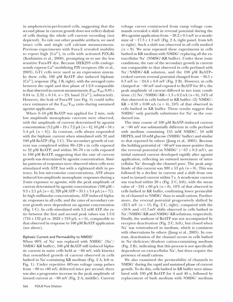
TA B L E I
Permeability of the WT and Mutant P2X 7 Receptors to Organic Cations
Receptors I (pA/pF)
KR
RP (mV)
NMDG +
RP (mV)
NMDG + -KR
RP (mV)
NMEA + -KR
WT 122 ± 8 � 66.9 ± 0.2 � 45.4 ± 2.5 b � 36.5 ± 0.3 � 24.6 ± 0.8 b � 20.0 ± 0.5 � 4.6 ± 1.0 b
T15A 30 ± 3 a � 68.9 ± 0.4 � 53.8 ± 1.2 b � 36.2 ± 0.8 � 36.5 ± 0.5 � 18.9 ± 0.7 � 17.1 ± 1.1
T15E 94 ± 10 � 54.9 ± 1.2 a � 53.7 ± 1.5 � 22.4 ± 0.7 a � 21.8 ± 0.9 � 14.8 ± 0.7 a � 12.0 ± 0.5
T15K 187 ± 45 � 35.5 ± 0.9 a � 36.7 ± 0.6 � 19.7 ± 0.6 a � 17.1 ± 0.6 � 14.9 ± 0.5 a � 11.0 ± 0.6 b
T15W 142 ± 11 � 52.6 ± 0.9 a � 47.4 ± 1.3 � 23.0 ± 0.3 a � 22.4 ± 0.5 � 16.4 ± 0.8 a � 14.4 ± 0.7
T15V 33 ± 4 a � 65.3 ± 0.6 � 48.5 ± 1.9 b � 36.1 ± 0.3 � 32.1 ± 0.6 b � 20.6 ± 0.4 � 17.5 ± 0.5 b
T15S 47 ± 8 a � 65.8 ± 0.3 � 53.4 ± 0.8 b � 36.6 ± 0.2 � 31.7 ± 1.1 b � 20.9 ± 0.4 � 12.9 ± 1.7 b
K17A 250 ± 25 a � 65.5 ± 0.4 � 45.2 ± 3.6 b � 35.3 ± 0.2 a � 23.9 ± 1 b � 18.2 ± 0.6 � 7.4 ± 0.4 b
� 18 180 ± 21 � 50.3 ± 0.9 a � 47.9 ± 1.2 � 28.1 ± 0.6 a � 28.1 ± 1.1 � 17.2 ± 0.4 � 15.6 ± 1.1
Peak amplitude of current (I) reached during a 4-s agonist application and reversal potential (RP) for WT and mutant receptors bathed in KR, NMDG + ,
NMDG + -KR, or NMEA + -KR solutions at the beginning (0.75 s; left columns) and end (right columns) of a 40-s application of 100 μ M BzATP. Data shown
are mean ± SEM values from at least six replicates per mutant.
a Signifi cant differences between WT and mutant receptors during a 40-s agonist application; P < 0.01.
b Signifi cant shifts in the reversal potential during a 40-s agonist application; P < 0.01.
570 P2X 7 R Pore Dilation
pressing WT receptors. In contrast to controls, pro-
longed application of agonist was not associated with a
rightward shift in reversal potential ( Fig. 6, D and E ),
indicating that the T15A mutant was not permeable
to larger organic cations. In cells bathed in NMDG + buf-
fer, there was a positive shift in reversal potential by
( Fig. 6 B ). As in control cells ( Fig. 2 C , left), a rapid and
transient outward current was observed in cells bathed
in NMDG + medium, followed by a shift from outward to
inward current ( Fig. 6 C ). The initial reversal potential
for this mutant bathed in NMDG + -KR and NMEA + -KR
buffers was comparable to that observed in cells ex-
Figure 5. Characterization of N-terminal P2X 7 R mutants. (Left) Representative traces of currents generated by a 40-s application of 100 μ M BzATP by WT and mutant receptors expressed in HEK293 cells. Notice that BzATP induced monophasic currents in cells ex-pressing T15E, T15K, and T15W mutants and biphasic current in all other mutants. Receptor deactivation was delayed in cells expressing T15E, T15K, T15W, and K17A mutants. Cells were bathed in KR buffer. (Middle and right) Comparison of the permeability of channels to NMDG + (middle) and NMEA + (right). Change in time courses are shown from left to right, and vertical dotted lines indicate the reversal potential for WT receptors at the beginning and at the end of a 40-s application of 100 μ M BzATP. Cells were bathed in NMDG + - or NMEA + -containing KR buffer. Mean ± SEM values are shown in Table I .

Yan et al. 571
activation of P2X 7 Rs ( Pelegrin and Surprenant, 2006 ).
However, several lines of evidence presented here argue
against the contribution of these channels or another
permeation pathway to sustained current growth. First,
the rates of leak of Fura-2 and Fura-FF were comparable
to the rate of YO-PRO-1 uptake and slower than the time
needed to reach the steady current during prolonged
exposure to agonist. Second, the uptake of YO-PRO-1
occurs in cells stimulated with ATP in a concentration
that was unable to generate the biphasic current. Third,
C6 glioma cells do not express pannexin1 – 3 channels
endogenously ( Lai et al., 2007 ), but respond with gen-
eration of biphasic current during sustained agonist
application. Fourth, the pattern of biphasic response was
not altered in cells bathed in medium containing CBX,
a pannexin-1 channel blocker. Consistently with our
conclusions with P2X 7 R, it has been shown recently
that both sustained rise in P2X 2 R current and permeabil-
ity of channels to NMDG + are independent of pannexin-1
channels ( Chaumont and Khakh, 2008 ).
In our experiments, the rates of sustained rise in current
from cells bathed in KR buffer were highly comparable to
the rates observed in cells perfused with Na + /NMDG + -KR
and NMDG + -KR media. Additionally, we observed that the
rates of positive shifts in reversal potentials were compara-
ble to the rates of delayed current growth in cells clamped
at � 60 mV. In previous reports, positive shifts in reversal
potential during sustained agonist application in cells
bathed in the presence of larger organic cations were used
as indicators of the P2X 7 R pore dilation ( Khakh et al.,
1999 ; Virginio et al., 1999 ; North, 2002 ). More recently,
however, Jiang et al. (2005) questioned the hypothesis that
P2XR pore dilation occurs under physiological conditions.
The authors observed that replacement of Na + with
NMDG + in activated cells caused the current to reverse its
direction from inward to outward. They also found that
deletion of a cysteine-rich 18 – amino acid segment in the
intracellular juxtamembrane region of the P2X 7 R results
in a mutant that is not permeable to NMDG + ( Jiang et al.,
2005 ). Single channel measurements also revealed the
+15.1 mV ( Fig. 6 F ) compared with the +22.5 mV shift
observed in cells expressing the WT receptor.
D I S C U S S I O N
Here, we have shown that the pattern of current response
by rat P2X 7 R at higher agonist concentrations is deter-
mined by the duration and the number of applications.
Repetitive brief applications (2 s or less) of BzATP gener-
ated inward currents of highly comparable peak am-
plitudes. However, during the 4-s repeated agonist
application, we consistently observed growth of current.
Furthermore, in the continued presence of agonist, initial
currents were always followed by slowly increasing cur-
rents, with time courses but not amplitudes determined
by agonist concentration. Previous studies have also shown
comparable amplitudes of P2X 7 R current during repeated
2-s agonist applications ( Hibell et al., 2000 ). Generation
of biphasic currents during prolonged stimulation of cells
expressing native and recombinant P2X 7 Rs sometimes re-
sulted in less clear separation of the two phases, probably
refl ecting the speed of application systems used ( Chessell
et al., 2001 ; Li et al., 2003 ; Ma et al., 2006 ). Biphasic cur-
rent was also observed in P2X 4 R when bathed in Ca 2+ -defi -
cient medium ( Khakh et al., 1999 ), as well as in Xenopus oocytes expressing human P2X 7 R ( Klapperstuck et al.,
2001 ). The occurrence of biphasic current response only
at high agonist concentrations in our experiments is in
accordance with a hypothesis of two distinct ATP activa-
tion sites at P2X 7 R ( Klapperstuck et al., 2001 ).
In general, the biphasic response could result from
dilation of the integral ion channel pore that opens
within milliseconds and/or from integration of pan-
nexin-1 channels or another permeation pathway that
opens more slowly. Our results are also consistent with
the expression of native pannexin-1 channels in both
HEK293 and GT1 cells. Furthermore, our data do not
detract from previous work on the role of these chan-
nels in fl uorescent dye uptake and their possible con-
tribution to uptake/leak of fluorescent dyes during
Figure 6. Characterization of the T15A-P2X 7 R mutant expressed in HEK293 cells. (A) Patterns of current in response to repetitive application of 100 μ M BzATP, followed by a washout period of 5 min. (B and C) 100 μ M BzATP-induced biphasic response from cells bathed in NMDG + -KR buffer (B) and NMDG + buf-fer (C). ( D and E) Lack of channel per-meability to NMDG + (D) and NMEA + (E) when cells were bathed in KR buffer in which all Na + was substituted with or-ganic cations. (F) A shift in the reversal potential observed during a 40-s applica-tion of 100 μ M BzATP from cells bathed in NMDG + medium.

572 P2X 7 R Pore Dilation
nexin-1 channels requires time ( Pelegrin and Surprenant,
2006 ). Changes in cation permeability for these mutant
receptors refl ected on the pattern of current response;
all four generated high amplitude monophasic cur-
rents. The simplest explanation for changes in permea-
bility to organic cations in cells expressing these mutants
is that the channel pore immediately opens with the di-
lated mode, and that the diameter of the pore is suffi -
cient to permit permeation of larger cations. In contrast
to the � 18 mutant, these three mutants stay locked in
the initial dilated state. On the other hand, the biphasic
response was preserved in cells expressing T15S, T15V,
and T15A mutants. In the presence of divalent cations,
the permeability of T15S and T15V receptors to NMDG +
was signifi cantly reduced, whereas the T15A mutant was
not permeable to large organic cations. These results
suggest the importance of N-terminal residues in pore
dilation, but further experiments are required to clarify
the residues of the relevance for this regulation and the
structural details of conformation changes during pore
dilation, and what extent pore dilation contributes to
cell lysis. Consistent with our conclusion, it has been shown
recently that P2X 2 receptors display permeability dy-
namics, which are correlated with conformational changes
in the cytosolic domain remote from the selectivity fi lter
itself ( Chaumont and Khakh, 2008 ).
In summary, we suggest that channel pore opening of
the WT receptor, as well as of the T15A, T15V, and T15S
mutants, involves a two-step transition: (1) from closed
to opened, and (2) from opened to dilated, leading to
the generation of biphasic currents ( Fig. 7 A ). The
steady-state size of the pore varies among these recep-
tors, which is refl ected by organic cation permeability
( Fig. 7 B ). In contrast, the enlargement of the channel
lack of change in the channel kinetics during sustained ac-
tivation of human P2X 7 R expressed in Xenopus oocytes
( Riedel et al., 2007a ).
Consistent with Jiang ’ s experiments, we also observed
that substitution of KR buffer with NMDG + buffer after a
short (4-s) exposure to 100 μ M BzATP induced a shift
from inward to outward current in < 1 s. In contrast, when
KR buffer was replaced with NMDG + buffer after a pro-
longed (40-s) application, the amplitude of inward cur-
rent was dramatically reduced without shifting to outward
current, with the mean value of current comparable to
the steady-state current observed in cells continuously
perfused with NMDG + medium for 40 s. In the absence of
monovalent and divalent cations, the permeability of the
channel pore for NMDG + resulted in the current amp-
litude of only 10% of that observed in controls. Such
small amplitude of the whole cell current is consistent
with the low permeability to Tris + observed in single chan-
nel recording with human P2X 7 R ( Riedel et al., 2007a ).
Furthermore, the � 18-P2X 7 R mutant in our hands showed
an instantaneous positive shift in reversal potential when
recording was performed in NMDG + -containing medium
both in the presence and absence of divalent cations,
demonstrating that the initial opening of the channel
pore of this mutant is larger than in controls. We also ob-
served changes in permeability of this mutant during pro-
longed agonist application; an initial shrinking of the
pore followed by sustained dilation. Collectively, these re-
sults suggest that the P2X 7 R pore dilates during sustained
agonist application, reaching the permeability for organic
cations, and that the C-terminal infl uences this process.
In our experiments, three other P2X 7 R mutants, T15E,
T15K, and T15W, also showed instantaneous permeabil-
ity to NMDG + and NMEA + , whereas integration of pan-
Figure 7. Effects of Thr 15 substitution on the pattern of current and permeability to NMDG + schematic models. (A) Two patterns of currents: biphasic (left) and high amplitude monophasic (right). The bi-phasic response refl ects three steps in gating: closed (c), opened (o), and dilated (d), and is observed in cells expressing WT receptors and T15A, T15V, and T15S mutants. The high amplitude monophasic response refl ects instantaneous transition from the closed to the dilated stage, and is observed in cells expressing T15W, T15E, and T15K mutants. (B) A shift in the reversal potential (RP) for WT and mu-tant receptors during sustained agonist application. Mean values for the reversal potential at the end of a 40-s recording for WT and six T15 mutant recep-tors were subtracted from the initial reversal poten-tial value for the WT receptor ( � 36.5 mV; Table I , NMDG + -KR column). The side chains of amino acid residues used in substitution studies are shown at the bottom.

Yan et al. 573
Klapperstuck , M. , C. Buttner , G. Schmalzing , and F. Markwardt .
2001 . Functional evidence of distinct ATP activation sites at the
human P2X(7) receptor. J. Physiol. 534 : 25 – 35 .
Koshimizu , T.A. , F. Van Goor , M. Tomic , A.O. Wong , A. Tanoue , G.
Tsujimoto , and S.S. Stojilkovic . 2000 . Characterization of calcium
signaling by purinergic receptor-channels expressed in excitable
cells. Mol. Pharmacol. 58 : 936 – 945 .
Lai , C.P. , J.F. Bechberger , R.J. Thompson , B.A. MacVicar , R.
Bruzzone , and C.C. Naus . 2007 . Tumor-suppressive effects of pan-
nexin 1 in C6 glioma cells. Cancer Res. 67 : 1545 – 1554 .
Li , Q. , X. Luo , W. Zeng , and S. Muallem . 2003 . Cell-specifi c behav-
ior of P2X7 receptors in mouse parotid acinar and duct cells.
J. Biol. Chem. 278 : 47554 – 47561 .
Li , Q. , X. Luo , and S. Muallem . 2005 . Regulation of the P2X7 recep-
tor permeability to large molecules by extracellular Cl- and Na+.
J. Biol. Chem. 280 : 26922 – 26927 .
Ma , W. , A. Korngreen , S. Weil , E.B. Cohen , A. Priel , L. Kuzin ,
and S.D. Silberberg . 2006 . Pore properties and pharmacologi-
cal features of the P2X receptor channel in airway ciliated cells.
J. Physiol. 571 : 503 – 517 .
Michel , A.D. , I.P. Chessell , and P.P. Humphrey . 1999 . Ionic ef-
fects on human recombinant P2X7 receptor function. Naunyn Schmiedebergs Arch. Pharmacol. 359 : 102 – 109 .
Nicke , A. , H.G. Baumert , J. Rettinger , A. Eichele , G. Lambrecht , E.
Mutschler , and G. Schmalzing . 1998 . P2X1 and P2X3 receptors
form stable trimers: a novel structural motif of ligand-gated ion
channels. EMBO J. 17 : 3016 – 3028 .
Nicke , A. , D. Kerschensteiner , and F. Soto . 2005 . Biochemical and
functional evidence for heteromeric assembly of P2X1 and P2X4
subunits. J. Neurochem. 92 : 925 – 933 .
North , R.A. 2002 . Molecular physiology of P2X receptors. Physiol. Rev. 82 : 1013 – 1067 .
Pelegrin , P. , and A. Surprenant . 2006 . Pannexin-1 mediates large
pore formation and interleukin-1beta release by the ATP-gated
P2X7 receptor. EMBO J. 25 : 5071 – 5082 .
Petrou , S. , M. Ugur , R.M. Drummond , J.J. Singer , and J.V. Walsh Jr .
1997 . P2X7 purinoceptor expression in Xenopus oocytes is not
suffi cient to produce a pore-forming P2Z-like phenotype. FEBS Lett. 411 : 339 – 345 .
Riedel , T. , I. Lozinsky , G. Schmalzing , and F. Markwardt . 2007a .
Kinetics of P2X7 receptor-operated single channels currents.
Biophys. J. 92 : 2377 – 2391 .
Riedel , T. , G. Schmalzing , and F. Markwardt . 2007b . Infl uence of extra-
cellular monovalent cations on pore and gating properties of P2X7
receptor-operated single-channel currents. Biophys. J. 93 : 846 – 858 .
Smart , M.L. , B. Gu , R.G. Panchal , J. Wiley , B. Cromer , D.A. Williams ,
and S. Petrou . 2003 . P2X7 receptor cell surface expression and
cytolytic pore formation are regulated by a distal C-terminal re-
gion. J. Biol. Chem. 278 : 8853 – 8860 .
Surprenant , A. , F. Rassendren , E. Kawashima , R.A. North , and G.
Buell . 1996 . The cytolytic P2Z receptor for extracellular ATP
identifi ed as a P2X receptor (P2X7). Science . 272 : 735 – 738 .
Virginio , C. , D. Church , R.A. North , and A. Surprenant . 1997 .
Effects of divalent cations, protons and calmidazolium at the rat
P2X7 receptor. Neuropharmacology . 36 : 1285 – 1294 .
Virginio , C. , A. MacKenzie , F.A. Rassendren , R.A. North , and A.
Surprenant . 1999 . Pore dilation of neuronal P2X receptor chan-
nels. Nat. Neurosci. 2 : 315 – 321 .
Yan , Z. , Z. Liang , M. Tomic , T. Obsil , and S.S. Stojilkovic . 2005 .
Molecular determinants of the agonist binding domain of a P2X
receptor channel. Mol. Pharmacol. 67 : 1078 – 1088 .
Yan , Z. , Z. Liang , T. Obsil , and S.S. Stojilkovic . 2006 . Participation
of the Lys313-Ile333 sequence of the purinergic P2X4 receptor in
agonist binding and transduction of signals to the channel gate.
J. Biol. Chem. 281 : 32649 – 32659 .
pore for T15E, T15K, and T15W mutants involves a sin-
gle transition step, from closed to dilated, resulting in
the generation of high amplitude monophasic currents
( Fig. 7 A , right). In the absence of crystal structure data,
it is diffi cult to predict how Thr 15 contributes to the con-
trol of gating. The T15XK17 triplet represents a putative
protein kinase C phosphorylation site, but phosphoryl-
ation of the Thr 15 residue (if it occurs) does not account
for delayed current growth. There is a parallel between
changes in permeability of receptors to NMDG + and the
steric size of the side chains of these residues ( Fig. 7 B ),
suggesting that this phenomenon could underlie changes
in the receptor behavior.
We are thankful to Tomas Obsil for helpful discussion. This work is supported by the Intramural Research Program of
the National Institute of Child Health and Human Development, National Institutes of Health.
Edward N. Pugh Jr. served as editor.
Submitted: 3 June 2008 Accepted: 29 September 2008
R E F E R E N C E S Chaumont , S. , and B.S. Khakh . 2008 . Patch-clamp coordinated
spectroscopy shows P2X2 receptor permeability dynamics require
cytosolic domain rearrangements but not Panx-1 channels. Proc. Natl. Acad. Sci. USA . 105 : 12063 – 12068 .
Chessell , I.P. , A.D. Michel , and P.P. Humphrey . 1997 . Properties of
the pore-forming P2X7 purinoceptor in mouse NTW8 microglial
cells. Br. J. Pharmacol. 121 : 1429 – 1437 .
Chessell , I.P. , C.B.A. Grahames , A.D. Michel, and P.P.A. Humphrey.
2001 . Dynamics of P2X7 receptor pore dilation: pharmacological
and functional consequences. Drug Dev. Res. 53 : 60 – 65 .
Dubyak , G.R. 2007 . Go it alone no more – P2X7 joins the society of hetero-
meric ATP-gated receptor channels. Mol. Pharmacol. 72 : 1402 – 1405 .
Egan , T.M. , J.A. Cox , and M.M. Voigt . 2004 . Molecular structure of
P2X receptors. Curr. Top. Med. Chem. 4 : 821 – 829 .
Ferrari , D. , C. Pizzirani , E. Adinolfi , R.M. Lemoli , A. Curti , M. Idzko ,
E. Panther , and F. Di Virgilio . 2006 . The P2X7 receptor: a key
player in IL-1 processing and release. J. Immunol. 176 : 3877 – 3883 .
Gudipaty , L. , B.D. Humphreys , G. Buell , and G.R. Dubyak . 2001 .
Regulation of P2X(7) nucleotide receptor function in human
monocytes by extracellular ions and receptor density. Am. J. Physiol. Cell Physiol. 280 : C943 – C953 .
He , M.L. , H. Zemkova , T.A. Koshimizu , M. Tomic , and S.S. Stojilkovic .
2003 . Intracellular calcium measurements as a method in studies
on activity of purinergic P2X receptor channels. Am. J. Physiol. Cell Physiol. 285 : C467 – C479 .
Hibell , A.D. , E.J. Kidd , I.P. Chessell , P.P. Humphrey , and A.D.
Michel . 2000 . Apparent species differences in the kinetic prop-
erties of P2X(7) receptors. Br. J. Pharmacol. 130 : 167 – 173 .
Jiang , L.H. , F. Rassendren , A. Mackenzie , Y.H. Zhang , A. Surprenant ,
and R.A. North . 2005 . N-methyl-D-glucamine and propidium
dyes utilize different permeation pathways at rat P2X(7) recep-
tors. Am. J. Physiol. 289 : C1295 – C1302 .
Khakh , B.S. , X.R. Bao , C. Labarca , and H.A. Lester . 1999 . Neuronal
P2X transmitter-gated cation channels change their ion selectiv-
ity in seconds. Nat. Neurosci. 2 : 322 – 330 .
Klapperstuck , M. , C. Buttner , T. Bohm , G. Schmalzing , and F.
Markwardt . 2000 . Characteristics of P2X7 receptors from human
B lymphocytes expressed in Xenopus oocytes. Biochim. Biophys. Acta . 1467 : 444 – 456 .