Acute Restraint Stress in Zebrafish: Behavioral Parameters and Purinergic Signaling
Upload
khangminh22Category
view
1download
0
�����������������
Citation: Rotondo, J.C.; Mazziotta, C.;
Lanzillotti, C.; Stefani, C.; Badiale, G.;
Campione, G.; Martini, F.; Tognon, M.
The Role of Purinergic P2X7 Receptor
in Inflammation and Cancer: Novel
Molecular Insights and Clinical
Applications. Cancers 2022, 14, 1116.
https://doi.org/10.3390/
cancers14051116
Academic Editor: Alain P. Gobert
Received: 20 January 2022
Accepted: 17 February 2022
Published: 22 February 2022
Publisher’s Note: MDPI stays neutral
with regard to jurisdictional claims in
published maps and institutional affil-
iations.
Copyright: © 2022 by the authors.
Licensee MDPI, Basel, Switzerland.
This article is an open access article
distributed under the terms and
conditions of the Creative Commons
Attribution (CC BY) license (https://
creativecommons.org/licenses/by/
4.0/).
cancers
Review
The Role of Purinergic P2X7 Receptor in Inflammation andCancer: Novel Molecular Insights and Clinical ApplicationsJohn Charles Rotondo 1,2,† , Chiara Mazziotta 1,2,†, Carmen Lanzillotti 1,2, Chiara Stefani 1, Giada Badiale 1 ,Giulia Campione 1, Fernanda Martini 1,2,3 and Mauro Tognon 1,*
1 Laboratories of Cell Biology and Molecular Genetics, Section of Experimental Medicine, Department ofMedical Sciences, School of Medicine, University of Ferrara, 44121 Ferrara, Italy; [email protected] (J.C.R.);[email protected] (C.M.); [email protected] (C.L.); [email protected] (C.S.);[email protected] (G.B.); [email protected] (G.C.); [email protected] (F.M.)
2 Centre for Studies on Gender Medicine, Department of Medical Sciences, University of Ferrara,44121 Ferrara, Italy
3 Laboratory for Technologies of Advanced Therapies (LTTA), University of Ferrara, 44121 Ferrara, Italy* Correspondence: [email protected]† These authors contributed equally to this work.
Simple Summary: The purinergic P2X7 receptor (P2X7R) is a cell membrane protein whose activa-tion has been related to a variety of cellular processes, while its dysregulation has been linked toinflammation and cancer. ATP plays a key role in numerous metabolic processes due to its abundancein the tumour microenvironment. P2X7R plays an important role in cancer by interacting with ATP.The unusual property of P2X7R is that stimulation with low ATP doses causes the opening of apermeable channel for sodium, potassium, and calcium ions, whereas continued stimulation withhigh ATP doses leads to the formation of a non-selective pore. The latter effect induces the cell death.This evidence suggests that P2X7R has both pro- and anti-tumour potential. In this review, we aimedto describe the most relevant characteristics of P2X7R function, activation, and its ligands, while alsosummarising the role of P2X7R activation in the context of inflammation and cancer. The currentlyused therapeutic approaches and clinical trials of P2X7R modulators is also described.
Abstract: The purinergic P2X7 receptor (P2X7R) is a transmembrane protein whose expression hasbeen related to a variety of cellular processes, while its dysregulation has been linked to inflammationand cancer. P2X7R is expressed in cancer and immune system cell surfaces. ATP plays a key rolein numerous metabolic processes due to its abundance in the tumour microenvironment. P2X7Rplays an important role in cancer by interacting with ATP. The unusual property of P2X7R is thatstimulation with low doses of ATP causes the opening of a permeable channel for sodium, potassium,and calcium ions, whereas sustained stimulation with high doses of ATP favours the formation ofa non-selective pore. The latter effect induces a change in intracellular homeostasis that leads tocell death. This evidence suggests that P2X7R has both pro- and anti-tumour proprieties. P2X7R isincreasingly recognised as a regulator of inflammation. In this review, we aimed to describe the mostrelevant characteristics of P2X7R function, activation, and its ligands, while also summarising therole of P2X7R activation in the context of inflammation and cancer. The currently used therapeuticapproaches and clinical trials of P2X7R modulators are also described.
Keywords: P2X7 receptor; P2X7R; ATP; BzATP; tumour microenvironment; inflammation; immunesystem; colon cancer; melanoma; infection; malignant pleural mesothelioma
1. Background
Extracellular nucleotides, including adenosine triphosphate (ATP), play a fundamentalrole in various biological functions, such as cell growth, differentiation, and migration, aswell as tissue homeostasis, immunity, inflammation, and cancer [1]. These molecules act
Cancers 2022, 14, 1116. https://doi.org/10.3390/cancers14051116 https://www.mdpi.com/journal/cancers
Cancers 2022, 14, 1116 2 of 21
as messengers in cell-to-cell communication by stimulating the release of other extracel-lular factors, which in turn activate additional signalling pathways by interacting withtheir own receptors [2]. One of the main functions of these molecules is the modulationof purinergic signal transduction [2]. ATP is found at high concentrations (5–10 mM) inphysiological conditions [3]. It interacts with immune cells, the vascular endothelium,and the surrounding matrix. When ATP is transferred from the cell cytoplasm to theextracellular environment, it can act as a damage-associated molecular pattern (DAMP),i.e., it acts as a signal molecule that induces an inflammatory response [4]. Consistently,extracellular ATP (eATP) accumulates near the inflamed tissues [4]. Moreover, both adeno-sine and ATP act as regulators in tumour growth processes. Both molecules are abundantin the tumour microenvironment (TME), comprising tumour-surrounding blood vessels,immune cells, fibroblasts, signalling molecules, and the extracellular matrix [5]. A key ATPtarget is represented by the purinergic P2X receptor (P2XR) family members [6]. Thesereceptors are trimeric ATP-activated ion channels that are permeable to Na+, K+, and Ca+2,which open when interacting with eATP [7]. The opening of non-selective pores causesthe dysregulation of cellular homeostasis with consequent cell death [8]. However, P2XRactivation causes the release of cytokines, guides survival, metabolic adaptation to nutrientdeprivation, and migration and invasion of cancer cells [9]. Particular attention has beenpaid to the P2X7 receptor (P2X7R) [10], whose expression is associated with inflammation,survival, proliferation, angiogenesis, and metastasis [11,12]. This receptor, which is foundon both cancer and immune system cell surfaces, is characterised by a biphasic response toATP: (i) a short stimulation allows the influx of sodium and calcium ions into the cell; (ii) along period of stimulation of the receptor triggers the opening of a non-selective pore thatallows the passage of molecules whose molecular weight is lower than 900 Da (Figure 1).
Cancers 2022, 14, x FOR PEER REVIEW 2 of 20
1. Background
Extracellular nucleotides, including adenosine triphosphate (ATP), play a
fundamental role in various biological functions, such as cell growth, differentiation, and
migration, as well as tissue homeostasis, immunity, inflammation, and cancer [1]. These
molecules act as messengers in cell-to-cell communication by stimulating the release of
other extracellular factors, which in turn activate additional signalling pathways by
interacting with their own receptors [2]. One of the main functions of these molecules is
the modulation of purinergic signal transduction [2]. ATP is found at high concentrations
(5–10 mM) in physiological conditions [3]. It interacts with immune cells, the vascular
endothelium, and the surrounding matrix. When ATP is transferred from the cell
cytoplasm to the extracellular environment, it can act as a damage-associated molecular
pattern (DAMP), i.e., it acts as a signal molecule that induces an inflammatory response
[4]. Consistently, extracellular ATP (eATP) accumulates near the inflamed tissues [4].
Moreover, both adenosine and ATP act as regulators in tumour growth processes. Both
molecules are abundant in the tumour microenvironment (TME), comprising tumour-
surrounding blood vessels, immune cells, fibroblasts, signalling molecules, and the
extracellular matrix [5]. A key ATP target is represented by the purinergic P2X receptor
(P2XR) family members [6]. These receptors are trimeric ATP-activated ion channels that
are permeable to Na+, K+, and Ca+2, which open when interacting with eATP [7]. The
opening of non-selective pores causes the dysregulation of cellular homeostasis with
consequent cell death [8]. However, P2XR activation causes the release of cytokines,
guides survival, metabolic adaptation to nutrient deprivation, and migration and invasion
of cancer cells [9]. Particular attention has been paid to the P2X7 receptor (P2X7R) [10],
whose expression is associated with inflammation, survival, proliferation, angiogenesis,
and metastasis [11,12]. This receptor, which is found on both cancer and immune system
cell surfaces, is characterised by a biphasic response to ATP: (i) a short stimulation allows
the influx of sodium and calcium ions into the cell; (ii) a long period of stimulation of the
receptor triggers the opening of a non-selective pore that allows the passage of molecules
whose molecular weight is lower than 900 Da (Figure 1).
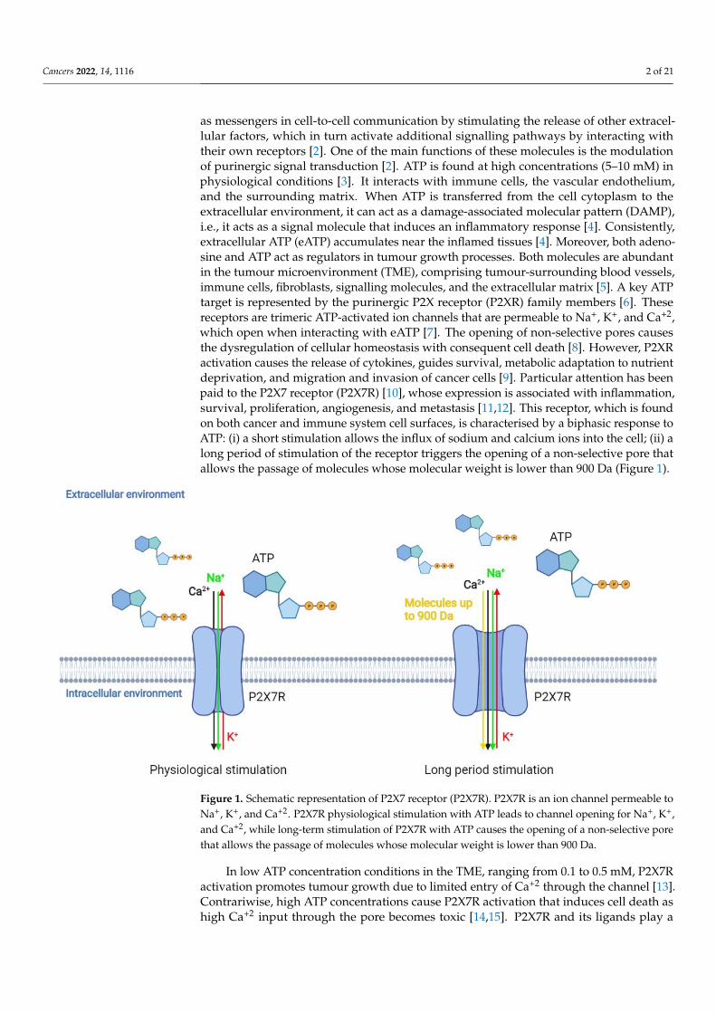
Figure 1. Schematic representation of P2X7 receptor (P2X7R). P2X7R is an ion channel permeable to
Na+, K+, and Ca+2. P2X7R physiological stimulation with ATP leads to channel opening for Na+, K+,
Figure 1. Schematic representation of P2X7 receptor (P2X7R). P2X7R is an ion channel permeable toNa+, K+, and Ca+2. P2X7R physiological stimulation with ATP leads to channel opening for Na+, K+,and Ca+2, while long-term stimulation of P2X7R with ATP causes the opening of a non-selective porethat allows the passage of molecules whose molecular weight is lower than 900 Da.
In low ATP concentration conditions in the TME, ranging from 0.1 to 0.5 mM, P2X7Ractivation promotes tumour growth due to limited entry of Ca+2 through the channel [13].Contrariwise, high ATP concentrations cause P2X7R activation that induces cell death ashigh Ca+2 input through the pore becomes toxic [14,15]. P2X7R and its ligands play a
Cancers 2022, 14, 1116 3 of 21
key role in tumorigenesis, development, and metastasis in a variety of cancers. Its role ininflammation is not fully understood.
In this review, we aimed to describe P2X7R activation and functions, and the effectsof P2X7R interaction with its ligands on cancer and immune response, as well as its rolein inflammation and tumour-associated inflammation. We also describe the currentlyemployed therapeutic approaches and the clinical trials of P2X7R modulators.
2. P2X7 Receptor Genetics, Characteristics, Function, and Tissue Distribution
The P2XR family includes seven different subtypes, i.e., P2X1-7, which are typedaccording to their pharmacological properties and structures. These receptors are cation-permeable ligand-gated ion channels that open in response to the binding of eATP consist-ing of three protein subunits (homotrimers or heterotrimers) activated by ATP permeableto Na+, K+, and Ca+2. The P2XR coding genes map in different chromosome loci, whilsthomologous genes have also been identified in other vertebrates [16]. The P2X7R (orP2RX7) gene maps in the 12q24.31 chromosome [17], containing 13 exons and 10 splicingvariants (P2X7A-J) [18], while the full-length isoform is the P2X7A form. Evidence indicatesthat P2X7R expression appears to be epigenetically regulated by DNA methylation [19]and miRNA regulation [20], which are important processes for gene expression regulationin a variety of cell types [21–25]. The P2X7R full-length isoform consists of a sequenceof 595 amino acid (a.a.) residues, in which two hydrophobic regions crossing the plasmamembrane can be identified: (i) N-terminus (N-ter), which is located in the cytoplasm;(ii) C-terminus (C-ter), which contains 70 to 200 additional a.a. residues compared toother P2X family receptors [26,27]. The extracellular domain contains the ATP binding siteand 10 cysteine residues whose oxidation contributes to the sulphide bridge formation,which is necessary for the tertiary structure [26]. The C-ter domain is involved in themajority of functions related to P2X7R, such as post-translational modifications (PTMs),cell localisation, protein–protein interaction, and initiation of the signal transduction cas-cade [26]. PTMs occurring at this domain play a role in the receptor function throughregulating its signalling pathways. For instance, a tyrosine phosphorylation site within theC-ter domain has been related to the P2X7R-induced release of TNF-α [28]. The cysteineresidues located at the P2X7R domain are susceptible to palmitoylation, which is a PTMcapable in influencing both receptor function and trafficking to and/or from the cell surfacemembrane [29]; P2X7R C-ter palmitoylation in particular facilitates the receptor large poreopening and enhances the association of the C-ter a.a. residues to the plasma membranecholesterol [30]. Mutations at this domain can reduce both cation channel activity and poreformation [31], while also influencing, at the same time, P2X7R trafficking [32]. Likewise,mutations at the C-ter domain can result in a decreased receptor function through defectiveN-linked glycosylation processing and oligomerisation [31,33–35]. P2X7R C-ter domain isinvolved in the interaction between the receptor and membrane structural factors and/orintracellular signalling messengers in order to mediate signalling transduction pathwayactivation [32,36]. For instance, the P2X7R C-ter domain is capable of interacting with theadaptor protein of the NF-κB, i.e., MyD88, in order to provoke NF-κB activation, ultimatelyleading to the positive regulation of cytokine expression such as immature pro-interleukin-1β (pro-IL-1β) [37]. Moreover, the cysteine-rich domain located within the C-ter of thereceptor prevents receptor desensitisation [38] while contributing to Ca2+-independentfacilitation current [36]. Different proteins interacting with P2X7R C-ter domain have beenidentified with different assays [39], including cytoskeletal proteins and enzymes such askinases, intracellular/transmembrane heat shock proteins other than the P2X4 receptor(P2X7R), and pannexin-1 plasma membrane hemichannels. The latter in particular mediatesthe P2X7-dependent pore opening as well as IL-1β and ATP release [40,41].
Crystallographic studies have shown that P2X7R consists of three subunits, whichinclude the ATP binding site in the pocket structure [38]. The ATP binding site has sevenpositively charged a.a. residues and two hydrophobic a.a. residues. There are four lysineresidues (Lys64, Lys66, Lys193, and Lys311) that are responsible for the inhibition of

Cancers 2022, 14, 1116 4 of 21
the receptor following binding with oxidised ATP (oATP). P2X7R sequencing/structuralanalysis suggested that the ATP binding site allows small molecule access [42]. The receptoralso presents an allosteric site in the interface between the subunits located adjacent tothe ATP binding site [38]. When the allosteric site is occupied, a conformational changein the receptor is induced, leading to the channel opening. This site is absent in P2X3-4receptors [42]. P2RX7 has a consensus sequence (Ans-X-Ser/Thr) being located within thethree glycosylation sites of the protein. Glycosylation is a post-translational modification,essential for the molecular transport of the receptor on the cell surface and for the receptorfunction. If two-thirds of sites are glycosylated, the receptor is fully functional. If onlyone site is glycosylated, the response to ATP is barely detectable, while in conditions ofno glycosylation, the receptor does not respond to stimulation [43]. This characteristic isshared with all P2X family receptors [43]. The human P2X7R is a highly polymorphic gene.Several single-nucleotide polymorphisms (SNPs) have been identified within the P2X7Rcoding gene. Most SNPs have been found as being located within the intronic regions,while about 150 non-synonymous SNPs have also been identified [26]. Mutations can beneutral or can positively or negatively affect P2X7R activity. The combination of theseSNPs generates different haplotypes with effects on the receptor functionality. Thus, it isimpossible to predict receptor functionality by identifying only one SNP. The most commonSNPs and related diseases/conditions, including inflammation-related diseases and cancer,are listed in Table 1 [42].
Table 1. Main human P2X7 single-nucleotide polymorphisms and related diseases/conditions.
dbSNP ID Base Amino Acid Effect on Implicated ConditionsSubstitution Substitution P2X7 Function
rsl7525809 370T > C A76V Loss Multiple sclerosisrs28360447 474G > A G150R Loss Osteoporosis
rs208294 489C > T H155Y Gain Multiple sclerosis, chronic pain, severe sepsis,children’s febrile seizures
rs7958311 835G > A R270H Loss Chronic painrs28360457 946G > A R307Q Loss Osteoporosisrs1718119 1068G > A A348T Gain Osteoporosis, anxiety disorder, toxoplasmosisrs2230911 1096C > G T357S Loss Osteoporosis
rs2230912 1405A > G Q460R Loss Osteoporosis, severe sepsis, bipolar disorders,major depressive disorders
rs3751143 1513A > C E496A Loss Osteoporosis, tuberculosis,cardiovascular risks
rs2230913 1563C > G H521Q Neutral -rs1653624 1729T > A I568N Loss Osteoporosis
The role of SNPs within the P2X7R coding gene is not yet completely clear. However,it is known that in the case of loss-of-function SNPs, the receptor may promote cell growthby inhibiting apoptosis, while in the case of gain-of-function SNPs, P2X7R induces cellproliferation and the release of factors, such as vascular endothelial growth factor (VEGF)and transforming growth factor beta (TGF-β) [44]. In addition, only a few gene muta-tions clearly interfere with receptor functionality [45]. Indeed, not only is P2X7R highlyexpressed in cancer, but tumour tissues express the functional form of the receptor [46,47].The unusual characteristic of P2X7R is that stimulation with low doses of ATP causes theopening of a permeable channel to Na+, K+, and Ca+2 ions [48], but continuous stimulationat high doses of ATP leads to the formation of a non-selective pore, which is permeableto molecules with a molecular weight of up to 900 Da (Figure 1) [49,50]. The mechanismresponsible for the channel-to-pore transition is not completely clear. It has been hypothe-sised that continuous stimulation with ATP recruits accessory proteins involved in poreformation [51]. Another hypothesis is that the trimer can change its conformation, enlargingthe diameter of the channel [52]. Indeed, previous data suggest that the non-selective poreformation is due to both the recruitment of accessory molecules and the dilation of the
Cancers 2022, 14, 1116 5 of 21
channel itself. Thus, both hypotheses could be considered valid. A possible explanationcould be that the single binding between ATP molecule and P2X7R and the subsequentconformational change of the binding domain makes the second binding with anotherATP molecule more difficult. When a second ATP molecule binds to P2X7R, the channelpermeable to cations opens alongside another conformational change, which increases thedifficulty of binding a third ATP molecule. However, binding with the third ATP moleculeis still possible. When this interaction occurs, P2X7R undergoes another conformationalchange, thus causing the dilation of the channel, which becomes permeable to moleculeswith molecular weight of up to 900 Da. In this condition, there is an increase in the flow ofcations into the cell [53]. This process activates other pores, which are responsible for thetransport of large molecules. An additional characteristic of P2X7R is that the receptor isnot desensitised in the presence of the agonist, even after several minutes [26]. Sustainedagonist stimulation increases receptor sensitivity and the amplitude of the response. Con-trariwise, P2X1/-3 receptors are desensitised in a few seconds, while P2X2/-4/-5 receptorsare desensitised after less than a minute [26]. Cell membrane composition affects the poreopening. For instance, high cholesterol content in the membrane inhibits pore openings [42].Indeed, studies aimed at identifying the mechanism of cholesterol action have suggestedthat the inhibition of pore openings does not depend on changes in membrane fluidity;rather, it is likely that cholesterol directly interacts with the P2X7R trans-membrane do-main [42]. P2X7R is expressed in hematopoietic cells, such as monocytes, macrophages,dendritic cells, B and T lymphocytes, and Langerhans epidermal cells, but also in thebasolateral membrane cells in the liver, as well as in osteoclasts and osteoblasts [53–56].P2X7R overexpression/activation is involved in many different pathological processes,such as inflammation [57], proliferation, invasion and migration, metabolism, autophagy,and cell death [53,58,59]. Receptor activation can either present positive or negative effectsin relation to the duration and intensity of the stimulation, the cell type, the ion extracellularconcentration, and the phospholipid composition of the membrane plasma [26,46,53,60].
3. The Role of P2X7 Receptor in Cancer
A crucial P2X7R characteristic is that prolonged receptor stimulation leads to the open-ing of a non-selective pore, allowing for the passage of several molecules with molecularweights of up to 900 Da. This event causes an alteration in the intracellular homeostasis,which can lead to cell death. However, as it has been reported that P2X7R activation confersan advantage for cancer cell survival and growth, the role of the receptor in cancer is enig-matic [61]. Moreover, when the cell type is considered, the effect of receptor activation isdifferent, and, in some cases, even contrary, thus indicating that the receptor presents bothpro- and anti-tumour activities [26]. However, a tumour diagnostic/prognostic role forP2X7R has been remarked upon [62,63], as it has been found that P2X7R is overexpressedin a variety of cancers, such as lung, colon, thyroid, pancreatic, prostate, and breast cancer,as well as lymphoma and glioma [8,64].
Overexpression of P2X7R leads to an increase in tumour growth, metastasis, produc-tion of VEGF, and release of matrix metalloproteinases (MMPs) [65], which are proteolyticenzymes that degrade extracellular matrix proteins and play a key role in metastasis [47].Tumours expressing P2X7R are also characterised by increased proliferation, decreasedapoptosis, and high levels of the transcription factor nuclear factor of activated T cells 1(NFATc1). P2X7R-related tumours also show alteration of intracellular Ca2+ homeostasisand stimulation of mitochondrial metabolism [66]. At the same time, NF-κB accumulatesin the nucleus and contributes to cell invasiveness and survival by maintaining high P2X7Rexpression and inducing synthesis of MMP-3 and MMP-9 [67]. Under hypoxic conditions,the expression of P2X7R increases. Knockdown of hypoxia-inducible factor 1α (HIF-1α)decreases P2X7R expression, which also decreases nuclear factor kappa light chain en-hancer of activated B cells (NF-κB), thus suggesting that HIF-1α controls P2X7R and NF-kBexpression under hypoxia conditions.

Cancers 2022, 14, 1116 6 of 21
P2X7R induces apoptosis by activating caspases 3 and 7 following a massive Ca+2
intake [68,69]. At low ATP concentration, ranging from 0.1 to 0.5 mM, the receptor promotestumour growth and metastasis formation, whilst strong P2X7R activation leads to cell deathdue to the cytotoxic effect of excessive Ca+2 influx [8]. Activation of P2X7R in melanoma,colon cancer, and neuroblastoma cancer cell lines induces secretion of VEGF [14,70,71],while its activation in breast cancer cell lines and leukocytes leads to an increase in the activ-ity of MMP-3, MMP-9, and MMP-13 [14,72–74]. Specifically, MMP-9 plays an important rolein intravasation, i.e., invasion of tumour cells through the basement membrane into a bloodor lymphatic vessel [75]. One mechanism by which P2X7R regulates MMP activity is therelease of cathepsin B, an enzyme that inactivates metalloprotease inhibitors [76]. P2X7Ralso induces intravasation by promoting epithelial–mesenchymal transition (EMT) [77].Indeed, activation of the receptor after binding to MMP-3 leads to the loss of E-cadherin, amolecule that promotes cell adhesion. It has also been suggested that P2X7R may play arole in actin remodelling, a mechanism associated with EMT [14].
Lung cancer animal models indicate that P2X7R inhibition leads to a dramatic reduc-tion in the migration of transplanted tumour cells into immunocompromised mice [78,79].Pharmacological receptor blockade decreases the invasiveness of breast cancer cells [73,80],while ATP induces increased cell migration and metastasis by increasing the release ofproteolytic enzymes that degrade the extracellular matrix, thereby promoting tumourinvasiveness [14]. A study conducted with malignant pleural mesothelioma (MPM) celllines reported that P2X7R stimulation induces cell proliferation even in the absence of cellnutrients [81]. This effect is not present in healthy mesothelial cells without P2X7R expres-sion. Moreover, stimulation of P2X7R with agonists leads to an increase in intracellularCa+2 in the absence of the pore. This is understandable, as despite P2X7R being expressedin MPM cells, the concentration of this receptor is insufficient in detecting the effects ofpore opening. Another explanation might be that the cells may express a receptor isoformthat does not favour pore opening [81].
In neuroblastoma, overexpression of P2X7R is involved in maintaining an undiffer-entiated cell state and decreases apoptosis. It also allows cell growth, even in the absenceof serum or glucose [82]. Moreover, P2X7R is overexpressed in neuroblastoma cells and isassociated with a negative prognosis [71]. Stimulation of P2X7R, and consequent activationof phosphoinositide 3-kinase (PI3K)/Akt/glycogen synthase kinase 3 beta (GSK-3β) [83],regulates aerobic glycolysis and cell cycle progression [71]. In particular, PI3K/Akt ac-tivation leads to an increase in GSK-3β phosphorylation, which is accompanied by adecrease in GSK-3β activity [84]. The non-phosphorylated form of GSK-3β mediates MYCproto-oncogene, bHLH transcription factor (MYCN) degradation. MYCN is an oncogeneoverexpressed in neuroblastoma [85]. PI3K activation is also associated with an increase inVEGF and HIF-1 in vivo and in vitro [71]. Moreover, P2X7R activation in neuroblastomainduces the release of substance P in a paracrine manner, which is associated with tumourgrowth [26].
The anti-proliferative/pro-apoptotic effect of activated P2X7R is associated withplasma membrane permeabilisation in several cancer types. However, in other cell types,the receptor functions as an ion channel but no pore formation occurs because a non-functional form of the receptor with a truncated C-ter is expressed. In this case, P2X7Rcontrols other processes such as proliferation or invasiveness [26]. Another interestingeffect of P2X7R stimulation is its ability to support and stimulate cell growth in the absenceof serum [66].
4. P2X7 Receptor Activation in Inflammation
ATP and adenosine are present in modest amounts (nmol/L) in the interstitium ofhealthy tissues, while high levels of both molecules can be reached in the inflammatorymicroenvironment (IME), which is an essential component of the TME [86]. In physiologicalconditions, the concentration of eATP is in the nanomolar range, but in nearby inflamedsites, its concentration has been reported to be considerably higher. In addition, in TME,
Cancers 2022, 14, 1116 7 of 21
eATP concentrations can excess 700 µM [87]. The accumulation of ATP in IME and TMEhas several effects, including promotion of inflammatory cell migration; redirection ofT helper cell differentiation; and activation of NLRP3 inflammasome, which is the mostversatile and clinically significant inflammasome. The release promotion of chemokines,cytokines, and growth factors has also been reported. Additional effects comprise oxygenand nitrogen radical generation, stromal and/or tumour cell growth stimulation, potentia-tion of intracellular killing of pathogens, and direct cytotoxicity [88]. Furthermore, sinceP2X7R is upregulated on immune cells during inflammation process, the receptor can beconsidered as an immunomodulatory receptor [89]. ATP released from injured cells actsas a danger signal by targeting the upregulated P2X7R and enhancing immune responsesinvolving the secretion of inflammatory cytokines, which occurs through the cell membraneby exocytosis [89–91]. Extracellular ATP is considered an endogenous adjuvant that cantrigger inflammation by acting as a danger signal via stimulation of P2Rs. Among P2Rs,P2X7R is the most studied from an immunological point of view, being found to be involvedin both innate and adaptive immune responses [88]. ATP acting through P2X7R is thesecond signal for inflammasome activation, inducing both the maturation and release ofproinflammatory cytokines IL-1β and IL-18 and the production of oxygen and nitrogenradicals [88]. In addition, during the adaptive immune response, P2X7R modulates thebalance between the generation of type 17 T helper lymphocytes (Th17) and regulatory Tlymphocytes (Treg) [88].
Several diseases have been associated with P2X7R-mediated inflammation: diabetesgut, liver, kidney, respiratory tract, and cardiovascular diseases [88]. As a result, a largenumber of clinical trials based on the use of P2X7R inhibitors for the treatment of inflam-mation and related diseases have been developed. Indeed, since 1999, when AstraZenecafirst reported the potential therapeutic application of P2X7R inhibitors for the treatment ofinflammation-related diseases, several patents have been developed [92].
4.1. P2X7R in Pathogenic Infection-Driven Inflammation
The ATP release from immune and non-immune cells can increase as a responseto pathogenic infections, while subsequent activation of P2X7R following ATP bindingleads to the modulation of innate/adaptive immune responses [93,94]. For this reason,growing evidence indicates that P2X7R plays an important role in pathogen infection-driven inflammation [88,95,96]. The receptor has therefore been described as involvedin the host response to various pathogenic infections including viruses, bacteria, fungi,protozoa, and even helminths [96].
ATP-P2X7R signalling modulates immune responses against several virus types. Forinstance, in vitro data indicated that P2X7R, when interacting with ATP, is able to protectmarrow-derived macrophages from vesicular stomatitis virus (VSV) infection. The mech-anism behind this process comprise the induction of IFN-β release via P38/JNK/ATF-2signalling activation, which leads to the reduction of VSV replication levels [97]. Similareffects have also been demonstrated in vivo in VSV-infected WT mice, but not in P2X7RKO mice [97]. P2X7R activation plays a role in controlling infection of human monocytesby dengue virus-2 [98]. In addition, it seems that P2X7R favours an exacerbated immuneresponse acting as a positive regulator of inflammation, according to the severity of theviral infection [99]. The purinergic pathway and in particular eATP-P2X7R pathway isimplicated in human immunodeficiency virus (HIV) infection, in different cellular pro-cesses of the acquired immunodeficiency syndrome (AIDS) [100–102], and in hepatotropicvirus infections such as hepatitis B virus (HBV) and hepatitis delta virus (HDV) [103].In particular, hepatotropic viruses require the activity of P2X1 receptor (P2X1R), P2X4R,and P2X7R to infect primary human hepatocytes since the expression of these purinergicreceptors in peripheral blood mononuclear cells (PBMCs) was found to be increased inchronic hepatitis C virus (HCV)-infected patients in comparison to healthy subjects [103].A possible role of P2X7R signalling has been hypothesised in the etiopathogenesis of coro-navirus disease (COVID-19) caused by severe acute respiratory syndrome coronavirus

Cancers 2022, 14, 1116 8 of 21
2 (SARS-CoV-2) [104–106]. Indeed, P2X7R hyperactivation and consequent NLRP3 in-flammasome activation has been found in response to SARS-CoV-2 infection [104]. Forthis reason, pharmacological P2X7R inhibition could be considered a helpful therapeuticapproach for COVID-19 management.
An important antimicrobial activity through the production of inflammatory mediatorsin phagocytic cells and the modulation of the adaptive immune response against bacterialinfections has been attributed to P2X7R [88]. The most studied pathogenic infectionsin the context of P2X7R pathway comprise (i) Chlamydia trachomatis, (ii) Mycobacteriumtuberculosis and bovis, and (iii) Gram-positive bacteria. Regarding the bacterium, which is acommon sexually transmitted pathogen [107], it has been reported that ATP-dependentP2X7R activation is capable of decreasing the bacterial DNA load in both epithelial cells andmacrophages infected with several types of Chlamydia species [108] while boosting the anti-Chlamydia immune response via NLRP3 inflammasome activation and IL-1b release [109].The inhibition of P2X7R signalling can also lead to antimycobacterial activities. For instance,P2X7R inhibition with Brilliant blue G (BBG) can prevent the development of a severeform of Micobacterium tuberculosis-driven tuberculosis in mice with experimental advancedpulmonary tuberculosis [110], while the adoptive transfer of P2X7R KO hematopoietic cellsin mice can lead to a reduced pneumonia as well as a lower lung Micobacterium bovis burdenin comparison with WT mice [95]. Furthermore, P2X7R has been reported to be implicatedduring the Gram-positive bacterial infection responsible for sepsis. Data obtained withan animal model of sepsis based on an ADORA2b KO mouse indicated that P2X7R onmacrophages can reduce the bacterial DNA load and increase the release of inflammatorychemokines/cytokines while also improving mouse survival [111]. An additional studyindicated that P2X7R inhibition with BBG can negatively modulate inflammation, cytokineproduction, and apoptosis of sinusoidal cells [112].
The implication of P2X7R signalling on pathogenic fungal infections has been de-scribed. For instance, P2X7R blockade can reduce the IL-1b levels in Candida albicans-stimulated PBMCs [113]. Data obtained from animal models indicated that P2X7R KOmice with pulmonary paracoccidioidomycosis, an infection caused by the fungus Paracoc-cidioides, presented reduced inflammation in comparison with WT mice [114]. In anotherstudy, Alternaria alternata-exposed mice under treatment with P2RX7 inhibitors showedan attenuated T helper 2 (TH2) response [115], which therefore reflected a reduction of theanti-parasite defence. Inversely, data obtained from a P2X7R KO mice model indicated thelack of a significant decrease in secretion of TH2 cytokines IL-5 and IL-13 [115].
The protective role of P2X7R for multiple protozoa, including Plasmodium chabaudi,Leishmania amazonensis, Toxoplasma gondii, and Trypanosoma cruzi, has been remarked upon.For instance, P2X7R overexpression/activation has been reported to be effective in counter-acting several protozoal infections by (i) favouring NLRP3 inflammasome activation andstimulating IL-1b secretion, as occurs during Toxoplasma gondii infection [116] and (ii) me-diating ATP-dependent host cell death, as demonstrated during Leishmania amazonensisinfection [117].
Helminth is a group of large microparasitic worms that mainly infect the gastroin-testinal tract and the blood vessels [118]. Current research has highlighted the protectiverole of P2X7R against helminth infection for the capacity of this purinergic receptor inboosting the anti-helminth host immunity [119]. During Schistosoma mansoni infection, thedownregulation of P2X7R in mesenteric endothelial cells has been reported [118], while thesimultaneous downregulation and overexpression of P2X7R and TGF-β, respectively, weredetermined in peritoneal macrophages [119]. Moreover, animal models conducted withP2X7R KO mice indicated a significantly higher fatality during Schistosoma mansoni infectioncompared to WT mice, who exhibited a complete survival [119]. It should be underlinedthat protozoa and helminths are eukaryotic organisms that both express P2X7R. As a conse-quence, modulators of P2X7R activity should be carefully investigated, being potentiallyeffective against the parasitic PRX7R and that one expressed in the host [120,121].
Cancers 2022, 14, 1116 9 of 21
In summary, these studies cumulatively indicate that the P2X7R signalling pathwayplays a role in numerous aspects of pathogenic infection-driven inflammation, includingimmunomodulatory and proinflammatory activities. P2X7R can therefore be considered apossible therapeutic target for pathogenic infections management.
4.2. P2X7 Role in Tumour-Associated Inflammation
The TME comprises a large variety of immune cells, including monocytes, macrophages,dendritic cells, lymphocytes, and myeloid-derived suppressor cells, that mediate the induc-tion of inflammatory processes and enable tumour growth and progression through directinteraction with cancer cells. This highly inflammatory milieu modulates the immuneresponse against tumours, while P2X7R activation, expressed on dendritic cells, play a keyrole in this context by activating NLRP3 inflammasome [88]. NLRP3 have been shown topromote the onset/development of several cancers, including breast cancer, where it pro-motes the infiltration of myeloid cells, such as tumour-associated macrophages and myeloid-derived suppressor cells (MDSCs), which are key components of the immunosuppressiveTME, favouring an IME and therefore promoting tumour initiation/progression [122].The P2X7R-induced activation of NLRP3 also leads to IL-1β production and subsequentstimulation of CD4+ and CD8+ T lymphocytes, which mediate anti-tumour responses.
NLRP3 inflammasome activation also encompasses a functional interplay occurringbetween P2X7R, Pannexin-1 channel, and P2X4R [39,123]. These receptors/channels caninteract with each other to form a membrane complex [124]; upon binding to ATP, theP2X4R/P2X7R/pannexin-1 complex can stimulate the production of active oxygen species(ROS), leading to the activation of NLRP3 inflammasome [124]. In vitro data obtained inbreast cancer cells also indicated that the P2X4R/P2X7R/Pannexin-1 sensitivity to ATPcan be modulated by a Food and Drug Administration (FDA)-approved anti-parasiticagent named ivermectin [125]. This interaction can prompt the channel opening, thusleading to the induction of mixed apoptotic and necrotic modalities of cell death alongsidethe activation of caspase-1 [125], which is a pro-inflammatory protein known to initiatepyroptotic cell death pathway activation [126]. These data cumulatively underline theimportance of P2X4R/P2X7R/Pannexin-1 interplay upon inflammation and cancer, whileopening a way for novel integrated cancer immunotherapy approaches.
The response following P2X7R activation not only affects cancer cells but also modu-lates the activity of cells with anti-tumour/immunosuppressive activity, or can stimulatethe release of growth factors [44]. It seems that cancer cells benefit from receptor activationwithout, however, responding to the signal that should lead to cell death following the for-mation of the non-selective pore. This aspect is particularly intriguing, as the concentrationof ATP is high in TME and therefore the formation of a pore would be expected.
Understanding the intracellular signalling pathways activated by P2X7R is still inthe early stages. Numerous cell-specific signal transduction pathways are associated withreceptor activation, such as PKC/MEK/ERK/FOS/JUN, PI3K/AKT/Mtor, MyD88/NF-κB, MMP-2/9, and calcineurin/NFATc1 [58,127]. Many of these signalling pathways areassociated with the release of inflammatory mediators, such as caspase-1, IL-1β, IL-6, andNLRP3/ASC. Specifically, IL-1 β is a potent proinflammatory cytokine [128] whose ATP-induced release is regulated by alpha-1 antitrypsin (AAT) [129], a plasma protease inhibitorencoded by the SERPINA1 gene [130,131]. By activating signal transduction pathways,P2X7R contributes to angiogenesis, invasion, and metastasis. Moreover, in vitro studieshave shown that P2X7R expression causes cell infiltration and release of overexpressedextracellular matrix proteases in cancer cells [1,46]. P2X7R appears to inhibit tumour growthby promoting interaction between dendritic cells and tumour cells, pro-inflammatorycytokine release, chemotaxis, and infiltration of immune cells into the TME [1,71]. Italso stimulates the release of immunosuppressive factors from MDSCs and modulatesmacrophages into the immunosuppressive phenotype M2, thus preventing the attack ofnatural killer and T cells on tumour cells [88].

Cancers 2022, 14, 1116 10 of 21
High concentrations of ATP in the TME leads to an increase in the concentration ofadenosine, which has immunosuppressive and anti-inflammatory effects [132]. Underthese conditions, recruitment of immune system cells occurs, promoting anti-tumourimmunity [132]. Indeed, T lymphocytes are activated against tumour cells by dendriticcells expressing P2X7R. Studies performed on various cancers, such as melanoma andcolon cancer, show that tumour growth is accelerated in mice due to a reduced immuneresponse. A functional P2X7R is required to activate the immune response [1,46]. Theimportance of P2X7R for an efficient anti-tumour response of immune system cells has beenconfirmed by bone marrow transplantation experiments from P2X7 WT mice to P2X7 KOmice, in which the anti-tumour response was restored [4]. Other data have reported thatMDSCs overexpress P2X7R, thereby inducing the release of immunosuppressive factors,such as the potent anti-inflammatory cytokine IL-10 and TGF-β [133]. Therefore, treatmentwith antagonists acting on MDSC membrane receptors could restore an efficient antitumorresponse induced by the release of immunosuppressive factors [1]. P2X7R also plays a rolein osteosarcoma progression and contributes to tumour-related pain perception [134]. Ingliomas, P2X7R activation is associated with an increase in the expression of monocytechemoattractant protein 1 (MCP-1), IL-8, and VEGF; increased inflammation; tumour cellmigration; and caspase-3 and caspase-7 activation [44,135]. The role of the ectonucleotidesCD39 and CD73, which hydrolyse ATP to adenosine, are interesting [136]. Adenosinehas long been considered one of the main drivers of immunosuppression [137]. However,recent data show that P2X7R/CD39/CD73 axes also play important roles in modulating theTME immune component. P2X7R activation can be promoted by targeting CD39, which hasa positive effect on antitumor responses and cannot be explained by inhibition of adenosineformation alone [138].
Additionally, a recent patent proposed a method of treating cancer by administeringpolymyxin B in combination with ATP as a novel tool to Treg CD4+ CD25+ lymphocytes,which prevent excessive immune responses and are known T-cell suppressors. In particular,the role of ATP in this patent is to stimulate P2X7R on Treg to promote apoptosis [139].
5. P2X7R as a Therapeutic Target in Cancer and Inflammation5.1. P2X7R Agonists
P2X7R is differentially regulated depending on different ATP concentrations. At ATPconcentrations above 3 mM, P2X7R can co-induce cell death. However, the receptor is alsoactivated at low ATP concentrations, i.e., between 1 and 3 mM, which is typically foundwithin the TME, especially near necrotic areas [140]. These low ATP concentrations are thereason for morphological changes leading to the acquisition of a pro-migratory phenotype,which is due to the activation of small conductance calcium-activated potassium channel 3(SK3) following P2X7R stimulation and the consequent influx of calcium ions [74]. More-over, P2X7R antagonist treatment has been shown to induce TGF-β1-induced reductionin cell migration in lung cancer cells [79]. In turn, TGF-β1 induces the release of ATP byactivating the receptor in an autocrine/paracrine manner [79]. ATP inhibits the growthof prostate cancer cells, while activation of P2X7R in breast cancer cells induces apopto-sis, thus negatively affecting cell proliferation [26]. Moreover, it has been reported thatthe anti-migratory effect induced by ATP is prevented under hypoxic conditions [141].However, the identification of an alternative form of P2X7R, the so-called non-pore func-tional P2X7R (nfP2X7R) [142], whose stimulation is not associated with the opening ofnon-selective pores, complicates the situation [61]. It has been hypothesised that the highATP concentration in the TME is responsible for the high nfP2X7R expression in cancercells [61]. Indeed, experiments with myeloma cell lines have shown that incubation withATP at a concentration between 0.1 and 0.25 mM overnight leads to a progressive decreasein pore function, which is completely abolished at a concentration of 0.5 mM [61]. Theseresults indicate that cells are able to adapt to high ATP concentrations typical of TME afterswitching from the normal P2X7 form to nfP2X7. Incubation with ATP promotes a rapiddownregulation of P2X7R, followed by a slow increase in the appearance of nfP2X7Rs

Cancers 2022, 14, 1116 11 of 21
on the surface [61]. In this way, cells benefit from the stimulation of nfP2X7Rs withoutcell death due to the opening of the pore [61]. It is still unclear as to which splice variantencodes the non-functional form of the receptor, but phase I clinical trials are ongoing totest the safety and tolerability of antibodies capable of recognising specific splice variantsfor the treatment of basal cell carcinoma [143].
The most potent P2X7R agonist is 2′,3′-benzoyl-4-benzoyl-ATP (BzATP) [144]. Al-though BzATP is an agonist, one that is 10–30 times more powerful than ATP, it is notselective for P2X7R as it also activates other P2XRs [144,145]. Both ADP and AMP are veryweak P2X7R agonists, but prolonged ATP administration causes a conformational change,by which P2X7R loses the ability to distinguish among these three molecules [43]. Recentevidence from animal models indicated that NAD+ appears to be involved in P2X7R activa-tion as CD38-mediated NAD+ degradation causes a decrease in receptor activation [42]. Itis also conceivable that autocrine/paracrine NAD+ stimulates ATP secretion that, in turn,activates P2X7R [146]. Among the non-nucleotide agonists, β-amyloid peptide promptsresponses normally associated with P2X7R stimulation in mouse microglial cells, whichinduces an influx of Ca2+, release of the pro-inflammatory cytokine IL-1β, and favourscytotoxicity [147]. P2X7R also seems to be activated by the bacterial peptide LL-37 [42],which induces the uptake of YO-Pro and the release of IL-1β in monocytes [148]. Theseeffects are inhibited by administering receptor antagonists. The Polymyxin-β antibiotic actsas a positive allosteric modulator in HEK293 and K562 cells, which expresses P2X7R. Thismolecule increases the flow of Ca2+ ions, as well as the cytotoxic effect and the permeabili-sation of the plasma membrane [149]. P2X7R also appears to be activated by liposaccharide,an endotoxin typical of Gram-negative bacteria [42].
5.2. P2X7R Antagonists
P2X7R blockers can be divided according to the P2X7R binding site. Some moleculesshare the same ATP binding site and therefore act as competitive antagonists, while othermolecules bind other binding sites, thereby acting as allosteric modulators. P2X7R antag-onists of the first generation include BBG and periodate-oxidised ATP. Although thesecompounds have been tested in several studies, they are not P2X7R-specific [150]. Indeed,they also inhibit P2X1 and -4 receptor activity [150]. Over the years, other antagoniststhat show greater selectivity for P2X7R have been developed, including AZ10606120 andA740003 [151,152]. Treatment with blockers is effective and well tolerated in differentmouse models of inflammatory diseases, muscular dystrophy, and cardiomyopathy, as wellas cancer [88,153]. Efficacy has also been proven in the treatment of psychiatric diseasessuch as Alzheimer’s and depression [154].
Different effects have been demonstrated on wild-type (WT) mice treated with P2X7Rantagonists and P2X7R KO mice upon comparing the anti-tumour response of immunesystem cells [4]. In P2X7R KO mice, TME is characterised by a strong reduction in CD andTeff cells and an increase in Treg cells [4]. However, in the case of P2X7 WT mice, treatmentwith antagonists induces an increase in Teff cells but leaves the number of CD and Tregunaltered [4]. These data demonstrate that the use of antagonists has no side effects on theefficiency of the anti-tumour response in immune system cells that infiltrate the tumour.The effects of gene silencing and the pharmacological blockade of the receptor also haveeffects on the expression of ectoenzymes CD39 and CD73. These two ectoenzymes areinvolved in the formation of adenosine from ATP, which drives a shift from an ATP-inducedproinflammatory milieu to an adenosine-driven anti-inflammatory environment [4]. TheCD73 ectoenzyme has been found to be overexpressed in Treg cells infiltrating tumoursin P2X7 KO mice, thus providing further evidence of the immunosuppressive role of Tregcells. CD73 is responsible for the hydrolysis of AMP into adenosine, which is knownfor its immunosuppressive and anti-inflammatory activity in TME [155]. Moreover, inP2X7 KO mice, there is an increase in CD73 and CD39 expression even in monocytes andTeff, which infiltrate the tumour. In case of P2X7 WT mice with implanted tumours, theuse of antagonists, such as A740003, causes a downregulation of CD39 and CD73 in Teff

Cancers 2022, 14, 1116 12 of 21
and dendritic cells, suggesting that blocking the receptor may lead to a reduction in thedegradation of ATP in adenosine [4], which triggers, in turn, an anti-inflammatory effect.In both melanoma and leukaemia cell lines, it has been seen that ATP levels are lower inmice with a silenced P2X7R coding gene [4]. This is due to on a defect in the release ofATP by cells from the immune system and an increase in ATP degradation mediated byinfiltrating Treg cells. Treatment by AZ10606120 and/or A740003 in P2X7 WT mice causesan increase in the ATP released by the tumour cells, and therefore the use of these moleculescan induce activation of an anti-tumour response [50]. Moreover, the use of antagonistsdoes not cause an increase in the release of ATP by the immune cells that infiltrate thetumour [4]. The use of AZ10606120 and AZ740003 have also shown promising results inneuroblastoma and glioblastoma/glioma, respectively [71,156]. In neuroblastoma cells,P2X7R activation induces PI3K/Akt/GSK-3β/MYCN activation as well as HIF-1α/VEGFpathways. Additional molecules developed to inhibit PI3K/Akt activity have been shownto be effective in treating cancer.
P2X7R activation in melanoma, colon cancer, and neuroblastoma cells induces VEGFsecretion [66]. A significant reduction in VEGF secretion and consequential reduction inthe formation of new blood vessels was demonstrated in mice with melanoma treatedwith the antagonist AZ10606120 [71]. In a mouse model, the use of the periodate-oxidisedadenosine 5′-triphosphate (oATP) antagonist induced blockage of tumour growth followinga reduction in VEGF secretion. Similarity, an additional study found that implanting ACNcells in immunocompromised mice resulted in a significant reduction in VEGF [14]. Anadditional has been study conducted on several malignant pleural mesothelioma (MPM)cell lines immortalised with the T antigen (Tag) of simian virus (SV40), a small DNA tumourvirus belonging to the polyomaviridae family [157–159]. It was shown that the effects of thepharmacological blockade of the receptor are variable according to the cell type examined.In SV40-Tag cells [160], receptor blockade with the selective agonist AZ10606120 or withthe less specific antagonist oATP induces inhibition of cell proliferation and increases therelease of lactate dehydrogenase (LHD), a common inflammation biomarker, associatedwith growth inhibition [81]. A similar result was also obtained in in vitro experimentsin an additional study. BzATP showed cell growth-promoting activity. However, cellproliferation was fully blocked by oATP or by AZ10606120. To obtain more informationon the role of the receptor in tumour growth, researchers conducted in vivo experimentsby implanting MPM cells in nude/nude mice [81]. The P2X7R antagonist AZ10606120 wasfound to inhibit MPM growth, confirming the effects observed in vitro.
With the aim of developing novel MPM therapeutic approaches, researchers recentlytested AZ10606120 in vitro in combination with an agonist of the adenosine A3 receptor(A3AR) [161]. The A3AR agonist Cl-IB-MECA and the P2X7R antagonist AZ10606120were evaluated for their anti-tumour activities in vitro in MPM cells, including MPP89 andIST-Mes2, as a single agent or in combination [162]. Treatment with Cl-IB-MECA alonedecreased cell proliferation and promoted apoptosis in both MPP89 and IST-Mes2, whileAZ10606120 inhibited cell proliferation and induced apoptosis only in IST-Mes2 [162]. Inaddition, combined treatment with Cl-IB-MECA and AZ10606120 reduced cell proliferationand promoted apoptosis in MPP89 and IST-Mes2 cell lines, while no synergistic effect wasdetected [162].
The use of P2X7R antagonists induced the decrease in tumour size; the upregulationof the anti-oncogenic kinase GSK-3β; and the downregulation of MYCN, HIF-1α, andVEGF [71]. These data obtained from preclinical studies demonstrate the efficacy of P2X7Rantagonists in the treatment of neuroblastoma [71]. In glioblastoma, P2X7R inhibition withAZ10606120 decreases granulocyte-macrophage colony-stimulating factor expression inU251 cells, thereby highlighting the potential therapeutic benefit of this P2X7R blocker [163].Positive results on the efficacy of AZ10606120 have also been obtained using the PancTu-1 cell line in pancreatic cancer [164]. This model shows that migration and invasion isreduced in cells treated with AZ10606120. In addition, the use of the antagonist caused areduction in the fibrous stroma associated with pancreatic cancer. Similar results have been

Cancers 2022, 14, 1116 13 of 21
obtained in other tumour types [81], thus validating the hypothesis that AZ10606120 hasan anti-proliferative effect in vitro/in vivo.
Clinical trials have been designed to test the safety, tolerability, and clinical applica-tion of several P2X7R antagonists such as AZD9056, CE-224535, and GSK1482160 for thetreatment of autoimmune and inflammatory diseases (www.clinicaltrials.gov, accessed on1 February 2022). AZD9056 is a P2X7R inhibitor capable in counteracting the positive effectof BzATP on P2X7R activation [165]. The inhibitor has been tested for safety and efficacy indifferent phase I and II clinical trials for the treatment of rheumatoid arthritis (NCT00908934,NCT00700986, and NCT00520572), while an additional phase I clinical trial, which hasbeen conducted in a cohort of healthy individuals, evaluated its pharmacokinetics incombination with simvastatin (NCT007366069), a compound used for reducing choles-terol/lipid levels [166]. The P2X7R inhibitor CE-224,535 has been repeatedly evaluated forthe treatment of autoimmune diseases, including rheumatoid arthritis and osteoarthritis,in several phase I, II, and III clinical trials (NCT00418782, NCT00782600, NCT00446784,NCT00838058, and NCT00628095). Notably, the efficacy and safety of CE-224,535 hasbeen tested without success in a phase II clinical trial (NCT00628095) for the treatmentof patients with rheumatoid arthritis inadequately controlled by methotrexate, which isan immune system suppressant used for cancer and autoimmune diseases therapy [167].Although the researchers hypothesised a possible anti-inflammatory effect of CE-224,535via P2X7R inhibition and consequent IL-1 and IL-18 reduction, the presence of multiplepathways involved in the release of additional proinflammatory cytokines [128] may havesustained a chronic inflammatory status in these patients [168]. Regarding GSK1482160,a phase I clinical trial has been completed to test its efficacy and tolerability for treatinginflammatory pain (NCT00849134). The use of GSK1482160 as a potential therapeutic agentfor the treatment of cancer has also been hypothesised [169].
Notably, to the best of our knowledge, no clinical trials have been started to test the useof antagonists for the treatment of cancer since P2X7R activation induces an anti-tumourresponse in cells in the immune system that infiltrate the tumour and therefore the use ofantagonists would seem to favour tumour progression. However, the data reported frompreclinical studies are very promising, and therefore further tests could be conducted tovalidate the use of antagonists in the treatment of cancer. The antitumor effect of P2X7Rantagonists has also been reported as being greater in immune-competent mice than inimmune-deficient mice. This could also be due to the blocking of receptors expressed inMDSCs, in which activation of the receptor induces the release of immunosuppressivefactors [71].
6. Conclusions and Future Perspectives
P2X7R is a purinergic receptor involved in inflammation and cancer. Its activationcan have both pro- and antitumor effects, depending on the cell type and a variety ofdifferent factors. It is considered a key mediator of the antitumor immune response, but itsexpression on cancer cell membranes appears to mediate a dual role. It has been reportedthat P2X7R and its ligands play a central role in cancer development, progression, andmetastasis. Previous in vitro studies and animal models have increasingly demonstratedthe paramount importance of P2X7R in a variety of cancers, including lung, colon, thyroid,pancreatic, prostate, breast, lymphoma, MPM, and glioma. On the basis of these findings,P2X7R has recently emerged as an attractive potential diagnostic/prognostic marker andtarget for cancer therapy.
Clinical trials based on P2X7R antagonists are currently ongoing for the managementof inflammatory diseases. In addition, although clinical trials have not yet been initiated totest the effect of P2X7R antagonists in the treatment of cancer, the preclinical data are veryencouraging. In fact, a number of ligands have already shown promising results in animalmodels, while their clinical efficacy requires further investigation.
Investigating the role of P2X7R and its ligands in inflammation and in cancer devel-opment/progression is an important area of research. Since a considerable number of

Cancers 2022, 14, 1116 14 of 21
signalling pathways are involved in the stimulation of P2X7R, understanding the mecha-nism behind its activation is essential to deepen the role of this receptor in inflammation,tumour development, progression, and metastasis. In addition, the rationale for the dualnature of P2X7R in mediating cell proliferation and apoptosis under different pathophysio-logical conditions should be further investigated. The role of P2X7R in immune responseis well established [170,171]. Therefore, targeting ATP/adenosine signalling may be apromising strategy to improve antitumor immunity in IME and TME. Further studies areneeded to develop new strategies to block this signalling for the next generation of cancerimmunotherapy. Therefore, further studies are needed to investigate the mechanisms ofinteraction between P2X7R and its ligands upon inflammation and cancer.
Author Contributions: Writing—original draft preparation, J.C.R., C.M., C.L. and G.C.; writing—review and editing, J.C.R., C.M., G.B. and G.C.; visualisation, C.S. and G.B.; supervision, F.M.; projectadministration, M.T. and F.M.; funding acquisition, M.T. and J.C.R. All authors contributed substan-tially to the work. All authors have read and agreed to the published version of the manuscript.
Funding: The works of John Charles Rotondo and Mauro Tognon have been supported by Asso-ciazione Italiana per la Ricerca sul Cancro (AIRC), grants MFAG 21956 and IG 21617, respectively.Chiara Mazziotta is supported by a AIRC fellowship for Italy (ID: 26829).
Acknowledgments: We thank Georgia Emma Gili for revising the English text of the manuscript.
Conflicts of Interest: The authors declare no conflict of interest.
References1. Di Virgilio, F.; Adinolfi, E. Extracellular purines, purinergic receptors and tumor growth. Oncogene 2017, 36, 293–303. [CrossRef]2. Giuliani, A.L.; Sarti, A.C.; Di Virgilio, F. Extracellular nucleotides and nucleosides as signalling molecules. Immunol. Lett. 2019,
205, 16–24. [CrossRef] [PubMed]3. Greiner, J.V.; Glonek, T. Intracellular ATP Concentration and Implication for Cellular Evolution. Biology 2021, 10, 1166. [CrossRef]4. De Marchi, E.; Orioli, E.; Pegoraro, A.; Sangaletti, S.; Portararo, P.; Curti, A.; Colombo, M.P.; Di Virgilio, F.; Adinolfi, E. The
P2X7 receptor modulates immune cells infiltration, ectonucleotidases expression and extracellular ATP levels in the tumormicroenvironment. Oncogene 2019, 38, 3636–3650. [CrossRef]
5. Joyce, J.A.; Fearon, D.T. T cell exclusion, immune privilege, and the tumor microenvironment. Science 2015, 348, 74–80. [CrossRef][PubMed]
6. Inami, Y.; Fukushima, M.; Kume, T.; Uta, D. Histamine enhances ATP-induced itching and responsiveness to ATP in keratinocytes.J. Pharmacol. Sci. 2022, 148, 255–261. [CrossRef] [PubMed]
7. North, R.A. P2X receptors. Philos. Trans. R. Soc. Lond. B Biol. Sci. 2016, 37, 20150427. [CrossRef]8. Lara, R.; Adinolfi, E.; Harwood, C.A.; Philpott, M.; Barden, J.A.; Di Virgilio, F.; McNulty, S. P2X7 in Cancer: From Molecular
Mechanisms to Therapeutics. Front. Pharmacol. 2020, 11, 793. [CrossRef] [PubMed]9. Burnstock, G.; Kennedy, C. P2X Receptors in Health and Disease. Adv. Pharmacol. 2011, 61, 333–372.10. Xu, X.Y.; He, X.T.; Wang, J.; Li, X.; Xia, Y.; Tan, Y.Z.; Chen, F.M. Role of the P2X7 receptor in inflammation-mediated changes in the
osteogenesis of periodontal ligament stem cells. Cell Death Dis. 2019, 10, 20. [CrossRef]11. Yang, C.; Shi, S.; Su, Y.; Tong, J.S.; Li, L. P2X7R promotes angiogenesis and tumour-associated macrophage recruitment by
regulating the NF-κB signalling pathway in colorectal cancer cells. J. Cell. Mol. Med. 2020, 24, 10830–10841. [CrossRef]12. Adinolfi, E.; De Marchi, E.; Orioli, E.; Pegoraro, A.; Di Virgilio, F. Role of the P2X7 receptor in tumor-associated inflammation.
Curr. Opin. Pharmacol. 2019, 47, 59–64. [CrossRef] [PubMed]13. Popper, L.D.; Batra, S. Calcium mobilization and cell proliferation activated by extracellular ATP in human ovarian tumour cells.
Cell Calcium 1993, 14, 209–218. [CrossRef]14. Hope, J.M.; Greenlee, J.D.; King, M.R. Mechanosensitive Ion Channels: TRPV4 and P2X7 in Disseminating Cancer Cells. Cancer J.
2018, 24, 84–92. [CrossRef] [PubMed]15. Notomi, S.; Hisatomi, T.; Kanemaru, T.; Takeda, A.; Ikeda, Y.; Enaida, H.; Kroemer, G.; Ishibashi, T. Critical Involvement of
Extracellular ATP Acting on P2RX7 Purinergic Receptors in Photoreceptor Cell Death. Am. J. Pathol. 2011, 179, 2798–2809.[CrossRef]
16. Hou, Z.; Cao, J. Comparative study of the P2X gene family in animals and plants. Purinergic Signal. 2016, 12, 269–281. [CrossRef]17. Burnstock, G.; Knight, G.E. Cellular distribution and functions of P2 receptor subtypes in different systems. Int. Rev. Cytol. 2004,
240, 301–304.18. Benzaquen, J.; Heeke, S.; Janho dit Hreich, S.; Douguet, L.; Marquette, C.H.; Hofman, P.; Vouret-Craviari, V. Alternative splicing
of P2RX7 pre-messenger RNA in health and diseases: Myth or reality? Biomed. J. 2019, 42, 141–151. [CrossRef]

Cancers 2022, 14, 1116 15 of 21
19. Jimenez-Mateos, E.M.; Smith, J.; Nicke, A.; Engel, T. Regulation of P2X7 receptor expression and function in the brain. Brain Res.Bull. 2019, 151, 153–163. [CrossRef]
20. Zhou, L.; Qi, X.; Potashkin, J.A.; Abdul-Karim, F.W.; Gorodeski, G.I. MicroRNAs miR-186 and miR-150 Down-regulate Expressionof the Pro-apoptotic Purinergic P2X7 Receptor by Activation of Instability Sites at the 3′-Untranslated Region of the Gene ThatDecrease Steady-state Levels of the Transcript. J. Biol. Chem. 2008, 283, 28274–28286. [CrossRef]
21. Oton-Gonzalez, L.; Rotondo, J.C.; Cerritelli, L.; Malagutti, N.; Lanzillotti, C.; Bononi, I.; Ciorba, A.; Bianchini, C.; Mazziotta, C.;De Mattei, M.; et al. Association between oncogenic human papillomavirus type 16 and Killian polyp. Infect. Agent. Cancer 2021,16, 3. [CrossRef]
22. Rotondo, J.C.; Lanzillotti, C.; Mazziotta, C.; Tognon, M.; Martini, F. Epigenetics of male infertility: The role of DNA methylation.Front. Cell Dev. Biol. 2021, 9, 689624. [CrossRef] [PubMed]
23. Rotondo, J.C.; Mazziotta, C.; Lanzillotti, C.; Tognon, M.; Martini, F. Epigenetic Dysregulations in Merkel Cell Polyomavirus-DrivenMerkel Cell Carcinoma. Int. J. Mol. Sci. 2021, 22, 11464. [CrossRef]
24. Lanzillotti, C.; De Mattei, M.; Mazziotta, C.; Taraballi, F.; Rotondo, J.C.; Tognon, M.; Martini, F. Long Non-coding RNAs andMicroRNAs Interplay in Osteogenic Differentiation of Mesenchymal Stem Cells. Front. Cell Dev. Biol. 2021, 9, 646032. [CrossRef][PubMed]
25. Rotondo, J.C.; Giari, L.; Guerranti, C.; Tognon, M.; Castaldelli, G.; Fano, E.A.; Martini, F. Environmental doses of perfluorooctanoicacid change the expression of genes in target tissues of common carp. Environ. Toxicol. Chem. 2018, 37, 942–948. [CrossRef]
26. Roger, S.; Jelassi, B.; Couillin, I.; Pelegrin, P.; Besson, P.; Jiang, L.H. Understanding the roles of the P2X7 receptor in solid tumourprogression and therapeutic perspectives. Biochim. Biophys. Acta—Biomembr. 2015, 1848, 2584–2602. [CrossRef]
27. Li, M.; Luo, S.; Zhang, Y.; Jia, L.; Yang, C.; Peng, X.; Zhao, R. Production, characterization, and application of a monoclonalantibody specific for the extracellular domain of human P2X7R. Appl. Microbiol. Biotechnol. 2020, 104, 2017–2028. [CrossRef]
28. Dalgarno, R.; Leduc-Pessah, H.; Pilapil, A.; Kwok, C.H.T.; Trang, T. Intrathecal delivery of a palmitoylated peptide targetingY382-384 within the P2X7 receptor alleviates neuropathic pain. Mol. Pain 2018, 14, 1744806918795793. [CrossRef] [PubMed]
29. Karasawa, A.; Michalski, K.; Mikhelzon, P.; Kawate, T. The P2X7 receptor forms a dye-permeable pore independent of itsintracellular domain but dependent on membrane lipid composition. eLife 2017, 6, e31186. [CrossRef]
30. Gonnord, P.; Delarasse, C.; Auger, R.; Benihoud, K.; Prigent, M.; Cuif, M.H.; Lamaze, C.; Kanellopoulos, J.M. Palmitoylationof the P2X7 receptor, an ATP-gated channel, controls its expression and association with lipid rafts. FASEB J. 2009, 23, 795–805.[CrossRef]
31. Wickert, L.E.; Blanchette, J.B.; Waldschmidt, N.V.; Bertics, P.J.; Denu, J.M.; Denlinger, L.C.; Lenertz, L.Y. The C-Terminus ofHuman Nucleotide Receptor P2X7 Is Critical for Receptor Oligomerization and N-Linked Glycosylation. PLoS ONE 2013, 8,e63789. [CrossRef]
32. Martínez-cuesta, M.Á.; Blanch-ruiz, M.A.; Ortega-luna, R.; Sánchez-lópez, A.; Álvarez, Á.; Karasawa, A.; Kawate, T. Structuraland Functional Basis for Understanding the Biological Significance of P2X7 Receptor. Int. J. Mol. Sci. 2020, 21, 8454. [CrossRef][PubMed]
33. Wiley, J.S.; Dao-Ung, L.P.; Li, C.; Shemon, A.N.; Gu, B.J.; Smart, M.L.; Fuller, S.J.; Barden, J.A.; Petrou, S.; Sluyter, R. An Ile-568 toAsn Polymorphism Prevents Normal Trafficking and Function of the Human P2X7 Receptor. J. Biol. Chem. 2003, 278, 17108–17113.[CrossRef] [PubMed]
34. Bradley, H.J.; Liu, X.; Collins, V.; Owide, J.; Goli, G.R.; Smith, M.; Surprenant, A.; White, S.J.; Jiang, L.H. Identification of anintracellular microdomain of the P2X7 receptor that is crucial in basolateral membrane targeting in epithelial cells. FEBS Lett.2010, 584, 4740–4744. [CrossRef] [PubMed]
35. Smart, M.L.; Gu, B.; Panchal, R.G.; Wiley, J.; Cromer, B.; Williams, D.A.; Petrou, S. P2X7 Receptor Cell Surface Expression andCytolytic Pore Formation Are Regulated by a Distal C-terminal Region. J. Biol. Chem. 2003, 278, 8853–8860. [CrossRef] [PubMed]
36. Costa-Junior, H.M.; Vieira, F.S.; Coutinho-Silva, R. C terminus of the P2X7 receptor: Treasure hunting. Purinergic Signal. 2011, 7,7–19. [CrossRef]
37. Liu, Y.; Xiao, Y.; Li, Z. P2X7 receptor positively regulates MyD88-dependent NF-κB activation. Cytokine 2011, 55, 229–236.[CrossRef]
38. Jiang, L.H.; Caseley, E.A.; Muench, S.P.; Roger, S. Structural basis for the functional properties of the P2X7 receptor for extracellularATP. Purinergic Signal. 2021, 17, 331–344. [CrossRef]
39. Orioli, E.; De Marchi, E.; Giuliani, A.L.; Adinolfi, E. P2X7 Receptor Orchestrates Multiple Signalling Pathways TriggeringInflammation, Autophagy and Metabolic/Trophic Responses. Curr. Med. Chem. 2017, 24, 2261–2275. [CrossRef]
40. Pelegrin, P.; Surprenant, A. Pannexin-1 Couples to Maitotoxin- and Nigericin-induced Interleukin-1β Release through a DyeUptake-independent Pathway. J. Biol. Chem. 2007, 282, 2386–2394. [CrossRef]
41. Baroja-Mazo, A.; Barberà-Cremades, M.; Pelegrín, P. The participation of plasma membrane hemichannels to purinergic signaling.Biochim. Biophys. Acta—Biomembr. 2013, 1828, 79–93. [CrossRef] [PubMed]
42. Di Virgilio, F.; Giuliani, A.L.; Vultaggio-Poma, V.; Falzoni, S.; Sarti, A.C. Non-nucleotide agonists triggering P2X7 receptoractivation and pore formation. Front. Pharmacol. 2018, 9, 39. [CrossRef] [PubMed]
43. North, R.A. Molecular physiology of P2X receptors. Physiol. Rev. 2002, 82, 1013–1067. [CrossRef]
Cancers 2022, 14, 1116 16 of 21
44. Bergamin, L.S.; Capece, M.; Salaro, E.; Sarti, A.C.; Falzoni, S.; Pereira, M.S.L.; De Bastiani, M.A.; Scholl, J.N.; Battastini, A.M.O.; DiVirgilio, F. Role of the P2X7 receptor in in vitro and in vivo glioma tumor growth. Oncotarget 2019, 10, 4840–4856. [CrossRef][PubMed]
45. Roger, S.; Mei, Z.Z.; Baldwin, J.M.; Dong, L.; Bradley, H.; Baldwin, S.A.; Surprenant, A.; Jiang, L.H. Single nucleotide polymor-phisms that were identified in affective mood disorders affect ATP-activated P2X7 receptor functions. J. Psychiatr. Res. 2010, 44,347–355. [CrossRef]
46. Young, C.N.J.; Górecki, D.C. P2RX7 purinoceptor as a therapeutic target-The second coming? Front. Chem. 2018, 6, 248. [CrossRef]47. Young, C.N.J.; Chira, N.; Róg, J.; Al-Khalidi, R.; Benard, M.; Galas, L.; Chan, P.; Vaudry, D.; Zabłocki, K.; Górecki, D.C. Sustained
activation of P2X7 induces MMP-2-evoked cleavage and functional purinoceptor inhibition. J. Mol. Cell Biol. 2018, 10, 229–242.[CrossRef]
48. Wang, Z.; Ren, W.; Zhao, F.; Han, Y.; Liu, C.; Jia, K. Curcumin amends Ca2+ dysregulation in microglia by suppressing theactivation of P2X7 receptor. Mol. Cell. Biochem. 2020, 465, 65–73. [CrossRef]
49. Furini, F.; Giuliani, A.L.; Parlati, M.E.; Govoni, M.; Di Virgilio, F.; Bortoluzzi, A. P2X7 receptor expression in patients with serositisrelated to systemic lupus erythematosus. Front. Pharmacol. 2019, 10, 435. [CrossRef]
50. Burnstock, G.; Knight, G.E. The potential of P2X7 receptors as a therapeutic target, including inflammation and tumourprogression. Purinergic Signal. 2018, 14, 1–18. [CrossRef]
51. Petrou, S.; Ugur, M.; Drummond, R.M.; Singer, J.J.; Walsh, J.V. P2X7 purinoceptor expression in Xenopus oocytes is not sufficientto produce a pore-forming P2Z-like phenotype. FEBS Lett. 1997, 411, 339–345. [CrossRef]
52. Browne, L.E.; Compan, V.; Bragg, L.; North, R.A. P2X7 Receptor Channels Allow Direct Permeation of Nanometer-Sized Dyes. J.Neurosci. 2013, 33, 3557–3566. [CrossRef] [PubMed]
53. Di Virgilio, F.; Jiang, L.H.; Roger, S.; Falzoni, S.; Sarti, A.C.; Vultaggio-Poma, V.; Chiozzi, P.; Adinolfi, E. Structure, function andtechniques of investigation of the P2X7 receptor (P2X7R) in mammalian cells. Methods Enzymol. 2019, 629, 115–150. [PubMed]
54. Rossi, L.; Salvestrini, V.; Ferrari, D.; Di Virgilio, F.; Lemoli, R.M. The sixth sense: Hematopoietic stem cells detect danger throughpurinergic signaling. Blood 2012, 120, 2365–2375. [CrossRef] [PubMed]
55. Jiang, L.H.; Hao, Y.; Mousawi, F.; Peng, H.; Yang, X. Expression of P2 Purinergic Receptors in Mesenchymal Stem Cells and TheirRoles in Extracellular Nucleotide Regulation of Cell Functions. J. Cell. Physiol. 2017, 232, 287–297. [CrossRef] [PubMed]
56. Agrawal, A.; Gartland, A. P2x7 receptors: Role in bone cell formation and function. J. Mol. Endocrinol. 2015, 54, R75–R88.[CrossRef]
57. Fan, X.; Ma, W.; Zhang, Y.; Zhang, L. P2X7 Receptor (P2X7R) of microglia mediates neuroinflammation by regulating (NOD)-likereceptor protein 3 (NLRP3) inflammasome-dependent inflammation after spinal cord injury. Med. Sci. Monit. 2020, 21, e925491.[CrossRef]
58. Kopp, R.; Krautloher, A.; Ramírez-Fernández, A.; Nicke, A. P2X7 Interactions and Signaling—Making Head or Tail of It. Front.Mol. Neurosci. 2019, 12, 183. [CrossRef]
59. Giannuzzo, A.; Pedersen, S.F.; Novak, I. The P2X7 receptor regulates cell survival, migration and invasion of pancreatic ductaladenocarcinoma cells. Mol. Cancer 2015, 14, 203. [CrossRef]
60. Di Virgilio, F.; Ferrari, D.; Adinolfi, E. P2X7: A growth-promoting receptor—Implications for cancer. Purinergic Signal. 2009, 5,251–256. [CrossRef]
61. Gilbert, S.; Oliphant, C.; Hassan, S.; Peille, A.; Bronsert, P.; Falzoni, S.; Di Virgilio, F.; McNulty, S.; Lara, R. ATP in the tumourmicroenvironment drives expression of nfP2X 7, a key mediator of cancer cell survival. Oncogene 2019, 38, 194–208. [CrossRef][PubMed]
62. Zhang, Y.; Ding, J.; Wang, L. The role of P2X7 receptor in prognosis and metastasis of colorectal cancer. Adv. Med. Sci. 2019, 64,388–394. [CrossRef]
63. Calik, I.; Calik, M.; Turken, G.; Ozercan, I.H. A promising independent prognostic biomarker in colorectal cancer: P2X7 receptor.Int. J. Clin. Exp. Pathol. 2020, 13, 107–1214.
64. Li, Q.; Zhu, X.; Song, W.; Peng, X.; Zhao, R. The P2X7 purinergic receptor: A potential therapeutic target for lung cancer. J. CancerRes. Clin. Oncol. 2020, 146, 2731–2741. [CrossRef]
65. Maxová, H.; Bacáková, L.; Lisá, V.; Novotná, J.; Tomásová, H.; Vízek, M.; Herget, J. Production of proteolytic enzymes in mastcells, fibroblasts, vascular smooth muscle and endothelial cells cultivated under normoxic or hypoxic conditions. Physiol. Res.2010, 59, 711–719. [CrossRef] [PubMed]
66. Adinolfi, E.; Raffaghello, L.; Giuliani, A.L.; Cavazzini, L.; Capece, M.; Chiozzi, P.; Bianchi, G.; Kroemer, G.; Pistoia, V.; Di Virgilio,F. Expression of P2X7 receptor increases in vivo tumor growth. Cancer Res. 2012, 72, 2957–2969. [CrossRef]
67. Tafani, M.; Schito, L.; Pellegrini, L.; Villanova, L.; Marfe, G.; Anwar, T.; Rosa, R.; Indelicato, M.; Fini, M.; Pucci, B.; et al. Hypoxia-increased RAGE and P2X7R expression regulates tumor cell invasion through phosphorylation of Erk1/2 and Akt and nucleartranslocation of NF-κB. Carcinogenesis 2011, 32, 1167–1175. [CrossRef] [PubMed]
68. Zhang, Y.; Jiang, M.; Cui, B.W.; Jin, C.H.; Wu, Y.L.; Shang, Y.; Yang, H.X.; Wu, M.; Liu, J.; Qiao, C.Y.; et al. P2X7 receptor-targetedregulation by tetrahydroxystilbene glucoside in alcoholic hepatosteatosis: A new strategy towards macrophage–hepatocytecrosstalk. Br. J. Pharmacol. 2020, 177, 2793–2811. [CrossRef]

Cancers 2022, 14, 1116 17 of 21
69. Souza, C.O.; Santoro, G.F.; Figliuolo, V.R.; Nanini, H.F.; De Souza, H.S.P.; Castelo-Branco, M.T.L.; Abalo, A.A.; Paiva, M.M.;Coutinho, C.M.L.M.; Coutinho-Silva, R. Extracellular ATP induces cell death in human intestinal epithelial cells. Biochim. Biophys.Acta—Gen. Subj. 2012, 1820, 1867–1878. [CrossRef]
70. Hattori, F.; Ohshima, Y.; Seki, S.; Tsukimoto, M.; Sato, M.; Takenouchi, T.; Suzuki, A.; Takai, E.; Kitani, H.; Harada, H.; et al.Feasibility study of B16 melanoma therapy using oxidized ATP to target purinergic receptor P2X7. Eur. J. Pharmacol. 2012, 695,20–26. [CrossRef]
71. Amoroso, F.; Capece, M.; Rotondo, A.; Cangelosi, D.; Ferracin, M.; Franceschini, A.; Raffaghello, L.; Pistoia, V.; Varesio, L.;Adinolfi, E. The P2X7 receptor is a key modulator of the PI3K/GSK3β/VEGF signaling network: Evidence in experimentalneuroblastoma. Oncogene 2015, 34, 5240–5251. [CrossRef]
72. Gu, B.J.; Wiley, J.S. Rapid ATP-induced release of matrix metalloproteinase 9 is mediated by the P2X7 receptor. Blood 2006, 107,4946–4953. [CrossRef]
73. Xia, J.; Yu, X.; Tang, L.; Li, G.; He, T. P2X7 receptor stimulates breast cancer cell invasion and migration via the AKT pathway.Oncol. Rep. 2015, 34, 103–110. [CrossRef]
74. Jelassi, B.; Chantme, A.; Alcaraz-Pérez, F.; Baroja-Mazo, A.; Cayuela, M.L.; Pelegrin, P.; Surprenant, A.; Roger, S. P2X 7 receptoractivation enhances SK3 channels- and cystein cathepsin-dependent cancer cells invasiveness. Oncogene 2011, 30, 2108–2122.[CrossRef] [PubMed]
75. Bekes, E.M.; Schweighofer, B.; Kupriyanova, T.A.; Zajac, E.; Ardi, V.C.; Quigley, J.P.; Deryugina, E.I. Tumor-recruited neutrophilsand neutrophil TIMP-free MMP-9 regulate coordinately the levels of tumor angiogenesis and efficiency of malignant cellintravasation. Am. J. Pathol. 2011, 179, 1455–1470. [CrossRef] [PubMed]
76. Murphy, N.; Lynch, M.A. Activation of the P2X7 receptor induces migration of glial cells by inducing cathepsin B degradation oftissue inhibitor of metalloproteinase 1. J. Neurochem. 2012, 123, 761–770. [CrossRef]
77. Arnaud-Sampaio, V.F.; Rabelo, I.L.A.; Ulrich, H.; Lameu, C. The P2X7 Receptor in the Maintenance of Cancer Stem Cells,Chemoresistance and Metastasis. Stem Cell Rev. Rep. 2020, 16, 288–300. [CrossRef]
78. Schneider, G.; Glaser, T.; Lameu, C.; Abdelbaset-Ismail, A.; Sellers, Z.P.; Moniuszko, M.; Ulrich, H.; Ratajczak, M.Z. Extracellularnucleotides as novel, underappreciated pro-metastatic factors that stimulate purinergic signaling in human lung cancer cells. Mol.Cancer 2015, 14, 1–15. [CrossRef] [PubMed]
79. Takai, E.; Tsukimoto, M.; Harada, H.; Kojima, S. Autocrine signaling via release of ATP and activation of P2X7 receptor influencesmotile activity of human lung cancer cells. Purinergic Signal. 2014, 10, 487–497. [CrossRef]
80. Jelassi, B.; Anchelin, M.; Chamouton, J.; Cayuela, M.L.; Clarysse, L.; Li, J.; Goré, J.; Jiang, L.H.; Roger, S. Anthraquinone emodininhibits human cancer cell invasiveness by antagonizing P2X7 receptors. Carcinogenesis 2013, 34, 1487–1496. [CrossRef]
81. Amoroso, F.; Salaro, E.; Falzoni, S.; Chiozzi, P.; Giuliani, A.L.; Cavallesco, G.; Maniscalco, P.; Puozzo, A.; Bononi, I.; Martini,F.; et al. P2X7 targeting inhibits growth of human mesothelioma. Oncotarget 2016, 7, 49664–49676. [CrossRef] [PubMed]
82. Gómez-Villafuertes, R.; García-Huerta, P.; Díaz-Hernández, J.I.; Miras-Portugal, M.T. PI3K/Akt signaling pathway triggers P2X7receptor expression as a pro-survival factor of neuroblastoma cells under limiting growth conditions. Sci. Rep. 2015, 5, 18417.[CrossRef]
83. Amoroso, F.; Falzoni, S.; Adinolfi, E.; Ferrari, D.; Di Virgilio, F. The P2X7 receptor is a key modulator of aerobic glycolysis. CellDeath Dis. 2012, 3, e370. [CrossRef]
84. Liu, M.; Huang, X.; Tian, Y.; Yan, X.; Wang, F.; Chen, J.; Zhang, Q.; Zhang, Q.; Yuan, X. Phosphorylated GSK-3β protectsstress-induced apoptosis of myoblasts via the PI3K/Akt signaling pathway. Mol. Med. Rep. 2020, 22, 317–327. [CrossRef]
85. Kunnimalaiyaan, S.; Schwartz, V.K.; Jackson, I.A.; Clark Gamblin, T.; Kunnimalaiyaan, M. Antiproliferative and apoptotic effectof LY2090314, a GSK-3 inhibitor, in neuroblastoma in vitro. BMC Cancer 2018, 18, 560. [CrossRef]
86. Qin, L.X. Inflammatory Immune Responses in Tumor Microenvironment and Metastasis of Hepatocellular Carcinoma. CancerMicroenviron. 2012, 5, 203–209. [CrossRef] [PubMed]
87. Pellegatti, P.; Raffaghello, L.; Bianchi, G.; Piccardi, F.; Pistoia, V.; Di Virgilio, F. Increased Level of Extracellular ATP at Tumor Sites:In Vivo Imaging with Plasma Membrane Luciferase. PLoS ONE 2008, 3, e2599. [CrossRef] [PubMed]
88. Savio, L.E.B.; Mello, P.d.A.; da Silva, C.G.; Coutinho-Silva, R. The P2X7 receptor in inflammatory diseases: Angel or demon?Front. Pharmacol. 2018, 9, 52. [CrossRef]
89. Burnstock, G. P2X ion channel receptors and inflammation. Purinergic Signal. 2016, 12, 59–67. [CrossRef]90. Lopez-Castejon, G.; Brough, D. Understanding the mechanism of IL-1β secretion. Cytokine Growth Factor Rev. 2011, 22, 189–195.
[CrossRef]91. Carta, S.; Rubartelli, R.L. Different Members of the IL-1 Family Come Out in Different Ways: DAMPs vs. Cytokines? Front.
Immunol. 2013, 4, 123. [CrossRef]92. Scarpellino, G.; Genova, T.; Munaron, L. Purinergic P2X7 Receptor: A Cation Channel Sensitive to Tumor Microenvironment.
Recent Pat. Anticancer. Drug Discov. 2019, 14, 32–38. [CrossRef]93. Di Virgilio, F.; Dal Ben, D.; Sarti, A.C.; Giuliani, A.L.; Falzoni, S. The P2X7 Receptor in Infection and Inflammation. Immunity 2017,
47, 15–31. [CrossRef] [PubMed]94. Killeen, M.E.; Ferris, L.; Kupetsky, E.A.; Falo, L.; Mathers, A.R. Signaling through Purinergic Receptors for ATP Induces Human
Cutaneous Innate and Adaptive Th17 Responses: Implications in the Pathogenesis of Psoriasis. J. Immunol. 2013, 190, 4324–4336.[CrossRef] [PubMed]
Cancers 2022, 14, 1116 18 of 21
95. Eberhardt, N.; Bergero, G.; Mazzocco Mariotta, Y.L.; Aoki, M.P. Purinergic modulation of the immune response to infections.Purinergic Signal. 2022, 1, 1–21. [CrossRef] [PubMed]
96. Soare, A.Y.; Freeman, T.L.; Min, A.K.; Malik, H.S.; Osota, E.O.; Swartz, T.H. P2RX7 at the Host-Pathogen Interface of InfectiousDiseases. Microbiol. Mol. Biol. Rev. 2021, 85, e00055-20. [CrossRef]
97. Zhang, C.; He, H.; Wang, L.; Zhang, N.; Huang, H.; Xiong, Q.; Yan, Y.; Wu, N.; Ren, H.; Han, H.; et al. Virus-Triggered ATPRelease Limits Viral Replication through Facilitating IFN-β Production in a P2X7-Dependent Manner. J. Immunol. 2017, 199,1372–1381. [CrossRef] [PubMed]
98. Corrêa, G.; Marques da Silva, C.; de Abreu Moreira-Souza, A.C.; Vommaro, R.C.; Coutinho-Silva, R. Activation of the P2X(7)receptor triggers the elimination of Toxoplasma gondii tachyzoites from infected macrophages. Microbes Infect. 2010, 12, 497–504.[CrossRef]
99. Lee, B.H.; Hwang, D.M.; Palaniyar, N.; Grinstein, S.; Philpott, D.J.; Hu, J. Activation of P2X7 Receptor by ATP Plays an ImportantRole in Regulating Inflammatory Responses during Acute Viral Infection. PLoS ONE 2012, 7, e35812. [CrossRef]
100. Paoletti, A.; Raza, S.Q.; Voisin, L.; Law, F.; Pipoli da Fonseca, J.; Caillet, M.; Kroemer, G.; Perfettini, J.L. Multifaceted roles ofpurinergic receptors in viral infection. Microbes Infect. 2012, 14, 1278–1283. [CrossRef]
101. Pacheco, P.A.F.; Faria, R.X.; Ferreira, L.G.B.; Paixão, I.C.N.P. Putative roles of purinergic signaling in human immunodeficiencyvirus-1 infection. Biol. Direct 2014, 9, 21. [CrossRef] [PubMed]
102. Barat, C.; Gilbert, C.; Imbeault, M.; Tremblay, M.J. Extracellular ATP reduces HIV-1 transfer from immature dendritic cells toCD4+ T lymphocytes. Retrovirology 2008, 5, 1–15. [CrossRef] [PubMed]
103. Taylor, J.M.; Han, Z. Purinergic Receptor Functionality Is Necessary for Infection of Human Hepatocytes by Hepatitis Delta Virusand Hepatitis B Virus. PLoS ONE 2010, 5, e15784. [CrossRef]
104. Ribeiro, D.E.; Oliveira-Giacomelli, Á.; Glaser, T.; Arnaud-Sampaio, V.F.; Andrejew, R.; Dieckmann, L.; Baranova, J.; Lameu, C.;Ratajczak, M.Z.; Ulrich, H. Hyperactivation of P2X7 receptors as a culprit of COVID-19 neuropathology. Mol. Psychiatry 2020, 26,1044–1059. [CrossRef] [PubMed]
105. Franciosi, M.L.M.; Lima, M.D.M.; Schetinger, M.R.C.; Cardoso, A.M. Possible role of purinergic signaling in COVID-19. Mol. Cell.Biochem. 2021, 476, 2891–2898. [CrossRef]
106. Rotondo, J.C.; Martini, F.; Maritati, M.; Mazziotta, C.; Di Mauro, G.; Lanzillotti, C.; Barp, N.; Gallerani, A.; Tognon, M.; Contini, C.SARS-CoV-2 Infection: New Molecular, Phylogenetic, and Pathogenetic Insights. Efficacy of Current Vaccines and the PotentialRisk of Variants. Viruses 2021, 13, 1687. [CrossRef] [PubMed]
107. Contini, C.; Rotondo, J.C.; Magagnoli, F.; Maritati, M.; Seraceni, S.; Graziano, A.; Poggi, A.; Capucci, R.; Vesce, F.; Tognon, M.; et al.Investigation on silent bacterial infections in specimens from pregnant women affected by spontaneous miscarriage. J. Cell.Physiol. 2018, 234, 100–107. [CrossRef]
108. Coutinho-Silva, R.; Perfettini, J.L.; Persechini, P.M.; Dautry-Varsat, A.; Ojcius, D.M. Modulation of P2Z/P2X7 receptor activity inmacrophages infected with Chlamydia psittaci. Am. J. Physiol.—Cell Physiol. 2001, 280, C81–C89. [CrossRef]
109. Abdul-Sater, A.A.; Saïd-Sadier, N.; Padilla, E.V.; Ojcius, D.M. Chlamydial infection of monocytes stimulates IL-1β secretionthrough activation of the NLRP3 inflammasome. Microbes Infect. 2010, 12, 652–661. [CrossRef]
110. Santiago-Carvalho, I.; de Almeida-Santos, G.; Bomfim, C.C.B.; Souza, P.C.D.; Melo, B.M.S.D.; Amaral, E.P.; Cione, M.V.P.;Lasunskaia, E.; Hirata, M.H.; Alves-Filho, J.C.F.; et al. P2x7 Receptor Signaling Blockade Reduces Lung Inflammation andNecrosis During Severe Experimental Tuberculosis. Front. Cell. Infect. Microbiol. 2021, 11, 385. [CrossRef]
111. Csóka, B.; Németh, Z.H.; Töro, G.; Idzko, M.; Zech, A.; Koscsó, B.; Spolarics, Z.; Antonioli, L.; Cseri, K.; Erdélyi, K.; et al.Extracellular ATP protects against sepsis through macrophage P2X7 purinergic receptors by enhancing intracellular bacterialkilling. FASEB J. 2015, 29, 3626–3637. [CrossRef]
112. Savio, L.E.B.; de Andrade Mello, P.; Figliuolo, V.R.; de Avelar Almeida, T.F.; Santana, P.T.; Oliveira, S.D.S.; Silva, C.L.M.;Feldbrügge, L.; Csizmadia, E.; Minshall, R.D.; et al. CD39 limits P2X7 receptor inflammatory signaling and attenuates sepsis-induced liver injury. J. Hepatol. 2017, 67, 716–726. [CrossRef]
113. Van De Veerdonk, F.L.; Joosten, L.A.B.; Devesa, I.; Héctor, M.M.M.; Kanneganti, T.D.; Dinarello, C.A.; Van Der Meer, J.W.M.; Gow,N.A.R.; Kullberg, B.J.; Netea, M.G. Bypassing Pathogen-Induced Inflammasome Activation for the Regulation of Interleukin-1βProduction by the Fungal Pathogen Candida albicans. J. Infect. Dis. 2009, 199, 1087–1096. [CrossRef]
114. Feriotti, C.; de Araújo, E.F.; Loures, F.V.; da Costa, T.A.; Galdino, N.A.D.L.; Zamboni, D.S.; Calich, V.L.G. NOD-like receptor P3inflammasome controls protective Th1/Th17 immunity against pulmonary paracoccidioidomycosis. Front. Immunol. 2017, 8, 786.[CrossRef]
115. Snelgrove, R.J.; Gregory, L.G.; Peiró, T.; Akthar, S.; Campbell, G.A.; Walker, S.A.; Lloyd, C.M. Alternaria-derived serine proteaseactivity drives IL-33–mediated asthma exacerbations. J. Allergy Clin. Immunol. 2014, 134, 583–592.e6. [CrossRef]
116. Quan, J.H.; Huang, R.; Wang, Z.; Huang, S.; Choi, I.W.; Zhou, Y.; Lee, Y.H.; Chu, J.Q. P2X7 receptor mediates NLRP3-dependentIL-1β secretion and parasite proliferation in Toxoplasma gondii-infected human small intestinal epithelial cells. Parasites Vectors2018, 11, 1–10. [CrossRef] [PubMed]
117. Chaves, S.P.; Torres-Santos, E.C.; Marques, C.; Figliuolo, V.R.; Persechini, P.M.; Coutinho-Silva, R.; Rossi-Bergmann, B. Modulationof P2X7 purinergic receptor in macrophages by Leishmania amazonensis and its role in parasite elimination. Microbes Infect. 2009,11, 842–849. [CrossRef]
Cancers 2022, 14, 1116 19 of 21
118. Oliveira, S.D.A.d.S.; Coutinho-Silva, R.; Silva, C.L.M. Endothelial P2X7 receptors’ expression is reduced by schistosomiasis.Purinergic Signal. 2012, 9, 81–89. [CrossRef] [PubMed]
119. Oliveira, S.D.A.S.; Nanini, H.F.; Savio, L.E.B.; Waghabi, M.C.; Silva, C.L.M.; Coutinho-Silva, R. Macrophage P2X7 ReceptorFunction is Reduced during Schistosomiasis: Putative Role of TGF-β 1. Mediators Inflamm. 2014, 2014, 134974. [CrossRef][PubMed]
120. Cruz, L.N.; Wu, Y.; Ulrich, H.; Craig, A.G.; Garcia, C.R.S. Tumor necrosis factor reduces Plasmodium falciparum growth andactivates calcium signaling in human malaria parasites. Biochim. Biophys. Acta—Gen. Subj. 2016, 1860, 1489–1497. [CrossRef][PubMed]
121. Levano-Garcia, J.; Dluzewski, A.R.; Markus, R.P.; Garcia, C.R.S. Purinergic signalling is involved in the malaria parasitePlasmodium falciparum invasion to red blood cells. Purinergic Signal. 2010, 6, 365–372. [CrossRef] [PubMed]
122. Hamarsheh, S.; Zeiser, R. NLRP3 Inflammasome Activation in Cancer: A Double-Edged Sword. Front. Immunol. 2020, 11, 1444.[CrossRef]
123. Makarenkova, H.P.; Shah, S.B.; Shestopalov, V.I. The two faces of pannexins: New roles in inflammation and repair. J. Inflamm.Res. 2018, 11, 273–288. [CrossRef]
124. Hung, S.C.; Choi, C.H.; Said-Sadier, N.; Johnson, L.; Atanasova, K.R.; Sellami, H.; Yilmaz, Ö.; Ojcius, D.M. P2X4 Assembleswith P2X7 and Pannexin-1 in Gingival Epithelial Cells and Modulates ATP-induced Reactive Oxygen Species Production andInflammasome Activation. PLoS ONE 2013, 8, e70210. [CrossRef] [PubMed]
125. Draganov, D.; Gopalakrishna-Pillai, S.; Chen, Y.R.; Zuckerman, N.; Moeller, S.; Wang, C.; Ann, D.; Lee, P.P. Modulation ofP2X4/P2X7/Pannexin-1 sensitivity to extracellular ATP via Ivermectin induces a non-apoptotic and inflammatory form of cancercell death. Sci. Rep. 2015, 5, 1–17. [CrossRef]
126. Miao, E.A.; Rajan, J.V.; Aderem, A. Caspase-1-induced pyroptotic cell death. Immunol. Rev. 2011, 243, 206–214. [CrossRef]127. Lin, L.; Huang, S.; Zhu, Z.; Han, J.; Wang, Z.; Huang, W.; Huang, Z. P2X7 receptor regulates EMMPRIN and MMP-9 expression
through AMPK/MAPK signaling in PMA-induced macrophages. Mol. Med. Rep. 2018, 18, 3027–3033. [CrossRef]128. Corazza, M.; Oton-Gonzalez, L.; Scuderi, V.; Rotondo, J.C.; Lanzillotti, C.; Di Mauro, G.; Tognon, M.; Martini, F.; Borghi, A.
Tissue cytokine/chemokine profile in vulvar lichen sclerosus: An observational study on keratinocyte and fibroblast cultures. J.Dermatol. Sci. 2020, 100, 223–226. [CrossRef] [PubMed]
129. Siebers, K.; Fink, B.; Zakrzewicz, A.; Agné, A.; Richter, K.; Konzok, S.; Hecker, A.; Zukunft, S.; Küllmar, M.; Klein, J.; et al. Alpha-1antitrypsin inhibits ATP-mediated release of interleukin-1β via CD36 and nicotinic acetylcholine receptors. Front. Immunol. 2018,9, 877. [CrossRef]
130. Rotondo, J.C.; Oton-Gonzalez, L.; Selvatici, R.; Rizzo, P.; Pavasini, R.; Campo, G.C.; Lanzillotti, C.; Mazziotta, C.; De Mattei, M.;Tognon, M.; et al. SERPINA1 Gene Promoter Is Differentially Methylated in Peripheral Blood Mononuclear Cells of PregnantWomen. Front. Cell Dev. Biol. 2020, 8, 5505. [CrossRef]
131. Rotondo, J.C.; Aquila, G.; Oton-Gonzalez, L.; Selvatici, R.; Rizzo, P.; De Mattei, M.; Pavasini, R.; Tognon, M.; Campo, G.C.; Martini,F. Methylation of SERPINA1 gene promoter may predict chronic obstructive pulmonary disease in patients affected by acutecoronary syndrome. Clin. Epigenet. 2021, 13, 79. [CrossRef] [PubMed]
132. Lotfi, R.; Steppe, L.; Hang, R.; Rojewski, M.; Massold, M.; Jahrsdörfer, B.; Schrezenmeier, H. ATP promotes immunosuppressivecapacities of mesenchymal stromal cells by enhancing the expression of indoleamine dioxygenase. Immun. Inflamm. Dis. 2018, 6,448–455. [CrossRef] [PubMed]
133. Di Virgilio, F.; Sarti, A.C.; Falzoni, S.; De Marchi, E.; Adinolfi, E. Extracellular ATP and P2 purinergic signalling in the tumourmicroenvironment. Nat. Rev. Cancer 2018, 18, 601–618. [CrossRef]
134. Zhang, Y.; Cheng, H.; Li, W.; Wu, H.; Yang, Y. Highly-expressed P2X7 receptor promotes growth and metastasis of humanHOS/MNNG osteosarcoma cells via PI3K/Akt/GSK3β/β-catenin and mTOR/HIF1α/VEGF signaling. Int. J. Cancer 2019, 145,1068–1082. [CrossRef] [PubMed]
135. Ji, Z.; Xie, Y.; Guan, Y.; Zhang, Y.; Cho, K.S.; Ji, M.; You, Y. Involvement of P2X7 Receptor in Proliferation and Migration of HumanGlioma Cells. Biomed Res. Int. 2018, 9, 8591397. [CrossRef]
136. Vijayan, D.; Young, A.; Teng, M.W.L.; Smyth, M.J. Targeting immunosuppressive adenosine in cancer. Nat. Rev. Cancer 2017, 17,709–724. [CrossRef]
137. Antonioli, L.; Blandizzi, C.; Pacher, P.; Haskó, G. Immunity, inflammation and cancer: A leading role for adenosine. Nat. Rev.Cancer 2013, 13, 842–857. [CrossRef]
138. Li, M.; Toombes, G.E.S.; Silberberg, S.D.; Swartz, K.J. Physical basis of apparent pore dilation of ATP-activated P2X receptorchannels. Nat. Neurosci. 2015, 18, 1577–1583. [CrossRef]
139. Acuna, C.; Capelli, C.; Coddou, C.; Escobar, A.; Imarai, M.; Yohana, L.; Lopex, X.; Rios, M.; Lopez, M. WO2013023319A1—InVitro Method for Modifying the Depletion Profile of Treg Cells Present in a Total Splenocyte Population of a Biological Sampleby Means of the Isolation, Culturing and Exposure Thereof to an Atp and Polymixin b Medium. U.S. Patent 14/181,810, 18December 2014.
140. Feng, L.L.; Cai, Y.Q.; Zhu, M.C.; Xing, L.J.; Wang, X. The yin and yang functions of extracellular ATP and adenosine in tumorimmunity. Cancer Cell Int. 2020, 20, 110. [CrossRef] [PubMed]

Cancers 2022, 14, 1116 20 of 21
141. Avanzato, D.; Genova, T.; Fiorio Pla, A.; Bernardini, M.; Bianco, S.; Bussolati, B.; Mancardi, D.; Giraudo, E.; Maione, F.; Cassoni,P.; et al. Activation of P2X7 and P2Y11 purinergic receptors inhibits migration and normalizes tumor-derived endothelial cells viacAMP signaling. Sci. Rep. 2016, 6, 1–16.
142. Barden, J.A. Non-Functional P2X7: A Novel and Ubiquitous Target in Human Cancer. J. Clin. Cell. Immunol. 2014, 5, 4. [CrossRef]143. Stokes, L.; Bidula, S.; Bibic, L.; Allum, E. To Inhibit or Enhance? Is There a Benefit to Positive Allosteric Modulation of P2X
Receptors? Front. Pharmacol. 2020, 11, 627. [CrossRef]144. Donnelly-Roberts, D.L.; Namovic, M.T.; Han, P.; Jarvis, M.F. Mammalian P2X7 receptor pharmacology: Comparison of recombi-
nant mouse, rat and human P2X7 receptors. Br. J. Pharmacol. 2009, 157, 1203–1214. [CrossRef] [PubMed]145. Solini, A.; Santini, E.; Chimenti, D.; Chiozzi, P.; Pratesi, F.; Cuccato, S.; Falzoni, S.; Lupi, R.; Ferrannini, E.; Pugliese, G.; et al.
Multiple P2X receptors are involved in the modulation of apoptosis in human mesangial cells: Evidence for a role of P2X4. Am. J.Physiol.—Ren. Physiol. 2007, 292, 1537–1547. [CrossRef] [PubMed]
146. Adriouch, S.; Hubert, S.; Pechberty, S.; Koch-Nolte, F.; Haag, F.; Seman, M. NAD + Released during Inflammation Participates inT Cell Homeostasis by Inducing ART2-Mediated Death of Naive T Cells In Vivo. J. Immunol. 2007, 179, 186–194. [CrossRef]
147. Sanz, J.M.; Chiozzi, P.; Ferrari, D.; Colaianna, M.; Idzko, M.; Falzoni, S.; Fellin, R.; Trabace, L.; Di Virgilio, F. Activation ofMicroglia by Amyloid β Requires P2X 7 Receptor Expression. J. Immunol. 2009, 182, 4378–4385. [CrossRef] [PubMed]
148. Elssner, A.; Duncan, M.; Gavrilin, M.; Wewers, M.D. A Novel P2X 7 Receptor Activator, the Human Cathelicidin-Derived PeptideLL37, Induces IL-1β Processing and Release. J. Immunol. 2004, 172, 4987–4994. [CrossRef]
149. Ferrari, D.; Pizzirani, C.; Gulinelli, S.; Callegari, G.; Chiozzi, P.; Idzko, M.; Panther, E.; Di Virgilio, F. Modulation of P2X 7 receptorfunctions by polymyxin B: Crucial role of the hydrophobic tail of the antibiotic molecule. Br. J. Pharmacol. 2007, 150, 445–454.[CrossRef]
150. Greve, A.S.; Skals, M.; Fagerberg, S.K.; Tonnus, W.; Ellermann-Eriksen, S.; Evans, R.J.; Linkermann, A.; Praetorius, H.A. P2X1,P2X4, and P2X7 Receptor Knock Out Mice Expose Differential Outcome of Sepsis Induced by α-Haemolysin Producing Escherichiacoli. Front. Cell. Infect. Microbiol. 2017, 7, 113. [CrossRef]
151. Allsopp, R.C.; Dayl, S.; Schmid, R.; Evans, R.J. Unique residues in the ATP gated human P2X7 receptor define a novel allostericbinding pocket for the selective antagonist AZ10606120. Sci. Rep. 2017, 7, 725. [CrossRef] [PubMed]
152. Allsopp, R.C.; Dayl, S.; Dayel, A.B.; Schmid, R.; Evans, R.J. Mapping the allosteric action of antagonists A740003 and A438079reveals a role for the left flipper in ligand sensitivity at P2X7 receptorss. Mol. Pharmacol. 2018, 93, 553–562. [CrossRef] [PubMed]
153. Bartlett, R.; Stokes, L.; Sluyter, R. The p2x7 receptor channel: Recent developments and the use of p2x7 antagonists in models ofdisease. Pharmacol. Rev. 2014, 66, 638–675. [CrossRef] [PubMed]
154. Karoutzou, O.; Kwak, S.H.; Lee, S.D.; Martínez-Falguera, D.; Sureda, F.X.; Vázquez, S.; Kim, Y.C.; Barniol-Xicota, M. Towards aNovel Class of Multitarget-Directed Ligands: Dual P2X7–NMDA Receptor Antagonists. Molecules 2018, 23, 230. [CrossRef]
155. Chhabra, P.; Linden, J.; Lobo, P.; Douglas Okusa, M.; Lewis Brayman, K. The Immunosuppressive Role of Adenosine A2AReceptors in Ischemia Reperfusion Injury and Islet Transplantation. Curr. Diabetes Rev. 2012, 8, 419–433. [CrossRef]
156. Kan, L.K.; Seneviratne, S.; Drummond, K.J.; Williams, D.A.; O’Brien, T.J.; Monif, M. P2X7 receptor antagonism inhibits tumourgrowth in human high-grade gliomas. Purinergic Signal. 2020, 16, 327–336. [CrossRef] [PubMed]
157. Tognon, M.; Tagliapietra, A.; Magagnoli, F.; Mazziotta, C.; Oton-Gonzalez, L.; Lanzillotti, C.; Vesce, F.; Contini, C.; Rotondo, J.C.;Martini, F.; et al. Investigation on Spontaneous Abortion and Human Papillomavirus Infection. Vaccines 2020, 8, 473. [CrossRef]
158. Tognon, M.; Luppi, M.; Corallini, A.; Taronna, A.; Barozzi, P.; Rotondo, J.C.; Comar, M.; Casali, M.V.; Bovenzi, M.; D’Agostino,A.; et al. Immunologic evidence of a strong association between non-Hodgkin lymphoma and simian virus 40. Cancer 2015, 121,2618–2626. [CrossRef]
159. Mazziotta, C.; Pellielo, G.; Tognon, M.; Martini, F.; Rotondo, J.C. Significantly low levels of IgG antibodies against oncogenicMerkel cell polyomavirus in sera from females affected by spontaneous abortion. Front. Microbiol. 2021, 12, 789991. [CrossRef][PubMed]
160. Rotondo, J.C.; Mazzoni, E.; Bononi, I.; Tognon, M.G.; Martini, F. Association Between Simian Virus 40 and Human Tumors. Front.Oncol. 2019, 9, 670. [CrossRef]
161. Mazziotta, C.; Rotondo, J.C.; Lanzillotti, C.; Campione, G.; Martini, F.; Tognon, M. Cancer biology and molecular genetics of A3adenosine receptor. Oncogene 2021, 41, 301–308. [CrossRef]
162. Vincenzi, F.; Rotondo, J.C.; Pasquini, S.; Di Virgilio, F.; Varani, K.; Tognon, M. A3 Adenosine and P2X7 Purinergic Receptorsas New Targets for an Innovative Pharmacological Therapy of Malignant Pleural Mesothelioma. Front. Oncol. 2021, 11, 3918.[CrossRef] [PubMed]
163. Drill, M.; Powell, K.L.; Kan, L.K.; Jones, N.C.; O’Brien, T.J.; Hamilton, J.A.; Monif, M. Inhibition of purinergic P2X receptor 7(P2X7R) decreases granulocyte-macrophage colony-stimulating factor (GM-CSF) expression in U251 glioblastoma cells. Sci. Rep.2020, 10, 14844. [CrossRef]
164. Giannuzzo, A.; Saccomano, M.; Napp, J.; Ellegaard, M.; Alves, F.; Novak, I. Targeting of the P2X7 receptor in pancreatic cancerand stellate cells. Int. J. Cancer 2016, 139, 2540–2552. [CrossRef] [PubMed]
165. Zhang, W.J.; Luo, C.; Huang, C.; Pu, F.Q.; Zhu, J.F.; Zhu, Z. ming PI3K/Akt/GSK-3β signal pathway is involved in P2X7receptor-induced proliferation and EMT of colorectal cancer cells. Eur. J. Pharmacol. 2021, 899, 174041. [CrossRef] [PubMed]
166. Talreja, O.; Kerndt, C.C.; Cassagnol, M. Simvastatin. xPharm Compr. Pharmacol. Ref. 2021, 1–4.

Cancers 2022, 14, 1116 21 of 21
167. Stock, T.C.; Bloom, B.J.; Wei, N.; Ishaq, S.; Park, W.; Wang, X.; Gupta, P.; Mebus, C.A. Efficacy and Safety of CE-224,535, anAntagonist of P2X7 Receptor, in Treatment of Patients with Rheumatoid Arthritis Inadequately Controlled by Methotrexate. J.Rheumatol. 2012, 39, 720–727. [CrossRef]
168. McInnes, I.B.; Schett, G. Cytokines in the pathogenesis of rheumatoid arthritis. Nat. Rev. Immunol. 2007, 7, 429–442. [CrossRef]169. Drill, M.; Jones, N.C.; Hunn, M.; O’Brien, T.J.; Monif, M. Antagonism of the ATP-gated P2X7 receptor: A potential therapeutic
strategy for cancer. Purinergic Signal. 2021, 17, 215–227. [CrossRef]170. Deng, Y.; Zhou, M.; Zhao, X.; Xue, X.; Liao, L.; Wang, J.; Li, Y. Immune response studies based on P2X7 receptors: A Mini-Review.
Curr. Pharm. Des. 2022, 28, 35100953. [CrossRef]171. Sheng, D.; Hattori, M. Recent progress in the structural biology of P2X receptors. Proteins Struct. Funct. Bioinform. 2022. [CrossRef]
Copyright © 2022 FDOKUMEN