
Flagellin of Pseudomonas aeruginosa inhibits Na+ transport in airway epithelia
Upload
khangminh22Category
view
2download
0
�����������������
Citation: Grigoryan, E.N. Pigment
Epithelia of the Eye: Cell-Type
Conversion in Regeneration and
Disease. Life 2022, 12, 382. https://
doi.org/10.3390/life12030382
Academic Editor: Akio Oishi, Yohei
Tomit, Zhongjie Fu and Ayumi Ouchi
Received: 8 February 2022
Accepted: 3 March 2022
Published: 6 March 2022
Publisher’s Note: MDPI stays neutral
with regard to jurisdictional claims in
published maps and institutional affil-
iations.
Copyright: © 2022 by the author.
Licensee MDPI, Basel, Switzerland.
This article is an open access article
distributed under the terms and
conditions of the Creative Commons
Attribution (CC BY) license (https://
creativecommons.org/licenses/by/
4.0/).
life
Review
Pigment Epithelia of the Eye: Cell-Type Conversion inRegeneration and DiseaseEleonora N. Grigoryan
Kol’tsov Institute of Developmental Biology, Russian Academy of Sciences, 119334 Moscow, Russia;[email protected]; Tel.: +7-(499)-1350052
Abstract: Pigment epithelial cells (PECs) of the retina (RPE), ciliary body, and iris (IPE) are capableof altering their phenotype. The main pathway of phenotypic switching of eye PECs in vertebratesand humans in vivo and/or in vitro is neural/retinal. Besides, cells of amphibian IPE give rise to thelens and its derivatives, while mammalian and human RPE can be converted along the mesenchymalpathway. The PECs’ capability of conversion in vivo underlies the lens and retinal regeneration inlower vertebrates and retinal diseases such as proliferative vitreoretinopathy and fibrosis in mammalsand humans. The present review considers these processes studied in vitro and in vivo in animalmodels and in humans. The molecular basis of conversion strategies in PECs is elucidated. Beingpredetermined onto- and phylogenetically, it includes a species-specific molecular context, differentialexpression of transcription factors, signaling pathways, and epigenomic changes. The accumulatedknowledge regarding the mechanisms of PECs phenotypic switching allows the development ofapproaches to specified conversion for many purposes: obtaining cells for transplantation, creatingconditions to stimulate natural regeneration of the retina and the lens, blocking undesirable con-versions associated with eye pathology, and finding molecular markers of pathology to be targetsof therapy.
Keywords: eye; pigment epithelia; cell-type conversion; regeneration; disease; molecular mechanisms
1. Introduction
The pigment epithelial cells (PECs) in the vertebrate eye are considered both a potentialsource of eyes tissues regeneration and a cause of a number of eye disorders. The cellsof retinal pigment epithelium (RPE), ciliary body pigment epithelium (CBPE), and irispigment epithelium (IPE) are distinguished by the capability of altering their phenotype(Figure 1). Therefore, the analysis of the conversion specifics that underlie both the biologyof these cells and the mechanisms of regulation of cell differentiation is of particular value.
Life 2022, 12, 382. https://doi.org/10.3390/life12030382 www.mdpi.com/journal/life
Review
Pigment Epithelia of the Eye: Cell-Type Conversion in Regeneration and Disease Eleonora N. Grigoryan
Kol’tsov Institute of Developmental Biology, Russian Academy of Sciences, 119334 Moscow, Russia; [email protected]; Tel.: +7-(499)-1350052
Abstract: Pigment epithelial cells (PECs) of the retina (RPE), ciliary body, and iris (IPE) are capable of altering their phenotype. The main pathway of phenotypic switching of eye PECs in vertebrates and humans in vivo and/or in vitro is neural/retinal. Besides, cells of amphibian IPE give rise to the lens and its derivatives, while mammalian and human RPE can be converted along the mesenchy-mal pathway. The PECs’ capability of conversion in vivo underlies the lens and retinal regeneration in lower vertebrates and retinal diseases such as proliferative vitreoretinopathy and fibrosis in mam-mals and humans. The present review considers these processes studied in vitro and in vivo in an-imal models and in humans. The molecular basis of conversion strategies in PECs is elucidated. Being predetermined onto- and phylogenetically, it includes a species-specific molecular context, differential expression of transcription factors, signaling pathways, and epigenomic changes. The accumulated knowledge regarding the mechanisms of PECs phenotypic switching allows the de-velopment of approaches to specified conversion for many purposes: obtaining cells for transplan-tation, creating conditions to stimulate natural regeneration of the retina and the lens, blocking un-desirable conversions associated with eye pathology, and finding molecular markers of pathology to be targets of therapy.
Keywords: eye; pigment epithelia; cell-type conversion; regeneration; disease; molecular mecha-nisms
1. Introduction The pigment epithelial cells (PECs) in the vertebrate eye are considered both a po-
tential source of eyes tissues regeneration and a cause of a number of eye disorders. The cells of retinal pigment epithelium (RPE), ciliary body pigment epithelium (CBPE), and iris pigment epithelium (IPE) are distinguished by the capability of altering their pheno-type (Figure 1). Therefore, the analysis of the conversion specifics that underlie both the biology of these cells and the mechanisms of regulation of cell differentiation is of partic-ular value.
Figure 1. Pigment epithelia in the structure of the eye of vertebrates. RPE—retinal pigment epithe-lium; CB—ciliary body, CBPE—Pigment epithelium of ciliary body; IPE—iris pigment epithelium.
Citation: Grigoryan, E.N. Pigment
Epithelia of the Eye: Cell-Type
Conversion in Regeneration and
Disease. Life 2022, 12, 382. https://
doi.org/10.3390/life12030382
Academic Editor: Akio Oishi
Received: 8 February 2022
Accepted: 3 March 2022
Published: 6 March 2022
Publisher’s Note: MDPI stays neu-
tral with regard to jurisdictional
claims in published maps and institu-
tional affiliations.
Copyright: © 2022 by the authors. Li-
censee MDPI, Basel, Switzerland.
This article is an open access article
distributed under the terms and con-
ditions of the Creative Commons At-
tribution (CC BY) license (https://cre-
ativecommons.org/licenses/by/4.0/). Figure 1. Pigment epithelia in the structure of the eye of vertebrates. RPE—retinal pigment epithelium;CB—ciliary body, CBPE—Pigment epithelium of ciliary body; IPE—iris pigment epithelium.
Life 2022, 12, 382. https://doi.org/10.3390/life12030382 https://www.mdpi.com/journal/life
Life 2022, 12, 382 2 of 31
The term “cell-type conversion”, concerning a process that occurs both in vivo andin vitro, means an alteration of somatic cells’ identity, a change in the differentiationtype, and is also referred to as transdifferentiation [1–3]. Currently, the terms “cell-typeconversion”, “transdifferentiation”, and “cell-type switching” are used in various contextsdepending on changes in phenotypes of various cells, ranging from fully differentiatedand specialized to stem cells. Cell-type conversion can be much more frequently observedin vitro than in a live organism where conditions are known to rigidly stabilize the terminalcell differentiation. The in vitro cell conversion described in the literature is achieved by itsinduction in targeted experiments with the exposure of cells to transcription factors (TFs),regulatory signaling molecules, and also with changes in the physicochemical propertiesof culture substrates. The induced pluripotent stem cells (iPSCs) obtained from culturedembryonic and mature mouse fibroblasts in vitro under conditions of overexpression ofsuch TFs as Oct4, Sox2, Klf4, and c-myc (OSKM) in cells is the most commonly knownexample [4]. A less known one is the induction of cell-type conversion in vitro using theTF MyoD. Transfections of the MyoD gene contribute to the transformation of RPE cellsand also mammalian fibroblasts and chondrocytes into muscle cells [5,6]. In a culture ofchick RPE cells, the TF Sox2 promotes the expression of the retinal ganglion and amacrinecell markers [7]. The use of signaling regulatory molecules is a mandatory attribute of thecell–phenotype switching induction during cell cultivation. Thus, e.g., FGF2, TGF-beta,and Wnt are widely known as stimulators of RPE conversion in vitro [8–12].
Natural cell-type conversion in vertebrates in vivo also provides ample examples.Besides those related to pigment epithelia of the eye, which are considered in detail in thepresent review, the modes of natural cell-type conversion in vivo are diverse as regardsthe initial cell type, the eventual result of the process, and also the class of organisms inwhich the phenomenon is observed. The known examples for mammals are as follows: theappearance of hepatic foci in the pancreas, the development of intestinal tissue at the lowerend of the esophagus, and the formation of muscle cells, chondrocytes, and neurons fromneural precursor cells (reviews: [13–15]). Considering these examples is beyond the scopeof the present review, where major focus is on the pigment epithelia of the eye.
Having similar ectodermal origins, pigment epithelia of the vertebrate eye—IPE,CBPE, and RPE—are examples of cell-type conversions in vertebrates in vivo and in vitro.The phenomenon has been shown and studied for members of various vertebrate classesincluding humans. The IPE cells are capable of phenotype conversion in some fish andamphibian species in vivo, and also in amphibians, birds, and mammals in vitro. Naturalconversion of RPE cells was found not only in amphibian and avian, but also mammalianembryos. The RPE cell conversion in vitro can be revealed by culturing cells of bothembryos and adults from different vertebrate classes, including humans.
It is important to note that there are differences in the pathways of conversion chosenby different animals and its outputs, resulting in the type of new differentiation acquired.The known examples are conversions of eye’s pigment tissues into lens cells, retinal cells,glial cells, and also cells of the mesenchymal spectrum of differentiation. All of theseexamples are discussed in detail in the respective chapters of the review. It should also benoted that pigment epithelia in the process of transdifferentiation in vivo passes through atransitory, “dedifferentiated” state, necessary for the loss of the original phenotypic traits bycells, and also through the proliferation and manifestation of traits of another cell type. Thededifferentiation stage includes dynamic reprogramming of the genome and epigenomeand, as a result, alterations in the cell morphology and metabolome. During the conversion,at the stage of transition from one differentiation to another, some specific properties of theoriginal cells, traits of multipotency, and certain traits of the new emerging differentiationmay coexist (reviews: [3,16,17]).
The phenomenon of cell-type conversion with the acquisition of a new identity bycells is directly associated with two key issues in ophthalmology: the eye tissue regenera-tion and the treatment of eye tissue pathologies in the case of retinal disorders associatedwith cell-type conversion. In mammals and humans, retinal detachment and rupture

Life 2022, 12, 382 3 of 31
are known to lead to RPE cell conversion along the mesenchymal pathway, and to dis-eases such as proliferative retinopathy [18–20], proliferative diabetic retinopathy [21], andsubretinal fibrosis [22]. Currently, the potential of the retinal regeneration from endoge-nous cell sources related to eye’s pigment epithelia is being widely studied and discussed(reviews: [23–28]).
The study and understanding of the IPE, CBPE, and RPE conversion along the neu-ral/retinal pathway are crucial to stimulate the process for providing cell replacementin the damaged retina. Endogenous cell sources for eye retina repair are an importantand attractive alternative to the use of cells derived from induced pluripotent stem orembryonic cells (iPSCs, ESCs) [29–31].
The review considers the course of conversion of pigment epithelia in vertebratesand humans, and also their comprehensive, dynamic molecular–genetic and epigeneticregulation. An attempt has been made to identify the initial properties of gene expressionand epigenome, conditioned by phylo- and ontogenetic development, that determine theconversion of pigment epithelia of the eye, and also the similarities/differences in thepatterns of conversion and the methods to regulate and control it. The analysis of theconversion of different pigment epithelia, having, however, a common origin and similarphenotypes, makes it possible to understand the factors that dictate a particular cell-typeconversion strategy.
The interest in the phenomenon of cell-type conversion and the further developmentof knowledge about it are relevant for many reasons. These are required to understand towhat extent the mechanisms of this process are common and what regulatory mechanismsprovide similarities and differences between the positive (regeneration) and negative(pathology) results of transdifferentiation of the same tissue in different vertebrates. Thisknowledge is of practical value to be potentially applied in regenerative medicine for thepurpose of protecting eye tissues, in particular the retina, from degenerative disorders andcreating conditions for regeneration. The resulting data help in modeling a number ofdiseases associated with pigment epithelium conversion and serves to prevent uncontrolledtransformations of eye tissues accompanied by active cell proliferation and posing a riskto human health. At last, the identification of key molecules and links in the regulatorymechanisms of cell-type conversion identifies the targets and factors influencing them,which allows prevention and treatment of pathology.
2. Cell-Type Conversion of Iris Pigment Epithelial (IPE) Cells2.1. Cell-Type Conversion of IPE Cells In Vivo
The iris plays an important role in the visual function of the eye. In vertebrates andhumans, it regulates the flux of incoming light and serves for the focal adjustment ofcloser objects. The iris is included in the circulation of the aqueous humor and is, thus,involved in the regulation of intraocular pressure [32]. During embryogenesis, the irisarises from both the optic cup and the periocular mesenchyme [33], which may affect IPE’scapability of cell-type conversion in vitro and in vivo. Despite the structural and functionalspecialization, IPE demonstrates, as shown below, the ability to convert into RPE cells, lenscells, and retinal neurons.
In a number of caudate amphibians [34–38], some fish species [39], and avian em-bryos [40–42], the lost lens can regenerate through the conversion of IPE cells into lensfibers. For mammals, the lens regeneration was reported for mice [43]. The authors causedthe lens to be formed in 7 day-old and adult mice by stimulating regeneration with in-traperitoneal injections of retinoic acid (RA). The resulting regenerates were identical to thenormal organs in their morphological properties.
The Wolffian lens regeneration from IPE cells in newts (Urodela, Salamandridae) is aclassic example of IPE cell-type conversion (Figure 2). The process has been studied compre-hensively, from the morphology of certain stages to their molecular mechanisms [2,38,44–48].The organization of the cytoskeleton of IPE cells in newts allows attributing them to themyoepithelial type [49]. After removing the lens in newts, the cells of the pupillary mar-
Life 2022, 12, 382 4 of 31
gin in the dorsal region of IPE disconnect, lose their original specific traits, remodel thecytoskeleton, proliferate, and produce a population of dedifferentiated descendant cellsthat are a source of the lens regenerate (lens vesicle) to form. The latter, multiplying thenumber of cells, grows, undergoes morphogenesis, forms lens epithelium and nucleus, andthen is separated from its source, IPE. One of the trigger signals to recovery after the lensremoval is the expression of thrombin in the distal part of the dorsal region of the iris, thesite of lens regeneration [50]. Leukocytes, attracted by the fibrin clot containing thrombinand the transmembrane protein tissue factor (clotting factor III), activate the expressionof FGF2, which, in turn, induces the entry of IPE cells into the cell cycle [51]. During thelens regeneration from IPE cells, FGF2 is a key regulatory factor whose role is shown tobe essential during the reprogramming of IPE cells and at the proliferative stage of lensregeneration. FGF2 controls the process through the up-regulation of a number of genesassociated with cell cycle regulators, cytoskeleton alterations, and transcription [52–54].Besides FGF2, other signaling pathways—BMP, Wnt, Retinoids, and their receptors—havebeen shown to also play their roles in lens regeneration [55,56]. The significant similarity ofthe signaling pathways that regulate the regeneration of the lens and its development inontogenesis is emphasized in discussions [55].
Life 2022, 12, 382 4 of 32
them to the myoepithelial type [49]. After removing the lens in newts, the cells of the pu-pillary margin in the dorsal region of IPE disconnect, lose their original specific traits, remodel the cytoskeleton, proliferate, and produce a population of dedifferentiated de-scendant cells that are a source of the lens regenerate (lens vesicle) to form. The latter, multiplying the number of cells, grows, undergoes morphogenesis, forms lens epithelium and nucleus, and then is separated from its source, IPE. One of the trigger signals to re-covery after the lens removal is the expression of thrombin in the distal part of the dorsal region of the iris, the site of lens regeneration [50]. Leukocytes, attracted by the fibrin clot containing thrombin and the transmembrane protein tissue factor (clotting factor III), ac-tivate the expression of FGF2, which, in turn, induces the entry of IPE cells into the cell cycle [51]. During the lens regeneration from IPE cells, FGF2 is a key regulatory factor whose role is shown to be essential during the reprogramming of IPE cells and at the pro-liferative stage of lens regeneration. FGF2 controls the process through the up-regulation of a number of genes associated with cell cycle regulators, cytoskeleton alterations, and transcription [52–54]. Besides FGF2, other signaling pathways—BMP, Wnt, Retinoids, and their receptors—have been shown to also play their roles in lens regeneration [55,56]. The significant similarity of the signaling pathways that regulate the regeneration of the lens and its development in ontogenesis is emphasized in discussions [55].
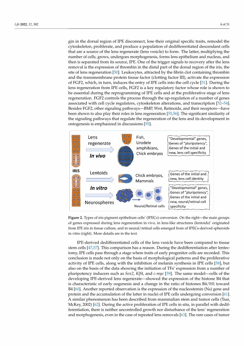
Figure 2. Types of iris pigment epithelium cells’ (IPECs) conversion. On the right—the main groups of genes expressed during lens regeneration in vivo, in lens-like structures (lentoids)’ originated from IPE iris in tissue culture, and in neural/retinal cells emerged from of IPECs-derived spheroids in vitro (right). More details are in the text.
IPE-derived dedifferentiated cells of the lens vesicle have been compared to tissue stem cells [47,57]. This comparison has a reason. During the dedifferentiation after lentectomy, IPE cells pass through a stage where traits of early progenitor cells are rec-orded. This conclusion is made not only on the basis of morphological patterns and the proliferative activity of IPE cells, along with the inhibition of melanin synthesis in IPE cells [58], but also on the basis of the data showing the initiation of TFs’ expression from a number of pluripotency inducers such as Sox2, Klf4, and c-myc [59]. The same model—cells of the developing IPE-derived lens regenerate—showed the expression of the histone B4 that is characteristic of early oogenesis and a change in the ratio of histones B4/H1 toward B4 [60]. Another reported observation is the expression of the nucleostemin (Ns) gene and protein and the accumulation of the latter in nuclei of IPE cells undergoing con-version [61]. A similar phenomenon has been described from mammalian stem and tumor cells (Tsai, McKey, 2002) [62]. During the active proliferation of IPE cells in situ, in parallel with dedifferentiation, there is neither uncontrolled growth nor disturbance of the lens’
Figure 2. Types of iris pigment epithelium cells’ (IPECs) conversion. On the right—the main groupsof genes expressed during lens regeneration in vivo, in lens-like structures (lentoids)’ originatedfrom IPE iris in tissue culture, and in neural/retinal cells emerged from of IPECs-derived spheroidsin vitro (right). More details are in the text.
IPE-derived dedifferentiated cells of the lens vesicle have been compared to tissuestem cells [47,57]. This comparison has a reason. During the dedifferentiation after lentec-tomy, IPE cells pass through a stage where traits of early progenitor cells are recorded. Thisconclusion is made not only on the basis of morphological patterns and the proliferativeactivity of IPE cells, along with the inhibition of melanin synthesis in IPE cells [58], butalso on the basis of the data showing the initiation of TFs’ expression from a number ofpluripotency inducers such as Sox2, Klf4, and c-myc [59]. The same model—cells of thedeveloping IPE-derived lens regenerate—showed the expression of the histone B4 thatis characteristic of early oogenesis and a change in the ratio of histones B4/H1 towardB4 [60]. Another reported observation is the expression of the nucleostemin (Ns) gene andprotein and the accumulation of the latter in nuclei of IPE cells undergoing conversion [61].A similar phenomenon has been described from mammalian stem and tumor cells (Tsai,McKey, 2002) [62]. During the active proliferation of IPE cells in situ, in parallel with dedif-ferentiation, there is neither uncontrolled growth nor disturbance of the lens’ regenerationand morphogenesis, even in the case of repeated lens removals [63]. The rare cases of tumor
Life 2022, 12, 382 5 of 31
formation are shown to be associated with exposure to carcinogens [64] and also occur innewts exposed to low gravity conditions [65].
The common ectodermal origin of the eye retina, iris, and lens probably explains thecases of formation of lentoids, the lens-like structures synthesizing crystallins, in tissuecultures from the retina and iris of birds [66] and rodents [67] (Figure 2). Currently, lentoidbodies expressing crystallins are obtained from human embryonic (hESC) and inducedhuman (iPSC) stem cells [68,69]. An ultrastructure analysis of hESC- and iPSC-derivedlentoid bodies has identified closely packed lens epithelial and differentiating fiber cells.A comparison of the mouse lens epithelial and fiber cell transcriptomes with hESC- andiPSC-derived lentoid body transcriptomes has shown a more then 96% overlap [69].
2.2. Cell-Type Conversion of IPE Cells In Vitro
IPE has a common origin and some similarity with RPE: it has an apical–basal axis ofpolarity, tight junctions, microvilli, and contains pigment in cytoplasm. IPE cells culturedin vitro have shown a potential to carry out many functions characteristic of RPE cells, e.g.,specific phagocytosis of rod outer segments and retinol metabolism. These observationshave served as a basis for an attempt to accumulate IPE-derived cells in vitro for subsequenttransplantation in pathologies associated with RPE cells’ death [70].
Of major interest, however, is the IPE cells’ capability of conversion into neural cell-types which could be used for transplantation in case of degenerative retinal diseases inhumans without gene transfer [71] (Figure 2). The data on the isolated chick IPE cells(2 days post hatching) that formed spheres containing cells with proliferative potential andexpressing TFs, which are markers of cell progenitors, gave some hope. After transferringthe spheres to laminin, cells of neural phenotypes resembling retinal ones were detected.When co-cultured with the chicken embryonic retina, these cells exhibited the propertiesof photoreceptors and Müller glial cells. In contrast, in adherent culture on collagen-coated dishes, IPE-derived progenitors could revert to the pigment epithelial phenotype,indicating the reversible state of IPE-derived cells during reprogramming [71]. Recentstudies on the same model, chick eye IPE in vitro, have indicated the role of the Wntsignaling pathway in the negative regulation of the process of IPE cell conversion along theneuronal pathway [72].
Isolated iris cells of postnatal and adult mice also exhibit properties of stem-like cellsin vitro: the expression of nestin and Pax6 [73]. IPE cells of adult rats in vitro can bereprogrammed along the neuronal/retinal pathway, producing photoreceptor-like cells.This occurs in the presence of FGF2 in the medium [74]. In the case of adult pig IPEcells cultured in vitro, the appearance of proneural cell progenitors with the ability todifferentiate into photoreceptor-like cells was recorded. The process was initiated withoutthe involvement of fetal serum and growth factors, but the presence of FGF2 and IGF2 inthe medium significantly stimulated their conversion [12].
In a comparative study of IPE-derived progenitor cells, cultured IPE cells of adult ratsand primates proved to exhibit similar characteristics of photoreceptor phenotypes [75].The cell phenotypes composing the spheres obtained in vitro from human IPE cells arenot an exception. They exhibit proliferative activity and express markers of progenitorssuch as Sox2, Nanog, nestin, and also GFAP. However, it is noted that many cells retain theproperties of differentiated epithelial cells and lack the key properties of stem cells [76]. Thestudy by Seko et al. [77] has demonstrated that iris cells of human infants in vitro expressstem cell markers such as Nestin, N-cadherin, SOX2, Musashi-1, and PAX6. To producelight-responsive photoreceptor-like cells, Seko and co-authors used combinations of “de-velopmental” TFs. Expression of such photoreceptor molecules as rhodopsin, blue opsin,and green/red opsin in induced photoreceptor cells were dependent on TFs’ combinations:CRX and NEUROD induced rhodopsin and blue opsin, but did not induce green opsin; acombination of CRX and RX induced blue opsin and green/red opsin, but did not inducerhodopsin. Phototransduction-related genes, as well as opsin genes, were up-regulatedin those cells. The combination of CRX and RX generated immature photoreceptors, and
Life 2022, 12, 382 6 of 31
additional NEUROD promoted maturation. These data suggest that photosensitive pho-toreceptor cells can be generated through manipulations with TF combinations [77]. Tocontinue studying the potential of mammalian IPE cells’ conversion along the retinal path-way, Bennis et al. [78] compared the expression profiles of human RPE and IPE genes. Ananalysis of transcriptomes has indicated significant molecular similarities, but, neverthe-less, revealed some differences. The latter included, along with the enriched RPE geneexpression, prominent features which were involved in the phototransduction cascade.Differences were also found in the activity of the Wnt signaling pathway: it was active inIPE, but not in RPE. However, when considered together, the data suggest that both IPE andRPE cells retain developmental or functional plasticity and can serve to develop strategiesfor replacement of lens and retinal cells [78]. In the work based on human IPE in vitro,Yamamoto et al. [79] identified cells that were positive for p75NTR, a marker of retinaltissue stem and progenitor cells. The p75NTR+H-iris cells, selected by sorting, expressedmarkers of retinal neurons. Among them, recoverin-positive cells and photoreceptor-likecells with electrophysiological functions were isolated and accumulated. Retinal ganglioncells with electrophysiological functions were also differentiated by changing the culturemethod [79].
As can be seen from the above information, the IPE cell-type conversion processincludes a stage where reversible and incomplete inhibition of the expression of genesresponsible for IPE specialization is assumed, which occurs along with the initiation ofthe expression of genes that determine a new differentiation. This assumption, previouslymade for amphibian IPE [3], is now confirmed for both IPE and other pigment epitheliaof the vertebrate eye, RPE and CBPE [16,17]. The above observations also indicate thatthe expression of principal genes characteristic for stem/progenitor cells occurs duringreprogramming. This information allows raising and answering the main question asto how and at what point the desired IPE cells conversion pathway should not only beset, but also fixed experimentally. This will make it possible to use IPE-derived cellswith a specified and stable in vitro differentiation for the transplantation of the obtainedcells in retinal pathologies. The development of approaches to the transplantation ofautologous IPE-derived cells for the treatment of degenerative diseases of the human retinais underway [80,81].
Among the congenital and acquired diseases of the human iris proper, those associatedwith iris cell-type conversion have not been reported to date [82]. The iridocorneal fibro-plasias leading to iridal–corneal adhesion may be remotely associated with cell conversionalong the mesenchymal pathway. Adhesion may be congenital or acquired and may causeopacities in either the cornea or the lens. Spontaneous adhesions are observed in albino rats,but are rare in mice [83]. These changes are assumed to be associated with the productionof fibrils, which are ECM molecules synthesized by the transforming IPE-derived cells thathave acquired the properties of mesenchymal differentiation. However, fibrotic changesin the lens capsule cells may also be, to an equal extent, a cause of the accumulation ofmesenchymal fibers between the lens and the iris [84].
The spectrum of cell-type conversions for IPE cells in pathology includes cases of uvealmelanomas, which is a rather rare malignant transformation of these cells in humans [85].Neoplastic degenerations of the eye PECs, having their own epidemiological, clinical,physiopathological, and molecular features, as well as mechanisms involved in metastasis,are beyond of the scope of the present review. Also, the cases of the formation of iriscysts [86] are not considered in the review’s discussion. Primary cysts arise either fromthe iris pigment epithelium or from the iris stroma. The potential of iris stroma cells’conversion into neuronal cells was also discussed in the above-cited study on cultured pigIPE cells [12].
Life 2022, 12, 382 7 of 31
3. Cell-Type Conversion of Ciliary Body Epithelial Cells3.1. Cell-Type Conversion of CB Cells In Vivo
The mammalian ciliary body (CB), located between ora serrata of the neural retina (NR)and the iris, has an anatomical homology with the ciliary marginal zone (CMZ) of fish andamphibians, an undifferentiated region known as a source of new neurons and glial cellsof the retina in development and regeneration (reviews: [28,87,88]). However, Miles andTropepe [88] insist not to confuse the CB region with the ciliary, non-stratified region of theretina, which contains low-differentiated cells in vertebrates.
The mammalian CB, unlike the fish and amphibian CMZ, is a highly specializedand multifunctional eye structure [89,90]. It includes ciliary muscles and two foldedepithelial layers: unpigmented and pigmented (ciliary body pigment epithelium, CBPE).The former is a continuation of the ciliary NR region; the latter is a continuation of RPE. CBis known to be responsible for secreting the aqueous humor in the anterior chamber andthe vitreous humor and protein components, including crystallin, Optc (opticin), collagen,and laminin proteins [91,92]. The CB muscles, supporting the iris, are involved in the lensaccommodation along with the latter. A mass spectrometry study of the CB proteomein humans has revealed a wide range of specific proteins that reflect CB functions [93].The profile of gene expression in the cells of pigment and non-pigment epithelial layersof the human CB was studied by microarray [94]. The observed similarity of both layers,in turn, indicated the similar functions performed by them. The differences were in thespecifics of cell phenotypes. CBPE cells expressed pigment cell-specific genes involved inpigmentation, cytoskeleton, endocrine and metabolic pathways, and NR developmentalproperties. With respect to them, it was found that the pathway of “human embryonicstem cell pluripotency” and Wnt/β-catenin signaling differed significantly between thenon-pigment and pigment epithelia of CB [94]. Differences were also found betweenthe two CB layers on the level of expression of a number of markers: P-Cad (Cdh3) isexpressed in CBPE, while the expression of N-Cad (Cdh2), Zic2, and Chx10 occurs in thenon-pigment layer [95]. It is important that, in the analysis of certain cell types, the CBPEcells that demonstrated the expression of P-Cadherin were the source of the neurogenicpotential in vitro. Nevertheless, there was an ambiguity in the question as to how muchthe P-Cadherin expression is required to maintain CBPE cells’ “stemness” in vivo and forthe formation of the clonal stem cell spheres in vitro [95].
Studies of CB cell conversion in vivo showed that the proliferation was activated, andthat the phenotype of CB cells changed in response to the optic nerve axotomy, whichcaused the death of retinal ganglion cells in adult mice. The subpopulation of thosecells was positively stained for homeodomain protein Chx10 and recoverin, a markerfor photoreceptors and bipolar cells in the retina [96]. When studying the molecularmechanisms responsible for the regulation of proliferation and progenitor genes’ expressionin adult mouse CB cells, Del Debbio et al. [97] performed intraocular injections of activityregulators of Rho GTPases, which are participants in the regulation of signaling pathwaysthat control cell proliferation and transcription. Activation of Rho GTPases increased theco-expression of Pax6 and Chx10, but had no significant effect on the proliferation of CBPE.In contrast, the Rho GTPases’ inactivation increased cell proliferation and potentiatedthe proliferative effect of growth factors. The authors suggest that the modulation of CBcells’ proliferation and reprogramming may provide a potentially new approach to retinalrepair [97].
3.2. Cell-Type Conversion of CB Cells In Vitro
When compared to in vivo studies, in vitro experiments provide much more exten-sive information about the conversion capabilities of CB cells. In studies of the initialproliferative potential of mammalian eye CB cells, Coles et al. [98] used mutant mice withabnormalities of eye development, in particular, RPE. Mitfmi/mi mutation caused completeloss of differentiated RPE population, while Chx10orJ/orJ mutant mice had a small eye pheno-type, characterized by a reduction in proliferation of the neural retina progenitors, a loss of
Life 2022, 12, 382 8 of 31
mature bipolar cells, and a severely hypocellular neural retina in adulthood. In adult micewith these eye development abnormalities caused by mutations in two “developmental”genes, CB cells showed higher proliferative activity with three to eight-fold increasedretinal stem cell population when compared to wild-type control eye. Cells isolated fromthe CB of Mitfmi/mi mice and exposed to in vitro conditions during sphere formation hadthe ability to differentiate to neural cell types including photoreceptor and glial lineages.CB-derived sphere cells of Chx10orJ/orJ mice gave rise to Müller glia and neural cell typesincluding ganglion, amacrine cells, and photoreceptors [98]. Thus, a direct correlation wasfound between the proliferative activity and cell-type switching of CB cells.
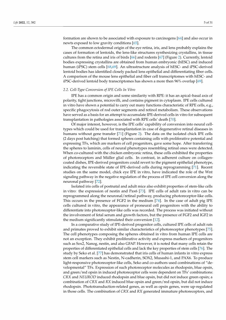
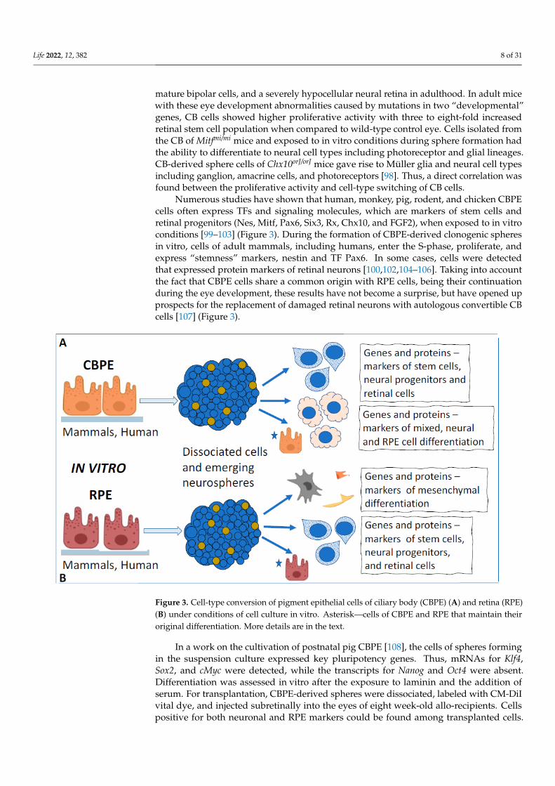
Numerous studies have shown that human, monkey, pig, rodent, and chicken CBPEcells often express TFs and signaling molecules, which are markers of stem cells andretinal progenitors (Nes, Mitf, Pax6, Six3, Rx, Chx10, and FGF2), when exposed to in vitroconditions [99–103] (Figure 3). During the formation of CBPE-derived clonogenic spheresin vitro, cells of adult mammals, including humans, enter the S-phase, proliferate, andexpress “stemness” markers, nestin and TF Pax6. In some cases, cells were detectedthat expressed protein markers of retinal neurons [100,102,104–106]. Taking into accountthe fact that CBPE cells share a common origin with RPE cells, being their continuationduring the eye development, these results have not become a surprise, but have opened upprospects for the replacement of damaged retinal neurons with autologous convertible CBcells [107] (Figure 3).
1
Figure 3. Cell-type conversion of pigment epithelial cells of ciliary body (CBPE) (A) and retina (RPE)(B) under conditions of cell culture in vitro. Asterisk—cells of CBPE and RPE that maintain theiroriginal differentiation. More details are in the text.
In a work on the cultivation of postnatal pig CBPE [108], the cells of spheres formingin the suspension culture expressed key pluripotency genes. Thus, mRNAs for Klf4,Sox2, and cMyc were detected, while the transcripts for Nanog and Oct4 were absent.Differentiation was assessed in vitro after the exposure to laminin and the addition ofserum. For transplantation, CBPE-derived spheres were dissociated, labeled with CM-DiIvital dye, and injected subretinally into the eyes of eight week-old allo-recipients. Cellspositive for both neuronal and RPE markers could be found among transplanted cells.
Life 2022, 12, 382 9 of 31
Large clusters of transplanted cells integrated into the RPE layer and multilayered RPE-likestructures positive for RPE65 were observed. Grafted cells were also identified in the NRwhere 5–10% of them were positive for recoverin, protein kinase C alpha (PKCα), andcalbindin [108]. Isolation of human pigment and non-pigment epithelium from CB hasdemonstrated that, like in other mammals, human CBPE cells are more efficient than cellsof non-pigmented layers in generating neurogenic spheres [109].
Studies by Cicero et al., Gualdoni et al., and then by Coles and van der Kooy [110–112]have clarified the behavior and phenotype of cells present in CBPE-derived cellular spheresin vitro. They have shown that these cells exhibit certain properties of both stem andepithelial pigment cells, in which, in addition to the pigment, there is also the interdigitationof plasmalemma and the epithelial pattern of contacts. Nevertheless, despite the observedproliferation, the partial loss of pigment, and the initiation of conversion to a neuralphenotype detected using neuronal markers, no differentiation of retinal cell types occurredin CBPE-derived spheres. Apparently, the differentiation required other, permissive for this,conditions in vitro. Currently, researchers tend to conclude that the adult human CB mostlikely does not contain bona fide neural stem cells but rather consists of a population ofepithelial cells which display a remarkable plasticity in vitro reflecting their neuroepithelialdevelopmental origin [87,113]. It should be noted again that the combination of somebehavioral, morphological, and molecular properties of progenitors and differentiatedpigmented cells in CBPE during reprogramming is a feature characteristic also of otherpigment epithelia of the eye considered in the review. This largely determines their potencyto produce cells of retinal phenotypes or to return to the original differentiation.
When studying the regulators of CBPE cells’ conversion under in vitro conditions, itis still poorly understood where their potencies to manifest neural/retinal differentiationproperties are fulfilled. Cells have been shown to initially express molecules involvedin the Notch signaling pathway. A disturbance in the work of this pathway blocks theneurogenic sphere formation [114,115]. A recently published study has revealed the roleof this signaling pathway in CB development [116]. It has been reported that the Notchsignaling in CB maintains the vitreous, as well as intraocular, pressure and eye structuresby regulating the CB morphogenesis, aqueous humor secretion, and vitreous proteinexpression. Notch 3 functions redundantly with Notch 2, driving the CB morphogenesis byregulating the expression of adhesion molecule Nectin1. Available data indicate the role ofthe Notch signaling not only in controlling the CB cell differentiation, but also, as a result, inthe key functions of this eye structure, whose disorders form a basis of, e.g., glaucoma [116].The similarity in the work of this signaling pathway in development and in CB cell-typeconversion once again confirms the rule about the involvement of developmental regulatorymechanisms in the cell-type switching of the eye’s pigment epithelium. The involvementof a signaling pathway, including the receptor tyrosine kinase c-Kit and its ligand stem cellfactor (SCF), is also assumed to occur along and in interaction with the Notch signaling. Ashas been shown, these components are able to maintain not only the proliferative activityof cells in CBPE-derived cell spheres, but also the differentiation of their cells along theretinal pathway [114,115].
In addition to TFs and molecular links of regulatory signaling pathways, the epigeneticregulators of CB-derived cells have also been preliminarily studied. Using the materialof human cadaveric eyes, Jasty and Krishnakumar [117] analyzed DNA methylation andhistone methylation: H3K4me3 and H3K27me3 in a CB-derived lineage of committedprogenitors to terminally differentiated cells in vitro. The results showed that severalpromoters, including pluripotency and lineage-specific genes, become methylated in thedifferentiating cell population. The authors suggested that methylation may repress thepluripotency, earlier observed in vitro, in this differentiating population. They found alsothe bivalent modifications involved in the process of differentiation of stem/progenitorcells [117]. Therefore, CB-derived progenitor cells and their progeny that have undergonedifferentiation into retinal neurons/glial cells show that specific DNA methylation andhistone modifications are involved in the gene expression reprogramming during their
Life 2022, 12, 382 10 of 31
conversion in vitro. The current epigenetic studies on the conversion of CB- and CBPE-derived cells have implications for understanding not only this process, but also a widerange of pathogenic processes, since the aberrant epigenetic regulation is known to oftenunderlie them [118–120].
There is no available information in the literature about the conversion of cells of CBand its pigment layer into any other phenotypes in the spectrum of pathologies, except forneoplastic changes [82,121–123]. However, these, as well as changes in the cases of otherpigment epithelia, are beyond discussion of the present review.
Apart from tumor formation in CB, the competence of these cells to convert thephenotype in vivo and in vitro is limited to the neural/retinal pathway. Despite the existingsimilarity between CBPE and RPE, and also the involvement of mesenchyme in the CBdevelopment [124], there is no evidence of CBPE cell conversion towards mesenchymalphenotype in the literature. Nevertheless, CB epithelial cells have been reported to exhibita higher reprogramming efficiency to iPSC than fibroblasts and have a reduced exogeneousrequirement. It is expected that the enhanced reprogramming efficiency may be due toincreased basal levels of Sox2 in CB cells [125].
Thus, the potencies to the cell-type conversion of CBPE in mammals and humans arebased on the presence of a molecular genetic context characteristic of progenitor neuroep-ithelial cells against the background of a specialization-providing context in the tissue [28].The former one becomes expressed mainly in conditions permissive for it in vitro, alongwith the retention and maintenance of some traits of the original CBPE cell phenotype.The cause of the combination of two, seemingly mutually exclusive states, as well as theepigenome’s permission for this condition, may be associated with (1) the late timing of de-velopment of this eye’s region relative to the equatorial and central regions of the posterioreye wall, (2) its border position between the posterior and anterior parts of the eye, (3) andalso with the molecular specifics of the regulation of this eye region development [87].
4. Cell-Type Conversion of the Retinal Pigment Epithelial (RPE) Cells4.1. RPE Cell-Type Conversion as a Basis of NR Regeneration in Amphibians and Birds
Retinal pigment epithelium (RPE) in vertebrates and humans have been studied to agreater extent than other pigment epithelia of the eye as regards cell functions, differen-tiation, its changes during regeneration and retinal pathologies, and cell transplantation(see, e.g., reviews [17,126–129]). This is due to the fact that many severe eye disorders(e.g., age-related macular dystrophy (AMD), proliferative vitreoretinopathy (PVR), andretinitis pigmentosa (RP)), and other types of retinal degenerations are associated with dis-turbances in the RPE layer, changes in the phenotype and behavior of its cells, and, as aresult, a disturbance of topological, trophic, regulatory, and functional relationships withretinal photoreceptors. Furthermore, some amphibian species and avian embryos manifestthe ability to regenerate the retina through RPE cells conversion up to the formation of anew NR at the site of the surgically removed original one.
In adult vertebrates, RPE is an epithelial monolayer of pigmented, polarized, andspecialized cells. RPE, lining the NR, is externally bounded by the Bruch’s membraneand choroidal coat (choroid), which supplies essential substances and oxygen to theretina [130,131]. Besides the transport of substances from the choroid to NR, RPE per-forms a number of other, equally important functions. They include protection againstoxidative stress, production and isolation of growth factors, and storage and metabolismof vitamin A derivatives [126,130,131]. The major RPE function is phagocytosis of theouter segments of photoreceptors, their digestion by lysosomes, and retinoid exchange, i.e.,providing the processes necessary for light perception.
RPE cells in caudate amphibians (Urodela) are the most evident example of a naturalconversion into retinal cells in vivo. After experimental damage to the NR, optic nervetransection, and artificial detachment or surgical removal of NR, RPE becomes a source ofa new, complete, and functioning retina [132–137]. The major events of the process are asfollows: the exit of RPE cells from the layer, the loss of initial traits and properties (dediffer-
Life 2022, 12, 382 11 of 31
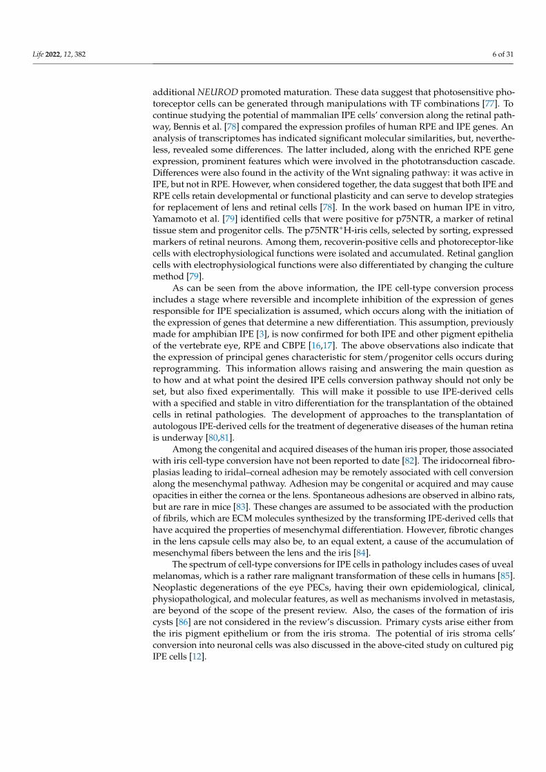
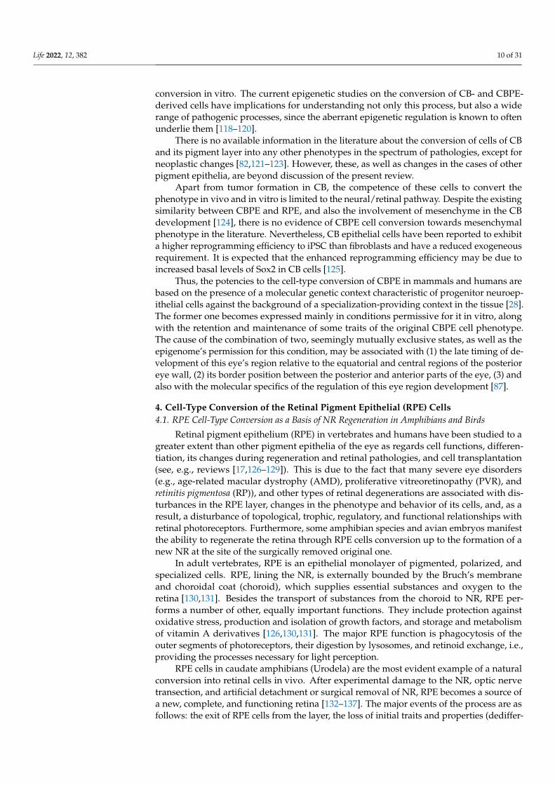
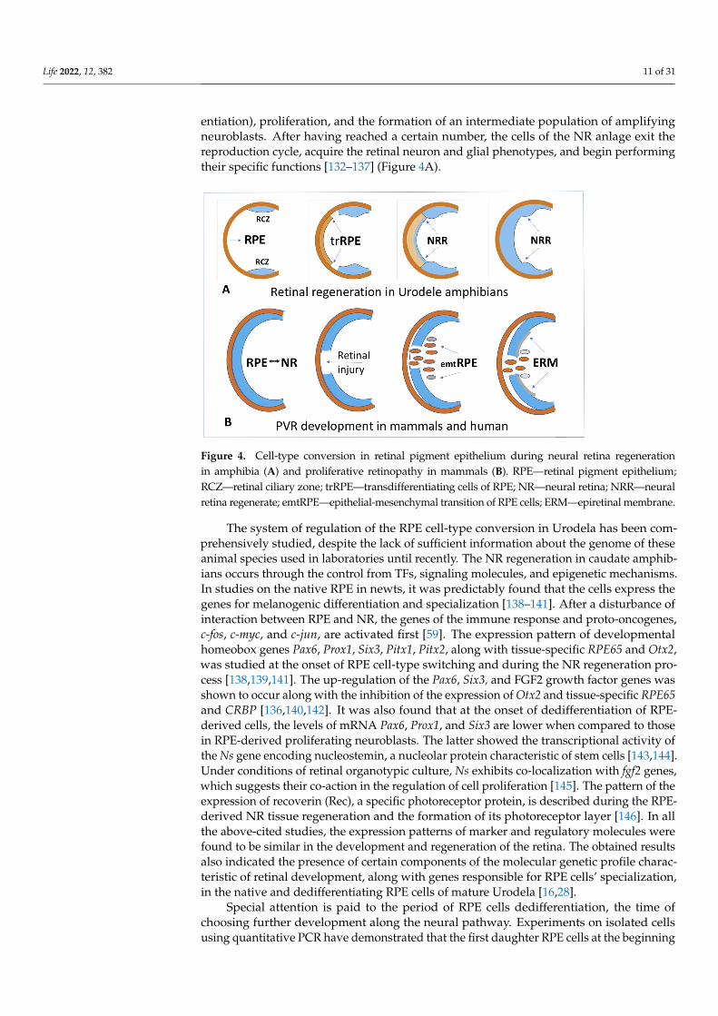
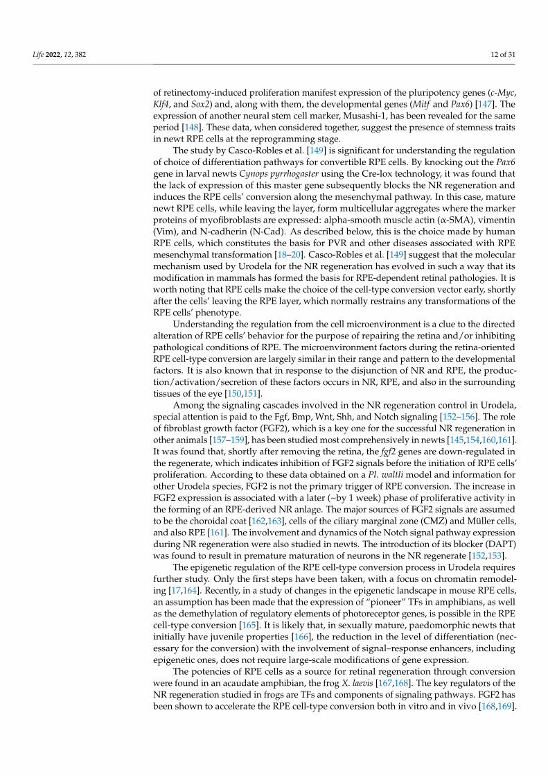
entiation), proliferation, and the formation of an intermediate population of amplifyingneuroblasts. After having reached a certain number, the cells of the NR anlage exit thereproduction cycle, acquire the retinal neuron and glial phenotypes, and begin performingtheir specific functions [132–137] (Figure 4A).
Life 2022, 12, 382 11 of 32
(dedifferentiation), proliferation, and the formation of an intermediate population of am-plifying neuroblasts. After having reached a certain number, the cells of the NR anlage exit the reproduction cycle, acquire the retinal neuron and glial phenotypes, and begin performing their specific functions [132–137] (Figure 4A).
Figure 4. Cell-type conversion in retinal pigment epithelium during neural retina regeneration in amphibia (A) and proliferative retinopathy in mammals (B). RPE—retinal pigment epithelium; RCZ—retinal ciliary zone; trRPE—transdifferentiating cells of RPE; NR—neural retina; NRR—neu-ral retina regenerate; emtRPE—epithelial-mesenchymal transition of RPE cells; ERM—epiretinal membrane. (A,B).
The system of regulation of the RPE cell-type conversion in Urodela has been com-prehensively studied, despite the lack of sufficient information about the genome of these animal species used in laboratories until recently. The NR regeneration in caudate am-phibians occurs through the control from TFs, signaling molecules, and epigenetic mech-anisms. In studies on the native RPE in newts, it was predictably found that the cells ex-press the genes for melanogenic differentiation and specialization [138–141]. After a dis-turbance of interaction between RPE and NR, the genes of the immune response and proto-oncogenes, c-fos, c-myc, and c-jun, are activated first [59]. The expression pattern of developmental homeobox genes Pax6, Prox1, Six3, Pitx1, Pitx2, along with tissue-specific RPE65 and Otx2, was studied at the onset of RPE cell-type switching and during the NR regeneration process [138,139,141]. The up-regulation of the Pax6, Six3, and FGF2 growth factor genes was shown to occur along with the inhibition of the expression of Otx2 and tissue-specific RPE65 and CRBP [136,140,142]. It was also found that at the onset of dedif-ferentiation of RPE-derived cells, the levels of mRNA Pax6, Prox1, and Six3 are lower when compared to those in RPE-derived proliferating neuroblasts. The latter showed the transcriptional activity of the Ns gene encoding nucleostemin, a nucleolar protein charac-teristic of stem cells [143,144]. Under conditions of retinal organotypic culture, Ns exhibits co-localization with fgf2 genes, which suggests their co-action in the regulation of cell pro-liferation [145]. The pattern of the expression of recoverin (Rec), a specific photoreceptor protein, is described during the RPE-derived NR tissue regeneration and the formation of its photoreceptor layer [146]. In all the above-cited studies, the expression patterns of marker and regulatory molecules were found to be similar in the development and regen-eration of the retina. The obtained results also indicated the presence of certain compo-nents of the molecular genetic profile characteristic of retinal development, along with genes responsible for RPE cells’ specialization, in the native and dedifferentiating RPE cells of mature Urodela [16,28].
Figure 4. Cell-type conversion in retinal pigment epithelium during neural retina regenerationin amphibia (A) and proliferative retinopathy in mammals (B). RPE—retinal pigment epithelium;RCZ—retinal ciliary zone; trRPE—transdifferentiating cells of RPE; NR—neural retina; NRR—neuralretina regenerate; emtRPE—epithelial-mesenchymal transition of RPE cells; ERM—epiretinal membrane.
The system of regulation of the RPE cell-type conversion in Urodela has been com-prehensively studied, despite the lack of sufficient information about the genome of theseanimal species used in laboratories until recently. The NR regeneration in caudate amphib-ians occurs through the control from TFs, signaling molecules, and epigenetic mechanisms.In studies on the native RPE in newts, it was predictably found that the cells express thegenes for melanogenic differentiation and specialization [138–141]. After a disturbance ofinteraction between RPE and NR, the genes of the immune response and proto-oncogenes,c-fos, c-myc, and c-jun, are activated first [59]. The expression pattern of developmentalhomeobox genes Pax6, Prox1, Six3, Pitx1, Pitx2, along with tissue-specific RPE65 and Otx2,was studied at the onset of RPE cell-type switching and during the NR regeneration pro-cess [138,139,141]. The up-regulation of the Pax6, Six3, and FGF2 growth factor genes wasshown to occur along with the inhibition of the expression of Otx2 and tissue-specific RPE65and CRBP [136,140,142]. It was also found that at the onset of dedifferentiation of RPE-derived cells, the levels of mRNA Pax6, Prox1, and Six3 are lower when compared to thosein RPE-derived proliferating neuroblasts. The latter showed the transcriptional activity ofthe Ns gene encoding nucleostemin, a nucleolar protein characteristic of stem cells [143,144].Under conditions of retinal organotypic culture, Ns exhibits co-localization with fgf2 genes,which suggests their co-action in the regulation of cell proliferation [145]. The pattern of theexpression of recoverin (Rec), a specific photoreceptor protein, is described during the RPE-derived NR tissue regeneration and the formation of its photoreceptor layer [146]. In allthe above-cited studies, the expression patterns of marker and regulatory molecules werefound to be similar in the development and regeneration of the retina. The obtained resultsalso indicated the presence of certain components of the molecular genetic profile charac-teristic of retinal development, along with genes responsible for RPE cells’ specialization,in the native and dedifferentiating RPE cells of mature Urodela [16,28].
Special attention is paid to the period of RPE cells dedifferentiation, the time ofchoosing further development along the neural pathway. Experiments on isolated cellsusing quantitative PCR have demonstrated that the first daughter RPE cells at the beginning
Life 2022, 12, 382 12 of 31
of retinectomy-induced proliferation manifest expression of the pluripotency genes (c-Myc,Klf4, and Sox2) and, along with them, the developmental genes (Mitf and Pax6) [147]. Theexpression of another neural stem cell marker, Musashi-1, has been revealed for the sameperiod [148]. These data, when considered together, suggest the presence of stemness traitsin newt RPE cells at the reprogramming stage.
The study by Casco-Robles et al. [149] is significant for understanding the regulationof choice of differentiation pathways for convertible RPE cells. By knocking out the Pax6gene in larval newts Cynops pyrrhogaster using the Cre-lox technology, it was found thatthe lack of expression of this master gene subsequently blocks the NR regeneration andinduces the RPE cells’ conversion along the mesenchymal pathway. In this case, maturenewt RPE cells, while leaving the layer, form multicellular aggregates where the markerproteins of myofibroblasts are expressed: alpha-smooth muscle actin (α-SMA), vimentin(Vim), and N-cadherin (N-Cad). As described below, this is the choice made by humanRPE cells, which constitutes the basis for PVR and other diseases associated with RPEmesenchymal transformation [18–20]. Casco-Robles et al. [149] suggest that the molecularmechanism used by Urodela for the NR regeneration has evolved in such a way that itsmodification in mammals has formed the basis for RPE-dependent retinal pathologies. It isworth noting that RPE cells make the choice of the cell-type conversion vector early, shortlyafter the cells’ leaving the RPE layer, which normally restrains any transformations of theRPE cells’ phenotype.
Understanding the regulation from the cell microenvironment is a clue to the directedalteration of RPE cells’ behavior for the purpose of repairing the retina and/or inhibitingpathological conditions of RPE. The microenvironment factors during the retina-orientedRPE cell-type conversion are largely similar in their range and pattern to the developmentalfactors. It is also known that in response to the disjunction of NR and RPE, the produc-tion/activation/secretion of these factors occurs in NR, RPE, and also in the surroundingtissues of the eye [150,151].
Among the signaling cascades involved in the NR regeneration control in Urodela,special attention is paid to the Fgf, Bmp, Wnt, Shh, and Notch signaling [152–156]. The roleof fibroblast growth factor (FGF2), which is a key one for the successful NR regeneration inother animals [157–159], has been studied most comprehensively in newts [145,154,160,161].It was found that, shortly after removing the retina, the fgf2 genes are down-regulated inthe regenerate, which indicates inhibition of FGF2 signals before the initiation of RPE cells’proliferation. According to these data obtained on a Pl. waltli model and information forother Urodela species, FGF2 is not the primary trigger of RPE conversion. The increase inFGF2 expression is associated with a later (~by 1 week) phase of proliferative activity inthe forming of an RPE-derived NR anlage. The major sources of FGF2 signals are assumedto be the choroidal coat [162,163], cells of the ciliary marginal zone (CMZ) and Müller cells,and also RPE [161]. The involvement and dynamics of the Notch signal pathway expressionduring NR regeneration were also studied in newts. The introduction of its blocker (DAPT)was found to result in premature maturation of neurons in the NR regenerate [152,153].
The epigenetic regulation of the RPE cell-type conversion process in Urodela requiresfurther study. Only the first steps have been taken, with a focus on chromatin remodel-ing [17,164]. Recently, in a study of changes in the epigenetic landscape in mouse RPE cells,an assumption has been made that the expression of “pioneer” TFs in amphibians, as wellas the demethylation of regulatory elements of photoreceptor genes, is possible in the RPEcell-type conversion [165]. It is likely that, in sexually mature, paedomorphic newts thatinitially have juvenile properties [166], the reduction in the level of differentiation (nec-essary for the conversion) with the involvement of signal–response enhancers, includingepigenetic ones, does not require large-scale modifications of gene expression.
The potencies of RPE cells as a source for retinal regeneration through conversionwere found in an acaudate amphibian, the frog X. laevis [167,168]. The key regulators of theNR regeneration studied in frogs are TFs and components of signaling pathways. FGF2 hasbeen shown to accelerate the RPE cell-type conversion both in vitro and in vivo [168,169].
Life 2022, 12, 382 13 of 31
After retinectomy, FGF2 activates the MAPK pathway, thus, providing the proliferationof RPE cells. In addition, the factor acts as a promoter of differentiation of RPE-derivedproliferating retinal progenitors into new retinal cells and is necessary for maintaining Pax6expression [170]. Inhibition of the MAPK pathway significantly decreases the amount ofretina regenerated [168]. It was tested also whether the up-regulation of matrix metallo-proteinases (Mmps) triggers the retinal regeneration [171]. In a tissue culture, soon afterthe retinal removal, the Mmps expression increased in the RPE cells and corresponded totheir migration from the choroid. In parallel, IL-1β and TNF-α were up-regulated bothin vivo and in vitro. The results suggested that inflammatory cytokines triggered the Mmps’upregulation. The role of TFs in the specification of NR progenitors during regenerationin frogs was studied using the example of the rax gene, whose expression is up-regulatedin RPE-derived neuroblasts, while the rax knock-down impairs retinal cell types in retinaregeneration in tadpoles [172,173]. Hothem et al. [174] and Martinez-De Luna et al. [175]reported about the necessity of the retinal homeobox Rx gene expression for the retinalregeneration in pre-metamorphic X. laevis.
The model of retinal regeneration in tadpoles and adult X. laevis is currently verypromising to be used in studies. Due to the good knowledge of the genome of these animals,the feasibility of using new techniques and approaches for the study of NR regeneration andfor testing strategies aimed at its promoting is under discussion now [176,177]. In particular,X. laevis is suggested to be used to study the molecular mechanisms responsible for thereactivation of expression of “developmental genes”, which constitute a basis of the process.By postulating their evolutionary conservativeness and similar activation mechanisms,Suzuki and Ochi [178] assign an important role to the signal–response enhancers of geneexpression, including also the epigenetic modification of DNA and histones.
The process of retinal regeneration through RPE cell-type conversion in chicks wasdiscovered by Coulombre J.L. and Coulombre A.J. [42]. The regeneration occurs in the earlystages of development (up to E4–E4.5); as a result, a new NR with an inverted structure(with photoreceptors facing the eye cavity, rather than the RPE) is formed [179]. As regardsthe immune system, the C3a complement has been shown as an inducer of NR regenerationthrough the activation of the STAT3 transcription regulator, which, in turn, activates thedamage response factors IL-6, IL-8, and TNF. This eventually leads to the regulation ofthe Wnt2b signaling pathway genes, and also to the expression of the Six3 and Sox2 genescharacteristic of retinal progenitors [180]. A study by Zhu et al. [181] on the expression ofβ-catenin, a well-known coordinator of cell–cell adhesion and gene transcription, showedthat nuclear β-catenin positive cells remain in the RPE in response to injury. These cells wereBrdU-negative and p27-positive, which suggested that nuclear β-catenin prevents themfrom entering the cell cycle. In the presence of FGF2, the RPE cells, while dedifferentiatingand proliferating, lost the expression of nuclear β-catenin. A retinectomy, followed bydisruption of active β-catenin using a signaling inhibitor (XAV939), induced regenerationfrom RPE in the absence of FGF2. Thus, inactivation of β-catenin seems a necessary pre-requisite for the NR regeneration in chicks [181]. At the stage of the proliferative phase ofRPE cells’ conversion in birds, as in other animals, FGF2 activates the cell multiplicationand, as a result, the subsequent histogenesis of the NR regenerate. In the layer of amplifyingneuroblasts, there is an expression of proneural markers, which are TFs (Pax6 and Chx10)regulated by the FGF2-FGFR/MEK/Erk signaling cascade [182,183]. In chicks, as well as innewts, a down-regulation of RPE65 specific protein and the TF Mitf, which are participantsin the RPE specialization of eye development, occurs during the RPE cells’ conversion [182].These data were supplemented by the results showing the expression of the factors klf4,c-myc, and lin-28, which are markers of cell pluripotency. The RNA binding proteinlin-28 is a target for the action of FGF2, and its overexpression results in the RPE cell-typeconversion in the absence of FGF2 as well [184]. The use of the model of NR regenerationby RPE in chicks has also revealed the involvement of Shh [185], BMP, and Wnt signalingcascades [186]. It is worth noting that at the stage of two prospective layers of the retinaanlage (RPE and NR) at the optic cup stages in chicken embryo, a low concentration of BMP
Life 2022, 12, 382 14 of 31
leads to the RPE cell-type conversion in NR, while a high concentration, on the contrary,leads to the conversion of embryonic NR cells into pigmented RPE cells [186]. A studyof the loss of function showed that the NR conversion into RPE requires both BMP andWnt signaling. Steinfeld et al. [186] suggest that manipulations with these key signalingpathways may contribute to the development of efficient standardized protocols for RPEand NR generation required to create cell replacement therapies.
To characterize the mechanisms underlying the RPE cell conversion in the chickenembryo, RNA was extracted by laser capture microdissection from an intact RPE tissue,tissues at 6 h post retinectomy, and cells converted in the presence of FGF2 [183]. UsingRNA-seq, the authors observed the repression of genes related to cell cycle progressionin the early-stage converting RPE, and also the up-regulation of injury-associated genes.In contrast, the RPE, converted in a FGF2-dependent manner, was enriched in MAPK-responsive genes and retina development factors, thus, confirming that FGF2 and thedownstream MAPK cascade are the main drivers of the embryonic chick RPE reprogram-ming. Tangeman et al. [183] made an attempt to understand the role that the mechanismsresponsible for epithelial–mesenchymal transition (EMT), which is a RPE cell-type con-version underlying retinal pathologies in mammals, can play in the conversion of chickenembryo RPE cells. Since the EMT program is accompanied by an extensive, coordinatedregulation of ECM regulators (see below), the major focus is on the regulators of ECM stateand composition [183].
As has been repeatedly mentioned, a switch of cell types is accompanied by resets ofthe epigenetic landscape that drive shifts in transcriptional programs and, eventually, incell identity. Currently, the whole genome bisulfite sequencing (WGBS) analysis of DNAmethylation in parallel with the up-to-date sequencing technology is used to study them.These methods were applied to describe global epigenetic patterns and fluctuations duringthe development of the chicken embryo retina. This allowed for comparing the globalchanges in DNA methylation to differential gene expression during chick developmentalretinogenesis [187]. Luz-Madrigal et al. [188] applied the same approach to the study ofthe NR regeneration process in a chicken embryo, where RPE prior to injury and duringdifferent stages of RPE reprogramming were considered. The changes in the expression ofgenes associated with epigenetic modifications during RPE reprogramming, the dynamicchanges in histone marks associated with bivalent chromatin (H3K27me3/H3K4me3),and intermediates of the process of DNA demethylation, including 5hmC and 5caC, werestudied. The data confirmed the extensive rearrangements of DNA methylation patterns,including differentially methylated regions found at promoters of genes associated withchromatin organization and FGF2 production. The role of FGF2, according to the authorsof the study, consists of supporting these dynamic modifications that are further sustainedin the commitment to form a new NR. In general, the findings reveal the active DNAdemethylation as an important process that may be applied to remove epigenetic barriersin order to regenerate retina in mammals [188]. Thus, in studies based on animal modelssuch as amphibians and birds that demonstrate NR regeneration through the RPE cell-typeconversion, a significant similarity can be found between the molecular mechanisms thataccompany the process, including regulatory ones, and those operating during the normaldevelopment of the eye’s retina. The similarity is found both in the conversion triggersand in the expression of conserved, “developmental” TFs, the expression of genes from the“pluripotency induction factors” group, and also in the set and up-regulation of expressionin a number of signaling networks. The competence of RPE cells to the cell-type conversionin amphibians and birds along the retinal pathway and its implementation are based onextracellular and intracellular specifics of cell behavior regulation, including epigeneticones. The same is applicable to mammals and humans, as shown below.
4.2. RPE Cell-Type Conversion In Vitro
Over the past 20 years, extensive information has accumulated, indicating the RPEcell-type plasticity and a capability of conversion under in vitro conditions. RPE cells of
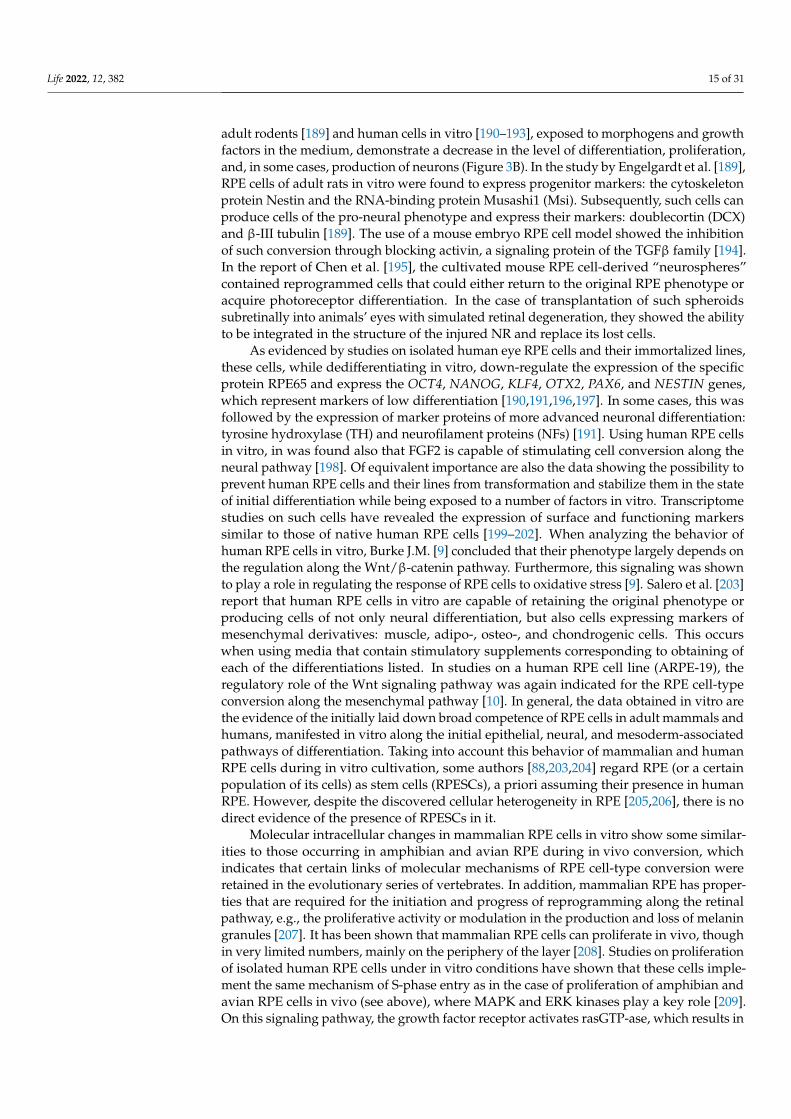
Life 2022, 12, 382 15 of 31
adult rodents [189] and human cells in vitro [190–193], exposed to morphogens and growthfactors in the medium, demonstrate a decrease in the level of differentiation, proliferation,and, in some cases, production of neurons (Figure 3B). In the study by Engelgardt et al. [189],RPE cells of adult rats in vitro were found to express progenitor markers: the cytoskeletonprotein Nestin and the RNA-binding protein Musashi1 (Msi). Subsequently, such cells canproduce cells of the pro-neural phenotype and express their markers: doublecortin (DCX)and β-III tubulin [189]. The use of a mouse embryo RPE cell model showed the inhibitionof such conversion through blocking activin, a signaling protein of the TGFβ family [194].In the report of Chen et al. [195], the cultivated mouse RPE cell-derived “neurospheres”contained reprogrammed cells that could either return to the original RPE phenotype oracquire photoreceptor differentiation. In the case of transplantation of such spheroidssubretinally into animals’ eyes with simulated retinal degeneration, they showed the abilityto be integrated in the structure of the injured NR and replace its lost cells.
As evidenced by studies on isolated human eye RPE cells and their immortalized lines,these cells, while dedifferentiating in vitro, down-regulate the expression of the specificprotein RPE65 and express the OCT4, NANOG, KLF4, OTX2, PAX6, and NESTIN genes,which represent markers of low differentiation [190,191,196,197]. In some cases, this wasfollowed by the expression of marker proteins of more advanced neuronal differentiation:tyrosine hydroxylase (TH) and neurofilament proteins (NFs) [191]. Using human RPE cellsin vitro, in was found also that FGF2 is capable of stimulating cell conversion along theneural pathway [198]. Of equivalent importance are also the data showing the possibility toprevent human RPE cells and their lines from transformation and stabilize them in the stateof initial differentiation while being exposed to a number of factors in vitro. Transcriptomestudies on such cells have revealed the expression of surface and functioning markerssimilar to those of native human RPE cells [199–202]. When analyzing the behavior ofhuman RPE cells in vitro, Burke J.M. [9] concluded that their phenotype largely depends onthe regulation along the Wnt/β-catenin pathway. Furthermore, this signaling was shownto play a role in regulating the response of RPE cells to oxidative stress [9]. Salero et al. [203]report that human RPE cells in vitro are capable of retaining the original phenotype orproducing cells of not only neural differentiation, but also cells expressing markers ofmesenchymal derivatives: muscle, adipo-, osteo-, and chondrogenic cells. This occurswhen using media that contain stimulatory supplements corresponding to obtaining ofeach of the differentiations listed. In studies on a human RPE cell line (ARPE-19), theregulatory role of the Wnt signaling pathway was again indicated for the RPE cell-typeconversion along the mesenchymal pathway [10]. In general, the data obtained in vitro arethe evidence of the initially laid down broad competence of RPE cells in adult mammals andhumans, manifested in vitro along the initial epithelial, neural, and mesoderm-associatedpathways of differentiation. Taking into account this behavior of mammalian and humanRPE cells during in vitro cultivation, some authors [88,203,204] regard RPE (or a certainpopulation of its cells) as stem cells (RPESCs), a priori assuming their presence in humanRPE. However, despite the discovered cellular heterogeneity in RPE [205,206], there is nodirect evidence of the presence of RPESCs in it.
Molecular intracellular changes in mammalian RPE cells in vitro show some similar-ities to those occurring in amphibian and avian RPE during in vivo conversion, whichindicates that certain links of molecular mechanisms of RPE cell-type conversion wereretained in the evolutionary series of vertebrates. In addition, mammalian RPE has proper-ties that are required for the initiation and progress of reprogramming along the retinalpathway, e.g., the proliferative activity or modulation in the production and loss of melaningranules [207]. It has been shown that mammalian RPE cells can proliferate in vivo, thoughin very limited numbers, mainly on the periphery of the layer [208]. Studies on proliferationof isolated human RPE cells under in vitro conditions have shown that these cells imple-ment the same mechanism of S-phase entry as in the case of proliferation of amphibian andavian RPE cells in vivo (see above), where MAPK and ERK kinases play a key role [209].On this signaling pathway, the growth factor receptor activates rasGTP-ase, which results in

Life 2022, 12, 382 16 of 31
the MAPK/ERK phosphorylation [210]. MAPK/ERK, in turn, up-regulates the expressionof transcripts (c-myc, Pax6, klf4, and Mitf ), which indicates a decrease in the level of RPEcells’ differentiation [211]. However, these potencies are not implemented in vivo eitherfor the regeneration of the RPE layer or for the conversion aimed at NR regeneration. It isobvious that mammalian RPE appears to be poor in the regulatory elements required tocontrol cell division and induction of transdifferentiation along the neuronal pathway [208].According to Rzhanova et al. [212], terminally differentiated mammalian RPE is rich inepigenetic regulatory mechanisms that fix specific patterns of gene expression.
4.3. RPE Cell-Type Conversion In Vivo as a Basis of Retinal Diseases in Mammals and Humans
In mammals, RPE-dependent NR regeneration is extremely limited. The mammalianretina does not regenerate spontaneously after injury or in disease [17,213,214]. An injuryresults in cell death, loss of the affected neurons and RPE cells, and, in some cases, in RPEcells’ conversion into cells with mesenchymal phenotype. The RPE transdifferentiationalong the neural pathway was observed in vivo, in embryos of a mutant mouse line afterelimination of the RPE-specific TFs Mitf or the Wnt signaling effector β-catenin [215]. Thesame could be observed in microphthalmia (mi/mi) mice [216]. Other data suggests thatthe mammalian RPE has lost its latent capacity to generate retinal cells in vivo.
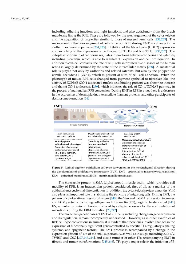
In a case of retinal pathology such as retinal detachment and rupture in mammals andhumans in vivo, similarly to that in amphibians, part of the RPE cells at the first stage losetheir epithelial properties, leave the layer, migrate, and proliferate. These processes areaccompanied by death or conversion of part of the cells with the acquisition of myofibroblastproperties (Figure 4B). These cells, when moving beyond RPE and NR and being exposedto the vitreous humor factors and the factors secreted by surrounding tissues, initiatethe synthesis of ECM components, proliferate, and become involved in the formationof the epiretinal membrane (ERM) [217–220] (Figures 4B and 5). RPE cells are the mainparticipants in the ERM formation that play a crucial role in proliferative retinopathy (PVR).Furthermore, fibroblasts and macrophages also make a significant contribution to the ERMformation [20,221]. The formation of ERM and the intrinsic contraction of its cells lead tothe tractional forces responsible for the clinical features of PVR. Descriptions of the stagesand mechanisms of regulation of this process are available in the literature [19,20,219,222].Attention to them is associated mainly with PVR, an unwelcome wound healing process ofthe retina and the cause of approximately 10% of all retinal detachments [151]. PVR withrecurrent retinal detachments requires additional surgical interventions and is associatedwith poor visual recovery. The process of RPE cells’ conversion in PVR is described in termsof epithelial–mesenchymal transition (EMT) or “RPE dysfunction” [223], although thedefinition “RPE cell-type conversion into mesenchymal phenotype” is also valid (Figure 5).This process occurs not only in PVR, but also in the case of attempts to restore the RPElayer after laser-induced damage [224], and also in proliferative diabetic retinopathy [21]and subretinal fibrosis [225,226]. The latter case, similar to that in PVR, is characterizedby proliferation and/or infiltration of RPE cells, glial cells, fibroblasts, myofibroblast-like cells, and macrophages, which, when interacting with inflammatory cytokines andgrowth factors, begin to synthesize and remodel the EMC. The above events are the finalaccompaniment of neovascular age-related macular degeneration (nAMD) [226,227].
A detailed consideration of EMT as a phenomenon is impossible within the frame-work of this review, due to the broad occurrence of this phenomenon in animals andhumans: EMT is the basis for laying down organs in development and regeneration ininvertebrates and vertebrates, cell reprogramming in fibrosis and tumor changes, and alsometastasizing [228–230].
The stability of RPE cell differentiation in the layer is supported by the coordinatedinteraction of genes with the regulatory network that controls the homeostasis processesand tissue functions. EMT of RPE cells in situ is an aberrant tissue response, an attempt torestore it that ends with ERM formation [219,231]. Specifics of this response of mammalianRPE cells are manifested as the loss of polarity, destruction of cell–cell junctional complexes,
Life 2022, 12, 382 17 of 31
including adhering junctions and tight junctions, and also detachment from the Bruchmembrane lining the RPE. These are followed by the rearrangement of the cytoskeletonand the acquisition of properties similar to those of mesenchymal cells [232,233]. Themajor event of the rearrangement of cell contacts in RPE during EMT is a change in thecadherin expression patterns [234,235]: inhibition of the N-cadherin (CDH2) expressionand switching to the expression of cadherins E (CDH1) and R (CDH3) [236,237]. Thecytoplasmic domain of cadherins regulates interactions between cadherins and catenins,including β-catenin, which is able to regulate TF expression and cell proliferation. Inaddition to cell–cell contacts, the fate of RPE cells in proliferative diseases of the humanretina is largely determined by the state of the intercellular matrix [238]. A substantialrole is played not only by cadherins and related catenins, but also by the polypeptidezonula occludens-1 (ZO-1), which is present at sites of cell–cell adhesion. When thephenotype of mouse RPE cells changed from pigment epithelial to fibroblast-like, theactivity of ZONAB (ZO-1-associated nucleic acid binding protein) was shown to increaseand that of ZO-1 to decrease [239], which indicates the role of ZO-1/ZONAB pathway inthe process of mammalian RPE conversion. During EMT in RPE in vivo, there is a decreasein the expression of desmoplakin, intermediate filament proteins, and other participants ofdesmosome formation [240].
Life 2022, 12, 382 17 of 32
Figure 5. Retinal pigment epithelium cell-type conversion in the mesenchymal direction during the development of proliferative retinopathy (PVR). EMT—epithelial-to-mesenchymal transition; ERM—epiretinal membrane; MMPs—matrix metalloproteinases.
A detailed consideration of EMT as a phenomenon is impossible within the frame-work of this review, due to the broad occurrence of this phenomenon in animals and hu-mans: EMT is the basis for laying down organs in development and regeneration in inver-tebrates and vertebrates, cell reprogramming in fibrosis and tumor changes, and also me-tastasizing [228–230].
The stability of RPE cell differentiation in the layer is supported by the coordinated interaction of genes with the regulatory network that controls the homeostasis processes and tissue functions. EMT of RPE cells in situ is an aberrant tissue response, an attempt to restore it that ends with ERM formation [219,231]. Specifics of this response of mammalian RPE cells are manifested as the loss of polarity, destruction of cell–cell junctional com-plexes, including adhering junctions and tight junctions, and also detachment from the Bruch membrane lining the RPE. These are followed by the rearrangement of the cyto-skeleton and the acquisition of properties similar to those of mesenchymal cells [232,233]. The major event of the rearrangement of cell contacts in RPE during EMT is a change in the cadherin expression patterns [234,235]: inhibition of the N-cadherin (CDH2) expres-sion and switching to the expression of cadherins E (CDH1) and R (CDH3) [236,237]. The cytoplasmic domain of cadherins regulates interactions between cadherins and catenins, including β-catenin, which is able to regulate TF expression and cell proliferation. In ad-dition to cell–cell contacts, the fate of RPE cells in proliferative diseases of the human ret-ina is largely determined by the state of the intercellular matrix [238]. A substantial role is played not only by cadherins and related catenins, but also by the polypeptide zonula occludens-1 (ZO-1), which is present at sites of cell–cell adhesion. When the phenotype of mouse RPE cells changed from pigment epithelial to fibroblast-like, the activity of ZONAB (ZO-1-associated nucleic acid binding protein) was shown to increase and that of ZO-1 to decrease [239], which indicates the role of ZO-1/ZONAB pathway in the process of mam-malian RPE conversion. During EMT in RPE in vivo, there is a decrease in the expression of desmoplakin, intermediate filament proteins, and other participants of desmosome for-mation [240].
The contractile protein α-SMA (alpha-smooth muscle actin), which provides cell mo-bility of RPE, is an intracellular protein considered, first of all, as a marker of the epithe-lial–mesenchymal differentiation. In addition, the cytoskeletal protein vimentin (Vim) also plays an important role in stabilizing the structure of migrating cells. During EMT, the pattern of cytokeratin expression changes [240], the Vim and α-SMA expression in-creases, and ECM proteins, including collagen and fibronectin (FN), begin to be deposited [241]. FN, a marker protein of fibrosis produced by cells, is necessary for the accumulation of microfibrils during the ERM formation [20,242].
Figure 5. Retinal pigment epithelium cell-type conversion in the mesenchymal direction duringthe development of proliferative retinopathy (PVR). EMT—epithelial-to-mesenchymal transition;ERM—epiretinal membrane; MMPs—matrix metalloproteinases.
The contractile protein α-SMA (alpha-smooth muscle actin), which provides cellmobility of RPE, is an intracellular protein considered, first of all, as a marker of theepithelial–mesenchymal differentiation. In addition, the cytoskeletal protein vimentin (Vim)also plays an important role in stabilizing the structure of migrating cells. During EMT, thepattern of cytokeratin expression changes [240], the Vim and α-SMA expression increases,and ECM proteins, including collagen and fibronectin (FN), begin to be deposited [241].FN, a marker protein of fibrosis produced by cells, is necessary for the accumulation ofmicrofibrils during the ERM formation [20,242].
The molecular–genetic bases of EMT of RPE cells, including changes in gene expressionand its regulation, remain incompletely understood. However, as in other examples ofRPE cell-type conversions in animals, it is evident that these ones involve changes in theexpression of functionally significant genes controlled by specific TFs, regulatory signalingsystems, and epigenetic factors. The EMT process is accompanied by a change in theexpression pattern of TFs of the snail superfamily, as well as in slugs, including ZEB1/2,TWIST, and GSC [227,243,244], and also a number of other TFs accompanying EMT infibrotic and tumor transformations [245,246]. TFs play a major role in the initiation of E-
Life 2022, 12, 382 18 of 31
cadherin expression, which is the key mechanism of EMT. Significant TFs in the regulationof the RPE cells’ phenotype were identified using bioinformatic and biochemical analysismethods [247]. Specific elements that are responsible for transcription during binding toTFs (regulators of EMT in RPE) and, accordingly, are potential targets for the preventionand treatment of pathologies associated with proliferating RPE cells were identified in thepromoters of some genes. These include Oct-1, hepatocyte nuclear factor 1 (HNF-1), nucleartranscription factor GATA-1, SMAD3, transcription factor E (TFE), interferon regulatoryfactor-1 (IRF), HNF3alpha, E2F, CDP, SP3, homeobox-containing gene NKX3A, sterolregulatory element-binding protein-1 (SREBP-1), lymphocyte enhancer-binding factor-1(LEF-1), etc. [227,247].
RPE cells obtained from human embryonic stem cells (hESCs), embryonic RPE, andthe ARPE19 cell line are often used as models to study the molecular mechanisms of RPEcell-type conversion by EMT [223,248–250]. It was found that the FOXM1 proto-oncogeneis a participant of EMT in the above-noted cells [251]. Its overexpression “enhances”the epithelial phenotype of RPE, as evidenced by the level of expression of the marker,the premelanosomal protein PMEL17. FOXM1 is also known to directly regulate theproliferation of RPE cells due to its association with genes that are cell cycle regulators [252].The knockdown of FOXM1 using siRNA leads to a decrease in the expression of positive cellcycle regulators (CDC5L, CDK12, and FZR1) and an increase in the expression of the cellcycle inhibitor CDKN1A [253,254]. The EMT mechanism also includes the action of FOXM1in modulating the expression of the signaling proteins BMP7 and Wnt5B. The exogenousrecombinant Wnt5B is known to significantly reduce the expression of epithelial markerswhen the epithelial phenotype changes towards the mesenchymal one [243]. As regards thegene expression in PVR, the role of microRNAs (miRNAs), small, endogenous, noncodingRNAs that negatively regulate gene expression within cell types, has been preliminarilystudied. In the work of Toro et al. [255], a total of 754 miRNAs were subjected to real-timePCR expression profiling in order to identify the differentially expressed miRNAs in thevitreous humor of patients diagnosed as having primary retinal detachment and PVRprogression. The study showed that the expression of miR-143-3p, miR-224-5p, miR-361-5p,miR-452-5p, miR-486-3p, and miR-891a-5p increased with the worsening of PVR gradingand could be considered good candidates for PVR biomarkers [255].
Both growth factors and inflammatory factors are the mediators of the EMT processin PVR. The list of important EMT regulators includes TGF-β (master regulator EMT),PDGF, EGF, FGF, VEGF, CTGF, IGF2, IL-1a,β, IL-2,3,6,8, TNF-a, and also the adhesionfactors ICAM-1, etc. Among them, TGF-β, TNF-a, PDGF, IL-6, and IL-8 are consideredto be particularly important in this process (reviews: [227,256,257]). The occurrence ofERM in PVR is associated with the activation of the most comprehensively studied TGF-βand TNF-a signaling pathways that are key ones in this process [258–260]. These factorssynergistically activate the EMT program in adult human RPE cells. Increased levels ofTGF-β [259] and TNF-a [261] were recorded from the vitreous body of patients with PVRand observed to correlate with severity of this disease.
Attempts are also made to compose a map of related epigenetic and transcriptionalchanges in RPE in normal conditions and in the case of EMT [256,262]. It has been shownthat the most pronounced epigenomic changes accompanying EMT are associated withthe increase in active chromatin tags on many elements of enhancers of the presumptiveTF binding targets, which, in turn, are key regulators of the process. The gene regulationlandscape accompanying EMT has been identified through comparing the data of the studyof the RPE epigenome and transcriptome in normal conditions and after treatment withthe TGF-β1 and TNF-a factors. Among the strategies for inhibiting signaling pathwaysinvolved in RPE pathology, the modulation of RhoA/Rho-kinase, Smad, or MAPK signalingis also considered [263–265]. In RPE cells derived from human pluripotent stem cells andinduced towards EMT, the TGF-β pathway was inhibited after treatment with nicotinamide(NAM), a derivative of vitamin B3 [266]. The exposure to NAM enhances the epithelialphenotype and prevents EMT, which allows considering this approach as a method of
Life 2022, 12, 382 19 of 31
therapy for patients with ERM [265]. Other approaches suggest blocking the expression ofone of the receptors of the canonical TGF-β signaling pathway (TGF-β receptor 1) using itsinhibitor LY-364947 (LY) to prevent PVR [267].
5. Overall Discussion
An attempt to summarize data on the RPE cell conversion along the mesenchymalpathway in vivo, which occurs in mammalian and human retinal pathologies such as PVR,has demonstrated that the main stages of this process are similar to those observed duringthe RPE cell transdifferentiation along the retinal pathway in vivo, which underlies the NRregeneration in amphibians. Similar features are the initiation of conversion, the displace-ment of RPE cells from the stabilizing environment (niche), the loss of cell–cell contactsand contacts with surrounding tissues, the remodeling of cell surface and cytoskeleton,the proliferation, migration, and change of metabolome, the genome reprogramming, and,eventually, the consistent acquisition of another phenotype. However, when discussing themolecular and genetic mechanisms of mesenchymal and neural conversion strategies ofRPE, we find only minor similarities and substantial differences.
When the conversion is initiated as a result of disjunction in the single functionalRPE–NR system, the changes in cadherin/catenin protein compositions of contacts andassociated signal cascades are observed to play a similar trigger role. The launch of themolecular “rescue mechanisms”, discussed in detail in the review by Grigoryan E. [150],is also a common feature. The similarity can also be seen in the trigger effect of thecomponents of the immune and circulatory systems [256,268], which, however, differsignificantly between lower and higher vertebrates. The development of RPE cell-typeconversions along the neural and mesenchymal pathways from the aspect of changes in themolecular genetic internal and external patterns occurs in different ways. These includedifferent dynamic regulatory networks formed by microenvironment factors, TFs, andthe epigenome.
As regards changes in the epigenome, its well-known general mechanism, accompa-nying all stages of conversion and very preliminarily studied for the examples given inthe review, is the chromatin remodeling and the active DNA demethylation along withthe initiation of “pioneer” factors’ expression. However, it should be noted that little dataexplaining the epigenetic regulation of cell-type conversions of pigment epithelia of theeye in lower vertebrates are available to date. So far, it can only be assumed that, in theseanimals, the enhancers of genes responsible for reprogramming are not strictly silencedby epigenetic modifications. All these interdependent regulatory factors (TFs, signalingpathways, and epigenomic changes), exhibiting certain evolutionary conservatism, gatherin different regulatory networks with different RPE conversion strategies.
Recently, Hoang et al. [269] have attempted to summarize information about the oper-ation of such networks on the Müller cell conversion model (another well-known potentialsource of NR regeneration, [270]) from studies on zebrafish, chicks, and mice. Using cross-species RNA-seq and ATAC-seq, and also the comparative bioinformatic analysis, the au-thors of the study aimed to identify the evolutionarily conserved and species-specific genenetworks controlling Müller glia quiescence, reactivity, and neurogenesis. Authors [269]suggested that such an approach in the future would make it possible to directionallychange the targeting gene regulatory networks that repress neurogenic competence.
A return to the active expression of many developmental genes was revealed in studieson amphibian IPE and RPE cells during in vivo conversions along the lens and retinalpathways that led to the regeneration of lens (IPE) and NR (RPE). The studies also showthat these genes, involved in regeneration, are frequently conserved among vertebrates.The same pattern—up-regulation of the expression of “developmental” genes—is found inthe processes of IPE, CBPE, and RPE cell-type conversions of mammalian and human cellsalong the neural/retinal pathway in vitro. Besides “developmental” genes, the expressionof TFs, the known inducers of pluripotency (OSKM) [4], was observed in the process ofneuronal conversion in IPE, CBPE, and RPE of vertebrates (including humans) regardless
Life 2022, 12, 382 20 of 31
of their taxonomic affiliation. Of particular note is the similarity of the active key signalingpathways where FGF2 plays a dominant role during the IPE, CBPE, and RPE cell-typeconversions along the neural and lens pathways. The canonical mechanism of cell entryinto the proliferative phase, where MAPK/ERK is the FGF2-downstream signaling cascade,is also uniform. These molecular genetic mechanisms are not observed in cases of RPEconversion along the mesenchymal pathway in vivo. As mentioned above, there is anopinion [149] that the molecular mechanism used by amphibians for the NR regenerationhas evolved and significantly modified in such a way that eventually formed a basis ofRPE-dependent retinal pathologies in mammals and humans.
The pigment epithelia considered in the article—IPE, CBPE, and RPE—have a commonorigin from neuroepithelium that originates from the cephalic ectoderm [211,271]. For thisreason, the potential of cells of these eye tissues to be converted along the neural andlens pathways is not unexpected. The manifestation of the competence of mammalianand human RPE cells to convert along the mesenchymal pathway under experimentalconditions or in the case of eye damage/pathology requires a different explanation. One ofthem, in addition to the effects of the above-mentioned various specific signaling systems,is a deeper differentiation of RPE in its central region, where damages are more oftendetected when compared to the eye “periphery”, NR ciliary zone, and CB, regions that formlater in the eye development. According to data in the literature available to date, CBPE,while remaining a relatively young, “transitory” compartment of the eye [211,271], does notdemonstrate conversion along the mesenchymal pathway either in vivo or in vitro. Anotherexplanation is associated with the taxonomic position of mammals and humans. The eventsthat occur in vivo in the RPE of humans in above-discussed diseases are specific to thistaxon. In animals related to it, tissue regeneration in most cases has the pattern of scarring,aimed at rapid wound healing. It is assumed that in the course of evolution, the longer intime tissue regeneration in poikilothermic animals and rapid scarring in homeothermicanimals were in a competitive relationship. As a result, the latter animals have acquiredmethods for rapid protection against infections with a more mature immune system andrapid healing through scarring, while, simultaneously, losing the ability to form tissueand whole structures de novo [272]. At last, a cause of the RPE cell conversion along themesenchymal pathway may be the EMT phenomenon proper, which is an aberrant responsethat can affect epithelia of various origins, including RPE, undergoing a drastic change inregulatory programs in response to the changes in the signaling environment [262,273].
6. Conclusions
In the present review, we have considered the examples of cell-type conversions ofpigment epithelia of the eye (IPE, CBPE, and RPE) in various animals and in humans, inthe case of pathologies associated with the conversion into mesenchymal cell phenotypes.Studies conducted on animal models, as well as in vitro, currently provide a general idea ofhow the conversion is carried out, and also what determines and controls it. The pathway ofphenotypic switching inherent in all the pigment epithelia of the eye is neural/retinal. Thistype of conversion underlying the retinal regeneration is found in amphibians and birdsin vivo. In mammals and humans, the conversion along the retinal pathway occurs only andexclusively in vitro, with targeted stimulation of the process. Amphibian IPE also shows theability to convert into lens cells, being a source of lens regeneration in vivo in some species;in vitro it can be reprogrammed into both the neural and lens cell phenotypes. Mammalianand human RPE in vivo demonstrates conversion along the mesenchymal pathway, leadingto PVR and other eye pathologies associated with fibrosis. However, RPE implements abroader range of potencies in vitro, converting additionally into neural/retinal cell types.
Molecular genetic studies of conversion processes have made it possible to identify themechanisms of their implementation, the causes and the timing of selection of which being aparticular strategy for changes in cell differentiation and their reprogramming pathways. Italso became possible to determine the molecular and genetic bases of conversion processesand their regulation, including species-specific molecular context, differential expression of
Life 2022, 12, 382 21 of 31
transcription factors and signaling pathways, and substantial epigenomic changes as well.These bases were formed during phylogeny and develop each time into a pattern typicalof each taxon during ontogeny. This pattern, while demonstrating some evolutionaryconservatism, has specifics that determine the strategy (strategies) and the conversionoutcome: manifestation of regenerative capabilities or progression of pathology of theretina and other eye tissues.
The key molecular participants in the implementation of inherent competencies can beidentified on animal models in vivo and also on in vitro models that allow for experimen-tally reproducing the cell-type switching processes by various biotechnological approachesand methods. Stimulation of the regenerative capabilities of eye’s pigment epithelia,i.e., their conversion along the retinal pathway, requires the conditions that stimulate pro-liferative activity, restore the expression pattern of developmental genes and multipotencygenes (i.e., “rejuvenation” conditions), and also provide control from the microenvironmentfor further development of emerging cell lines along the desired pathway. Some of theseconditions are known and can be reproduced in vitro and, to a limited extent, in vivo, but,nevertheless, much remains to be learned. Thus, the miRNA-based regulatory mechanismsof cell response to injury and the influence of inflammatory signals on the regenerativeprocess are still far from being fully understood. There is also a need for a cross-speciescomparison of the eye pigment epithelium transcriptomes using single-cell technologies.It is also necessary to establish new strategies for promoting both the proliferative andneurogenic potentials with a strict prevention of unwanted transformations. It has becomeevident that the loss of regenerative potential and the frequent pathological changes intissues lead to inactivation of certain genes as the organism ages. For this reason, fur-ther comparisons of the epigenetic controls over the expression of regeneration-relatedenhancers between different taxa and throughout the lifespan are required.
Views on the phenomenon of eye pigment epithelial tissues conversion urgentlyneed further development on the basis of accumulated data and using novel technologies.This may provide a clue to understanding the relatedness or heterogeneity of the cell-type switching mechanisms leading to positive (regeneration) and negative (pathology)outcomes in the same pigment epithelial tissues in different groups of vertebrates and inhumans. This knowledge can eventually be applied in regenerative medicine to preventdegenerative disturbances in damaged or diseased human retina and to create adequateand safe conditions for its regeneration. The key molecules and links in the mechanisms ofcell-type conversion regulation discovered to date currently allow identification of targetsand factors to have an effect on them for the therapeutic prevention of retinal pathologiesassociated with the proliferation and conversion of RPE and other pigment tissues ofthe eye.
Funding: This work has been funded by Government program of basic research in Koltzov Instituteof Developmental Biology of the Russian Academy of Sciences in 2022, no. 0088-2021-0017.
Institutional Review Board Statement: The study was conducted in accordance with the Declarationof Helsinki, and approved by the Institutional Review Board of Kol’tsov Institute of DevelopmentalBiology, Russian Academy of Sciences (protocol code 5; 7 February 2022).
Informed Consent Statement: Not applicable.
Data Availability Statement: Not applicable.
Conflicts of Interest: The author declares no conflict of interest. The funders had no role in thedesign of the study; in the collection, analyses, or interpretation of data; in the writing of themanuscript, or in the decision to publish the results. The author states that the manuscript has notbeen published previously.
Life 2022, 12, 382 22 of 31
References1. Selman, K.; Kafatos, F.C. Transdifferentiation in the labial gland of silk moths: Is DNA required for cellular metamorphosis? Cell
Diff. 1974, 3, 81–94. [CrossRef]2. Yamada, T. Control mechanisms in cell-type conversion in newt lens regeneration. In Monographs in Developmental Biology; Wolsky,
A., Ed.; S. Karger: Basel, Switzerland, 1977; Volume 13, pp. 1–126.3. Okada, T.S. Transdifferentiation; Clarendon Press: Oxford, UK, 1991; pp. 1–238.4. Takahashi, K.; Yamanaka, S. Induction of Pluripotent Stem Cells from Mouse Embryonic and Adult Fibroblast Cultures by
Defined Factors. Cell 2006, 126, 663–676. [CrossRef] [PubMed]5. Tapscott, S.J.; Davis, R.L.; Thayer, M.J.; Cheng, P.F.; Weintraub, H.; Lassar, A.B. MyoD1: A nuclear phosphoprotein requiring a
Myc homology region to convert fibroblasts to myoblasts. Science 1988, 242, 405–411. [CrossRef] [PubMed]6. Weintraub, H.; Tapscott, S.J.; Davis, R.L.; Thayer, M.J.; Adam, M.A.; Lassar, A.B.; Miller, A.D. Activation of muscle-specific genes
in pigment, nerve, fat, liver, and fibroblast cell lines by forced expression of MyoD. Proc. Natl. Acad. Sci. USA 1989, 86, 5434–5438.[CrossRef] [PubMed]
7. Ma, W.; Yan, R.-T.; Li, X.; Wang, S.-Z. Reprogramming RPE to differentiate towards retinal neurons with Sox2. Stem Cells 2009, 27,1376–1387. [CrossRef]
8. Yan, R.-T.; Wang, S.-Z. Differential induction of gene expression by basic fibroblast growth factor and neuroD in cultured retinalpigment epithelial cells. Vis. Neurosci. 2000, 17, 157–164. [CrossRef]
9. Burke, J.M. Epithelial phenotype and the RPE: Is the answer blowing in the Wnt? Prog. Retin. Eye Res. 2008, 27, 579–595.[CrossRef]
10. Chen, H.-C.; Zhu, Y.-T.; Chen, S.-Y.; Tseng, S.C.G. Wnt Signaling Induces Epithelial-Mesenchymal Transition with Proliferation inARPE-19 Cells upon Loss of Contact Inhibition. Lab. Investig. 2012, 92, 676–687. [CrossRef]
11. Xiao, W.; Chen, X.; Liu, X.; Luo, L.; Ye, S.; Liu, Y. Trichostatin A, a histone deacetylase inhibitor, suppresses proliferation andepithelial-mesenchymal transition in retinal pigment epithelium cells. Cell Mol. Med. 2014, 18, 646–655. [CrossRef]
12. Royall, L.N.; Lea, D.; Matsushita, T.; Takeda, T.-A.; Taketani, S.; Araki, M. A novel culture method reveals unique neuralstem/progenitors in mature porcine iris tissues that differentiate into neuronal and rod photoreceptor-like cells. Brain Res. 2017,167515, 51–60. [CrossRef]
13. Shen, C.-N.; Burke, Z.D.; Tosh, D. Transdifferentiation, metaplasia and tissue regeneration. Organogenesis 2004, 1, 36–44. [CrossRef][PubMed]
14. Szibor, M.; Poling, J.; Warnecke, H.; Kubin, T.; Braun, T. Remodeling and dedifferentiation of adult cardiomyocytes during diseaseand regeneration. Cell Mol. Life Sci. 2014, 71, 1907–1916. [CrossRef] [PubMed]
15. Yao, Y.; Wang, C.H. Dedifferentiation: Inspiration for devising engineering strategies for regenerative medicine. NPJ Regen. Med.2020, 5, 14. [CrossRef]
16. Grigoryan, E.N.; Markitantova, Y.V. Cellular and molecular preconditions for retinal pigment epithelium (RPE) natural repro-gramming during retinal regeneration in Urodela. Biomedicines 2016, 4, 28. [CrossRef] [PubMed]
17. Grigoryan, E.N.; Markitantova, Y.V. Molecular Strategies for Transdifferentiation of Retinal Pigment Epithelial Cells in Amphibiansand Mammals In Vivo. Russ. J. Dev. Biol. 2021, 52, 220–243. [CrossRef]
18. Morescalchi, F.; Duse, S.; Gambicorti, E.; Romano, M.R.; Costagliola, C.; Semeraro, F. Proliferative Vitreoretinopathy after EyeInjuries: An Overexpression of Growth Factors and Cytokines Leading to a Retinal Keloid. Mediat. Inflamm. 2013, 2013, 269787.[CrossRef]
19. Idrees, S.; Sridhar, J.; Kuriyan, A.E. Proliferative Vitreoretinopathy: A Review. Int. Ophthalmol. Clin. 2019, 59, 221–240. [CrossRef]20. Zou, H.; Shan, C.; Ma, L.; Liu, J.; Yang, N.; Zhao, J. Polarity and epithelial-mesenchymal transition of retinal pigment epithelial
cells in proliferative vitreoretinopathy. PeerJ 2020, 8, e10136. [CrossRef]21. Abu El-Asrar, A.M.; Midena, E.; Al-Shabrawey, M.; Mohammad, G. New Developments in the Pathophysiology and Management
of Diabetic Retinopathy. J. Diabetes Res. 2013, 2013, 424258. [CrossRef]22. Lopez, P.F.; Yan, Q.; Kohen, L.; Rao, N.A.; Spee, C.; Black, J.; Oganesian, A. Retinal pigment epithelial wound healing in vivo.
Arch. Ophthalmol. 1995, 113, 1437–1446. [CrossRef]23. Yu, H.; Vu, T.H.K.; Cho, K.-S.; Guo, C.; Chen, D.F. Mobilizing endogenous stem cells for retinal repair. Transl. Res. 2014, 163,
387–398. [CrossRef] [PubMed]24. Karl, M.O.; Reh, T.A. Regenerative medicine for retinal diseases: Activating the endogenous repair mechanisms. Trends Mol. Med.
2010, 16, 193–202. [CrossRef] [PubMed]25. Wohl, S.G.; Schmeer, C.W.; Isenmann, S. Neurogenic potential of stem/progenitor-like cells in the adult mammalian eye. Prog.
Retin. Eye Res. 2012, 31, 213–242. [CrossRef]26. Aladdad, A.M.; Kador, K.E. Adult Stem Cells, Tools for Repairing the Retina. Curr. Ophthalmol. Rep. 2019, 7, 21–29. [CrossRef]
[PubMed]27. Grigoryan, E.N. Endogenous cell sources for eye retina regeneration in vertebrate animals and human. Russ. J. Dev. Biol. 2018, 49,
314–326. [CrossRef]28. Grigoryan, E.N. Potential Endogenous Cell Sources for Retinal Regeneration in Vertebrates and Humans: Progenitor Traits and
Specialization. Biomedicines 2020, 8, 208. [CrossRef]
Life 2022, 12, 382 23 of 31
29. Pan, C.K.; Heilweil, G.; Lanza, R.; Schwartz, S.D. Embryonic stem cells as a treatment for macular degeneration. Expert Opin. Biol.Ther. 2013, 13, 1125–1133. [CrossRef]
30. Singh, M.S.; Park, S.S.; Albini, T.A.; Canto-Soler, M.V.; Klassen, H.; MacLaren, R.E.; Takahashi, M.; Nagiel, A.; Schwartz, S.D.;Bharti, K. Retinal Stem Cell Transplantation: Balancing Safety and Potential. Prog. Retin. Eye Res. 2020, 75, 100779. [CrossRef]
31. Wang, Y.; Tang, Z.; Gu, P. Stem/progenitor cell-based transplantation for retinal degeneration: A review of clinical trials. CellDeath Dis. 2020, 11, 793. [CrossRef]
32. Land, M.F.; Fernald, R.D. The evolution of eyes. Annu. Rev. Neurosci. 1992, 15, P1–P29. [CrossRef]33. Davis-Silberman, N.; Ashery-Padan, R. Iris development in vertebrates: Genetic and molecular considerations. Brain Res. 2008,
1192, 17–28. [CrossRef]34. Reyer, R.W. Regeneration of the lens in the amphibian eye. Q. Rev. Biol. 1954, 29, 1–46. [CrossRef]35. Stone, L.S. An investigation recording all salamanders which can and cannot regenerate a lens from the dorsal iris. J. Exp. Zool.
1967, 164, 87–103. [CrossRef]36. Tsonis, P.A. Regeneration in vertebrates. Dev. Biol. 2000, 221, 273–284. [CrossRef]37. Tsonis, P.A.; Madhavan, M.; Tancous, E.E.; del Rio-Tsonis, K. A newt’s eye view of lens regeneration. Int. J. Dev. Biol. 2004, 48,
975–980. [CrossRef]38. Vergara, M.N.; Tsissios, G.; del Rio-Tsonis, K. Lens regeneration: A historical perspective. Int. J. Dev. Biol. 2018, 62, 351–361.
[CrossRef]39. Sato, T. Uber die Linsen Regeneration bei Cobiditien Fishen. I. Misgurnus anguilicaudatus (Contor). Embriologia 1961, 6, 251–290.40. Van Deth, J.H.M.G. Induction et regeneration du cristallin chez l’embryon de la poule. Acta Neerl. Morphol. 1940, 3, 151–169.41. Genis-Galvez, J.M.G. The result of the total and partial removal of the lens primordium in the chick embryo. Contribution to the
study of lens regeneration. An. Dessarollo 1962, 10, 249–267.42. Coulombre, J.L.; Coulombre, A.J. Regeneration of neural retina from the pigmented epithelium in the chick embryo. Dev. Biol.
1965, 12, 79–92. [CrossRef]43. Shekhawat, D.S.; Jangir, O.P.; Prakash, A.; Pawan, S. Lens regeneration in mice under the influence of vitamin A. J. Biosci. 2001,
26, 571–576. [CrossRef]44. Eguchi, G. Electron microscopic studies on lens regeneration. Embryologia 1964, 8, 247–287. [CrossRef]45. Eguchi, G. Cellular and molecular background of Wolffian lens regeneration. Cell Differ. Dev. 1988, 25, 147–158. [CrossRef]46. Roddy, M.; Fox, T.P.; McFadden, J.P.; Nakamura, K.; Del Rio-Tsonis, K.; Tsonis, P.A. A comparative proteomic analysis during
urodele lens regeneration. Biochem. Biophys. Res. Commun. 2008, 377, 275–279. [CrossRef]47. Tsonis, P.A.; del Rio-Tsonis, K. Lens and retina regeneration: Transdifferentiation, stem cells and clinical applications. Exp. Eye
Res. 2004, 78, 161–172. [CrossRef]48. Henry, J.J.; Tsonis, P.A. Molecular and cellular aspects of amphibian lens regeneration. Prog. Retin. Eye Res. 2010, 29, 543–555.
[CrossRef]49. Yang, Y.; Zalik, S.E. The cells of the dorsal iris involved in lens regeneration are myoepithelial cells whose cytoskeleton changes
during cell-type conversion. Anat. Embryol. 1994, 189, 475–487. [CrossRef]50. Imokawa, Y.; Simon, A.; Brockes, J.P. A critical role for thrombin in vertebrate lens regeneration. Philos. Trans. R. Soc. Lond. Ser. B
Biol. Sci. 2004, 359, 765–776. [CrossRef]51. Godwin, J.W.; Liem, K.F.; Brockes, J.P. Tissue factor expression in newt iris coincides with thrombin activation and lens
regeneration. Mech. Dev. 2010, 127, 321–328. [CrossRef]52. Del Rio-Tsonis, K.; Trombley, M.T.; McMahon, G.; Tsonis, P.A. Regulation of lens regeneration by fibroblast growth factor receptor
1. Dev. Dyn. 1998, 213, 140–146. [CrossRef]53. Makarev, E.; Call, M.K.; Grogg, M.W.; Atkinson, D.L.; Milash, B.; Odelberg, S.J.; Tsonis, P.A. Gene expression signatures in the
newt irises during lens regeneration. FEBS Lett. 2007, 581, 1865–1870. [CrossRef]54. Sousounis, K.; Looso, M.; Maki, N.; Ivester, C.J.; Braun, T.; Tsonis, P.A. Transcriptome analysis of newt lens regeneration reveals
distinct gradients in gene expression patterns. PLoS ONE 2013, 8, e61445. [CrossRef]55. Grogg, M.W.; Call, M.K.; Tsonis, P.A. Signaling during lens regeneration. Semin. Cell Dev. Biol. 2006, 17, 753–758. [CrossRef]56. Hayashi, T.; Mizuno, N.; Kondoh, H. Determinative roles of FGF and Wnt signals in iris-derived lens regeneration in newt eye.
Dev. Growth Differ. 2008, 50, 279–287. [CrossRef]57. Del Rio-Tsonis, K.; Tsonis, P.A. Eye regeneration at the molecular age. Dev. Dyn. Off. Publ. Am. Assoc. Anat. 2003, 226, 211–224.
[CrossRef]58. Mitashov, V.I.; Grigoryan, E.N. Radiographic study of proliferation and specific proteins’ synthesis in iris cells during eye
regeneration in the newt. Ontogenez 1980, 11, 160–167.59. Maki, N.; Suetsugu-Maki, R.; Tarui, H.; Agata, K.; del Rio-Tsonis, K.; Tsonis, P.A. Expression of stem cell pluripotency factors
during regeneration in newts. Dev. Dyn. 2009, 238, 1613–1616. [CrossRef]60. Maki, N.; Suetsugu-Maki, R.; Sano, S.; Nakamura, K.; Nishimura, O.; Tarui, H.; del Rio-Tsonis, K.; Ohsumi, K.; Agata, K.; Tsonis,
P.A. Oocyte-type linker histone B4 is required for transdifferentiation of somatic cells in vivo. FASEB J. 2010, 24, 3462–3467.[CrossRef]
61. Maki, N.; Takechi, K.; Sano, S.; Tarui, H.; Sasai, Y.; Agata, K. Rapid accumulation of nucleostemin in nucleolus during newtregeneration. Dev. Dyn. 2007, 236, 941–950. [CrossRef]

Life 2022, 12, 382 24 of 31
62. Tsai, R.Y.L.; McKay, R.D.G. A nucleolar mechanism controlling cell proliferation in stem cells and cancer cells. Genes Dev. 2002, 16,2991–3003. [CrossRef]
63. Eguchi, G.; Eguchi, Y.; Nakamura, K.; Yadav, M.C.; Millan, J.L.; Tsonis, P.A. Regenerative capacity in newts is not altered byrepeated regeneration and ageing. Nat. Commun. 2011, 2, 384. [CrossRef]
64. Okamoto, M. Simultaneous demonstration of lens regeneration from dorsal iris and tumor production from ventral iris in thesame newt eye after carcinogen administration. Differentiation 1997, 61, 285–292. [CrossRef]
65. Grigoryan, E.N.; Radugina, E.A. Behavior of stem-like cells, precursors for tissue regeneration in Urodela, under conditions ofmicrogravity. Stem Cells Dev. 2019, 28, 423–437. [CrossRef]
66. Clayton, R.M.; Jeanny, J.-C.; Bower, D.J.; Errington, L.H. The presence of extralenticular crystallins and its relationship withtransdifferentiation to lens. Curr. Top. Dev. Biol. 1986, 20, 137–151. [CrossRef]
67. Juric-Lekic, G.; Svajger, A. Lentoid formation in ectopic grafts of lentectomized eyes of rat fetuses. Cell Differ. Dev. 1989, 27,225–232. [CrossRef]
68. Yang, C.; Yang, Y.; Brennan, L.; Bouhassira, E.E.; Kantorow, M.; Cvekl, A. Efficient generation of lens progenitor cells and lentoidbodies from human embryonic stem cells in chemically defined conditions. FASEB J. 2010, 24, 3274–3283. [CrossRef]
69. Ali, M.; Kabir, F.; Thomson, J.J.; Ma, Y.; Qiu, C.; Delannoy, M.; Khan, S.Y.; Riazuddin, S.A. Comparative transcriptome analysis ofhESC- and iPSC-derived lentoid bodies. Sci. Rep. 2019, 9, 18552. [CrossRef]
70. Thumann, G. Development and Cellular Functions of the Iris Pigment Epithelium. Surv. Ophthalmol. 2001, 45, 345–354. [CrossRef]71. Sun, G.; Asami, M.; Ohta, H.; Kosaka, J.; Kosaka, M. Retinal stem/progenitior properties of iris pigment epithelial cells. Dev. Biol.
2006, 289, 243–252. [CrossRef]72. Matsushita, T.; Steinfeld, J.; Fujihara, A.; Urayama, S.; Taketani, S. Regulation of neuronal and photoreceptor cell differentiation
by Wnt signaling from iris-derived stem/progenitor cells of the chick in flat vs. matrigel-embedding cultures. Brain Res. 2019,1704, 207–218. [CrossRef]
73. Asami, M.; Sun, G.; Yamaguchi, M.; Kosaka, M. Multipotent cells from mammalian iris pigment epithelium. Dev. Biol. 2007, 304,433–446. [CrossRef] [PubMed]
74. Haruta, M.; Kosaka, M.; Kanegae, Y.; Saito, I.; Inoue, T.; Kageyama, R.; Nishida, A.; Honda, Y.; Takahashi, M. Induction ofphotoreceptor-specific phenotypes in adult mammalian iris tissue. Nat. Neurosci. 2001, 4, 1163–1164. [CrossRef] [PubMed]
75. Akagi, T. Photoreceptors derived from adult iris tissue: Prospects for retinal transplantation. Semin. Ophthalmol. 2005, 20, 11–15.[CrossRef] [PubMed]
76. Frøen, R.C.; Johnsen, E.O.; Petrovski, G.; Berényi, E.; Facskó, A.; Berta, A.; Nicolaissen, B.; Moe, M.C. Pigment epithelial cellsisolated from human peripheral iridectomies have limited properties of retinal stem cells. Acta Ophthalmol. 2011, 89, e635–e644.[CrossRef] [PubMed]
77. Seko, Y.; Azuma, N.; Kaneda, M.; Nakatani, K.; Miyagawa, Y.; Noshiro, Y.; Kurokawa, R.; Okano, H.; Umezawa, A. Derivation ofhuman differential photoreceptor-like cells from the iris by defined combinations of CRX, RX and NEUROD. PLoS ONE 2012, 7,e35611. [CrossRef]
78. Bennis, A.; ten Brink, J.B.; Moerland, P.D.; Heine, V.M.; Bergen, A.A. Comparative gene expression study and pathway analysis ofthe human iris and the retinal pigment epithelium. PLoS ONE 2017, 12, e0182983. [CrossRef]
79. Yamamoto, N.; Hiramatsu, N.; Ohkuma, M.; Hatsusaka, N.; Takeda, S.; Nagai, N.; Miyachi, E.; Kondo, M.; Imaizumi, K.;Horiguchi, M.; et al. Novel Technique for Retinal Nerve Cell Regeneration with Electrophysiological Functions Using HumanIris-Derived iPS Cells. Cells 2021, 10, 743. [CrossRef]
80. Abe, T.; Yoshida, M.; Youshioka, Y.; Wakusawa, R.; Tokita-Ishikawa, Y.; Seto, H.; Tamai, M.; Nishida, K. Iris pigment epithelial celltransplantation for degenerative retinal diseases. Prog. Retin. Res. 2007, 26, 302–321. [CrossRef]
81. Yosukawa, T.; Hirano, Y.; Kato, A.; Ohashi, Y.; Ogura, Y. Experimental transplantation of autologous iris pigment epithelial cellsheets to treat chorioretinal atrophy and tests for clinical applications. Cytotherapy 2021, 23, 135. [CrossRef]
82. Ramos, M.F.; Baker, J.; Atzpodien, E.-A.; Bach, U.; Brassard, J.; Cartwright, J.; Farman, C.; Fishman, C.; Jacobsen, M.; Junker-Walker, U.; et al. Nonproliferative and Proliferative Lesions of the Ratand Mouse Special Sense Organs (Ocular [eye and glands],Olfactory and Otic). J. Toxicol. Pathol. 2018, 31 (Suppl. 3), 97S–214S. [CrossRef]
83. Taradach, C.; Greaves, P. Spontaneous eye lesions in laboratory animals: Incidence in relation to age. Crit. Rev. Toxicol. 1984, 12,121–147. [CrossRef] [PubMed]
84. Wormstone, I.M. The human capsular bag model of posterior capsule opacification. Eye 2020, 34, 225–231. [CrossRef] [PubMed]85. Ortega, M.A.; Fraile-Martínez, O.; García-Honduvilla, N.; Coca, S.; Álvarez-Mon, M.; Buján, J.; Teus, M.A. Update on uveal
melanoma: Translational research from biology to clinical practice (Review). Int. J. Oncol. 2020, 57, 1262–1279. [CrossRef][PubMed]
86. Georgalas, I.; Petrou, M.D.; Papaconstantonou, D.; Brouzas, D.; Koutsandrea, C.; Kanakis, M. Iris cysts: A comprehensive reviewon diagnosis and treatment. Surv. Ophthalmol. 2018, 63, 347–364. [CrossRef]
87. Fernández-Nogales, M.; Murcia-Belmonte, V.; Chen, H.Y.; Herrera, E. The peripheral eye: A neurogenic area with potential totreat retinal pathologies? Prog. Retin. Eye Res. 2019, 68, 110–123. [CrossRef]
88. Miles, A.; Tropepe, V. Retinal Stem Cell ‘Retirement Plans’: Growth, Regulation and Species Adaptations in the Retinal CiliaryMarginal Zone. Int. J. Mol. Sci. 2021, 22, 6528. [CrossRef]
Life 2022, 12, 382 25 of 31
89. Napier, H.R.; Kidson, S.H. Molecular events in early development of the ciliary body: A question of folding. Exp. Eye Res. 2007,84, 615–625. [CrossRef]
90. McDougal, D.H.; Gamlin, P.D. Autonomic control of the eye. Comp. Physiol. 2015, 5, 439–473. [CrossRef]91. Delamere, N.A. Ciliary Body and Ciliary Epithelium. Adv. Organ Biol. 2005, 10, 127–148. [CrossRef]92. Coca-Prados, M.; Escribano, J. New perspectives in aqueous humor secretion and in glaucoma: The ciliary body as a multifunc-
tional neuroendocrine gland. Prog. Retin. Eye Res. 2007, 26, 239–262. [CrossRef]93. Goel, R.; Murthy, K.R.; Srikanth, S.M.; Pinto, S.M.; Bhattacharjee, M.; Kelkar, D.S.; Madugundu, A.K.; Dey, G.; Mohan, S.S.;
Krishna, V.; et al. Characterizing the normal proteome of human ciliary body. Clin. Proteom. 2013, 10, 9. [CrossRef] [PubMed]94. Janssen, S.F.; Gorgels, T.G.M.F.; Bossers, K.; ten Brink, J.B.; Essing, A.H.W.; Nagtegaal, M.; van der Spek, P.; Jansonius, N.M.;
Bergen, A.A.B. Gene Expression and Functional Annotation of the Human Ciliary Body Epithelia. PLoS ONE 2012, 7, e44973.[CrossRef] [PubMed]
95. Coles, B.L.K.; van der Kooy, D. P-Cadherin is necessary for retinal stem cell behavior in vitro, but not in vivo. Stem Cell Res. 2017,21, 141–147. [CrossRef] [PubMed]
96. Nickerson, P.E.; Emsley, J.G.; Myers, T.; Clarke, D.B. Proliferation and expression of progenitor and mature retinal phenotypes inthe adult mammalian ciliary body after retinal ganglion cell injury. Investig. Ophthalmol. Vis. Sci. 2007, 48, 5266–5275. [CrossRef]
97. Del Debbio, C.B.; Santos, M.F.; Yan, C.Y.; Ahmad, I.; Hamassaki, D.E. Rho GTPases control ciliary epithelium cells proliferationand progenitor profile induction in vivo. Investig. Ophthalmol. Vis. Sci. 2014, 55, 2631–2641. [CrossRef]
98. Coles, B.L.; Horsford, D.J.; McInnes, R.R.; van der Kooy, D. Loss of retinal progenitor cells leads to an increase in the retinal stemcell population in vivo. Eur. J. Neurosci. 2006, 23, 75–82. [CrossRef]
99. Ahmad, I.; Tang, L.; Pham, H. Identification of neural progenitors in the adult mammalian eye. Biochem. Biophys. Res. Commun.2000, 270, 517–521. [CrossRef]
100. Das, A.V.; James, J.; Rahnenfuhrer, J.; Thoreson, W.B.; Bhattacharya, S.; Zhao, X.; Ahmad, I. Retinal properties and potential of theadult mammalian ciliary epithelium stem cells. Vis. Res. 2005, 45, 1653–1666. [CrossRef]
101. Lord-Grignon, J.; Abdouh, M.; Bernier, G. Identification of genes expressed in retinal progenitor/stem cell colonies isolated fromthe ocular ciliary body of adult mice. Gene Expr. Patterns 2006, 6, 992–999. [CrossRef]
102. MacNeil, A.; Pearson, R.A.; MacLaren, R.E.; Smith, A.J.; Sowden, J.C.; Ali, R.R. Comparative analysis of progenitor cells isolatedfrom the iris, pars plana, and ciliary body of the adult porcine eye. Stem Cells 2007, 25, 2430–2438. [CrossRef]
103. Martinez-Navarrete, G.C.; Angulo, A.; Martin-Nieto, J.; Cuenca, N. Gradual morphogenesis of retinal neurons in the peripheralretinal margin of adult monkeys and humans. J. Comp. Neurol. 2008, 511, 557–580. [CrossRef]
104. Tropepe, V.; Coles, B.L.; Chiasson, B.J.; Horsford, D.J.; Elia, A.J.; McInnes, R.R.; van der Kooy, D. Retinal stem cells in the adultmammalian eye. Science 2000, 287, 2032–2036. [CrossRef]
105. Coles, B.L.; Angenieux, B.; Inoue, T.; del Rio-Tsonis, K.; Spence, J.R.; McInnes, R.R.; Arsenijevic, Y.; van der Kooy, D. Facileisolation and the characterization of human retinal stem cells. Proc. Natl. Acad. Sci. USA 2004, 101, 15772–15777. [CrossRef]
106. Kohno, R.; Ikeda, Y.; Yonemitsu, Y.; Hisatomi, T.; Yamaguchi, M.; Miyazaki, M.; Takeshita, H.; Ishibashi, T.; Sueishi, K. Sphereformation of ocular epithelial cells in the ciliary body is a reprogramming system for neural differentiation. Brain Res. 2006, 1093,54–70. [CrossRef]
107. Bertolotti, E.; Neri, A.; Camparini, M.; Macaluso, C.; Marigo, V. Stem cells as source for retinal pigment epithelium transplantation.Prog. Retin. Eye Res. 2014, 42, 130–144. [CrossRef]
108. Guduric-Fuchs, J.; Chen, W.; Price, H.; Archer, D.B.; Cogliati, T. RPE and neuronal differentiation of allotransplantated porcineciliary epithelium-derived cells. Mol. Vis. 2011, 17, 2580.
109. Jasty, S.; Srinivasan, P.; Pasricha, G.; Chatterjee, N.; Subramanian, K. Gene expression profiles and retinal potential ofstem/progenitor cells derived from human iris and ciliary pigment epithelium. Stem Cell Rev. 2012, 8, 1163–1177. [CrossRef]
110. Cicero, S.A.; Johnson, D.; Reyntjens, S.; Frase, S.; Connell, S.; Chow, L.M.; Baker, S.J.; Sorrentino, B.P.; Dyer, M.A. Cells previouslyidentified as retinal stem cells are pigmented ciliary epithelial cells. Proc. Natl. Acad. Sci. USA 2009, 106, 6685–6690. [CrossRef]
111. Gualdoni, S.; Baron, M.; Lakowski, J.; Decembrini, S.; Smith, A.J.; Pearson, R.A.; Ali, R.R.; Sowden, J.C. Adult ciliary epithelialcells, previously identified as retinal stem cells with potential for retinal repair, fail to differentiate into new rod photoreceptors.Stem Cells 2010, 28, 1048–1059. [CrossRef]
112. Coles, B.L.; van der Kooy, D. Isolation of retinal stem cells from the mouse eye. J. Vis. Exp. 2010, 43, 2209. [CrossRef]113. Frøen, R.; Johnsen, E.O.; Nicolaissen, B.; Facskó, A.; Petrovski, G.; Moe, M.C. Does the adult human ciliary body epithelium
contain “true” retinal stem cells? Biomed. Res. Int. 2013, 2013, 531579. [CrossRef]114. Ahmad, I.; Das, A.V.; James, J.; Bhattacharya, S.; Zhao, X. Neural stem cells in the mammalian eye: Types and regulation. Semin.
Cell Dev. Biol. 2004, 15, 53–62. [CrossRef]115. Das, A.V.; James, J.; Zhao, X.; Rahnenführer, J.; Ahmad, I. Identification of c-Kit receptor as a regulator of adult neural stem cells
in the mammalian eye: Interactions with Notch signaling. Dev. Biol. 2004, 273, 87–105. [CrossRef]116. Pang, J.; Le, L.; Zhou, Y.; Tu, R.; Hou, Q.; Tsuchiya, D.; Thomas, N.; Wang, Y.; Yu, Z.; Alexander, R.; et al. NOTCH Signaling
Controls Ciliary Body Morphogenesis and Secretion by Directly Regulating Nectin Protein Expression. Cell Rep. 2021, 34, 108603.[CrossRef]
Life 2022, 12, 382 26 of 31
117. Jasty, S.; Krishnakumar, S. Profiling of DNA and histone methylation reveals epigenetic-based regulation of gene expressionduring retinal differentiation of stem/progenitor cells isolated from the ciliary pigment epithelium of human cadaveric eyes.Brain Res. 2016, 165115, 1–10. [CrossRef]
118. Oliver, V.F.; van Bysterveldt, K.A.; Merbs, S.L. Epigenetics in ocular medicine. In Medical Epigenetics; Elsevier Inc.: Amsterdam,The Netherlands, 2016; Volume 22, pp. 391–412.
119. Aldiri, I.; Xu, B.; Wang, L.; Chen, X.; Hiler, D.; Griffiths, L.; Valentine, M.; Shirinifard, A.; Thiagarajan, S.; Sablauer, A.; et al.The Dynamic Epigenetic Landscape of the Retina During Development, Reprogramming, and Tumorigenesis. Neuron 2017, 94,550–568. [CrossRef]
120. Chen, E.; Bohm, K.; Rosenblatt, M.; Kang, K. Epigenetic regulation of anterior segment diseases and potential therapeutics. Ocul.Surf. 2020, 18, 383–395. [CrossRef]
121. Kumar, J.B.; Proia, A.D.; Sharma, S. Primary adenocarcinoma of pigmented ciliary epithelium in a phthisical eye. Surv. Ophthalmol.2016, 61, 502–505. [CrossRef]
122. Li, Y.; Shi, J.; Yang, J.; Ge, S.; Zhang, J.; Jia, R.; Fan, X. Uveal melanoma: Progress in molecular biology and therapeutics. Ther. Adv.Med. Oncol. 2020, 12, 1758835920965852. [CrossRef]
123. Fallico, M.; Raciti, G.; Longo, A.; Reibaldi, M.; Bonfiglio, V.; Russo, A.; Caltabiano, R.; Gattuso, G.; Falzone, L.; Avitabile, T.Current molecular and clinical insights into uveal melanoma (Review). Int. J. Oncol. 2021, 58, 10. [CrossRef]
124. Hägglund, A.C.; Jones, I.; Carlsson, L. A novel mouse model of anterior segment dysgenesis (ASD): Conditional deletion of Tsc1disrupts ciliary body and iris development. Dis. Model. Mech. 2017, 10, 245–257. [CrossRef]
125. Ni, A.; Wu, M.J.; Nakanishi, Y.; Chavala, S.H. Facile and efficient reprogramming of ciliary body epithelial cells into inducedpluripotent stem cells. Stem Cells Dev. 2013, 22, 2543–2550. [CrossRef]
126. Sparrow, J.R.; Hicks, D.; Hamel, C.P. The Retinal Pigment Epithelium in Health and Disease. Curr. Mol. Med. 2010, 10, 802–823.[CrossRef]
127. Koster, C.; Wever, K.E.; Wagstaff, P.E.; van den Hurk, K.T.; Hooijmans, C.R.; Bergen, A.A. A Systematic Review on TransplantationStudies of the Retinal Pigment Epithelium in Animal Models. Int. J. Mol. Sci. 2020, 21, 2719. [CrossRef]
128. Caceres, P.S.; Rodriguez-Boulan, E. Retinal Pigment Epithelium Polarity in Health and Blinding Diseases. Curr. Opin. Cell Biol.2020, 62, 37–45. [CrossRef]
129. Markitantova, Y.; Simirskii, V. Inherited Eye Diseases with Retinal Manifestations through the Eyes of Homeobox Genes. Int. J.Mol. Sci. 2020, 21, 1602. [CrossRef]
130. Strauss, O. The Retinal Pigment Epithelium in Visual Function. Physiol. Rev. 2005, 85, 845–881. [CrossRef]131. Fuhrmann, S.; Zou, C.J.; Levine, E.M. Retinal pigment epithelium development, plasticity, and tissue homeostasis. Exp. Eye Res.
2014, 123, 141–150. [CrossRef]132. Hasegawa, M. Restitution of the eye after removal of the retina and lens in the newt Triturus pyrrhogaster. Embryologia 1958, 4,
1–32. [CrossRef]133. Keefe, J.R. An analysis of urodelean retinal regeneration. I–IV. J. Exp. Zool. 1973, 184, 185–257. [CrossRef]134. Mitashov, V.I. Mechanisms of retina regeneration in vertebrates. Int. J. Dev. Biol. 1996, 40, 833–844.135. Mitashov, V.I. Retinal regeneration in amphibians. Int. J. Dev. Biol. 1997, 41, 893–905.136. Chiba, C.; Mitashov, V.I. Cellular and molecular events in the adult newt retinal regeneration. In Strategies for Retinal Tissue Repair
and Regeneration in Vertebrates: From Fish to Human; Chiba, C., Ed.; Research Signpost: Kerala, India, 2007; pp. 15–33.137. Yasumuro, H.; Sakurai, K.; Toyama, F.; Maruo, F.; Chiba, C. Implications of a Multi-Step Trigger of Retinal Regeneration in the
Adult Newt. Biomedicines 2017, 5, 25. [CrossRef]138. Markitantova, Y.V.; Makar’ev, E.O.; Smirnova, Y.A.; Zinov’eva, R.D.; Mitashov, V.I. Analysis of the expression pattern of regulatory
genes pax6, prox1, and six3 during regeneration of eye structures in the newt. Biol. Bull. 2004, 31, 428–436. [CrossRef]139. Markitantova, Y.V.; Avdonin, P.P.; Grigoryan, E.N.; Zinovieva, R.D. Identification of the pitx1 embryogenesis regulatory gene in a
regenerating newt retina. Dokl. Biol. Sci. 2010, 435, 421–424. [CrossRef]140. Avdonin, P.P.; Markitantova, Y.V.; Zinovieva, R.D.; Mitashov, V.I. Expression of regulatory genes Pax6, Otx2, Six3, and FGF2
during newt retina regeneration. Biol. Bull. 2008, 35, 355. [CrossRef]141. Avdonin, P.P.; Grigoryan, E.N.; Markitantova, Y.V. Transcriptional factor Pitx2: Localization during newt retina regeneration. Biol.
Bull. 2010, 37, 231–235. [CrossRef]142. Sakami, S.; Hisatomi, O.; Sakakibara, S.; Liu, J. Downregulation of Otx2 in the dedifferentiated RPE cells of regenerating newt
retina. Dev. Brain Res. 2005, 155, 49–59. [CrossRef]143. Markitantova, Y.V.; Avdonin, P.P.; Grigoryan, E.N. Nucleostemin expression in the process of reprogramming of pigment
epithelium cells in situ during retinal regeneration in an adult newt. Tsitologiya 2014, 56, 671–672.144. Markitantova, Y.V.; Avdonin, P.P.; Grigoryan, E.N. Identification of the gene encoding nucleostemin in the eye tissues of Pleurodeles
waltl. Biol. Bull. 2015, 42, 379–386. [CrossRef]145. Markitantova, Y.V.; Novikova, Y.P.; Poplinskaya, V.A.; Grigoryan, E.N. Expression of FGF2 and nucleostemin in models of retinal
regeneration in the newt under conditions of 3D organotypic culture in vitro. EC Ophthalmol. 2020, 10, 1–9. [CrossRef]146. Grigoryan, E.N.; Bazhin, A.V.; Krasnov, M.S.; Philippov, P.P. Study of calcium-binding protein recoverin expression in normal,
surviving and regenerating retina of the newt Pleurodeles waltl. Klet. Tekhnol. Biol. Med. 2009, N3, 169–173.
Life 2022, 12, 382 27 of 31
147. Islam, M.R.; Nakamura, K.; Casco-Robles, M.M.; Kunahong, A.; Inami, W.; Toyama, F.; Maruo, F.; Chiba, C. The newt reprogramsmature RPE cells into a unique multipotent state for retinal regeneration. Sci. Rep. 2014, 4, 6043. [CrossRef] [PubMed]
148. Kaneko, J.; Chiba, C. Immunohistochemical analysis of Musashi-1 expression during retinal regeneration of adult newt. Neurosci.Lett. 2009, 450, 252–257. [CrossRef] [PubMed]
149. Casco-Robles, M.M.; Islam, M.R.; Inami, W.; Tanaka, H.V.; Kunahong, A.; Yasumuro, H.; Hanzawa, S.; Casco-Robles, R.M.;Toyama, F.; Maruo, F.; et al. Turning the fate of reprogramming cells from retinal disorder to regeneration by Pax6 in newts. Sci.Rep. 2016, 6, 33761. [CrossRef] [PubMed]
150. Grigoryan, E.N. Shared triggering mechanisms of retinal regeneration in lower vertebrates and retinal rescue in higher ones. InTissue Regeneration—From Basic Biology to Clinical Application; Davies, J., Ed.; In Tech: Rijeka, Croatia, 2012; pp. 145–164.
151. Pastor, J.C.; Rojas, J.; Pastor-Idoate, S.; di Lauro, S.; Gonzalez-Buendia, L.; Delgado-Tirado, S. Proliferative vitreoretinopathy: Anew concept of disease pathogenesis and practical consequences. Prog. Retin. Eye Res. 2016, 51, 125–155. [CrossRef]
152. Kaneko, Y.; Hirota, K.; Matsumoto, G.; Hanyu, Y. Expression pattern of a newt Notch homologue in regenerating newt retina.Brain Res. Dev. Brain Res. 2001, 31, 53–62. [CrossRef]
153. Nakamura, K.; Chiba, C. Evidence for Notch signaling involvement in retinal regeneration of adult newt. Brain Res. 2007, 1136,28–42. [CrossRef]
154. Mercer, S.E.; Cheng, C.-H.; Atkinson, D.L.; Krcmery, J.; Guzman, C.E.; Kent, D.T.; Zukor, K.; Marx, K.A.; Odelberg, S.J.; Simon,H.-J. Multi-tissue microarray analysis identifies a molecular signature of regeneration. PLoS ONE 2012, 7, e52375. [CrossRef]
155. Grigoryan, E.N. Molecular factors of the maintenance and activation of the juvenile phenotype of cellular sources for eye tissueregeneration. Biochemistry 2018, 83, 1627–1642. [CrossRef]
156. Sherpa, T.; Lankford, T.; McGinn, T.E.; Hunter, S.S.; Frey, R.A.; Sun, C.; Ryan, M.; Robison, B.D.; Stenkamp, D.L. Retinalregeneration is facilitated by the presence of surviving neurons. Dev. Neurobiol. 2014, 74, 851–876. [CrossRef] [PubMed]
157. Spence, J.R.; Aycinena, J.-C.; del Rio-Tsonis, K. FGF-Hedgehog Interdependence During Retina Regeneration. Dev. Dyn. 2007, 236,1161–1174. [CrossRef] [PubMed]
158. Qin, Z.; Kidd, A.R., 3rd; Thomas, J.L.; Poss, K.D.; Hyde, D.R.; Raymond, P.A.; Thummel, R. FGF signaling regulates rodphotoreceptor cell maintenance and regeneration zebrafish. Exp. Eye Res. 2011, 93, 726–734. [CrossRef] [PubMed]
159. Fukui, L.; Henry, J.J. FGF signaling is required for lens regeneration in Xenopus laevis. Biol. Bull. 2011, 221, 137–145. [CrossRef]160. Susaki, K.; Chiba, C. Pigment MEK mediates in vitro neural transdifferentiation of the adult newt retinal pigment epithelium
cells: Is FGF2 an induction factor? Cell Res. 2007, 20, 364–379. [CrossRef]161. Markitantova, Y.V.; Avdonin, P.P.; Grigoryan, E.N. FGF2 signaling pathway components in tissues of the posterior eye sector in
the adult newt Pleurodeles waltl. Biol. Bull. 2014, 41, 297–305. [CrossRef]162. Mitsuda, S.; Yoshii, C.; Ikegami, Y.; Araki, M. Tissue interaction between the retinal pigment epithelium and the choroid triggers
retinal regeneration of the newt Cynops pyrrhogaster. Dev. Biol. 2005, 280, 122–132. [CrossRef]163. Araki, M. Regeneration of the amphibian retina: Role of tissue interaction and related signaling molecules on RPE transdifferenti-
ation. Dev. Growth Differ. 2007, 49, 109–120. [CrossRef]164. Nikolaev, A.A. Epigenetic features of pigment epithelium reprogramming during retinal regeneration after photo-induced
detachment in Pleurodeles waltl newt. In Bachelor’s Final Qualification Work; Moscow State University: Moscow, Russia, 2018.165. Dvoriantchikova, D.; Seemungal, R.J.; Ivanov, D. The epigenetic basis for the impaired ability of adult murine retinal pigment
epithelium cells to regenerate retinal tissue. Sci. Rep. 2019, 9, 3860. [CrossRef]166. Grigoryan, E.N. Study of natural long-life juvenility and tissue regeneration in caudate amphibians and potential application of
resulting data in biomedicine. J. Dev. Biol. 2021, 9, 2. [CrossRef]167. Yoshii, C.; Ueda, Y.; Okamoto, M.; Araki, M. Neural retinal regeneration in the anuran amphibian Xenopus laevis post-
metamorphosis: Transdifferentiation of retinal pigmented epithelium regenerates the neural retina. Dev. Biol. 2007, 303,45–56. [CrossRef] [PubMed]
168. Vergara, M.N.; del Rio-Tsonis, K. Retinal regeneration in the Xenopus laevis tadpole: A new model system. Mol. Vis. 2009, 15,1000–1013. [PubMed]
169. Sakaguchi, D.S.; Janick, L.M.; Reh, T.A. Basic Fibroblast Growth Factor (FGF-2) induced transdifferentiation of retinal pigmentepithelium: Generation of retinal neurons and glia. Dev. Dyn. 1997, 209, 387–398. [CrossRef]
170. Kuriyama, F.; Ueda, Y.; Araki, M. Complete reconstruction of the retinal laminar structure from a cultured retinal pigmentepithelium is triggered by altered tissue interaction and promoted by overlaid extracellular matrices. Dev. Neurobiol. 2009, 69,950–958. [CrossRef] [PubMed]
171. Naitoh, H.; Suganuma, Y.; Ueda, Y.; Sato, T.; Hiramuki, Y.; Fujisawa-Sehara, A.; Taketani, S.; Araki, M. Upregulation of matrixmetalloproteinase triggers transdifferentiation of retinal pigmented epithelial cells in Xenopus laevis: A Link between inflammatoryresponse and regeneration. Dev. Neurobiol. 2017, 77, 1086–1100. [CrossRef]
172. Arresta, E.; Bernardini, S.; Bernardini, E.; Filoni, S.; Cannata, S.M. Pigmented epithelium to retinal transdifferentiation and Pax6expression in larval Xenopus laevis. J. Exp. Zool. A Comp. Exp. Biol. 2005, 303, 958–967. [CrossRef]
173. Nabeshima, A.; Nishibayashi, C.; Ueda, Y.; Ogino, H.; Araki, M. Loss of cell-extracellular matrix interaction triggers retinalregeneration accompanied by Rax and Pax6 activation. Genesis 2013, 51, 410–419. [CrossRef]
174. El-Hodiri, H.M.; Martinez-De Luna, R.I.; Kelly, L.E. The retinal homeobox (Rx) gene is necessary for retinal regeneration inXenopus laevis tadpoles. Dev. Biol. 2010, 344, 423. [CrossRef]
Life 2022, 12, 382 28 of 31
175. Martinez-De Luna, R.I.; Kelly, L.E.; El-Hodiri, H.M. The Retinal Homeobox (Rx) gene is necessary for retinal regeneration. Dev.Biol. 2011, 353, 10–18. [CrossRef]
176. Tseng, A.-S. Seeing the future: Using Xenopus to understand eye regeneration. Genesis 2017, 55, e23003. [CrossRef]177. Kha, C.X.; Son, P.H.; Son, P.H.; Tseng, K.A.-S. A model for investigating developmental eye repair in Xenopus laevis. Exp. Eye Res.
2018, 169, 38–47. [CrossRef] [PubMed]178. Suzuki, N.; Ochi, H. Regeneration enhancers: A clue to reactivation of developmental genes. Dev. Growth Differ. 2020, 62, 343–354.
[CrossRef] [PubMed]179. Park, C.M.; Hollenberg, M.J. Growth factor-induced retinal regeneration in vivo. Int. Rev. Cytol. 1993, 146, 49–74. [CrossRef]
[PubMed]180. Haynes, T.; Luz-Madrigal, A.; Reis, E.S.; Echeverri Ruiz, N.P.; Grajales-Esquivel, E.; Tzekou, A.; Tsonis, P.A.; Lambris, J.D.; del
Rio-Tsonis, K. Complement anaphylatoxin C3a is a potent inducer of embryonic chick retina regeneration. Nat. Commun. 2013, 4,2312. [CrossRef] [PubMed]
181. Zhu, J.; Luz-Madrigal, A.; Haynes, T.; Zavada, J.; Burke, A.K.; del Rio-Tsonis, K. β-Catenin Inactivation Is a Pre-Requisite forChick Retina Regeneration. PLoS ONE 2014, 9, e101748. [CrossRef]
182. Spence, J.R.; Madhavan, M.; Aycinena, J.-C.; del Rio-Tsonis, K. Retina regeneration in the chick embryo is not induced byspontaneous Mitf downregulation but requires FGF/FGFR/MEK/Erk dependent upregulation of Pax6. Mol. Vis. 2007, 13, 57–65.
183. Tangeman, J.R.; Luz-Madrigal, A.; Sreeskandarajan, S.; Grajales-Esquivel, E.; Liu, L.; Liang, C.; Tsonis, P.A.; del Rio-Tsonis, K.Transcriptome Profiling of Embryonic Retinal Pigment Epithelium Reprogramming. Genes 2021, 12, 840. [CrossRef]
184. Luz-Madrigal, A.; Grajales-Esquivel, E.; McCorkle, A.; DiLorenzo, A.M.; Barbosa-Sabanero, K.; Tsonis, P.A.; del Rio-Tsonis, K.Reprogramming of the chick retinal pigmented epithelium after retinal injury. BMC Biol. 2014, 12, 28. [CrossRef]
185. Spence, J.R.; Madhavan, M.; Ewing, J.D.; Jones, D.K.; Lehman, B.M.; del Rio-Tsonis, K. The hedgehog pathway is a modulator ofretina regeneration. Development 2004, 131, 4607–4621. [CrossRef]
186. Steinfeld, J.; Steinfeld, I.; Bausch, A.; Coronato, N.; Hampel, M.-L.; Depner, H.; Layer, P.G.; Vogel-Höpker, A. BMP-inducedreprogramming of the neural retina into retinal pigment epithelium requires Wnt signalling. Biol. Open. 2017, 6, 979–992.[CrossRef]
187. Lee, I.; Rasoul, B.A.; Holub, A.S.; Lejeune, A.; Enke, R.A.; Timp, W. Whole genome DNA methylation sequencing of the chickenretina, cornea and brain. Sci. Data 2017, 4, 170148. [CrossRef] [PubMed]
188. Luz-Madrigal, A.; Grajales-Esquivel, E.; Tangeman, J.; Kosse, S.; Liu, L.; Wang, K.; Fausey, A.; Liang, C.; Tsonis, P.A.; delRio-Tsonis, K. DNA demethylation is a driver for chick retina regeneration. Epigenetics 2020, 15, 998–1019. [CrossRef] [PubMed]
189. Engelhardt, M.; Bogdahn, U.; Aigner, L. Adult retinal pigment epithelium cells express neural progenitor properties and theneuronal precursor protein doublecortin. Brain Res. 2005, 1040, 98–111. [CrossRef]
190. Kuznetsova, A.V.; Rzhanova, L.A.; Aleksandrova, M.A. Small non-coding RNA in regulation of differentiation of retinal pigmentepithelium. Russ. J. Dev. Biol. 2021, 52, 305–314. [CrossRef]
191. Milyushina, L.A.; Verdiev, B.I.; Kuznetsova, A.V.; Aleksandrova, M.A. Expression of multipotent and retinal markers in pigmentepithelium of adult human in vitro. Bull. Exp. Biol. Med. 2012, 153, 157. [CrossRef]
192. Amemiya, K.; Haruta, M.; Takahashi, M.; Kosaka, M.; Eguchi, G. Adult human retinal pigment epithelial cells capable ofdifferentiating into neurons. Biochem. Biophys. Res. Commun. 2004, 316, 1–5. [CrossRef]
193. Li, S.; Zhang, H.; Wang, A.; Liu, Y.; Liu, H.; Yue, F.; Abulaiti, X.; Zhang, C.; Li, L. Differentiation of adult human retinal pigmentepithelial cells into dopaminergic-like cells in vitro and in the recipient monkey brain. Mol. Med. 2019, 25, 9. [CrossRef]
194. Sakami, S.; Etter, P.; Reh, T.A. Activin signaling limits the competence for retinal regeneration from the pigmented epithelium.Mech. Dev. 2008, 125, 106–116. [CrossRef]
195. Chen, F.; Liu, X.; Chen, Y.; Liu, J.Y.; Lu, H.; Wang, W.; Lu, X.; Dean, K.C.; Gao, L.G.; Kaplan, H.J.; et al. Sphere-inducedreprogramming of RPE cells into dual-potential RPE stem-like cells. EBioMedicine 2020, 52, 102618. [CrossRef]
196. Milyushina, L.A.; Kuznetsova, A.V.; Grigoryan, E.N.; Aleksandrova, M.A. Phenotypic plasticity of retinal pigment epitheliumcells of adult human eye in vitro. Klet. Tekhnol. Biol. Med. 2011, 2, 71–76.
197. Kuznetsova, A.V.; Kurinov, A.M.; Aleksandrova, M.A. Cell models to study regulation of cell transformation in pathologies ofretinal pigment epithelium. J. Ophthalmol. 2014, 2014, 801787. [CrossRef] [PubMed]
198. Shafei, E.V.; Kurinov, A.M.; Kuznetsova, A.V.; Aleksandrova, M.A. Reprogramming of human retinal pigment epithelial cellsunder the effect of bFGF in vitro. Bull. Exp. Biol. Med. 2017, 163, 574–582. [CrossRef] [PubMed]
199. Hu, J.; Bok, D. A cell culture medium that supports the differentiation of human retinal pigment epithelium into functionallypolarized monolayers. Mol. Vis. 2001, 7, 14–19. [PubMed]
200. Blenkinsop, T.A.; Salero, E.; Stern, J.H.; Temple, S. The culture and maintenance of functional retinal pigment epithelial monolayersfrom adult human eye. Methods Mol. Biol. 2013, 945, 45–55. [CrossRef]
201. Blenkinsop, T.A.; Saini, J.S.; Maminishkis, A.; Bharti, K.; Wan, Q.; Banzon, T.; Lotfi, M.; Davis, J.; Singh, D.; Rizzolo, L.J.; et al.Human Adult Retinal Pigment Epithelial Stem Cell-Derived RPE Monolayers Exhibit Key Physiological Characteristics of NativeTissue. Investig. Ophthalmol. Vis. Sci. 2015, 56, 7085–7099. [CrossRef]
202. Samuel, W.; Jaworski, C.; Postnikova, O.A.; Kutty, R.K.; Duncan, T.; Tan, L.X.; Poliakov, E.; Lakkaraju, A.; Redmond, T.M.Appropriately differentiated ARPE-19 cells regain phenotype and gene expression profiles similar to those of native RPE cells.Mol. Vis. 2017, 23, 60–89.
Life 2022, 12, 382 29 of 31
203. Salero, E.; Blenkinsop, T.A.; Corneo, B.; Harris, A.; Rabin, D.; Stern, J.H.; Temple, S. Adult human RPE can be activated into amultipotent stem cell that produces mesenchymal derivatives. Cell Stem Cell 2012, 10, 88–95. [CrossRef]
204. Saini, J.S.; Temple, S.; Stern, J.H. Human Retinal Pigment Epithelium Stem Cell (RPESC). Adv. Exp. Med. Biol. 2016, 854, 557–562.[CrossRef]
205. Burke, J.M.; Hjelmeland, L.M. Mosaicism of the retinal pigment epithelium: Seeing the small picture. Mol. Interv. 2005, 5, 241–249.[CrossRef]
206. Hjelmeland, L.M.; Fujikawa, A.; Oltjen, S.L.; Smit-McBride, Z.; Braunschweig, D. Quantification of retinal pigment epithelialphenotypic variation using laser scanning cytometry. Mol. Vis. 2010, 16, 1108–1121.
207. Boulton, M.E. Studying melanin and lipofuscin in RPE cell culture models. Exp. Eye Res. 2014, 126, 61–67. [CrossRef] [PubMed]208. Al-Hussaini, H.; Vugler, A.; Semo, M.; Jeffery, G. Mature mammalian retinal pigment epithelium cells proliferate in vivo. Mol. Vis.
2008, 14, 1784–1791. [PubMed]209. Zhang, W.; Liu, H.T. MAPK signal pathways in the regulation of cell proliferation in mammalian cells. Cell Res. 2002, 12, 9–18.
[CrossRef] [PubMed]210. Hecquet, C.; Lefevre, G.; Valtink, M.; Engelmann, K.; Mascarelli, F. Activation and role of MAP kinase-dependent pathways in
retinal pig-ment epithelial cells: ERK and RPE cell proliferation. Investig. Ophthalmol. Vis. Sci. 2002, 43, 3091–3098.211. Bharti, K.; Nguyen, M.-T.T.; Skuntz, S.; Bertuzzi, S.; Arnheiter, H. The other pigment cell: Specification and development of the
pigmented epithelium of the vertebrate eye. Pigment Cell Res. 2006, 19, 380–394. [CrossRef]212. Rzhanova, L.A.; Kuznetsova, A.V.; Aleksandrova, M.A. Reprogramming of differentiated mammalian and human retinal pigment
epithelium: Current achievements and prospects. Russ. J. Dev. Biol. 2020, 51, 212–230. [CrossRef]213. Martinez-De Luna, R.I.; Zuber, M.E. Putting Regeneration into Regenerative Medicine. J. Ophthalmic Vis. Res. 2014, 9, 126–133.214. George, S.M.; Lu, F.; Rao, M.; Leach, L.L. Gross JM. The retinal pigment epithelium: Development, injury responses, and
regenerative potential in mammalian and non-mammalian systems. Prog. Retin. Eye Res. 2021, 23, 100969. [CrossRef]215. Westenskow, P.; Piccolo, S.; Fuhrmann, S. Beta-catenin controls differentiation of the retinal pigment epithelium in the mouse
optic cup by regulating Mitf and Otx2 expression. Development 2009, 136, 2505–2510. [CrossRef]216. Bumsted, K.M.; Barnstable, C.J. Dorsal retinal pigment epithelium differentiates as neural retina in the microphthalmia (mi/mi)
mouse. Investig. Ophthalmol. Vis. Sci. 2000, 41, 903–908.217. Kirchhof, B.; Sorgente, N. Pathogenesis of proliferative vitreoretinopathy. Modulation of retinal pigment epithelial cell functions
by vitreous and macrophages. Dev. Ophthalmol. 1989, 16, 1–53. [PubMed]218. Abe, T.; Sato, M.; Tamai, M. Dedifferentiation of the retinal pigment epithelium compared to the proliferative membranes of
proliferative vitreoretinopathy. Curr. Eye Res. 1998, 17, 1103–1109. [CrossRef] [PubMed]219. Tamiya, S.; Kaplan, H.J. Role of epithelial-mesenchymal transition in proliferative vitreoretinopathy. Exp. Eye Res. 2016, 142,
26–31. [CrossRef] [PubMed]220. Wu, J.; Chen, X.; Liu, X.; Huang, S.; He, C.; Chen, B.; Liu, Y. Autophagy regulates TGF-beta2-induced epithelial-mesenchymal
transition in human retinal pigment epithelium cells. Mol. Med. Rep. 2018, 17, 3607–3614. [CrossRef]221. Tikhonovich, M.V.; Iojleva, E.J.; Gavrilova, S.A. The role of inflammation in the development of proliferative vitreoretinopathy.
Klin. Med. 2015, 93, 14–20.222. Garweg, J.G.; Tappeiner, C.; Halberstadt, M. Pathophysiology of proliferative vitreoretinopathy in retinal detachment. Surv.
Ophthalmol. 2013, 58, 321–329. [CrossRef]223. Zhou, M.; Geathers, J.S.; Grillo, S.L.; Weber, S.R.; Wang, W.; Zhao, Y.; Sundstrom, J.M. Role of epithelial-mesenchymal transition
in retinal pigment epithelium dysfunction. Front. Cell Dev. Biol. 2020, 8, 501. [CrossRef]224. Han, J.W.; Lyu, J.; Park, Y.J.; Jang, S.Y.; Park, T.K. Wnt/β-catenin signaling mediates regeneration of retinal pigment epithelium
after laser photocoagulation in mouse eye. Investig. Ophthalmol. Vis. Sci. 2015, 56, 8314–8324. [CrossRef]225. Kent, D.; Sheridan, C. Choroidal neovascularization: A wound healing perspective. Mol. Vis. 2003, 9, 747–755.226. Ishikawa, K.; Kannan, R.; Hinton, D.R. Molecular mechanisms of subretinal fibrosis in age-related macular degeneration. Exp.
Eye Res. 2016, 142, 19–25. [CrossRef]227. Shu, D.Y.; Butcher, E.; Saint-Geniez, M. EMT and EndMT: Emerging Roles in Age-Related Macular Degeneration. Int. J. Mol. Sci.
2020, 21, 4271. [CrossRef] [PubMed]228. Santamaria, P.G.; Moreno-Bueno, G.; Portillo, F.; Cano, A. EMT: Present and future in clinical oncology. Mol. Oncol. 2017, 11,
718–738. [CrossRef] [PubMed]229. Francou, A.; Anderson, K.V. The Epithelial-to-Mesenchymal Transition (EMT) in Development and Cancer. Annu. Rev. Cancer
Biol. 2020, 4, 197–220. [CrossRef] [PubMed]230. Sisto, M.; Ribatti, D.; Lisi, S. Organ Fibrosis and Autoimmunity: The Role of Inflammation in TGFβ-Dependent EMT. Biomolecules
2021, 11, 310. [CrossRef]231. Snead, D.R.J.; James, S.; Snead, M.P. Pathological changes in the vitreoretinal junction 1: Epiretinal membrane formation. Eye
2008, 22, 1310–1317. [CrossRef]232. Philp, N.J.; Nachmias, V.T. Polarized distribution of integrin and fibronectin in retinal pigment epithelium. Investig. Ophthalmol.
Vis. Sci. 1987, 28, 1275–1280.233. Huang, X.; Wei, Y.; Ma, H.; Zhang, S. Vitreous-induced cytoskeletal rearrangements via the Rac1 GTPase- dependent signaling
pathway in human retinal pigment epithelial cells. Biochem. Biophys. Res. Commun. 2012, 419, 395–400. [CrossRef]
Life 2022, 12, 382 30 of 31
234. Thiery, J.P.; Sleeman, J.P. Complex networks orchestrate epithelial-mesenchymal transitions. Nat. Rev. Mol. Cell Biol. 2006, 7,131–142. [CrossRef]
235. Tamiya, S.; Liu, L.; Kaplan, H.J. Epithelial-mesenchymal transition and proliferation of retinal pigment epithelial cells initiatedupon loss of cell-cell contact. Investig. Ophthalmol. Vis. Sci. 2010, 51, 2755–2763. [CrossRef]
236. Maeda, M.; Johnson, K.R.; Wheelock, M.J. Cadherin switching: Essential for behavioral but not morphological changes during anepithelium-to-mesenchyme transition. J. Cell Sci. 2005, 118, 873–887. [CrossRef]
237. Nieto, M.A. The ins and outs of the epithelial to mesenchymal transition in health and disease. Annu. Rev. Cell Dev. Biol. 2011, 27,347–376. [CrossRef] [PubMed]
238. Hiscott, P.; Sheridan, C.; Magee, R.M.; Grierson, I. Matrix and the retinal pigment epithelium in proliferative retinal disease. Prog.Ret. Eye Res. 1999, 18, 167–190. [CrossRef]
239. Georgiadis, A.; Tschernutter, M.; Bainbridge, J.W.B.; Balaggan, K.S.; Mowat, F.; West, E.; Munro, P.M.G.; Thrasher, A.J.; Matter,K.; Balda, M.S.; et al. The tight junction associated signalling proteins ZO-1 and ZONAB regulate retinal pigment epitheliumhomeostasis in mice. PLoS ONE 2010, 5, e15730. [CrossRef] [PubMed]
240. Sheridan, C.; Hiscott, P.; Grierson, I. Retinal Pigment Epithelium Differentiation and Dedifferentiation—Vitreo-Retinal Surgery; Kirchhof,B., Wong, D., Eds.; Springer: Berlin/Heidelberg, Germany, 2005; pp. 101–119.
241. Imamichi, Y.; Menke, A. Signaling pathways involved in collagen-induced disruption of the E-cadherin complex during epithelial-mesenchymal transition. Cells Tissues Organs 2007, 185, 180–190. [CrossRef] [PubMed]
242. Lee, H.; O’Meara, S.J.; O’Brien, C.; Kane, R. The role of gremlin, a bmp antagonist, and epithelial-to-mesenchymal transition inproliferative vitreoretinopathy. Investig. Ophthalmol. Vis. Sci. 2007, 48, 4291–4299. [CrossRef]
243. Choudhary, P.; Dodsworth, B.T.; Sidders, B.; Gutteridge, A.; Michaelides, C.; Duckworth, J.K.; Sidders, B.; Gutteridge, A.;Michaelides, C.; Duckworth, J.K.; et al. A FOXM1 dependent mesenchymal-epithelial transition in retinal pigment epitheliumcells. PLoS ONE 2015, 10, e0130379. [CrossRef]
244. Shu, D.Y.; Lovicu, F.J. Myofibroblast transdifferentiation: The dark force in ocular wound healing and fibrosis. Prog. Retin. EyeRes. 2017, 60, 44–65. [CrossRef]
245. Hua, X.; Liu, X.; Ansari, D.O.; Lodish, H.F. Synergistic cooperation of TFE3 and SMAD proteins in TGF-beta-induced transcriptionof the plasminogen activator inhibitor-1 gene. Genes Dev. 1998, 12, 3084–3095. [CrossRef]
246. Kang, Y.; Massague, J. Epithelial-mesenchymal transitions: Twist in development and metastasis. Cell 2004, 118, 277–279.[CrossRef]
247. Pratt, C.H.; Vadigepalli, R.; Chakravarthula, P.; Gonye, G.E.; Philp, N.J.; Grunwald, G.B. Transcriptional regulatory networkanalysis during epithelial-mesenchymal transformation of retinal pigment epithelium. Mol. Vis. 2008, 14, 1414–1428.
248. Vaajasaari, H.; Ilmarinen, T.; Juuti-Uusitalo, K.; Rajala, K.; Onnela, N.; Narkilahti, S.; Suuronen, R.; Hyttinen, J.; Uusitalo, H.;Skottman, H. Toward the defined and xeno-free differentiation of functional human pluripotent stem cell-derived retinal pigmentepithelial cells. Mol. Vis. 2011, 17, 558–575. [PubMed]
249. Buchholz, D.E.; Pennington, B.O.; Croze, R.H.; Hinman, C.R.; Coffey, P.J.; Clegg, D.O. Rapid and efficient directed differentiationof human pluripotent stem cells into retinal pigmented epithelium. Stem Cells Transl. Med. 2013, 2, 384–393. [CrossRef] [PubMed]
250. Lynn, S.A.; Gareth, W.; Keeling, E.; Scott, J.A.; Cree, A.J.; Johnston, D.A.; Page, A.; Cuan-Urquizo, E.; Bhaskar, A.; Grossel, M.C.;et al. Ex-vivo models of the retinal pigment epithelium (RPE) in long-term culture faithfully recapitulate key structural andphysio-logical features of native RPE. Tissue Cell 2017, 49, 447–460. [CrossRef]
251. Benayoun, B.A.; Caburet, S.; Veitia, R.A. Forkhead transcription factors: Key players in health and disease. Trends Genet. 2011, 27,224–232. [CrossRef] [PubMed]
252. Chen, X.; Muller, G.A.; Quaas, M.; Fischer, M.; Han, N.; Stutchbury, B.; Sharrocks, A.D.; Engeland, K. The forkhead transcriptionfactor FoxM1 controls cell cycle-dependent gene expression through an atypical chromatin binding mechanism. Mol. Cell. Biol.2013, 33, 227–236. [CrossRef]
253. Wang, I.C.; Chen, Y.J.; Hughes, D.; Petrovic, V.; Major, M.L.; Park, H.J.; Tan, Y.; Ackerson, T.; Costa, R.H. Forkhead box M1regulates the transcriptional network of genes essential for mitotic progression and genes encoding the SCF (Skp2-Cks1) ubiquitinligase. Mol. Cell. Biol. 2005, 25, 10875–10894. [CrossRef]
254. Qu, K.; Xu, X.; Liu, C.; Wu, Q.; Wei, J.; Meng, F.; Zhou, L.; Wang, Z.; Lei, L.; Liu, P. Negative regulation of transcription factorFoxM1 by p53 enhances oxaliplatin-induced senescence in hepatocellular carcinoma. Cancer Lett. 2013, 331, 105–114. [CrossRef]
255. Toro, M.D.; Reibaldi, M.; Avitabile, T.; Bucolo, C.; Salomone, S.; Rejdak, R.; Nowomiejska, K.; Tripodi, S.; Posarelli, C.; Ragusa,M.; et al. MicroRNAs in the Vitreous Humor of Patients with Retinal Detachment and a Different Grading of ProliferativeVitreoretinopathy: A Pilot Study. Transl. Vis. Sci. Technol. 2020, 9, 23. [CrossRef]
256. Chaudhary, R.; Scott, R.A.H.; Wallace, G.; Berry, M.; Logan, A.; Blanch, R.J. Inflammatory and Fibrogenic Factors in ProliferativeVitreoretinopathy Development. Transl. Vis. Sci. Technol. 2020, 9, 23. [CrossRef]
257. Dai, Y.; Dai, C.; Sun, T. Inflammatory mediators of proliferative vitreoretinopathy: Hypothesis and review. Int. Ophthalmol. 2020,40, 1587–1601. [CrossRef]
258. Saika, S. TGF-beta pathobiology in the eye. Lab. Investig. 2006, 86, 106–115. [CrossRef] [PubMed]259. Kita, T.; Hata, Y.; Arita, R.; Kawahara, S.; Miura, M.; Nakao, S.; Mochizuki, Y.; Enaida, N.; Goto, Y.; Shimokawa, H.; et al. Role of
TGF-β in proliferative vitreoretinal diseases and ROCK as a therapeutic target. Proc. Natl. Acad. Sci. USA 2008, 105, 17504–17509.[CrossRef] [PubMed]
Life 2022, 12, 382 31 of 31
260. Nakazawa, T.; Kayama, M.; Ryu, M.; Kunikata, H.; Watanabe, R.; Yasuda, M.; Kinugawa, J.; Vavvas, D.; Miller, J.W. TumorNecrosis Factor-α Mediates Photoreceptor Death in a Rodent Model of Retinal Detachment. Invest. Ophthalmol. Vis. Sci. 2011, 52,1384–1391. [CrossRef] [PubMed]
261. Korthagen, N.M.; van Bilsen, K.; Swagemakers, S.M.; van de Peppel, J.; Bastiaans, J.; van der Spek, P.J.; van Hagen, P.M.; Dik,W.A. Retinal pigment epithelial cells display specific transcriptional responses upon TNF-alpha stimulation. Br. J. Ophthalmol.2015, 99, 700–704. [CrossRef]
262. Boles, N.C.; Fernandes, M.; Swigut, T.; Srinivasan, R.; Schiff, L.; Rada-Iglesias, A.; Wang, Q.; Saini, J.S.; Kiehl, T.; Stern, J.H.; et al.Epigenomic and Transcriptomic Changes During Human RPE EMT in a Stem Cell Model of Epiretinal Membrane Pathogenesisand Prevention by Nicotinamide. Stem Cell Rep. 2020, 14, 631–647. [CrossRef]
263. Saika, S.; Yamanaka, O.; Okada, Y.; Tanaka, S.; Miyamoto, T.; Sumioka, T.; Kitano, A.; Shirai, K.; Ikeda, K. TGFβ in fibroproliferativediseases in the eye. Front. Biosci. 2009, 1, 376–390. [CrossRef]
264. Yan, X.; Liu, Z.; Chen, Y. Regulation of TGF-β signaling by Smad7. Acta Biochim. Biophys. Sin. 2009, 41, 263–272. [CrossRef]265. Schiff, L.; Boles, N.C.; Fernandes, M.; Nachmani, B.; Gentile, R.; Blenkinsop, T.A. P38 inhibition reverses TGFb1 and TNFa-induced
contraction in a model of proliferative vitreoretinopathy. Commun. Biol. 2019, 2, 162. [CrossRef]266. Saini, J.S.; Corneo, B.; Miller, J.D.; Kiehl, T.R.; Wang, Q.; Boles, N.C.; Blenkinsop, T.A.; Stern, J.H.; Temple, S. Nicotinamide
Ameliorates Disease Phenotypes in a Human iPSC Model of Age-Related Macular Degeneration. Cell Stem Cell 2017, 20, 635–647.[CrossRef]
267. Nassar, K.; Grisanti, S.; Tura, A.; Lüke, J.; Lüke, M.; Soliman, M.; Grisanti, S. A TGF-β receptor 1 inhibitor for prevention ofproliferative vitreoretinopathy. Exp. Eye Res. 2014, 123, 72–86. [CrossRef]
268. Sherpa, R.D.; Hui, S.P. An insight on established retinal injury mechanisms and prevalent retinal stem cell activation pathways invertebrate models. Anim. Models Exp. Med. 2021, 4, 1–15. [CrossRef] [PubMed]
269. Hoang, T.V.; Wang, J.; Boyd, P.; Wang, F.; Santiago, C.; Sooyeon, J.L.; Jiang, L.; Lahne, M.; Todd, L.J.; Jia, M.; et al. Gene regulatorynetworks controlling vertebrate retinal regeneration. Science 2020, 370, eabb8598. [CrossRef] [PubMed]
270. Hamon, A.; Roger, J.E.; Yang, X.J.; Perron, M. Muller glial cell dependent regeneration of the neural retina: An overview acrossvertebrate model systems. Dev. Dyn. 2016, 245, 727–738. [CrossRef] [PubMed]
271. Miesfeld, J.B.; Brown, N.L. Eye organogenesis: A hierarchical view of ocular development. Curr. Top. Dev. Biol. 2019, 132, 351–393.[CrossRef] [PubMed]
272. Godwin, J.W.; Rosenthal, N. Scar-free wound healing and regeneration in amphibians: Immunological influences on regenerativesuccess. Differentiation 2014, 87, 66–75. [CrossRef] [PubMed]
273. Thiery, J.P.; Acloque, H.; Huang, R.Y.; Nieto, M.A. Epithelial-mesenchymal transitions in development and disease. Cell 2009, 139,871–890. [CrossRef]
Copyright © 2022 FDOKUMEN