OSI-930 analogues as novel reversal agents for ABCG2-mediated multidrug resistance
51
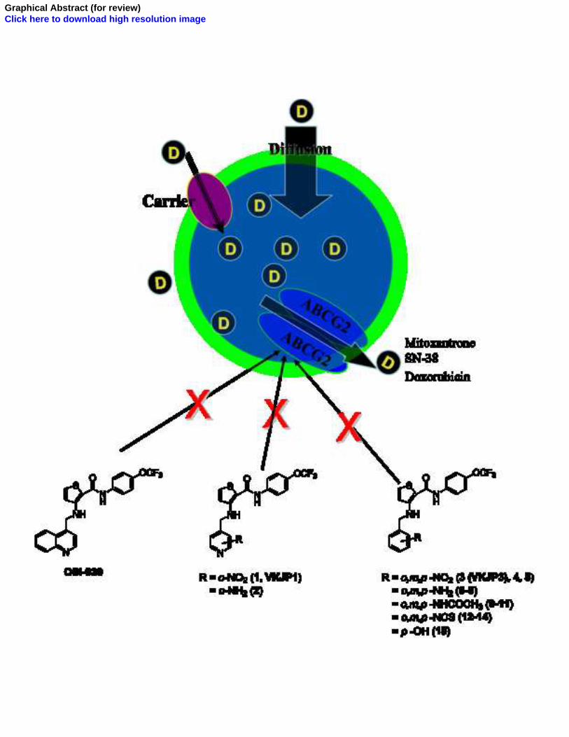
Elsevier Editorial System(tm) for Biochemical Pharmacology Manuscript Draft Manuscript Number: Title: OSI-930 ANALOGUES AS POTENT REVERSAL AGENTS FOR ABCG2-MEDIATED MULTIDRUG RESISTANCE Article Type: Full Length Article Section/Category: Antibiotics and Chemotherapeutics Keywords: OSI-930; ABC transporters; ABCG2; multidrug resistance (MDR); Tyrosine kinase inhibitor Corresponding Author: Associate Professor Zhe-Sheng Chen, MD. Ph. D Corresponding Author's Institution: College of Pharmacy and Allied Health Professions, St. John's University First Author: Ye-Hong Kuang, MD., Ph. D. Order of Authors: Ye-Hong Kuang, MD., Ph. D.; Jay P Patel, Master; Kamlesh Sodani, Master; Chun-Pu Wu, Ph. D.; Li-Qiu Liao, MD., Ph. D.; Atish Patel, Master; Amit K Tiwari, Ph. D.; Chun-Ling Dai, MD., Ph. D.; Xiang Chen, MD., Ph. D.; Li-Wu Fu, MD., Ph. D.; Suresh V Ambudkar, Ph. D.; Vijaya L Korlipara, Ph. D.; Zhe-Sheng Chen, MD. Ph. D Manuscript Region of Origin: America Abstract: OSI-930, a dual c-Kit and KDR tyrosine kinase inhibitor, is reported to have undergone a Phase I dose escalation study in patients with advanced solid tumors. A series of fifteen pyridyl and phenyl analogues of OSI-930 were designed and synthesized. Extensive screening of these compounds led to the discovery that nitropyridyl and ortho-nitrophenyl analogues, VKJP1 and VKJP3, were effective in reversing ABC subfamily G member 2 (ABCG2) transporter-mediated multidrug resistance (MDR). VKJP1 and VKJP3 significantly sensitized ABCG2-expressing cells to established substrates of ABCG2 including mitoxantrone, SN-38, and doxorubicin in a concentration-dependent manner, but not to the non-ABCG2 substrate cisplatin. However, they were unable to reverse ABCB1- or ABCC1- mediated MDR indicating their selectivity for ABCG2. Western blotting analysis was performed to evaluate ABCG2 expression and it was found that neither VKJP1 nor VKJP3 significantly altered ABCG2 protein expression for up to 72 h. [3H]-mitoxantrone accumulation study demonstrated that VKJP1 and VKJP3 increased the intracellular accumulation of [3H]-mitoxantrone, a substrate of ABCG2. VKJP1 and VKJP3 also remarkably inhibited the transport of [3H]-methotrexate by ABCG2 membrane vesicles. Importantly, both VKJP1 and VKJP3 were efficacious in stimulating the activity of ATPase of ABCG2 and inhibited the photoaffinity labeling of this transporter by its substrate [125I]- iodoarylazidoprazosin. The results suggested that VKJP1 and VKJP3, specifically inhibit the function of ABCG2 through direct interaction with its substrate binding site(s). Thus VKJP1 and VKJP3 represent a new class of reversal agents for treatment of MDR in ABCG2 over-expressing tumors.
-
Upload
independent -
Category
Documents
-
view
5 -
download
0
Transcript of OSI-930 analogues as novel reversal agents for ABCG2-mediated multidrug resistance
Elsevier Editorial System(tm) for Biochemical Pharmacology Manuscript Draft Manuscript Number: Title: OSI-930 ANALOGUES AS POTENT REVERSAL AGENTS FOR ABCG2-MEDIATED MULTIDRUG RESISTANCE Article Type: Full Length Article Section/Category: Antibiotics and Chemotherapeutics Keywords: OSI-930; ABC transporters; ABCG2; multidrug resistance (MDR); Tyrosine kinase inhibitor Corresponding Author: Associate Professor Zhe-Sheng Chen, MD. Ph. D Corresponding Author's Institution: College of Pharmacy and Allied Health Professions, St. John's University First Author: Ye-Hong Kuang, MD., Ph. D. Order of Authors: Ye-Hong Kuang, MD., Ph. D.; Jay P Patel, Master; Kamlesh Sodani, Master; Chun-Pu Wu, Ph. D.; Li-Qiu Liao, MD., Ph. D.; Atish Patel, Master; Amit K Tiwari, Ph. D.; Chun-Ling Dai, MD., Ph. D.; Xiang Chen, MD., Ph. D.; Li-Wu Fu, MD., Ph. D.; Suresh V Ambudkar, Ph. D.; Vijaya L Korlipara, Ph. D.; Zhe-Sheng Chen, MD. Ph. D Manuscript Region of Origin: America Abstract: OSI-930, a dual c-Kit and KDR tyrosine kinase inhibitor, is reported to have undergone a Phase I dose escalation study in patients with advanced solid tumors. A series of fifteen pyridyl and phenyl analogues of OSI-930 were designed and synthesized. Extensive screening of these compounds led to the discovery that nitropyridyl and ortho-nitrophenyl analogues, VKJP1 and VKJP3, were effective in reversing ABC subfamily G member 2 (ABCG2) transporter-mediated multidrug resistance (MDR). VKJP1 and VKJP3 significantly sensitized ABCG2-expressing cells to established substrates of ABCG2 including mitoxantrone, SN-38, and doxorubicin in a concentration-dependent manner, but not to the non-ABCG2 substrate cisplatin. However, they were unable to reverse ABCB1- or ABCC1-mediated MDR indicating their selectivity for ABCG2. Western blotting analysis was performed to evaluate ABCG2 expression and it was found that neither VKJP1 nor VKJP3 significantly altered ABCG2 protein expression for up to 72 h. [3H]-mitoxantrone accumulation study demonstrated that VKJP1 and VKJP3 increased the intracellular accumulation of [3H]-mitoxantrone, a substrate of ABCG2. VKJP1 and VKJP3 also remarkably inhibited the transport of [3H]-methotrexate by ABCG2 membrane vesicles. Importantly, both VKJP1 and VKJP3 were efficacious in stimulating the activity of ATPase of ABCG2 and inhibited the photoaffinity labeling of this transporter by its substrate [125I]-iodoarylazidoprazosin. The results suggested that VKJP1 and VKJP3, specifically inhibit the function of ABCG2 through direct interaction with its substrate binding site(s). Thus VKJP1 and VKJP3 represent a new class of reversal agents for treatment of MDR in ABCG2 over-expressing tumors.
CO L L E G E O F PH A R M A C Y A N D
AL L I E D HE A LT H PR O F E S S I O N S DE PA RT M E N T O F P H A R M AC E U T I C A L SC I E N C E S
8000 UTOPIA PARKWAY JAM AICA, NY 11439 (718) 990-1408 FAX: (718) 990-1877
CAM PUSES : QUEENS, NY STATEN ISLAND, NY MANHATTAN, NY EASTERN LONG ISLAND, NY ROM E, ITALY
March. 22, 2012
Dr. S. J. Enna,
Editor-in-Chief, Biochemical Pharmacology
University of Kansas, USA
Dear Dr. Enna:
Enclosed is our manuscript by Kuang et al., entitled “OSI-930 ANALOGUES AS
POTENT REVERSAL AGENTS FOR ABCG2-MEDIATED MULTIDRUG
RESISTANCE” for consideration for publication as an original research article in
“Biochemical Pharmacology”.
All authors have contributed significantly and are in agreement with the content of the
manuscript, which has not been published or submitted for publication elsewhere.
In this study, we discovered that some analogues of OSI930 have potential effects on
reversing ABCG2/BCRP-mediated drug resistance. We found that both VKJP1 and VKJP3
are selective inhibitors of ABCG2 by inhibiting ABCG2 transporter. We believe that this
study is of significance and will lead to further discussion and investigations on the use of
Tyrosine kinase inhibitors as the modulators of ABC transporters in cancer chemotherapy.
As this may be the first publication to show that potent and selective dual inhibitor of the
closely related receptor tyrosine kinases (RTKs) Kit (c-Kit, a type III RTK over-expressed in
seminoma, acute myeloid leukemia and in gastrointestinal stromal tumor) and kinase insert
domain receptor (KDR, a type V RTK as a regulator of tumor angiogenesis) can reverse
MDR, we believe that this finding makes a contribution to the scientific literature. We are
quite sure that Pharmacologists, Biochemists, Oncologists and other cancer researchers in the
field of cancer therapeutics field will be most interested in this study.
The subject category that applies to this manuscript is “Antibiotics and Chemotherapeutics”.
I hope you will find our manuscript acceptable for publication in Biochemical
Pharmacology.
I thank you for your time and consideration!
Sincerely yours,
Cover Letter
- 2 -
Zhe-Sheng (Jason) Chen, MD., Ph.D.
Professor with tenure, Director of Biochemical Pharmacology Lab
Department of Pharmaceutical Sciences
College of Pharmacy and Allied Health Professions
St. John’s University
8000 Utopia Parkway
Jamaica, NY 11439
Phone: (718)990-1432
FAX: (718)990-1877
Email: [email protected]
1 2 3 4 5 6 7 8 9 10 11 12 13 14 15 16 17 18 19 20 21 22 23 24 25 26 27 28 29 30 31 32 33 34 35 36 37 38 39 40 41 42 43 44 45 46 47 48 49 50 51 52 53 54 55 56 57 58 59 60 61 62 63 64 65
1
Ye-Hong Kuanga, b
, Jay P. Patela, Kamlesh Sodani
a, Chun-Pu Wu
c, Li-Qiu Liao
d, Atish
Patela, Amit K. Tiwari
a, Chun-Ling Dai
a, e, Xiang Chen
b,*, Li-Wu Fue, Suresh V.
Ambudkarc, Vijaya L. Korlipara
a,*, Zhe-Sheng Chena,*
aDepartment of Pharmaceutical Sciences, College of Pharmacy and Allied Health Professions, St.
John’s University, Queens, NY 11439,USA; b
Department of Dermatology, Xiang Ya Hospital,
Central South University, Changsha, 410008, China; cLaboratory of Cell Biology, Center for
Cancer Research, National Cancer Institute, NIH, Bethesda MD 20892, USA; dDepartment of
Breast Surgery, Xiang Ya Hospital, Central South University, Changsha, 410008, China; eState
Key Laboratory for Oncology in South China, Cancer Center, Sun Yat-Sen University,
Guangzhou 510060, China
*To whom correspondence should be addressed:
Zhe-Sheng Chen, MD, PhD, 8000 Utopia Parkway, Queens, NY 11439, Fax: 1-718-990-1877;
Email: [email protected]; Xiang Chen: MD, PhD., 87 Xiang Ya Road, Changsha, 410008,
China, Fax: +86 731 432 8478; E-mail: [email protected]; Vijaya Korlipara, PhD., 8000
Utopia Parkway, Queens, NY 11439, Fax: 1-718-990-1877; Email: [email protected]
Running title: Reversal of MDR by OSI-930 analogues
Abbreviations: OSI-930: N-(4-trifluoromethoxyphenyl) 3-((quinolin-4-ylmethyl) amino)
thiophene -2-carboxamide; ABC: ATP binding cassette; ABCG2 (BCRP/MXR/ABCP): ABC
subfamily G member 2; MDR: Multidrug resistance; P-gp: P-glycoprotein; ABCCs (MRPs):
RTK: receptor tyrosine kinases; ABC subfamily C member; KDR: kinase insert domain receptor;
*ManuscriptClick here to view linked References
1 2 3 4 5 6 7 8 9 10 11 12 13 14 15 16 17 18 19 20 21 22 23 24 25 26 27 28 29 30 31 32 33 34 35 36 37 38 39 40 41 42 43 44 45 46 47 48 49 50 51 52 53 54 55 56 57 58 59 60 61 62 63 64 65
2
TKIs: tyrosine kinase inhibitors; IAAP: [125
I]-iodoarylazidoprazosin; FTC: Fumitremorgin C;
MTT: 3-(4, 5-dimethylthiazol -2-yl)-2, 5-diphenyltetrazolium bromide; DMSO:
dimethylsulfoxide.
1 2 3 4 5 6 7 8 9 10 11 12 13 14 15 16 17 18 19 20 21 22 23 24 25 26 27 28 29 30 31 32 33 34 35 36 37 38 39 40 41 42 43 44 45 46 47 48 49 50 51 52 53 54 55 56 57 58 59 60 61 62 63 64 65
3
Abstract:
OSI-930, a dual c-Kit and KDR tyrosine kinase inhibitor, is reported to have undergone a Phase I
dose escalation study in patients with advanced solid tumors. A series of fifteen pyridyl and
phenyl analogues of OSI-930 were designed and synthesized. Extensive screening of these
compounds led to the discovery that nitropyridyl and ortho-nitrophenyl analogues, VKJP1 and
VKJP3, were effective in reversing ABC subfamily G member 2 (ABCG2) transporter-mediated
multidrug resistance (MDR). VKJP1 and VKJP3 significantly sensitized ABCG2-expressing
cells to established substrates of ABCG2 including mitoxantrone, SN-38, and doxorubicin in a
concentration-dependent manner, but not to the non-ABCG2 substrate cisplatin. However, they
were unable to reverse ABCB1- or ABCC1-mediated MDR indicating their selectivity for
ABCG2. Western blotting analysis was performed to evaluate ABCG2 expression and it was
found that neither VKJP1 nor VKJP3 significantly altered ABCG2 protein expression for up to
72 h. [3H]-mitoxantrone accumulation study demonstrated that VKJP1 and VKJP3 increased
the intracellular accumulation of [3H]-mitoxantrone, a substrate of ABCG2. VKJP1 and VKJP3
also remarkably inhibited the transport of [3H]-methotrexate by ABCG2 membrane vesicles.
Importantly, both VKJP1 and VKJP3 were efficacious in stimulating the activity of ATPase of
ABCG2 and inhibited the photoaffinity labeling of this transporter by its substrate
[125
I]-iodoarylazidoprazosin. The results suggested that VKJP1 and VKJP3, specifically inhibit
the function of ABCG2 through direct interaction with its substrate binding site(s). Thus
VKJP1 and VKJP3 represent a new class of reversal agents for treatment of MDR in ABCG2
over-expressing tumors.
1 2 3 4 5 6 7 8 9 10 11 12 13 14 15 16 17 18 19 20 21 22 23 24 25 26 27 28 29 30 31 32 33 34 35 36 37 38 39 40 41 42 43 44 45 46 47 48 49 50 51 52 53 54 55 56 57 58 59 60 61 62 63 64 65
4
Keywords: OSI-930; ABC transporters; ABCG2; multidrug resistance (MDR); tyrosine kinase
inhibitor

1 2 3 4 5 6 7 8 9 10 11 12 13 14 15 16 17 18 19 20 21 22 23 24 25 26 27 28 29 30 31 32 33 34 35 36 37 38 39 40 41 42 43 44 45 46 47 48 49 50 51 52 53 54 55 56 57 58 59 60 61 62 63 64 65
5
1. Introduction
Multidrug resistance (MDR) is a major obstacle to successful chemotherapy in cancer
treatment. It is a phenomenon where cancer cells become resistant to a wide spectrum of anti
cancer drugs belonging to different chemical or pharmacological classes, or cellular targets and
results in decreased cytotoxic activity of these anticancer drugs [1, 2]. The mechanisms of
MDR are multifactorial and include alteration in the permeability of lipid bilayer membrane,
increased DNA repair in cancer cells, increased inactivation or detoxification of drugs, or
inhibition of apoptosis [3, 4]. However, Over-expression of multiple ATP binding cassette (ABC)
transporters in cancer cells still remains the leading cause of MDR [5-7]. The term ABC
transporter, is based on the fact that almost all members of the ABC superfamily, from yeast and
bacteria to man, share a conserved consensus sequence of 90-110 amino acids, where ATP binds
and is hydrolyzed by the ATPase enzyme. This consensus has Walker A and B motifs and
another C region called ABC cassette. ABC transporters is the largest trans-membrane protein
family encoded in the human genome, which are divided into 7 subfamilies ABC-A to G and
subdivided into sub-subfamilies depending on their structural similarity or difference in their
trans-membrane domain. The prominent ABC transporters playing a role in MDR development
are ABCB1, also called P-glycoprotein (P-gp) [8, 9], multidrug resistance proteins (MRPs,
ABCCs) [10], and ABCG2 (BCRP/MXR/ABCP) [11, 12]. This ABC cassette is involved in
transportation of not only toxic metabolites or xenobiotics out of the cell, but also a multitude of
anticancer agents across extracellular membranes, decreasing cellular drug disposition and their
1 2 3 4 5 6 7 8 9 10 11 12 13 14 15 16 17 18 19 20 21 22 23 24 25 26 27 28 29 30 31 32 33 34 35 36 37 38 39 40 41 42 43 44 45 46 47 48 49 50 51 52 53 54 55 56 57 58 59 60 61 62 63 64 65
6
cytotoxicity. Therefore, their expression is believed to be the major mechanism by which MDR
to anticancer drugs develops. Initially, verapamil was found by Tsuruo et. al. to be the first
modulator which could inhibit the function of ABCB1 and increase drug accumulation in the
cancer cells [13]. Later, cyclosporine A (CsA), an immunosuppressant, was discovered to
inhibit the function of ABCB1 and was used as another modulator of ABCB1 [14]. After that,
many other compounds have been studied for their inhibitory effects to other members of ABC
transporter family [15-18], raising hopes that ABC transporter mediated MDR could be reversed.
At present, as the newly synthesized molecule-targeting agents such as tyrosine kinase inhibitors
(TKIs) are being introduced into the clinic, research is being conducted to determine whether
these novel compounds could represent a new class of ABC transporter inhibitors. TKIs such
as imatinib, nilotinib, erlotinib, dasatinib and lapatinib have been extensively investigated as
modulators of ABC transporters-mediated MDR, which most likely act as competitive inhibitors
[19-23]. More recently, pre-clinical research and clinical trials have been conducted to study
the efficacy of the combination of some TKIs with other anticancer drugs in improving the
therapeutic outcome of cancer patients. Therefore, discovering novel inhibitors of ABC
transporters to reverse the drug resistance can lead to improvement in the use of currently
available anticancer drugs.
OSI-930 N-(4-trifluoromethoxyphenyl) 3-((quinolin-4-ylmethyl) amino) thiophene
-2-carboxamide is a heterocyclic anthranilamide analogue synthesized by OSI Pharmaceuticals
Inc. It is a potent and selective dual inhibitor of the closely related receptor tyrosine kinases
(RTKs) Kit (c-Kit, a type III RTK over-expressed in seminoma, acute myeloid leukemia and in
1 2 3 4 5 6 7 8 9 10 11 12 13 14 15 16 17 18 19 20 21 22 23 24 25 26 27 28 29 30 31 32 33 34 35 36 37 38 39 40 41 42 43 44 45 46 47 48 49 50 51 52 53 54 55 56 57 58 59 60 61 62 63 64 65
7
gastrointestinal stromal tumor) and kinase insert domain receptor (KDR, a type V RTK as a
regulator of tumor angiogenesis) [24,25]. OSI-930 acts by these two distinct mechanisms in
appropriate tumor types: i.e., direct effects on the tumor cell phenotype through inhibition of
c-Kit and indirect effects via disruption of endothelial cell function by inhibition of KDR and is
reported to be under phase I clinical trials. The quinoline domain of OSI-930 was modified
with heteroatom substituted pyridyl and phenyl ring systems by our group to obtain a series of
fifteen compounds (Figure 1) [26]. The effects of these changes on their activities and the
binding site characteristics of c-Kit and KDR were evaluated using a kinase assay. In the present
study, we report the screening of our newly designed and synthesized OSI 930 analogues to
determine their ability to reverse ABC transporter-mediated MDR. The reversal mechanisms of
the most potent analogues VKJP1 and VKJP3 were further evaluated by membrane vesicle
transport assay, photolabeling affinity assay, intracellular drug accumulation assay and ATPase
assay, etc.

1 2 3 4 5 6 7 8 9 10 11 12 13 14 15 16 17 18 19 20 21 22 23 24 25 26 27 28 29 30 31 32 33 34 35 36 37 38 39 40 41 42 43 44 45 46 47 48 49 50 51 52 53 54 55 56 57 58 59 60 61 62 63 64 65
8
1 2 3 4 5 6 7 8 9 10 11 12 13 14 15 16 17 18 19 20 21 22 23 24 25 26 27 28 29 30 31 32 33 34 35 36 37 38 39 40 41 42 43 44 45 46 47 48 49 50 51 52 53 54 55 56 57 58 59 60 61 62 63 64 65
9
2. Materials and Methods
2.1 Chemicals and reagents
[3H]-mitoxantrone (4 Ci/mmol) and [
3H]-methotrexate (23 Ci/mmol) were purchased from
Moravek Biochemicals, Inc. [125
I]-iodoarylazidoprazosin (IAAP, 2,200 Ci/mmol) were
obtained from Perkin-Elmer Life Sciences. BXP-21 (against ABCG2) was acquired from
Signet Laboratories, Inc. Anti-actin monoclonal antibody was obtained from Santa Cruz
Biotechnology, Inc. OSI-930 was gift from Selleck Chemical. OSI-930 analogues were
successfully designed and synthesized by our group at St. John’s University. PAK104P was a
kind gift from Prof. Shin-Ichi Akiyama (Kagoshima University) from Nissan Chemical Ind. Co.
Ltd (Chiba, Japan). Fumitremorgin C (FTC) was synthesized by Thomas McCloud
Developmental Therapeutics Program, Natural Products Extraction Laboratory, National Cancer
Institute, NIH. Mitoxantrone, SN-38, doxorubicin, cisplatin, colchicine, vincristine, 3-(4,
5-dimethylthiazol -2-yl)-2, 5-diphenyltetrazolium bromide (MTT), dimethylsulfoxide (DMSO)
and other chemicals were purchased from Sigma Chemical Co.
2.2 Cell lines and cell culture
HEK293/pcDNA3.1, ABCG2-482-R2, and ABCG2-482-T7 cells were established by
selection with G418 after transfecting HEK293 with either the empty pcDNA3.1 vector or
pcDNA3.1 vector containing the full length ABCG2 coding either arginine (R) or threonine (T)
at amino acid 482, respectively [27]. They were cultured in medium with 2 mg/ml of G418
[27]. The parental human epidermoid carcinoma cell line KB-3-1 was selected in a stepwise
manner using increasing concentration of colchicine to establish the ABCB1-overexpressing
1 2 3 4 5 6 7 8 9 10 11 12 13 14 15 16 17 18 19 20 21 22 23 24 25 26 27 28 29 30 31 32 33 34 35 36 37 38 39 40 41 42 43 44 45 46 47 48 49 50 51 52 53 54 55 56 57 58 59 60 61 62 63 64 65
10
drug resistant cell line KB-C2 and was cultured in the medium with 2 µg/ml of colchicine [28].
An ABCC1- overexpressing MDR cell line, KB-CV60, was also cloned from KB-3-1 cells and
was maintained in medium with 1 µg/ml cepharanthine and 60 ng/ml vincristine [29]. All the cell
lines were grown in Dulbecco’s modified Eagle’s medium (DMEM) supplemented with 10%
bovine serum, 100 units/ml penicillin, and 100 mg/ml streptomycin in a humidified incubator
containing 5% CO2 at 37°C.
2.3 Cell cytotoxicity by MTT assay
Drug sensitivity was analyzed using an MTT colorimetric assay [19]. Cells were harvested
with trypsin and resuspended in a final concentration 8 x 10³ cells/ml for KB-C2 and 5 x 10
3 for
all the other cell lines. Cells were seeded evenly into (160 µl/well) 96-well multiplates. After
24 h of incubation, the test VKJP compounds (20 µl/well) was added 1 h prior to different
concentrations of chemotherapeutic drugs (20 µl/well) into designated wells. FTC, verapamil
or PAK-104 (20 µl/well) was added as positive control inhibitors of ABCG2, ABCB1 or ABCC1,
respectively. After 72 h of incubation, 20 µl of MTT solution (4 mg/ml) was added to each well,
further incubated for 4 h, the medium was discarded, and 100 l of DMSO was added into each
well to dissolve the formazan crystals. The absorbance was determined at 570 nm by an
OPSYS microplate Reader from DYNEX Technologies, Inc. (Chantilly, VA). The
concentrations required to inhibit growth by 50% (IC50) were calculated from survival curves.
The degree of resistance was calculated by dividing the IC50 of the MDR cells by that of the
parental sensitive cells.
2.4 Preparation of cell lysates
1 2 3 4 5 6 7 8 9 10 11 12 13 14 15 16 17 18 19 20 21 22 23 24 25 26 27 28 29 30 31 32 33 34 35 36 37 38 39 40 41 42 43 44 45 46 47 48 49 50 51 52 53 54 55 56 57 58 59 60 61 62 63 64 65
11
Cells in T-25 flask treated with 10 µM VKJP1 or VKJP3 for different time periods (0, 36, 72
h), then were harvested and rinsed twice with cold PBS. The cell extracts were prepared by
incubating the cells with the Radioimmunoprecipitation assay (RIPA) buffer [1 × PBS, 1%
Nonidet P-40, 0.5% sodium deoxycholate, 0.1% SDS, 100 μM p-aminophenylmethylsulfonyl
fluoride (p-APMSF), 10 μM leupeptin, and 10 μM aprotinin] for 30 min on ice with occasional
rocking, followed by centrifugation at 12,000 x g at 4°C for 15 min. The supernatant
containing total cell lysates were collected and stored at -80°C until future experiments. The
protein concentration was determined by bicinchoninic acid (BCA™)-based protein assay
(Thermo Scientific, Rockford, IL).
2.5 Western blotting
Equal amounts of total cell lysates (40 μg) were resolved by 4-12% sodium dodecyl sulfate
polyacrylamide gel electrophoresis (SDS-PAGE) and electrophoretically transferred onto PVDF
membrane, then immersed in blocking solution (5% skim milk in TBST) for 1 h at room
temperature. The membrane was then immunoblotted overnight with monoclonal anti-ABCG2
antibody (BXP-21) at 1:500 dilution at 4°C. The following day, the membrane was washed
with TBST buffer (0.3% Tris, 0.8% NaCl, 0.02% KCl, 0.05% Tween 20) three times and
followed by 3 h incubation with horseradish peroxide (HRP)-conjugated secondary anti-mouse
IgG at 1:1000 for 3 h. Proteins were detected by enhanced chemiluminescence detection
system (Amersham, NJ). β-Actin was used to confirm equal loading in each lane in the samples
prepared from cell lysates.

1 2 3 4 5 6 7 8 9 10 11 12 13 14 15 16 17 18 19 20 21 22 23 24 25 26 27 28 29 30 31 32 33 34 35 36 37 38 39 40 41 42 43 44 45 46 47 48 49 50 51 52 53 54 55 56 57 58 59 60 61 62 63 64 65
12
2.6 [3H]-mitoxantrone accumulation
The effect of VKJP1 or VKJP3 on the intracellular accumulation of [3H]-mitoxantrone in
HEK293/pcDNA3.1, ABCG2-482-R2, and ABCG2-482-T7 cells was determined as previously
described [30]. Confluent cells in 24-well plates were preincubated with or without VKJP1,
VKJP3 (FTC as positive control) for 1 h at 37°C. Intracellular drug accumulation was
measured by incubating cells with 0.2 µM [3H]-mitoxantrone for another 2 h. The cells were
washed three times with ice-cold PBS, trypsinized and lysed in 10 mM lysis buffer (pH 7.4,
containing 1% Triton X-100 and 0.2% SDS). Each sample was placed in scintillation fluid and
radioactivity was measured in a Packard TRI-CARB® 1900CA liquid scintillation analyzer from
Packard Instrument Company, Inc (Downers Grove, IL).
2.7 Preparation of membrane vesicles and in vitro transport assays
Membrane vesicles were prepared by using the nitrogen cavitation method [31]. Cells were
rinsed twice with PBS and then scraped into PBS containing 1% aprotinin. Cells were then
washed at 4°C in PBS, collected by centrifugation (4000 x g for 10 min), suspended in buffer A
(10 mM Tris-HCl, pH 7.4, 0.25 M sucrose, 1 mM p-APMSF, and 0.2 mM CaCl2) and
equilibrated at 4°C for 15 min under a nitrogen pressure of 500 psi. EDTA was added to the
suspension of lysed cells to make a final concentration of 1 mM, and the suspension was then
diluted 1:4 with buffer B (10 mM Tris-HCl, pH 7.4, 0.25 M sucrose, and 1 mM p-APMSF) and
centrifuged at 4000 x g for 10 min at 4°C to remove nuclei and unlysed cells. The supernatant
was layered onto a sucrose cushion (35% sucrose, 10 mM Tris-HCl, pH 7.4, and 1 mM EDTA)
and centrifuged for 30 min at 16,000 x g at 4°C. The interface was collected and centrifuged
1 2 3 4 5 6 7 8 9 10 11 12 13 14 15 16 17 18 19 20 21 22 23 24 25 26 27 28 29 30 31 32 33 34 35 36 37 38 39 40 41 42 43 44 45 46 47 48 49 50 51 52 53 54 55 56 57 58 59 60 61 62 63 64 65
13
for 45 min at 100,000 x g at 4°C. The pellet was resuspended in buffer B by repeated passage
through a 25-gauge needle. The protein concentration was determined by bicinchoninic acid
(BCA™)-based protein assay (Thermo Scientific, Rockford, IL).
Transport assays were performed using the rapid filtration method as previously described [19].
Membrane vesicles were incubated with VKJP1 or VKJP3 (FTC as positive control) for 1 h on
ice, and then transport experiments were carried out at 37°C for 10 min in a total volume of 50 µl
medium (membrane vesicles 10 µg, 0.25 M sucrose, 10 mM Tris-HCl, pH 7.4, 10 mM MgCl2, 4
mM ATP or 4 mM AMP, 10 mM phosphocreatine, 100 µg/ml creatine phosphokinase, and 0.5
µM [3H]-methotrexate). Reactions were stopped by the addition of 3 ml of ice-cold stop
solution (0.25 M sucrose, 100 mM NaCl, and 10 mM Tris-HCl, pH 7.4). During the rapid
filtration step, samples were passed through 0.22 µM GVWP filters (Millipore Corporation,
Billerica, MA) that were presoaked in the stop solution. The filters were washed three times
with 3 ml of ice-cold stop solution. Radioactivity was measured by the use of a liquid
scintillation analyzer from Packard Instrument Company, Inc (Downers Grove, IL).
2.8 Effect of VKJP1 and VKJP3 on ATPase activity of ABCG2
ATPase activities of ABCG2 in High Five insect cell crude membranes were measured by
endpoint inorganic phosphate Pi assay as described previously with minor modifications [32].
ABCG2-specific ATPase activity was recorded as BeFx-sensitive ATPase activity. Briefly, The
membrane vesicles (100 µg protein/ml) were incubated in ATPase assay buffer (50 mM
MES-Tris, pH 6.8, 50 mM KCl, 5 mM NaN3, 1 mM EGTA, 1 mM ouabain, 2 mM dithiothreitol
and 10 mM MgCl2) for 20 min at 37°C in the absence or presence of 0.2 mM beryllium sulfate
1 2 3 4 5 6 7 8 9 10 11 12 13 14 15 16 17 18 19 20 21 22 23 24 25 26 27 28 29 30 31 32 33 34 35 36 37 38 39 40 41 42 43 44 45 46 47 48 49 50 51 52 53 54 55 56 57 58 59 60 61 62 63 64 65
14
and 2.5 mM sodium fluoride (BeFX), then incubated with different concentrations of VKJP1 or
VKJP3 at 37°C for 3 min. The reaction was initiated by the addition of 5 mM ATP and
terminated by the addition of SDS (2.5% final concentration). The amount of Pi released was
quantified using a colorimetric method.
2.9 Effect of VKJP1 and VKJP3 on photoaffinity labeling with [125
I]-iodoarylazidoprazosin
(IAAP ) of ABCG2
Crude membranes of High Five insect cells (250 µg protein/mL) were incubated with
increasing concentrations of VKJP1 or VKJP3 at room temperature in 50 mM Tris-HCl, pH 7.5,
for 3 min. [125
I]-IAAP (3-6 nM) was added and further incubated for additional 5 min under
subdued light. The samples were exposed to a UV lamp (365 nm) for 10 min at room
temperature and processed as described previously [33]. The samples were separated on a 7 %
Tris-acetate gel at a constant voltage. Gel was dried and exposed to Bio-Max MR film
(Eastman Kodak, Rochester, NY) for 9 hours at -80 ºC. The incorporation of [125
I]-IAAP into
ABCG2 band was quantified by estimating the radioactivity of this band using the STORM 860
phosphorimager (Molecular Dynamics, Sunnyvale, CA) and ImageQuaNT.
Statistical analysis- Unless otherwise indicated, all experiments were repeated at least three
times and the differences were determined by the two-tailed Student's t-test. When statistical
differences among more than 2 groups were analyzed, one-way ANOVA followed by Tukey’s
multiple comparison tests were performed. Results were presented as means ± standard
deviations (SD). The statistical significance was determined to be P < 0.05.

1 2 3 4 5 6 7 8 9 10 11 12 13 14 15 16 17 18 19 20 21 22 23 24 25 26 27 28 29 30 31 32 33 34 35 36 37 38 39 40 41 42 43 44 45 46 47 48 49 50 51 52 53 54 55 56 57 58 59 60 61 62 63 64 65
15
1 2 3 4 5 6 7 8 9 10 11 12 13 14 15 16 17 18 19 20 21 22 23 24 25 26 27 28 29 30 31 32 33 34 35 36 37 38 39 40 41 42 43 44 45 46 47 48 49 50 51 52 53 54 55 56 57 58 59 60 61 62 63 64 65
16
3. Results
3.1 Structure of VKJP1 and VKJP3
A series of OSI-930 analogues 1-15 (Figure 1) were successfully designed, synthesized, and
evaluated for their reversal effect of ABC tranpsorter-mediated MDR.
3.2 The effect of VKJP compounds on sensitivity of ABC-overexpressing cells to
chemotherapeutic drugs
Prior to determining the effects of VKJP1 and VKJP3 in reversing the MDR, the effect of
OSI-930 and its analogues on cell viability was examined. The IC50 values of these compounds
in ABC–overexpressing cells were in the range of 100 µM (data not shown). In order to avoid
cellular toxicity, the highest concentration of the test compounds used in subsequent reversal
experiments was 10 M.
Subsequently, we screened the 15 compounds in the test series and determined whether they
could sensitize the transfected wild-type ABCG2-overexpressing MDR cells to certain
chemotherapeutic drugs. VKJP1 and VKJP3 were found to be more potent inhibitors than other
members of the test series. For this reason, VKJP1 and VKJP3 were chosen for further study.
Compound 13 had no significant reversing ability, while compound 14 was found to protect cells
from cytotoxicity by mitoxantrone (Table 1). As shown in Table 2, VKJP1 or VKJP3 at 1, 3
and 10 μM, concentration-dependently decreased the IC50 values of ABCG2 substrates including
mitoxantrone, SN-38, and doxorubicin forof the transfected wild-type (R2) or mutant (T7)
ABCG2-overexpressing cells. VKJP1 was shown to be a more potent inhibitor of ABCG2 than
VKJP3 at all concentrations. The magnitude of the sensitization produced by 10 µM VKJP1 was
1 2 3 4 5 6 7 8 9 10 11 12 13 14 15 16 17 18 19 20 21 22 23 24 25 26 27 28 29 30 31 32 33 34 35 36 37 38 39 40 41 42 43 44 45 46 47 48 49 50 51 52 53 54 55 56 57 58 59 60 61 62 63 64 65
17
similar to that induced by the known specific ABCG2 inhibitor FTC at 2.5 µM. In addition, 10
μM VKJP1 could significantly sensitize the parental HEK293-pcDNA3.1 cells to all the
aforementioned chemotherapeutic drugs. However, this effect was significantly lower than
measured in both ABCG2-482-R2 and ABCG2-482-T7 cells. In contrast, there was no
significant effect in the IC50 values for all the cell lines when cisplatin (which is not a substrate
of ABCG2) was incubated with VKJP1, VKJP3 or FTC (Table 2). Besides, VKJP1 or VKJP3
at 10 μM did not significantly alter the cytotoxicity of colchicine (a substrate of ABCB1) on
KB-C2 cells which is a drug seletced cell line overexpressing ABCB1 (Supplemental Table 1) or
vincristine (a substrate of ABCC1) on KB-CV60 cells which is a drug seletced cell line
overexpresses ABCC1 (Supplemental Table 2). These results suggest that VKJP1 and VKJP3
selectively sensitize chemotherapeutic drugs that are the substrates of ABCG2 but not ABCB1 or
ABCC1.
3.3 Effects of VKJP1 and VKJP3 on the expression of ABCG2 in ABCG2-overexpressing
MDR cell
The reversal of ABCG2-mediated MDR can be achieved either by decreasing ABCG2 expression
or by inhibiting its efflux function. To study the effects of VKJP1 and VKJP3 on ABCG2
expression, ABCG2-482-R2 and ABCG2-482-T7 cells were treated with VKJP1 or VKJP3 at 10
µM for different time periods (0, 36, 72 h), and ABCG2 expression levels were examined by
Western blotting analysis. As shown in Figs. 2B & C, the protein levels of ABCG2 in both cell
lines were not significantly altered by VKJP1 or VKJP3 treatment, suggesting that the regulatory
mechanisms of VKJP1 and VKJP3 are other than alteration of ABCG2 expression.
1 2 3 4 5 6 7 8 9 10 11 12 13 14 15 16 17 18 19 20 21 22 23 24 25 26 27 28 29 30 31 32 33 34 35 36 37 38 39 40 41 42 43 44 45 46 47 48 49 50 51 52 53 54 55 56 57 58 59 60 61 62 63 64 65
18
3.4 Effect of [3H]-mitoxantrone intracellular accumulation and [
3H]-methotrexate transport
by VKJP1 and VKJP3
In order to ascertain the mechanisms of VKJP1 and VKJP3 on the modulation of ABCG2
function, the intracellular levels of [3H]-mitoxantrone, a known substrate of ABCG2 was
measured in the presence or absence of VKJP1 or VKJP3 in HEK293/pcDNA3.1,
ABCG2-482-R2 and ABCG2-482-T7 cells (Fig. 3A). Both VKJP1 and VKJP3 at 10 µM, were
found to produce a significant increase in the intracellular levels of [3H]-mitoxantrone in both
wild-type and mutant ABCG2 transfected cell lines. VKJP1 and VKJP3 at 10 M possess
almost the equal potent ability compared to the specific ABCG2 inhibitor FTC at 2.5 µM.
There was no significant change in the intracellular levels of [3H]-mitoxantrone in parental
HEK293/pcDNA3.1 cells incubated either with VKJP1 or VKJP3. Due to the short time course,
we did not see any difference between the potency of VKJP1 and VKJP3 on [3H]-mitoxantrone
accumulation.
To further validate the ability of VKJP1 and VKJP3 to inhibit the transport activity of ABCG2,
we performed transport experiments using membrane vesicles to determine the rate of
ATP-dependent uptake of [3H]-methotrexate, another substrate of ABCG2. Previously, we have
reported that only wild-type ABCG2 was able to robustly transport [3H]-methotrexate in the in
vitro transport system [26,15]. Thus, in this study, we only used the membrane vesicles
prepared from HEK293/pcDNA3.1 and ABCG2-482-R2 cell lines. Our data showed that
VKJP1 and VKJP3 at 10 µM produced a significant inhibition of the ATP-energized uptake rate
of [3H]-methotrexate in ABCG2-482-R2 membrane vesicles (Fig. 3B), confirming that both
1 2 3 4 5 6 7 8 9 10 11 12 13 14 15 16 17 18 19 20 21 22 23 24 25 26 27 28 29 30 31 32 33 34 35 36 37 38 39 40 41 42 43 44 45 46 47 48 49 50 51 52 53 54 55 56 57 58 59 60 61 62 63 64 65
19
VKJP1 and VKJP3 are able to directly inhibit the transport function of ABCG2-overexpressing
membrane vesicles.
3.5 Effect of VKJP1 and VKJP3 on the ATPase activity of ABCG2
Since ABCG2 is an ATP-dependent membrane efflux pump which posses ATPase activity, we
next measured the ABCG2-mediated ATP hydrolysis to investigate the effect of VKJP1 and
VKJP3 on its ATPase activity. As shown in Figs. 4A & B, VKJP1 and VKJP3
concentration-dependently enhanced the ATPase activity of ABCG2. The concentrations of
VKJP1 or VKJP3 required for 50% stimulation of ATPase activity of ABCG2 were 0.024 ±
0.004 and 0.057 ± 0.016 µM, respectively, indicating that VKJP1 and VKJP3 may be the
substrates of ABCG2.
3.6 Effect of VKJP1 and VKJP3 on photoaffinity labeling of ABCG2 with [125
I]-IAAP
ABCG2 is able to be photo-labeled by its substrate [125
I]-IAAP, and its substrates as well as
inhibitors can compete with [125
I]-IAAP for binding to ABCG2. To understand whether there is
physical interaction of VKJP1 or VKJP3 with the substrate interaction sites of ABCG2, we
examined the photoaffinity labeling of ABCG2 with [125
I]-IAAP by using the membranes in the
presence of various concentrations of VKJP1 and VKJP3. As indicated in Fig. 5A & B, VKJP1
and VKJP3 significantly inhibited the photoaffinity labeling of ABCG2 with [125
I]-IAAP in a
concentration-dependent manner. The concentrations of VKJP1 and VKJP3 required for 50%
inhibition of photoaffinity labeling were 6.89 ± 0.12 µM and 9.32 ± 2.64 µM, respectively.
Therefore, these results suggest that VKJP1 and VKJP3 could bind to ABCG2 substrate-binding
sites with different affinity.

1 2 3 4 5 6 7 8 9 10 11 12 13 14 15 16 17 18 19 20 21 22 23 24 25 26 27 28 29 30 31 32 33 34 35 36 37 38 39 40 41 42 43 44 45 46 47 48 49 50 51 52 53 54 55 56 57 58 59 60 61 62 63 64 65
20

1 2 3 4 5 6 7 8 9 10 11 12 13 14 15 16 17 18 19 20 21 22 23 24 25 26 27 28 29 30 31 32 33 34 35 36 37 38 39 40 41 42 43 44 45 46 47 48 49 50 51 52 53 54 55 56 57 58 59 60 61 62 63 64 65
21
4. Discussion
ABCG2 is a 655-amino acid membrane protein, located on 4q22 chromosome, with a
molecular weight of 72 KDa. It is a half transporter in since it has one single N-terminal
nucleotide binding domain and one trans-membrane domain. Its unique structure indicates that
it might have to dimerize with itself (homodimerize) or other members of ABC-G subfamily
(hetrodimerize) to function as an efflux transporter [34, 35]. ABCG2 is expressed
endogenously in many tissues, including placental syncytiotrophoblast cells, epithelial cells of
gastrointestinal tract and liver, and endothelial cells of blood brain barrier [36]. It is also
over-expressed in many kinds of MDR cancer tissues or established MDR cell lines. In recent
years, ABCG2 has been shown to confer resistance to a diverse group of chemotherapeutic drugs,
including nucleoside analogues, anthracyclines, camptothecin-derived indolocarbazole
topoisomerase I inhibitors, methotrexate, and flavopiridols [37]. Several compounds have been
identified that serve as ABCG2 inhibitors and have been shown to overcome ABCG2-mediated
MDR [38]. Additionally, many newly designed compounds are under investigation for their
inhibitory effects in in vitro experiments. Several tyrosine kinase inhibitors (TKIs), including
imatinib (Gleevec, STI571), AG1478, erlotinib (Tarceva, OSI-774), and lapatinib (Tykerb,
GW572016) [19, 20-23] have been identified as novel ABCG2 reversal agents by our group and
others. These TKIs interact with ABCG2 and block its efflux function; increasing the
intracellular accumulation of anticancer drugs and reverse ABCG2- mediated MDR.
In the current study, we found that nearly all the OSI-930 and its pyridyl and phenyl analogues
were shown to reverse drug resistance in wild-type ABCG2 transfected MDR ABCG2-482-R2
1 2 3 4 5 6 7 8 9 10 11 12 13 14 15 16 17 18 19 20 21 22 23 24 25 26 27 28 29 30 31 32 33 34 35 36 37 38 39 40 41 42 43 44 45 46 47 48 49 50 51 52 53 54 55 56 57 58 59 60 61 62 63 64 65
22
cells. Among which, VKJP1 and VKJP3 had greater potency for increasing the cytotoxicity of
ABCG2 substrate mitoxantrone. For this reason, we chose to conduct further mechanistic studies
on these analogues. We found that meta-isothiocyanate derivative 13 had no significant
reversing ability, while the para isothiocyanate derivative 14 interestingly had an opposite effect
of protecting the cells from cytotoxicity by mitoxantrone (Table. 1). We assume that these 15
OSI-930 analogues resulted in variant reversal or protective effects due to their structural
differences. The isothiocyanate analogues in phenyl series were less potent in reversing the MDR.
Based on the present results, OSI-930 analogues such as VKJP1, VKJP3 also showed the
enhancement of cytotoxicity of mitoxantrone in the parental HEK293/pcDNA3.1 cells. One
explanation could be that the parental cells might also have other endogenous drug transporters
that we could not detect, or some other mechanisms, which were interfered by the compounds,
we tested.
Our previous data showed that amino acid residues at position 482 of ABCG2 is essential for
substrate recognition [30]. In order to further study whether the reversal abilities of VKJP1 and
VKJP3 is related to the amino acid residue, we used wild-type and mutant transfectants,
ABCG2-482-R2 and ABCG2-482-T7 cells, in our experiments. Our results showed that VKJP1
and VKJP3 markedly increased the cytotoxicity of several ABCG2 anticancer substrates
including mitoxantrone, SN-38, and doxorubicin in a concentration-dependent manner in both
cell lines implying that the amino acid residues at ABCG2 482 position did not play an important
role in the reversal abilities of VKJP1 and VKJP3. The reversal effect of VKJP1 on
ABCG2-mediated MDR is more potent than that of VKJP3. The sensitization produced by 10
1 2 3 4 5 6 7 8 9 10 11 12 13 14 15 16 17 18 19 20 21 22 23 24 25 26 27 28 29 30 31 32 33 34 35 36 37 38 39 40 41 42 43 44 45 46 47 48 49 50 51 52 53 54 55 56 57 58 59 60 61 62 63 64 65
23
µM VKJP1 was similar to that induced by the known specific ABCG2 inhibitor FTC at 2.5 µM,
showing that the use of VKJP1 as an ABCG2 modulator is very promising. Besides, both
VKJP1 and VKJP3 were found to be ABCG2 selective inhibitors as demonstrated by their
inability to significantly increase the sensitivity of all cell lines to a non ABCG2 substrate,
cisplatin (Table. 2) and inability to reverse ABCB1 or ABCC1 mediated drug resistance
(Supplemental Table. 1& 2).
Neither VKJP1 nor VKJP3 at 10 µM altered the expression levels of ABCG2 in the
ABCG2-482-R2 and ABCG2-482-T7 cells suggesting that VKJP1 and VKJP3 exerted their
inhibitory abilities by directly interacting with ABCG2 transporter. Consistent with the above
hypothesis, we found that when ABCG2-482-R2 and ABCG2-482-T7 MDR cells were incubated
concomitantly with [3H]-mitoxantrone and VKJP1 or VKJP3 at 10 µM, a higher intracellular
drug accumulation was seen than when the cells were incubated with [3H]-mitoxantrone alone
(Fig. 3A). A similar result was obtained when we examined the transport of [3H]-methotrexate
using wild-type ABCG2 overexpressing membrane vesicles, the rates of [3H]-methotrexate
uptake were significantly inhibited by VKJP1 or VKJP3 (Fig. 3B). These results were similar
to some of the ABCG2 inhibitors which were previously reported by our research group. For
example, we previously discovered that several TKIs such as AG1478, erlotinib, lapatinib could
selectively modulate MDR protein-ATPase activity, by competitively inhibiting the efflux
function of ABCG2 and ABCB1 and thus increase the intracellular accumulation of certain
substrates into the MDR cells19, 22-23
. We then examined whether VKJP1 and VKJP3 have the
ability to interact with the ABCG2 transporter substrate binding sites and stimulate ATP

1 2 3 4 5 6 7 8 9 10 11 12 13 14 15 16 17 18 19 20 21 22 23 24 25 26 27 28 29 30 31 32 33 34 35 36 37 38 39 40 41 42 43 44 45 46 47 48 49 50 51 52 53 54 55 56 57 58 59 60 61 62 63 64 65
24
hydrolysis. Indeed this was confirmed by the finding that VKJP1 and VKJP3 significantly
stimulated the ATP hydrolysis and inhibited the binding of [125
I]-IAAP to the substrate binding
sites of ABCG2 (Figs. 4 & 5).
In conclusion, our study shows that VKJP1 and VKJP3 have specific and the most potential
reversal activity on ABCG2-mediated MDR by directly inhibiting the drug efflux function,
resulting in an increase in the intracellular drug accumulation. The mechanistic study found
that VKJP1 and VKJP3 stimulated the activity of ATPase and inhibited the photoaffinity labeling
of ABCG2 with [125
I]-IAAP. The pyridyl and phenyl analogues of OSI-930 are novel reversal
agents of ABCG2 transporter with a potential to increase clinical response when combined with
conventional chemotherapeutic agents.
Acknowledgements: We thank Drs. S.E. Bates and R.W. Robey (National Cancer Institute, NIH)
for the ABCB1, ABCG2 and ABCC1 transfectant cell lines and FTC (NIH, MD.); Drs. Michael
M. Gottesman (NCI, NIH, Bethesda, USA) for KB-3-1 cells; Shin-ichi Akiyama (Kagoshima
University, Japan) for KB-C2 and KB-CV60 cell lines and PAK104P and Selleck Chemical for
OSI-930 (http://www.selleckchem.com/). This work was supported by funds from NIH R15 No.
1R15CA143701 (Z. S. Chen), St. John’s University Seed Grant No.579-1110, (Z.S. Chen), the
National Natural Science Foundation of China No. 81000690 (YH. Kuang), the Millions of
Strategic Project of Xiang Ya hospital (YH. Kuang), the Freedom Explore Program of Central
South University (YH. Kuang).
1 2 3 4 5 6 7 8 9 10 11 12 13 14 15 16 17 18 19 20 21 22 23 24 25 26 27 28 29 30 31 32 33 34 35 36 37 38 39 40 41 42 43 44 45 46 47 48 49 50 51 52 53 54 55 56 57 58 59 60 61 62 63 64 65
25
References
[1] Pratt WB, Ruddon RW (eds). The anticancer drugs. New York, Oxford University Press,
1979.
[2] Zhou, J., Multidrug Resistance in Cancer. Humana Press, 2010.
[3] Gottesman MM. Mechanisms of cancer drug resistance. Annu Rev Med 2002;53:615-27.
[4] Beck WT. Mechanisms of multidrug resistance in human tumor cells. The roles of
P-glycoprotein, DNA topoisomerase II, and other factors. Cancer Treat Rev 1990;17Suppl
A:11-20.
[5] Gottesman MM, Fojo T. Bates SE. Multidrug resistance in cancer: role of ATP-dependent
transporters. Nat Rev Cancer 2002;2:48-58.
[6] Schinkel AH, Jonker JW. Mammalian drug efflux transporters of the ATP binding cassette
(ABC) family: an overview. Adv Drug Deliv Rev 2003;55:3-29.
[7] Dean M, Annilo T. Evolution of the ATP-binding cassette (ABC) transporter superfamily in
vertebrates. Annu Rev Genomics Hum Genet 2005;6:123-42.
[8] Juliano RL, Ling V. A surface glycoprotein modulating drug permeability in Chinese hamster
ovary cell mutants. Biochim Biophys Acta 1976;455:152-62.
[9] Dean M, Rzhetsky A, Allikmets R. The human ATP-binding cassette (ABC) transporter
superfamily. Genome Res 2001 Jul;11:1156-66.
[10] Kruh GD, Belinsky MG. The MRP family of drug efflux pumps. Oncogene
2003;22:7537-52.
[11] Doyle LA, Yang W, Abruzzo LV, Krogmann T, Gao Y, Rishi AK, et al. A multidrug
1 2 3 4 5 6 7 8 9 10 11 12 13 14 15 16 17 18 19 20 21 22 23 24 25 26 27 28 29 30 31 32 33 34 35 36 37 38 39 40 41 42 43 44 45 46 47 48 49 50 51 52 53 54 55 56 57 58 59 60 61 62 63 64 65
26
resistance transporter from human MCF-7 breast cancer cells. Proc Natl Acad Sci USA
1998;95:15665-70.
[12] Maliepaard M, van Gastelen MA, de Jong LA, Pluim D, van Waardenburg RC,
Ruevekamp-Helmers MC, et al. Overexpression of the BCRP/MXR/ABCP gene in a
topotecan-selected ovarian tumor cell line. Cancer Res 1999;59:4559-63.
[13] Tsuruo T, Iida H, Tsukagoshi S, Sakurai Y. Overcoming of vincristine resistance in P388
leukemia in vivo and in vitro through enhanced cytotoxicity of vincristine and vinblastine by
verapamil. Cancer Res 1981;41:1967-72.
[14] Schinkel AH, Jonker JW. Mammalian drug efflux transporters of the ATP binding cassette
(ABC) family: an overview. Adv Drug Deliv Rev 2003;55:3-29.
[15] Tan B, Piwnica-Worms D, Ratner L. Multidrug resistance transporters and modulation. Curr
Opin Oncol 2000;12:450-8.
[16] Robert J, Jarry C. Multidrug resistance reversal agents. J Med Chem 2003 Nov
6;46:4805-17.
[17] Dantzig AH, de Alwis DP, Burgess M. Considerations in the design and development of
transport inhibitors as adjuncts to drug therapy. Adv Drug Deliv Rev 2003;55:133-50.
[18] Chen ZS, Kawabe T, Ono M, Aoki S, Sumizawa T, Furukawa T, et al. Effect of multidrug
resistance-reversing agents on transporting activity of human canalicular multispecific
organic anion transporter. Mol Pharmacol 1999;56:1219-28.
[19] Shi Z, Peng XX, Kim IW, Shukla S, Si QS, Robey RW, et al. Erlotinib (Tarceva, OSI-774)
antagonizes ATP-binding cassette subfamily B member 1 and ATP-binding cassette

1 2 3 4 5 6 7 8 9 10 11 12 13 14 15 16 17 18 19 20 21 22 23 24 25 26 27 28 29 30 31 32 33 34 35 36 37 38 39 40 41 42 43 44 45 46 47 48 49 50 51 52 53 54 55 56 57 58 59 60 61 62 63 64 65
27
subfamily G member 2-mediated drug resistance. Cancer Res 2007;67:11012-20.
[20] Liu W, Baer MR, Bowman MJ, Pera P, Zheng X, Morgan J, et al. The tyrosine kinase
inhibitor imatinib mesylate enhances the efficacy of photodynamic therapy by inhibiting
ABCG2. Clin Cancer Res 2007;13:2463-70.
[21] Houghton PJ, Germain GS, Harwood FC, Schuetz JD, Stewart CF, Buchdunger E, et al.
Imatinib mesylate is a potent inhibitor of the ABCG2 (BCRP) transporter and reverses
resistance to topotecan and SN-38 in vitro. Cancer Res 2004;64:2333-7.
[22] Shi Z, Tiwari AK, Shukla S, Robey RW, Kim IW, Parmar S, et al. Inhibiting the function of
ABCB1 and ABCG2 by the EGFR tyrosine kinase inhibitor AG1478. Biochem Pharmacol
2009;77:781-93.
[23]Dai CL, Tiwari AK, Wu CP, Su XD, Wang SR, Liu DG, et al. Lapatinib (Tykerb, GW572016)
reverses multidrug resistance in cancer cells by inhibiting the activity of ATP-binding
cassette subfamily B member 1 and G member 2. Cancer Res 2008;68:7905-14.
[24] Garton AJ, Crew AP, Franklin M, Cooke AR, Wynne GM, Castaldo L, et al. OSI-930: a
novel selective inhibitor of Kit and kinase insert domain receptor tyrosine kinases with
antitumor activity in mouse xenograft models. Cancer Res 2006;66:1015-24.
[25] Petti F, Thelemann A, Kahler J, McCormack S, Castaldo L, Hunt T, et al. Temporal
quantitation of mutant Kit tyrosine kinase signaling attenuated by a novel thiophene kinase
inhibitor OSI-930. Mol Cancer Ther 2005;4:1186-97.
[26] Patel JP, Kuang YH, Chen ZS, Korlipara VL. Inhibition of c-Kit, VEGFR-2 (KDR), and
ABCG2 by analogues of OSI-930. Bioorg Med Chem Lett 2011; 21(21):6495-9.
1 2 3 4 5 6 7 8 9 10 11 12 13 14 15 16 17 18 19 20 21 22 23 24 25 26 27 28 29 30 31 32 33 34 35 36 37 38 39 40 41 42 43 44 45 46 47 48 49 50 51 52 53 54 55 56 57 58 59 60 61 62 63 64 65
28
[27] Robey RW, Honjo Y, Morisaki K, Nadjem TA, Runge S, Risbood M, et al. Mutations at
amino-acid 482 in the ABCG2 gene affect substrate and antagonist specificity. Br J Cancer
2003;89:1971-8.
[28] Akiyama S, Fojo A, Hanover JA, Pastan I, Gottesman MM. Isolation and genetic
characterization of human KB cell lines resistant to multiple drugs. Somat Cell Mol Genet
1985;11:117-26.
[29] Nagayama S, Chen ZS, Kitazono M, Takebayashi Y, Niwa K, Yamada K, et al. Increased
sensitivity to vincristine of MDR cells by the leukotriene D4 receptor antagonist, ONO-1078.
Cancer Lett 1998;130:175-82.
[30] Chen ZS, Robey RW, Belinsky MG, Shchaveleva I, Ren XQ, Sugimoto Y. Transport of
methotrexate, methotrexate polyglutamates, and 17beta-estradiol 17-(beta-D-glucuronide) by
ABCG2: effects of acquired mutations at R482 on methotrexate transport. Cancer Res
2003;63:4048-54.
[31] Cornwell MM, Gottesman MM, Pastan IH. Increased vinblastine binding to membrane
vesicles from multidrug-resistant KB cells. J Biol Chem 1986;261:7921–8.
[32] Ambudkar SV. Drug-stimulatable ATPase activity in crude membranes of human
MDR1-transfected mammalian cells. Meth Enzymol 1998;292:504–14.
[33] Sauna ZE, Ambudkar SV. Evidence for a requirement for ATP hydrolysis at two distinct
steps during a single turnover of the catalytic cycle of human P-glycoprotein. Proc Natl Acad
Sci USA 2000;97:2515–20.
[34] Ejendal KF, Hrycyna CA. Multidrug resistance and cancer: The role of the human ABC

1 2 3 4 5 6 7 8 9 10 11 12 13 14 15 16 17 18 19 20 21 22 23 24 25 26 27 28 29 30 31 32 33 34 35 36 37 38 39 40 41 42 43 44 45 46 47 48 49 50 51 52 53 54 55 56 57 58 59 60 61 62 63 64 65
29
transporter ABCG2. Curr Prot Pep Sci 2002;503: 503-11.
[35] Mao Q, Unadkat JD. Role of the Breast Cancer Resistance Protein (ABCG2) in Drug
Transport. AAPS J 2005;7:E118-33.
[36] Deeley RG, Westlake C, Cole SP. Transmembrane transport of endo- and xenobiotics by
mammalian ATP-binding cassette multidrug resistance proteins. Physiol Rev 2006;86:849-99.
[37] Mao Q, Unadkat JD. Role of the breast cancer resistance protein (ABCG2) in drug transport.
AAPS J 2005;7:E118-33.
[38] Allen JD, van Loevezijn A, Lakhai JM, van der Valk M, van Tellingen O, Reid G, et al.
Potent and specific inhibition of the breast cancer resistance protein multidrug transporter in
vitro and in mouse intestine by a novel analogue of fumitremorgin C. Mol Cancer Ther
2002;1:417-25.

1 2 3 4 5 6 7 8 9 10 11 12 13 14 15 16 17 18 19 20 21 22 23 24 25 26 27 28 29 30 31 32 33 34 35 36 37 38 39 40 41 42 43 44 45 46 47 48 49 50 51 52 53 54 55 56 57 58 59 60 61 62 63 64 65
30

1 2 3 4 5 6 7 8 9 10 11 12 13 14 15 16 17 18 19 20 21 22 23 24 25 26 27 28 29 30 31 32 33 34 35 36 37 38 39 40 41 42 43 44 45 46 47 48 49 50 51 52 53 54 55 56 57 58 59 60 61 62 63 64 65
31
FIGURE LEGENDS
Fig. 1. Chemical structures of OSI-930 and VKJP compounds 1-15
Fig. 2. Western blot detection of ABCG2 in cells
A: The expression of ABCG2 in HEK293/pcDNA3.1, ABCG2-482-R2, ABCG2-482-T7 cells.
Equal amounts (40 µg protein) of total cell lysates were used for each sample. The membranes
were immunoblotted with primary antibody against ABCG2 at 1:500 dilutions at 4°C overnight,
then incubated with HRP-conjugated secondary antibody at 1:1000 dilutions at room temperature
for 3 h. Proteins were detected by enhanced chemoluminescence detection system (Amersham,
NJ). β-Actin was used to confirm equal loading in each lane in the samples prepared from cell
lysates. Representative result is shown out of three experiments. B: Representative result
showed the ABCG2 expression of ABCG2-482-R2 cells when treated with VKJP1 or VKJP3 (10
µM) at 0, 36, and 72 h. C: Representative result showed the ABCG2 expression of
ABCG2-482-T7 cells when treated with VKJP1 or VKJP3 (10 µM) at 0, 36, and 72 h.
Fig. 3. Effect of VKJP1 and VKJP3 on the intracellular accumulation of [3H]-mitoxantrone
and ABCG2-mediated transport of [3H]-methotrexate
A: The accumulation of [3H]-mitoxantrone was measured after cells were preincubated with or
without VKJP1, VKJP3 (10 M), or FTC (2.5 M) for 1 h at 37°C and then incubated with 0.2
µM [3H]-mitoxantrone for another 2 h at 37°C. Columns, mean of triplicate determinations;

1 2 3 4 5 6 7 8 9 10 11 12 13 14 15 16 17 18 19 20 21 22 23 24 25 26 27 28 29 30 31 32 33 34 35 36 37 38 39 40 41 42 43 44 45 46 47 48 49 50 51 52 53 54 55 56 57 58 59 60 61 62 63 64 65
32
bars, SD; *, P < 0.05 versus the control group. Experiments were repeated at least three times
and a representative experiment is shown. B: Membrane vesicles (10 µg) were prepared from
HEK293/pcDNA3.1 and ABCG2-482-R2 cells. The uptake rates of [3H]-methotrexate into
membrane vesicles were measured for 10 min at 37°C in uptake medium containing 4 mM of
ATP or AMP. For inhibition experiments, membrane vesicles were incubated with VKJP1,
VKJP3, or FTC for 1 h on ice, and then transport reactions were carried out for 10 min at 37°C in
uptake medium containing 4 mM ATP. Columns, mean of triplicate determinations; bars, SD; *,
P < 0.05; **, P < 0.01, versus the control group. Experiments were repeated at least three times
and a representative experiment is shown.
Fig. 4. Effect of VKJP1 and VKJP3 on the ATPase activity of ABCG2
ATPase activity of ABCG2 in membrane vesicles was measured with different concentrations of
VKJP1 (panel A) and VKJP3 (panel B) as described in “Experimental Procedures”. The lines
represent the best fit for the data either by linear or non-linear leastsquares regression analysis
using GraphPad Prism version 2.0. Spot, mean of triplicate determinations; bars, SD. In both
panels A and B, inset shows ATP hydrolysis in the presence of 0 to 0.5 M VKJP1 and VKJP3.
Experiments were repeated at least three times and a representative experiment is shown.
Fig. 5. Effect of VKJP1 and VKJP3 on the photoaffinity labeling of ABCG2 by [125
I]-IAAP
The photoaffinity labeling of ABCG2 with [125
I]-IAAP was performed with different
concentration of VKJP1 or VKJP3. The radioactivity incorporated into ABCG2 was
1 2 3 4 5 6 7 8 9 10 11 12 13 14 15 16 17 18 19 20 21 22 23 24 25 26 27 28 29 30 31 32 33 34 35 36 37 38 39 40 41 42 43 44 45 46 47 48 49 50 51 52 53 54 55 56 57 58 59 60 61 62 63 64 65
33
determined by exposing the gel to an X-ray film at –70°C. An autoradiogram and
quantification of incorporation of IAAP into the ABCG2 band from at least three independent
experiments.
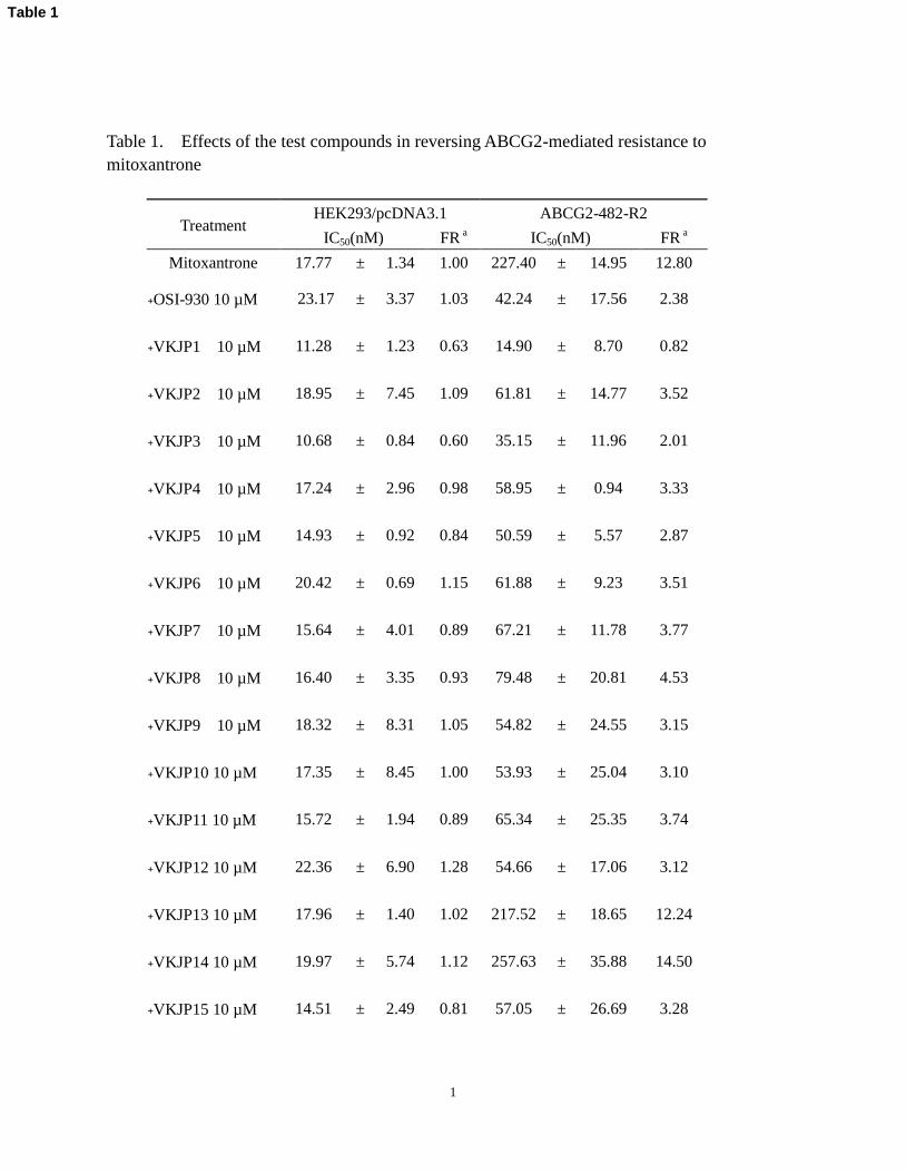
1
Table 1. Effects of the test compounds in reversing ABCG2-mediated resistance to
mitoxantrone
Treatment HEK293/pcDNA3.1 ABCG2-482-R2
IC50(nM) FR a IC50(nM) FR
a
Mitoxantrone 17.77 ± 1.34 1.00 227.40 ± 14.95 12.80
﹢OSI-930 10 µM 23.17 ± 3.37 1.03 42.24 ± 17.56 2.38
﹢VKJP1 10 µM 11.28 ± 1.23 0.63 14.90 ± 8.70 0.82
﹢VKJP2 10 µM 18.95 ± 7.45 1.09 61.81 ± 14.77 3.52
﹢VKJP3 10 µM 10.68 ± 0.84 0.60 35.15 ± 11.96 2.01
﹢VKJP4 10 µM 17.24 ± 2.96 0.98 58.95 ± 0.94 3.33
﹢VKJP5 10 µM 14.93 ± 0.92 0.84 50.59 ± 5.57 2.87
﹢VKJP6 10 µM 20.42 ± 0.69 1.15 61.88 ± 9.23 3.51
﹢VKJP7 10 µM 15.64 ± 4.01 0.89 67.21 ± 11.78 3.77
﹢VKJP8 10 µM 16.40 ± 3.35 0.93 79.48 ± 20.81 4.53
﹢VKJP9 10 µM 18.32 ± 8.31 1.05 54.82 ± 24.55 3.15
﹢VKJP10 10 µM 17.35 ± 8.45 1.00 53.93 ± 25.04 3.10
﹢VKJP11 10 µM 15.72 ± 1.94 0.89 65.34 ± 25.35 3.74
﹢VKJP12 10 µM 22.36 ± 6.90 1.28 54.66 ± 17.06 3.12
﹢VKJP13 10 µM 17.96 ± 1.40 1.02 217.52 ± 18.65 12.24
﹢VKJP14 10 µM 19.97 ± 5.74 1.12 257.63 ± 35.88 14.50
﹢VKJP15 10 µM 14.51 ± 2.49 0.81 57.05 ± 26.69 3.28
Table 1
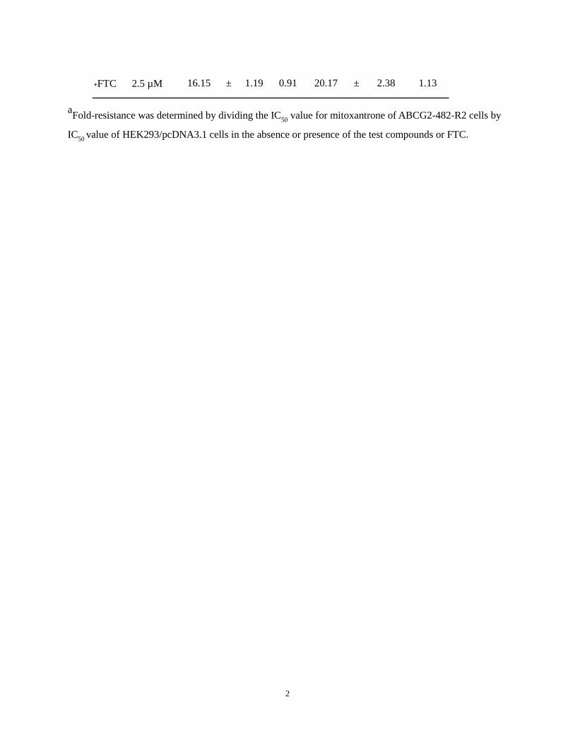
2
﹢FTC 2.5 µM 16.15 ± 1.19 0.91 20.17 ± 2.38 1.13
aFold-resistance was determined by dividing the IC
50 value for mitoxantrone of ABCG2-482-R2 cells by
IC50 value of HEK293/pcDNA3.1 cells in the absence or presence of the test compounds or FTC.

1
Table 2. Effect of VKJP1 and VKJP3 in reversing ABCG2-mediated MDR
Treatment HEK293/pcDNA3.1 ABCG2-482-R2 ABCG2-482-T7
IC50(nM) FR a IC50(nM) FR a IC50(nM) FR a
Mitoxantrone 24.78 ± 0.68 1 277.91 ± 35.02 11.21 814.61 ± 3.05 32.87
﹢VKJP1 1 µM 21.34 ± 4.65 0.86 109.68 ± 95.05 4.43 235.57 ± 97.66 9.51
﹢VKJP1 3 µM 23.91 ± 0.93 0.96 28.52 ± 3.59 1.15 118.47 ± 67.94 4.78
﹢VKJP1 10 µM 18.97 ± 5.41 0.77 16.75 ± 9.1 0.68 24.06 ± 0.49 0.97
﹢VKJP3 1 µM 21.84 ± 7.17 0.88 108.58 ± 1.7 4.38 313.45 ± 194.86 12.65
﹢VKJP3 3 µM 21.43 ± 7.75 0.86 46.94 ± 11.55 1.89 163.69 ± 37.21 6.6
﹢VKJP3 10 µM 17.78 ± 6.88 0.72 53.19 ± 30.81 2.15 68.19 ± 0.08 2.75
﹢FTC 2.5 µM 27.16 ± 3.43 1.1 9.03 ± 0.09 0.36 28.19 ± 1.97 1.14
SN-38 63.08 ± 0.94 1 2456.15 ± 376.77 38.94 2750.44 ± 180.13 43.6
﹢VKJP1 1 µM 64.28 ± 7.45 1.02 641.69 ± 405.07 10.17 684.43 ± 40.57 10.85
﹢VKJP1 3 µM 58.59 ± 21.93 0.93 310.36 ± 177.14 4.92 172.93 ± 16.78 2.74
﹢VKJP1 10 µM 45.1 ± 18.13 0.72 213.17 ± 120.53 3.38 67.4 ± 5.01 1.07
﹢VKJP3 1 µM 76.81 ± 17 1.22 931.17 ± 814.32 14.76 1164.95 ± 61.44 18.47
﹢VKJP3 3 µM 73.12 ± 7.24 1.16 229.58 ± 23.21 3.64 403.42 ± 38.39 6.4
﹢VKJP3 10 µM 68.49 ± 13.5 1.09 137.06 ± 39 2.17 191.8 ± 16.45 3.04
﹢FTC 2.5 µM 61.88 ± 13.4 0.98 106.03 ± 63.17 1.68 61.33 ± 3.64 0.97
Doxorubicin 4.39 ± 0.21 1 15.02 ± 0.08 3.42 80.52 ± 5.96 18.33
﹢VKJP1 1 µM 4.56 ± 0.69 1.04 10.5 ± 2.57 2.39 29.72 ± 3.35 6.77
Table 2
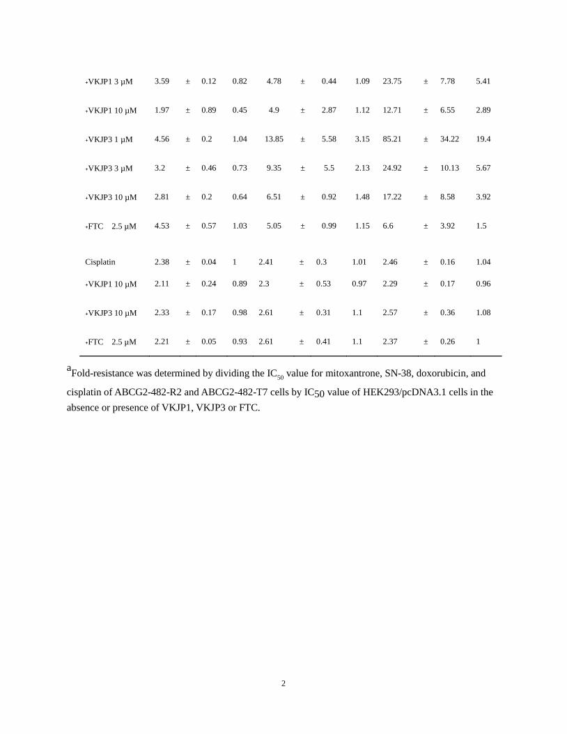
2
﹢VKJP1 3 µM 3.59 ± 0.12 0.82 4.78 ± 0.44 1.09 23.75 ± 7.78 5.41
﹢VKJP1 10 µM 1.97 ± 0.89 0.45 4.9 ± 2.87 1.12 12.71 ± 6.55 2.89
﹢VKJP3 1 µM 4.56 ± 0.2 1.04 13.85 ± 5.58 3.15 85.21 ± 34.22 19.4
﹢VKJP3 3 µM 3.2 ± 0.46 0.73 9.35 ± 5.5 2.13 24.92 ± 10.13 5.67
﹢VKJP3 10 µM 2.81 ± 0.2 0.64 6.51 ± 0.92 1.48 17.22 ± 8.58 3.92
﹢FTC 2.5 µM 4.53 ± 0.57 1.03 5.05 ± 0.99 1.15 6.6 ± 3.92 1.5
Cisplatin 2.38 ± 0.04 1 2.41 ± 0.3 1.01 2.46 ± 0.16 1.04
﹢VKJP1 10 µM 2.11 ± 0.24 0.89 2.3 ± 0.53 0.97 2.29 ± 0.17 0.96
﹢VKJP3 10 µM 2.33 ± 0.17 0.98 2.61 ± 0.31 1.1 2.57 ± 0.36 1.08
﹢FTC 2.5 µM 2.21 ± 0.05 0.93 2.61 ± 0.41 1.1 2.37 ± 0.26 1
aFold-resistance was determined by dividing the IC
50 value for mitoxantrone, SN-38, doxorubicin, and
cisplatin of ABCG2-482-R2 and ABCG2-482-T7 cells by IC50 value of HEK293/pcDNA3.1 cells in the
absence or presence of VKJP1, VKJP3 or FTC.

Figure 1Click here to download high resolution image
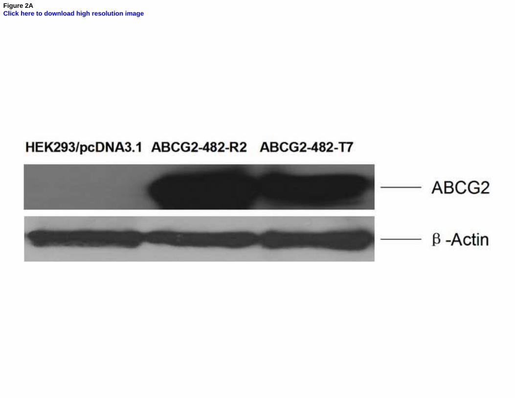
Figure 2AClick here to download high resolution image

Figure 2BClick here to download high resolution image

Figure 2CClick here to download high resolution image

Figure 3AClick here to download high resolution image

Figure 3BClick here to download high resolution image

Figure 4AClick here to download high resolution image
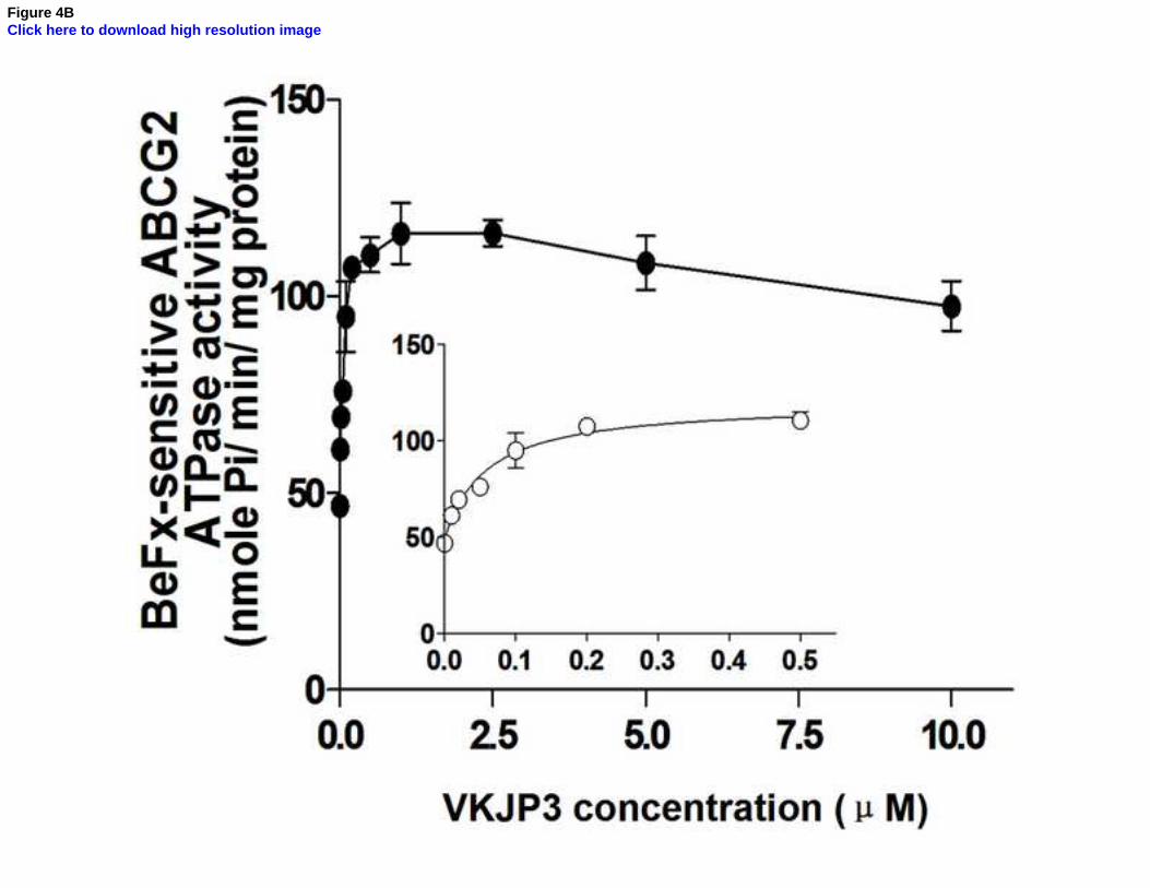
Figure 4BClick here to download high resolution image
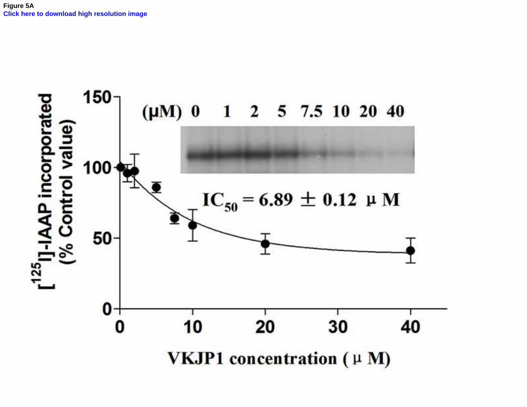
Figure 5AClick here to download high resolution image
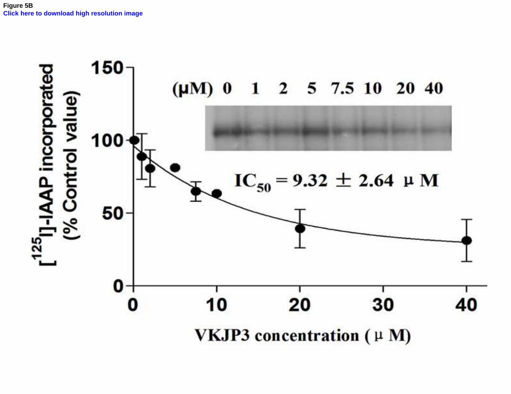
Figure 5BClick here to download high resolution image
Graphical Abstract (for review)Click here to download high resolution image