Discovery of Mitogen-Activated Protein Kinase-Interacting Kinase 1 Inhibitors by a Comprehensive...
11
pubs.acs.org/jmc Published on Web 08/19/2010 r 2010 American Chemical Society 6618 J. Med. Chem. 2010, 53, 6618–6628 DOI: 10.1021/jm1005513 Discovery of Mitogen-Activated Protein Kinase-Interacting Kinase 1 Inhibitors by a Comprehensive Fragment-Oriented Virtual Screening Approach Julen Oyarzabal,* Natasha Zarich, Marı´a Isabel Albarran, Irene Palacios, Manuel Urbano-Cuadrado, Genoveva Mateos, Isabel Reymundo, Obdulia Rabal, Antonio Salgado, Ana Corrionero, Jes us Fominaya, Joaquin Pastor, and James R. Bischoff* Experimental Therapeutics Programme, Spanish National Cancer Research Centre (CNIO), Melchor Fernandez Almagro 3, 28029 Madrid, Spain Received May 6, 2010 Mitogen-activated protein kinase-interacting kinases 1 and 2 (MNK1 and MNK2) phosphorylate the oncogene eIF4E on serine 209. This phosphorylation has been reported to be required for its oncogenic activity. To investigate if pharmacological inhibition of MNK1 could be useful for the treatment of cancers, we pursued a comprehensive virtual screening approach to rapidly identify pharmacological tools for target validation and to find optimal starting points for a plausible medicinal chemistry project. A collection of 1236 compounds, selected from a library of 42 168 compounds and a database of 18.8 million structures, were assayed. Of the identified hits, 26 were found to have IC 50 values less than 10 μM (2.10% hit rate). The most potent compound had an IC 50 value of 117 nM, and 73.1% of these hits were fragments. The hits were characterized by a high ligand efficiency (0.32-0.52 kcal/mol per heavy atom). Ten different chemical scaffolds were represented, giving a chemotype/hit ratio of 0.38. Introduction The initiation of translation factor eIF4E is a partner in a key signaling node that integrates inputs from the PI3K/Akt and Ras/MAPK signal transduction pathways. 1 eIF4E is a compo- nent of the eIF4F complex that controls the rate-limiting step of the recruitment of ribosomes to mRNAs. eIF4E binds to the 7-methylguanosine of the mRNA cap which brings the mRNA in proximity to the ribosome in concert with eIF4G, the ribo- some binding component of eIF4F. 2 Overexpression of eIF4E is oncogenic and can suppress apoptosis. 3 High levels of eIF4E expression have been linked to the progression of head and neck, colon, breast, and bladder cancer. 1 Wendel et al. 4 demonstrated that the phosphorylaton status of serine 209 is key to eIF4E- driven lymphogenesis. They observed that a catalytically inactive dominant negative form of MNK1, a the kinase responsible for the phosphorylation of eIF4E on serine 209, blocks eIF4E- driven lymphomageneis. 4 MNK1 was first identified as a protein kinase that associated and activated by the MAP kinase ERK. 5 Subsequently it was found that MNK1 and MNK2 were the protein kinases responsible for the phosphorylation of eIF4E. 6 Thus far, no other MNK1 or MNK2 substrates have been described. Mice in which both MNK1 and MNK2 have been knocked out in the embryo are viable and show no gross defects, 7 implying that inhibitors targeting MNK1 and MNK2 would be well tolerated. In order to investigate if pharmacological inhibi- tion of MNK1 could be useful for the treatment of cancer, we pursued the following strategy to rapidly identify inhibitors. The goal was to identify pharmacological tools for target validation and at the same time find optimal starting points for medicinal chemistry. Small chemical structures, fragment-like molecules (MW < 300 Da), 8 were preferred because of their potential for rapid expansion and their potential for optimization into leads and then into preclinical candidates. To accelerate the process and minimize costs, our strategy involved the screening of a representative set of chemical structures from our compound collection and a focused set of molecules, fragment-oriented, which were selected using a comprehensive computational app- roach. Several complementary virtual screening strategies were used, together with in silico physicochemical profiling with a special emphasis on fragment-like structures with optimal pre- dicted solubility (>100 μM at pH 7.4). In this study we report the identification of 26 novel MNK1 inhibitors, one of which, 29 (Table 1, ETP-45835), can be used as pharmacological tool for MNK1 validation. Compound 29 had IC 50 for inhibition of MNK1 of 646 nM and was selective against a panel 24 protein kinases. This hit inhibited MNK1 in intact cells, decreasing phosphorylation of eIF4E on serine 209 with an EC 50 of 4.7 μM. This exercise also identified expandable and viable starting points for medicinal chemistry. In fact, 73.1% of hits were frag- ments with high ligand efficiency (LE) values, 9 between 0.32 and 0.52 kcal/mol per heavy atom. There were 10 scaffolds repre- sented by the 26 hits which provides an additional qualitative value in terms of diversity, not only from an intellectual property perspective but also considering their physicochemical profiling and related ADME/Tox properties. 10 Methods Fragment-based drug discovery has proven to be a very useful approach in the hit finding process to identify optimal chemistry starting points for drug discovery programs. 11 Fragments with *To whom correspondence should be addressed. For J.O.: phone, þ34 91 7328000; fax, þ34 91 2246976; e-mail, [email protected]. For J.R.B.: phone, þ34 91 7328000; fax, þ34 91 7328051; e-mail, jrbischoff@ cnio.es. a Abbreviations: MNK1, mitogen-activated protein kinase-interacting kinase 1; MNK2, mitogen-activated protein kinase-interacting kinase 2; ADME/Tox, adsorption, distribution, metabolism, excretion, and toxi- city; VS, virtual screening; PDB, Protein Data Bank; rmsd, root-mean- square deviation; 2D, bidimensional; SSS, substructural searches; ECFP, extended connectivity fingerprints; eMaps, electrostatic maps; LE, ligand efficiency; AML, acute myeloid leukemia; HTS, high-throughput screen- ing; IP, intellectual property; SAR, structure-activity relationship; DB, database; DoE, design of experiments.
-
Upload
independent -
Category
Documents
-
view
0 -
download
0
Transcript of Discovery of Mitogen-Activated Protein Kinase-Interacting Kinase 1 Inhibitors by a Comprehensive...
pubs.acs.org/jmc Published on Web 08/19/2010 r 2010 American Chemical Society
6618 J. Med. Chem. 2010, 53, 6618–6628
DOI: 10.1021/jm1005513
Discovery of Mitogen-Activated Protein Kinase-Interacting Kinase 1 Inhibitors by a Comprehensive
Fragment-Oriented Virtual Screening Approach
Julen Oyarzabal,* Natasha Zarich, Marıa Isabel Albarran, Irene Palacios, Manuel Urbano-Cuadrado, Genoveva Mateos,IsabelReymundo,ObduliaRabal,Antonio Salgado,AnaCorrionero, Jes�usFominaya, Joaquin Pastor, and JamesR.Bischoff*
Experimental Therapeutics Programme, SpanishNational CancerResearchCentre (CNIO),Melchor FernandezAlmagro 3, 28029Madrid, Spain
Received May 6, 2010
Mitogen-activated protein kinase-interacting kinases 1 and 2 (MNK1 and MNK2) phosphorylate theoncogene eIF4E on serine 209. This phosphorylation has been reported to be required for its oncogenicactivity. To investigate if pharmacological inhibition of MNK1 could be useful for the treatment ofcancers, we pursued a comprehensive virtual screening approach to rapidly identify pharmacologicaltools for target validation and to find optimal starting points for a plausiblemedicinal chemistry project.A collection of 1236 compounds, selected from a library of 42 168 compounds and a database of 18.8million structures, were assayed. Of the identified hits, 26were found to have IC50 values less than 10 μM(2.10% hit rate). Themost potent compound had an IC50 value of 117 nM, and 73.1% of these hits werefragments. The hits were characterized by a high ligand efficiency (0.32-0.52 kcal/mol per heavy atom).Ten different chemical scaffolds were represented, giving a chemotype/hit ratio of 0.38.
Introduction
The initiation of translation factor eIF4E is a partner in a keysignaling node that integrates inputs from the PI3K/Akt andRas/MAPK signal transduction pathways.1 eIF4E is a compo-nent of the eIF4F complex that controls the rate-limiting step ofthe recruitment of ribosomes to mRNAs. eIF4E binds to the7-methylguanosine of the mRNA cap which brings the mRNAin proximity to the ribosome in concert with eIF4G, the ribo-some binding component of eIF4F.2Overexpression of eIF4E isoncogenic and can suppress apoptosis.3 High levels of eIF4Eexpressionhave been linked to the progressionof headandneck,colon, breast, and bladder cancer.1Wendel et al.4 demonstratedthat the phosphorylaton status of serine 209 is key to eIF4E-driven lymphogenesis.Theyobserved thatacatalytically inactivedominant negative form of MNK1,a the kinase responsible forthe phosphorylation of eIF4E on serine 209, blocks eIF4E-driven lymphomageneis.4MNK1was first identifiedas aproteinkinase that associated and activated by theMAP kinase ERK.5
Subsequently it was found that MNK1 and MNK2 were theprotein kinases responsible for the phosphorylation of eIF4E.6
Thus far, no other MNK1 or MNK2 substrates have beendescribed. Mice in which both MNK1 and MNK2 have beenknockedout in theembryoareviableandshownogrossdefects,7
implying that inhibitors targetingMNK1 andMNK2would be
well tolerated. In order to investigate if pharmacological inhibi-tion of MNK1 could be useful for the treatment of cancer, wepursued the following strategy to rapidly identify inhibitors. Thegoal was to identify pharmacological tools for target validationand at the same time find optimal starting points for medicinalchemistry. Small chemical structures, fragment-like molecules(MW< 300 Da),8 were preferred because of their potential forrapid expansion and their potential for optimization into leadsand then into preclinical candidates. To accelerate the processand minimize costs, our strategy involved the screening of arepresentative set of chemical structures from our compoundcollection and a focused set of molecules, fragment-oriented,which were selected using a comprehensive computational app-roach. Several complementary virtual screening strategies wereused, together with in silico physicochemical profiling with aspecial emphasis on fragment-like structures with optimal pre-dicted solubility (>100 μM at pH 7.4). In this study we reportthe identification of 26novelMNK1 inhibitors, one ofwhich, 29(Table 1, ETP-45835), can be used as pharmacological tool forMNK1 validation. Compound 29 had IC50 for inhibition ofMNK1 of 646 nM and was selective against a panel 24 proteinkinases. This hit inhibited MNK1 in intact cells, decreasingphosphorylationof eIF4Eon serine 209with anEC50 of 4.7μM.This exercise also identified expandable and viable startingpoints for medicinal chemistry. In fact, 73.1% of hits were frag-mentswith high ligand efficiency (LE) values,9 between 0.32 and0.52 kcal/mol per heavy atom. There were 10 scaffolds repre-sented by the 26 hits which provides an additional qualitativevalue in termsof diversity, not only froman intellectual propertyperspective but also considering their physicochemical profilingand related ADME/Tox properties.10
Methods
Fragment-baseddrugdiscoveryhasproven tobeaveryusefulapproach in the hit finding process to identify optimal chemistrystarting points for drug discovery programs.11 Fragments with
*To whom correspondence should be addressed. For J.O.: phone,þ34 91 7328000; fax, þ34 91 2246976; e-mail, [email protected]. ForJ.R.B.: phone, þ34 91 7328000; fax, þ34 91 7328051; e-mail, [email protected].
aAbbreviations: MNK1, mitogen-activated protein kinase-interactingkinase 1; MNK2, mitogen-activated protein kinase-interacting kinase 2;ADME/Tox, adsorption, distribution, metabolism, excretion, and toxi-city; VS, virtual screening; PDB, Protein Data Bank; rmsd, root-mean-square deviation; 2D, bidimensional; SSS, substructural searches; ECFP,extended connectivity fingerprints; eMaps, electrostatic maps; LE, ligandefficiency; AML, acute myeloid leukemia; HTS, high-throughput screen-ing; IP, intellectual property; SAR, structure-activity relationship; DB,database; DoE, design of experiments.

Article Journal of Medicinal Chemistry, 2010, Vol. 53, No. 18 6619
Table 1. The 26 Compounds Identified as MNK1 Inhibitors
aAll these active compounds were obtained from external sources (more details in Materials and Methods). b Identifiers, ETP numbers, forcompounds with molecular weight lower than 300 are labeled with letter b as superscript, fragments. c IC50 values are represented as mean values of twoindependent experiments. Only differences in pIC50 up to 0.6 (SD < 0.5) were considered as reproducible and were maintained (see Materials andMethods formore details). dExperimental LE is defined as LE=ΔG/NHA,
9a whereΔG is the free energy of ligand binding (ΔG=-RT lnKi),NHA is thenumber of heavy atoms in the compound, andKi values are estimated from the corresponding IC50 experimental data andMNK1assay conditions.ATPconcentration was 100 μM, and the Km for ATP of MNK1 was determined to be 120 μM; thus, Ki = IC50/(1 þ ([ATP]/Km)).
eVS strategy, from thecomprehensive approach, driving to its identification as hit.

6620 Journal of Medicinal Chemistry, 2010, Vol. 53, No. 18 Oyarzabal et al.
their low-molecular-mass structure are likely to have high bind-ing energies per unit of molecular mass and in addition aresimple structures that allow for enhancement during the multi-factorial lead optimization process.12 Therefore, we assembled ahit finding collection that is heavily represented by fragments.The compounds represent themedicinal chemistry spacedefinedbyour libraryofmolecules togetherwitha complementary set ofchemically diverse structures, and a focused set of compoundswere selected by a comprehensive fragment-oriented virtualscreening. Overall, the selected compounds were skewed 2:1toward focused fragment-oriented molecules against the repre-sentative selection of compounds from the CNIO library.
Virtual Screening (VS). Because of their complementarynature, both structure-based and ligand-based virtual screeningstrategies were applied to find inhibitors of MNK1. Thesescreens were performed against our CNIO library containing42168 unique compounds and our virtual CNIO library com-posedof 18.8million unique real compounds, i.e., commerciallyavailable or/and reported (e.g., in PubChem, ChemBank, ...).Different but complementary criteria were considered for com-pound selection and purchase to build our CNIO library of42168 compounds: (a) chemistry perspective, druglike com-pounds, and (b) biological point of view, structures focused onkinases (kinases libraries) and a diverse set of compoundsexploring different chemical spaces with a potential biologicalactivity.
The virtual screening was followed by in silico profiling fo-cused on molecular weight and solubility.13 According to ourinitial criteria, molecular weight values for selected compoundshad to be preferably lower than 300 or fit to a Gaussian curvearound this figure, andestimatedsolubilityvalues, in logarithmicterms, had to be as close as possible to-4mol/L or higher. Thisthreshold for estimated solubility values was adapted per chemi-cal series once experimental values were available.14
Structure-Based Virtual Screening. Crystal structures forboth isoforms, MNK1 and MNK2, have been deposited inthe ProteinDataBank (PDB).MNK1andMNK2have 78%sequence identity within their catalytic domains.15 Thisstructural information was used to identify compoundsfitting the key structural requirements to achieve an optimalbinding to MNK1 and MNK2.
Three published crystal structures were considered. Twostructures correspond to the apo-form for MNK1 (2hw6.pdb)15 and MNK2 (2ac3.pdb),16 both in the inactive confor-mation (DFG/Dout) inwhich autoinhibition is due to Phe230(MNK1) or Phe265 (MNK2) from the activation segment,pushing Phe192 (MNK1) or Phe227 (MNK2) away fromDFG/D toward the ATP binding site where adenosyl moietyof ATP should be accommodated. The third crystal structurecorresponds to the halo form for MNK2 (2hw7.pdb)15 com-plexed with staurosporine. Staurosporine binds in the ATPbinding site; therefore, kinase active conformation involves amovement towardDFG/D in. There are three key interactionsbetween staurosporine and MNK2: two with residues fromhinge region, Glu160 and Met162, and the third between thetitrablenitrogenbornby staurosporine andGlu209 (Figure 1).In the case ofMNK1, only the crystal structure for the inactiveform is available in which the size of the ATP binding site isreduced because of Phe192 being pushed toward the ATPbinding pocket. Since it is possible that inhibitorsmaywork ineither the inactive or active conformations, the initial goalbefore performing any virtual screening was to elucidate thoseresidues thatmight be critical for the binding of ligands in bothinactive and active conformations.
The information concerning the ligand-receptor interac-tions described above for MNK2 and staurosporine wastransferred to MNK1 taking into account the degree ofsequence identity and structural differences between the in-active and active conformations. The first step was to deter-mine if the docking software GOLD17 could recapitulate thedatapublished about theMNK2-staurosporine complex (seeMaterials and Methods for details). Docking was performedwithout constraints in order to obtain an unbiased resultand to explore all possible binding modes of the stauro-sporine. GOLD was found to reliably redock the crystallo-graphic ligand (rmsd=0.65 A); therefore, this was used as thereference for the rest of docking experiments. By use of asimilar docking setup, the staurosporine was docked inMNK1, and as expected, it did not fit into the ATP bindingpocket of inactiveMNK1 probably because of spatial restric-tions derived from inactive conformation mentioned above.Consequently, the next step involved the definition of anartificial structure, substructure 1, with the critical structuralrequirements for binding identified in the ligand-receptormap of the staurosporine-MNK2 complex described inFigure 1. This substructure 1, atom type colored in Figure 1,was then docked into MNK1 to check if the previously iden-tified key chemical features, to interact with MNK2, mightalso bind to MNK1. A consensus result was obtained fromdocking studies confirming knowledge transfer: three key in-teractions, two with residues from the hinge region, Leu127and Gly130, and the third between the titrable nitrogen andbackbone from Phe192 (Figure 2) (also quite close toGlu174,equivalent to Glu209 in MNK2, around 5.5 A). Thus, on thebasis of this result, those three key chemical features may berequired for ligands tobind toMNK1and led to thedefinitionofthe pharmacophore that can bind to MNK1 (Figure 2). Addi-tional factors were contemplated to fine-tune the pharmaco-phoric requirements such as exclusion of volume toward thegatekeeper Phe124 in order to avoid a potential clash withthis side chain and use of a a larger sphere than usual to definethe plausible spatial position for titrable nitrogen. The latterpoint considered protein plasticity in the transition from theinactive to the active conformation, because if the active con-formation takes place, the highlighted interaction predictedwith Phe192 backbone might not occur but will occur insteadwith Glu174.
Figure 1. (A) Interaction map, extracted from 2hw7.pdb, where keychemical features from ligand (staurosporine) andMNK2 amino acidsinvolved in binding are highlighted. (B) Minimal substructure, fromstaurosporine, required to get key interactions withMNK2 (atom typecolor-coded). (C) Substructure 1 from part B.

Article Journal of Medicinal Chemistry, 2010, Vol. 53, No. 18 6621
Once the residues and chemical features that may be criticalfor binding ligands in active or inactive MNK1 conformationswere identified and the pharmacophore was defined (seeMaterials andMethods for details), pharmacophore fittingwasutilized as an alternative to docking in structure-based virtualscreening, since implicitly this considers protein plasticity whichallows for different alternatives for the titrable nitrogen spatialposition and no shape restriction for the ligand; thus, both largeand small cavities in the ATP binding site are accepted (activeand inactive forms). This pharmacophorewas utilized to screenthe CNIO library, and a total of 92 compounds were selectedthat fit all the requirements and possessed an optimal physico-chemical in silico profiling.
Ligand-Based Virtual Screening. Four different virtualscreening strategies based on ligand-based knowledge wereused. Examples ofMNK ligands have been published,18 severalofwhich inhibitMNKas a secondary activity19 (see SupportingInformation Figure SI1). Six knownMNK-oriented inhibitors(Figure 3) were selected to use as references for ligand-basedvirtual screening: the well-knownMNK1 inhibitor CGP57380,2,20 together with five additional compounds, MNK1 or/andMNK2 inhibitors 3,21 4,22 5,23 6,24 and 7.25 The first two ligand-based strategies were utilized to screen both the CNIO libraryand the proprietary virtual database of external compounds.For the last two cases only the CNIO library was used.
Strategy 1 involved substructural searches together with phar-macophoric requirements (2DSubStruct) (Figure4).Theprocessinvolved two sequential steps: (1) identification of compoundsbearing the structural requirements defined by the queries; (2)within these compounds, identification of those meeting theprevious pharmacophore criteria. In total 249 structures wereselected that had all the key pharmacophoric features.
Strategy 2 involved two-dimensional structural similarity(2DTanimoto). These similarity analyses were based on circu-lar molecular fingerprints using ECFP_6 descriptors,26 imple-mented in Pipeline Pilot,27 that defined themolecular structureusing radial atom neighborhoods. These analyses used fourcompounds as reference structures: two MNK1 inhibitors(structures 2 and 3) and two MNK2 ligands (compounds 4
and 5) (Figure 3). This approach identified 140 molecules asstructurally similar to the reference compounds. In the case ofthe CNIO library, 0.05%of structures with the best Tanimotosimilarity values were selected (top values were around 0.40depending on reference structure) and just 0.0001% of com-pounds from the virtual CNIO library with best similarityvalues were selected (top Tanimoto values of around 0.35,again depending on reference compound).
Strategy 3 involved Feature Trees28 similarity (FTrees, seeMaterials and Methods for its definition). The six ligandsreported in Figure 3 were utilized as references, and thosestructures with similarity values greater than 0.90, comparedto the corresponding reference, were selected. In total, 124compounds, 0.05% of chemical structures from CNIO li-brary, with the best FTree similarity values were selected.
Strategy 4 involved three-dimensional similarity based onshape29 and electrostatics30 (3D eMaps, see Materials andMethods for details). The two reported MNK1 inhibitorswere utilized as reference structures (Figure 5). In this case,compounds from CNIO library with the best Tanimotoelectrostatic similarity values (top values are around 0.60and 0.80 for structure 2 and compound 3, respectively) and aTanimoto shape similarity value greater than 0.80 werefound. A total of 231 compounds were selected.
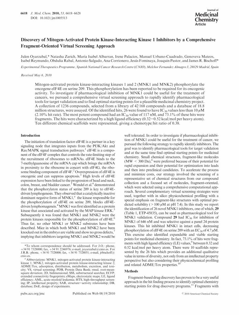
The above approaches yielded a final set of 836 chemicalstructures; 73 of these compounds were purchased from com-mercial suppliers. 70.0% of these selected structures haveestimated solubility values greater than 100 μM, and 74.4% ofthem have a molecular weight lower than 300 (Figure 6).
In addition, a representative set of compounds from theCNIO collection complementing the focused selection wasprepared (Rep. CNIO). This selection consisted of 320 com-pounds representing, from a medicinal chemistry point ofview, as many key chemotypes as possible. In order to selectthis diverse set of compounds, all molecules from our librarywere computationally fragmented.10 Then scaffolds wereselected on the basis of the following criteria: (i) frequencyof representation in the library, (ii) number of fused rings,(iii) number of diversity points, (iv) growing vectors(orientation) for the diversity points, and (v) similarity basedon electrostatics30 and Tanimoto distance (2D Tanimoto),described above, usingATPpurine as reference. Then, on thebasis of “maximum common substructure” around eachselected chemotype, as implemented in Pipeline Pilot,27
R-group analyses were performed selecting compounds forwhich the R-groups provide diversity.
Finally, a D-optimal design strategy (see Materials andMethods)31 was utilized to select an additional set of 80 com-pounds from the CNIO library. D-Optimal designs can be usedin cases in which the data set is composed of discrete mole-cular structures, thus facilitating the selection a few moleculesoptimally spanning the domain of interest. This chemometricstrategy is particularly useful because it permits the selectionof asmall number of molecules that cover the entire chemical spacerepresented by our library which can be useful for derivingreliable QSAR (quantitative structure-activity relationship)models. This designed set of informativemolecules representingthe structural variation in our library collection ensures thereliability of derived models. In addition, once biological dataare available for these selected compounds, we can exploitthose models using the response surface modeling (RSM)32
technique to point out trends, favorable directions, and acti-vity regions.33 All these calculations were performed withSIMCA-Pþ34 and MODDE.35
Figure 2. (A) Interaction map, top ranked answer for dockedcomplex, between substructure 1 andMNK1. (B)Key chemical fea-tures derived from docked complex, those highlighted in Figure 1B(hydrogen-bond donor in light blue, projection point for hydrogen-bond acceptor in red, and titratable nitrogen in blue) together witharomatic ring (in green) and excluded volume (in gray). (C) 5-pointspharmacophore, using CHD scheme, as implemented in MOE(in this case, hydrogen-bond donor is magenta, hydrogen-bondacceptor is light-blue, excluded volume is represented by a graysphere, aromatic ring is green, and titrable nitrogen is blue).

6622 Journal of Medicinal Chemistry, 2010, Vol. 53, No. 18 Oyarzabal et al.
Results and Discussion
Hit Identification. In total, 1236 compounds selected bymethods described above were assayed for the ability tobiochemically inhibit MNK1. A total of 26 compounds wereconfirmed as hits with IC50 values less than 10 μM, giving ahit rate of 2.10%.Ten different chemotypeswere representedin the 26 hits. In addition, these 26 compounds are char-acterized by a high ligand efficiency, between 0.32 and 0.52kcal/mol per heavy atom (Table 1). Six compounds had IC50
values superior to that of the reference compound 2, whichhad an IC50 value of 830 nM in our assay. It is noted that
73.1% of these hits were fragments with a molecular weightless than 300.
In only 1 case out of those 26 hits, compound 28 (ETP-43474), we obtained a selective MNK1 inhibitor vs MNK2;ΔpIC50 was greater than 1 log unit. In this case, IC50 forMNK1 was 847 nM and bigger than 100 μM for MNK2.
The initial goal of this exercise was to identify a compoundthat could be used as a pharmacological tool as well as repre-senting a reasonable starting point for medicinal chemistryexpansion. Compound 29 met these criteria. This compoundwas selected by ligand-based virtual screening based on 2Dstructural similarity within the 18.8 million compounds in thevirtual CNIO library of unique real compounds. It was withinthe top 0.0001% similar structures to reference compound 3
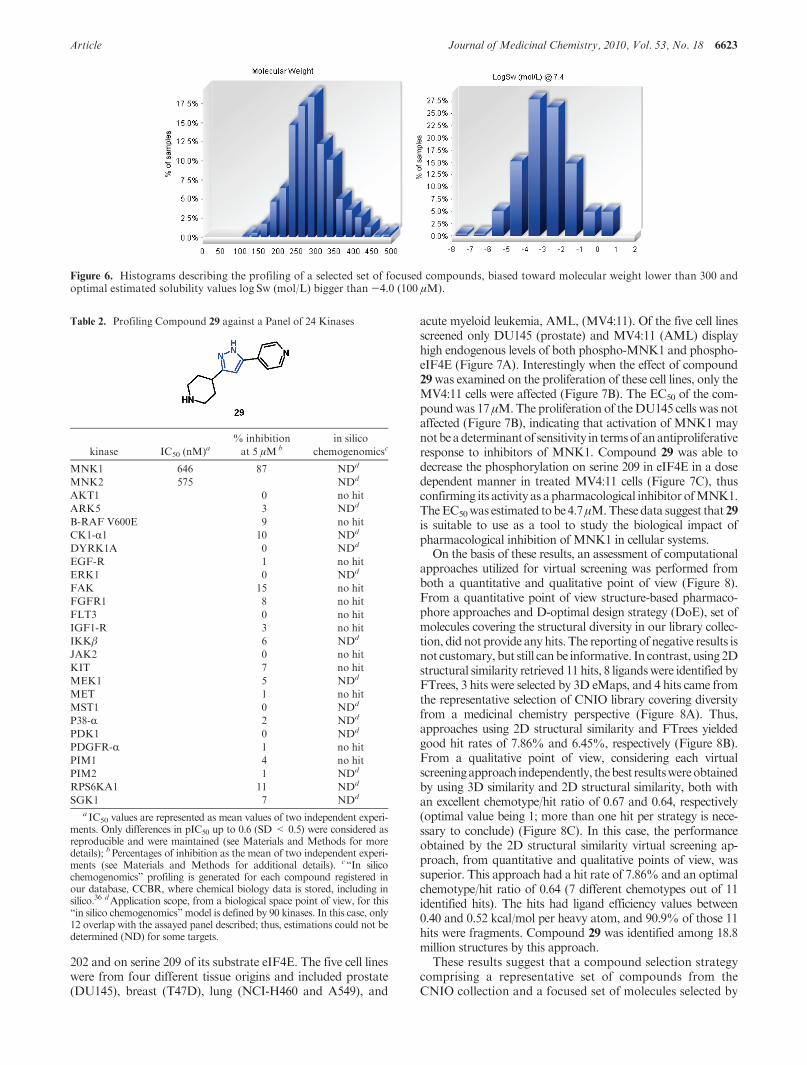
(Figure 3) with a Tanimoto similarity value of 0.30. Thiscompound has an IC50 of 646 nM toward MNK1 and a mole-cularweight of 228,which results in a very high ligand efficiencyof 0.52 kcal/mol per heavy atom. Compound 29was also activetowardMNK2,with IC50 of 575nM. Inorder todetermine if 29was a selective MNK1/2 inhibitor, it was assayed in duplicateagainst a panel of 24 protein kinases at 5 μM. This panel of 24kinases included upstreamMNK1/2 activating kinases such asB-raf, MEK1, ERK, and p38 to ensure that the observedcellular activity was likely due to MNK inhibition and not tooff target activities.Compound29was relatively inactiveagainstthe 24 kinases tested; in all cases the corresponding percentagesof inhibition were less than 16%. These experimental values arein agreement with the estimated “in silico chemogenomics”profiling generated for this compound during its registrationprocess in our database, the Common Chemical BiologyRepository (CCBR)36 (Table 2). Compound 29 had an esti-mated solubilityvaluegreater than100μMatpH7.4.13Thiswasconfirmed experimentally through a kinetic solubility assay (seeMaterials andMethods for solubility assaydetails); its solubilityat pH 7.4 was 125 μM.
Five human tumor cell lines were screened to determine ifany had activated MNK1. Activation was determined by thelevel of phosphorylation of the MNK1 on threonines 197 and
Figure 3. ReportedMNK1 andMNK2 inhibitors together with their corresponding activities in those cases where they are explicitly described(only for compounds 2, 3, and 4). Compounds 5, 6, and 7 are preferred structures among those reported in their corresponding patents;however, no activity value is described for them.
Figure 4. Example of a general query utilized for substructuralsearches, together with pharmacophoric requirements utilized torefine the final set of selected compounds. Hydrogen-bond donor isin light blue, projection point for hydrogen-bond acceptor in red,and titratable nitrogen in blue together with aromatic ring (in green)and excluded volume (in gray).
Figure 5. Electrostatic maps, obtained with EON,30 for the refer-ence structures 2 and 3.
Article Journal of Medicinal Chemistry, 2010, Vol. 53, No. 18 6623
202 and on serine 209 of its substrate eIF4E. The five cell lineswere from four different tissue origins and included prostate(DU145), breast (T47D), lung (NCI-H460 and A549), and
acute myeloid leukemia, AML, (MV4:11). Of the five cell linesscreened only DU145 (prostate) and MV4:11 (AML) displayhigh endogenous levels of both phospho-MNK1 and phospho-eIF4E (Figure 7A). Interestingly when the effect of compound29was examined on the proliferation of these cell lines, only theMV4:11 cells were affected (Figure 7B). The EC50 of the com-poundwas 17 μM.The proliferation of theDU145 cells was notaffected (Figure 7B), indicating that activation of MNK1 maynotbeadeterminantof sensitivity in termsofanantiproliferativeresponse to inhibitors of MNK1. Compound 29 was able todecrease the phosphorylation on serine 209 in eIF4E in a dosedependent manner in treated MV4:11 cells (Figure 7C), thusconfirming its activity as a pharmacological inhibitor ofMNK1.TheEC50was estimated tobe4.7μM.Thesedata suggest that29is suitable to use as a tool to study the biological impact ofpharmacological inhibition of MNK1 in cellular systems.
On the basis of these results, an assessment of computationalapproaches utilized for virtual screening was performed fromboth a quantitative and qualitative point of view (Figure 8).From a quantitative point of view structure-based pharmaco-phore approaches and D-optimal design strategy (DoE), set ofmolecules covering the structural diversity in our library collec-tion, did not provide any hits. The reporting of negative results isnot customary, but still canbe informative. In contrast, using 2Dstructural similarity retrieved 11 hits, 8 ligandswere identified byFTrees, 3 hits were selected by 3D eMaps, and 4 hits came fromthe representative selection of CNIO library covering diversityfrom a medicinal chemistry perspective (Figure 8A). Thus,approaches using 2D structural similarity and FTrees yieldedgood hit rates of 7.86% and 6.45%, respectively (Figure 8B).From a qualitative point of view, considering each virtualscreeningapproach independently, thebest resultswereobtainedby using 3D similarity and 2D structural similarity, both withan excellent chemotype/hit ratio of 0.67 and 0.64, respectively(optimal value being 1; more than one hit per strategy is nece-ssary to conclude) (Figure 8C). In this case, the performanceobtained by the 2D structural similarity virtual screening ap-proach, from quantitative and qualitative points of view, wassuperior. This approach had a hit rate of 7.86% and an optimalchemotype/hit ratio of 0.64 (7 different chemotypes out of 11identified hits). The hits had ligand efficiency values between0.40 and 0.52 kcal/mol per heavy atom, and 90.9% of those 11hits were fragments. Compound 29 was identified among 18.8million structures by this approach.
These results suggest that a compound selection strategycomprising a representative set of compounds from theCNIO collection and a focused set of molecules selected by
Figure 6. Histograms describing the profiling of a selected set of focused compounds, biased toward molecular weight lower than 300 andoptimal estimated solubility values log Sw (mol/L) bigger than -4.0 (100 μM).
Table 2. Profiling Compound 29 against a Panel of 24 Kinases
kinase IC50 (nM)a% inhibition
at 5 μM bin silico
chemogenomicsc
MNK1 646 87 NDd
MNK2 575 NDd
AKT1 0 no hit
ARK5 3 NDd
B-RAF V600E 9 no hit
CK1-R1 10 NDd
DYRK1A 0 NDd
EGF-R 1 no hit
ERK1 0 NDd
FAK 15 no hit
FGFR1 8 no hit
FLT3 0 no hit
IGF1-R 3 no hit
IKKβ 6 NDd
JAK2 0 no hit
KIT 7 no hit
MEK1 5 NDd
MET 1 no hit
MST1 0 NDd
P38-R 2 NDd
PDK1 0 NDd
PDGFR-R 1 no hit
PIM1 4 no hit
PIM2 1 NDd
RPS6KA1 11 NDd
SGK1 7 NDd
a IC50 values are represented as mean values of two independent experi-ments. Only differences in pIC50 up to 0.6 (SD < 0.5) were considered asreproducible and were maintained (see Materials and Methods for moredetails); bPercentages of inhibition as the mean of two independent experi-ments (see Materials and Methods for additional details). c“In silicochemogenomics” profiling is generated for each compound registered inour database, CCBR, where chemical biology data is stored, including insilico.36 dApplication scope, from a biological space point of view, for this“in silico chemogenomics” model is defined by 90 kinases. In this case, only12 overlap with the assayed panel described; thus, estimations could not bedetermined (ND) for some targets.

6624 Journal of Medicinal Chemistry, 2010, Vol. 53, No. 18 Oyarzabal et al.
a comprehensive fragment-oriented virtual screening wasoptimal for the discovery of novel, efficient, and diverseMNK1 inhibitors. More than 70% of the identified MNK1ligands selected by this strategy were fragments that dis-played submicromolar activities and therefore had highligand efficiency values (LE, 0.32-0.52 kcal/mol per heavyatom). Therefore, considering that typically the primary hitsobtained froma fragment screen have biochemical activity ofmillimolar to high micromolar range with rare exceptions inthe 50-200 μM range,8,37 we can consider these reportedresults as good. A recent report11b by AstraZeneca describesthe performance of 12 real cases for which fragment screen-ing was used in its discovery process. These cases includeddifferent screening methods, sizes of assayed libraries used,and different target classes and gave hit rates between 0%and 15% (the criteria for hit definition were not reported).
As reported above, the hit rate for identification of MNK1inhibitors was 2.10%. A hit was defined as a ligand with IC50
lower than 10 μM. If a rigorous comparison is performedwhere only fragments are considered, the hit rate was 2.54%.These hit rates were better than 8 out of those 12 casesreported by AstraZeneca. An additional benchmark used byAstraZeneca was to evaluate the ligand efficacy of fragmenthits (frits)11b considering the ratio pIC50/NHA. This valuewas utilized to identify those fragments that should or shouldnot progress in their drug discovery programs. AstraZenecarecommends not pursuing a frit for which the pIC50/NHA
ratio is less than 0.25 unless the low value can be clearlyunderstood in terms of its interaction with the target.11b Inour case the best value for pIC50/NHA (frit) corresponded to0.37 (details in Table SI1, Supporting Information) whichplaces the results from our fragment-oriented screeningcampaign in the fourth position relative to the 21 fragmentscreening campaigns reported by AstraZeneca where fritratio was explicitly described.11b In those cases wherepIC50/NHA ratios are less than 0.25 but greater than 0.20,such as the hit bearing the chemotype IV, structural informa-tion for the complex target-frit might be critical to clearlyunderstand its interaction with the target in order to recon-sider this frit for further optimization. Ligand efficiencyvalues exhibited by our hits, in kcal/mol per heavy atom,were comparable to those recently described as highly effi-cient hits for which LE values were from 0.30 to 0.50.38
Advanced fragment hits, such as 29 with a potency profilemore similar to those of classical high-throughput screening(HTS) hits, are likely to be easier to progress than traditionalfragment hits (usually in the high micromolar) because thekey chemical features for its interaction with the criticalresidues in the binding pocket of MNK1 are already incor-porated into the hit molecule.8
The identified hits came from 10 different chemical series(Figure 9), which provides an additional value in terms ofchemical diversity. From our point of view, the chemotype/hit ratio should also be routinely quantified in screeningcampaigns, as a third parameter together with hit rate andligand efficiencies, in order to properly assess results ob-tained from screening campaigns from a qualitative perspec-tive. In our case the chemotype/hit ratio was 0.38. If only thefragments were considered, then there were 19 hits represent-ing 7 different chemical series, yielding a chemotype/hit ratiofor fragments of 0.37.
The chemotypes represented by these hits cover a broaddiversity from a structural point of view and include (i) simplefive- or six-member heteroaromatic rings, (ii) bicycles with allpossible combinations of two fused five- and/or six-memberaromatic rings, including heteroaromatics, and (iii) tricycleswhere different options for five and six-member fused ringsare covered. In addition, as Figure 8 shows, all these scaffoldscontainmore than one diversity point fromwhere they can befunctionalized not only to optimize primary activity but alsoto overcome potential ADME/Tox issues. This diversity inchemotypes provides compounds with similar primary acti-vities but different potential pharmacokinetics and off-targetprofiles10 and increases the chance of finding chemotypeswith some room from an Intellectual property (IP) perspec-tive. Therefore, obtaining an optimal chemotype/hit ratio iscritical to drawing conclusions about the performance of ascreening campaign. In theory the higher the ratio is, thebetter, but a balance with the corresponding hit rate valueshould be maintained. In a case such as ours, which used
Figure 7. Compound 29 inhibits MNK1 in human tumor cell lines.(A) Determination of the levels of phospho-MNK1 and phospho-eIF4E in human tumor cell lines. Lysates (25 μg of total protein) ofthe indicated cell lines were analyzed by immunoblotting with anti-P-eIF4E (Ser209), anti-eIF4E, anti-P-MNK1 (Thr197/202), anti-MNK1, and anti-tubulin antibodies as described in Materials andMethods. (B) Effect of treatment with 29 on the proliferation ofMV4:11 (solid black circles) and DU145 (solid gray squares).Percent proliferation was calculated from the DMSO treated con-trol. Data from two independent experiments performed in dupli-cate are presented. (C) MV4.11 cells were treated with DMSO orwith 0.21, 0.62, 1.85, or 5.56 μM 29 for 4 h. Phospho-eIF4E, totaleIF4E, and tubulin were detected by immunoblotting as describedin Materials and Methods. The blot is representative of threeindependent experiments.

Article Journal of Medicinal Chemistry, 2010, Vol. 53, No. 18 6625
commercially available compounds for the screening cam-paign, the possibility of achieving IP is unlikely. However,because the chemical feasibility is guaranteed and someanalogues often are also commercially available, preliminary
studies for their structure-activity relationships (SAR) canbeeasily performed. As a next step, one can use bioisostericreplacements using IP free chemotypes through fragment-hopping strategies10,12 in order to obtain IP.
Figure 8. Histograms comparing the performance of computational approaches utilized for this virtual screening. Quantitative point of view: (A)numberofhit compoundsand (B) hit ratebyVSstrategy,where eachcolor code representsadifferentVSstrategy:orange, 2Dstructural similarity; blue,FTrees similarity; dark green, 3D eMaps and shape; green, CNIO library (MedChem representatives). Zero hitswere obtained for 3Dpharmacophore,SSS and pharmacophore, and CNIO library (DoE representatives). Qualitative assessment was based on (C) chemotype/hit ratio.
Figure 9. Chemotypes borne by the 26 MNK1 hits reported in Table 1. Those 7 scaffolds in blue are borne by the 19 fragments identified as hits.
6626 Journal of Medicinal Chemistry, 2010, Vol. 53, No. 18 Oyarzabal et al.
Conclusion
The implemented strategy was based on two complemen-taryapproachesused to select a limited set of compounds tobeused for hit identification: (i) a representative set of com-pounds from our collection and (ii) a focused set of fragment-oriented molecules, selected from the CNIO library andfrom a virtual DB of external compounds (18.8 million) byboth structure- and ligand-based virtual screening strategiestogether with an in silico physicochemical and chemo-genomics profiling. This comprehensive strategy presentedherein for the identification of MNK1 inhibitors gave satis-factory results: 26 inhibitors (hit rate of 2.10%)with very highligand efficiency (0.32-0.52 kcal/mol per heavy atom) werediscovered. More than 73% of these identified hits are frag-ments. Comparison of these results with recent reports11b,38
highlights their value. In addition, these 26 inhibitors bear 10different chemotypes with good diversity, from five- and six-member rings to bicycles and tricycles, and had an acceptablechemotype/hit ratio: 0.38. Six of these hits had a better IC50
value than the best reference structure 2, and the best com-pound had a binding affinity of 117 nM toward MNK1.
Apharmacological tool compound,29,wasdiscoveredby thefragment-oriented virtual screening strategy utilized during thishit finding process. Compound 29 is a soluble fragment withveryhigh ligand efficiency (0.52kcal/molper heavyatom) and isselective against a panel of 24 kinases including upstreamMNK1/2 activating kinases. The compound is active in cellsbeing able to inhibit the phosphorylationof eIF4Ewith anEC50
of 4.7 μM in acute myeloid leukemia cell line (MV4:11) andtherefore is suitable to use as a pharmacological tool to evaluatethe potential of MNK1/2 inhibition. Thus, our two goals wereachieved: the identification of a pharmacological tool for targetvalidation and identification of optimal starting points for aplausible medicinal chemistry program.
These results suggest the applicabilityof this comprehensivevirtual screening approach to rapidly identify not only opti-mal pharmacological tools for target validation that arepotent and selective but also soluble fragments, bearingdiverse chemotypes, with high binding affinities that representoptimal starting points for medicinal chemistry.
Materials and Methods
Docking.TheGold 3.1 program17was used to carry out dockingof starurosporine and substructure 1 to MNK1 (2hw6.pdb)15 andMNK2 (2hw7.pdb).15 The binding site was defined using theavailable experimental information; thus, the docking region usedfor MNK2 was a 12 A sphere around the carbon CD2 of Leu212(atom2301).GoldScore scoring functionwas used to rank dockingposes without constraints. Per each ligand, the top five best dockedstructures out of 20 independent genetic algorithm runs wereretrieved. For validation purposes, we compared the data forstaurosporine docked to MNK2 with the corresponding crystalstructure (PDB entry 2hw7.pdb).15 The rmsd between reportedstaurosporine and the pose obtained, unique consensus answer, is0.65 A. Once the setting of GOLD, using previous complex, wasvalidated, then a similar setupwas utilizedwithMNK1as receptor.In this case, the binding sitewas definedusing a 12 A sphere aroundthe carbonCD1 of Leu177 (atom 2234). Again, GoldScore scoringfunction was used to rank docking poses without constraints. Pereach ligand, the top five best docked structures out of 20 indepen-dent genetic algorithm runs were retrieved; in this case, the con-sensus answer in the top three ranked results out of the five.
Pharmacophore. On the basis of the proposed binding modefor substructure 1, the key chemical features providing criticalinteractions with MNK1 were identified as well as exclude
volumes; therefore, the corresponding pharmacophore annota-tion was performed. In this case, by using MOE,39 the charged-hydrophobic-direction (CHD) scheme was utilized to build a5-points pharmacophore.
Sampling the Conformational Space. Software developed byOpenEye,40 Omega,41 was utilized to explore the conforma-tional space around each molecule within an energetic windowof 25 kcal/mol, where conformers with a root of mean squaredeviation (rmsd) of>0.4 A are stored, until a maximum of 500,to ensure adequate and diverse conformational representation.Thus, multiconformer databases for CNIO library and selectedcompounds from substructural searches were ready for pharmaco-phore query searching.
Feature Trees Similarity. Features Trees (FTrees)28 are de-scriptors that represent the molecule as a reduced graph. Thegraph encodes functional groups and rings to single nodes.FTree descriptor is a rather fuzzy description of the molecule,ignoring its three-dimensional structure and chirality. It pre-serves only the overall topology of the FTree nodes, but withinthe nodes FTrees compare only property profiles without takinginto account the exact positions of certain functionalities.FTrees focus basically only on the presence or absence of certainfunctionalities in roughly the right position, and thus, it is able todetect remote similarities.
3D Similarity Analysis. Once all conformers per each struc-ture were generated, we had to align them with the correspond-ing reference compound before doing any further analysis. Inthis case, we utilize the ROCS (rapid overlay of chemicalstructures) package,29 by Open Eye, to perform the alignmentbecause this is a fast and accurate method for superimposingmolecules. Shape similarity can be determined, in part, bycomparing the shapes of those molecules. This effectively re-duces to calculating the overlap volume between two moleculesand has been previously reported. Once the overlap has beenoptimized, the shape similarity may be computed by Tanimotoequation. However, ROCS does not contain an accurate notionof charge distribution and therefore is not a complete solution tothe search for molecular similarity. The electrostatic fields ofmolecules can be calculated and the similarity between fieldsexpressed as the electrostatic Tanimoto. Electrostatics Tanimoto(ET) scores are calculated using the EON program,30 by OpenEye.Molecules are superimposed using ROCS, providing shapesimilarities vs reference compound, and electrostatic potentialsare calculated using OpenEye’s ZAP Poisson-Boltzmann sol-ver implemented in EON. In this case ET is calculated using anexternal dielectric of 80, thus accounting for dampening ofelectrostatic field by aqueous solvent, and may better representthe field experienced upon binding a protein.42 A normalizedshape Tanimoto of unity indicates that the molecules areidentical, and the Tanimoto tends to zero as molecules becomeless similar; the electrostatic Tanimoto ranges from-0.3 toþ1,indicating dissimilar and similar electrostatic fields.40 From theCNIO library, 0.30% top ranking compounds, in terms of shapeand electrostatics, are selected.
Design of Experiments (DoE). Three principal components(scores t[1], t[2], and t[3]) derived from 315 1D and 2D descrip-tors (including electrotopological state keys, VSA descriptors,and molecular property counts as implemented in Pipeline Pilot)27
cover 73.1%of the chemical space defined byCNIO library (42168compounds). Onion design is an addition to the design of experi-ments toolbox, and MODDE software35 is utilized. An oniondesign represents the experimental space as comprising a numberof subspaces. The experimental domain is constructed from scorevariables derived from a multivariate projection, principal compo-nent analysis (PCA), model. Here we refer to such a subspace as a“layer” or “shell”. A local design is associated with a layer. Theselocal designs can be classical or D-optimal. We utilize onion designin statisticalmolecular design; thus, a subset ofmolecules is selectedto represent the variation across the entire candidate set, the wholeCNIO library.One critical step in the generation of an onion design

Article Journal of Medicinal Chemistry, 2010, Vol. 53, No. 18 6627
is the divisionof the design space into smaller parts.The subdivisionemanates from the defined center-point, and in our case, by use ofscores variables drawn from the PCA projection model, the subsetselection is distance-based. This means that each subdivision of theexperimental domain can be interpreted as a spherical “shell”surrounding the center-point coordinate. Thus, the structure ofthese designs resembles an onion. The onionD-optimal design wasbased on those three scores obtained from the PCA describedabove, covering 73.1% of the total variance, and three layers wereutilized. As regression, in this RSMapplication, a linearmodel wasselected to be supported in each layer, with the exception of theoutermost one, which supports a quadraticmodel.After generatinga number of tentative D-optimal designs, tailored to the variouslayers, MODDE displays the recommended number of runs pereach layer. This proposed set of selected compounds, runs, is desi-gned by G-efficiency. Thus, in our case, the final onion D-optimaldesign had 80 compounds.
Biochemical Assays. MNK1 and MNK2 were purchasedfrom CARNABiosciences. The biochemical assay activity usedthe ADP Hunter assay kit (DiscoveRx Corp., catalogue no. 90-0077 which determines the amount of ADP as direct product ofthe kinase catalytic activity. The reaction mixture consisted of15 mM HEPES, pH 7.4, 20 mM NaCl, 1 mM EGTA, 0.02%Tween 20, 10 mM MgCl2, and 0.1 mg/mL bovine γ-globulins.MNK1 or MNK2 were included at a final concentration of4 μg/mL (MNK1) or 0.7 μg/mL (MNK2). The final concentra-tion of ATP (Sigma) was 100 μM. The peptide substrate(TATKSGSTTKNR) was designed from the amino acid resi-dues surrounding serine 209 of human eIF4E and was includedat a final concentration of 300 μM. The final reaction mixturewas incubated for 60min at 30 �C. The assays were performed in384-well plates (Corning 3575 or 3573). The product of thecoupled reactions was the release of the fluorescent productresorufin and was measured with a multilabel HTS counterENVISION (PerkinElmer) using an excitation filter at 550 nmand an emission filter at 590 nm. The protein kinase assays, withthe exceptions of MNK1 and MNK2, depicted in Table II ofSupporting Information were performed at ProQinase, GmbH;visit www.proquinase.com for details.
Cell Lines, Immunoblots, and Proliferation Assays. Cell lineswere obtained from the American Type Culture Collection(ATTC; Manassas, VA). MV4:11 (human AML), T47D(human breast cancer), NCI-H460, A549 (human lung cancer),DU145 (human prostate cancer) cell lines were maintained inRPMI 1640 (Sigma). All media were supplemented with 10%fetal bovine serum (Sigma) and antibiotics-antimycotics(Gibco). Cells were maintained in a humidified incubator at37 �Cwith 5%CO2 and passaged when confluent using trypsin/EDTA. For immunobloting experiments, cells were plated insix-well tissue culture plates at 3 � 105 cells/well in 2 mL ofmedium. Rabbit polyclonal antibodies to phospho-eIF4E (S-209) (no. 9741), eIF4E (no. 9742), phospho-MNK1 (Thr197/202) (no. 2111), andMNK1 (no. 2195) proteins were purchasedfrom Cell Signaling Technology (Beverly, MA). Antitubulinmonoclonal antibody was purchased from Sigma-Aldrich.MNK1 inhibitors were dissolved in DMSO at 10 mmol/L, andaliquots were stored at -80 �C. Stock solutions were diluted tothe desired final concentrations with growthmedium just beforeuse. Following treatment cells were washed with ice-cold phos-phate-buffered saline (PBS) and lysed in buffer containing 50mMTris, pH 7.5, 150 mMNaCl, 1%NP-40 supplemented withprotease and phosphatase inhibitors (Roche). Cell lysates wereclarified by centrifugation at 12000g for 15 min at 4 �C, resolvedby SDS-polyacrylamyde gel electrophoresis (PAGE), and sub-sequently transferred onto nitrocellulose membranes. Blotswere probed with the indicated primary and appropriate AlexaFluor 680 conjugated or Alexa Fluor 800 conjugated secondaryantibodies (Molecular Probes) and were visualized and quanti-fied using the Odyssey infrared imaging system (Li-Cor,Biosciences). For proliferation assays cells were harvested just
prior to reaching confluency, counted with a hemocytometer,and diluted with media. Cells were then seeded in 96-well micro-titer plates at a density between 1000 and 4000 cells/well,depending on the cell size. Cells were incubated for 24 h beforeadding the compounds. Compounds were weighed out anddiluted with DMSO to a final concentration of 10 mM. Fromhere a “mother plate” with serial dilutions was prepared at 200�the final concentration in the culture. The final concentration ofDMSO in the tissue culture media should not exceed 0.5%. Theappropriate volume of the compound solution (usually 2 μL)was added automatically (BeckmanFX96 tip) tomedia tomakeit up to the final concentration for each drug. The medium wasremoved from the cells and replaced with 0.2 mL of mediumcontaining the compound. Each concentration was assayed induplicate. Two sets of control wells were left on each plate,containing either medium without drug or medium with thesame concentration of DMSO. A third control set was obtainedwith the cells untreated just before adding the drugs (seedingcontrol, number of cells starting the culture). Cells were exposedto the compounds for 72 h and then processed for MTTcolorimetric readout.
Solubility Assay. In 1 mL vials, test compound (5 μL of a10 mM stock solution in DMSO) was diluted with 245 μL of pH7.4 PBS buffer (A), pH 4 10mMphosphate buffer (B), or 10mMHCl (C). Each mixture was prepared in duplicate. After beingstirred at 30 rpm for 24 h at room temperature, mixtures werefiltered through 0.45 μm polypropylene filters and analyzed byLC-MS (method 1, details in Supporting Information). Solu-bility was estimated by comparing area integrals of chromato-grams (254 nm) with those of a standard sample prepared bydiluting 5 μLof test compound solution (10mM inDMSO)with245 μL of methanol-dimethyl ether 50:50 (v:v) and analyzed bythe same LC-MS method. The integral area of the standardsamples was given a 200 μM solubility value.
Compounds. Tested compounds were purchased from sevendifferent vendors: ChemDiv, BioFocus, Calbiochem, LifeChemicals,Asinex,Enamine, andAurora.Thevendorshadverifiedthat each compoundhadg95%purityby liquid chromatography-mass spectrometry (LC-MS) or/and nuclear magnetic resonance(NMR) experiments.An internal quality controlwasperformed forthose active molecules, 26 hit compounds; thus, evidence of theirpurity was checked by LC-MS confirming that in all casescompounds are g91% pure (details in Supporting Information,Table SI2).
Acknowledgment. We gratefully acknowledge supportfrom the Spanish Ministerio de Ciencia e Innovacion.
Supporting Information Available: Quality control for hits,HPLC-MSmethods, and additional details for hit compounds.Thismaterial is available free of charge via the Internet at http://pubs.acs.org.
References
(1) De Benedetti, A.; Graff, J. R. eIF-4E expression and its role inmalignancies and metastases. Oncogene 2004, 23, 3189–3199.
(2) Gingras, A. C.; Raught, B.; Sonenberg, N. eIF4 initiation factors:effectors of mRNA recruitment to ribosomes and regulators oftranslation. Annu. Rev. Biochem. 1999, 68, 913–963.
(3) Polunovsky, V. A.; Rosenwald, I. B.; Tan, A. T.;White, J.; Chiang,L.; Sonenberg, N.; Bitterman, P. B. Translational control ofprogrammed cell death: eukaryotic translation initiation factor4E blocks apoptosis in growth-factor-restricted fibroblasts withphysiologically expressed or deregulated Myc. Mol. Cell. Biol.1996, 16, 6573–6581.
(4) Wendel, H.-G.; Silva, R. L. A.; Malina, A.; Mills, J. R.; Zhu,H.; Ueda, T.; Watanabe-Fukunaga, R.; Fukunaga, R.; Teruya-Feldstein, J.; Pelletier, J.; Lowe, S. W. Dissecting eIF4E action intumorigenesis. Genes Dev. 2007, 21, 3232–3237.
(5) (a) Waskiewicz, A. J.; Flynn, A.; Proud, C. G.; Cooper, J. A.Mitogen-activated protein kinases activate the serine/threonine
6628 Journal of Medicinal Chemistry, 2010, Vol. 53, No. 18 Oyarzabal et al.
kinasesMnk1andMnk2.EMBOJ. 1997, 16, 1909–1920. (b) Fukunaga,R.; Hunter, T. MNK1, a newMAP kinase-activated protein kinase, isolatedby a novel expression screening method for identifying protein kinasesubstrates. EMBO J. 1997, 16, 1921–1933.
(6) Waskiewicz, A. J.; Johnson, J. C.; Penn, B.; Mahalingam, M.;Kimball, S. R.; Cooper, J. A. Phosphorylation of the cap-bindingprotein eukaryotic translation initiation factor 4E by proteinkinase Mnk1 in-vivo. Mol. Cell. Biol. 1999, 19, 1871–1880.
(7) Ueda, T.; Watanabe-Fukunaga, R.; Fukuyama, H.; Nagata, S.;Fukunaga, R. Mnk2 and Mnk1 are essential for constitutive andinducible phosphorylation of eukaryotic initiation factor 4E but notfor cell growth or development.Mol. Cell. Biol. 2004, 24, 6539–6549.
(8) Makara, G. M. On sampling of fragment space. J. Med. Chem.2007, 50, 3214–3221.
(9) (a) Hopkins, A. L.; Groom, C. R.; Alex, A. Ligand efficiency: a usefulmetric for lead selection. Drug Discovery Today 2004, 9, 430–431.(b) Abad-Zapatero, C.; Metz, J. T. Ligand efficiency indices as guidepostsfor drug discovery. Drug Discovery Today 2005, 10, 464–469.
(10) Oyarzabal, J; Howe, T.; Alcazar, J.; Andres, J. I.; Alvarez, R. M.;Dautzenberg, F.; Iturrino, L.; Martinez, S.; Van der Linden, I.Novel approach for chemotype hopping based on annotateddatabases of chemically feasible fragments and a prospective casestudy: new melanin concentrating hormone antagonists. J. Med.Chem. 2009, 52, 2076–2089.
(11) (a) Alex, A. A.; Flocco,M.M. Fragment-based drug discovery:Whathas it achieved so far? Curr. Top. Med. Chem. 2007, 7, 1544–1567.(b) Albert, J. S.; Blomberg, N.; Breeze, A. L.; Brown, A. J. H.; Burrows,J. N.; Edwards, P. D.; Folmer, R. H. A.; Geschwindner, S.; Griffen, E. J.;Kenny, P. W.; Nowak, T.; Olson, L.-L.; Sanganee, H.; Shapiro, A. Anintegrated approach to fragment-based lead generation: philosophy, strategyand case studies from AstraZeneca's drug discovery programmes. Curr.Top. Med. Chem. 2007, 7, 1600–1629.
(12) Ji, H.; Stanton, B. Z.; Igarashi, J.; Li, H.; Martasek, P.; Roman,L. J.; Poulos, T. L.; Silverman, R. B. Minimal pharmacophoricelements and fragment hopping, an approach directed atmoleculardiversity and isozyme selectivity.Design of selective neuronal nitricoxide synthase inhibitors. J. Am. Chem. Soc. 2008, 130, 3900–3914.
(13) ACD/SolubilityDB, version 12.0;AdvancedChemistryDevelopment,Inc.: Toronto, Canada, 2008; http://www.acdlabs.com.
(14) Oyarzabal, J; Pastor, J.; Howe, T. Optimizing the performance ofin silico ADMET general models according to local requirements:MARS approach. Solubility estimations as case study. J. Chem.Inf. Model. 2009, 49, 2837–2850.
(15) Jauch, R.; Cho, M. K.; J€akel, S.; Netter, C.; Schreiter, K.; Aicher,B.; Zweckstetter, M.; J€ackle, H.; Wahl, M. C. Mitogen-activatedprotein kinases interacting kinases are autoinhibited by a repro-grammed activation segment. EMBO J. 2006, 25, 4020–4032.
(16) Jauch, R.; Wahl, M. C.; Netter, C.; J€akel, S.; Schreiter, K.; Aicher,B.; J€ackle, H. Structures of MNK2 Kinase Domain and MNK2(D228G) Kinase Domain. Unpublished data deposited in theProtein Data Bank (PDB).
(17) Jones, G.; Willett, P.; Glen, R. C.; Leach, A. R.; Taylor, R.Development and validation of a genetic algorithm for flexibledocking. J. Mol. Biol. 1997, 267, 727–748.
(18) KinaseKnowledgebase (KKB) is available fromEidogen-Sertanty,Inc. at http://www.eidogen-sertanty.com (accessed May 2009).
(19) Karaman,M.W.;Herrgard, S.; Treiber, D.K.; Gallant, P.; Atteridge,C. E.; Campbell, B. T.; Chan, K. W.; Ciceri, P.; Davis, M. I.; Edeen,P. T.; Faraoni, R.; Floyd, M.; Hunt, J. P.; Lockhart, D. J.; Milanov,Z. V.; Morrison, M. J.; Pallares, G.; Patel, H. K.; Pritchard, S.;Wodicka, L. M.; Zarrinkar, P. P. A quantitative analysis of kinaseinhibitor selectivity. Nat. Biotechnol. 2008, 26, 127–132.
(20) Knauf, U.; Tschopp, C.; Gram, H. Negative regulation of proteintranslation by mitogen-activated protein kinase-interactingkinases 1 and 2. Mol. Cell. Biol. 2001, 21, 5500–5511.
(21) Anderson, D. R.; Meyers, M. J.; Vernier, W. F.; Mahoney, M.W.;Kurumbail, R. G.; Caspers, N.; Poda, G. I.; Schindler, J. F.; Reitz,D. B.; Mourey, R. J. Pyrrolopyridine inhibitors of mitogen-acti-vated protein kinase-activated protein kinase 2 (MK-2). J. Med.Chem. 2007, 50, 2647–2654.
(22) Wang, S.; Wood, G.; Duncan, K.; Meades, C.; Gibson, D.;Mclachlan, J.; Fischer, P. Preparation of 2-Substituted 4-Thiazolyl-pyrimidines as Protein Kinase Inhibitors with Improved SolubilityProperties. Patent WO05116025 A2, 2005; 216 pp.
(23) Jaekel, S.; Reuter, T.; Murfin, S.; Coulter, T. S.; Taylor, S.Pyrrolopyrimidines for Pharmaceutical Compositions. PatentWO08006547 A2, 2008; 60 pp.
(24) Aicher, B.; Coulter, T. S.; Jaekel, S.; Kelter, A.-R.; Murfin, S.;Reuter, T.; Taylor, S. Thienopyrimidines Having MNK1/MNK2Inhibiting Activity for Pharmaceutical Compositions. PatentWO07115822 A1, 2007; 95 pp.
(25) Coulter, T. S.; Taylor, S.; Murfin, S.; Thammalaksa, V.; Aicher,B.; Jaekel, S.; Reuter, T. Preparation of Pyrazolopyrimidinesas Inhibitors of Kinase Activity. Patent WO06066937 A2, 2006;122 pp.
(26) (a) Bender, A.; Glen, R. C. A discussion of measures of enrichmentin virtual screening: comparing the information content of descriptorswith increasing levels of sophistication. J. Chem. Inf. Model. 2005, 45,1369–1375. (b)Bender,A.;Mussa,H.Y.;Reiling, S.;Glen,R.C. Similaritysearching of chemical databases using atom environment descriptors(MOLPRINT 2D): evaluation of performance. J. Chem. Inf. Model.2004, 44, 1708–1718. (c) Glen, R. C.; Bender, A.; Arnby, C. H.; Carlsson,L.; Boyer, S.; Smith, J. Circular fingerprints: flexible molecular descriptorswith applications from physical chemistry to ADME. IDrugs 2006, 49,199–204.
(27) Pipeline Pilot, version 7.5; Accelrys, Inc. (10188 Telesis Court, Suite100, San Diego, CA 92121); http://accelrys.com/.
(28) FTrees, version 2.0. BioSolveIT GmbH (An der Ziegelei 79, 53757Sankt Augustin, Germany); http://www.biosolveit.de.
(29) Grant, J. A.; Gallardo, M. A.; Pickup, B. T. A fast method ofmolecular shape comparison. A simple application of Gaussiandescription of molecular shape. J. Comput. Chem. 1996, 17, 1653–1666.
(30) (a) Nicholls, A.; MacCuish, N. E.; MacCuish, J. D. Variableselection and model validation of 2-D and 3-D molecular descrip-tors. J. Comput.-AidedMol. Des. 2004, 18, 451–474. (b) Nicholls, A.;Grant, A. J. Molecular shape and electrostatics in the encoding ofrelevant chemical information. J. Comput.-Aided Mol. Des. 2005, 19(9-10), 661–686.
(31) (a) Mitchell, T. J. An algorithm for the construction of“D-optimal” experimental designs. Technometrics 1974, 16,203–210. (b) St. John, R. C.; Draper, N. R. D-Optimality forregression designs: a review. Technometrics 1975, 17, 15–23.(c) Dumouchel, W.; Jones, B. A simple bayesian modification ofD-optimal designs to reduce dependence on an assumed model.Technometrics 1994, 36, 37–47.
(32) Myers, R. H.Response SurfaceMethodology; Allyn and Bacon Inc.:Boston, MA, 1971.
(33) Giraud, E.; Luttmann, C.; Lavelle, F.; Riou, J.-L.; Mailliet, P.;Laoui, A. Multivariate data analysis using D-optimal designs,partial least squares, and response surface modeling: a directionalapproach for the analysis of farnesyltransferase inhibitors. J. Med.Chem. 2000, 43, 1807–1816.
(34) SIMCA-Pþ, version 11.0; Umetrics AB (Box 7960, SE-907 19 Umea,Sweden); www.umetrics.com.
(35) MODDE, version 8.0; Umetrics AB (Box 7960, SE-907 19 Umea,Sweden); www.umetrics.com.
(36) Urbano-Cuadrado, M.; Rabal, O.; Oyarzabal, J. Centralizingdiscovery information: from logistics to knowledge at a publicorganisation. Comb. Chem. High Throughput Screening, in press.
(37) Hajduk, P.; Huth, J. R.; Fesik, S. W. Druggability indices forprotein targets derived from NMR-based screening data. J. Med.Chem. 2005, 48, 2518–2525.
(38) Katritch, V.; Jaakola, V. P.; Lane, J. R.; Lin, J.; Ijzerman, A. P.;Yeager, M.; Kufareva, I.; Stevens, R. C.; Abagyan, R. Structure-based discovery of novel chemotypes for adenosineA(2A) receptorantagonists. J. Med. Chem. 2010, 53, 1799–1809.
(39) Molecular Operating Environment (MOE); Chemical ComputingGroup Inc. (1010 Sherbrooke St. W, Suite 910, Montreal, Quebec,Canada); http://www.chemcomp.com.
(40) OpenEye Scientific Software Inc. (9 Bisbee Court, Suite D, SantaFe, NM 87508); http://www.eyesopen.com.
(41) Bostr€om, J. Reproducing the conformations of protein-boundligands: a critical evaluation of several popular conformationalsearching tools. J. Comput.-Aided. Mol. Des. 2001, 15, 1137–1152.
(42) Jennings, A.; Tennant, M. Selection of molecules based on shapeand electrostatic similarity: proof of concept of “electroforms”.J. Chem. Inf. Model. 2007, 47, 1829–1838.