Nitric oxide stimulates tyrosine phosphorylation of focal adhesion kinase, SRC kinase, and...
7
Biochem. J. (1995) 305, 613-619 (Printed in Great Britain) Nitric oxide stimulates tyrosine phosphorylation in murine fibroblasts in the absence and presence of epidermal growth factor Tereza M. S. PERANOVICH,* Aline M. DA SILVA,t Diana M. FRIES,* Arnold STERN$ and Hugo P. MONTEIRO*§ *Fundaao Hemocentro Sao Paulo, Brazil, tDepartamento de Bioquimica, Instituto de Quimica da Universidade de Sao Paulo, Sao Paulo, Brazil, and tNew York University Medical Center, Department of Pharmacology, New York, NY, U.S.A. In the present study, utilizing anti-phosphotyrosine monoclonal antibodies, sodium nitroprusside (SNP) and S-nitroso-N-acetyl- penicillamine (SNAP) as sources of NO and murine fibroblasts expressing the human epidermal growth factor (EGF) receptor (HER14 cells), we showed that tyrosine phosphorylation of a set of proteins (126, 56 and 43 kDa) was stimulated when cells were incubated with either SNP or SNAP and abolished by Methylene Blue and oxyhaemoglobin. Inhibition by Methylene Blue sug- gested an involvement of cyclic GMP in the process, which was evidenced by the effects of 8-bromo cyclic GMP. This analogue of cyclic GMP stimulated tyrosine phosphorylation of the same set of proteins phosphorylated after incubation with the NO INTRODUCTION The mitogenic response to epidermal growth factor (EGF) is mediated by a 170 kDa transmembrane glycoprotein, the EGF receptor. The EGF receptor is a protein tyrosine kinase (PTK) receptor that catalyses the phosphorylation of exogenous sub- strates as well as tyrosine residues within its own polypeptide chains. Tyrosine phosphorylation associated with the control of both mitogenic signal pathways and oncogenic transformation [1,2] is a reversible process controlled by the opposing actions of PTK and protein tyrosine phosphatases (PTPs). Together, PTK and PTP activities control the amount of phosphotyrosine in the cell [3,4]. Sequence analysis of PTP isoforms from several sources revealed the presence of a conserved catalytic domain and an essential cysteine residue in this domain [4]. Previously, it was shown that oxidative-stress-inducing sys- tems, such as the thiol-group oxidant diamide [5], the vanadate/ H202 system [6] and autoxidizing ascorbic acid in the presence of sub-micromolar concentrations of iron [7], were able to inhibit PTP activities in a number of cell lines. PTK activities are also subject to redox regulation. Chan et al. [8] have shown that oxygen-derived free radicals generated through redox cycling of naphthoquinones were stimulatory of tyrosine phosphorylation in rat liver plasma membranes. The redox regulation of a transmembrane PTK homologous to the cytoplasmic portion of the insulin receptor and localized in the endoplasmic reticulum of B lymphocytes, the Ltk receptor, has been reported [9]. The thiol-group-oxidizing and -alkylating agent N-ethylmaleimide was shown to be a modulator of PTK activity in T lymphocytes [10]. Thus PTP activities and PTK activities could be redox modulated. source. Tyrosine phosphorylation of the same set of proteins was stimulated when cells were incubated simultaneously with SNP and EGF, showing that NO also potentiates EGF-evoked tyrosine kinase activity in HER14 cells. However, stimulation of the autophosphorylation of the EGF receptor, above the levels obtained for EGF alone, was not observed under those con- ditions. Additionally, we investigated the effects of NO on EGF- receptor tyrosine phosphatase activities in HER14 cells. In- creasing concentrations of NO correlate with a gradual inhibition of these activities in HER14 cells, either in intact cells or in cell lysates. Taken together, these observations suggest that NO modulates tyrosine phosphorylation in HER14 cells. NO, a reactive free-radical gas, was found to be generated enzymically from L-arginine and molecular oxygen by NO synthases in activated macrophages, endothelium, adrenal glands, neutrophils, platelets, hepatocytes, Kupffer cells and neuronal cells [11]. NO plays several physiological roles in mammalian systems: (a) mediator of endothelial-derived relax- ation of smooth-muscle cells in the artery; (b) synaptic neuronal messenger; (c) cytotoxic agent released by macrophages; and (d) signalling molecule that acts by binding the haem iron at the active site of soluble guanylate cyclase, stimulating the enzyme to generate cyclic GMP [12]. Furthermore, under appropriate conditions, NO reacts with thiol groups to form S-nitroso compounds in a reversible manner [13]. These observations led us to investigate whether, like the previously described oxidative systems, NO could also modulate PTP and PTK activities. Using sodium nitroprusside (SNP) and S-nitroso-N-acetylpenicillamine (SNAP) as NO donors, we have analysed its effects on tyrosine- phosphorylation levels in NIH 3T3 cells expressing the human EGF receptor, HER14 cells [14], in the absence and presence of EGF. The effects of NO on PTP activities evaluated as the EGF- receptor tyrosine phosphatase activity, from whole extracts of these cells, were also investigated. MATERIALS AND METHODS Reagents EGF from mouse submaxillary glands was obtained from Toyobo Co. (New York, NY, U.S.A.). SNP, 8-bromo cyclic GMP (8-Br-cGMP), Protein A-Sepharose CL4B and other Abbreviations used: EGF, epidermal growth factor; PTK, protein tyrosine kinase; PTP, protein tyrosine phosphatase; SNP, sodium nitroprusside; SNAP, S-nitroso-N-acetylpenicillamine; MB, Methylene Blue; Hb, oxyhaemoglobin; 8-Br-cGMP, 8-bromo cyclic GMP; anti-P-Tyr, anti-phosphotyrosine; DMEM, Dulbecco's modified Eagle's medium. § To whom correspondence should be addressed, at: Fundarao Hemocentro Sao Paulo, Av. Dr. En6as de Carvalho Aguiar 255, PAMB CEP 05403- 900, Sao Paulo, SP, Brazil. 613
-
Upload
independent -
Category
Documents
-
view
0 -
download
0
Transcript of Nitric oxide stimulates tyrosine phosphorylation of focal adhesion kinase, SRC kinase, and...

Biochem. J. (1995) 305, 613-619 (Printed in Great Britain)
Nitric oxide stimulates tyrosine phosphorylation in murine fibroblasts in theabsence and presence of epidermal growth factorTereza M. S. PERANOVICH,* Aline M. DA SILVA,t Diana M. FRIES,* Arnold STERN$ and Hugo P. MONTEIRO*§*Fundaao Hemocentro Sao Paulo, Brazil, tDepartamento de Bioquimica, Instituto de Quimica da Universidade de Sao Paulo, Sao Paulo, Brazil,and tNew York University Medical Center, Department of Pharmacology, New York, NY, U.S.A.
In the present study, utilizing anti-phosphotyrosine monoclonalantibodies, sodium nitroprusside (SNP) and S-nitroso-N-acetyl-penicillamine (SNAP) as sources of NO and murine fibroblastsexpressing the human epidermal growth factor (EGF) receptor(HER14 cells), we showed that tyrosine phosphorylation of a setof proteins (126, 56 and 43 kDa) was stimulated when cells wereincubated with either SNP or SNAP and abolished by MethyleneBlue and oxyhaemoglobin. Inhibition by Methylene Blue sug-gested an involvement of cyclic GMP in the process, which wasevidenced by the effects of 8-bromo cyclic GMP. This analogueof cyclic GMP stimulated tyrosine phosphorylation of the sameset of proteins phosphorylated after incubation with the NO
INTRODUCTION
The mitogenic response to epidermal growth factor (EGF) ismediated by a 170 kDa transmembrane glycoprotein, the EGFreceptor. The EGF receptor is a protein tyrosine kinase (PTK)receptor that catalyses the phosphorylation of exogenous sub-strates as well as tyrosine residues within its own polypeptidechains. Tyrosine phosphorylation associated with the control ofboth mitogenic signal pathways and oncogenic transformation[1,2] is a reversible process controlled by the opposing actions ofPTK and protein tyrosine phosphatases (PTPs). Together, PTKand PTP activities control the amount of phosphotyrosine in thecell [3,4]. Sequence analysis ofPTP isoforms from several sourcesrevealed the presence of a conserved catalytic domain and an
essential cysteine residue in this domain [4].Previously, it was shown that oxidative-stress-inducing sys-
tems, such as the thiol-group oxidant diamide [5], the vanadate/H202 system [6] and autoxidizing ascorbic acid in the presence ofsub-micromolar concentrations of iron [7], were able to inhibitPTP activities in a number of cell lines. PTK activities are alsosubject to redox regulation. Chan et al. [8] have shown thatoxygen-derived free radicals generated through redox cycling ofnaphthoquinones were stimulatory of tyrosine phosphorylationin rat liver plasma membranes. The redox regulation of a
transmembrane PTK homologous to the cytoplasmic portion ofthe insulin receptor and localized in the endoplasmic reticulum ofB lymphocytes, the Ltk receptor, has been reported [9]. Thethiol-group-oxidizingand -alkylating agent N-ethylmaleimide wasshown to be a modulator of PTK activity in T lymphocytes [10].Thus PTP activities and PTK activities could be redox modulated.
source. Tyrosine phosphorylation of the same set of proteins wasstimulated when cells were incubated simultaneously with SNPand EGF, showing that NO also potentiates EGF-evokedtyrosine kinase activity in HER14 cells. However, stimulation ofthe autophosphorylation of the EGF receptor, above the levelsobtained for EGF alone, was not observed under those con-ditions. Additionally, we investigated the effects ofNO on EGF-receptor tyrosine phosphatase activities in HER14 cells. In-creasing concentrations ofNO correlate with a gradual inhibitionof these activities in HER14 cells, either in intact cells or in celllysates. Taken together, these observations suggest that NOmodulates tyrosine phosphorylation in HER14 cells.
NO, a reactive free-radical gas, was found to be generatedenzymically from L-arginine and molecular oxygen by NOsynthases in activated macrophages, endothelium, adrenalglands, neutrophils, platelets, hepatocytes, Kupffer cells andneuronal cells [11]. NO plays several physiological roles inmammalian systems: (a) mediator of endothelial-derived relax-ation of smooth-muscle cells in the artery; (b) synaptic neuronalmessenger; (c) cytotoxic agent released by macrophages; and (d)signalling molecule that acts by binding the haem iron at theactive site of soluble guanylate cyclase, stimulating the enzyme togenerate cyclic GMP [12]. Furthermore, under appropriateconditions, NO reacts with thiol groups to form S-nitrosocompounds in a reversible manner [13]. These observations ledus to investigate whether, like the previously described oxidativesystems, NO could also modulate PTP and PTK activities. Usingsodium nitroprusside (SNP) and S-nitroso-N-acetylpenicillamine(SNAP) as NO donors, we have analysed its effects on tyrosine-phosphorylation levels in NIH 3T3 cells expressing the humanEGF receptor, HER14 cells [14], in the absence and presence ofEGF. The effects ofNO on PTP activities evaluated as the EGF-receptor tyrosine phosphatase activity, from whole extracts ofthese cells, were also investigated.
MATERIALS AND METHODSReagentsEGF from mouse submaxillary glands was obtained fromToyobo Co. (New York, NY, U.S.A.). SNP, 8-bromo cyclicGMP (8-Br-cGMP), Protein A-Sepharose CL4B and other
Abbreviations used: EGF, epidermal growth factor; PTK, protein tyrosine kinase; PTP, protein tyrosine phosphatase; SNP, sodium nitroprusside;SNAP, S-nitroso-N-acetylpenicillamine; MB, Methylene Blue; Hb, oxyhaemoglobin; 8-Br-cGMP, 8-bromo cyclic GMP; anti-P-Tyr, anti-phosphotyrosine;DMEM, Dulbecco's modified Eagle's medium.
§ To whom correspondence should be addressed, at: Fundarao Hemocentro Sao Paulo, Av. Dr. En6as de Carvalho Aguiar 255, PAMB CEP 05403-900, Sao Paulo, SP, Brazil.
613
614 T. M. S. Peranovich and others
routine reagents were from Sigma Chemical Co. (St. Louis, MO,U.S.A.). Methylene Blue (MB) was from Merck (Darmstadt,Germany). SNAP was kindly provided by Dr. Lakshmi Devi(New York University Medical Center, NY, U.S.A.). Oxyhaemo-globin (Hb) was obtained from haemolysates of freshly drawnhuman blood donated by healthy volunteers. Briefly, 20 ml ofblood was centrifuged to obtain the erythrocytes. Erythrocytes(10 ml) were washed twice with phosphate-buffered saline andlysed with an equal volume of ultrapure deionized cold water.Haemolysates were centrifuged, and the supernatant was col-lected and eluted through a Sephadex G-50 column equilibratedwith 20 mM Hepes buffer, pH 7.5. The Hb concentration of theeluate was determined by reading the A577 (577=15 300 M-1 * cm-1 [15]). Cell culture reagents and tissue-cultureflasks were from Gibco/BRL (Gaithersburg, MD, U.S.A.).Electrophoresis reagents were from Pharmacia Biotechnology(Uppsala, Sweden). Autoradiographic films X-OMAT AR andX-RP were from Eastman Kodak (Rochester, NY, U.S.A.). [y-32P]ATP (250 #uCi/ml), 1251-labelled Protein A (100 uCi/ml) and1251-labelled EGF (20 IuCi/ml) were from Amersham Inter-national (Amersham, Bucks., U.K.). A monoclonal antibodyspecific for the human EGF receptor (mAB 108.1) was kindlyprovided by Dr. Yuri Ivaschenko of Rorer Rhone-PoulencCentral Research (King of Prussia, PA, U.S.A.). Affinity-purifiedanti-phosphotyrosine (anti-P-Tyr) antibodies were generouslygiven by Dr. Ben Margolis (New York University MedicalCenter).
Cell culturesHER14 cells were grown in Dulbecco's modified Eagle's medium(DMEM) supplemented with 10% fetal-bovine serum in 75 cm2plastic culture flasks and maintained in a humidified atmosphereof air/CO2 (19:1) at 37 'C.
Treatment of intact cellsCells grown in DMEM supplemented with 100% fetal-bovineserum had their medium changed and were then starved forapprox. 12 h or for 48 h in DMEM supplemented with 0.5 %fetal-bovine serum before they were incubated with or withoutagents in DMEM supplemented with 20 mM Hepes, pH 7.5, and1 mg/ml BSA for 30 min at 25 'C.
Immunoprecipitation of the EGF receptorCells were solubilized in lysis buffer A (20 mM Hepes, pH 7.5,150 mM NaCl, 10% glycerol, 1 % Triton X-100, 1.5 mM MgCl2,1 mM EGTA, 1 ag/ml aprotinin, 1 ,ug/ml leupeptin and 1 mMphenylmethanesulphonyl fluoride) and immunoprecipitated withmAB 108.1 against the human EGF receptor, conjugated withProtein A-Sepharose CL4B [14].
Phosphorylation of the EGF receptor in vitroAfter washing the immunoprecipitated receptors with kinasebuffer (20 mM Hepes, pH 7.5, 5 mM MnCl2 and 2 mM MgCl2),8 ,Ci of [y-32P]ATP was added to immunoprecipitates, and themixture was incubated for 5 min at 25 'C. The reaction wasstopped by addition of HNTG buffer (20 mM Hepes, pH 7.5,150 mM NaCl, 10% glycerol, 0.1 % Triton X- 100), followed bythree washes in this buffer. The labelled immunoprecipitate wasresuspended in the same buffer and kept at 4 'C for further use.
lmmunoblottingFor immunoblotting experiments cells were lysed with lysisbuffer A containing tyrosine as well as serine/threoninephosphatase inhibitors (2 mM sodium orthovanadate, 50 mMNaF and 10 mM sodium pyrophosphate). The protein content oflysates was determined by the Bradford method [16], and 75 ,tg
of protein was boiled with 3-fold-concentrated SDS/PAGEsample buffer and resolved by SDS/PAGE (7.5 % or 10% gels)[17]. The gels were transferred to 0.45,tm-pore nitrocellulosesheets by using the Pharmacia Novablot system. The transferwas carried out for 2 h at 200 mA in 48 mM Tris base/39 mMglycine/0.037% SDS/20% methanol. The nitrocellulose blotswere saturated with TBS (10 mM Tris, pH 7.5, 150 mM NaCl)and 5 % BSA for 2 h at room temperature and incubatedovernight with anti-P-Tyr antibodies at room temperature. Afterfour washes with TBS and TBS/0.05 % Triton X-100, the blotswere incubated for 1 h with 251I-labelled Protein A (approx.300000 c.p.m./ml) in TBS containing 5% BSA. The blots were
subsequently washed with the TBS solutions described above.After drying, the nitrocellulose sheets were exposed to auto-radiographic films for 2-7 days at -80 0C. Band intensities werequantified by densitometric analysis of linear-range autoradio-grams by using a LKB Ultroscan enhanced laser densitometer.
Assay of PTP activityCells were either untreated or treated with agents as describedabove and lysed with lysis buffer A. After incubation for 30 minon ice, lysates were centrifuged at 4 'C for 10 min at 12000 g.
The supernatants were collected and served as the source of PTPactivities. Protein concentration was determined, and the volumeof cell lysate corresponding to 200 ,ug of protein was mixed with25 4u1 of a suspension containing [32P]phosphorylated immuno-precipitated EGF receptors, prepared as described above. Re-actions were carried out for 30 min at 37 'C with agitation, andterminated by centrifugation (12000 g for 1 min) of the immuno-precipitates. After centrifugation, the supernatant was immedi-ately aspirated and 50 ,l of 2-fold-concentrated SDS/PAGEsample buffer was added to the immunoprecipitates. Sampleswere boiled for 5 min and resolved by SDS/PAGE (7.5 % gels).After drying, gels were exposed to autoradiographic films for4-12 h at room temperature. The intensity of the band corres-
ponding to the EGF receptor was determined by enhancedlaser densitometry as above.
Binding studies1251-EGF-binding studies in intact HER14 cells were performedessentially as described by Lax et al. [18].
Assay for NONO generation by autoxidation ofSNP and SNAP was quantifiedby measuring the accumulation of nitrite in the reaction mediumby using the Greiss reagent [19].
RESULTSProduction of NO by autoxidation of SNP and SNAPSNP and SNAP, which are two NO donors, autoxidize andgenerate NO [20,21]. Autoxidation of both compounds wasstudied in 20 mM Hepes, pH 7.5, supplemented with 1 mg/mlBSA. DMEM was not added to the reaction medium because itcontains Phenol Red, which interferes with the spectrophoto-

Nitric oxide and tyrosine phosphorylation 615
Table 1 Effects of SNP on tyrosine-phosphorylation levels In HER14 cells
HER14 cells were starved for 12 or 48 h and treated with increasing concentrations of SNP,as described in the legend of Figure 2. Tyrosine-phosphorylation levels in each protein bandwere determined by laser-densitometry analysis of the autoradiographed immunoblots. The datarepresent the means+S.E.M. of three independent experiments. Results are expressed aspercentage of the area for each protein band from untreated cell lysates (% of control). Valuesare significantly different from control (P< 0.05), unless indicated by * (n.d. = notdetermined).
Tyrosine phosphorylation (% of control)
Starvation 12 h Starvation 48 hSNP(mM) Protein ... 126 kDa 56 kDa 43 kDa 126 kDa 56 kDa 43 kDa
0.1 156+15 192+21 218+20 295+24 186+2 260+140.5 100+7* 207+15 196+25 235+31 199+10 206+161.0 95+11* 184+25 146+10 222+16 185+20 249+265.0 100+8* 181 +10 146+ 11 n.d n.d n.d
Figure 1 NO generation by autoxidation of SNP (0) and SNAP (0)
Increasing concentrations of both NO donors (0.1-5 mM) were incubated for 30 min at 25 °Cin 20 mM Hepes, pH 7.5, supplemented with 1 mg/ml BSA. After this period, the nitrite contentof the reaction medium was assayed as described in the Materials and methods section.
2 3 4 5(kDa)
-4 170
-4 126
4 56
-4 43-4 40
Figure 2 Effects of SNP on tyrosine phosphorylation in HER14 cellsstarved for 12 h
HER14 cells were starved for 12 h, and after this period were incubated in the presence ofincreasing concentrations (0-5 mM) of SNP. After 30 min incubation, cells were lysed. Thesame amount of protein from lysates (75,ug) was resolved by SDS/PAGE (7.5% gel) andtransferred to nitrocellulose membranes. Immunoblotting was performed with anti-P-Tyrantibodies as described in the text. The autoradiograph of one out of three independentexperiments is shown. Lane 1, control; lane 2, 0.1 mM SNP; lane 3, 0.5 mM SNP; lane 4,1 mM SNP; lane 5, 5 mM SNP.
metric determination. NO production, determined as accumu-
lation of nitrite in the reaction medium, was linearly dependenton the concentration of the NO donors (Figure 1).
Effects of SNP and SNAP on levels of phosphotyrosine-containingproteins in HER14 cellsHER14 cells were allowed to grow in DMEM supplemented with10% fetal-bovine serum for 24 h and then subjected to starvationin DMEM supplemented with 0.5% fetal-bovine serum forapprox. 12 h. Cells were subsequently treated with increasingconcentrations of SNP (0.1-5 mM) and were analysed for theircontent on tyrosine-phosphorylated proteins by immunoblottingwith anti-P-Tyr antibodies. The specificity of the antibody usedin these experiments was demonstrated by competitive displace-ment from the blots by I mM phosphotyrosine, but not byI mM phosphoserine, I mM phosphothreonine or 1 mM nitro-tyrosine (results not shown). Incubation of cells with 0.1-5 mMSNP stimulated tyrosine phosphorylation of 126, 56 and 43 kDaproteins to different extents (Figure 2). At 0.1 mM, SNP stimu-lated tyrosine phosphorylation of the 126 kDa protein abovebasal levels, by approx. 1.4-fold; at higher concentrations stimu-lation was not observed (Table 1). The 56 kDa protein had itstyrosine-phosphorylation levels enhanced to approx. 2.0-foldabove basal levels by 0.1-5 mM SNP (Table 1). Stimulation toapprox. 1.5-2.0-fold above basal levels of tyrosine phosphoryl-ation was also observed for the 43 kDa protein on treatmentwith the four different SNP concentrations. SNP-dependentstimulation of tyrosine phosphorylation was also investigated incells starved for a longer period (48 h). As shown on Table 1, thiscondition caused a greater stimulation of tyrosine phosphoryl-ation. After incubation with 0.1, 0.5 and 1 mM SNP, thetyrosine phosphorylation of the three proteins was enhanced, onaverage, 2.0-3.0-fold above basal levels. A protein of40 kDa alsoappears to have its tyrosine-phosphorylation level enhancedafter treatment with SNP. However, the changes were not asreproducible as those observed for the 126, 56 and 43 kDa bands.In either starvation condition (12 or 48 h), treatment with SNPconcentrations lower than 0.1 mM had no detectable effect onthe tyrosine-phosphorylation levels of the bands analysed. Inorder to exclude a direct effect of the nitroprusside anion ontyrosine phosphorylation, we performed the experiments withSNAP (0.1-1 mM), a NO-releasing substance which does notcontain cyanide or ferricyanide groups in its structure. SNAPalso promoted tyrosine phosphorylation of the same group ofproteins phosphorylated after incubation with SNP (Figure 3).Stimulation of tyrosine-phosphorylation levels of the threeproteins ranged from 2.0- to 4.0-fold above basal levels (Figure 3).
10
8
0EC
0
E.I 4z
2
0
NO donor (mM)
616 T. M. S. Peranovich and others
2M. _ .
.......
3 4_D ]z.S;s.: :? 3!_!.! "° j_
yes_?:.o.
}S&B.
i. :mi s.;. s b:s
1
4-_ 43
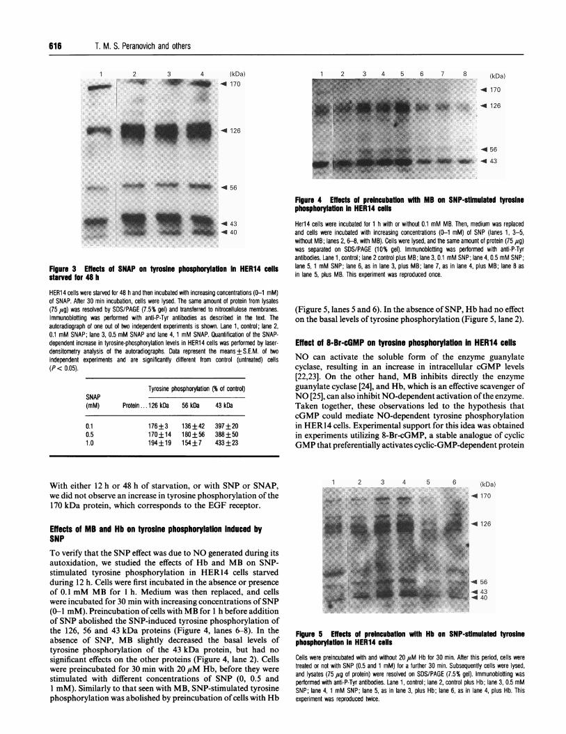
Figure 3 Effects of SNAP on tyrosine phosphorylation in HER14 cellsstarved for 48 h
HER14 cells were starved for 48 h and then incubated with increasing concentrations (0-1 mM)of SNAP. After 30 min incubation, cells were lysed. The same amount of protein from lysates(75 ,ug) was resolved by SDS/PAGE (7.5% gel) and transferred to nitrocellulose membranes.Immunoblotting was performed with anti-P-Tyr antibodies as described in the text. Theautoradiograph of one out of two independent experiments is shown. Lane 1, control; lane 2,0.1 mM SNAP; lane 3, 0.5 mM SNAP and lane 4,1 mM SNAP. Quantification of the SNAP-dependent increase in tyrosine-phosphorylation levels in HER14 cells was performed by laser-densitometry analysis of the autoradiographs. Data represent the means+S.E.M. of twoindependent experiments and are significantly different from control (untreated) cells(P < 0.05).
Tyrosine phosphorylation (% of control)SNAP(mM) Protein ...126 kDa 56 kDa 43 kDa
0.10.51.0
176+3 136+42 397+20170+14 180+56 388+50194+19 154+7 433+23
Figure 4 Effects of preincubation with MB on SNP-stimulated tyrosinephosphorylation In HER14 cells
Her14 cells were incubated for 1 h with or without 0.1 mM MB. Then, medium was replacedand cells were incubated with increasing concentrations (0-1 mM) of SNP (lanes 1, 3-5,without MB; lanes 2, 6-8, with MB). Cells were lysed, and the same amount of protein (75 jug)was separated on SDS/PAGE (10% gel). Immunoblotting was performed with anti-P-Tyrantibodies. Lane 1, control; lane 2 control plus MB; lane 3, 0.1 mM SNP; lane 4, 0.5 mM SNP;lane 5, 1 mM SNP; lane 6, as in lane 3, plus MB; lane 7, as in lane 4, plus MB; lane 8 asin lane 5, plus MB. This experiment was reproduced once.
(Figure 5, lanes 5 and 6). In the absence of SNP, Hb had no effecton the basal levels of tyrosine phosphorylation (Figure 5, lane 2).
Effect of 8-Br-cGMP on tyrosine phosphorylation In HER14 cells
NO can activate the soluble form of the enzyme guanylatecyclase, resulting in an increase in intracellular cGMP levels[22,23]. On the other hand, MB inhibits directly the enzyme
guanylate cyclase [24], and Hb, which is an effective scavenger ofNO [25], can also inhibit NO-dependent activation of the enzyme.Taken together, these observations led to the hypothesis thatcGMP could mediate NO-dependent tyrosine phosphorylationin HER14 cells. Experimental support for this idea was obtainedin experiments utilizing 8-Br-cGMP, a stable analogue of cyclicGMP that preferentially activates cyclic-GMP-dependent protein
With either 12 h or 48 h of starvation, or with SNP or SNAP,we did not observe an increase in tyrosine phosphorylation of the170 kDa protein, which corresponds to the EGF receptor.
Effects of MB and Hb on tyrosine phosphorylation induced bySNP
To verify that the SNP effect was due to NO generated during itsautoxidation, we studied the effects of Hb and MB on SNP-stimulated tyrosine phosphorylation in HER14 cells starvedduring 12 h. Cells were first incubated in the absence or presenceof 0.1 mM MB for 1 h. Medium was then replaced, and cellswere incubated for 30 min with increasing concentrations of SNP(0-1 mM). Preincubation ofcells with MB for 1 h before additionof SNP abolished the SNP-induced tyrosine phosphorylation ofthe 126, 56 and 43 kDa proteins (Figure 4, lanes 6-8). In theabsence of SNP, MB slightly decreased the basal levels oftyrosine phosphorylation of the 43 kDa protein, but had no
significant effects on the other proteins (Figure 4, lane 2). Cellswere preincubated for 30 min with 20 ,uM Hb, before they were
stimulated with different concentrations of SNP (0, 0.5 and1 mM). Similarly to that seen with MB, SNP-stimulated tyrosinephosphorylation was abolished by preincubation of cells with Hb
1 2! 3s 4 b b (kDa).. : . ... ..... .............. ......... .~~~~~~~~~~~~~~~~~~. ... ...........-.-..... ...... , .... . . : .......4 126~~~~~~~~~~~~~~~~~~~~~~~~~~~~~~~~. ... **!:t. . :i:3,....
-4 56
43
-44 40
Figure 5 Effects of preincubation with Hb on SNP-stimulated tyrosine
phosphorylatlon in HER14 cells
Cells were preincubated with and without 20 ,zM Hb for 30 min After this period, cells were
treated or not with SNP (0.5 and 1 mM) for a further 30 min. Subsequently cells were lysed,
and lysates (75 tZg ot protein) were resolved on SDS/PAGE (7.5% gel). Immunoblofting was
performed with anti-P-Tyr antibodies. Lane 1,. control; lane 2, control plus Hb; lane 3, 0.5 mM
SNP; lane 4,1 mM SNP; lane 5, as in lane 3, plus Hb; lane 6, as in lane 4, plus Hb. This
experiment was reproduced twice.
(kDa)4 170
-4 126
8:::. ::
*:
*...0.... ,...
..;.<. 9,-.....
(kDa)
4 170
4 126
4 56.4 43
Nitric oxide and tyrosine phosphorylation 617
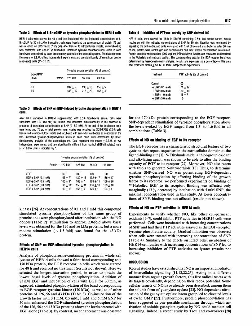
Table 2 Effects of 8-Br-cGMP on tyrosine phosphorylation in HER14 cells Table 4 Inhibition of PTPase acffvity by SNP-derived NOHER14 cells were starved for 48 h and then incubated with the indicated concentrations of 8-Br-cGMP for 30 min. After incubation, cells were lysed and the same amount of protein (75 jug)was resolved on SDS/PAGE (7.5% gel). After transfer to nitrocellulose sheets, immunoblottingwas performed with anti-P-Tyr antibodies. Increased tyrosine-phosphorylation levels in eachband were determined by laser-densitometry analysis of the autoradiographs. The data representthe means + S.E.M. of two independent experiments and are significantly different from control(untreated) cells (P < 0.05).
Tyrosine phosphorylation (% of control)8-Br-cGMP(mM) Protein ... 126 kDa 56 kDa 43 kDa
0.11.0
207+5 195+18 155+5188+12 218+20 158+4
HER14 cells were starved for 48 h in DMEM containing 0.5% fetal-bovine serum, beforeincubation with the indicated concentrations of SNP for 30 min. Reaction was terminated byaspirating the cell media, and cells were lysed with 1 ml of ice-cold lysis buffer A. After 30 minon ice, lysates were centrifuged and supernatants had their protein concentration determined.Protein contents were matched (200 ,ug) and PTP activity in lysates was measured as describedin the Materials and methods section. The corresponding area for the EGF-receptor band wasdetermined by laser-densitometry analysis. Results are expressed as a percentage of this areaand represent means+ S.E.M. of three independent experiments.
Treatment PTP activity (% of control)
Control+SNP (0.1 mM)+SNP (0.5 mM)+ SNP (1.0 mM)+SNP (5.0 mM)
10071 +1750 +1016+70
Table 3 Effects of SNP on EGF-induced tyrosine phosphorylailon In HER14cellsAfter 48 h starvation in DMEM supplemented with 0.5% fetal-bovine serum, cells werestimulated with EGF (83 nM) for 30 min and incubated simultaneously in the absence orpresence of increasing concentrations of SNP (0-5.0 mM). At the end of the incubation, cellswere lysed and 75 ,cg of total protein from lysates was resolved by SDS/PAGE (7.5% gel),transferred to nitrocellulose sheets and incubated with anti-P-Tyr antibodies as described in thetext. Increased tyrosine-phosphorylation levels in each band were determined by laser-densitometry analysis of the autoradiographs. Data represent the means+S.E.M. of twoindependent experiments and are significantly different from control (EGF-stimulated) cells(P < 0.05) unless indicated by *.
Tyrosine phosphorylation (% of control)
Protein ... 170 kDa 126 kDa 56 kDa 43 kDa
EGFEGF+ SNP (0.1 mM)EGF+SNP (0.5 mM)EGF+ SNP (1.0 mM)EGF+ SNP (5.0 mM)
10095 + 7*
105 _3*98 +11*99+13*
100 100 100130+16 132+17 136+17168+7 163+11 158+20153+31 136+10 133+10156+5 125+1 124+1
for the 170 kDa protein corresponding to the EGF receptor.SNP-dependent stimulation of tyrosine phosphorylation abovethe levels evoked by EGF ranged from 1.3- to 1.6-fold in allcombinations (Table 3).
Effects of NO on binding of EGF to Its receptorThe EGF receptor has a characteristic structural feature of twocysteine-rich repeat sequences in the extracellular domain at theligand-binding site [1]. N-Ethylmaleimide, a thiol-group oxidantand alkylating agent, was shown to be able to alter the bindingcapacity of EGF to its receptor [27]. Moreover, NO also reactswith thiols to generate S-nitrosothiols [13]. Thus, to determinewhether SNP-derived NO was potentiating EGF-dependenttyrosine phosphorylation by affecting binding of the growthfactor to its receptor, we performed experiments on binding of'l25-labelled EGF to its receptor. Binding was affected onlymarginally (13 % decrease) by incubation with 5 mM SNP, themaximal concentration used in this study. At lower concentra-tions of SNP, binding was not affected (results not shown).
kinases [26]. At concentrations of 0.1 and 1 mM this compoundstimulated tyrosine phosphorylation of the same group ofproteins that were phosphorylated after incubation with the NOdonors (Table 2): stimulation to approx. 2.0-fold above basallevels was obtained for the 126 and 56 kDa proteins, but a moremodest stimulation ( 1.5-fold) was found for the 43 kDaprotein.
Effects of SNP on EGF-stimulated tyrosine phosphorylation inHER14 cellsAnalysis of phosphotyrosine-containing proteins in whole celllysates of HER14 cells showed a faint band corresponding to a170 kDa protein, the EGF receptor, in cells which were starvedfor 48 h and received no treatment (results not shown). Here weselected the longest starvation period, in order to obtain thelowest basal levels of tyrosine phosphorylation. Addition of83 nM EGF and incubation of cells with EGF for 30 min, asexpected, stimulated phosphorylation of the band correspondingto EGF-receptor tyrosine kinase (170 kDa), as well as of otherproteins of 126, 56 and 43 kDa (Table 3). Co-incubation of thegrowth factor with 0.1 mM, 0.5 mM, 1 mM and 5 mM SNP for30 min enhanced the EGF-stimulated tyrosine phosphorylationof the 126, 56 and 43 kDa proteins above the levels detected withEGF alone (Table 3). By contrast, no enhancement was observed
Effects of NO on PTP activities in HER14 cellsExperiments to verify whether NO, like other cell-permeantoxidants [5-7], could inhibit PTP activities in HER14 cells wereperformed. Cells were incubated with increasing concentrationsof SNP and had their PTP activities assayed as the EGF-receptortyrosine phosphatase activity. Gradual inhibition was observedwhen cells were treated with increasing concentrations of SNP(Table 4). Similarly to the effects on intact cells, incubation ofHER14-cell lysates with increasing concentrations of SNP led toincreasing inhibition of PTP activities on lysates (results notshown).
DISCUSSIONRecent studies have established thatNO is an important mediatorof intercellular signalling [11,12,22,23]. Acting in a differentmanner from regular growth factors, this free radical reacts withits targets covalently, depending on their redox potential. Somecellular targets of NO have already been described, among themthe soluble form of guanylate cyclase [23]. NO-dependent nitro-sation of the guanylate cyclase haem group led to elevated levelsof cyclic GMP [22]. Furthermore, protein phosphorylation hasbeen suggested as one possible mechanism through which ac-tivation of soluble guanylate cyclase by NO could mediate cellsignalling. Indeed, a recent study by Tsou and co-workers [28]

618 T. M. S. Peranovich and others
demonstrated that SNP-derived NO increased the threonine-phosphorylation levels of the dopamine- and cyclic AMP-regulated 32 kDa phosphoprotein from brain tissue. The presentpaper showed for the first time that NO derived from two NO-releasing compounds, SNP and SNAP, stimulated tyrosinephosphorylation levels on a series of endogenous proteins inmurine fibroblasts. Essentially, three proteins, 126, 56 and 43 kDahad their levels of phosphotyrosine increased by the action ofNO. However, stimulation patterns were quite different, depend-ing on the NO donor used. SNAP strongly stimulated tyrosinephosphorylation of the 43 kDa protein (- 4.0-fold), but stimu-lated phosphorylation of the 126 and 56 kDa proteins to a lesserextent. On the other hand, under the same experimental context,SNP was less efficient as a stimulant of tyrosine phosphorylation.Two basic features could account for the observed differences:(a) the amount of NO released by the NO donor in the reactionmedium; (b) the nature of the redox-related forms of NOgenerated after autoxidation of each compound. Under our
experimental conditions, SNAP was a better releaser ofNO thanwas SNP, as showed by the nitrite accumulation in the reactionmedium. However, in any case, stimulation of tyrosinephosphorylation was not directly proportional to the con-
centration of the NO donor used in the experiment. Consideringthe redox forms ofNO released by each compound, SNP donatesprimarily the nitrosonium cation, NO' [18], whereas SNAPdelivers NO as a S-nitrosothiol, RS-NO [21]. Under neutralphysiological conditions the NO' species is very reactive, andwill participate in addition and substitution reactions withnucleophiles, producing nitrosamines, alkyl and aryl nitrites,nitrogen oxides and S-nitrosothiols [18]. On the other hand, S-nitrosothiols are a more stable redox form of NO which can
participate in S-nitrosylation and thiol/disulphide exchangereactions [18], and are the predominant redox form of NO inhuman plasma [29]. Thus SNAP, which is a S-nitrosothiol, coulddeliver this stable redox form of NO into cells more efficientlythan SNP.
Stimulation by NO was obtained in cells which were in eithera quiescent or a non-quiescent stage. Others [5,30] have shownthat oxidants such as vanadate, H202 and sodium selenate were
capable of stimulating tyrosine phosphorylation in cell lineswhich respond to insulin or EGF. Nevertheless, differently fromNO, this stimulation was only achieved in cells which were in a
non-quiescent stage.SNP-evoked tyrosine phosphorylation was abolished by pre-
treatment of the cells either with Hb, a haem protein which is anefficient NO scavenger [25], or with MB, an inhibitor of solubleguanylate cyclase [24]. Abolition by Hb indicates that the SNPeffect is due to NO. Abolition by MB would suggest that theobserved NO-stimulated tyrosine phosphorylation could berelated to guanylate cyclase activation. This hypothesis is sup-
ported by the experiments with the cGMP analogue 8-Br-cGMP,showing that the activation of cGMP-dependent protein kinasesalone is sufficient to produce a pattern of tyrosine phosphoryl-ation similar to that observed when cells were incubated with theNO donors.We also observed that NO can stimulate EGF-induced protein
tyrosine phosphorylation in HER14 cells. Co-incubation of theNO source together with the growth factor led to an enhancementofphosphotyrosine cellular content above the levels observed forincubation with the growth factor alone. NO neither stimulatednor inhibited EGF-dependent receptor autophosphorylation, butenhanced EGF-dependent protein tyrosine phosphorylationtowards endogenous substrates. These observations suggest thatNO could regulate distal components ofthe EGF-initiated signal-transduction pathway. The mechanism of action of NO in this
situation is not understood, but may be related to its reactionwith essential thiol groups in regulatory proteins. Reactions ofNO with thiol groups have been described in the literature[13,18]. However, the absence of significant effects on EGFbinding, which is dependent on cysteine-rich domains, suggeststhat NO would target preferentially intracellular critical thiolgroups. PTP activities, by promoting tyrosine dephosphoryl-ation, are regulatory of growth-factor PTK activity [3,4]. Fur-thermore, PTPs have a conserved cysteine residue in theircatalytic domains, which is essential for their activities (3,4]. Asmentioned above, the NO+ species is released during SNPautoxidation and can be promptly reduced by thiols, generatingS-nitrosothiols [13,18]. Thus NO produced from SNP autoxid-ation possibly inhibits PTP activities through S-nitrosylationreactions with their critical cysteine residue. However, by contrastwith the NO-evoked inhibition of PTP activities, the stimulationof tyrosine phosphorylation was not linearly dependent on theconcentration of SNP. These observations imply that NO couldexert either inhibitory effects towards PTP activities or itsstimulatory effects on tyrosine phosphorylation, independently.The effect of NO on tyrosine phosphorylation and PTP
activities reported here may have some implications on cellproliferation. NO has been reported to inhibit proliferation ofkeratinocytes and smooth-muscle cells, and to be involved inneuronal cell death [31-33]. Here we report that NO is able toinduce tyrosine phosphorylation in a number of endogenousproteins in murine fibroblasts, but not at the EGF-receptor level.These results suggest that NO could exert its anti-proliferativeeffects by subverting the regular growth-factor-associated signal-transduction pathway. EGF otherwise counteracts the NO-derived growth-inhibitory effects [31,33]. Accordingly, we showthat NO in the presence of EGF had no effects on the growth-factor-mediated tyrosine autophosphorylation levels of its re-ceptor, and stimulated tyrosine phosphorylation of other pro-teins. Thus, in the vasculature, the simultaneous presence of NOsynthesized by endothelial cells and of EGF may have con-sequences on the modulation ofsmooth-muscle-cell proliferation.
This work was supported by Fundaao de Amparo a Pesquisa do Estado De SaoPaulo (FAPESP) grants 92/3018-0 to H. P. M. and 90/3875-4 to A. M. S., and N.I.H.grant ES03425 to A.S.
REFERENCESI UJlirich, A. and Schiessinger, J. (1990) Gell 61, 203-2122 Yarden, Y. and Ulirich, A. (1988) Annu. Rev. Biochem. 57, 443-4783 Fischer, E. H., Charbonneau, H. and Tonks, N. K. (1991) Science 253, 401-4064 Walton, K. M. and Dixon, J. E. (1993) Annu. Rev. Biochem. 62, 101-1205 Monteiro, H. P., Ivaschenko, Y., Fischer, R. and Stern, A. (1991) FEBS Lett. 295,
146-1486 Heffetz, D., Bushkin, I., Dror, R. and Zick, Y. (1990) J. Biol. Chem. 265, 2896-29027 Monteiro, H. P., Ivaschenko, Y., Fischer, R. and Stern, A. (1993) Int. J. Biochem. 25,
1859-18648 Chan, T. M., Chen, E., Tatoyan, A., Shargill, N. S., Pleta, M. and Hochstein, P. (1986)
Biochem. Biophys. Res. Commun. 137, 286-2949 Bauskin, A. R., Alkalay, I. and Ben-Neriah, Y. (1991) Cell 66, 685-696
10 Kanner, S. B., Kavanagh, T. J., Grossmann, A., Hu, S. L., Bolen, J. B., Rabinovitch, P.and Ledbetter, J. A. (1992) Proc. Natl. Acad. Sci. U.S.A. 89, 300-304
11 Nathan, C. F. (1992) FASEB J. 6, 3051-306412 Lowenstein, C. J. and Synder, S. H. (1992) Cell 70, 705-70713 Stamler, J. S., Singel, D. J. and Loscalzo, J. (1992) Science 258, 1898-190214 Honegger, A. M., Dull, J. S., Felder, S., van Obberghen, E., Bellot, F., Szapary, D.,
Schmidt, A., Ullrich, A. and Schlessinger, J. (1987) Cell 51,199-20915 Riggs, A. (1981) Methods Enzymol. 76, 5-2816 Bradford, M. M. (1976) Anal. Biochem. 72, 248-25417 Lammli, U. K. (1970) Nature (London) 227, 680-68518 Lax, I., Johnson, A., Howk, R., Sap, J., Bellot, F., Winkler, M., Ulirich, A., Vennstrom,
B., Schessinger, J. and Givol, . (1988) Mo Ll. D-lBiol. 8,1970197819 Stuehr, D. J. and Nathan, C. F. (1989) J. Exp. Med. 169, 1543-1545
Nitric oxide and tyrosine phosphorylation
20 Lipton, S. A., Choi, Y., Pan, Z., Lei, S. Z., Chen, H. V., Sucher, N. J., Loscalzo, J.,Singel, D. J. and Stamler, J. S. (1993) Nature (London) 364, 626-632
21 Ignarro, L. J., Lippton, H., Edwards, J. C., Baricos, W. H., Hyman, A. L., Kadowitz,P. J. and Gruetter, C. A. (1981) J. Pharmacol. Exp. Ther. 218, 739-749
22 Ignarro, L. J. (1990) Annu. Rev. Pharmacol. Toxicol. 30, 535-56023 Wong, S. K. F. and Garbers, D. L. (1992) J. Clin. Invest. 90, 299-30524 Martin, W., Villani, G. M., Jothianandan, D. and Furchgott, R. F. (1985) J. Pharmacol.
Exp. Ther. 232, 708-71625 Ignarro, L. J., Buga, G. M., Wood, K. S., Byrns, R. E. and Chaudhuri, G. (1987) Proc.
Natl. Acad. Sci. U.S.A. 84, 9265-926926 Geiger, J., Nolte, C., Butt, E., Sage, S. 0. and Walter, U. (1992) Proc. Natl. Acad. Sci.
U.S.A. 89, 1031-1035
27 Cassel, D., Pike, L. J., Grant, G. A., Krebs, E. G. and Glaser, L. (1983) J. Biol. Chem.258, 2945-2950
28 Tsou, K., Snyder, G. L. and Greengard, P. (1993) Proc. Natl. Acad. Sci. U.S.A. 90,3462-3465
29 Stamler, J. S., Simon, D. I., Osborne, J. A., Mullins, M. E., Jaraki, O., Michel, T.,Singel, D. J. and Loscalzo, J. (1992) Proc. Natl. Acad. Sci. U.S.A. 89, 444-448
30 Pillay, T. S. and Makgoba, M. W. (1992) FEBS Lett. 308, 38-4231 Heck, D. E., Laskin, D. L., Gardner, C. R. and Laskin, J. D. (1992) J. Biol. Chem.
267, 21 277-21 28032 Garg, U. C. and Hassid, A. (1989) J. Clin. Invest. 83, 1774-177733 Maiese, K., Boniece, I., DeMeo, D. and Wagner, J. A. (1993) J. Neurosci. 13,
3034-3040
619
Received 28 June 1994/13 September 1994; accepted 16 September 1994