Web oriented applications generator development through reengineering process
Upload
independentCategory
view
0download
0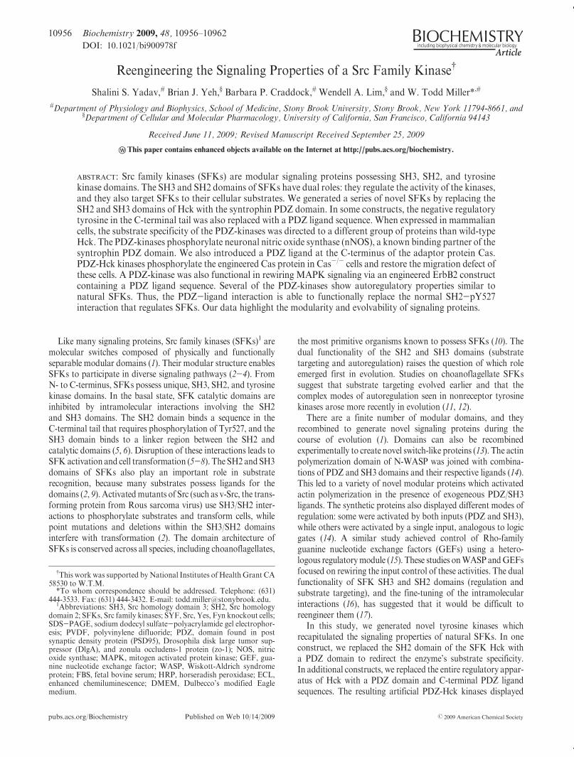
pubs.acs.org/Biochemistry Published on Web 10/14/2009 r 2009 American Chemical Society
10956 Biochemistry 2009, 48, 10956–10962
DOI: 10.1021/bi900978f
Reengineering the Signaling Properties of a Src Family Kinase†
Shalini S. Yadav,# Brian J. Yeh,§ Barbara P. Craddock,# Wendell A. Lim,§ and W. Todd Miller*,#
#Department of Physiology and Biophysics, School of Medicine, Stony Brook University, Stony Brook, New York 11794-8661, and§Department of Cellular and Molecular Pharmacology, University of California, San Francisco, California 94143
Received June 11, 2009; Revised Manuscript Received September 25, 2009
wnThis paper contains enhanced objects available on the Internet at http://pubs.acs.org/biochemistry.
ABSTRACT: Src family kinases (SFKs) are modular signaling proteins possessing SH3, SH2, and tyrosinekinase domains. The SH3 and SH2 domains of SFKs have dual roles: they regulate the activity of the kinases,and they also target SFKs to their cellular substrates. We generated a series of novel SFKs by replacing theSH2 and SH3 domains of Hck with the syntrophin PDZ domain. In some constructs, the negative regulatorytyrosine in the C-terminal tail was also replaced with a PDZ ligand sequence. When expressed in mammaliancells, the substrate specificity of the PDZ-kinases was directed to a different group of proteins than wild-typeHck. The PDZ-kinases phosphorylate neuronal nitric oxide synthase (nNOS), a known binding partner of thesyntrophin PDZ domain. We also introduced a PDZ ligand at the C-terminus of the adaptor protein Cas.PDZ-Hck kinases phosphorylate the engineered Cas protein in Cas-/- cells and restore the migration defect ofthese cells. A PDZ-kinase was also functional in rewiring MAPK signaling via an engineered ErbB2 constructcontaining a PDZ ligand sequence. Several of the PDZ-kinases show autoregulatory properties similar tonatural SFKs. Thus, the PDZ-ligand interaction is able to functionally replace the normal SH2-pY527interaction that regulates SFKs. Our data highlight the modularity and evolvability of signaling proteins.
Like many signaling proteins, Src family kinases (SFKs)1 aremolecular switches composed of physically and functionallyseparable modular domains (1). Their modular structure enablesSFKs to participate in diverse signaling pathways (2-4). FromN- to C-terminus, SFKs possess unique, SH3, SH2, and tyrosinekinase domains. In the basal state, SFK catalytic domains areinhibited by intramolecular interactions involving the SH2and SH3 domains. The SH2 domain binds a sequence in theC-terminal tail that requires phosphorylation of Tyr527, and theSH3 domain binds to a linker region between the SH2 andcatalytic domains (5, 6). Disruption of these interactions leads toSFK activation and cell transformation (5-8). The SH2 and SH3domains of SFKs also play an important role in substraterecognition, because many substrates possess ligands for thedomains (2, 9). Activatedmutants of Src (such as v-Src, the trans-forming protein from Rous sarcoma virus) use SH3/SH2 inter-actions to phosphorylate substrates and transform cells, whilepoint mutations and deletions within the SH3/SH2 domainsinterfere with transformation (2). The domain architecture ofSFKs is conserved across all species, including choanoflagellates,
the most primitive organisms known to possess SFKs (10). Thedual functionality of the SH2 and SH3 domains (substratetargeting and autoregulation) raises the question of which roleemerged first in evolution. Studies on choanoflagellate SFKssuggest that substrate targeting evolved earlier and that thecomplex modes of autoregulation seen in nonreceptor tyrosinekinases arose more recently in evolution (11, 12).
There are a finite number of modular domains, and theyrecombined to generate novel signaling proteins during thecourse of evolution (1). Domains can also be recombinedexperimentally to create novel switch-like proteins (13). The actinpolymerization domain of N-WASP was joined with combina-tions of PDZ and SH3 domains and their respective ligands (14).This led to a variety of novel modular proteins which activatedactin polymerization in the presence of exogeneous PDZ/SH3ligands. The synthetic proteins also displayed different modes ofregulation: some were activated by both inputs (PDZ and SH3),while others were activated by a single input, analogous to logicgates (14). A similar study achieved control of Rho-familyguanine nucleotide exchange factors (GEFs) using a hetero-logous regulatorymodule (15). These studies onWASP andGEFsfocused on rewiring the input control of these activities. The dualfunctionality of SFK SH3 and SH2 domains (regulation andsubstrate targeting), and the fine-tuning of the intramolecularinteractions (16), has suggested that it would be difficult toreengineer them (17).
In this study, we generated novel tyrosine kinases whichrecapitulated the signaling properties of natural SFKs. In oneconstruct, we replaced the SH2 domain of the SFK Hck witha PDZ domain to redirect the enzyme’s substrate specificity.In additional constructs, we replaced the entire regulatory appar-atus of Hck with a PDZ domain and C-terminal PDZ ligandsequences. The resulting artificial PDZ-Hck kinases displayed
†This work was supported by National Institutes of Health Grant CA58530 to W.T.M.*To whom correspondence should be addressed. Telephone: (631)
444-3533. Fax: (631) 444-3432. E-mail: [email protected]: SH3, Src homology domain 3; SH2, Src homology
domain 2; SFKs, Src family kinases; SYF, Src, Yes, Fyn knockout cells;SDS-PAGE, sodium dodecyl sulfate-polyacrylamide gel electrophor-esis; PVDF, polyvinylene difluoride; PDZ, domain found in postsynaptic density protein (PSD95), Drosophila disk large tumor sup-pressor (DlgA), and zonula occludens-1 protein (zo-1); NOS, nitricoxide synthase; MAPK, mitogen activated protein kinase; GEF, gua-nine nucleotide exchange factor; WASP, Wiskott-Aldrich syndromeprotein; FBS, fetal bovine serum; HRP, horseradish peroxidase; ECL,enhanced chemiluminescence; DMEM, Dulbecco’s modified Eaglemedium.
Article Biochemistry, Vol. 48, No. 46, 2009 10957
three salient features of modular signaling proteins: (i) theirsubstrate specificity was governed by the PDZ domain; (ii) theydisplayed autoregulatory properties similar to natural SFKs; and(iii) they were versatile and could be used to rewire two separatesignaling pathways. Our data highlight the modularity andevolvability of signaling proteins and suggest that the targetingfunction of modular domains is most amenable to manipulation.
MATERIALS AND METHODS
Reagents and Antibodies. DMEM, trypsin-EDTA, penicil-lin, streptomycin, and amphotericin B were purchased fromGIBCO (Cellgro). FBS, Polybrene, anti-Flag HRP, and anti-tubulin antibodies were fromSigma.CasC-20 antibodywas fromSanta Cruz Biotech (Santa Cruz, CA), and Cas monoclonalantibody was from BD Biosciences (San Jose, CA). Anti-phosphotyrosine 4G10 mouse monoclonal and ErbB2 antibodywas from Millipore, and anti-pY416 antibody was from Bio-source. The ErbB2 hybridoma 4D10 was a kind gift fromDr. Deborah Brown (Stony Brook University). Erk and p-Erkantibodies were from Cell Signaling Technology. HRP conju-gated secondary antimouse and antirabbit antibodies, and ECLand ECLþ kits were from Amersham.Plasmid Construction. For mammalian expression, all con-
structs were subcloned into pCMV-Tag2B (Stratagene) betweenthe EcoRI and XhoI sites. The neuronal nitric oxide synthase(nNOS) construct was subcloned into pXJ-HA between theBamHI and XhoI sites. For retroviral expression, PDZ-kinaseconstructs were subcloned into the EcoRI and XhoI sites ofpMSCV-IRES-GFP (a kind gift from Dr. Nicholas Carpino,Stony Brook University). To generate Cas-VKESLV, site-direc-ted mutagenesis was performed on wild-type Cas cloned intopCDNA6. Cas-VKESLV was further subjected to site-directedmutagenesis to delete the Src binding sequence of Cas. Toproduce the kinase-dead ErbB2 construct with a C-terminalPDZ ligand, pCDNA3-kdErbB2 (Dr. Len Neckers, NIH) wassubcloned into pBS-SK(() and site-directed mutagenesis wasperformed. The kdErbB2-VKESLV was subcloned back intopCDNA3 for mammalian expression. All constructs were con-firmed by restriction digestion and sequencing.Cell Culture and Transient Transfection. SYF cells were
purchased from ATCC (Manassas, VA) and gradually adaptedfor maintenance in DMEM containing 10% FBS and 1�antibiotic/antimycotic at 37 �C in a humidified 5% CO2 incu-bator. Cells were transiently transfected using TransIT LT1(Mirus) transfection reagent following the manufacturer’s re-commendations. Typically, 1-10 mg of DNA was transfectedusing a DNA:TransIT ratio of 1:2.Western Blotting and Immunoprecipitation. Cells were
transiently transfected and harvested 40 h post transfectionin RIPA buffer containing 1 mM sodium orthovanadate and10 mM sodium fluoride. Protein concentration was estimatedusing the Bradford assay (Bio-Rad). For analysis of whole celllysates, lysates (10-50 μg) were separated on 10% SDS-PAGEand transferred onto PVDF membranes. The membranes wereprobed with anti-Flag, anti-pY416, anti-tubulin and 4G10 anti-bodies. For immunoprecipitation, lysates (500 μg) were pre-cleared for 1 h at 4 �C, followed by incubation with antibody(1-2 μg) for 2 h or overnight at 4 �C on a rotator. Immunopre-cipitates were washed three times with buffer, and proteins wereeluted in Laemmli buffer, separated by SDS-PAGE, transferredto PVDF, and analyzed by Western blotting as shown in thefigures. Quantificationwas carried out with the LI-COROdyssey
Quantification software package (version 2.0). All Western blotexperiments were performed at least three times, and typicalresults are shown.Retrovirus Generation and Infection. Retroviruses were
derived essentially as described (18, 19). Briefly, Phoenix retro-virus producer cell lines were obtained from Dr. NicholasCarpino (Stony Brook) andweremaintained inDMEMcontain-ing 10% FBS and 1� antibiotic/antimycotic solution. Cells weretransiently transfected with 10 μg of the retroviral constructsalong with 10 μg helper plasmid. Retroviruses were collected at32 �C for 2 days at 12 h intervals. Aliquots were frozen at-80 �C.Cas-/- cells were transiently transfected with Cas-VKESLV andselected with 5 μg/mL blasticidin (Sigma) for one week. Forinfection, 2 � 105 Cas-/- cells stably expressing Cas-VKESLVwere plated in a 6well plate and spin infectedwith the retrovirusesalong with Polybrene (4 μg/mL). Two days postinfection >90%cells were GFP positive. The cells were harvested and used forwound healing assays.Migration and Wound Healing Assays. For migration
assays, Cas-/- cells were transiently transfected with Cas-VKESLV along with PDZ-kinase or Hck. The cells wereharvested 24 h post transfection and 2 � 105 cells were placedin a 12 well transwell migration assay plate. Cells were allowed tomigrate for 12 h in a 5% CO2, humidified incubator maintainedat 37 �C. Cells were washedwith PBS, harvested from the bottomof the transwell insert and the bottomof the well in a final volumeof 100 μL of DMEM, and counted using a hemocytometer. Eachcondition was processed in triplicate, and the percentage migra-tion was calculated using the formula:
%migration ¼ ½ðnumber of cells migratedÞ=ðtotal number of cellsÞ� � 100
Wound healing assays were performed essentially as des-cribed (20). Retrovirally infected Cas-/- cells were plated on60 mm dishes and grown to confluency. The plates were washedwith 1� PBS and multiple wounds were scratched using a P200pipet tip across the plate. Phase contrast images of wounds wereimaged at 10min intervals for 10-12 h using aCarl Zeiss invertedmicroscope with 20� objective.
RESULTS
Domain Architecture of Novel Kinases. We generated apanel of novel kinases in which the SH2 and SH3 domains of theSFK Hck were replaced by the syntrophin PDZ domain. ThePDZ domain was chosen because it is similar in size to the SH2domain and because it binds to ligands at the C-termini of targetproteins. Thus, it could potentially recapitulate both functions ofthe SH2 domain (substrate targeting and pY527 tail binding).Construct ZK represents the most basic PDZ-kinase, with onlythe substrate targeting function intact. In constructs ZKVweak
and ZKVstrong, the SH2 domain of Hckwas replaced by the PDZdomain, and the C-terminal pTyr sequence that normally inter-acts with the SH2 domain of Hck was replaced by a PDZ ligand(Figure 1). Construct ZKVstrong contains the high-affinity PDZligand sequence VKESLV, which binds to the syntrophin PDZdomain with a Kd of 8 μM (21). ZKVweak contains the sequenceVKQSLL, which binds with a Kd of 100 μM (14). These valuesare comparable to the Kd for the Src SH2 domain binding tothe relatively low-affinity pY527 tail sequence (29 μM) (22). Thelength and sequence of the linker between PDZ and kinasedomains were designed to provide sufficient flexibility for an
10958 Biochemistry, Vol. 48, No. 46, 2009 Yadav et al.
intramolecular interaction between PDZ and ligand to occur.In construct 3ZKV, the SH3 domain was retained, while theSH2-pY527 interaction was replaced by the nonnative PDZ-ligand pair. Because 3ZKV contained the polyproline linkersequence of WT Hck between the PDZ and catalytic domains,this kinase potentially has two autoinhibitory interactions, onenatural (via the SH3 domain) and one artificial (via the PDZdomain). We also generated the control construct KV, whichpossesses a kinase domain and C-terminal PDZ ligand sequencebut lacks the PDZ domain (Figure 1). All kinase constructscontained an N-terminal FLAG tag, and as a consequence theylacked the normal SFK N-terminal fatty acylation motif. Thus,the targeting signal for membrane-associated substrates was alsoremoved in the PDZ-kinases.PDZDomainRedirects Specificity ofHck.We first tested
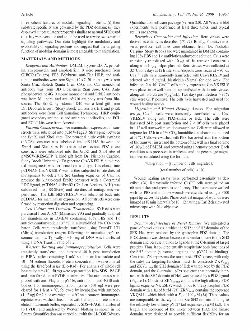
whether the presence of the PDZ domain altered Hck substratespecificity. To examine phosphorylation of cellular proteins, weexpressed the PDZ-kinases ZKVstrong and ZK in Src/Yes/Fyntriple-knockout (SYF) cells (23) and measured global tyrosinephosphorylation by antiphosphotyrosine immunoblotting oflysates. The pattern of cellular phosphoproteins was markedlydifferent when the PDZ domain was attached to the catalyticdomain, as compared to WT Hck or the KV control (Figure 2).A smaller number of proteins are phosphorylated by the KVcontrol than the PDZ-containing kinases ZKVstrong and ZK,consistent with the role of associated domains in directing SFKsubstrate recognition. We also compared the PDZ-containingkinases to an activated (tail tyrosine mutated) form of Hck(Supplemental Figure 1, Supporting Information). Some cellularproteins appeared to be phosphorylated both byYF-Hck and the
PDZ-kinases, but there were clear differences in the lowermolecular weight range. The data suggest that the PDZ domainretargeted Hck to an alternative group of substrates. We alsoobserved that the ZKVstrong and 3ZKV constructs, whichpossessed C-terminal PDZ ligands, were less active than theZK construct lacking the ligand (Figure 2 and SupplementalFigure 1, Supporting Information), raising the possibility that theengineered PDZ-ligand interaction was functioning to represskinase activity (see below).Syntrophin PDZ Domain Directs Phosphorylation of
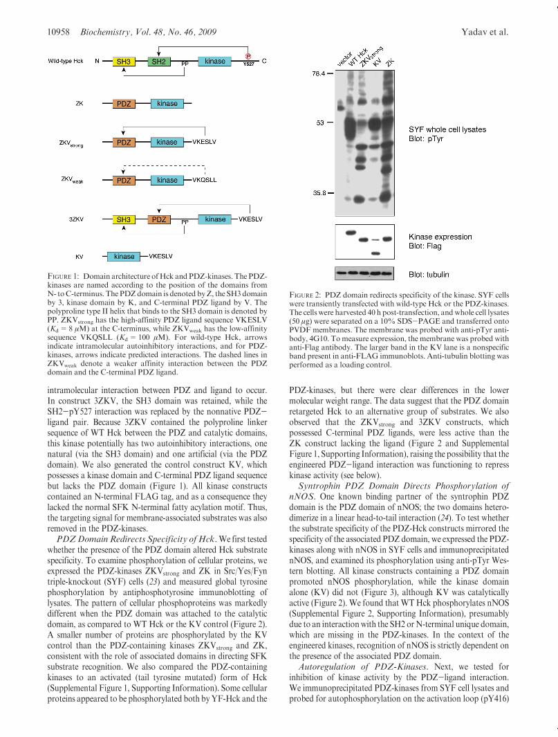
nNOS. One known binding partner of the syntrophin PDZdomain is the PDZ domain of nNOS; the two domains hetero-dimerize in a linear head-to-tail interaction (24). To test whetherthe substrate specificity of the PDZ-Hck constructs mirrored thespecificity of the associated PDZ domain, we expressed the PDZ-kinases along with nNOS in SYF cells and immunoprecipitatednNOS, and examined its phosphorylation using anti-pTyr Wes-tern blotting. All kinase constructs containing a PDZ domainpromoted nNOS phosphorylation, while the kinase domainalone (KV) did not (Figure 3), although KV was catalyticallyactive (Figure 2). We found that WTHck phosphorylates nNOS(Supplemental Figure 2, Supporting Information), presumablydue to an interactionwith the SH2 orN-terminal unique domain,which are missing in the PDZ-kinases. In the context of theengineered kinases, recognition of nNOS is strictly dependent onthe presence of the associated PDZ domain.Autoregulation of PDZ-Kinases. Next, we tested for
inhibition of kinase activity by the PDZ-ligand interaction.We immunoprecipitated PDZ-kinases from SYF cell lysates andprobed for autophosphorylation on the activation loop (pY416)
FIGURE 1: Domain architecture ofHck and PDZ-kinases. The PDZ-kinases are named according to the position of the domains fromN- toC-terminus. The PDZdomain is denoted byZ, the SH3domainby 3, kinase domain by K, and C-terminal PDZ ligand by V. Thepolyproline type II helix that binds to the SH3 domain is denoted byPP. ZKVstrong has the high-affinity PDZ ligand sequence VKESLV(Kd=8 μM) at the C-terminus, while ZKVweak has the low-affinitysequence VKQSLL (Kd=100 μM). For wild-type Hck, arrowsindicate intramolecular autoinhibitory interactions, and for PDZ-kinases, arrows indicate predicted interactions. The dashed lines inZKVweak denote a weaker affinity interaction between the PDZdomain and the C-terminal PDZ ligand.
FIGURE 2: PDZ domain redirects specificity of the kinase. SYF cellswere transiently transfected with wild-type Hck or the PDZ-kinases.The cells were harvested 40 h post-transfection, andwhole cell lysates(50 μg) were separated on a 10% SDS-PAGE and transferred ontoPVDF membranes. The membrane was probed with anti-pTyr anti-body, 4G10. Tomeasure expression, the membrane was probed withanti-Flag antibody. The larger band in the KV lane is a nonspecificband present in anti-FLAG immunoblots. Anti-tubulin blotting wasperformed as a loading control.
Article Biochemistry, Vol. 48, No. 46, 2009 10959
as a measure of kinase activity. Construct ZK (lacking a C-terminal PDZ ligand) displayed strong activity, and ZKVweak
was comparable to ZK (Figure 4). ZKVstrong and 3ZKV, withhigh-affinity PDZ ligands at the C-terminus, were inhibitedrelative to ZK. Thus, the degree of regulation correlatedwith the strength of the autoinhibitory interaction. The auto-phosphorylation of 3ZKV was comparable to WT Hck(Figure 4), suggesting that transplantation of the PDZ ligandcan yield a kinase with similar autoregulatory properties asWT Hck.PDZ-Kinase Leads to Phosphorylation of EngineeredCas.
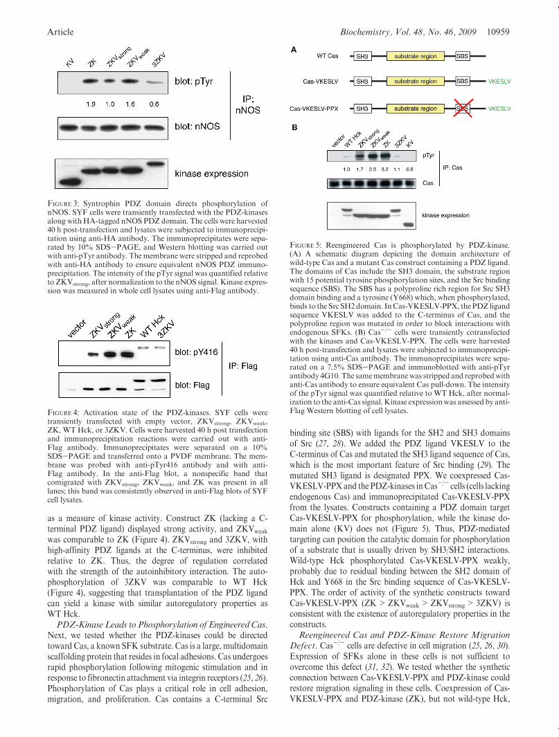
Next, we tested whether the PDZ-kinases could be directedtoward Cas, a known SFK substrate. Cas is a large, multidomainscaffolding protein that resides in focal adhesions. Cas undergoesrapid phosphorylation following mitogenic stimulation and inresponse to fibronectin attachment via integrin receptors (25, 26).Phosphorylation of Cas plays a critical role in cell adhesion,migration, and proliferation. Cas contains a C-terminal Src
binding site (SBS) with ligands for the SH2 and SH3 domainsof Src (27, 28). We added the PDZ ligand VKESLV to theC-terminus of Cas and mutated the SH3 ligand sequence of Cas,which is the most important feature of Src binding (29). Themutated SH3 ligand is designated PPX. We coexpressed Cas-VKESLV-PPXand the PDZ-kinases inCas-/- cells (cells lackingendogenous Cas) and immunoprecipitated Cas-VKESLV-PPXfrom the lysates. Constructs containing a PDZ domain targetCas-VKESLV-PPX for phosphorylation, while the kinase do-main alone (KV) does not (Figure 5). Thus, PDZ-mediatedtargeting can position the catalytic domain for phosphorylationof a substrate that is usually driven by SH3/SH2 interactions.Wild-type Hck phosphorylated Cas-VKESLV-PPX weakly,probably due to residual binding between the SH2 domain ofHck and Y668 in the Src binding sequence of Cas-VKESLV-PPX. The order of activity of the synthetic constructs towardCas-VKESLV-PPX (ZK>ZKVweak>ZKVstrong> 3ZKV) isconsistent with the existence of autoregulatory properties in theconstructs.Reengineered Cas and PDZ-Kinase Restore Migration
Defect. Cas-/- cells are defective in cell migration (25, 26, 30).Expression of SFKs alone in these cells is not sufficient toovercome this defect (31, 32). We tested whether the syntheticconnection between Cas-VKESLV-PPX and PDZ-kinase couldrestore migration signaling in these cells. Coexpression of Cas-VKESLV-PPX and PDZ-kinase (ZK), but not wild-type Hck,
FIGURE 3: Syntrophin PDZ domain directs phosphorylation ofnNOS. SYF cells were transiently transfected with the PDZ-kinasesalong withHA-tagged nNOS PDZ domain. The cells were harvested40 h post-transfection and lysates were subjected to immunoprecipi-tation using anti-HA antibody. The immunoprecipitates were sepa-rated by 10% SDS-PAGE, and Western blotting was carried outwith anti-pTyr antibody. Themembrane were stripped and reprobedwith anti-HA antibody to ensure equivalent nNOS PDZ immuno-precipitation. The intensity of the pTyr signal was quantified relativeto ZKVstrong, after normalization to the nNOS signal. Kinase expres-sion was measured in whole cell lysates using anti-Flag antibody.
FIGURE 4: Activation state of the PDZ-kinases. SYF cells weretransiently transfected with empty vector, ZKVstrong, ZKVweak,ZK, WTHck, or 3ZKV. Cells were harvested 40 h post transfectionand immunoprecipitation reactions were carried out with anti-Flag antibody. Immunoprecipitates were separated on a 10%SDS-PAGE and transferred onto a PVDF membrane. The mem-brane was probed with anti-pTyr416 antibody and with anti-Flag antibody. In the anti-Flag blot, a nonspecific band thatcomigrated with ZKVstrong, ZKVweak, and ZK was present in alllanes; this band was consistently observed in anti-Flag blots of SYFcell lysates.
FIGURE 5: Reengineered Cas is phosphorylated by PDZ-kinase.(A) A schematic diagram depicting the domain architecture ofwild-type Cas and a mutant Cas construct containing a PDZ ligand.The domains of Cas include the SH3 domain, the substrate regionwith 15 potential tyrosine phosphorylation sites, and the Src bindingsequence (SBS). The SBS has a polyproline rich region for Src SH3domain binding and a tyrosine (Y668) which, when phosphorylated,binds to the Src SH2domain. InCas-VKESLV-PPX, the PDZ ligandsequence VKESLV was added to the C-terminus of Cas, and thepolyproline region was mutated in order to block interactions withendogenous SFKs. (B) Cas-/- cells were transiently cotransfectedwith the kinases and Cas-VKESLV-PPX. The cells were harvested40 h post-transfection and lysates were subjected to immunoprecipi-tation using anti-Cas antibody. The immunoprecipitates were sepa-rated on a 7.5% SDS-PAGE and immunoblotted with anti-pTyrantibody 4G10. The samemembranewas stripped and reprobedwithanti-Cas antibody to ensure equivalent Cas pull-down. The intensityof the pTyr signal was quantified relative to WT Hck, after normal-ization to the anti-Cas signal.Kinase expressionwas assessed by anti-Flag Western blotting of cell lysates.
10960 Biochemistry, Vol. 48, No. 46, 2009 Yadav et al.
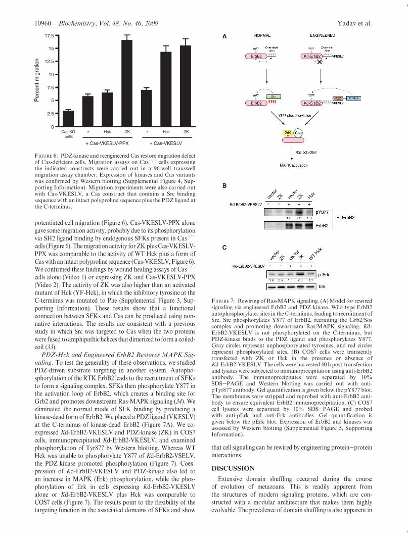
potentiated cell migration (Figure 6). Cas-VKESLV-PPX alonegave somemigration activity, probably due to its phosphorylationvia SH2 ligand binding by endogenous SFKs present in Cas-/-
cells (Figure 6). Themigration activity for ZKplus Cas-VKESLV-PPX was comparable to the activity of WT Hck plus a form ofCaswith an intact polyproline sequence (Cas-VKESLV,Figure 6).We confirmed these findings by wound healing assays of Cas-/-
cells alone (Video 1) or expressing ZK and Cas-VKESLV-PPX(Video 2). The activity of ZK was also higher than an activatedmutant of Hck (YF-Hck), in which the inhibitory tyrosine at theC-terminus was mutated to Phe (Supplemental Figure 3, Sup-porting Information). These results show that a functionalconnection between SFKs and Cas can be produced using non-native interactions. The results are consistent with a previousstudy in which Src was targeted to Cas when the two proteinswere fused to amphipathic helices that dimerized to forma coiled-coil (33).PDZ-Hck and Engineered ErbB2 Restores MAPK Sig-
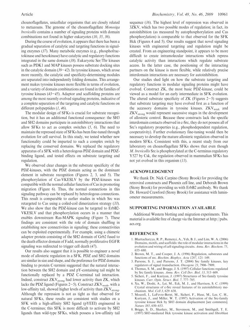
naling. To test the generality of these observations, we studiedPDZ-driven substrate targeting in another system. Autopho-sphorylation of the RTKErbB2 leads to the recruitment of SFKsto form a signaling complex. SFKs then phosphorylate Y877 inthe activation loop of ErbB2, which creates a binding site forGrb2 and promotes downstream Ras-MAPK signaling (34). Weeliminated the normal mode of SFK binding by producing akinase-dead form of ErbB2.We placed a PDZ ligand (VKESLV)at the C-terminus of kinase-dead ErbB2 (Figure 7A). We co-expressed Kd-ErbB2-VKESLV and PDZ-kinase (ZK) in COS7cells, immunoprecipitated Kd-ErbB2-VKESLV, and examinedphosphorylation of Tyr877 by Western blotting. Whereas WTHck was unable to phosphorylate Y877 of Kd-ErbB2-VSELV,the PDZ-kinase promoted phosphorylation (Figure 7). Coex-pression of Kd-ErbB2-VKESLV and PDZ-kinase also led toan increase in MAPK (Erk) phosphorylation, while the phos-phorylation of Erk in cells expressing Kd-ErbB2-VKESLValone or Kd-ErbB2-VKESLV plus Hck was comparable toCOS7 cells (Figure 7). The results point to the flexibility of thetargeting function in the associated domains of SFKs and show
that cell signaling can be rewired by engineering protein-proteininteractions.
DISCUSSION
Extensive domain shuffling occurred during the courseof evolution of metazoans. This is readily apparent fromthe structures of modern signaling proteins, which are con-structed with a modular architecture that makes them highlyevolvable. The prevalence of domain shuffling is also apparent in
FIGURE 6: PDZ-kinase and reengineered Cas restore migration defectof Cas-deficient cells. Migration assays on Cas-/- cells expressingthe indicated constructs were carried out in a 96-well transwellmigration assay chamber. Expression of kinases and Cas variantswas confirmed by Western blotting (Supplemental Figure 4, Sup-porting Information). Migration experiments were also carried outwith Cas-VKESLV, a Cas construct that contains a Src bindingsequence with an intact polyproline sequence plus the PDZ ligand atthe C-terminus.
FIGURE 7: Rewiring ofRas-MAPK signaling. (A)Model for rewiredsignaling via engineered ErbB2 and PDZ-kinase. Wild-type ErbB2autophosphorylates sites in the C-terminus, leading to recruitment ofSrc. Src phosphorylates Y877 of ErbB2, recruiting the Grb2/Soscomplex and promoting downstream Ras/MAPK signaling. Kd-ErbB2-VKESLV is not phosphorylated on the C-terminus, butPDZ-kinase binds to the PDZ ligand and phosphorylates Y877.Gray circles represent unphosphorylated tyrosines, and red circlesrepresent phosphorylated sites. (B) COS7 cells were transientlytransfected with ZK or Hck in the presence or absence ofKd-ErbB2-VKESLV. The cells were harvested 40 h post-transfectionand lysates were subjected to immunoprecipitation using anti-ErbB2antibody. The immunoprecipitates were separated by 10%SDS-PAGE and Western blotting was carried out with anti-pTyr877 antibody. Gel quantification is given below the pY877 blot.The membranes were stripped and reprobed with anti-ErbB2 anti-body to ensure equivalent ErbB2 immunoprecipitation. (C) COS7cell lysates were separated by 10% SDS-PAGE and probedwith anti-pErk and anti-Erk antibodies. Gel quantification isgiven below the pErk blot. Expression of ErbB2 and kinases wasassessed by Western blotting (Supplemental Figure 5, SupportingInformation).
Article Biochemistry, Vol. 48, No. 46, 2009 10961
choanoflagellates, unicellular organisms that are closely relatedto metazoans. The genome of the choanoflagellate Monosigabrevicollis contains a number of signaling proteins with domaincombinations not found in higher eukaryotes (10, 35, 36).
During the course of evolution, it appears that there has been agradual separation of catalytic and targeting functions in signal-ing enzymes (37). Many metabolic enzymes (e.g., phosphofruc-tokinase and hexokinase) have catalytic and regulatory functionsintegrated in the same domain (38). Eukaryotic Ser/Thr kinasessuch as PDK1 and MAP kinases possess substrate docking sitesin the catalytic domain (39-42). In tyrosine kinases, which arosemore recently, the catalytic and specificity-determining modulesare separated into independently folding domains. This arrange-ment makes tyrosine kinases more flexible in terms of evolution,and a variety of domain combinations are found in the families oftyrosine kinases (43-45). Adaptor and scaffolding proteins areamong the most recently evolved signaling proteins, indicative ofa complete separation of the targeting and catalytic functions ondifferent polypeptides (1, 46).
The modular design of SFKs is crucial for substrate recogni-tion, but it has an additional functional consequence: the SH3and SH2 domains participate in autoinhibitory interactions thatallow SFKs to act as complex switches (5, 6). The need tomaintain the repressed state of SFKs has been fine-tuned throughevolution for cell survival. In this study, we tested whether newfunctionality could be imparted to such a complex switch byreplacing the conserved domains. We replaced the regulatoryapparatus of a SFK with a heterologous PDZ domain and PDZ-binding ligand, and tested effects on substrate targeting andregulation.
We observed clear changes in the substrate specificity of thePDZ-kinases, with the PDZ domain acting as the dominantelement in substrate recognition (Figures 2, 3, and 5). Thephosphorylation of Cas-VKESLV by the PDZ-kinases wascompatible with the normal cellular function of Cas in promotingmigration (Figure 6). Thus, the normal connections in thissignaling pathway can be replaced by heterologous connections.This result is comparable to earlier studies in which Src wasretargeted to Cas using a coiled-coil dimerization strategy (33).We also show that the PDZ-kinase can be targeted to ErbB2-VKESLV and that phosphorylation occurs in a manner thatenables downstream Ras-MAPK signaling (Figure 7). Thesefindings are consistent with the role of domain shuffling inestablishing new connectivities in signaling; these connectivitiescan be exploited experimentally. For example, using a chimericadaptor protein consisting of the SH2 domain of Grb2 fused tothe death effector domain of Fadd, normally proliferative EGFRsignaling was redirected to trigger cell death (47).
Our results also suggest that it is possible to engineer a novelmode of allosteric regulation in a SFK. PDZ and SH2 domainsare similar in size and shape, and the preference for PDZdomainsbinding to protein C-termini suggested that the natural interac-tion between the SH2 domain and pY-containing tail might befunctionally replaced by a PDZ C-terminal tail interaction.Indeed, construct ZKVstrong had lower activity than ZK, whichlacks the PDZ ligand (Figures 2-5). Construct ZKVweak, with alow-affinity tail, showed higher levels of activity than ZKVstrong.Although the repression is not as tight as that observed fornatural SFKs, these results are consistent with studies on aSFK with a high-affinity SH2 ligand (pYEEI) engineered inthe C-terminus; this SFK is more difficult to activate by SH2ligands than wild-type SFKs, which possess a low-affinity tail
sequence (16). The highest level of repression was observed in3ZKV, which has two possible modes of regulation; in fact, itsautoinhibition (as measured by autophosphorylation and Casphosphorylation) is comparable to that observed for the SFKHck (Figures 4 and 5). Our results suggest that novel signalingkinases with engineered targeting and regulation might becreated. From an engineering standpoint, it appears to be moredifficult to create intramolecular interactions which represscatalytic activity than interactions which regulate substrateaccess. In the latter case, the positioning of the interactingpartners on the kinase is more flexible, while relatively preciseinterdomain interactions are necessary for autoinhibition.
Our studies shed light on how the substrate targeting andregulatory functions in modular signaling proteins might haveevolved. Construct ZK, the most basic PDZ-kinase, could beviewed as a model for an early intermediate in SFK evolution.The altered substrate specificity of ZK (Figures 2-5) suggeststhat substrate targeting may have evolved first as a function ofthe accessory domains in tyrosine kinases. ZKVweak andZKVstrong would represent successive stages in the developmentof allosteric control. Because these constructs lack the specificinterdomain contacts observed in c-Src, they do not possess all ofSrc’s regulatory properties (e.g., phosphodependent regulation,cooperativity). Further evolutionary fine-tuning would then benecessary to develop the mature allosteric regulation observed inmodern SFKs. Consistent with this, a recent study from ourlaboratory on choanoflagellate SFKs shows that even thoughM. brevicollis Src is phosphorylated at the C-terminus regulatoryY527 by Csk, the regulation observed in mammalian SFKs hasnot yet evolved in this organism (11).
ACKNOWLEDGMENT
We thank Dr. Nick Carpino (Stony Brook) for providing theretroviral constructs and Phoenix cell line, and Deborah Brown(Stony Brook) for providing us with ErbB2 antibody. We thankDr. Howard Crawford (Stony Brook) for assistance with lumin-ometer measurements.
SUPPORTING INFORMATION AVAILABLE
Additional Western blotting and migration experiments. Thismaterial is available free of charge via the Internet at http://pubs.acs.org.
REFERENCES
1. Bhattacharyya, R. P., Remenyi, A., Yeh, B. J., and Lim,W. A. (2006)Domains,motifs, and scaffolds: the role ofmodular interactions in theevolution and wiring of cell signaling circuits.Annu. Rev. Biochem. 75,655–680.
2. Brown, M. T., and Cooper, J. A. (1996) Regulation, substrates andfunctions of src. Biochim. Biophys. Acta 1287, 121–149.
3. Parsons, S. J., and Parsons, J. T. (2004) Src family kinases, keyregulators of signal transduction. Oncogene 23, 7906–7909.
4. Thomas, S. M., and Brugge, J. S. (1997) Cellular functions regulatedby Src family kinases. Annu. Rev. Cell Dev. Biol. 13, 513–609.
5. Sicheri, F., and Kuriyan, J. (1997) Structures of Src-family tyrosinekinases. Curr. Opin. Struct. Biol. 7, 777–785.
6. Xu, W., Doshi, A., Lei, M., Eck, M. J., and Harrison, S. C. (1999)Crystal structures of c-Src reveal features of its autoinhibitory me-chanism. Mol. Cell 3, 629–638.
7. Moarefi, I., LaFevre-Bernt, M., Sicheri, F., Huse, M., Lee, C. H.,Kuriyan, J., and Miller, W. T. (1997) Activation of the Src-familytyrosine kinase Hck by SH3 domain displacement [see comments].Nature 385, 650–653.
8. Briggs, S. D., Sharkey, M., Stevenson, M., and Smithgall, T. E.(1997) SH3-mediated Hck tyrosine kinase activation and fibroblast
10962 Biochemistry, Vol. 48, No. 46, 2009 Yadav et al.
transformation by the Nef protein of HIV-1. J. Biol. Chem. 272,17899–17902.
9. Miller, W. T. (2003) Determinants of substrate recognition in non-receptor tyrosine kinases. Acc. Chem. Res. 36, 393–400.
10. King, N., Westbrook, M. J., Young, S. L., Kuo, A., Abedin, M.,Chapman, J., Fairclough, S., Hellsten, U., Isogai, Y., Letunic, I.,Marr, M., Pincus, D., Putnam, N., Rokas, A., Wright, K. J., Zuzow,R., Dirks, W., Good, M., Goodstein, D., Lemons, D., Li, W., Lyons,J. B.,Morris, A., Nichols, S., Richter, D. J., Salamov, A., Sequencing,J. G., Bork, P., Lim, W. A., Manning, G., Miller, W. T., McGinnis,W., Shapiro, H., Tjian, R., Grigoriev, I. V., and Rokhsar, D. (2008)The genome of the choanoflagellate Monosiga brevicollis and theorigin of metazoans. Nature 451, 783–788.
11. Li, W., Young, S. L., King, N., and Miller, W. T. (2008) Signalingproperties of a non-metazoan Src kinase and the evolutionary historyof Src negative regulation. J. Biol. Chem. 283, 15491–15501.
12. Segawa, Y., Suga, H., Iwabe, N., Oneyama, C., Akagi, T.,Miyata, T.,andOkada,M. (2006) Functional development of Src tyrosine kinasesduring evolution from a unicellular ancestor to multicellular animals.Proc. Natl. Acad. Sci. U. S. A. 103, 12021–12026.
13. Dueber, J. E., Yeh, B. J., Bhattacharyya, R. P., and Lim,W. A. (2004)Rewiring cell signaling: the logic and plasticity of eukaryotic proteincircuitry. Curr. Opin. Struct. Biol. 14, 690–699.
14. Dueber, J. E., Yeh, B. J., Chak, K., and Lim, W. A. (2003) Repro-gramming control of an allosteric signaling switch through modularrecombination. Science 301, 1904–1908.
15. Yeh, B. J., Rutigliano, R. J., Deb, A., Bar-Sagi, D., and Lim, W. A.(2007) Rewiring cellular morphology pathways with synthetic gua-nine nucleotide exchange factors. Nature 447, 596–600.
16. Porter, M., Schindler, T., Kuriyan, J., and Miller, W. T. (2000)Reciprocal regulation of Hck activity by phosphorylation of Tyr(527)andTyr(416). Effect of introducing a high affinity intramolecular SH2ligand. J. Biol. Chem. 275, 2721–2726.
17. Harrison, S. C. (2003) Variation on an Src-like theme.Cell 112, 737–740.18. Mikhailik, A., Ford, B., Keller, J., Chen, Y., Nassar, N., andCarpino,
N. (2007) A phosphatase activity of Sts-1 contributes to the suppres-sion of TCR signaling. Mol. Cell 27, 486–497.
19. Pear, W. S., Nolan, G. P., Scott, M. L., and Baltimore, D. (1993)Production of high-titer helper-free retroviruses by transient transfec-tion. Proc. Natl. Acad. Sci. U.S.A. 90, 8392–8396.
20. Valster, A., Tran, N. L., Nakada,M., Berens,M. E., Chan, A. Y., andSymons, M. (2005) Cell migration and invasion assays.Methods (SanDiego, Calif 37, 208–215.
21. Harris, B. Z., Hillier, B. J., and Lim, W. A. (2001) Energeticdeterminants of internal motif recognition by PDZ domains. Bio-chemistry 40, 5921–5930.
22. Bradshaw, J. M., Grucza, R. A., Ladbury, J. E., and Waksman, G.(1998) Probing the “two-pronged plug two-holed socket” model forthe mechanism of binding of the Src SH2 domain to phosphotyrosylpeptides: a thermodynamic study. Biochemistry 37, 9083–9090.
23. Klinghoffer, R. A., Sachsenmaier, C., Cooper, J. A., and Soriano, P.(1999) Src family kinases are required for integrin but not PDGFRsignal transduction. EMBO J. 18, 2459–2471.
24. Hillier, B. J.,Christopherson,K. S., Prehoda,K.E., Bredt,D. S., andLim,W. A. (1999) Unexpected modes of PDZ domain scaffolding revealed bystructure of nNOS-syntrophin complex. Science 284, 812–815.
25. Bouton, A. H., Riggins, R. B., and Bruce-Staskal, P. J. (2001)Functions of the adapter protein Cas: signal convergence and thedetermination of cellular responses. Oncogene 20, 6448–6458.
26. O’Neill, G. M., Fashena, S. J., and Golemis, E. A. (2000) Integrinsignalling: a new Cas(t) of characters enters the stage. Trends Cell Biol10, 111–119.
27. Nakamoto, T., Sakai, R., Ozawa, K., Yazaki, Y., andHirai, H. (1996)Direct binding of C-terminal region of p130Cas to SH2 and SH3domains of Src kinase. J. Biol. Chem. 271, 8959–8965.
28. Burnham, M. R., Bruce-Staskal, P. J., Harte, M. T., Weidow, C. L.,Ma, A., Weed, S. A., and Bouton, A. H. (2000) Regulation of c-SRCactivity and function by the adapter protein CAS.Mol. Cell. Biol. 20,5865–5878.
29. Pellicena, P., and Miller, W. T. (2001) Processive phosphorylation ofp130Cas by Src depends on SH3-polyproline interactions. J. Biol.Chem. 276, 28190–28196.
30. Klemke, R. L., Leng, J., Molander, R., Brooks, P. C., Vuori, K., andCheresh, D. A. (1998) CAS/Crk coupling serves as a “molecularswitch” for induction of cell migration. J. Cell Biol. 140, 961–972.
31. Patwardhan, P., Shen, Y., Goldberg, G. S., and Miller, W. T. (2006)Individual Cas phosphorylation sites are dispensable for processivephosphorylation by Src and anchorage-independent cell growth.J. Biol. Chem. 281, 20689–20697.
32. Goldberg, G. S., Alexander, D. B., Pellicena, P., Zhang, Z. Y., Tsuda,H., andMiller,W. T. (2003) Src phosphorylatesCas on tyrosine 253 topromote migration of transformed cells. J. Biol. Chem. 278, 46533–46540.
33. Sharma, A., Antoku, S., Fujiwara, K., and Mayer, B. J. (2003)Functional interaction trap: a strategy for validating the functionalconsequences of tyrosine phosphorylation of specific substratesin vivo. Mol Cell Proteomics 2, 1217–1224.
34. Xu, W., Yuan, X., Beebe, K., Xiang, Z., and Neckers, L. (2007) Lossof Hsp90 association up-regulates Src-dependent ErbB2 activity.Mol. Cell. Biol. 27, 220–228.
35. Manning, G., Young, S. L., Miller, W. T., and Zhai, Y. (2008) Theprotist, Monosiga brevicollis, has a tyrosine kinase signaling networkmore elaborate and diverse than found in any knownmetazoan.Proc.Natl. Acad. Sci. U. S. A. 105, 9674–9679.
36. Pincus, D., Letunic, I., Bork, P., and Lim, W. A. (2008) Evolution ofthe phospho-tyrosine signaling machinery in premetazoan lineages.Proc. Natl. Acad. Sci. U. S. A. 105, 9680–9684.
37. Lim, W. A. (2002) The modular logic of signaling proteins: buildingallosteric switches from simple binding domains. Curr. Opin. Struct.Biol. 12, 61–68.
38. Traut, T. (2008) Allosteric Regulatory Enzymes, SpringerLink.39. Biondi, R. M., Cheung, P. C., Casamayor, A., Deak, M., Currie,
R. A., and Alessi, D. R. (2000) Identification of a pocket in the PDK1kinase domain that interacts with PIF and the C-terminal residues ofPKA. Embo J. 19, 979–988.
40. Biondi, R. M., and Nebreda, A. R. (2003) Signalling specificity ofSer/Thr protein kinases through docking-site-mediated interactions.Biochem. J. 372, 1–13.
41. Frodin,M., Jensen, C. J., Merienne, K., and Gammeltoft, S. (2000) Aphosphoserine-regulated docking site in the protein kinase RSK2 thatrecruits and activates PDK1. Embo J 19, 2924–2934.
42. Holland, P. M., and Cooper, J. A. (1999) Protein modification:docking sites for kinases. Curr. Biol. 9, R329–331.
43. Robinson, D. R., Wu, Y. M., and Lin, S. F. (2000) The proteintyrosine kinase family of the human genome. Oncogene 19, 5548–5557.
44. Manning, G., Whyte, D. B., Martinez, R., Hunter, T., and Sudarsa-nam, S. (2002) The protein kinase complement of the human genome.Science 298, 1912–1934.
45. Krupa, A., Srinivasan, N. (2002) The repertoire of protein kinasesencoded in the draft version of the human genome: atypical variationsand uncommon domain combinations, Genome Biology 3, RE-SEARCH0066.
46. Morrison, D. K., and Davis, R. J. (2003) Regulation of MAP kinasesignaling modules by scaffold proteins in mammals. Annu. Rev. CellDev. Biol. 19, 91–118.
47. Howard, P. L., Chia, M. C., Del Rizzo, S., Liu, F. F., and Pawson, T.(2003) Redirecting tyrosine kinase signaling to an apoptotic caspasepathway through chimeric adaptor proteins. Proc. Natl. Acad. Sci.U. S. A. 100, 11267–11272.
Copyright © 2022 FDOKUMEN