Impaired inflammatory pain and thermal hyperalgesia in mice expressing neuron-specific dominant...
10
BioMed Central Page 1 of 10 (page number not for citation purposes) Molecular Pain Open Access Research Impaired inflammatory pain and thermal hyperalgesia in mice expressing neuron-specific dominant negative mitogen activated protein kinase kinase (MEK) Farzana Karim 1 , Hui-Juan Hu 1 , Hita Adwanikar 2 , David Kaplan 3 and Robert W Gereau IV* 1,4 Address: 1 Washington University Pain Center and Department of Anesthesiology, Washington University School of Medicine, 660 S. Euclid Ave, Campus Box 8054, St. Louis MO 63110, USA, 2 Department of Neuroscience and Cell Biology, University of Texas Medical Branch, Galveston, TX 77551, USA, 3 Department of Medical Genetics and Microbiology, University of Toronto and The Hospital for Sick Children, Toronto, ON, M5G 1X8, Canada and 4 Department of Anatomy and Neurobiology, Washington University School of Medicine, 660 S. Euclid Ave, St. Louis MO 63110, USA Email: Farzana Karim - [email protected]; Hui-Juan Hu - [email protected]; Hita Adwanikar - [email protected]; David Kaplan - [email protected]; Robert W Gereau* - [email protected] * Corresponding author Abstract Background: Numerous studies have implicated spinal extracellular signal-regulated kinases (ERKs) as mediators of nociceptive plasticity. These studies have utilized pharmacological inhibition of MEK to demonstrate a role for ERK signaling in pain, but this approach cannot distinguish between effects of ERK in neuronal and non-neuronal cells. The present studies were undertaken to test the specific role of neuronal ERK in formalin-induced inflammatory pain. Dominant negative MEK (DN MEK) mutant mice in which MEK function is suppressed exclusively in neurons were tested in the formalin model of inflammatory pain. Results: Formalin-induced second phase spontaneous pain behaviors as well as thermal hyperalgesia measured 1 – 3 hours post-formalin were significantly reduced in the DN MEK mice when compared to their wild type littermate controls. In addition, spinal ERK phosphorylation following formalin injection was significantly reduced in the DN MEK mice. This was not due to a reduction of the number of unmyelinated fibers in the periphery, since these were almost double the number observed in wild type controls. Further examination of the effects of suppression of MEK function on a downstream target of ERK phosphorylation, the A-type potassium channel, showed that the ERK-dependent modulation of the A-type currents is significantly reduced in neurons from DN MEK mice compared to littermate wild type controls. Conclusion: Our results demonstrate that the neuronal MEK-ERK pathway is indeed an important intracellular cascade that is associated with formalin-induced inflammatory pain and thermal hyperalgesia. Published: 16 January 2006 Molecular Pain 2006, 2:2 doi:10.1186/1744-8069-2-2 Received: 02 December 2005 Accepted: 16 January 2006 This article is available from: http://www.molecularpain.com/content/2/1/2 © 2006 Karim et al; licensee BioMed Central Ltd. This is an Open Access article distributed under the terms of the Creative Commons Attribution License (http://creativecommons.org/licenses/by/2.0 ), which permits unrestricted use, distribution, and reproduction in any medium, provided the original work is properly cited.
-
Upload
independent -
Category
Documents
-
view
1 -
download
0
Transcript of Impaired inflammatory pain and thermal hyperalgesia in mice expressing neuron-specific dominant...

BioMed CentralMolecular Pain
ss
Open AcceResearchImpaired inflammatory pain and thermal hyperalgesia in mice expressing neuron-specific dominant negative mitogen activated protein kinase kinase (MEK)Farzana Karim1, Hui-Juan Hu1, Hita Adwanikar2, David Kaplan3 and Robert W Gereau IV*1,4Address: 1Washington University Pain Center and Department of Anesthesiology, Washington University School of Medicine, 660 S. Euclid Ave, Campus Box 8054, St. Louis MO 63110, USA, 2Department of Neuroscience and Cell Biology, University of Texas Medical Branch, Galveston, TX 77551, USA, 3Department of Medical Genetics and Microbiology, University of Toronto and The Hospital for Sick Children, Toronto, ON, M5G 1X8, Canada and 4Department of Anatomy and Neurobiology, Washington University School of Medicine, 660 S. Euclid Ave, St. Louis MO 63110, USA
Email: Farzana Karim - [email protected]; Hui-Juan Hu - [email protected]; Hita Adwanikar - [email protected]; David Kaplan - [email protected]; Robert W Gereau* - [email protected]
* Corresponding author
AbstractBackground: Numerous studies have implicated spinal extracellular signal-regulated kinases(ERKs) as mediators of nociceptive plasticity. These studies have utilized pharmacological inhibitionof MEK to demonstrate a role for ERK signaling in pain, but this approach cannot distinguishbetween effects of ERK in neuronal and non-neuronal cells. The present studies were undertakento test the specific role of neuronal ERK in formalin-induced inflammatory pain. Dominant negativeMEK (DN MEK) mutant mice in which MEK function is suppressed exclusively in neurons weretested in the formalin model of inflammatory pain.
Results: Formalin-induced second phase spontaneous pain behaviors as well as thermalhyperalgesia measured 1 – 3 hours post-formalin were significantly reduced in the DN MEK micewhen compared to their wild type littermate controls. In addition, spinal ERK phosphorylationfollowing formalin injection was significantly reduced in the DN MEK mice. This was not due to areduction of the number of unmyelinated fibers in the periphery, since these were almost doublethe number observed in wild type controls. Further examination of the effects of suppression ofMEK function on a downstream target of ERK phosphorylation, the A-type potassium channel,showed that the ERK-dependent modulation of the A-type currents is significantly reduced inneurons from DN MEK mice compared to littermate wild type controls.
Conclusion: Our results demonstrate that the neuronal MEK-ERK pathway is indeed animportant intracellular cascade that is associated with formalin-induced inflammatory pain andthermal hyperalgesia.
Published: 16 January 2006
Molecular Pain 2006, 2:2 doi:10.1186/1744-8069-2-2
Received: 02 December 2005Accepted: 16 January 2006
This article is available from: http://www.molecularpain.com/content/2/1/2
© 2006 Karim et al; licensee BioMed Central Ltd. This is an Open Access article distributed under the terms of the Creative Commons Attribution License (http://creativecommons.org/licenses/by/2.0), which permits unrestricted use, distribution, and reproduction in any medium, provided the original work is properly cited.
Page 1 of 10(page number not for citation purposes)

Molecular Pain 2006, 2:2 http://www.molecularpain.com/content/2/1/2
BackgroundExtracellular signal-regulated kinases (ERKs) belong to acascade that is part of a phosphorelay system composed ofthree sequentially activated kinases regulated by phos-phorylation. Initiation of this cascade occurs via multiplemechanisms which ultimately activate raf kinases. Acti-vated raf phosphorylates MEK which phosphorylatesERK1 and ERK2 on tyrosine and threonine residues. Extra-cellular signal-regulated kinases are involved in the regu-lation of meiosis and mitosis, and in differentiated cells,ERKs integrate a wide variety of postmitotic functions [1-3]. Within the past decade, numerous studies in rodentshave elucidated the role of ERKs in nociceptive plasticity.ERK activation is activity-dependent, and occurs followingnoxious stimulation [4,5]. The role of ERK in nociceptiveplasticity has been extensively studied in the spinal cordand dorsal root ganglia, two important sites of nociceptivesensitization [6-9]. In addition to different types of nox-ious stimuli, high intensity electrical stimulation of C-fib-ers also activates ERK in the spinal cord dorsal horn,suggesting that C-fiber recruitment is crucial for release oftransmitters that activate ERK centrally in the spinal cord[6,10].
ERK is expressed in neuronal as well as non-neuronal cells[11] and the above mentioned studies have shown thatERK activation occurs in both neuronal and glial cells ofthe spinal cord. A recent study showed that ERK is sequen-tially activated first in spinal neurons, then in microglia,and then in astrocytes during the development of neuro-pathic pain [12]. Activated microglia and astrocytes in thespinal cord play a pivotal role in mediating enhanced painstates. Noxious stimulation, such as occurs with a subcu-taneous formalin injection in the paw, is associated withglial cell activation [13,14]. Inhibitors of microglial acti-vation can reduce persistent pain states [15]. It is thoughtthat glial cells may enhance pain states by releasing pro-inflammatory cytokines and other substances that facili-tate pain transmission [16]. Because ERK has been shownto promote glial activation [17], it is possible that activa-tion of ERK may lead to increased activity of spinal glialcells in persistent pain states. Taken together with previ-ous studies showing that ERK is strongly activated in dor-sal horn neurons in response to noxious stimuli, and ERKactivation in dorsal horn neurons leads to alterations inK+ channel function and enhanced excitability of thesecells [18,19], these data suggest that both neuronal andglial cells may contribute to enhanced pain transmissionvia ERK activation.
To study the importance of ERKs in nociception, moststudies mentioned above have utilized intrathecal phar-macological inhibition of MEK using either PD 98059 orU0126, which may inhibit MEK function in both neuro-nal and non-neuronal cells. In addition to inhibiting ERK
activation in multiple cell types, high doses of PD98059have direct inhibitory effects on Cam Kinase II [20] andcyclooxygenase II [21]. U0126 used at higher doses, andparticularly with continuous perfusions, may lead tomotor effects [22] which may result in misinterpretationof withdrawal responses. To address the above concerns,and to evaluate the specific contribution of neuronal ERKactivation to pain behavior, we aimed to test whetherselective suppression of neuronal MEK activity candecrease nociceptive plasticity using the formalin model.We tested mutant mice that express dominant negativeMEK, whose expression was driven by the pan-neuronaland neuron-specific Talpha1 alpha-tubulin promoter,such that the dominant negative MEK protein is expressedonly in neurons. Our findings suggest that the neuronalMEK-ERK cascade is required for inflammatory pain plas-ticity.
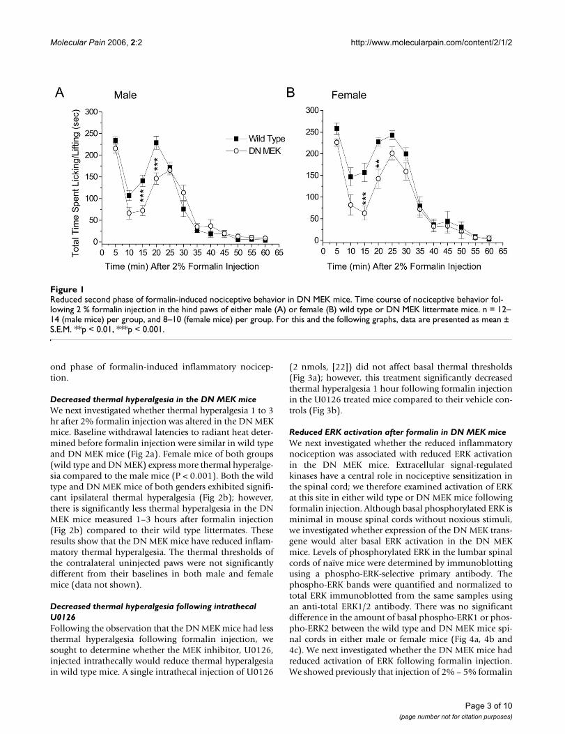
ResultsReduced second phase of formalin test in the DN MEK miceThe formalin model is frequently used in the study ofinflammatory pain states in rodents. Injection of 2 % for-malin subcutaneously in the hind paw of mice results in atypical biphasic nociceptive response [23]. The first phase,which usually lasts less than 5 minutes, occurs a few sec-onds after formalin injection and is characterized byintense spontaneous licking or lifting of the injected paw.This phase is due to acute stimulation of nociceptors. Thesecond phase is characterized by licking and lifting of theinjected paw beginning at about 15–20 minutes after for-malin injection and lasting until approximately 40–60minutes after formalin. This second phase of nociceptionis thought to involve central sensitization of dorsal hornneurons as well as peripheral sensitization associated withthe inflammation [24]. We previously showed that inmice, there is a reduced second phase of licking/liftingbehavior following attenuation of ERK activity by intrath-ecal injection of the MEK inhibitor, PD 98059 [7]. In thepresent study, we investigated the effects of reduced neu-ronal MEK function in the DN MEK mice in the formalintest. We performed experiments in male and female micesince the MEK-ERK signaling pathway is a more dominantcomponent of inflammatory hyperalgesia in females[25,26]. We find a significant gender difference in theresponse to formalin; the female mice of both groups havemore significant spontaneous nociceptive behavior thanthe male mice (p < 0.001, ANOVA). The first phase wasnot altered in either the male or female DN MEK micewhen compared to their wild type littermates. However,there was a significant reduction of the ascending part ofthe second phase of the formalin test in both male andfemale DN MEK mice (Fig 1). Thus, the neuronal MEK-ERK cascade is important for the development of the sec-
Page 2 of 10(page number not for citation purposes)
Molecular Pain 2006, 2:2 http://www.molecularpain.com/content/2/1/2
ond phase of formalin-induced inflammatory nocicep-tion.
Decreased thermal hyperalgesia in the DN MEK miceWe next investigated whether thermal hyperalgesia 1 to 3hr after 2% formalin injection was altered in the DN MEKmice. Baseline withdrawal latencies to radiant heat deter-mined before formalin injection were similar in wild typeand DN MEK mice (Fig 2a). Female mice of both groups(wild type and DN MEK) express more thermal hyperalge-sia compared to the male mice (P < 0.001). Both the wildtype and DN MEK mice of both genders exhibited signifi-cant ipsilateral thermal hyperalgesia (Fig 2b); however,there is significantly less thermal hyperalgesia in the DNMEK mice measured 1–3 hours after formalin injection(Fig 2b) compared to their wild type littermates. Theseresults show that the DN MEK mice have reduced inflam-matory thermal hyperalgesia. The thermal thresholds ofthe contralateral uninjected paws were not significantlydifferent from their baselines in both male and femalemice (data not shown).
Decreased thermal hyperalgesia following intrathecal U0126Following the observation that the DN MEK mice had lessthermal hyperalgesia following formalin injection, wesought to determine whether the MEK inhibitor, U0126,injected intrathecally would reduce thermal hyperalgesiain wild type mice. A single intrathecal injection of U0126
(2 nmols, [22]) did not affect basal thermal thresholds(Fig 3a); however, this treatment significantly decreasedthermal hyperalgesia 1 hour following formalin injectionin the U0126 treated mice compared to their vehicle con-trols (Fig 3b).
Reduced ERK activation after formalin in DN MEK miceWe next investigated whether the reduced inflammatorynociception was associated with reduced ERK activationin the DN MEK mice. Extracellular signal-regulatedkinases have a central role in nociceptive sensitization inthe spinal cord; we therefore examined activation of ERKat this site in either wild type or DN MEK mice followingformalin injection. Although basal phosphorylated ERK isminimal in mouse spinal cords without noxious stimuli,we investigated whether expression of the DN MEK trans-gene would alter basal ERK activation in the DN MEKmice. Levels of phosphorylated ERK in the lumbar spinalcords of naïve mice were determined by immunoblottingusing a phospho-ERK-selective primary antibody. Thephospho-ERK bands were quantified and normalized tototal ERK immunoblotted from the same samples usingan anti-total ERK1/2 antibody. There was no significantdifference in the amount of basal phospho-ERK1 or phos-pho-ERK2 between the wild type and DN MEK mice spi-nal cords in either male or female mice (Fig 4a, 4b and4c). We next investigated whether the DN MEK mice hadreduced activation of ERK following formalin injection.We showed previously that injection of 2% – 5% formalin
Reduced second phase of formalin-induced nociceptive behavior in DN MEK miceFigure 1Reduced second phase of formalin-induced nociceptive behavior in DN MEK mice. Time course of nociceptive behavior fol-lowing 2 % formalin injection in the hind paws of either male (A) or female (B) wild type or DN MEK littermate mice. n = 12–14 (male mice) per group, and 8–10 (female mice) per group. For this and the following graphs, data are presented as mean ± S.E.M. **p < 0.01, ***p < 0.001.
Page 3 of 10(page number not for citation purposes)

Molecular Pain 2006, 2:2 http://www.molecularpain.com/content/2/1/2
subcutaneously into the mouse hind paw induces a time-dependent activation of ERK in the lumbar spinal cord [7]which peaks at 3 minutes, remains sustained for up to 25minutes and diminishes by 60 minutes. In the currentexperiment, mice were killed 15 minutes after 2 % forma-lin injection in the right hind paw. In the wild type mice,blots of tissue taken from the side of the spinal cord ipsi-lateral to the formalin injection showed significant stimu-lation of both ERK1 and ERK2 when compared to thecontralateral side (Fig 5a and 5b), whereas ERK activationin the spinal cords from the DN MEK mice was not signif-icantly different from their contralateral sides. Further-more, ipsilateral ERK2 activation was significantly lowerin the DN MEK mice than ipsilateral ERK2 activation inthe wild type mice. Taken together, these results indicatethat DN MEK mice have reduced formalin-inducedinflammatory pain as well as reduced formalin-inducedERK activation in the spinal cord.
Unmyelinated fiber counts in cross sections of the sciatic nerveERKs also play an important role in development [2].Because recruitment of C-fibers is necessary for spinal ERKactivation [6,10], we asked whether the reduction of spi-nal neuronal ERK activation might be due to a reductionin the number of peripheral unmyelinated fibers in theDN MEK mice. Electron microscopy of sciatic nerve sec-tions of wild type or DN MEK mice revealed that the DNMEK mice had approximately twice the number of unmy-elinated fibers as those counted in the wild type mice (Fig6a, 6b, and 6c). Therefore, the reduction in spinal ERKactivation in the DN MEK mice is not due to reducednumber of unmyelinated peripheral fibers.
Decreased ERK-mediated modulation of A-type potassium currents in DN MEK miceTo further investigate whether there is a functional deficitof the MEK-ERK cascade specifically in spinal cord neu-rons of the DN MEK mice, we asked whether ERK regula-tion of a downstream target, the transient A-typepotassium channel, is altered in these mice. ERK is knownto phosphorylate Kv4.2, an A-type potassium channelsubunit [27], and we have previously shown that MEKinhibitors (U0126 or PD 98059) enhance A-type potas-sium currents in dorsal horn neurons of the spinal cord[19]. Dorsal horn cultures were prepared from either wildtype or DN MEK mice, and the effect of bath applicationof 20 µM PD 98059 was examined. Neurons from the DNMEK mice were significantly less sensitive to modulationby the MEK inhibitor PD 98059 (Fig 7). These results con-firm a reduced function of the MEK-ERK cascade in dorsalhorn neurons from the DN MEK mice.
DiscussionThe present study reports several important findingsregarding the role of the neuronal MEK-ERK cascade innociception. The DN MEK mutant mice present a func-tional reduction of the activity of neuronal MEK, thekinase that selectively activates ERK 1 and ERK 2 [28]. TheDN MEK mice have a reduced second phase of lickingbehavior following injection of 2% formalin in the hindpaw compared to the responses of their wild type litterma-tes. These data are in a sense similar to our previous phar-macological data where the intrathecally applied MEKinhibitor PD 98059, selectively reduced the second phaseof licking behavior in mice [7]. However, the pattern ofthe second phase reduction is different between the phar-macological [6,7] and genetic suppression of neuronalERK activation. PD 98059 provided a much stronger sup-pression of both the ascending and descending segmentsof the formalin second phase behavior. The current datashows a clear suppression, in both male and female mice,only during the ascending part of the second phase, sug-gesting that neuronal MEK-ERK cascade contributes to the
Reduced thermal hyperlagesia in DN MEK miceFigure 2Reduced thermal hyperlagesia in DN MEK mice. A, Baseline thermal thresholds of male and female wild type and DN MEK mice. B, Thermal thresholds taken 1 to 3 hours follow-ing injection of 2 % formalin, expressed as % of baseline val-ues (shown as 100%, dashed line). n = 11–16 mice per group (male mice), and 12–15 mice per group (female mice). *p < 0.05, ***p < 0.001, significant differences from baseline thresholds. #p < 0.05, significant differences between wild type and DN MEK mice.
Page 4 of 10(page number not for citation purposes)

Molecular Pain 2006, 2:2 http://www.molecularpain.com/content/2/1/2
development of the second phase spontaneous lickingbehavior. Perhaps the larger suppression induced byintrathecally applied MEK inhibitors is due to inhibitionof both neuronal and non-neuronal ERK activation.Indeed it has been shown recently using a neuropathicmodel that ERK is sequentially activated first in neurons,followed by microglia, and later in astrocytes [12], andtaken together with our present data, we suggest that neu-ronal ERK contributes to development of central sensitiza-tion, which may later be maintained by non-neuronalcells. Our data are also in agreement with a wealth of pre-vious data reporting that MEK inhibitors reduce inflam-matory pain using different pain models in rodents[6,29,30]. In the current study, we do not rule out the con-
tribution of other nervous system structures to thereduced behavioral effect in the DN MEK mice. However,we do show that the contribution of the spinal cord to thereduced behavioral effect is paramount since the activa-tion of ERK1 and ERK2 is also decreased following forma-lin injection in the DN MEK mice relative to wild typelittermates, and the behavioral and biochemical inhibi-tion can be mimicked by intrathecal administration ofMEK inhibitors.
A recent paper reported decreased basal ERK activity in thehippocampi of the DN MEK mice [31]. In the currentstudies, we do not observe suppressed basal ERK activa-tion in the spinal cords of the DN MEK mice. Basal ERKactivation is minimal in the spinal cord and spinal ERKactivation is activity-dependent and has been shown tooccur upon noxious or electrical stimulation of theperipheral nerves [5-7]. It is unlikely that the decrease inbasal hippocampal ERK activity could produce decreasednociception in the DN MEK mice. Shalin et al. (2004),showed that despite the deficits in contextual fear-condi-tioning in the DN MEK mice, these mice did not have sen-sory deficits but rather comparable activity and anxietylevels as that of the wild type mice. We show further in ourstudy, that there are no differences in basal thermalthresholds.
Injection of 2% formalin in mice produced thermalhyperalgesia (measured 1–3 hours after formalin injec-tion), and more so in female mice than in the male litter-mates. Ipsilateral thermal hyperalgesia was significantlyreduced in both the female and male DN MEK mice whencompared to littermate wild types. Parallel to these data, asingle intrathecal injection of U0126 reduced thermalhyperalgesia induced by 2 % formalin in wild type mice.Reduction of thermal hyperalgesia in the DN MEK mice ispossibly due to decreased central sensitization since weshowed clearly that spinal ERK activation following for-malin injection was decreased in these mice. Possiblereduction of upstream activation of ERKs by glutamatethrough either NMDA receptors [6], group I metabotropicglutamate receptors [7,32] and/or neurotrophins such asBDNF [33-35] could decrease central sensitization proc-esses leading to reduced thermal hyperalgesia. Althoughwe do not rule out possible contributions of peripheralactivation of ERK through activation of TRPV1 [6,36-40],this possibility seems unlikely because of the increasednumber of unmyelinated fibers in the DN MEK mice.However, future experiments will determine whetherTRPV1 channels and/or their functions are altered in theDN MEK mice.
In this study we examined cross sections of the sciaticnerves of the DN MEK mice in order to determine whetherreduced ERK activation following formalin injection was
Intrathecal injection of the MEK inhibitor, U0126, reduces thermal hyperalgesia in wild type miceFigure 3Intrathecal injection of the MEK inhibitor, U0126, reduces thermal hyperalgesia in wild type mice. A) Effect of intrathe-cal injection of vehicle (7.5 % DMSO in PBS, pH 7.4) or U0126 (2 nmols) on thermal thresholds in mice. B) Effect of intrathecal pretreatment of either vehicle or U0126 (2 nmols) 15 min prior to injection of 5 % formalin in the hind paw on thermal thresholds recorded 1 hr after formalin injection. n = 10 per group. *p < 0.05.
Page 5 of 10(page number not for citation purposes)

Molecular Pain 2006, 2:2 http://www.molecularpain.com/content/2/1/2
due to reduced number of unmyelinated peripheral fibers.We were surprised to observe more unmyelinated fibers inthe sciatic nerves of the DN MEK mice, particularly giventhat these mice showed no change in baseline thermalsensitivity. However, similar unexpected findings havebeen reported in the literature. For example, mice overex-pressing glial cell line-derived neurotrophic factor or
nerve growth factor possess increased numbers of unmy-elinated fibers, yet they do not display hyperalgesia [41].The increased number of unmyelinated fibers in the DNMEK mice could be a result of reduced ERK activity duringdevelopment. The MEK-ERK cascade has gained muchattention recently regarding the role of these kinases inpromoting neuronal cell death. Death of cerebellar gran-ule neurons (CGN) cultured in low potassium concentra-tions is accompanied by persistent ERK activation [42].Inhibition of persistent activation of ERK with either MEKinhibitors, or with overexpression of dominant negativeMEK in the cultures, resulted in a decrease in cell death ofthe CGN [42,43]. Our present data from the DN MEKmice are the first in vivo data that support a novel andimportant role of the MEK-ERK cascade promoting neuro-nal survival in the whole animal. Future experiments willbe designed to characterize this role in mice and especiallyhow the presence of the DN MEK affects the developmentof primary afferent nerve fibers and their receptors innociception.
The current studies further show that ERK-mediated mod-ulation of A-type potassium channels is impaired in spi-nal dorsal horn neurons from DN MEK mice. ERKs areknown to directly phosphorylate Kv4.2, a K+ channelalpha subunit that generates A-type potassium currents[27]. Reduced ERK modulation of A-type potassium chan-nels may contribute to decreased central sensitization ofspinal neurons leading to decreased pain after inflamma-tion.
ConclusionWe show here, using transgenic mice with reduced neuro-nal ERK activity, that neuronal ERK plays a key role in thedevelopment of inflammatory nociceptive behavior, andcontributes to the processing of thermal hyperalgesia. Fur-thermore, our results suggest that A-type potassium chan-nels may be possible downstream targets of ERKs in theregulation of inflammatory nociception.
Materials and methodsTα1 DN-MEK transgenic mice generationThe generation of the DN MEK mice has been previouslydescribed [31]. Briefly, a 1.1 kb Tα1 α-tubulin promoterelement that confers pan- neuronal and neuronal-specificexpression of the transgene [44,45] was used to drive theexpression of an HA-tagged K97M dominant negativeform of MEK [31,46]. The transgenic mice were estab-lished in a C3H background strain, and back-crossed sev-eral generations with C57 Bl/6 mice. For genotyping, tailDNA was extracted following standard procedures andused for PCR analysis. The primers used to amplify the436 bp HA-DN MEK transgene were sense 5'-CCC ATACGA TGT TCC AGA TTA CGC-3' and antisense 5'-CGCACC ATA GAA GCC CAC GAT G-3' (Sigma -Genosys).
Basal phosphorylation of ERK is similar in the wild type and DN MEK mice spinal cordsFigure 4Basal phosphorylation of ERK is similar in the wild type and DN MEK mice spinal cords. A, Representative immunoblot of mouse spinal cord homogenates using a phospho-ERK1/2 antibody (top) or a total ERK1/2 antibody (bottom). The arrows show the position of the 44 kDa ERK 1 and 42 kDa ERK 2 isoforms. Quantification of ERK activation in male (B) and female (C) mice. Phospho-ERK bands were densitized and normalized to total ERK from the same samples. n = 8–9 (male mice), and 8–9 (female mice).
Page 6 of 10(page number not for citation purposes)

Molecular Pain 2006, 2:2 http://www.molecularpain.com/content/2/1/2
Animal behavioral nociceptive testingAll experiments were done in accordance with the AnimalCare and Use Committee of Washington UniversitySchool of Medicine. Mice were housed in 12 hr/12 hrlight/dark cycles and given food ad libitum. Mice weighing20–25 g were used for experiments. All experiments weredone using littermate controls and were performed withthe experimenter blind to the genotype. The formalin testwas performed as described previously [7]. Mice werehabituated in a transparent Plexiglas test box (5 × 5 × 10inches) before any injections for I hr. 10 µl of 2 % forma-lin solution was injected subcutaneously into the righthind paw, and the mouse returned to the test box imme-diately. The total time spent in nociceptive behavior (lick-
ing and lifting of the injected paw) was recorded in blocksof five minutes for one hour. In separate experiments,mice were habituated in Plexiglas chambers for 2–3hours, and baseline thermal thresholds recorded. 10 µl of2 % formalin solution was injected subcutaneously intothe right hind paw, and the mice were returned to thechambers. Thermal thresholds were measured 1 hr fol-lowing injection of formalin, and recorded for up to 3hours. Thermal thresholds were measured as the latency(seconds) to withdraw or lick the paw in response to aconstant radiant heat source through the glass bottom ofa chamber to the plantar surface of the hind paw (IITC LifeSciences, Woodland hills, CA) [47].
Drug applicationFor electrophysiological recordings, the MEK inhibitorPD98059 (Sigma, St. Louis, MO) was dissolved in 100 %DMSO and diluted to the final concentration (20 µM) inHBSS (Invitrogen Life Technologies, Carlsbad, CA). PD98059 was applied by perfusion continuously at approxi-mately 2–3 ml/min. For behavioral experiments, U0126(Biomol, Plymouth Meeting, PA) was first dissolved in100 % DMSO and diluted with PBS, pH 7.4 to a final con-centration of 2 nmols in 3 µl. U0126 or the final concen-tration of vehicle (7.5 % DMSO in PBS, Ph 7.4) wasinjected intrathecally (i.t.) in a volume of 3 µl by lumbarpuncture using a Hamilton syringe and a 30 gauge needle.
Sample preparationMice were sacrificed 15 minutes after hind paw formalininjection (2 %). The spinal cords were isolated and lum-bar sections from individual mice were stored at -80°C.Lumbar spinal cord enlargements (L4-S1) where indi-cated, were separated into ipsilateral and contralateral sec-tions and each homogenized using a douncehomogenizer in ice-cold homogenization buffer (50 mMTris HCl, pH 7.5; 50 mM NaCl; 10 mM EGTA; 5 mMEDTA; 2 mM sodium pyrophosphate, 1 mM sodiumorthovanadate; paranitrophenylphosphate; 1 mM phe-nylmethylsulfonyl fluoride, 20 µg/ml leupeptin and 4 µg/ml aprotinin, Sigma-Aldrich, St. Louis, MO). Protein con-centrations were determined by the DC assay kit (Bio-Radlaboratories, Hercules, CA).
Immunoblotting for total and phospho-ERK10 µg of total protein was electrophoresed in 10% SDSpolyacrylamide gels. Proteins were transferred onto pro-tein sensitive nitrocellulose membranes and blocked in B-TTBS (3% bovine serum albumin (BSA); 50 mM Tris-HClpH 7.5; 150 NaCl; 0.02 mM Na Orthovanadate; 0.05%Tween 20; 0.01% Thimerosal, Sigma-Aldrich, St. Louis,MO) for 1 hour at room temperature. All antibody appli-cations were done in B-TTBS. An antiphospho-p44/42ERK primary antibody that detects ERK phosphorylationat both Thr202 and Tyr204 (1:1000 dilution in B-TTBS,
Reduced phospho-ERK in DN MEK mice spinal cords 15 minutes after 2 % formalin injection in the hind pawFigure 5Reduced phospho-ERK in DN MEK mice spinal cords 15 minutes after 2 % formalin injection in the hind paw. A, Rep-resentative immunoblots of ipsilateral (ipsi) and contralateral (contra) mouse spinal cord homogenates from a wild type or a DN MEK mouse using a phospho-ERK 1/2 antibody (top) or a total ERK 1/2 antibody (bottom). B, Quantification of ERK1 and ERK2 bands. Phospho-ERK bands were densitized and normalized to total ERK bands and expressed as % change of phospho-ERK on the ipsilateral side compared to the contralateral side (100 %). ***p < 0.001, significant differ-ences between ipsilateral phospho-ERK and contralateral phospho-ERK. #p < 0.05, significant differences in ipsilateral phospho-ERK between the wild type and DN MEK mice. n = 12 (wild type) and 7 (DN MEK).
Page 7 of 10(page number not for citation purposes)

Molecular Pain 2006, 2:2 http://www.molecularpain.com/content/2/1/2
Cell Signaling Technology, Beverly, MA), and an Anti-p44/42 ERK primary antibody (1:1000 dilution in 3%BSA, Cell Signaling Technology, Beverly, MA) that detectstotal p44/42 isoforms were used for immunoblottingovernight at 4°C. The blots were washed and incubated inHRP-conjugated secondary antibody for 1 hour at roomtemperature. Blots were developed with enhanced chemi-luminescence (ECL, Amersham, Arlington Heights, IL).Densitometric quantification of immunopositive bandsfor total or phospho-ERK 1/2 were performed using ScionImage software.
Cell cultureAll reagents for cell culture were purchased from Invitro-gen Life Technologies, Carlsbad, CA, except where other-wise mentioned. Primary cultures of spinal cord dorsalhorn were prepared from 3–7 day old mice using our pre-vious protocol [19]. Briefly, the mice were killed by decap-itation and a laminectomy performed to obtain the spinalcord. The spinal cord superficial dorsal horn was isolated
and chopped into several strips which were incubated for45 minutes at 37°C in Hank's balanced salt solution(HBSS) containing papain (15 U/ml: Worthington Bio-chemical, Lakewood, NJ). The strips were rinsed 3 timeswith HBSS, and placed in culture medium containingNeurobasal, 5% fetal calf serum, 5% heat-inactivatedhorse serum, 100 U/ml penicillin, 100 µg/ml streptomy-cin, 2 mM L-glutamax-1, 1% B-27 and 12 mM glucose(SIGMA-ALDRICH, St. Louis, MO). The cells were dissoci-ated by triturition with a fire-polished Pasteur pipette. Thecells were plated onto poly-D-lysine and collagen-coatedcoverslips, and cultured for 1 to 2 days in humidified airwith 5% CO2 at 37°C.
Electrophysiological recordingWhole cell recordings were performed as described in ourprevious work [19]. Briefly, whole cell recordings weremade by standard procedures at room temperature withan EPC-10 amplifier and PULSE software (HEKA Ele-ktronic, Lambrecht, Germany). Electrodes were pulledfrom filamented borosilicate glass and fire polished to aresistance of 3–6 MΩ. Most neurons had series resistancearound 6–10 MΩ (range, 5–18 MΩ), which was compen-sated ≥65%. Input resistance was 1.00 ± 0.05 GΩ. Mostneurons had leak currents < 100pA (-80 mV) which werenot subtracted on-line. The bath solution (HBSS) con-tained 500 nM TTX and 2 mM CoCl2 to block voltagegated Na+ currents, Ca2+ currents and Ca2+ -activated K+
currents. The electrode solution contained (in mM): 140KCl, 1 mgCl2, 0.5 CaCl2, 5 EGTA, 10 HEPES, 3 Na2ATP,0.3 Na2GTP, pH adjusted to 7.4 with KOH. The mem-brane voltage was held at -80 mV and potassium currentswere evoked by a command potential of +40 mV. Thetransient A-type current was isolated by subtracting thesustained current evoked by a step to +40 mV with a 150ms prepulse to -10 mV [19].
Unmyelinated fiber counts in cross sections of the sciatic nerveMice were anesthetized with 50 mg/kg pentobarbital andthe skin on the dorsal thigh was cut open. The muscleswere separated with blunt dissection and the sciatic nerveexposed. One centimeter of the nerve was removed andimmersed in a fixative (CAC) containing 2% paraformal-dehyde, 2.5% gluteraldehyde, 0.1 M cacodylic acid pH 7.2for 1 hour. After multiple rinses in CAC, samples werethen fixed in 1% osmium tetroxide in CAC for an addi-tional hour and then stained enblock in 1% uranyl acetatein h20 for 1 hour. Samples were then dehydrated througha series of ETOH, propylene oxide and then infiltrated andembedded in monomeric Embed 812 (Electron Micros-copy Sciences). Blocks were sectioned with an RMC MTXLultramicrotome at approximately 75–80 nm with a Dia-tome diamond knife, stained with Pb citrate, Uranyl ace-tate. Grids were viewed on a Hitachi H7500 transmission
Representative electron micrographs of cross sections of the sciatic nerves of A) wild type and B) DN MEK miceFigure 6Representative electron micrographs of cross sections of the sciatic nerves of A) wild type and B) DN MEK mice. Small diameter unmyelinated fibers (UM) are often present as encapsulated bunches of fibers in between the myelinated fib-ers. Thick arrowheads point to the Schwann cells of the large diameter myelinated fibers (LM). Small arrows point to the Schwann cells of the small diameter myelinated fibers (SM). S represents the Schwann cell of the unmyelinated fibers. C) Bar graph showing the mean number of unmyelinated fibers per 100 µm2. ***P < 0.001, n = 363 and 377 images from 2 wild type and 2 DN MEK mice sciatic nerves respectively.
Page 8 of 10(page number not for citation purposes)
Molecular Pain 2006, 2:2 http://www.molecularpain.com/content/2/1/2
electron microscope with Advanced Microscopy Tech-niques (AMT) digital image capturing software andHamamatsu digital camera. A total of 363 and 377 micro-graphs, from 2 wild type and 2 DN MEK mice respectively,were captured diagonally across the sciatic nerve by a per-son blind to the genotypes, and the images saved. Thenumber of unmyelinated fibers was counted and themean number of unmyelinated fibers was expressed per100 µm2.
Statistical analysisBehavioral and immunoblot data were analyzed usingone way or two way ANOVA on the Prism statistical pro-gram, followed by Bonferroni's or Newman-Keuls posthoc tests. Data from electrophysiology was evaluated off-line using Pulsefit software (HEKA Elektronic, Lambrecht,Germany) and Origin (Microcal Software, Northampton,MA) and Paired Student's t-test was used to compare drugeffects from controls. Unpaired Student's t-test was used
to compare the number of unmyelinated fibers betweenthe DN MEK and wild type mice.
Competing interestsThe author(s) declare that they have no competing inter-ests.
Authors' contributionsFK performed the behavior, biochemical and anatomicalstudies and drafted the manuscript. HH performed theelectrophysiological studies. HA participated in unpub-lished experiments and in drafting the manuscript. DKprovided the transgenic mice for the studies, and helpedto draft the manuscript. RWG conceived of the study, par-ticipated in its design and its coordination, and helped indrafting the manuscript. All authors read and approvedthe final manuscript.
AcknowledgementsThis work was supported by grants from the National Institutes of Mental Health (NS48602) to RWG, the Arthritis Foundation to FK and the Cana-dian Institutes for Health Research, National Cancer Institute, and the Neu-roscience Network Centre for Excellence to DK. We thank Joe Elias and Chang Shen Qiu for help with mouse genotyping, and Jamie Dant for help with electron microscopy.
References1. Chang L, Karin M: Mammalian MAP kinase signalling cascades.
Nature 2001, 410:37-40.2. Johnson GL, Lapadat R: Mitogen-activated protein kinase path-
ways mediated by ERK, JNK, and p38 protein kinases. Science2002, 298:1911-1912.
3. Sweatt JD: Mitogen-activated protein kinases in synaptic plas-ticity and memory. Curr Opin Neurobiol 2004, 14:311-317.
4. Ji RR: Peripheral and central mechanisms of inflammatorypain, with emphasis on MAP kinases. Curr Drug Targets InflammAllergy 2004, 3:299-303.
5. Obata K, Noguchi K: MAPK activation in nociceptive neuronsand pain hypersensitivity. Life Sci 2004, 74:2643-2653.
6. Ji RR, Baba H, Brenner GJ, Woolf CJ: Nociceptive-specific activa-tion of ERK in spinal neurons contributes to pain hypersensi-tivity. Nat Neurosci 1999, 2:1114-1119.
7. Karim F, Wang CC, Gereau RW: Metabotropic glutamate recep-tor subtypes 1 and 5 are activators of extracellular signal-regulated kinase signaling required for inflammatory pain inmice. J Neurosci 2001, 21:3771-3779.
8. Ma W, Quirion R: Partial sciatic nerve ligation induces increasein the phosphorylation of extracellular signal-regulatedkinase (ERK) and c-Jun N-terminal kinase (JNK) in astrocytesin the lumbar spinal dorsal horn and the gracile nucleus. Pain2002, 99:175-184.
9. Obata K, Yamanaka H, Dai Y, Mizushima T, Fukuoka T, Tokunaga A,Noguchi K: Activation of extracellular signal-regulated pro-tein kinase in the dorsal root ganglion following inflamma-tion near the nerve cell body. Neuroscience 2004, 126:1011-1021.
10. Lever IJ, Pezet S, McMahon SB, Malcangio M: The signaling compo-nents of sensory fiber transmission involved in the activationof ERK MAP kinase in the mouse dorsal horn. Mol Cell Neurosci2003, 24:259-270.
11. Kato T, Ohtani-Kaneko R, Ono K, Okado N, Shiga T: Developmen-tal regulation of activated ERK expression in the spinal cordand dorsal root ganglion of the chick embryo. Neurosci Res2005, 52:11-19.
12. Zhuang ZY, Gerner P, Woolf CJ, Ji RR: ERK is sequentially acti-vated in neurons, microglia, and astrocytes by spinal nerveligation and contributes to mechanical allodynia in this neu-ropathic pain model. Pain 2005, 114:149-159.
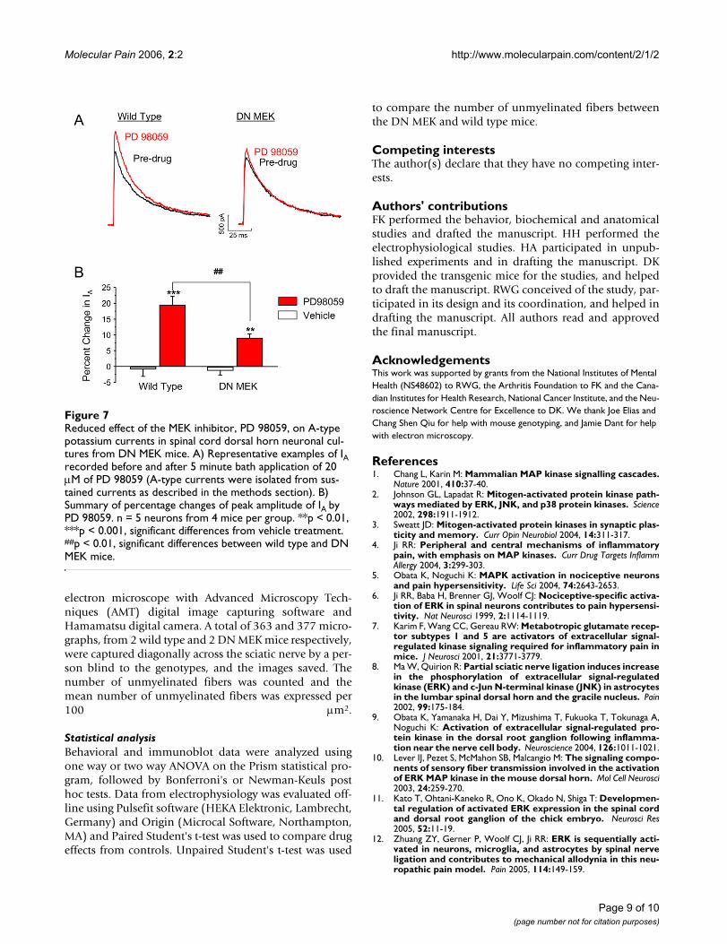
Reduced effect of the MEK inhibitor, PD 98059, on A-type potassium currents in spinal cord dorsal horn neuronal cul-tures from DN MEK miceFigure 7Reduced effect of the MEK inhibitor, PD 98059, on A-type potassium currents in spinal cord dorsal horn neuronal cul-tures from DN MEK mice. A) Representative examples of IA recorded before and after 5 minute bath application of 20 µM of PD 98059 (A-type currents were isolated from sus-tained currents as described in the methods section). B) Summary of percentage changes of peak amplitude of IA by PD 98059. n = 5 neurons from 4 mice per group. **p < 0.01, ***p < 0.001, significant differences from vehicle treatment. ##p < 0.01, significant differences between wild type and DN MEK mice.
Page 9 of 10(page number not for citation purposes)

Molecular Pain 2006, 2:2 http://www.molecularpain.com/content/2/1/2
13. Watkins LR, Martin D, Ulrich P, Tracey KJ, Maier SF: Evidence forthe involvement of spinal cord glia in subcutaneous formalininduced hyperalgesia in the rat. Pain 1997, 71:225-235.
14. Wiertelak EP, Roemer B, Maier SF, Watkins LR: Comparison of theeffects of nucleus tractus solitarius and ventral medialmedulla lesions on illness-induced and subcutaneous forma-lin-induced hyperalgesias. Brain Res 1997, 748:143-150.
15. Ledeboer A, Sloane EM, Milligan ED, Frank MG, Mahony JH, Maier SF,Watkins LR: Minocycline attenuates mechanical allodynia andproinflammatory cytokine expression in rat models of painfacilitation. Pain 2005, 115:71-83.
16. Wieseler-Frank J, Maier SF, Watkins LR: Central proinflamma-tory cytokines and pain enhancement. Neurosignals 2005,14:166-174.
17. Heffron DS, Mandell JW: Opposing roles of ERK and p38 MAPkinases in FGF2-induced astroglial process extension. Mol CellNeurosci 2005, 28:779-790.
18. Hu HJ, Gereau RW: ERK integrates PKA and PKC signaling insuperficial dorsal horn neurons. II. Modulation of neuronalexcitability. J Neurophysiol 2003, 90:1680-1688.
19. Hu HJ, Glauner KS, Gereau RW: ERK integrates PKA and PKCsignaling in superficial dorsal horn neurons. I. Modulation ofA-type K+ currents. J Neurophysiol 2003, 90:1671-1679.
20. Liu J, Fukunaga K, Yamamoto H, Nishi K, Miyamoto E: Differentialroles of Ca(2+)/calmodulin-dependent protein kinase II andmitogen-activated protein kinase activation in hippocampallong-term potentiation. J Neurosci 1999, 19:8292-8299.
21. Borsch-Haubold AG, Pasquet S, Watson SP: Direct inhibition ofcyclooxygenase-1 and -2 by the kinase inhibitors SB 203580and PD 98059. SB 203580 also inhibits thromboxane syn-thase. J Biol Chem 1998, 273:28766-28772.
22. Adwanikar H, Karim F, Gereau RW: Inflammation persistentlyenhances nocifensive behaviors mediated by spinal group ImGluRs through sustained ERK activation. Pain 2004,111:125-135.
23. Hunskaar S, Hole K: The formalin test in mice: dissociationbetween inflammatory and non-inflammatory pain. Pain1987, 30:103-114.
24. Coderre TJ, Fundytus ME, McKenna JE, Dalal S, Melzack R: The for-malin test: a validation of the weighted-scores method ofbehavioural pain rating. Pain 1993, 54:43-50.
25. Aley KO, Martin A, McMahon T, Mok J, Levine JD, Messing RO: Noci-ceptor sensitization by extracellular signal-regulatedkinases. J Neurosci 2001, 21:6933-6939.
26. Dina OA, Aley KO, Isenberg W, Messing RO, Levine JD: Sex hor-mones regulate the contribution of PKCepsilon and PKA sig-nalling in inflammatory pain in the rat. Eur J Neurosci 2001,13:2227-2233.
27. Adams JP, Anderson AE, Varga AW, Dineley KT, Cook RG, PfaffingerPJ, Sweatt JD: The A-type potassium channel Kv4.2 is a sub-strate for the mitogen-activated protein kinase ERK. J Neuro-chem 2000, 75:2277-2287.
28. Alessi DR, Cuenda A, Cohen P, Dudley DT, Saltiel AR: PD 098059is a specific inhibitor of the activation of mitogen-activatedprotein kinase kinase in vitro and in vivo. J Biol Chem 1995,270:27489-27494.
29. Ji RR, Befort K, Brenner GJ, Woolf CJ: ERK MAP kinase activationin superficial spinal cord neurons induces prodynorphin andNK-1 upregulation and contributes to persistent inflamma-tory pain hypersensitivity. J Neurosci 2002, 22:478-485.
30. Kominato Y, Tachibana T, Dai Y, Tsujino H, Maruo S, Noguchi K:Changes in phosphorylation of ERK and Fos expression indorsal horn neurons following noxious stimulation in a ratmodel of neuritis of the nerve root. Brain Res 2003, 967:89-97.
31. Shalin SC, Zirrgiebel U, Honsa KJ, Julien JP, Miller FD, Kaplan DR,Sweatt JD: Neuronal MEK is important for normal fear condi-tioning in mice. J Neurosci Res 2004, 75:760-770.
32. Guo W, Wei F, Zou S, Robbins MT, Sugiyo S, Ikeda T, Tu JC, WorleyPF, Dubner R, Ren K: Group I metabotropic glutamate recep-tor NMDA receptor coupling and signaling cascade mediatespinal dorsal horn NMDA receptor 2B tyrosine phosphoryla-tion associated with inflammatory hyperalgesia. J Neurosci2004, 24:9161-9173.
33. Kerr BJ, Bradbury EJ, Bennett DL, Trivedi PM, Dassan P, French J,Shelton DB, McMahon SB, Thompson SW: Brain-derived neuro-trophic factor modulates nociceptive sensory inputs and
NMDA-evoked responses in the rat spinal cord. J Neurosci1999, 19:5138-5148.
34. Kaplan DR, Miller FD: Neurotrophin signal transduction in thenervous system. Curr Opin Neurobiol 2000, 10:381-391.
35. Pezet S, Malcangio M, Lever IJ, Perkinton MS, Thompson SW, Wil-liams RJ, McMahon SB: Noxious stimulation induces Trk recep-tor and downstream ERK phosphorylation in spinal dorsalhorn. Mol Cell Neurosci 2002, 21:684-695.
36. Caterina MJ, Schumacher MA, Tominaga M, Rosen TA, Levine JD,Julius D: The capsaicin receptor: a heat-activated ion channelin the pain pathway. Nature 1997, 389:816-824.
37. Caterina MJ, Leffler A, Malmberg AB, Martin WJ, Trafton J, Petersen-Zeitz KR, Koltzenburg M, Basbaum AI, Julius D: Impaired nocicep-tion and pain sensation in mice lacking the capsaicin recep-tor. Science 2000, 288:306-313.
38. Davis JB, Gray J, Gunthorpe MJ, Hatcher JP, Davey PT, Overend P,Harries MH, Latcham J, Clapham C, Atkinson K, Hughes SA, Rance K,Grau E, Harper AJ, Pugh PL, Rogers DC, Bingham S, Randall A, Shear-down SA: Vanilloid receptor-1 is essential for inflammatorythermal hyperalgesia. Nature 2000, 405:183-187.
39. Ji RR, Samad TA, Jin SX, Schmoll R, Woolf CJ: p38 MAPK activa-tion by NGF in primary sensory neurons after inflammationincreases TRPV1 levels and maintains heat hyperalgesia.Neuron 2002, 36:57-68.
40. Tominaga M, Caterina MJ, Malmberg AB, Rosen TA, Gilbert H, Skin-ner K, Raumann BE, Basbaum AI, Julius D: The cloned capsaicinreceptor integrates multiple pain-producing stimuli. Neuron1998, 21:531-543.
41. Zwick M, Molliver DC, Lindsay J, Fairbanks CA, Sengoku T, AlbersKM, Davis BM: Transgenic mice possessing increased numbersof nociceptors do not exhibit increased behavioral sensitivityin models of inflammatory and neuropathic pain. Pain 2003,106:491-500.
42. Subramaniam S, Strelau J, Unsicker K: Growth differentiation fac-tor-15 prevents low potassium-induced cell death of cerebel-lar granule neurons by differential regulation of Akt and ERKpathways. J Biol Chem 2003, 278:8904-8912.
43. Subramaniam S, Zirrgiebel U, von Bohlen Und Halbach O, Strelau J,Laliberte C, Kaplan DR, Unsicker K: ERK activation promotesneuronal degeneration predominantly through plasmamembrane damage and independently of caspase-3. J Cell Biol2004, 165:357-369.
44. Gloster A, Wu W, Speelman A, Weiss S, Causing C, Pozniak C, Rey-nolds B, Chang E, Toma JG, Miller FD: The T alpha 1 alpha-tubulinpromoter specifies gene expression as a function of neuronalgrowth and regeneration in transgenic mice. J Neurosci 1994,14:7319-7330.
45. Bamji SX, Miller FD: Comparison of the expression of a T alpha1:nlacZ transgene and T alpha 1 alpha-tubulin mRNA in themature central nervous system. J Comp Neurol 1996, 374:52-69.
46. Mansour SJ, Candia JM, Gloor KK, Ahn NG: Constitutively activemitogen-activated protein kinase kinase 1 (MAPKK1) andMAPKK2 mediate similar transcriptional and morphologicalresponses. Cell Growth Differ 1996, 7:243-250.
47. Hargreaves K, Dubner R, Brown F, Flores C, Joris J: A new and sen-sitive method for measuring thermal nociception in cutane-ous hyperalgesia. Pain 1988, 32:77-88.
Page 10 of 10(page number not for citation purposes)
http://www.ncbi.nlm.nih.gov/entrez/query.fcgi?cmd=Retrieve&db=PubMed&dopt=Abstract&list_uids=9231865
http://www.ncbi.nlm.nih.gov/entrez/query.fcgi?cmd=Retrieve&db=PubMed&dopt=Abstract&list_uids=9231865
http://www.ncbi.nlm.nih.gov/entrez/query.fcgi?cmd=Retrieve&db=PubMed&dopt=Abstract&list_uids=9231865
http://www.ncbi.nlm.nih.gov/entrez/query.fcgi?cmd=Retrieve&db=PubMed&dopt=Abstract&list_uids=9067455
http://www.ncbi.nlm.nih.gov/entrez/query.fcgi?cmd=Retrieve&db=PubMed&dopt=Abstract&list_uids=9067455
http://www.ncbi.nlm.nih.gov/entrez/query.fcgi?cmd=Retrieve&db=PubMed&dopt=Abstract&list_uids=9067455
http://www.ncbi.nlm.nih.gov/entrez/query.fcgi?cmd=Retrieve&db=PubMed&dopt=Abstract&list_uids=9786874
http://www.ncbi.nlm.nih.gov/entrez/query.fcgi?cmd=Retrieve&db=PubMed&dopt=Abstract&list_uids=9786874
http://www.ncbi.nlm.nih.gov/entrez/query.fcgi?cmd=Retrieve&db=PubMed&dopt=Abstract&list_uids=9786874
http://www.ncbi.nlm.nih.gov/entrez/query.fcgi?cmd=Retrieve&db=PubMed&dopt=Abstract&list_uids=3614974
http://www.ncbi.nlm.nih.gov/entrez/query.fcgi?cmd=Retrieve&db=PubMed&dopt=Abstract&list_uids=3614974
http://www.ncbi.nlm.nih.gov/entrez/query.fcgi?cmd=Retrieve&db=PubMed&dopt=Abstract&list_uids=8378102
http://www.ncbi.nlm.nih.gov/entrez/query.fcgi?cmd=Retrieve&db=PubMed&dopt=Abstract&list_uids=8378102
http://www.ncbi.nlm.nih.gov/entrez/query.fcgi?cmd=Retrieve&db=PubMed&dopt=Abstract&list_uids=8378102
http://www.ncbi.nlm.nih.gov/entrez/query.fcgi?cmd=Retrieve&db=PubMed&dopt=Abstract&list_uids=7499206
http://www.ncbi.nlm.nih.gov/entrez/query.fcgi?cmd=Retrieve&db=PubMed&dopt=Abstract&list_uids=7499206
http://www.ncbi.nlm.nih.gov/entrez/query.fcgi?cmd=Retrieve&db=PubMed&dopt=Abstract&list_uids=7499206
http://www.ncbi.nlm.nih.gov/entrez/query.fcgi?cmd=Retrieve&db=PubMed&dopt=Abstract&list_uids=9349813
http://www.ncbi.nlm.nih.gov/entrez/query.fcgi?cmd=Retrieve&db=PubMed&dopt=Abstract&list_uids=9349813
http://www.ncbi.nlm.nih.gov/entrez/query.fcgi?cmd=Retrieve&db=PubMed&dopt=Abstract&list_uids=9768840
http://www.ncbi.nlm.nih.gov/entrez/query.fcgi?cmd=Retrieve&db=PubMed&dopt=Abstract&list_uids=9768840
http://www.ncbi.nlm.nih.gov/entrez/query.fcgi?cmd=Retrieve&db=PubMed&dopt=Abstract&list_uids=7996178
http://www.ncbi.nlm.nih.gov/entrez/query.fcgi?cmd=Retrieve&db=PubMed&dopt=Abstract&list_uids=7996178
http://www.ncbi.nlm.nih.gov/entrez/query.fcgi?cmd=Retrieve&db=PubMed&dopt=Abstract&list_uids=7996178
http://www.ncbi.nlm.nih.gov/entrez/query.fcgi?cmd=Retrieve&db=PubMed&dopt=Abstract&list_uids=8891946
http://www.ncbi.nlm.nih.gov/entrez/query.fcgi?cmd=Retrieve&db=PubMed&dopt=Abstract&list_uids=8891946
http://www.ncbi.nlm.nih.gov/entrez/query.fcgi?cmd=Retrieve&db=PubMed&dopt=Abstract&list_uids=8891946
http://www.ncbi.nlm.nih.gov/entrez/query.fcgi?cmd=Retrieve&db=PubMed&dopt=Abstract&list_uids=8822208
http://www.ncbi.nlm.nih.gov/entrez/query.fcgi?cmd=Retrieve&db=PubMed&dopt=Abstract&list_uids=8822208
http://www.ncbi.nlm.nih.gov/entrez/query.fcgi?cmd=Retrieve&db=PubMed&dopt=Abstract&list_uids=8822208
http://www.ncbi.nlm.nih.gov/entrez/query.fcgi?cmd=Retrieve&db=PubMed&dopt=Abstract&list_uids=3340425