Isolation and primary culture of Necturus maculosus (Amphibia: urodela) hepatocytes
Upload
independentCategory
view
3download
0
FEBS 18853 FEBS Letters 412 (1997) 9-14
Positive and negative regulation of JNKl by protein kinase C and p 4 2 M A P kinase [n ^ ^ m t hepatOCVteS
W. David Jarvisc, Kelly L. Auera, Mark Spectorb, George Kunosb, Steven Grantb,c, Philip Hylemond, Ross Mikkelsena, Paul Denta'b'*
Department of Radiation Oncology, Box 980058, Massey Cancer Center, Medical College of Virginia, Richmond, VA 23298-0058, USA hDepartment of Pharmacology and Toxicology, Medical College of Virginia, Richmond, VA 23298-0058, USA
cDepartment of Medicine, Medical College of Virginia, Richmond, VA 23298-0058, USA dDepartment of Microbiology and Immunology, Medical College of Virginia, Richmond, VA 23298-0058, USA
Received 2 May 1997; revised version received 29 May 1997
Abstract The role of protein kinase C (PKC) and p42MAP klnase signaling in the regulation of proliferation and apoptosis was investigated in freshly isolated and primary cultured rat hepatocytes. Acute treatment of freshly isolated hepatocytes with phenylephrine and EGF caused rapid phasic activations of p42MAP kinase and JNKl. Acute pre-treatment of hepatocytes with the PKC inhibitors sphingosine, chelerythrine and Ais-indolylmaleimide abolished the ability of phenylephrine, but not EGF, to activate p42MAP k!nase and JNKl. Acute pre-treatments with all of the PKC inhibitors alone increased JNKl basal activity ~ 2-fold. Acute treatments of primary cultures of hepatocytes with an inhibitor of MEK1 activation (PD98059) also caused inhibition of p42MAP klnase and a ~ 2-fold activation of JNKl. These data demonstrate that PKC can function as both a proximal activator and a distal inhibitor of signaling through the JNK1/SAP kinase pathway. Treatments (4 h) of primary cultured hepatocytes with sphingosine, chelerythrine, Ws-indo-lylmaleimide and PD98059 did not induce apoptosis as judged by propidium iodide staining. Similar acute treatments of HepG2 cells rapidly induced cell death. These data demonstrate that acute inhibition of either PKC or p 42 M A P k i n a s c function is sufficient to rapidly induce apoptosis in transformed, but not in non-transformed hepatocytes.
© 1997 Federation of European Biochemical Societies.
Key words: p42MAI 'kinasc; JNKl ; Protein kinase C; Sphingosine; /j«-Indolylmaleimide; Chelerythrine; PD98059
1. Introduction
The mechanisms by which non-transformed cells regulate their ability to differentiate, proliferate, or undergo apoptosis are poorly understood. To better understand these processes, we have examined the regulation of signal transduction cas-cades in primary cultures of rat hepatocytes.
The liver is the only organ of the body which can fully regenerate its cell mass after injury [1-3]. During regeneration, hepatocytes acquire altered sensitivity/responsiveness towards multiple agonists, including a reciprocal decrease and increase, respectively, in the levels of ai-adrenergic and f^-adrenergic receptors [4-6]. We recently demonstrated that such altera-
*Corresponding author. Fax: +1 (804) 828-6042. E-mail: [email protected]
Abbreviations: MAP kinase, mitogen-activated protein kinase; JNK, c-Jun NH2-terminal kinase; PKC, protein kinase C; SAP kinase, stress-activated protein kinase; AR, adrenergic receptor; EGF, epidermal growth factor
tions in signaling lead to a decrease in the ability of epineph-rine and other factors to activate protein kinase C (PKC), Raf-1, and p42/44MAPkinase, and concurrently lead to an in-crease in the ability of epinephrine and other factors to acti-vate JNKl (c-Jun NH2-terminal kinase), and p38S A P k i n a s c
(p38-reactivating kinase) [6]. The JNKl/stress-activated protein (SAP) kinase cascade
was initially discovered in yeast and has subsequently been characterized in mammalian cells [7-10]. Apoptotic responses have been asssociated with the cytotoxic lipid messengers cer-amide [11,12] and sphingosine [8,13] in human myeloid leuke-mia cells. Activation of the SAP kinases, JNKl and JNK2 have been directly implicated in the ceramide-related apopto-sis [14,15], although the pathway linking ceramide to the SAP kinase cascade has not been completely delineated. In con-trast, sphingosine-related cytotoxicity is most commonly at-tributed to proximal inhibition of PKC [16]. Recently we have found that, unlike ceramide, sphingosine does not engage JNKl to promote apoptosis [17], indicating that the lethal actions of ceramide and sphingosine are subserved by distinct signaling mechanisms. In hepatocytes, however, little is known about the co-ordinated mechanisms by which altera-tions in receptor signaling, PKC activity, and the activities of p42MAP kmasc and JNKl are altered upon liver regeneration or hepatocellular transformation. Still less is known as to how these alterations effect the actions of cytotoxic lipid second messengers and the contributions of these messengers towards regulating systems governing the initiation of apoptosis [18,19].
In the present studies, we have examined the role of PKC in the regulation of p42MAP kmase and JNKl by hormonal ago-nists and lipid signaling molecules, and the roles PKC and p42MAPkmase play in proliferative and apoptotic signaling processes in primary isolates of hepatocytes. Our findings strongly suggest that the balance of proliferative and apoptotic signals are integrated at multiple levels, and that examination of signaling via any single pathway cannot be used to explain definitively the physiological regulation of cell survival.
2. Materials and methods
2.1. Materials Male Sprague-Dawley rats (200 g) had access to food and water ad
libitum [5]. Anti-JNKl/2 (mostly immunoprecipitating JNKl, sc-571AC) antibody was from Santa Cruz Biotechnology (Santa Cruz Biotechnologies, CA) and anti-p42MAF hnaae antibodies were a kind gift from Dr. M.J. Weber (University of Virginia, Charlottesville, VA). Hormones, growth factors, and propidium iodide (PI) were pur-chased from Sigma Chemicals (St. Louis, MO). Radiolabelled
0014-5793/97/S17.00 © 1997 Federation of European Biochemical Societies. All rights reserved. P / /S0014-5793(97)00705-9
10
[y-32P]ATP and [3H]thymidine were from NEN. GST-c-Jun (aal-169) was synthesized in Escherichia coli and purified on GST-Sepharose. Protein preparations of all other reagents were as described in [6,21,22]. Specific kinase domain inhibitors of protein kinase C, bis-indolyl-maleimaide GF109203X [23,24], chelerythrine [18], and the MEK1 activation inhibitor PD98059 [25] were from Calbiochem. Sphingosine was from Biomol (Plymouth Meeting, MA).
2.2. Preparation of hepatocytes Rats were anesthetized by i.p. injection of sodium pentabarbital (50
mg/kg), and the lower thorax and abdomen was shaved to remove fur. A small (3 cm) vertical mid-line incision was made in the abdominal wall from just below the costal margin/xiphoid process. Hepatocytes were prepared by cannulation of the portal vein and collagenase per-fusion of the liver. A cannula was inserted into the portal vein and two ties tightened so as to occlude the inferior vena cava, and to secure the cannula in the portal vein. Gassed Krebs-Henseleit buffer (95% air, 5% C0 2 , 37°C) containing 0.3 mM EGTA (to prevent clot-ting), was passed through the liver (20 ml/min flow rate) to wash out erythrocytes; a second cannula was inserted into the inferior vena cava via the right atrium of the heart, and also secured with a tie, to act as a drain. Livers were washed sequentially with 200 ml of this buffer, followed by 200 ml of the same media omitting EGTA, and including 0.5 mg/ml collagenase. The liver was then removed and filtered through sterile muslin into a sterile container and washed (X 3) with Krebs-Henseleit buffer. Hepatocytes were then resuspended to 5 mg wet weight/ml serum-free Krebs-Henseleit buffer containing 1.5% (w/v) Difco gelatin, and were incubated at 37°C under an atmo-sphere of 5% CO2 in O2 with continuous rotory shaking at 100 cycles/ min. After a 30 min pre-incubation to stabilize basal levels of protein kinase activities (data not shown), aliquots were taken from the first portion and used immediately for experiments to determine hormonal effects on protein kinase activities [6].
2.3. Treatment of freshly isolated hepatocytes and primary cultures of hepatocytes and HepG2 cells with hormones and cell homogenization
Hormones were added to microfuge tubes containing hepatocytes (5 mg wet weight in 1 ml buffer, 1 mg total protein used per immuno-precipitate) in the absence of serum. Primary cultures of hepatocytes and HepG2 cells were cultured in 12-well plates or 20 mm dishes (2X10⁵ cells) and grown in DMEM containing 10% (w/v) fetal calf serum (2 mg wet weight cells/dish, 1 mg total protein used per immu-noprecipitate) and additions made to these cells after 30 min serum starvation in 5 ml of DMEM medium. Hormones were added to give final specified concentrations (see text and figure legends), vortexed/ mixed to ensure complete mixing, and incubated for the specified times at 37°C in a water bath or cell culture incubator. Cells were pretreated with protein kinase inhibitors (20 min) or lipids (15 min) prior to hormonal additions. Thirty seconds prior to termination, tubes were removed from the bath, placed in a microfuge, the cells pelleted, and the supernatant removed. Plated cells were aspirated followed by immediate homogenization. Cells were homogenized in 1 ml of ice-cold buffer A (25 mM HEPES, pH 7.4 at 4°C, 5 mM EDTA, 5 mM EGTA, 5 mM benzamidine, 1 mM phenylmethyl sul-phonylfluoride, 1 mg/ml soybean trypsin inhibitor, 40 ug/ml pepsta-tin A, 40 ug/ml E64, 40 ug/ml aprotinin, 1 uM Microcystin-LR, 0.5 mM sodium orthovanadate, 0.5 mM sodium pyrophosphate, 0.05% (w/v) sodium deoxycholate, 1% (v/v) Triton X100, 0.1% (v/v) 2-mer-captoethanol), with trituration using a P1000 pipette to lyse the cells. Homogenates were stored on ice (5 min) prior to clarification by centrifugation (4°C).
2.4. Immunoprecipitations from homogenates Fifty microliters of Protein A agarose (Ag) slurry (25 ja.1 bead vol-
ume) was washed twice with 1 ml of PBS containing 0.1% (v/v) Tween-20, and resuspended in 0.1 ml of the same buffer. Antibodies (2 ug, 20 ul) or serum (20 |xl) were added to each tube and incubated (3 h, 4°C). Clarified hepatocyte homogenates (0.5 ml, 1 mg total protein) were mixed with Protein A-Ag-conjugated antibody in dup-licate using gentle agitation (2.5 h, 4°C). Protein A-Ag was recovered by centrifugation, the supernatant discarded, and washed (10 min) sequentially with 0.5 ml buffer A (twice), PBS and buffer B (25 mM HEPES, pH 7.4, 15 mM MgCl2, 0.1 mM Na2V04 , 0.1% (v/v) 2-mer-captoenthanol).
W.D. Jarvis et al.lFEBS Letters 412 (1997) 9-14
2.5. Assay of p42MAF'kmase activity Immunoprecipitates were incubated (final volume 50 ill) with 50 ul
of buffer B containing 0.2 mM [y-32P]ATP (5000 cpm/pmol), 1 uM Microcystin-LR, 0.5 mg/ml myelin basic protein (MBP), which initi-ated reactions. After 20 min, 40 ul °f the reaction mixtures were spotted onto a 2 cm circle of P81 paper (Whatman, Maidstone, UK) and immediately placed into 180 mM phosphoric acid. Papers were washed 4 times (10 min each) with phosphoric acid, and once with acetone, and 32P-incorporation into MBP was quantified by liquid scintillation spectroscopy. Pre-immune controls were performed to ensure MBP phosphorylation was dependent upon specific immu-noprecipitation 0 f ^ 2 M " " " [21].
2.6. Assay of JNK1 activity Immunoprecipitates were incubated (final volume 100 ul) with 2 ul
(10 ug) of GST-c-Jun (aal-169), and reactions initiated with 98 ul of buffer B containing 0.2 mM [y-32P]ATP (5000 cpm/pmol) and 1 uM Microcystin-LR. After 30 min, reactions were terminated with sample buffer and prepared for SDS-PAGE (10% gel) to quantify 32P-incor-poration into excised, Coomassie blue-stained GST-c-Jun (aal-169) bands by liquid scintillation spectroscopy. Pre-immune control assays were performed to ensure GST-c-Jun (aal-169) phosphorylation was dependent upon specific immunoprecipitation of JNK1 in the assay [26].
2.7. Cell culture, and primary culture and assay for DNA synthesis in hepatocytes
HepG2 cells were cultured by established procedures in Dulbecco's modified Eagle's medium containing 10% (v/v) fetal calf serum (FCS) in 5% (v/v) C0 2 . Hepatocytes isolated from sham-operated or PHX rats were cultured on rat-tail collagen (Vitrogen)-coated plastic dishes (12x20 mm, 2X 10⁶ cells, 2 mg wet weight) in William's E medium and allowed to adhere to the dish. After 2 h the medium was replaced and hepatocytes cultured in William's E medium containing 20 uCi of [3H]thymidine, 1 nM dexamethasone and 100 nM insulin, in 5% (v/v) CO2. Adrenergic drugs, hormonal treatments, and/or protein kinase inhibitors were added at the time of the media change. Cells were cultured for 48 h, after which time cells were lysed with 0.5 N NaOH and DNA precipitated with 12.5% (w/v) TCA (final). Acid precipitable material was transferred to glass fiber filters, washed with 5% (w/v) TCA, and [3H]thymidine incorporation into DNA quantified by liquid scintillation spectrometry [27].
2.8. Transcription factor DNA binding assay Nuclear extracts are prepared as described [5]. Oligonucleotides are
32P-labelled with [y-32P]ATP using polynucleotide kinase. Binding re-action mixtures (20 ill) are incubated at room temperature for 45 min containing 1 ng DNA probe and 5 ug nuclear extract in 10 mM Tris-HC1, pH 7.5, at 25°C, 40 mM NaCl, 1 mM DTT, 1 mM EDTA, 5% (v/v) glycerol and 2 ug poly(dl-dC), to inhibit non-specific binding in the extract. DNA-protein complexes are resolved by electrophoresis through a 4%/8% non-denaturing polyacrylamide gel containing 50 mM Tris-HCl, 0.38 M glycine and 2 mM EDTA. Gels are dried and quantified/processed using a Phosphoimager.
2.9. Data analysis Comparison of the effects of various hormone treatments was done
using 1-way analysis of variance and a 2-tailed Mest. Differences with a P-value of < 0.05 (* and &) were considered statistically significant. All bar-graph fold-values and means shown are ± SD from an aver-age of 2-5 independent experiments (depending upon the figure) are shown.
3. Results and discussion
Freshly isolated hepatocytes were treated with phenyleph-rine or E G F , and p42 M A P klnase and J N K 1 activities measured in immune-complex kinase assays (Fig. 1). Phenylephrine (Fig. 1A) and E G F (Fig. IB) activated p42 M A P kinase and J N K 1 with rapid phasic kinetics, with maximal activations between 4 and 10 min; maximal stimulations by phenyleph-rine and E G F were titrated to 10 |0.M and 30 ng/ml for each hormone, respectively (data not shown). Acute (20 min) pre-

W.D. Jarvis et allFEBS Letters 412 (1997) 9-14 11
PL, 7
pa ' % a 6
_o
1 5
£ 4
£ 3 a, V a * o s-S 1 o
Phenylephrine
- f \ <r
J
^ . „ MAP kinase
—•— p42 v j N K i
v
10 20 30 40 50 60
Time (min)
Time (min)
Fig. 1. Time course of activation of p42MAPkmase and JNKI by phenylephrine (10 uM) (A) and epidermal growth factor (30 ng/ml) (B) in freshly isolated hepatocytes. Cells were treated with agonist for the indicated times at 37°C. Cells were pelleted, lysed and pro-tein kinases specifically immunoprecipitated as described Section 2. Data are an average of duplicates from representative experiments (n = 3) from immune-complex kinase assays and shown as -fold in-creases in 32P-incorporation into substrate. Vehicle control values were r)47^A P kinase
spectively. (25 500 ±750 cpm) and JNKI (950 ±70 cpm), re-
trated to 10 uM), or catalytic domain c/n/aPKC inhibitors GF109203X (titrated to 10 uM) and chelerythrine (titrated to 1 uM) blunted the ability of phenylephrine, but not E G F , to activate p 4 2 M A P k i ^ e (Fig. 2A). Pre-treatment of hepatocytes with the P K C inhibitors significantly reduced the ability of phenylephrine, but not E G F , to activate J N K I (Fig. 2B). Gq-coupled phenylephrine receptors are rapidly down-regulated in hepatocytes after isolation and primary culture [7], whereas Gq-coupled receptors for vasopressin are not. Pre-treatment of hepatocytes with the P K C inhibitors
B
%
I
Sphingosine GF109203X Chelerythrine
y £ S
treatment of cells with maximally inhibitory doses of the reg-ulatory domain cPKC inhibitory effector, sphingosine (ti-
V Sphingosine GF1O9203X Chelerythrine
Fig. 2. The role of PKC in the activation of (A) p42MAP kinase and (B) JNKI in freshly isolated hepatocytes, in response to phenyleph-rine (phe) and epidermal growth factor (EGF) treatments. Freshly isolated hepatocytes were pre-treated (20 min, 37°C) with the maxi-mally inhibitory doses of the PKC inhibitors sphingosine (10 |J,M), Ms-indolylmaleiamide (10 uM), chelerythrine (1.0 U.M), or vehicle before hormonal addition. After 20 min, cells were further treated with maximally stimulatory doses of phe (10 u,M) or EGF (30 ng/ ml) for 6 min at 37°C. Cells were pelleted, lysed and protein kinases specifically immunoprecipitated as described in Section 2. Data are an average of duplicates from three experiments using immune-com-plex kinase assays and shown as -fold increases in 32P-incorporation into substrate. Vehicle control values were p42MAPki™se (25 500 ±750 cpm) and JNKI (950 ± 70 cpm), respectively. (C) Treatment of hep-atocytes (20 min, 37°C) with ±PD98059 (50 iiM, +Lane 1, -Lane 2) activates JNKI and stimulates AP-1 binding. AP-1 complex DNA binding was performed as described in Section 2 on 5 |xg of cellular lysate using non-denaturing gel electrophoresis. Lane 3, probe alone.
'Si
3 C
2 "3 '—
1 2 3
A P I DNA binding^- i t
12 W.D. Jarvis et al.lFEBS Letters 412 (1997) 9-14
2 .
«
a c u o u d
C8 i> I . u S
o
1. e a
« i . u _c
'o In
<u -■ = 0)
>
ai S
o OX! B
J3 fi. («
O OH
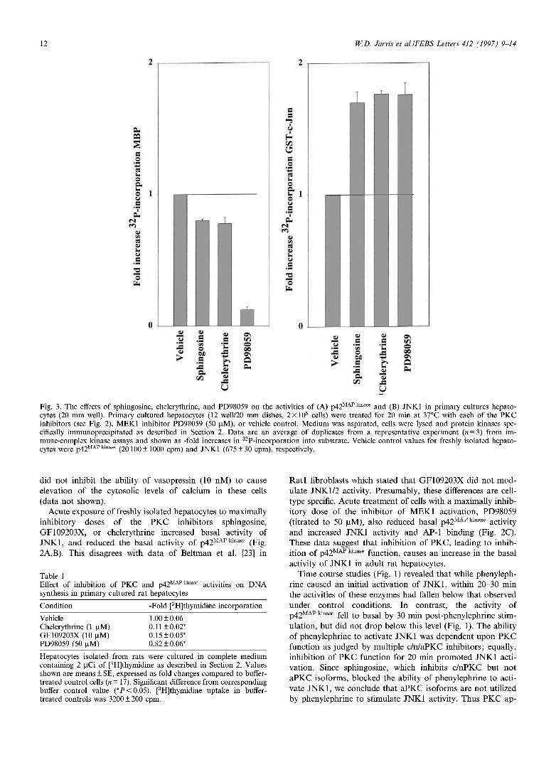
Fig. 3. The effects of sphingosine, chelerythrine, and PD98059 on the activities of (A) p42MAPkmase and (B) JNK1 in primary cultures hepato-cytes (20 mm well). Primary cultured hepatocytes (12 well/20 mm dishes, 2x 10⁵ cells) were treated for 20 min at 37°C with each of the PKC inhibitors (see Fig. 2), MEK1 inhibitor PD98059 (50 U.M), or vehicle control. Medium was aspirated, cells were lysed and protein kinases spe-cifically immunoprecipitated as described in Section 2. Data are an average of duplicates from a representative experiment (« = 3) from im-mune-complex kinase assays and shown as -fold increases in 32P-incorporation into substrate. Vehicle control values for freshly isolated hepato-cytes were p42MAP kinase (20100 ±1000 cpm) and JNK1 (675 ±30 cpm), respectively.
w .= <u >
u a •5 o DA
| Q«
C/3
<u c i .
J3
11 o
J=
u
0\ in o 00 0\
a.
did not inhibit the ability of vasopressin (10 nM) to cause elevation of the cytosolic levels of calcium in these cells (data not shown).
Acute exposure of freshly isolated hepatocytes to maximally inhibitory doses of the PKC inhibitors sphingosine, GF109203X, or chelerythrine increased basal activity of JNK1, and reduced the basal activity of p42M A P k i n a s e (Fig. 2A,B). This disagrees with data of Beltman et al. [23] in
Table 1 Effect of inhibition of PKC and p42MAP kinase
synthesis in primary cultured rat hepatocytes activities on DNA
Condition -Fold [3H]thymidine incorporation
Vehicle Chelerythrine (1 U.M) GF109203X (10 |xM) PD98059 (50 uM)
1.00 ±0.06 0.11 ±0.02* 0.15 ±0.05* 0.82 ±0.06*
Hepatocytes isolated from rats were cultured in complete medium containing 2 uCi of [3H]thymidine as described in Section 2. Values shown are means ± SE, expressed as fold changes compared to buffer-treated control cells (n = 17). Significant difference from corresponding buffer control value (*P<0.05). [3H]thymidine uptake in buffer-treated controls was 3200 ± 200 cpm.
Ratl fibroblasts which stated that GF109203X did not mod-ulate JNK1/2 activity. Presumably, these differences are cell-type specific. Acute treatment of cells with a maximally inhib-itory dose of the inhibitor of MEK1 activation, PD98059 (titrated to 50 uM), also reduced basal p42fc activity and increased JNK1 activity and AP-1 binding (Fig. 2C). These data suggest that inhibition of PKC, leading to inhib-ition of p42MAP kmase function, causes an increase in the basal activity of JNK1 in adult rat hepatocytes.
Time course studies (Fig. 1) revealed that while phenyleph-rine caused an initial activation of JNK1, within 20-30 min the activities of these enzymes had fallen below that observed under control conditions. In contrast, the activity of p42MAP kmase fell to basal by 30 min post-phenylephrine stim-ulation, but did not drop below this level (Fig. 1). The ability of phenylephrine to activate JNK1 was dependent upon PKC function as judged by multiple c/n/aPKC inhibitors; equally, inhibition of PKC function for 20 min promoted JNK1 acti-vation. Since sphingosine, which inhibits c/nPKC but not aPKC isoforms, blocked the ability of phenylephrine to acti-vate JNK1, we conclude that aPKC isoforms are not utilized by phenylephrine to stimulate JNK1 activity. Thus PKC ap-
W.D. Jarvis et al.lFEBS Letters 412 (1997) 9-14 13
•9 0
to " g £ R s " J5 = K e
o. to -Si "2 to .2 ™ O % t & o «
Hepatocyte HepG2
an
*3 14
B
■5 o
12
10
e
t/s
U u .2 0 ■o
-
1 1 1
T T
e to
ON
o 00
c
X © ON
o to O
U
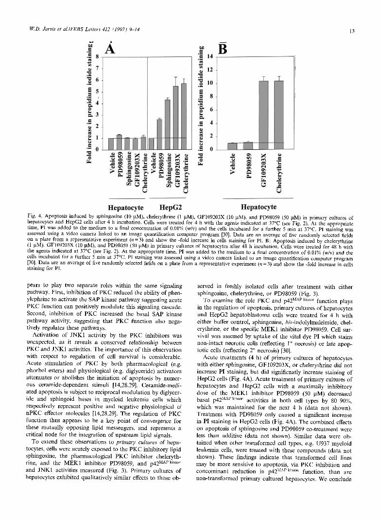
Hepatocyte Fig. 4. Apoptosis induced by sphingosine (10 uM), chelerythrine (1 uM), GF109203X (10 uM), and PD98059 (50 |xM) in primary cultures of hepatocytes and HepG2 cells after 4 h incubation. Cells were treated for 4 h with the agents indicated at 37°C (see Fig. 2). At the appropriate time, PI was added to the medium to a final concentration of 0.01% (w/v) and the cells incubated for a further 5 min at 37°C. PI staining was assessed using a video camera linked to an image quantification computer program [30]. Data are an average of five randomly selected fields on a plate from a representative experiment (« = 3) and show the -fold increase in cells staining for PI. B: Apoptosis induced by chelerythrine (1 uM), GF109203X (10 uM), and PD98059 (50 iiM) in primary cultures of hepatocytes after 48 h incubation. Cells were treated for 48 h with the agents indicated at 37°C (see Fig. 2). At the appropriate time, PI was added to the medium to a final concentration of 0.01% (w/v) and the cells incubated for a further 5 min at 37°C. PI staining was assessed using a video camera linked to an image quantification computer program [30]. Data are an average of five randomly selected fields on a plate from a representative experiment (n = 3) and show the -fold increase in cells staining for PI.
pears to play two separate roles within the same signaling pathway. First, inhibition of PKC reduced the ability of phen-ylephrine to activate the SAP kinase pathway suggesting acute PKC function can positively modulate this signaling cascade. Second, inhibition of PKC increased the basal SAP kinase pathway activity, suggesting that PKC function also nega-tively regulates these pathways.
Activation of JNK1 activity by the PKC inhibitors was unexpected, as it reveals a conserved relationship between PKC and JNK1 activities. The importance of this observation with respect to regulation of cell survival is considerable. Acute stimulation of PKC by both pharmacological (e.g. phorbol esters) and physiological (e.g. diglyceride) activators attenuates or abolishes the initiation of apoptosis by numer-ous ceramide-dependent stimuli [14,28,29]. Ceramide-medi-ated apoptosis is subject to reciprocal modulation by diglycer-ide and sphingoid bases in myeloid leukemia cells which respectively represent positive and negative physiological d nPKC effector molecules [14,28,29]. The regulation of PKC function thus appears to be a key point of convergence for these mutually opposing lipid messengers, and represents a critical node for the integration of upstream lipid signals.
To extend these observations to primary cultures of hepa-tocytes, cells were acutely exposed to the PKC inhibitory lipid sphingosine, the pharmacological PKC inhibitor cheleryth-rine, and the MEK1 inhibitor PD98059, and p42M A P k i n a s e
and JNK1 activities measured (Fig. 3). Primary cultures of hepatocytes exhibited qualitatively similar effects to those ob-
served in freshly isolated cells after treatment with either sphingosine, chelerythrine, or PD98059 (Fig. 3).
To examine the role PKC and p42MAP kinase function plays in the regulation of apoptosis, primary cultures of hepatocytes and HepG2 hepatoblastoma cells were treated for 4 h with either buffer control, sphingosine, iw-indolylmaleimide, chel-erythrine, or the specific MEK1 inhibitor PD98059. Cell sur-vival was assessed by uptake of the vital dye PI which stains non-intact necrotic cells (reflecting 1° necrosis) or late apop-totic cells (reflecting 2° necrosis) [30].
Acute treatments (4 h) of primary cultures of hepatocytes with either sphingosine, GF109203X, or chelerythrine did not increase PI staining, but did significnatly increase staining of HepG2 cells (Fig. 4A). Acute treatment of primary cultures of hepatocytes and HepG2 cells with a maximally inhibitory dose of the MEK1 inhibitor PD98059 (50 uM) decreased basal p42MAP killase activities in both cell types by 80-90%, which was maintained for the next 4 h (data not shown). Treatment with PD98059 only caused a significant increase in PI staining in HepG2 cells (Fig. 4A). The combined effects on apoptosis of sphingosine and PD98059 co-treatment were less than additive (data not shown). Similar data were ob-tained when other transformed cell types, e.g. U937 myeloid leukemia cells, were treated with these compounds (data not shown). These findings indicate that transformed cell lines may be more sensitive to apoptosis, via PKC inhibition and concominant reduction in p42MAPkinase function, than are non-transformed primary cultured hepatocytes. We conclude

14 W.D. Jarvis et allFEBS Letters 412 (1997) 9-14
that the sensitivities and mechanisms by which primary cul-tures of hepatocytes and transformed HepG2 cells undergo apoptosis are different.
Fur ther additional experiments then examined the abilities of chelerythrine, GF109203X, and PD98059 treatments to modulate proliferation of primary cultures of hepatocytes over a longer period of time. Maximally effective doses of chelerythrine and GF109203X which cause apoptosis in HepG2 cells but not primary cultures of hepatocytes after 4 h, reduced the ability of primary cultured hepatocytes to proliferate (Table 1) and caused apoptosis after 48 h (Fig. 4B). Treatment with PD98059, which reduced basal p42MAP kinase a c t j v j t y by 80-90%, had a small effect on the ability of hepatocytes to proliferate (Table 1), and did not cause apoptosis in these cells (Fig. 4B). These data demon-strate that (a) inhibition of P K C in primary cultures of hep-atocytes for 48 h, but not 4 h, causes apoptosis; (b) chronic inhibition of p42 M A P kinase by PD98059 does not dramatically reduce proliferative potential or cause apoptosis in primary cultures of hepatocytes; and (c) inhibition of p42 M A P k l n a s e
function in transformed hepatocytes is sufficient to cause cell death, but that inhibition of p42 M A P k m a s e in non-transformed hepatocytes does not. However, since chronic inhibition of P K C caused apoptosis in primary cultures of hepatocytes, we can dissociate inhibition of P K C function from inhibition of p42 M A P k m a s e function as the key step in causing apoptosis in non-transformed primary cultures of hepatocytes. Overall, data suggest that non-transformed primary cultures of hepa-tocytes are more resistant to apoptotic signals than are trans-formed cells, which may permit better design of chemothera-peutic treatments.
D a t a presented in the present report suggest that the activ-ities of P K C , p 4 2 M A P k i n a s e , and J N K 1 are in tightly regulated by multiple stimuli, and that it is the balance of regulatory signals emanating from these enzymes which determines cell fate. Our data suggests that the ability of P K C to modulate downstream signaling pathways is potentiated by cellular transformation. These mechanisms may thus represent a gen-eral signaling paradigm through which information flow along multiple signaling pathways is integrated to produce an ob-served physiological response.
Acknowledgements: Work was supported by the Department of Radi-ation Oncology (MCV) and aided by Grant IN-105V from the Amer-ican Cancer Society, to P.D. who wishes to thank Dr. M.J. Weber for antibody, and Mrs. Pat Bohdan and Dr. Edward J.N. Ishac for tech-nical assistance during these studies.
References
[1] Michalopoulos, G.K. and DeFrances, M.C. (1997) Science 276, 60-66.
[9:
[10[
[11
[12!
[13!
[14
[15!
[16! [17!
[is;
[19!
[21
[22
[23;
[24]
[25
[26; [27
[28;
[29
po;
Diehl, A.M. and Rai, R.M. (1996) FASEB J. 10, 215-227. Michalopoulos, G.K. (1990) FASEB J. 4, 176-187. Ishac, E.J.N., Lazar-Wesley, E. and Kunos, G. (1992) J. Cell. Physiol. 152, 79-86. Kunos, G., Ishac, E.J.N., Gao, B. and Jiang, L. (1995) Ann. NY Acad. Sci. 757, 261-270. M.S. Spector, K.L. Auer, W.D. Jarvis, G. Kunos, and P. Dent, Mol. Cell. Biol. 1997, in press. Bogoyevitch, M.A., Gillespie-Brown, J., Ketterman, A.J., Fuller, S.J., Ben-Levy, R., Ashworth, A., Marshall, C.J. and Sugden, P.H. (1996) Circ. Res. 79, 162-173. Verheij, M., Bose, R., Lin, X.H., Yao, B., Jarvis, W.D., Grant, S., Birrer, M.J., Szabo, E., Zon, L.I., Kyriakis, J.M., Haimovitz-Friedman, A., Fuks, Z. and Kolesnik, R.N. (1996) Nature 380, 75-79. Xia, Z., Dickens, M., Raingeaud, J., Davis, R.J. and Greenberg, M.E. (1995) Science 270, 1326-1331. Yao, B., Zhang, Y., Delikat, S., Mathias, S., Basu, S. and Ko-lesnik, R. (1995) Nature 378, 307-310. Jarvis, W.D., Kolesnik, R.N., Fornari, F.A., Traylor, R.S., Ge-writz, D.A. and Grant, S. (1994) Proc. Natl. Acad. Sci. USA 91, 73-77. Obeid, L.M., Linardic, CM., Karolak, L.A. and Hannun, Y.A. (1993) Science 259, 1769-1771. Ohta, H., Sweeny, E.A., Masamune, A., Yatomi, Y., Hakomori, S.-I. and Igarashi, Y. (1995) Cancer Res. 55, 691-697. Jarvis, W.D., Fornari, F.A., Traylor, R.S., Martin, H.A., Kramer, L.B., Erukulla, R.K., Bittman, R. and Grant, S. (1996) J. Biol. Chem. 271, 8275-8284. Westwick, J.K., Bielawska, A.E., Dhaibo, G.S., Hannun, Y.A. and Brenner, D.A. (1995) J. Biol. Chem. 270, 22689-22692. Hannun, Y.A. and Bell, R.M. (1987) Science 235, 670-674. W.D. Jarvis, F.A. Fornari, K. Auer, P. Dent, and S. Grant, submitted. Grant, S. and Jarvis, W.D. (1996) Clin. Cancer Res. 2, 1915-1920. Grant, S., Freemerman, A.J., Birrer, M.J., Martin, H.A., Turner, A.J., Szabo, E., Cheliah, J. and Jarvis, W.D. (1996) Cell Growth Diff. 7, 603-613. Dent, P., Reardon, D.B., Morrison, D.K. and Sturgill, T.W. (1995) Mol. Cell. Biol. 15, 4125^1135. Dent, P., Haser, W., Haystead, T.A.J., Vincent, L.A., Roberts, T.M. and Sturgill, T.W. (1992) Science 257, 1404-1407. Beltman, J., McCormick, F. and Cook, S.J. (1996) J. Biol. Chem. 271, 27018-27024. Kitakaze, M., Hori, M., Morioka, T., Minamino, T., Takashima, S., Okazaki, Y., Node, K., Komamura, K. and Itoh, T. (1995) Circulation 91, 2226-2234. Alessi, D.R., Cuenda, A., Cohen, P., Dudley, D. and Saltiel, A. (1995) J. Biol. Chem. 270, 12000-12008. Rosette, C. and Karin, M. (1996) Science 274, 1194-1197. Kolch, W., Heidecker, G., Lloyd, P. and Rapp, U.R. (1991) Nature 349, 426^128. Jarvis, W.D., Fornari, F.A., Browning, J.L., Gewritz, D.A., Ko-lesnik, R.N. and Grant, S. (1994) J. Biol. Chem. 269, 31685-31692. Jarvis, W.D., Grant, S. and Kolesnik, R.N. (1996) Clin. Cancer Res. 2, 1-6. Zhang, L., Rzigalinski, B.A., Ellis, E.F. and Satin, L.S. (1996) Science 274, 1921-1924.
Copyright © 2022 FDOKUMEN