Effects of human pharmaceuticals on cytotoxicity, EROD activity and ROS production in fish...
31
Effects of human pharmaceuticals on cytotoxicity, EROD activity and ROS production in fish hepatocytes. N. Laville, S. A¨ ıt-A¨ ıssa, E. Gomez, C. Casellas, J. M. Porcher To cite this version: N. Laville, S. A¨ ıt-A¨ ıssa, E. Gomez, C. Casellas, J. M. Porcher. Effects of human pharmaceu- ticals on cytotoxicity, EROD activity and ROS production in fish hepatocytes.. Toxicology, Elsevier, 2004, 196 (1-2), pp.41-55. <10.1016/j.tox.2003.11.002>. <ineris-00167560> HAL Id: ineris-00167560 http://hal-ineris.ccsd.cnrs.fr/ineris-00167560 Submitted on 21 Aug 2007 HAL is a multi-disciplinary open access archive for the deposit and dissemination of sci- entific research documents, whether they are pub- lished or not. The documents may come from teaching and research institutions in France or abroad, or from public or private research centers. L’archive ouverte pluridisciplinaire HAL, est destin´ ee au d´ epˆ ot et ` a la diffusion de documents scientifiques de niveau recherche, publi´ es ou non, ´ emanant des ´ etablissements d’enseignement et de recherche fran¸cais ou ´ etrangers, des laboratoires publics ou priv´ es.
-
Upload
independent -
Category
Documents
-
view
0 -
download
0
Transcript of Effects of human pharmaceuticals on cytotoxicity, EROD activity and ROS production in fish...

Effects of human pharmaceuticals on cytotoxicity,
EROD activity and ROS production in fish hepatocytes.
N. Laville, S. Aıt-Aıssa, E. Gomez, C. Casellas, J. M. Porcher
To cite this version:
N. Laville, S. Aıt-Aıssa, E. Gomez, C. Casellas, J. M. Porcher. Effects of human pharmaceu-ticals on cytotoxicity, EROD activity and ROS production in fish hepatocytes.. Toxicology,Elsevier, 2004, 196 (1-2), pp.41-55. <10.1016/j.tox.2003.11.002>. <ineris-00167560>
HAL Id: ineris-00167560
http://hal-ineris.ccsd.cnrs.fr/ineris-00167560
Submitted on 21 Aug 2007
HAL is a multi-disciplinary open accessarchive for the deposit and dissemination of sci-entific research documents, whether they are pub-lished or not. The documents may come fromteaching and research institutions in France orabroad, or from public or private research centers.
L’archive ouverte pluridisciplinaire HAL, estdestinee au depot et a la diffusion de documentsscientifiques de niveau recherche, publies ou non,emanant des etablissements d’enseignement et derecherche francais ou etrangers, des laboratoirespublics ou prives.

Effects of human pharmaceuticals on cytotoxicity, EROD activity and ROS production
in fish hepatocytes.
Laville N.a,b, Aït-Aïssa S.a, , Gomez, E.b and Casellas C.b, Porcher J.M.a
aUnité d’évaluation du risque écotoxicologique, INERIS, BP2, F60550, Verneuil-en-Halatte,
France
bDépartement Sciences de l'Environnement et Santé Publique, UMR, CNRS 5569 - Faculté de
Pharmacie Avenue Charles Flahault, BP 14 491, F34093 Montpellier, Cedex 5 France
Corresponding author:
Nathalie LAVILLE
INERIS – BP n°2
Parc Technologique ALATA
DRC / Ecotoxicology group
60550 Verneuil-en-halatte
France
Tel. (33) 344 55 67 90
Fax. (33) 344 55 67 67

2
Keywords: Pharmaceuticals; PLHC-1; Primary Rainbow Trout Hepatocytes; Cytotoxicity;
EROD; Oxidative Stress.

3
Abstract
Pharmaceuticals are found in the aquatic environment but their potential effects on non-target
species like fish remain unknown. This in vitro study is a first approach in the toxicity
assessment of human drugs on fish. Nine pharmaceuticals were tested on two fish hepatocyte
models: primary cultures of rainbow trout hepatocytes (PRTH) and on PLHC-1 fish cell line.
Cell viability, interaction with cytochrome P450 1A (CYP1A) enzyme and oxidative stress
were assessed by using 3-(4,5-dimethylthiazol-2-yl)-2,5-diphenyltetrasodium bromide
tetrazolium (MTT), 7-ethoxyresorufin-o-deethylase (EROD) and dichlorofluorescein (DCFH-
DA) assays respectively. The tested drugs were clofibrate (CF), fenofibrate (FF),
carbamazepine (CBZ), fluoxetine (FX), diclofenac (DiCF), propranolol (POH),
sulfamethoxazole (SFX), amoxicillin (AMX) and gadolinium chloride (GdCl3). All
substances were cytotoxic, except AMX at concentration up to 500 µM. The calculated MTT
EC50 values ranged from 2 µM (CF) to 651 µM (CBZ) in PLHC-1, and from 53 µM (FF) to
962 µM (GdCl3) in PRTH. CF, FF, and FX were the most cytotoxic drugs and induced
oxidative stress before being cytotoxic. In PLHC-1 cells none of the tested drugs induced the
EROD activity whereas on PRTH, POH appeared as a weak EROD inducer. Moreover, in
PRTH, SFX, DiCF, CBZ and to a lesser extend, FF and CF inhibited the basal EROD activity
at clearly sublethal concentrations which may be of concern at the biological and at the
chemical level in a multipollution context. Finally, even if effective concentrations remained
much higher than the environmental ones described in the literature, further in vivo
investigations under chronic exposures are required before concluding on the potential
toxicity of human pharmaceuticals to fish.
4
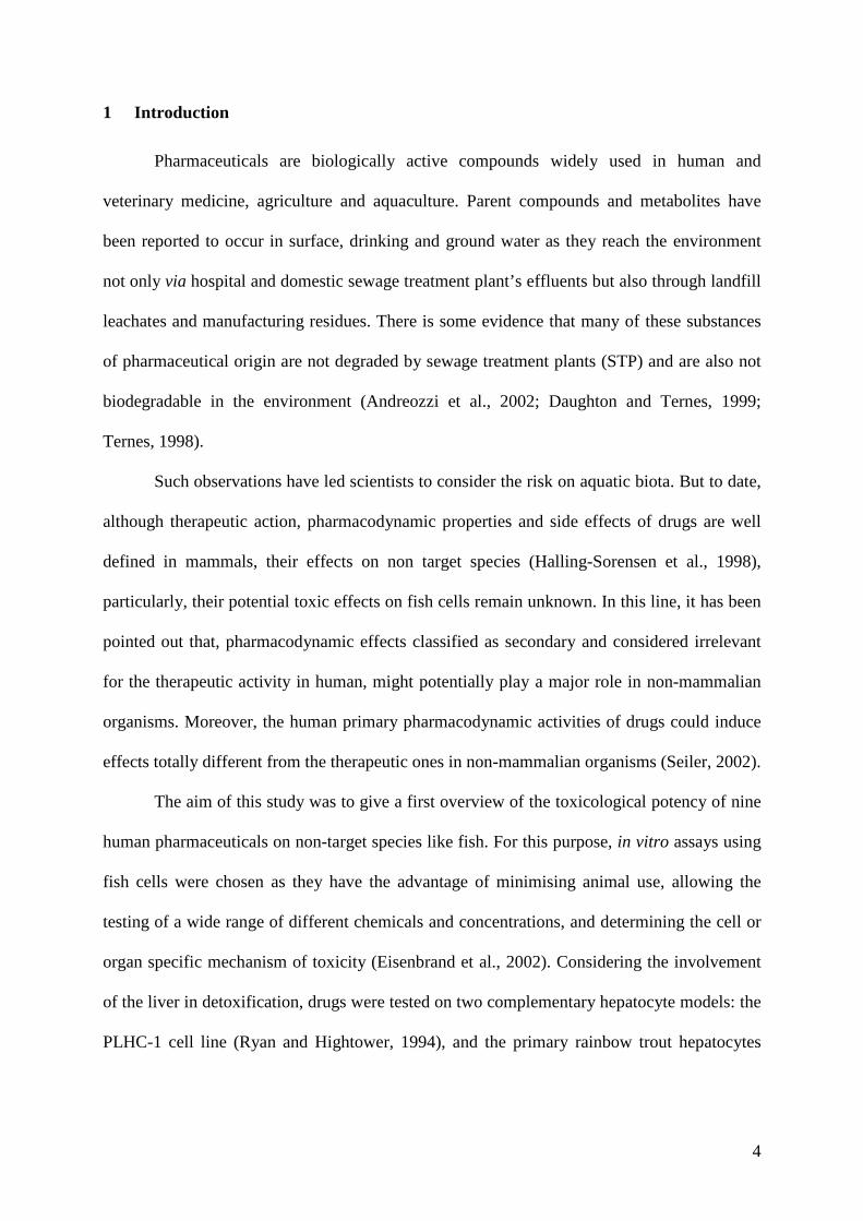
1 Introduction
Pharmaceuticals are biologically active compounds widely used in human and
veterinary medicine, agriculture and aquaculture. Parent compounds and metabolites have
been reported to occur in surface, drinking and ground water as they reach the environment
not only via hospital and domestic sewage treatment plant’s effluents but also through landfill
leachates and manufacturing residues. There is some evidence that many of these substances
of pharmaceutical origin are not degraded by sewage treatment plants (STP) and are also not
biodegradable in the environment (Andreozzi et al., 2002; Daughton and Ternes, 1999;
Ternes, 1998).
Such observations have led scientists to consider the risk on aquatic biota. But to date,
although therapeutic action, pharmacodynamic properties and side effects of drugs are well
defined in mammals, their effects on non target species (Halling-Sorensen et al., 1998),
particularly, their potential toxic effects on fish cells remain unknown. In this line, it has been
pointed out that, pharmacodynamic effects classified as secondary and considered irrelevant
for the therapeutic activity in human, might potentially play a major role in non-mammalian
organisms. Moreover, the human primary pharmacodynamic activities of drugs could induce
effects totally different from the therapeutic ones in non-mammalian organisms (Seiler, 2002).
The aim of this study was to give a first overview of the toxicological potency of nine
human pharmaceuticals on non-target species like fish. For this purpose, in vitro assays using
fish cells were chosen as they have the advantage of minimising animal use, allowing the
testing of a wide range of different chemicals and concentrations, and determining the cell or
organ specific mechanism of toxicity (Eisenbrand et al., 2002). Considering the involvement
of the liver in detoxification, drugs were tested on two complementary hepatocyte models: the
PLHC-1 cell line (Ryan and Hightower, 1994), and the primary rainbow trout hepatocytes

5
(PRTH) which maintain in vivo-like enzymatic activity during 3 to 8 days (Ferraris et al.,
2002).
The nine tested drugs were parent compounds of human pharmaceuticals from seven
different pharmaceutical classes. The two hypolipemiants, Fenofibrate (FF) and Clofibrate
(CF) were chosen because of their wide use in Europe and France respectively. The diagnostic
agent, Gadolinium Chloride (GdCl3), usually used in magnetic radio imaging (MRI), was
tested because of the presence of anomalies in coastal waters and rivers in the south of France
due to human use (Elbaz-Poulichet et al., 2002). The other tested drugs were chosen
according to their wide use in France. Antidepressants were represented by Fluoxetine (FX),
Non Steroidian Anti Inflammatory Drugs (NSAID) by Diclofenac (DiCF), β-blockers by
Propranolol (POH), antiepileptics by Carbamazepine (CBZ) and antibiotics by
Sulfamethoxazole (SFX) and Amoxicillin (AMX). In mammals most of these drugs are
metabolised by cytochromes P450 (CYP) enzymes and three of them, FF, CF and FX, induce
oxidative stress in human and rodent cells (Qu et al., 2001; Slamon and Pentreath, 2000). In
the present study, the toxicological potency of these pharmaceuticals on fish cells was
evaluated by cytotoxicity measurement, reactive oxygen species production and CYP1A
activity on two fish hepatocyte models PLHC-1 and PRTH.

6
2 Materials and methods
2.1 Chemicals
All chemicals were purchased from Sigma (France). Stock solutions of organic
compounds were prepared in dimethyl sulfoxyde (DMSO) at 200 times the desired
concentration. Concerning GdCl3, stock solutions were prepared in ultra-pure water at 100
times the desired final concentration.
2.2 Animals
Immature rainbow trout (Onchorynchus mykiss) were obtained from a local hatchery
(INRA, Gournay-sur-Aronde, France). Fish were kept in tanks with aerated charcoal filtered
tap-water at a temperature of 15°C. Rainbow trout were fed with commercial fish food and
acclimatised to laboratory conditions for a minimum of 2 weeks before use in the
experiments.
2.3 Trout hepatocyte isolation and culture
Hepatocytes were collected by the perfusion method described by Gagne et al. (1995).
In brief, rainbow trout were killed and the abdominal cavity was rapidly opened in aseptical
conditions. The liver was perfused via the hepatic portal vein with about 30 ml of perfusion
solution: Dulbecco’s Phosphate Buffered Saline (D-PBS) without calcium and magnesium
(Sigma, France), albumin 0.1 % and sodium citrate 10 mM adjusted to pH 7.5. The liver was
dissected, cut in small pieces and transferred to a sterile dissociation solution: D-PBS with
calcium and magnesium (Gibco, France), albumin 0.5 %, NaCl 120 mM and sodium citrate
15 mM. After 30 min agitation at 4 °C, the mix was passed 3 times through sterile 63 µm
nylon gauze. The cell suspension was washed 3 times by centrifugation at 2000 g, 4 °C during
4 min until the supernatant became clear. The supernatant was then removed and the cells
resuspended in 5 ml of D-PBS with calcium and magnesium. The cell viability was

7
determined using trypan blue exclusion test and was always found above 90 %. Freshly
isolated hepatocytes were seeded in 48 well untreated microplates (Iwaki, Japan) at a density
of 15.105 cells per well and cultured at 15°C in M199 cell culture medium (Sigma, France)
supplemented with 5 % decomplemented fœtal calf serum, penicillin and streptomycin (50
U/ml) and 10 mM hepes. Cells were left to incubate 24 hrs before exposure to chemicals.
2.4 Hepatocyte cell line
The PLHC-1 cell line, obtained from the American Type Culture Collection (ATCC
CRL 2406), is derived from the hepatocellular carcinoma of the topminnow Poeciliopsis
lucida (Ryan and Hightower, 1994). They were routinely grown at 30 °C in minimum
essential medium with Earle’s salts (E-MEM, Gibco, France) supplemented with 10 % (v/v)
decomplemented fœtal calf serum, 1 % v/v non-essential amino acids (Gibco, France) and 50
U.ml-1 of penicillin and streptomycin in a 5 % CO2 humidified atmosphere. For chemical
testing, cells were sub-cultured and seeded in 96-well plate (TPP, Switzerland) at a rate of
5.105 cells per well and left to grow up to confluency before adding chemicals.
2.5 Chemical exposure
Chemicals dissolved either in DMSO or in ultra-pure water were added to the medium
so that the final solvent concentration was always 0.5 or 1 % v/v respectively. Control wells
received the solvent only (carrier control). After exposure to pharmaceuticals and controls for
24 hrs , both cell models were subjected to MTT and EROD assays. Initially, the tested
concentrations were chosen according to the limit of solubility of each drug. Afterwards, a
range-finding study was conducted to determine the pharmaceuticals exposure that allowed
the modelling of the dose-effect curves.

8
2.6 MTT assay
Cytotoxic concentrations were determined by the MTT reduction test adapted from the
Mosmann's procedure (Mosmann, 1983). After chemical exposure the medium was removed
and cells were incubated for 3 hrs with 0.5 mg.ml-1 MTT dissolved in RPMI medium. MTT
was cleared out and the formazan salts were solubilized in 100 µl isopropanol. Plates were
read at 570 nm against a 660 nm reference wavelength on a microplate reader (BioTek
Instruments, France). Cell viability was expressed as a percentage of the corresponding
control value.
2.7 EROD assay
EROD activity in intact cells was assessed as previously described by Hahn et al.
(1996) and Kennedy et al. (1993). In short, medium was removed and replaced by 100 µl or
300 µl of culture medium containing 2 µM of 7-ethoxyresorufin in 96 and 48-well
microplates respectively. Kinetics of resorufin production were monitored during 15 min in a
microplate fluorimeter (Victor2, Perkin Elmer, France) at 530 nm and 590 nm excitation and
emission wavelengths, respectively, at 30°C for PLHC-1 cells and at room temperature for
PRTH. In each test, the positive control for EROD induction was 1 µM BaP. Then, the wells
were washed with PBS and total cellular proteins were determined with the fluorescamine
assay (Lorenzen and Kennedy, 1993), using bovine serum albumin (BSA) as a standard. The
EROD activity was expressed in pmol of resorufine per minute and per mg of proteins
(pmol.min-1.mg-1).
2.8 Reactive oxygen species (ROS) generation
The production of ROS in PLHC-1 cells was determined by using the method of
LeBel et al. (1983) with slight modifications. Cells were washed three times with PBS and
incubated with 1 µl of 40 µM DCFH-DA (dichlorofluorescamin diacetate) diluted in PBS

9
supplemented with 10 mM glucose (PBS-Gluc). After 30 min incubation at 30 °C, the cells
monolayers were washed twice with PBS and exposed to pharmaceuticals in PBS-Gluc. The
fluorescence of oxidised DCFH was measured immediately and every hour during 5 hrs with
a microplate fluorimeter (Victor2, Perkin Elmer, France), at excitation and emission
wavelengths of 490 nm and 535 nm respectively. Positive control was obtained using 5 µM
H2O2. The kinetics allowed the determination of the time of toxic exposure at which
maximum of ROS production occurred. Results were expressed as a percentage of the basal
fluorescence in the carrier control wells.
2.9 Data analysis
The toxicity of the tested substances was expressed by the concentration of drug
required to observe 50 % of the maximum effect (EC50). EC50 for MTT reduction and EROD
inhibition were calculated using the Microsoft Excell Regtox 6.3 macro (Vindimian et al.,
1983) freely available at www.perso.wanadoo.fr/eric.vindimian/en_index.html. All
experimental data were expressed as means ± standard deviation of 3 (PLHC-1) and 4
replicates (PRTH). Data were subjected to statistical analysis by one way analysis of variance
(ANOVA) followed by Dunnett's bilateral posthoc test. A value of p < 0.05 was considered
significant. The SPSS™ software version 10.1 for Windows was used for the statistical
analysis. All results are representative of at least three independent experiments.

10
3 Results
3.1 Cytotoxicity
The MTT EC50 values and the concentrations of pharmaceuticals tested on PRTH and
PLHC-1 are summarised in Table 1. The levels of toxicity varied not only according to the
substances tested but also to the biological model.
The most cytotoxic drugs to PLHC-1 cells were CF, FX, FF and DiCF with MTT EC50 values
of 2 µM, 5 µM, 9 µM and 19 µM respectively. On PRTH, the most cytotoxic drugs were FF,
FX and CF with EC50 of 53 µM, 66 µM and 133 µM respectively. For the other drugs, the
EC50 values ranged from 85 µM (POH) to 651 µM (CBZ) on PLHC-1 and from 453 µM
(POH) to 963 µM (GdCl3) on PRTH. Among the tested compounds, only the antibiotic AMX
showed no cytotoxicity neither on PLHC-1 nor on PRTH. For the sulfamide antibiotic, SFX,
cytotoxicity appeared only at the highest tested concentration. The overall comparison of the
MTT EC50 values obtained for both models showed that PRTH were somewhat less sensitive
than PLHC-1 to the pharmaceuticals cytotoxicity. Indeed, EC50 values on PRTH cells were
from 5 (FF, POH) to 50 (CF) orders of magnitude higher than those obtained for PLHC-1.
1.2 Effect on EROD activity
None of the tested drugs induced EROD in PLHC-1 hepatocytes due to a low basal
EROD expression, only induction could be studied in these cells. On the contrary, PRTH
allowed the detection of both induction and inhibition of EROD activity. As seen in Fig.1-4,
different patterns of responses were observed. First a significant increase of EROD activity
was observed in cells exposed to sublethal concentration of the beta-blocker POH (Fig. 1).
Secondly, all substances, except AMX (Fig.4.), were able to decrease EROD activity whether
at sublethal or lethal concentrations. The specific EROD inhibition, at clearly sublethal
concentrations, appeared after exposure to DiCF, SFX and CBZ (Fig. 2) whereas this

11
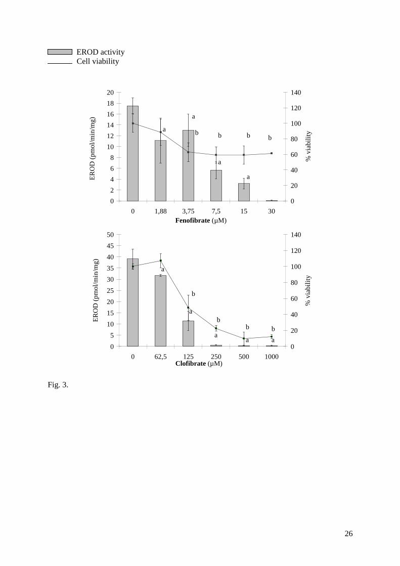
inhibition appeared to be less specific in the case of FF and CF (Fig. 3). Finally the EROD
inhibition was concommitant with cytotoxicity in cells exposed to FX, GdCl3 and POH
(Fig.4).
1.3 ROS production
Three (FF, CF and FX) out of nine pharmaceuticals induced significant ROS production
in PLHC-1 cells (Fig.5). FF was the most efficient in inducing an oxidant effect as, within the
first hour of exposure, ROS production increased in a dose-dependant manner (7.8 - 30 µM)
up to 4 times the basal level at the highest concentration tested. The ROS level, after CF and
FX exposure, was weaker as it reached only about 1.5 times the basal value after a 4 hrs
exposure at 1000 and 36 µM respectively.

12
4 Discussion
The purpose of this work was to evaluate the acute and sublethal effects of nine
pharmaceuticals on two fish hepatocyte models, PLHC-1 and PRTH. The main outcomes are:
(1) eight of the nine tested drugs were cytotoxic on both cell models; (2) for FF, CF and FX,
oxidative stress was involved in cytotoxicity process; (3) specific EROD inhibition was
observed with DiCF, CBZ and SFX, and, to a lesser extend, by FF and CF; (4) POH
significantly induced EROD activity.
Our results suggest that most of the tested drugs (FF, CF, FX, POH, DiCF, GdCl3) were
more toxic to PLHC-1 than to PRTH (Table 1). This difference could reflect the different
sensitivity of cell lines versus primary hepatocytes or the differences in the ability of each
model to metabolise human drugs as PRTH can inactivate toxicants (via metabolisation) and
develop defence against oxidants (Ferraris et al., 2002). Only CBZ was more toxic in PRTH,
probably because of its transformation into active metabolites CBZ-E and/or arene oxide
(Masubuchi et al., 2001; Tabatabaei et al., 1997). Overall, although no comparable data are
available in fish for all the tested substances, MTT EC50 measured in this study are in the
same range and sometimes slightly more sensitive than cytotoxic concentrations previously
described in human and rat. For example, the MTT EC50 found in human and rat hepatocytes
range between 176 and 341 µM for POH (Ponsoda et al., 1995), and between 61 and 64 µM
for DiCF (Bort et al., 1999b). In the same way the EC50s obtained with the acute Daphnia
magna test for FX (Brooks et al., 2003), POH (Huggett et al., 2002), CBZ and DiCF
(Cleuvers, 2003; Ferrari et al., 2003), are in the same order of magnitude as our cytotoxicity
results. In parallel, all the tested drugs except AMX, interacted with CYP1A metabolism
pathways in rainbow trout hepatocytes through an induction or an inhibition of the EROD

13
activity. Such effects are of environmental concern, as alteration of CYP1A activity has been
shown to affect xenobiotic metabolism and toxicity in rainbow trout (Hawkins et al., 2002).
The hypolipemiants FF and CF belong to peroxisome proliferators (PPs). This group of
structurally diverse compounds is known to bind and activate the peroxisome proliferator-
activated receptors (PPARs). Among the three main PPARs isoforms so far identified
(PPARα, PPARβ, PPARγ) PPARα seems to mediate the hypotriglyceridemic effect of
hypolipemiants by inducing high rates of mitochondrial and peroxisomal β-oxidation of lipids
in rodents and human (Mukherjee et al., 1994). Such activation leads to pro-oxidant effect via
the increase of H2O2 production rate in mice (Qu et al., 1999; Qu et al., 2001). In salmon
hepatocytes the activation of PPARγ (Andersen et al., 2000) after exposure to fibrates,
mediates enzymatic response like the increase of peroxisomal acyl-co-enzyme-A activity
(Ruyter et al., 1997). In addition FF has been described to be more efficient than CF in
inducing peroxisome proliferation in rat hepatocytes and in increasing characteristic
peroxisomal activities such as carnitine acetyl transferase (CAT) or CYP4A1 activities
(Hildebrand et al., 1999). In our experiments, FF rapidly induced ROS generation whereas its
homologue was much less efficient regarding ROS production (Fig. 3). Thus, our results
support the idea that CF and FF activate PPARs or at least peroxisomal activities in fish cells.
This finding is particularly interesting because differences between species have been
reported about sensitivity towards peroxisome proliferation. Indeed dogs and humans are
regarded as insensitive species (Ashby et al., 1994) whereas rat (Hildebrand et al., 1999) and
fish (Ruyter et al., 1997) would be sensitive ones.
ROS, such as H2O2, are known to specifically inhibit CYP enzymes mainly at the
transcriptional level by inhibiting the mRNA synthesis (Barker et al., 1994; Risso-de
Faverney et al., 2000). Sometimes this CYP inhibition also occurs at the posttranscriptional

14
level by the increasing the degradation of mRNA (Delaporte and Renton, 1997) or at the
protein level when toxicants act directly as mechanism-based inhibitors (suicide substrate)
(Watson et al., 1995),. Moreover Jiao and Zhao(2002) showed that FF inhibited the growth of
human HepG2 cells in a dose-related manner through the generation of oxidative stress.
Consequently, it is very likely that, in fish cells, specific EROD inhibition and loss of cell
viability by FF and CF are due to ROS overproduction through PPAR activation.
The antidepressant FX was among the most cytotoxic drugs in both cellular models
and induced ROS production in PLHC-1. The resulting hypothesis that, in fish hepatocytes,
FX cytotoxicity is mediated through oxidative stress is consistent with the previous study of
Slamon and Pentreath (2000) who showed the involvement of antioxidants such as glutathion
(GSH) in the protection of rat glioma C6 cells and human astrocytes cell line against this
antidepressant.
The DiCF exerted a cytotoxic effect at 500 µM, which is in agreement with previous
results on rat or human hepatocytes (Bort et al., 1999b). The mechanism of DiCF cytotoxicity
is not fully understood but there is some evidence that both uncoupling of mitochondrial
oxidative phosphorilation and CYP-mediated metabolism are involved in human and rat
hepatocytes acute toxicity(Bort et al., 1999a). In our study, we observed a clear inhibition of
EROD at 36 µM, suggesting a specific interaction between DiCF or its metabolites with this
enzyme in rainbow trout, but this interaction is apparently not related to the cytotoxicity of
DiCF to PRTH. Although the mechanism of CYP1A inhibition by NSAID is not known, the
ability of these drugs to decrease 3 methylcholanthrene-induced EROD activity has been
reported in rat hepatocytes (Pappas et al., 1998).
Among the tested drugs, the β-adrenergic receptor antagonist, POH, was the only
EROD inducer in PRTH. To our knowledge, there is no report on the ability of this compound
either to bind and activate the aryl hydrocarbon receptor (Ah-R), or to induce the CYP1A

15
enzyme. Nevertheless, the observed EROD induction might be related to POH metabolism by
CYP1A previously described in β-naphtoflavone-induced rat hepatic microsomes (Li and
Zeng, 2000).
In human lymphocytes, CBZ is metabolised by CYP dependant monooxygenase into
10,11-epoxide CBZ (CBZ-E), an active and toxic metabolite which is then transformed into
the corresponding diol by an epoxide hydrolase (Tabatabaei et al., 1997). It was clearly
established that in some cases, clinical toxicities were associated with CBZ-E production.
CBZ-E is also found in mice, dog, rat and rabbit (Rambeck et al., 1990), thus representing a
metabolic pathway common to these species. In human and rat, CYPs involved in CBZ
metabolism are diverse and include CYP1A2, CYP2C8, CYP3A4, CYP2D and CYP1A2
(Masubuchi et al., 2001; Mesdjian et al., 1999). Recently, it has been shown that CBZ
biotransformation into arene-oxide intermediates by CYP2D in rat and CYP1A2 in humans
results in inactivation of the enzymes themselves probably via the covalent binding of these
metabolites with CYP proteins (Masubuchi et al., 2001). Therefore, the strong and specific
CYP1A inhibition observed in our study results likely from the CBZ biotransformation into
reactive metabolites and thus suggests the ability of fish cells to metabolise this drug.
The contrast agent GdCl3 has been described as a CYP inhibitor in vivo in human and
rodents (Badger et al., 1997; Palasz and Czekaj, 2000). In goldfish, it has been shown to
accumulate in the liver and to induce antioxidant enzymes after 10 days of exposure (Chen et
al., 2000). Our results did not correlate with those data as GdCl3 neither inhibited specifically
EROD activity, nor induced oxidative stress in fish cells. This suggests a low sensitivity of
the fish hepatocytes PRTH and PLHC-1 to this rare earth element.
Although the sulfonamide antibiotic SFX was not cytotoxic enough to calculate EC50
values, it inhibited EROD activity right from 125 µM. In human liver microsomes, SFX is
described as a selective inhibitor of CYP2C8 and CYP2C9 that would loose selectivity for the

16
CYP isoforms at concentrations higher than 500 µM (Wen et al., 2002). As a result, SFX must
be a selective inhibitor of CYP1A enzymes in fish hepatocytes. Concerning the ampicillin
antibiotic, AMX is not described as a potential toxicant in literature and, accordingly, it didn’t
exert any particular effect on fish hepatocytes.
In summary, this study raises three interesting observations. First, a classification of
human drugs from the least to the most cytotoxic to fish hepatocytes was established: the two
antibiotics (AMX and SFX) showed no cytotoxic effect, whereas the hypolipemiants (CF and
FF) and the antidepressant (FX) were the most cytotoxic and induced oxidative stress at
sublethal concentrations. Secondly, the majority of the human drugs caused a specific EROD
inhibition. This may have relevance to the use of CYP1A as a biomarker for aquatic pollution.
In a multipollution context, considering the involvement of CYP1A in detoxification
pathways, its inhibition could increase the toxicity of the other toxicants. Thirdly, if the
effective concentrations were comparable with ecotoxicity tests found in the literature, they
remain much higher (one thousand times) than those found in the environment. Nevertheless,
before coming to a conclusion about the absence of toxicity in fish exposed to contaminated
waters in the environment, the in vivo potential adverse effects of drugs to fish under chronic
exposure should be assessed.
AKNOWLEDGEMENTS This work was partly supported by the French National Program
for Ecotoxicology (PNETOX) and by the “Budget Civil de Recherche et Développement”
(BCRD Grant 01-111).
References

17
Andersen, O., Eijsink, V.G., Thomassen, M., 2000. Multiple variants of the peroxisome
proliferator-activated receptor (PPAR) gamma are expressed in the liver of atlantic
salmon (Salmo salar). Gene 255, 411 - 418.
Andreozzi, R., Marotta, R., Pinto, G., Pollio, A., 2002. Carbamazepine in water: persistence
in the environment, ozonation treatment and preliminary assessment on algal toxicity.
Water. Res. 36, 2869 - 2877.
Ashby, J., Brady, A., Elcombe, C.R., Elliott, B.M., Ishmael, J., Odum, J., Tugwood, J.D.,
Kettle, S., Purchase, I.F., 1994. Mechanistically-based human hazard assessment of
peroxisome proliferator-induced hepatocarcinogenesis. Hum. Exp. Toxicol. 13 Suppl
2, S1-117.
Badger, D.A., Kuester, R.K., Sauer, J.M., Sipes, I.G., 1997. Gadolinium chloride reduces
cytochrome P450: relevance to chemical-induced hepatotoxicity. Toxicology 121, 143
- 153.
Barker, C.W., Fagan, J.B., Pasco, D.S., 1994. Down-regulation of P4501A1 and P4501A2
mRNA expression in isolated hepatocytes by oxidative stress. J. Biol. Chem. 269,
3985 - 3990.
Bort, R., Mace, K., Boobis, A., Gomez-Lechon, M.J., Pfeifer, A., Castell, J., 1999a. Hepatic
metabolism of diclofenac: role of human CYP in the minor oxidative pathways.
Biochem. Pharmacol. 58, 787 - 796.
Bort, R., Ponsoda, X., Jover, R., Gomez-Lechon, M.J., Castell, J.V., 1999b. Diclofenac
Toxicity to Hepatocytes: A Role for Drug Metabolism in Cell Toxicity. J. Pharmacol.
Exp. Ther. 288, 65 - 72.
Brooks, B.W., Turner, P.K., Stanley, J.K., Weston, J.J., Glidewell, E.A., Foran, C.M.,
Slattery, M., La Point, T.W., Huggett, D.B., 2003. Waterborne and sediment toxicity
of fluoxetine to select organisms. Chemosphere 52, 135 - 142.

18
Chen, Y., Cao, X.D., Lu, Y., Wang, R., 2000. Effects of rare earth metal ions and their EDTA
complexes on antioxydant enzymes of fish liver. Bull. Environ. Contam. Toxicol. 65,
357 - 365.
Cleuvers, M., 2003. Aquatic ecotoxicity of pharmaceuticals including the assessment of
combination effects. Toxicol. Lett. 142, 185 - 194.
Daughton, C.G., Ternes, T.A., 1999. Pharmaceuticals and personal care products in the
environment: agents of subtle change? Environ. Health. Perspect. 107, 907 - 938.
Delaporte, E., Renton, K.W., 1997. Cytochrome P4501A1 and cytochrome P4501A2 are
downregulated at both transcriptional and post-transcriptional levels by conditions
resulting in interferon-alpha/beta induction. Life Sci. 60, 787 - 796.
Eisenbrand, G., Pool-Zobel, B., Baker, V., Balls, M., Blaauboer, B.J., Boobis, A., Carere, A.,
Kevekordes, S., Lhuguenot, J.C., Pieters, R., Kleiner, J., 2002. Methods of in vitro
toxicology. Food Chem. Toxicol. 40, 193 - 236.
Elbaz-Poulichet, F., Seidel, J.L., Othoniel, C., 2002. Occurrence of an anthropogenic
gadolinium anomaly in river and coastal waters of southern France. Water Res. 36,
1102 - 1105.
Ferrari, B., Paxéus, N., Lo Giudice, R., Pollio, A., Garric, J., 2003. Ecotoxicological impact of
pharmaceuticals found in treated wastewaters: study of carbamazepine, clofibric acid,
and diclofenac. Ecotoxicological and environmental safety 55, 359 - 370.
Ferraris, M., Radice, S., Catalani, P., Francolini, M., Marabini, L., Chiesara, E., 2002. Early
oxidative damage in primary cultured trout hepatocytes: a time course study. Aquat.
Toxicol. 59, 283 - 296.
Gagne, F., Trottier, S., Blaise, C., Sproull, J., Ernst, B., 1995. Genotoxicity of sediment
extracts obtained in the vicinity of a creosote-treated wharf to rainbow trout
hepatocytes. Toxicol. Lett. 78, 175-82.

19
Hahn, M.E., Woodward, B.L., J., S.J., 1996. Rapid assessmant of induced cytochrome
P4501A protein and catalytic activity in fish hepatoma cells grown in multiwell
plates: response to TCDD, TCDF and two planar PCBS. Environmental Toxicology
and Chemistry 15, 582 - 591.
Halling-Sorensen, B., Nors Nielsen, S., Lanzky, P.F., Ingerslev, F., Holten Lutzhoft, H.C.,
Jorgensen, S.E., 1998. Occurrence, fate and effects of pharmaceutical substances in
the environment - a review. Chemosphere 36, 357 - 393.
Hawkins, S.A., Billiard, S.M., Tabash, S.P., Brown, R.S., Hodson, P.V., 2002. Altering
cytochrome P4501A activity affects polycyclic aromatic hydrocarbon metabolism and
toxicity in rainbow trout (Oncorhynchus mykiss). Environ. Toxicol. Chem. 21, 1845 -
1853.
Hildebrand, H., Schmidt, U., Kempka, G., Jacob, R., Ahr, H.J., Ebener, C., Goretzki, P.E.,
Bader, A., 1999. An in vitro model for peroxisome proliferation utilizing primary
hepatocytes in sandwich culture. Toxicol In vitro 13, 265 - 273.
Huggett, D.B., Brooks, B.W., Peterson, B., Foran, C.M., Schlenk, D., 2002. Toxicity of select
beta adrenergic receptor-blocking pharmaceuticals (B-Blockers) on aquatic organisms.
Arch. Environ. Contam. Toxicol. 43, 229 - 235.
Jiao, H.L., Zhao, B.L., 2002. Cytotoxic effect of peroxisome proliferator fenofibrate on
human HepG2 hepatoma cell line and relevant mechanisms. Toxicol. Appl.
Pharmacol. 185, 172 - 179.
Kennedy, S.W., Lorenzen, A., James, C.A., Collins, B.T., 1993. Ethoxyresorufin-O-
deethylase and porphyrin analysis in chicken embryo hepatocyte cultures with a
fluorescence multiwell plate reader. Anal. Biochem. 211, 102 - 112.

20
LeBel, C.P., Ischiropoulos, H., Bondy, S.C., 1983. Evaluation of the probe 2',7'-
dichlorofluorescin as an indicator of reactive oxygen species formation and oxidative
stress. Chem. Res. Toxicol. 5, 227 - 231.
Li, X., Zeng, S., 2000. Stereoselective propranolol metablism in two drug induced in rat
hepatic microsomes. World J. Gastroenterol. 6, 74 - 78.
Lorenzen, A., Kennedy, S.W., 1993. A fluorescence-based protein assay for use with a
microplate reader. Anal. Biochem. 214, 346 - 348.
Masubuchi, Y., Nakano, T., Ose, A., Horie, T., 2001. Differential selectivity in
carbamazepine-induced inactivation of cytochrome P450 enzymes in rat and human
liver. Arch. Toxicol. 75, 538 - 543.
Mesdjian, E., Seree, E., Charvet, B., Mirrione, A., Bourgarel-Rey, V., Desobry, A., Barra, Y.,
1999. Metabolism of carbamazepine by CYP3A6: a model for in vitro drug
interactions studies. Life Sci. 64, 827 - 835.
Mosmann, T., 1983. Rapid colorimetric assay for cellular growth and survival: application to
proliferation and cytotoxicity assays. J. Immunol. Methods 65, 55 - 63.
Mukherjee, R., Jow, L., Noonan, D., McDonnell, D.P., 1994. Human and rat peroxisome
proliferator activated receptors (PPARs) demonstrate similar tissue distribution but
different responsiveness to PPAR activators. J. Steroid Biochem. Mol. Biol. 51, 157 -
166.
Palasz, A., Czekaj, P., 2000. Toxicological and cytophysiological aspects of lanthanides
action. Acta Biochim. Pol. 47, 1107 - 1114.
Pappas, P., Stehanou, P., Vasiliou, V., Marselos, M., 1998. Antiinflammatory agents
andinducibility of hepatic drug metabolism. Eur. J. Drug Met. Pharma. 23, 457 - 460.
Ponsoda, X., Jover, R., Nunez, C., Royo, M., Castell, J.V., Gomez-Lechon, M.J., 1995.
Evaluation of the cytotoxicity of 10 chemicals in human and rat hepatocytes and in
21
cell lines: correlation between in vitro data and human lethal concentration. Toxicol.
In vitro 9, 959 - 966.
Qu, B., Li, Q.T., Wong, K.P., Ong, C.N., Halliwell, B., 1999. Mitochondrial damage by the
"pro-oxidant" peroxisomal proliferator clofibrate. Free Radic. Biol. Med. 27, 1095 -
1102.
Qu, B., Li, Q.T., Wong, K.P., Tan, T.M.C., Halliwell, B., 2001. Mechanism of clofibrate
hepatotoxicity: mitochondrial damage and oxidative stress in hepatocytes. Free Radic.
Biol. Med. 31, 659 - 669.
Rambeck, B., Salke-Treumann, A., May, T., Boenigk, H.E., 1990. Valproic acid-induced
carbamazepine-10,11-epoxide toxicity in children and adolescents. Eur. Neurol. 30, 79
- 83.
Risso-de Faverney, C., Lafaurie, M., Girard, J.P., Rahmani, R., 2000. The nitroxide stable
radical tempo prevents metal-induced inhibition of CYP1A1 expression and induction.
Toxicol. Lett. 111, 219 - 227.
Ruyter, B., Andersen, O., Dehli, A., Ostlund Farrants, A.K., Gjoen, T., Thomassen, M.S.,
1997. Peroxisome proliferator activated receptors in Atlantic salmon (Salmo salar):
effects on PPAR transcription and acyl-CoA oxidase activity in hepatocytes by
peroxisome proliferators and fatty acids. Biochim. Biophys. Acta. 1348, 331 - 338.
Ryan, J.A., Hightower, L.E., 1994. Evaluation of heavy-metal ion toxicity in fish cells using a
combined stress protein and cytotoxicity assay. Environ. Toxicol. Chem. 13, 1231 -
1240.
Seiler, J.P., 2002. Pharmacodynamic activity of drugs and ecotoxicology - can the two be
connected? Toxicol. Lett. 131, 105 - 115.
Slamon, N.D., Pentreath, V.W., 2000. Antioxidant defense against antidepressants in C6 and
1321N1 cells. Chem. Biol. Interact. 127, 181 - 199.

22
Tabatabaei, A.R., Thies, R.L., Farrell, K., Abbott, F.S., 1997. A rapid in vitro assay for
evaluation of metabolism-dependent cytotoxicity of antiepileptic drugs on isolated
human lymphocytes. Fundam. Appl. Tox. 37, 181 - 189.
Ternes, T., 1998. Occurrence of drugs in German sewage treatment plants and rivers. Water
Res. 32, 3245 - 3260.
Vindimian, E., Robaut, C., Fillion, G., 1983. A method for cooperative and non cooperative
binding studies using non linear regression analysis on a microcomputer. J. Appl.
Biochem. 5, 261 - 268.
Watson, D.E., Menard, L., Stegeman, J.J., Digiulio, R.T., 1995. Aminoanthracene is a
mechanism-based inactivator of CYP1A in channel catfish hepatic issue. Toxicol.
Appl. Pharmacol. 135, 208 - 215.
Wen, X., Wang, J.S., Backman, J.T., Laitila, J., Neuvonen, P.J., 2002. Trimethoprim and
sulfamethoxazole are selective inhibitors of CYP2C8 and CYP2C9, respectively. Drug
Metab. Dispos. 30, 631 - 635.

23
Figures
Fig. 1. Specific induction of EROD activity and viability of primary rainbow trout (PRTH)hepatocytes after 24 hrs of exposure to propranolol (POH). Values are means ± StandardDeviation.a Significant difference from basal EROD activity (p < 0.05).b Significant difference from the positive control of cell viability (p < 0.05).
Fig. 2. Specific inhibition of EROD activity at sublethal concentrations on primary rainbowtrout (PRTH) hepatcoytes after 24 hours of exposure. Values are means ± Standard Deviation.a Significant difference from basal EROD activity (p < 0.05).b Significant difference from the positive control of cell viability (p < 0.05).
Fig.3. Basal EROD inhibition and cell viability of primary rainbow trout (PRTH) hepatocytesafter 24 hrs of exposure to the two hypolipemiants clofibrate (CF) and fenofibrtae (FF).Values are means ± Standard Deviation.a Significant difference from basal EROD activity (p < 0.05).b Significant difference from the positive control of cell viability (p < 0.05).
Fig.4. Pharmaceuticals without specific effect on the EROD activity at sublethalconcentrations on primary rainbow trout (PRTH) hepatcoytes after 24 hours of exposure:cytotoxic and not cytotoxic ones. Values are means ± Standard Deviation.a Significant difference from basal EROD activity (p < 0.05).b Significant difference from the positive control of cell viability (p < 0.05).
Fig. 5. ROS production on PLHC-1 cells after 1 hr of exposure for fenofibrate (FF) and after4 hrs of exposure for clofibrate (CF) and fluoxetine (FX). Times of exposure were chosenafter the study of the kinetics of ROS production for each compound during 5 hours, andcorrespond to the time with the optimum of ROS production. The six other drugs (POH,SFX, CBZ, GCl3, AMX, DiCF) didn’t show any ROS production. Values are means ±Standard Deviation.* Significant difference from control (p < 0.05).

24
EROD activity Cell viability
0
5
10
15
20
25
30
35
0 31,25 62,5 125 250 500
Propranolol (µM)
ER
OD
(pm
ol/m
in/m
g)
0
20
40
60
80
100
120
140
% v
iabi
lity
a aa
b
b
a
a
Fig.1.

25
EROD activity Cell viability
0
2
4
6
8
10
12
14
0 31,25 62,5 125 250 500
Diclofenac (µM)
ER
OD
(pm
ol/m
in/m
g)
0
20
40
60
80
100
120
140
% v
iabi
lity
a
a a a
b
0
5
10
15
20
25
30
35
40
45
50
0 31,25 62,5 125 250 500
Carbamazepine (µM)
ER
OD
(pm
ol/m
in/m
g)
0
20
40
60
80
100
120
140
% v
iabi
lity
a
a
a
b
b
a a
0
5
10
15
20
25
30
35
40
45
50
0 31,25 62,5 125 250 500
Sulfamethoxazole (µM)
ER
OD
(po
ml/m
in/m
g)
0
20
40
60
80
100
120
140
% v
iabi
lity
a
a
a
a
b
Fig. 2.
26
EROD activity Cell viability
0
2
4
6
8
10
12
14
16
18
20
0 1,88 3,75 7,5 15 30Fenofibrate (µM)
ER
OD
(pm
ol/m
in/m
g)
0
20
40
60
80
100
120
140
% v
iabi
litya
a
a
a
b b bb
0
5
10
15
20
25
30
35
40
45
50
0 62,5 125 250 500 1000Clofibrate (µM)
ER
OD
(pm
ol/m
in/m
g)
0
20
40
60
80
100
120
140
% v
iabi
lity
a
b
aa
a
a
bb b
Fig. 3.

27
EROD activity Cell viability
0
5
10
15
20
25
30
0 9 18 36 70 140Fluoxetine (µM)
% v
iabi
lity
0
20
40
60
80
100
120
140
ER
OD
(pm
ol/m
in/m
g)
a
a
b
b
0
5
10
15
20
25
0 62,5 125 250 500 1000
Gadolinium chlorhyde (µM)
ER
OD
(pm
ol/m
in/m
g)
0
20
40
60
80
100
120
140
% v
iabi
lity
ba
b
a
0
5
10
15
20
25
0 31,25 62,5 125 250 500
Amoxicillin (µM)
ER
OD
(pm
ol/m
in/m
g)
0
20
40
60
80
100
120
140
% v
iabi
lity
Fig. 4.

28
0
50
100
150
200
250
300
350
400
450
0 5 10 15 20 25 30 35
Fenofibrate (µM)
% b
asa
l RO
S p
rodu
ctio
n*
*
*
0
20
40
60
80
100
120
140
160
0 200 400 600 800 1000
Clofibrate (µM)
% b
asa
l RO
S p
rodu
ctio
n
*
0
20
40
60
80
100
120
140
160
180
0 20 40 60 80 100 120 140
Fluoxetine (µM)
% b
asal
RO
S p
rod
uct
ion
*
Fig.5.

29
Tables
Table 1.
Tested concentrations of pharmaceuticals on primary rainbow trout hepatocytes(PRTH) and PLHC-1 cell line and EC50 of cytotoxicity.
Values are EC50 and confidence interval calculated with MTT results after 24 hours ofexposure.
n.m : not modelisable ; n.c : not cytotoxic
Pharmaceutical Tested concentrations (µM) EC 50 cytotoxicity (µM)
Confidence interval with α=5 %PLHC-1 PRTH PLHC-1 PRTH
Fenofibrate (FF) 0.1 – 15.6 1.9 - 30 98- 11
5328 – 102
Clofibrate (CF) 3.9 - 500 62.5 - 1000 22 – 3
133117 – 161
Fluoxetine (FX) 1.1 - 140 9 - 140 54 – 6
6644 - 83
Propranolol(POH)
3.9 - 500 31.3 - 500 8572 - 102
453334 - 690
Diclofenac(DiCF)
3.9 - 500 31.3 - 500 1911 - 29
420361 - 480
Gadoliniumchloride (GdCl3)
7.8 - 1000 62.5 - 1000 262112 - 789
963672 - 1516
Carbamazepine(CBZ)
3.9 500 31.3 – 500 651344 - 2127
318253 - 401
Sulfamethoxazole (SFX)
3.9 - 500 31.3 - 500 n.m n.m
Amoxicillin(AMX)
3.9 - 500 31.3 - 500 n.c n.c

30
Table 2.
EROD EC50 of 9 pharmaceuticals tested on primary rainbow trout hepatocytes(PRTH).
Values are EC50 with confidence interval calculated with EROD results after 24 hoursof exposure.
*Inhibitors of the basal EROD activity at sublethal concentration.
5 Pharmaceutical Effect on the basal ERODactivity
EC50 EROD
Fenofibrate (FF)* Inhibition 2513 - 30
Diclofenac (DiCF)* Inhibition 6362 – 68
Fluoxetine (FX) Inhibition 7758 – 95
Clofibrate (CF* Inhibition 9688 -104
Sulfamethoxazole (SFX)* Inhibition 10878 – 144
Carbamazepine (CBZ)* Inhibition 318253 – 401
Gadolinium chlorhyde(GdCl3)
Inhibition 371279 - 482
Propranolol (POH) Induction 271 - 44
Amoxicillin (AMX) No effect -